
Nanotechnology as a Novel Approach in Combating Microbes Providing an Alternative to Antibiotics

 , , , , , , , and
Abstract
:1. Introduction
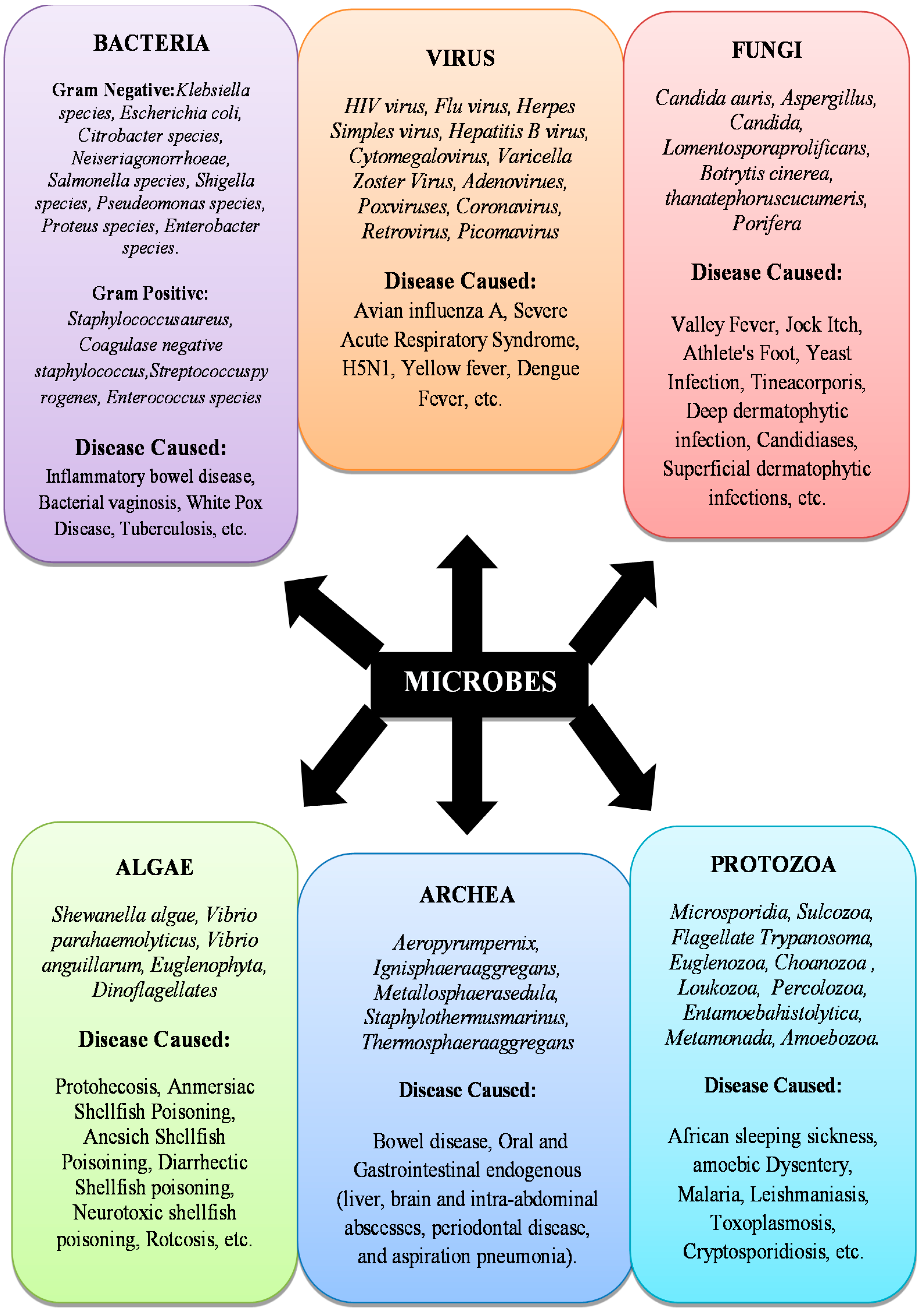
2. Microbes
Infectious Microbial Species
3. Conventional Antibiotics
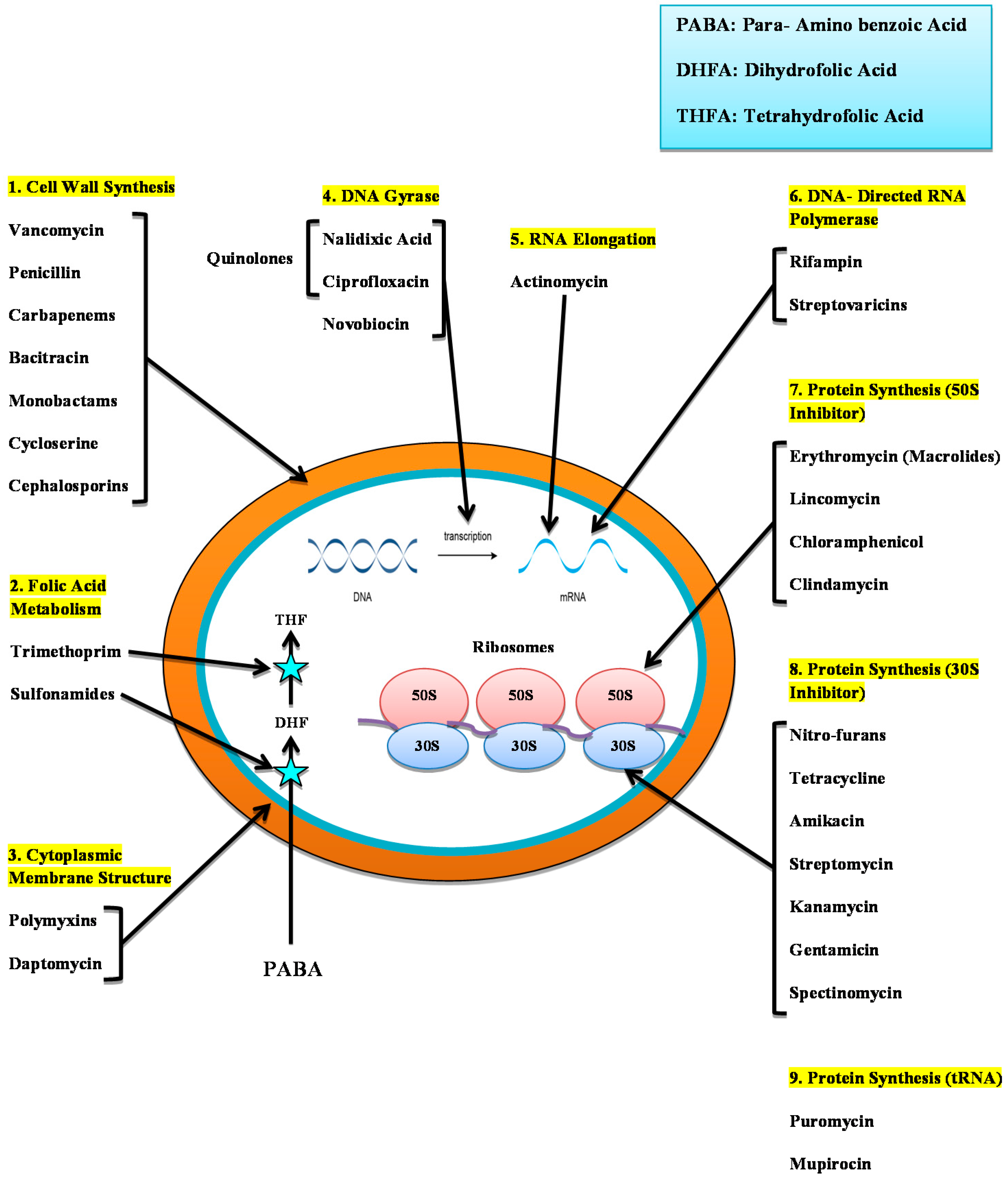
3.1. Mode of Action of Antibiotics
3.2. Origin of Antibiotic Resistance
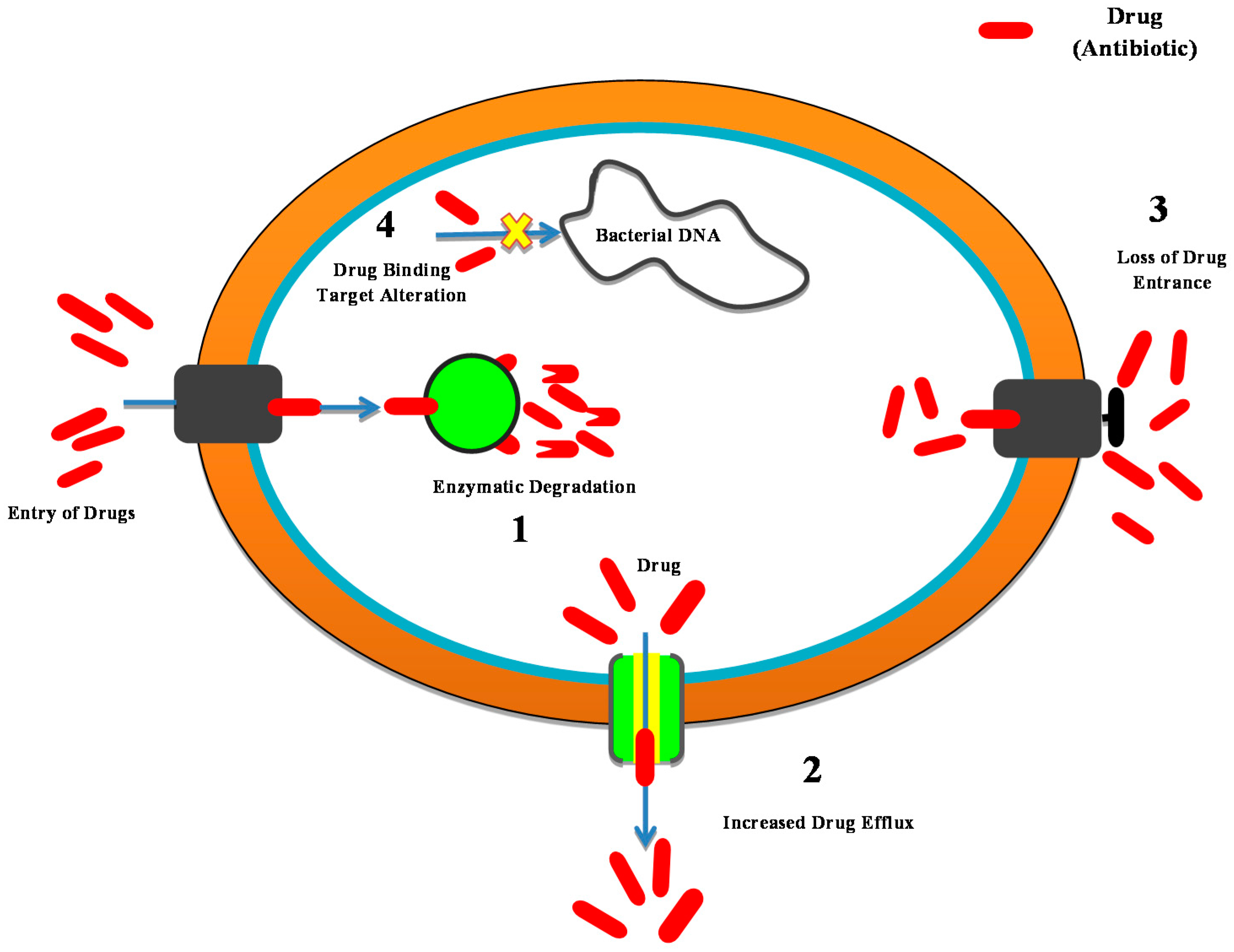
3.3. Development of Antibiotic Resistance
- Antibiotic enzyme inactivation/degradation; an endogenous cellular enzyme is modified to interact with that of the antibiotic in a manner in which the bacteria are no longer affected. B-lactamase enzymes are among the most important examples; they hydrolyze most commonly administered antibiotics, i.e., b-lactams (cephalosporin and penicillin), and are the most widespread source of antibiotic resistance in Gram-negative bacteria.
- The excretion of the drug through efflux pumps; Bacteria are triggered to eliminate the antibiotic by stimulating the proteins that can eradicate an extensive range of substances from the periplasm to the outside cell. This is a mechanism of resistance especially essential for P. aeruginosa and Acinetobacter spp.
- Reduced absorption by variations in the external membrane permeability; these changes inhibit the successful entry of antibiotics.
- Drug target modifications to weaken or demolish the antibiotic binding efficacy and thereby minimize its potential.
3.4. Availability of Antibiotics
4. Nanomaterials
4.1. History and Development of Nanomaterials
4.2. Nanoparticles Act as Antimicrobials
4.3. Classification of Nanomaterials
- Carbon-based nanomaterial
- Inorganic nanomaterial
- Organic-based nanomaterial
- Composite-based nanomaterial
4.3.1. Carbon-Based Nanomaterial
Carbon Nanotubes (CNTs)
Fullerenes
Graphene Oxide (GO)
4.3.2. Inorganic Nanomaterial
Silver Nanoparticles (AgNPs)
Gold Nanoparticles
Zinc Oxide Nanoparticles
Titanium Dioxide Nanoparticles
Copper Nanoparticles
Aluminum Oxide Nanoparticles
Nitric Oxide (NO)—Releasing Nanoparticles
Magnesium Oxide Nanoparticles
Iron Oxide Nanoparticles
Super-Paramagnetic Iron Oxide (SPION)
4.3.3. Organic-Based Nanomaterials
Poly-ε-Lysine
Quaternary Ammonium Compounds
N–Halamine Compounds
Polysiloxanes
Benzoic Acid, Phenol, and p-Hydroxy Benzoate Esters
Quaternary Phosphonium or Sulfonium Groups
Triclosan
Chitosan
- a.
- Adding it to the negatively loaded cell surface, inducing agglutination and even microbial cell permeation, allowing leaks of intracellular substances [309].
- b.
- Chitosan chelation characteristics used for the chelation of trace metals blocks the action of certain enzymes, causing cell death.
- c.
- Fungal chitosan produced through host hydrolytic enzymes from the fungal wall prevents RNA and protein synthesis [310].
4.3.4. Composite-Based Nanomaterial
Ceramic Matrix Nano-Composites (CMNC)
Metal Matrix Nanocomposites (MMNC)
Polymer Matrix Nano-Composites (PMNC)

| Nanoparticles | Particle Size (nm) | Targeted Bacteria and Antibiotic Resistance | Antibacterial Mechanisms | Factors Affecting Antimicrobial Activity | References |
|---|---|---|---|---|---|
| Inorganic Nanomaterials | |||||
| Fe2O3 NP | 1–100 |
|
|
| |
| Ag NP | 1–100 |
|
|
| |
| ZnO NP | 10–100 |
|
|
| |
| Cu NP | 2–350 |
|
|
| |
| Au NP | 1–100 |
|
|
| |
| TiO2 NP | 30–45 |
|
|
| |
| Si NP | 20–400 |
|
|
| |
| MgO NP | 15–100 |
|
|
|
|
| Al NP | 10–100 |
|
|
| |
| SPIONS | 15–25 |
|
|
|
|
| Organic Nanomaterials | |||||
| Poly-ε- lysine | 1–100 |
|
|
|
|
| Chitosan | 200 |
|
|
|
|
| Quaternary ammonium compounds | 1–100 |
|
|
|
|
| N-halamine compounds | 1–10 |
|
|
|
|
| Quaternary bis-phosphonium and ammonium | 1–100 |
|
|
|
|
| Carbon-Based Nanomaterials | |||||
| Fullerenes | 200 |
|
|
|
|
| CNTs | 1–100 |
|
|
| |
| Graphene Oxide NPs | 12 |
|
|
| |
| Composite-Based Nanomaterials | |||||
| Ceramic Matrix Nano-composites | 1–100 |
|
|
|
|
| Metal Matrix Nano-composites | 1–100 |
|
|
|
|
| Polymer Matrix Nano-composites | 1–100 |
|
|
|
|
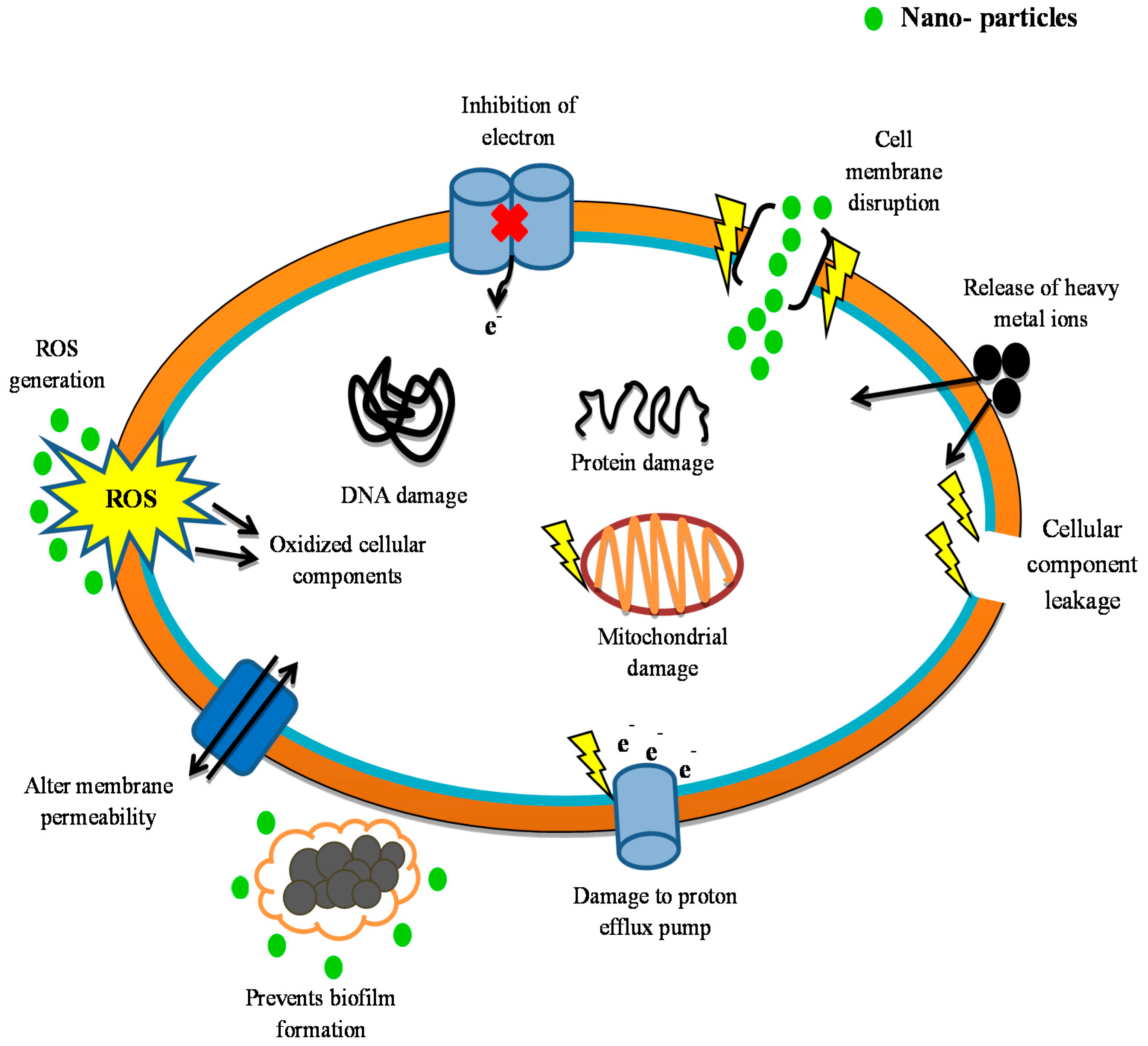
4.4. Mechanism of Action of Nanoparticles
4.5. Drug Release Kinetics of Nanoantibiotics
- i.
- Surface-bound or adsorbed product desorption
- ii.
- Drugs diffusion from polymeric NPs
- iii.
- Polymeric NP erosion and a cumulative erosion/diffusion effect
- i.
- Lipid membrane composition
- ii.
- Nature of drug involved
- iii.
- The percentage of drug’s permeability
- iv.
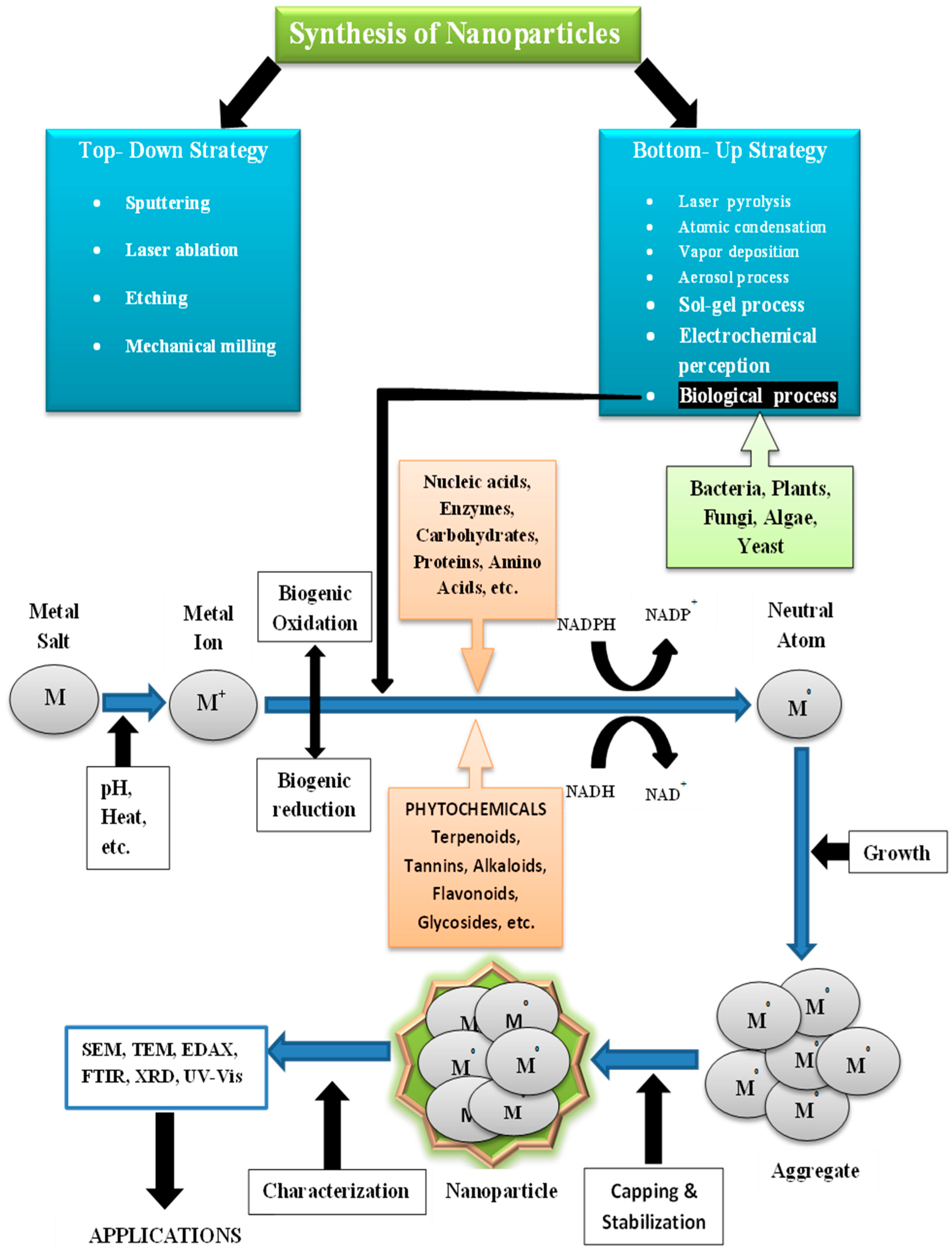
4.6. Synthesis of Nanoparticles
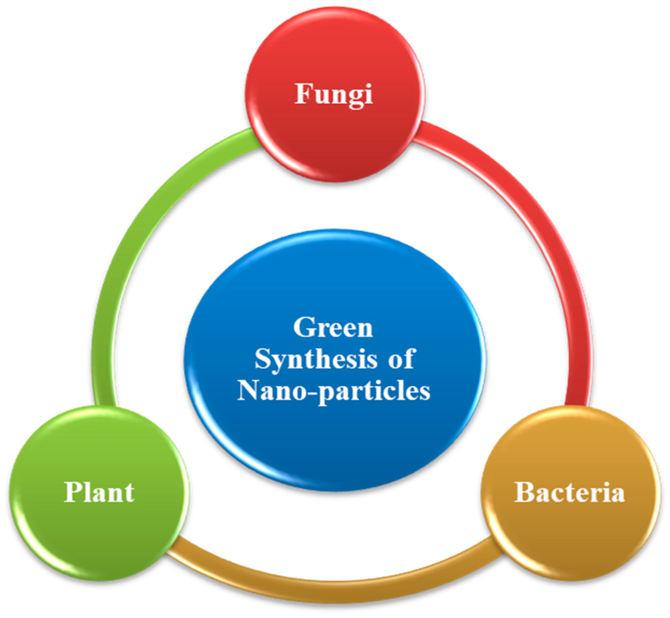
4.6.1. Green Synthesis of Nanoparticles
Fungi
Bacteria
Plant
4.6.2. Purification of Nanoparticles Extracted Biologically
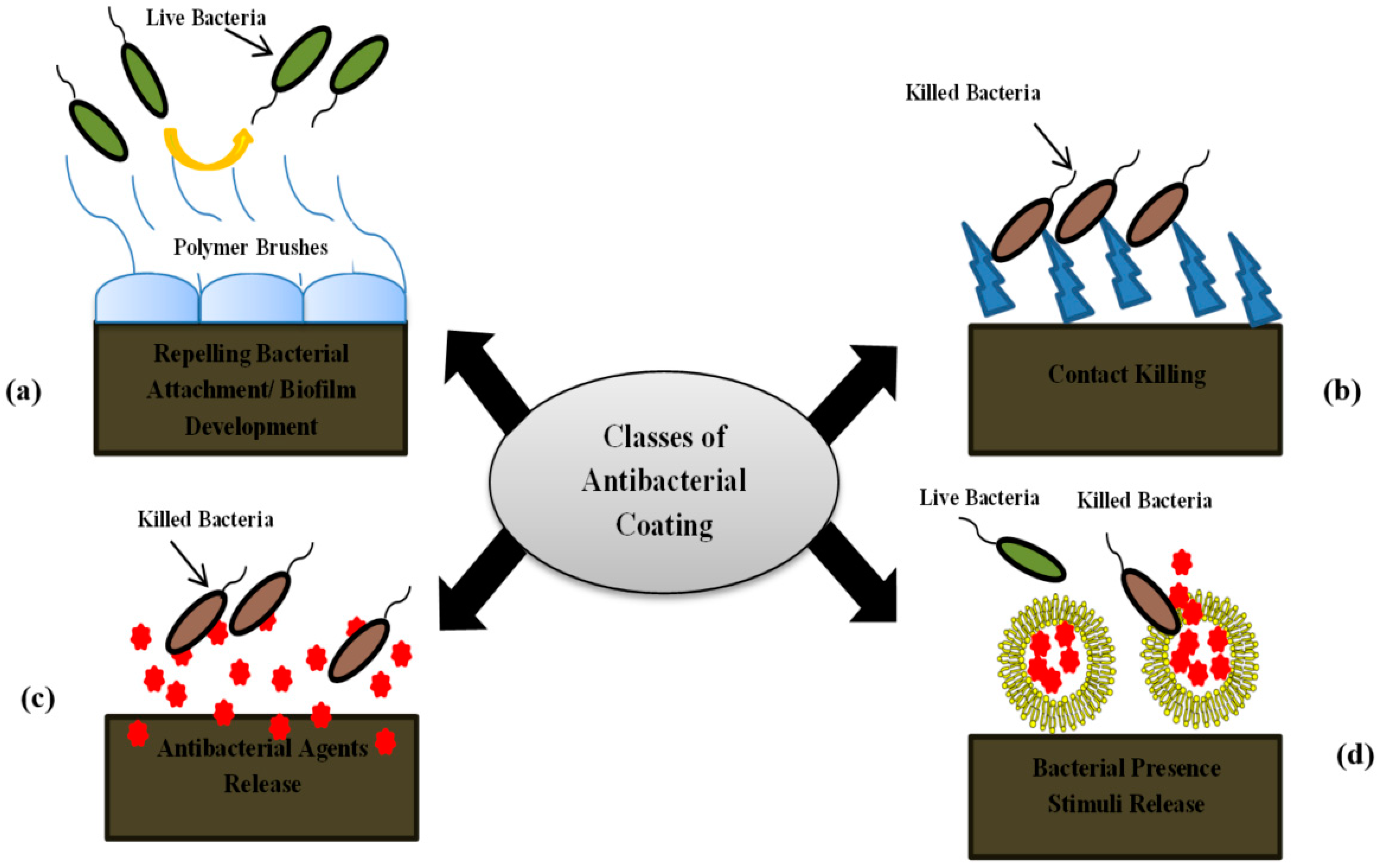
4.6.3. Nanoparticles Coating for Antibacterial Activity
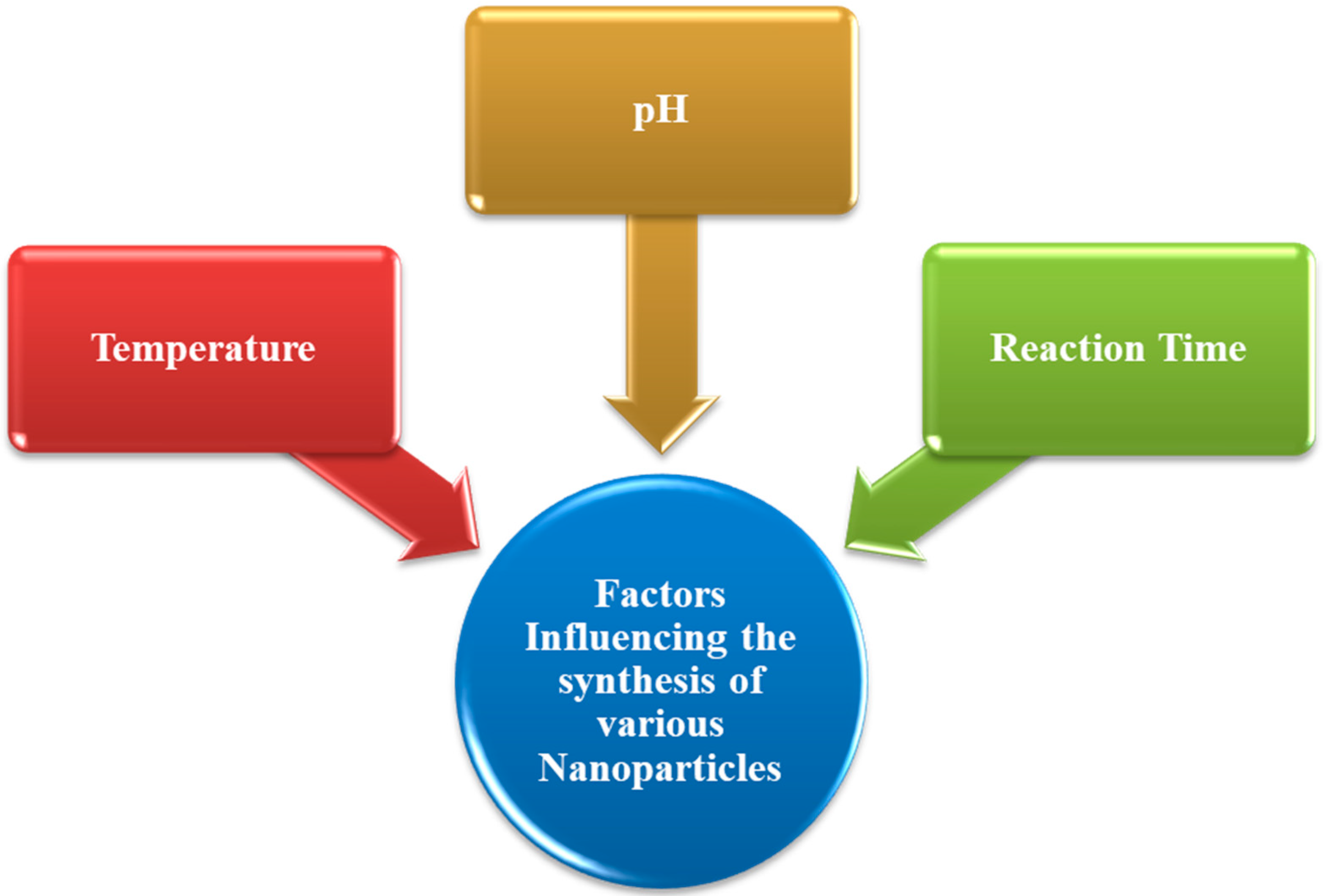
4.7. Factors Influencing the Synthesis of Various NPs
4.7.1. Temperature
4.7.2. pH
4.7.3. Reaction Time
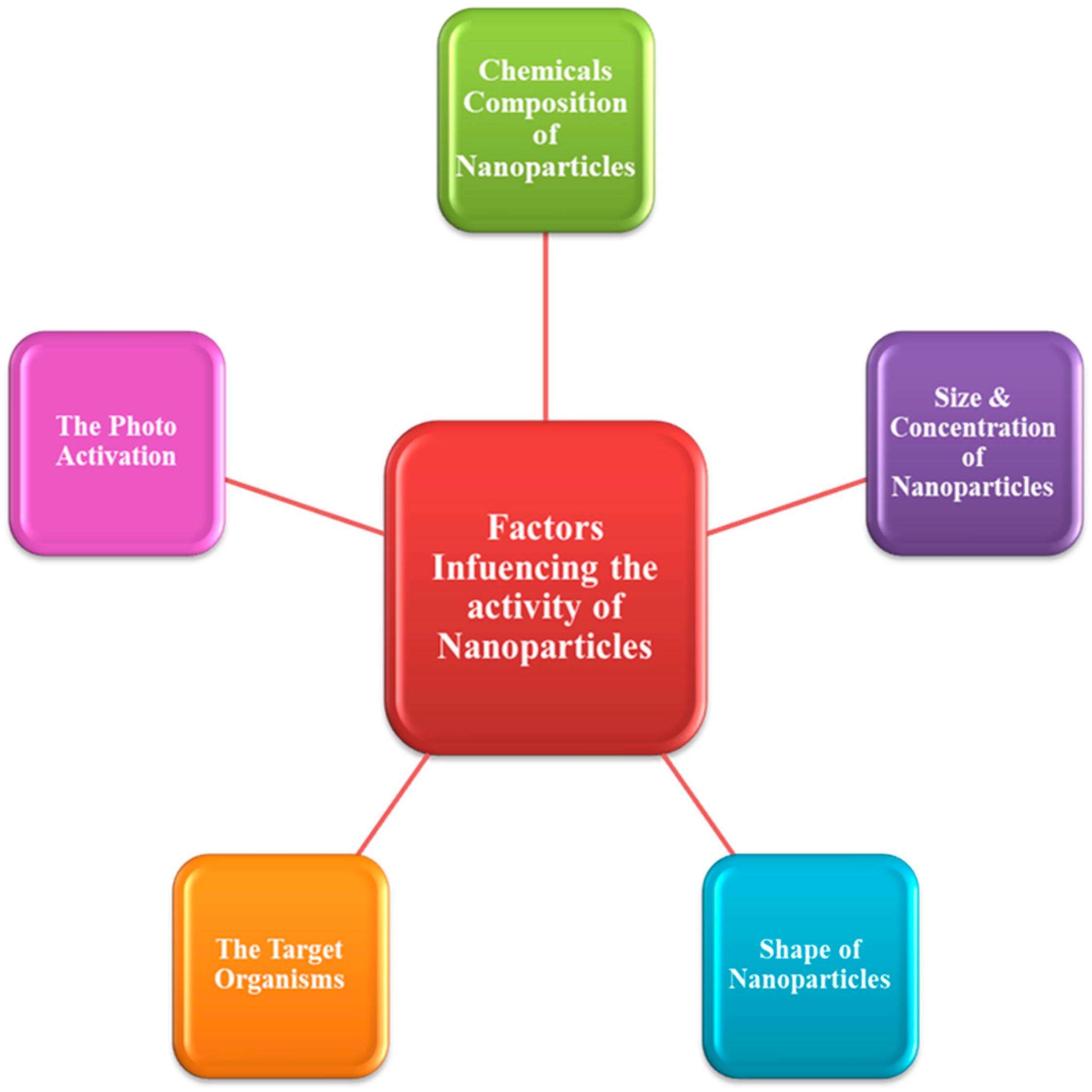
4.8. Factors Influencing the Activity of Nanoparticles
4.8.1. Chemical Composition of Nanoparticle
4.8.2. Shape of Nanoparticles
4.8.3. The Target Organisms
4.8.4. The Photoactivation
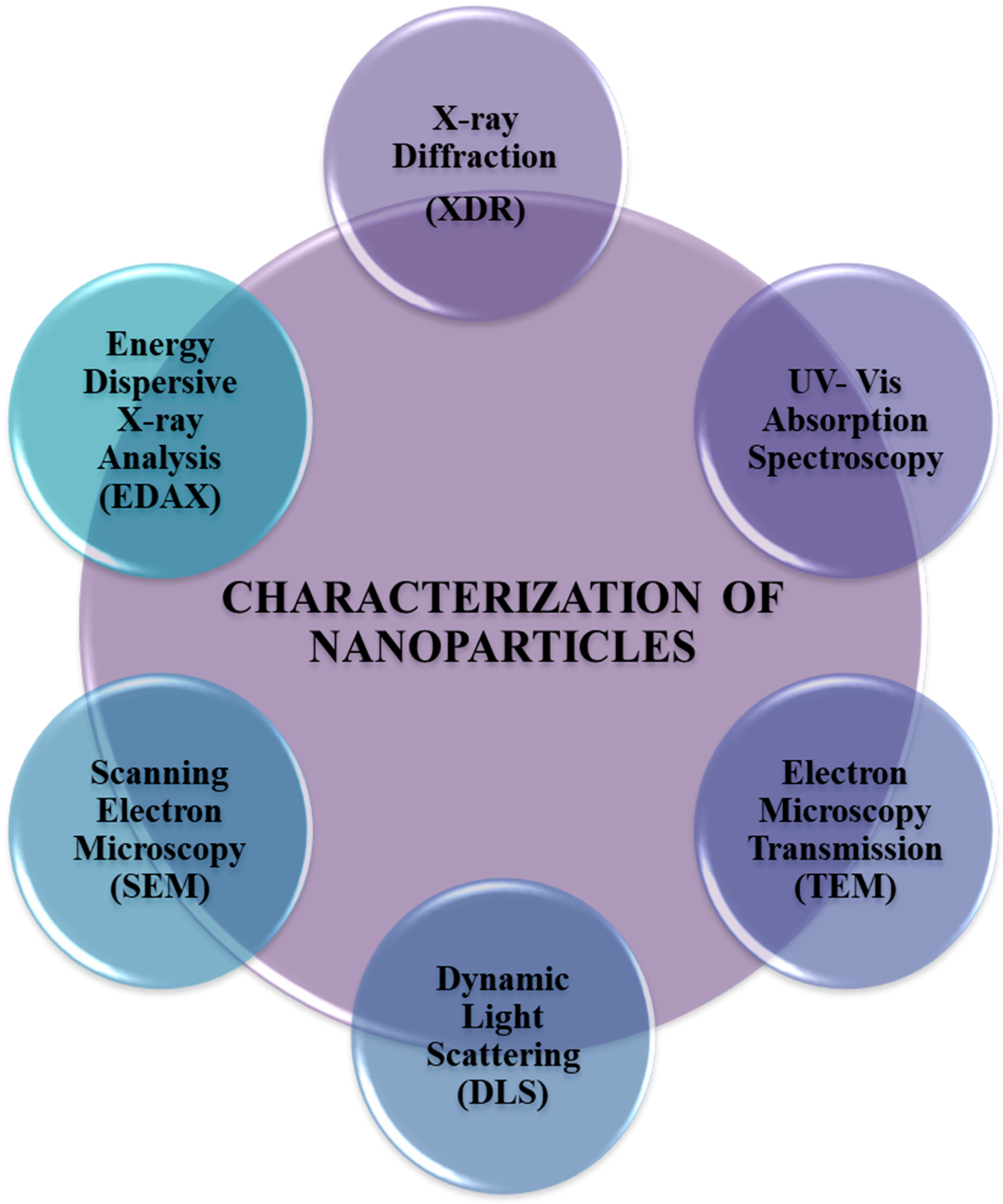
5. Characterization of Nanoparticles
6. Comparison of Antibiotics with Nanoparticles
7. Antimicrobials and Nanoparticles in Combination
8. Antimicrobial Applications of Nanoparticles on Animal Model
9. Challenges for Nanoparticle
10. Conclusions
Author Contributions
Funding
Acknowledgments
Conflicts of Interest
References
- Etebu, E.; Arikekpar, I. Antibiotics: Classification and mechanisms of action with emphasis on molecular perspectives. Int. J. Appl. Microbiol. Biotechnol. Res. 2016, 4, 90–101. [Google Scholar]
- Kohanski, M.A.; Dwyer, D.J.; Collins, J.J. How antibiotics kill bacteria: From targets to networks. Nat. Rev. Genet. 2010, 8, 423–435. [Google Scholar] [CrossRef] [PubMed] [Green Version]
- Padiyara, P.; Inoue, H.; Sprenger, M. Global Governance Mechanisms to Address Antimicrobial Resistance. Infect. Dis. Res. Treat. 2018, 11, 11. [Google Scholar] [CrossRef] [PubMed] [Green Version]
- Pelgrift, R.Y.; Friedman, A.J. Nanotechnology as a therapeutic tool to combat microbial resistance. Adv. Drug Deliv. Rev. 2013, 65, 1803–1815. [Google Scholar] [CrossRef] [PubMed]
- Ventola, C.L. The antibiotic resistance crisis: Part 1: Causes and threats. J. Manag. Care Hosp. Formul. Manag. 2015, 40, 277–283. [Google Scholar]
- Baluja, Z.; Nabi, N.; Ray, A. Challenges in Antimicrobial Resistance: An Update, EC Pharmacol. Toxicology 2018, 6, 865–877. [Google Scholar]
- Davis, M.D.M.; Whittaker, A.; Lindgren, M.; Djerf-Pierre, M.; Manderson, L.; Flowers, P. Understanding media publics and the antimicrobial resistance crisis. Glob. Public Health 2017, 13, 1158–1168. [Google Scholar] [CrossRef]
- Blecher, K.; Nasir, A.; Friedman, A. The growing role of nanotechnology in combating infectious disease. Virulence 2011, 2, 395–401. [Google Scholar] [CrossRef] [PubMed] [Green Version]
- Huh, A.J.; Kwon, Y.J. “Nanoantibiotics”: A new paradigm for treating infectious diseases using nanomaterials in the antibiotics resistant era. J. Control. Release 2011, 156, 128–145. [Google Scholar] [CrossRef]
- Teixeira, M.; Sanchez-Lopez, E.; Espina, M.; Calpena, A.; Silva, A.; Veiga, F.; Garcia, M.L.; Souto, E. Chapter 9—Advances in antibiotic nanotherapy: Overcoming antimicrobial resistance. In Emerging Nanotechnologies in Immunology; Elsevier: Boston, MA, USA, 2018; pp. 233–259. [Google Scholar]
- Bartlett, J.G.; Gilbert, D.N.; Spellberg, B. Seven Ways to Preserve the Miracle of Antibiotics. Clin. Infect. Dis. 2013, 56, 1445–1450. [Google Scholar] [CrossRef]
- Adeniji, F. Global analysis of strategies to tackle antimicrobial resistance. Int. J. Pharm. Pract. 2018, 26, 85–89. [Google Scholar] [CrossRef]
- Wong, I.Y.; Bhatia, S.N.; Toner, M. Nanotechnology: Emerging tools for biology and medicine. Genes Dev. 2013, 27, 2397–2408. [Google Scholar] [CrossRef] [Green Version]
- Jena, M.; Mishra, S.; Jena, S.; Mishra, S. Nanotechnology-future prospect in recent medicine: A review. Int. J. Basic Clin. Pharmacol. 2013, 2, 353–359. [Google Scholar] [CrossRef] [Green Version]
- Allen, H.; Moe, L.; Rodbumrer, J.; Gaarder, A.; Handelsman, J. Functional metagenomics reveals diverse Beta-lactamases in a remote Alaskan soil. ISME J. 2009, 3, 243–251. [Google Scholar] [CrossRef]
- Donato, J.J.; Moe, L.A.; Converse, B.J.; Smart, K.D.; Berklein, F.C.; McManus, P.S.; Handelsman, J. Metagenomic Analysis of Apple Orchard Soil Reveals Antibiotic Resistance Genes Encoding Predicted Bifunctional Proteins. Appl. Environ. Microbiol. 2010, 76, 4396–4401. [Google Scholar] [CrossRef] [Green Version]
- Georgiev, V.S. National Institute of Allergy and infectious diseases. In NIH: Volume 2: Impact on Global Health; Springer Science & Business Media: North Bethesda, MD, USA, 2009. [Google Scholar]
- Morgun, A.; Dzutsev, A.; Dong, X.; Greer, R.; Sexton, D.J.; Ravel, J.; Schuster, M.; Hsiao, W.; Matzinger, P.; Shulzhenko, N. Uncovering effects of antibiotics on the host and microbiota using transkingdom gene networks. Gut 2015, 64, 1732–1743. [Google Scholar] [CrossRef] [PubMed] [Green Version]
- Scully, J.L. What is a disease? Disease, disability and their definitions. Eur. Mol. Biol. Organ. 2004, 7, 650–653. [Google Scholar]
- Friswell, M.; Campbell, B.; Rhodes, J. The Role of Bacteria in the Pathogenesis of Inflammatory Bowel Disease. Gut Liver 2010, 4, 295–306. [Google Scholar] [CrossRef]
- Baker, S.J.; Hui, X.; Maibach, H.I. Progress on new therapeutics for fungal nail infections. Ann. Rep. Med. Chem. 2005, 40, 323. [Google Scholar]
- Peiris, J.S.; Guan, Y.; Yuen, K.Y. Severe acute respiratory syndrome. Nat. Med. 2004, 10, S88–S97. [Google Scholar] [CrossRef]
- Mosqueda, J.; Olvera-Ramirez, A.; Aguilar-Tipacamu, G.; Canto, G.J. Current Advances in Detection and Treatment of Babesiosis. Curr. Med. Chem. 2012, 19, 1504–1518. [Google Scholar] [CrossRef] [Green Version]
- Seok, J.Y.; Lee, Y.; Lee, H.; Yi, S.Y.; Oh, H.E.; Song, J.-S. Human Cutaneous Protothecosis: Report of a Case and Literature Review. Korean J. Pathol. 2013, 47, 575–578. [Google Scholar] [CrossRef]
- Eckburg, P.B.; Lepp, P.W.; Relman, D.A. Archaea and Their Potential Role in Human Disease. Infect. Immun. 2003, 71, 591–596. [Google Scholar] [CrossRef] [Green Version]
- Ishaq, S.L.; Moses, P.L.; Wright, A.D. The pathology of methanogenic archaea in human gastrointestinal tract disease. Gut Microbiome Implic. Hum. Dis. 2016. [Google Scholar] [CrossRef] [Green Version]
- De Jesus, C.; Whiting, R. Thermal inactivation, growth, and survival studies of Listeria monocytogenes strains belonging to three distinct genotypic lineages. J. Food Prot. 2003, 66, 1611–1617. [Google Scholar] [CrossRef]
- Mohr, K.I. History of Antibiotics Research. In Current Topics in Microbiology and Immunology; Springer: Cham, Switzerland, 2016; Volume 398, pp. 237–272. [Google Scholar]
- Bankova, V. Chemical diversity of propolis and the problem of standardization. J. Ethnopharmacol. 2005, 100, 114–117. [Google Scholar] [CrossRef]
- Aminov, R. History of antimicrobial drug discovery: Major classes and health impact. Biochem. Pharm. 2017, 133, 4–19. [Google Scholar] [CrossRef]
- Oliver, S.P.; Murinda, S.E.; Jayarao, B.M. Impact of Antibiotic Use in Adult Dairy Cows on Antimicrobial Resistance of Veterinary and Human Pathogens: A Comprehensive Review. Foodborne Pathog. Dis. 2011, 8, 337–355. [Google Scholar] [CrossRef]
- Armelagos, G.J.; Kolbacher, K.; Collins, K.; Cook, J.; Krafeld-Daugherty, M. Tetracycline consumption in prehistory. In Tetracyclines in Biology, Chemistry and Medicine; Springer: Cham, Switzerland, 2001; pp. 219–236. [Google Scholar]
- Ryu, C.; Lee, K.; Yoo, C.; Seong, W.K.; Oh, H.-B. Sensitive and Rapid Quantitative Detection of Anthrax Spores Isolated from Soil Samples by Real-Time PCR. Microbiol. Immunol. 2003, 47, 693–699. [Google Scholar] [CrossRef] [Green Version]
- Lipp, E.K.; Huq, A.; Colwell, R.R. Effects of Global Climate on Infectious Disease: The Cholera Model. Clin. Microbiol. Rev. 2002, 15, 757–770. [Google Scholar] [CrossRef] [PubMed] [Green Version]
- Ostensvik, O.; Skulberg, O.M.; Underdal, B.; Hormazabal, V. Antibacterial properties of extracts from selected planktonic freshwater cyanobacteria—A comparative study of bacterial bioassays. J. Appl. Microbiol. 1998, 84, 1117–1124. [Google Scholar] [CrossRef]
- Arai, T.; Mikami, Y.; Fukushima, K.; Utsumi, T.; Yazawa, K. A new antibiotic, leucinostatin, derived from penicillium lilacinum. J. Antibiot. 1973, 26, 157–161. [Google Scholar] [CrossRef]
- Nadar, V.S.; Chen, J.; Dheeman, D.S.; Galván, A.E.; Yoshinaga-Sakurai, K.; Kandavelu, P.; Sankaran, B.; Kuramata, M.; Ishikawa, S.; Rosen, B.P.; et al. Arsinothricin, an arsenic-containing non-proteinogenic amino acid analog of glutamate, is a broad-spectrum antibiotic. Commun. Biol. 2019, 2, 1–12. [Google Scholar] [CrossRef] [Green Version]
- Cole, H.N. The use of antisyphilitic remedies. J. Am. Med. Assoc. 1936, 107, 2123. [Google Scholar] [CrossRef]
- Xiong, G.M.; Venkatraman, K.; Venkatraman, S. The magic bullet as cancer therapeutic—Has nanotechnology failed to find its mark? Prog. Biomed. Eng. 2020, 2, 042004. [Google Scholar] [CrossRef]
- Giesbrecht, P.; Kersten, T.; Maidhof, H.; Wecke, J. Staphylococcal cell wall: Morphogenesis and fatal variations in the presence of penicillin. Microbiol. Mol. Biol. Rev. 1998, 62, 1371–1414. [Google Scholar] [CrossRef] [PubMed] [Green Version]
- Jenkinson, D. The effects of biocidal treatments on metabolism in soil—IV. The decomposition of fumigated organisms in soil. Soil Biol. Biochem. 1976, 8, 203–208. [Google Scholar] [CrossRef]
- Bentley, R. Different roads to discovery; Prontosil (hence sulfa drugs) and penicillin (hence β-lactams). J. Ind. Microbiol. Biotechnol. 2009, 36, 775–786. [Google Scholar] [CrossRef]
- Yousef, F.; Mansour, O.; Herbali, J. Sulfonamides: Historical Discovery Development (Structure-Activity Relationship Notes). In-Vitro In-Vivo In-Silico J. 2018, 1, 1–15. [Google Scholar]
- Genç, Y.; Özkanca, R.; Bekdemir, Y. Antimicrobial activity of some sulfonamide derivatives on clinical isolates of Staphylococus aureus. Ann. Clin. Microbiol. Antimicrob. 2008, 7, 17. [Google Scholar] [CrossRef] [Green Version]
- Wood, W.B.; Long, P.H. Observations upon the experimental and clinical use of sulfapyridine. III. The mechanism of recovery from pneumococcal pneumonia in patients treated with sulfapyridine. Ann. Intern. Med. 1939, 13, 612. [Google Scholar] [CrossRef]
- Stauss-Grabo, M.; Atiye, S.; Le, T.; Kretschmar, M. Decade-long use of the antimicrobial peptide combination tyrothricin does not pose a major risk of acquired resistance with gram-positive bacteria and Candida spp. Die Pharm. 2014, 69, 838–841. [Google Scholar]
- Takada, Y.; Itoh, H.; Paudel, A.; Panthee, S.; Hamamoto, H.; Sekimizu, K.; Inoue, M. Discovery of gramicidin A analogues with altered activities by multidimensional screening of a one-bead-one-compound library. Nat. Commun. 2020, 11, 1–10. [Google Scholar] [CrossRef]
- Stark, B.J.; Earl, H.S.; Gross, G.N.; Lumry, W.R.; Goodman, E.L.; Sullivan, T.J. Acute and chronic desensitization of penicillin-allergic patients using oral penicillin. J. Allergy Clin. Immunol. 1987, 79, 523–532. [Google Scholar] [CrossRef]
- Genvert, I.G.; Cohen, E.J.; Donnenfeld, E.D.; Blecher, M.H. Erythema multiforme after use of topical sulfacetamide. Am. J. Ophthalmol. 1985, 99, 465–468. [Google Scholar] [CrossRef]
- Singha, P.; Locklin, J.; Handa, H. A review of the recent advances in antimicrobial coatings for urinary catheters. Acta Biomater. 2017, 50, 20–40. [Google Scholar] [CrossRef] [PubMed] [Green Version]
- Abeylath, S.C.; Turos, E. Drug delivery approaches to overcome bacterial resistance to β-lactam antibiotics. Expert Opin. Drug Deliv. 2008, 5, 931–949. [Google Scholar] [CrossRef]
- Forrest, D.M.; Schellenberg, R.R.; Thien, V.V.S.; King, S.; Anis, A.H.; Dodek, P.M. Introduction of a Practice Guideline for Penicillin Skin Testing Improves the Appropriateness of Antibiotic Therapy. Clin. Infect. Dis. 2001, 32, 1685–1690. [Google Scholar] [CrossRef] [Green Version]
- Brooks, D.; Garrett, G.; Hollihead, R. Sulphadimidine, co-trimoxazole, and a placebo in the management of symptomatic urinary tract infection in general practice. J. R. Coll. Gen. Pract. 1972, 22, 695–703. [Google Scholar]
- Li, B.; Webster, T.J. Bacteria antibiotic resistance: New challenges and opportunities for implant-associated orthopedic infections. J. Orthop. Res. 2017, 36, 22–32. [Google Scholar] [CrossRef] [PubMed] [Green Version]
- Hancock, R.E.; Chapple, D.S. Peptide antibiotics. Antimicrob. Agents Chemother. 1999, 43, 1317–1323. [Google Scholar] [CrossRef] [Green Version]
- Lewis, K. Platforms for antibiotic discovery. Nat. Rev. Drug Discov. 2013, 12, 371–387. [Google Scholar] [CrossRef] [PubMed]
- Hossain, G.G.; Amoroso, A.; Banu, A.; Malik, K. Syntheses and characterisation of mercury complexes of sulfadiazine, sulfamerazine and sulfamethazine. Polyhedron 2007, 26, 967–974. [Google Scholar] [CrossRef]
- Shama, G.; Shama, G. Zones of inhibition? The transfer of information relating to penicillin in Europe during World War II. Adv. Appl. Microbiol. 2009, 69, 133–158. [Google Scholar]
- Lipsky, B.A.; Hoey, C. Topical Antimicrobial Therapy for Treating Chronic Wounds. Clin. Infect. Dis. 2009, 49, 1541–1549. [Google Scholar] [CrossRef] [Green Version]
- Harir, M.; Bendif, H.; Bellahcene, M.; Pogni, Z.F.A.R. Streptomyces Secondary Metabolites. Basic Biol. Appl. Actinobacteria 2018, 6, 99–122. [Google Scholar] [CrossRef] [Green Version]
- Strohl, W.R. Antimicrobials. In Microbial Diversity and Bioprospecting; ASM Press: Washington, DC, USA, 2014; pp. 336–355. [Google Scholar] [CrossRef]
- Figueroa, R.A.; Leonard, A.; Mackay, A.A. Modeling Tetracycline Antibiotic Sorption to Clays. Environ. Sci. Technol. 2004, 38, 476–483. [Google Scholar] [CrossRef]
- He, Z.; Kisla, D.; Zhang, L.; Yuan, C.; Green-Church, K.B.; Yousef, A.E. Isolation and Identification of a Paenibacillus polymyxa Strain That Coproduces a Novel Lantibiotic and Polymyxin. Appl. Environ. Microbiol. 2006, 73, 168–178. [Google Scholar] [CrossRef] [Green Version]
- Gadebusch, H.H.; Basch, H.I. New Antimicrobial Nitrofuran, trans-5-Amino-3-[2-(5-Nitro-2-Furyl) Vinyl]-Δ2-1,2,4-Oxadiazole: Antibacterial, Antifungal, and Antiprotozoal Activities In Vitro. Antimicrob. Agents Chemother. 1974, 6, 263–267. [Google Scholar] [CrossRef] [PubMed] [Green Version]
- Wulf, N.R.; Matuszewski, K.A. Sulfonamide cross-reactivity: Is there evidence to support broad cross-allergenicity? Am. J. Health Pharm. 2013, 70, 1483–1494. [Google Scholar] [CrossRef]
- Stypulkowska, K.; Blazewicz, A.; Fijalek, Z.; Warowna-Grześkiewicz, M.; Srebrzynska, K. Determination of neomycin and related substances in pharmaceutical preparations by reversed-phase high performance liquid chromatography with mass spectrometry and charged aerosol detection. J. Pharm. Biomed. Anal. 2013, 76, 207–214. [Google Scholar] [CrossRef]
- Das, S.; Al Faysal, M.N.; Ferdous, J.; Sachi, S.; Islam, M.S.; Sikder, M.H. Detection of oxytetracycline and doxycycline residue in different growth stages of commercial broiler. Bangladesh J. Vet. Med. 2009, 17, 7–14. [Google Scholar]
- George, A.M.; Levy, S.B. Amplifiable resistance to tetracycline, chloramphenicol, and other antibiotics in Escherichia coli: Involvement of a non-plasmid-determined efflux of tetracycline. J. Bacteriol. 1983, 155, 531–540. [Google Scholar] [CrossRef] [Green Version]
- Yoneyama, H.; Katsumata, R. Antibiotic Resistance in Bacteria and Its Future for Novel Antibiotic Development. Biosci. Biotechnol. Biochem. 2006, 70, 1060–1075. [Google Scholar] [CrossRef]
- Cannon, M.; Harford, S.; Davies, J. A comparative study on the inhibitory actions of chloramphenicol, thiamphenicol and some fluorinated derivatives. J. Antimicrob. Chemother. 1990, 26, 307–317. [Google Scholar] [CrossRef]
- Amin, M.M.; Zilles, J.L.; Greiner, J.; Charbonneau, S.; Raskin, L.; Morgenroth, E. Influence of the Antibiotic Erythromycin on Anaerobic Treatment of a Pharmaceutical Wastewater. Environ. Sci. Technol. 2006, 40, 3971–3977. [Google Scholar] [CrossRef] [PubMed]
- Crank, C.W.; O’Driscoll, T. Vancomycin-resistant enterococcal infections: Epidemiology, clinical manifestations, and optimal management. Infect. Drug Resist. 2015, 8, 217–230. [Google Scholar] [CrossRef] [Green Version]
- Abraham, E.P. Cephalosporins 1945–1986. Drugs 1987, 34, 1–14. [Google Scholar] [CrossRef]
- Zhong, P.; Shortridge, V.D. The role of efflux in macrolide resistance. Drug Resist. Updat. 2000, 3, 325–329. [Google Scholar] [CrossRef] [PubMed]
- Clement, M.E.; Okeke, N.L.; Hicks, C.B. Treatment of syphilis: A systematic review. JAMA 2014, 312, 1905–1917. [Google Scholar] [CrossRef]
- Li, Y.; Wang, F.; Wu, L.; Zhu, M.; He, G.; Chen, X.; Sun, F.; Liu, Q.; Wang, X.; Zhang, W. Cycloserine for treatment of multidrug-resistant tuberculosis: A retrospective cohort study in China. Infect. Drug Resist. 2019, 12, 721–731. [Google Scholar] [CrossRef] [PubMed] [Green Version]
- Valentini, P.; Annunziata, M.L.; Angelone, D.F.; Masini, L. Role of spiramycin/cotrimoxazole association in the mother-to-child transmission of toxoplasmosis infection in pregnancy. Eur. J. Clin. Microbiol. Infect. Dis. 2009, 28, 297–300. [Google Scholar] [CrossRef] [PubMed]
- Flynt, L.K.; Kenney, R.M.; Zervos, M.J.; Davis, S.L. The Safety and Economic Impact of Cefazolin versus Nafcillin for the Treatment of Methicillin-Susceptible Staphylococcus aureus Bloodstream Infections. Infect. Dis. Ther. 2017, 6, 225–231. [Google Scholar] [CrossRef] [Green Version]
- Weisblum, B. Insights into erythromycin action from studies of its activity as inducer of resistance. Antimicrob. Agents Chemother. 1995, 39, 797–805. [Google Scholar] [CrossRef] [Green Version]
- Gikalo, M.B.; Nosova, E.Y.; Krylova, L.Y.; Moroz, A.M. The role of eis mutations in the development of kanamycin resistance in Mycobacterium tuberculosis isolates from the Moscow region. J. Antimicrob. Chemother. 2012, 67, 2107–2109. [Google Scholar] [CrossRef]
- Schiewe, H.J.; Zeeck, A. Cineromycins, γ-butyrolactones and ansamycins by analysis of the secondary metabolite pattern created by a single strain of Streptomyces. J. Antibiot. 1999, 52, 635–642. [Google Scholar] [CrossRef] [Green Version]
- Cheng, I.-L.; Chen, Y.-H.; Lai, C.-C.; Tang, H.-J. Intravenous Colistin Monotherapy versus Combination Therapy against Carbapenem-Resistant Gram-Negative Bacteria Infections: Meta-Analysis of Randomized Controlled Trials. J. Clin. Med. 2018, 7, 208. [Google Scholar] [CrossRef] [Green Version]
- Müller, M. Reductive activation of nitroimidazoles in anaerobic microorganisms. Biochem. Pharm. 1986, 35, 37–41. [Google Scholar] [CrossRef]
- Enwemeka, C.S.; Williams, D.; Enwemeka, S.K.; Hollosi, S.; Yens, D. Blue 470-nm Light Kills Methicillin-Resistant Staphylococcus aureus (MRSA) in Vitro. Photomed. Laser Surg. 2009, 27, 221–226. [Google Scholar] [CrossRef]
- Mitchell, D.A. Metronidazole: Its use in clinical dentistry. J. Clin. Periodontol. 1984, 11, 145–158. [Google Scholar] [CrossRef]
- Harkins, C.P.; Pichon, B.; Doumith, M.; Parkhill, J.; Westh, H.; Tomasz, A.; De Lencastre, H.; Bentley, S.D.; Kearns, A.M.; Holden, M.T.G. Methicillin-resistant Staphylococcus aureus emerged long before the introduction of methicillin into clinical practice. Genome Biol. 2017, 18, 130. [Google Scholar] [CrossRef] [Green Version]
- Klein, J.O. Role of nontypeable Haemophilus influenzae in pediatric respiratory tract infections. Pediatr. Infect. Dis. J. 1997, 16, S5–S8. [Google Scholar] [CrossRef]
- Riedel, S.; Vijayakumar, D.; Berg, G.; Kang, A.D.; Smith, K.P.; Kirby, E.J. Evaluation of apramycin against spectinomycin-resistant and -susceptible strains of Neisseria gonorrhoeae. J. Antimicrob. Chemother. 2019, 74, 1311–1316. [Google Scholar] [CrossRef]
- Jönsson, S.; Davidse, A.; Wilkins, J.; Van Der Walt, J.-S.; Simonsson, U.S.H.; Karlsson, M.O.; Smith, P.; McIlleron, H. Population Pharmacokinetics of Ethambutol in South African Tuberculosis Patients. Antimicrob. Agents Chemother. 2011, 55, 4230–4237. [Google Scholar] [CrossRef] [Green Version]
- Werner, A.; Russell, A. Mupirocin, fusidic acid and bacitracin: Activity, action and clinical uses of three topical antibiotics. Veter Dermatol. 1999, 10, 225–240. [Google Scholar] [CrossRef]
- Andriole, V.T. The Quinolones: Past, Present, and Future. Clin. Infect. Dis. 2005, 41, S113–S119. [Google Scholar] [CrossRef] [Green Version]
- Imbuluzqueta, E.; Elizondo, E.; Gamazo, C.; Moreno-Calvo, E.; Veciana, J.; Ventosa, N.; Blanco-Prieto, M.; María, J. Novel bioactive hydrophobic gentamicin carriers for the treatment of intracellular bacterial infections. Acta Biomater. 2011, 7, 1599–1608. [Google Scholar] [CrossRef] [PubMed]
- Thomson, J.M.; Bonomo, A.R. The threat of antibiotic resistance in Gram-negative pathogenic bacteria: β-lactams in peril! Curr. Opin. Microbiol. 2005, 8, 518–524. [Google Scholar] [CrossRef]
- Shaikh, S.; Fatima, J.; Shakil, S.; Rizvi, S.M.D.; Kamal, M.A. Antibiotic resistance and extended spectrum beta-lactamases: Types, epidemiology and treatment. Saudi J. Biol. Sci. 2015, 22, 90–101. [Google Scholar] [CrossRef] [Green Version]
- Crumplin, G.C.; Smith, J.T. Nalidixic Acid: An Antibacterial Paradox. Antimicrob. Agents Chemother. 1975, 8, 251–261. [Google Scholar] [CrossRef] [Green Version]
- Holmes, N.E.; Charles, P.G. Safety and Efficacy Review of Doxycycline. Clin. Med. 2009, 1, CMT-S2035. [Google Scholar] [CrossRef] [Green Version]
- Momtaz, H.; Khamesipour, F.; Tavakol, M.; Awosile, B. Determination of Antimicrobial Resistance and Resistant Genes in Acinetobacter Baumannii from Human Clinical Samples. West Indian Med. J. 2015, 66, 56–64. [Google Scholar] [CrossRef] [Green Version]
- Kasten, M.J. Clindamycin, Metronidazole, and Chloramphenicol. Mayo Clin. Proc. 1999, 74, 825–833. [Google Scholar] [CrossRef]
- Korenromp, E.L.; Scano, F.; Williams, B.G.; Dye, C.; Nunn, P. Effects of Human Immunodeficiency Virus Infection on Recurrence of Tuberculosis after Rifampin-Based Treatment: An Analytical Review. Clin. Infect. Dis. 2003, 37, 101–112. [Google Scholar] [CrossRef]
- Speer, B.S.; Shoemaker, N.B.; Salyers, A.A. Bacterial resistance to tetracycline: Mechanisms, transfer, and clinical significance. Clin. Microbiol. Rev. 1992, 5, 387–399. [Google Scholar] [CrossRef]
- Brogden, R.N.; Carmine, A.A.; Heel, R.C.; Speight, T.M.; Avery, G.S. Trimethoprim: A review of its antibacterial activity, pharmacokinetics and therapeutic use in urinary tract infections. Drugs 1982, 23, 405–430. [Google Scholar] [CrossRef] [PubMed]
- Reffert, J.L.; Smith, W.J. Fosfomycin for the treatment of resistant gram-negative bacterial infections: Insights from the society of infectious diseases pharmacists. Pharmacother. J. Hum. Pharmacol. Drug Ther. 2014, 34, 845–857. [Google Scholar] [CrossRef]
- Ball, A.P.; Gray, J.A.; Murdoch, J.M. Antibacterial Drugs Today; Springer: New York, NY, USA, 2012. [Google Scholar]
- Lyu, Y.; Yang, X.; Goswami, S.; Gorityala, B.K.; Idowu, T.; Domalaon, R.; Zhanel, G.G.; Shan, A.; Schweizer, F. Amphiphilic Tobramycin–Lysine Conjugates Sensitize Multidrug Resistant Gram-Negative Bacteria to Rifampicin and Minocycline. J. Med. Chem. 2017, 60, 3684–3702. [Google Scholar] [CrossRef]
- Ward, A.; Campoli-Richards, D.M. Mupirocin. Drugs 1986, 32, 425–444. [Google Scholar] [CrossRef]
- Birnbaum, J.; Kahan, F.M.; Kropp, H.; Macdonald, J.S. Carbapenems, a new class of beta-lactam antibiotics: Discovery and development of imipenem/cilastatin. Am. J. Med. 1985, 78, 3–21. [Google Scholar] [CrossRef]
- Garrido-Mesa, N.; Camuesco, D.; Arribas, B.; Comalada, M.; Bailón, E.; Cueto-Sola, M.; Utrilla, P.; Nieto, A.; Zarzuelo, A.; Rodríguez-Cabezas, M.E.; et al. The intestinal anti-inflammatory effect of minocycline in experimental colitis involves both its immunomodulatory and antimicrobial properties. Pharmacol. Res. 2011, 63, 308–319. [Google Scholar] [CrossRef]
- Cañete, R.; Escobedo, A.A.; González, M.E.; Almirall, P.; Cantelar, N. A randomized, controlled, open-label trial of a single day of mebendazole versus a single dose of tinidazole in the treatment of giardiasis in children. Curr. Med. Res. Opin. 2006, 22, 2131–2136. [Google Scholar] [CrossRef]
- Klastersky, J.; Vamecq, G.; Cappel, R.; Swings, G.; Vandenborre, L. Effects of the Combination of Gentamicin and Carbenicillin on the Bactericidal Activity of Serum. J. Infect. Dis. 1972, 125, 183–186. [Google Scholar] [CrossRef]
- Liu, Q.; Li, M.; Zhang, F.; Yu, H.; Zhang, Q.; Liu, X. The removal of trimethoprim and sulfamethoxazole by a high infiltration rate artificial composite soil treatment system. Front. Environ. Sci. Eng. 2017, 11, 12. [Google Scholar] [CrossRef]
- Goldberg, E.; Bishara, J. Contemporary unconventional clinical use of co-trimoxazole. Clin. Microbiol. Infect. 2012, 18, 8–17. [Google Scholar] [CrossRef] [Green Version]
- Kawaguchi, H. Discovery, Chemistry, and Activity of Amikacin. J. Infect. Dis. 1976, 134, S242–S248. [Google Scholar] [CrossRef]
- Stapley, E.O.; Birnbaum, J.; Miller, A.K.; Wallick, H.; Hendlin, D.; Woodruff, H.B. Cefoxitin and Cephamycins: Microbiological Studies. Clin. Infect. Dis. 1979, 1, 73–87. [Google Scholar] [CrossRef] [PubMed]
- Butler, M.S.; A Hansford, K.; Blaskovich, M.A.T.; Halai, R.; Cooper, A.M. Glycopeptide antibiotics: Back to the future. J. Antibiot. 2014, 67, 631–644. [Google Scholar] [CrossRef]
- Bozkurt, A.; Deniz, M.; Yegen, B.C. Cefaclor, a cephalosporin antibiotic, delays gastric emptying rate by a CCK-A receptor-mediated mechanism in the rat. Br. J. Pharm. 2000, 131, 399–404. [Google Scholar] [CrossRef] [Green Version]
- Grover, N.; Sahni, A.; Bhattacharya, S. Therapeutic challenges of ESBLS and AmpC beta-lactamase producers in a tertiary care center. Med. J. Armed. Forces India 2013, 69, 4–10. [Google Scholar] [CrossRef] [Green Version]
- Owens, R.C., Jr.; Ambrose, P.G. Clinical use of the fluoroquinolones. Med. Clin. N. Am. 2000, 84, 1447–1469. [Google Scholar] [CrossRef]
- Sinha, M.; Srinivasa, H.; Macaden, R. Antibiotic resistance profile & extended spectrum beta-lactamase (ESBL) production in Acinetobacter species. Indian J. Med. Res. 2007, 126, 63. [Google Scholar] [PubMed]
- Chakravarty, I.; Kundu, K.; Kundu, S. Daptomycin: Discovery, development and perspectives. The battle against microbial pathogens: Basic science, technological advances and educational programs. Microbiology 2015, 2, 895–903. [Google Scholar]
- Bradley, J.; Garau, J.; Lode, H.; Rolston, K.; Wilson, S.; Quinn, J. Carbapenems in clinical practice: A guide to their use in serious infection. Int. J. Antimicrob. Agents 1999, 11, 93–100. [Google Scholar] [CrossRef]
- Bonten, M.J.; Willems, R.; A Weinstein, R. Vancomycin-resistant enterococci: Why are they here, and where do they come from? Lancet Infect. Dis. 2001, 1, 314–325. [Google Scholar] [CrossRef]
- Solomkin, J.S.; Mazuski, J. Intra-abdominal Sepsis: Newer Interventional and Antimicrobial Therapies. Infect. Dis. Clin. N. Am. 2009, 23, 593–608. [Google Scholar] [CrossRef]
- Ezelarab, H.A.A.; Abbas, S.H.; Hassan, H.A.; Abuo-Rahma, G.E.-D.A. Recent updates of fluoroquinolones as antibacterial agents. Arch. Pharm. 2018, 351, e1800141. [Google Scholar] [CrossRef] [PubMed]
- Pfeifer, Y.; Cullik, A.; Witte, W. Resistance to cephalosporins and carbapenems in Gram-negative bacterial pathogens. Int. J. Med. Microbiol. 2010, 300, 371–379. [Google Scholar] [CrossRef]
- El Amin, N.; Giske, C.G.; Jalal, S.; Keijser, B.; Kronvall, G.; Wretlind, B. Carbapenem resistance mechanisms in Pseudomonas aeruginosa: Alterations of porin OprD and efflux proteins do not fully explain resistance patterns observed in clinical isolates. APMIS 2005, 113, 187–196. [Google Scholar] [CrossRef]
- Hooper, D.C. Mechanisms of fluoroquinolone resistance. Drug Resist. Updat. 1999, 2, 38–55. [Google Scholar] [CrossRef]
- Harris, M.D. Infectious Disease in Athletes. Curr. Sports Med. Rep. 2011, 10, 84–89. [Google Scholar] [CrossRef] [PubMed]
- Marin, A.C.; McNicholl, A.G.; Gisbert, J.P. A review of rescue regimens after clarithromycin-containing triple therapy failure (for Helicobacter pylori eradication). Expert Opin. Pharmacother. 2013, 14, 843–861. [Google Scholar] [CrossRef]
- Huwyler, T.; Lenggenhager, L.; Abbas, M.; Lorenzini, K.I.; Hughes, S.; Huttner, B.; Karmime, A.; Uçkay, I.; Von Dach, E.; Lescuyer, P.; et al. Cefepime plasma concentrations and clinical toxicity: A retrospective cohort study. Clin. Microbiol. Infect. 2017, 23, 454–459. [Google Scholar] [CrossRef] [Green Version]
- Van Hal, S.J.; Paterson, D.L. Systematic Review and Meta-Analysis of the Significance of Heterogeneous Vancomycin-IntermediateStaphylococcus aureusIsolates. Antimicrob. Agents Chemother. 2010, 55, 405–410. [Google Scholar] [CrossRef] [PubMed] [Green Version]
- Lamb, H.M.; Figgitt, D.P.; Faulds, D. Quinupristin/Dalfopristin. Drugs 1999, 58, 1061–1097. [Google Scholar] [CrossRef] [PubMed]
- Chien, J.W.; Kucia, M.L.; Salata, R.A. Use of Linezolid, an Oxazolidinone, in the Treatment of Multidrug-Resistant Gram-Positive Bacterial Infections. Clin. Infect. Dis. 2000, 30, 146–151. [Google Scholar] [CrossRef] [Green Version]
- Krueger, W.A.; Unertl, K.E. New treatment option for gram-positive infections in critically ill patients-overview over linezolid. Anasthesiol. Intensivmed. Notf. Schmerzther. AINS 2002, 37, 199. [Google Scholar] [CrossRef] [PubMed]
- Shain, C.S.; Amsden, G.W. Telithromycin: The First of the Ketolides. Ann. Pharm. 2002, 36, 452–464. [Google Scholar] [CrossRef] [PubMed]
- Smith, A.; Pennefather, P.M.; Kaye, S.B.; Hart, C.A. Fluoroquinolones. Drugs 2001, 61, 747–761. [Google Scholar] [CrossRef]
- Mendes, R.E.; Deshpande, L.M.; Jones, R.N. Linezolid update: Stable in vitro activity following more than a decade of clinical use and summary of associated resistance mechanisms. Drug Resist. Updat. 2014, 17, 1–12. [Google Scholar] [CrossRef]
- Duval, R.E.; Grare, M.; Demoré, B. Fight against Antimicrobial Resistance: We Always Need New Antibacterials but for Right Bacteria. Molecular 2019, 24, 3152. [Google Scholar] [CrossRef] [Green Version]
- Chang, S.; Sievert, D.M.; Hageman, J.C.; Boulton, M.L. Infection with vancomycin-resistant Staphylococcus aureus containing the vanA resistance gene. N. Engl. J. Med. 2003, 348, 1342. [Google Scholar] [CrossRef]
- Pirri, G.; Giuliani, A.; Nicoletto, S.; Pizzuto, L.; Rinaldi, A. Lipopeptides as anti-infectives: A practical perspective. Open Life Sci. 2009, 4, 258–273. [Google Scholar] [CrossRef]
- Tally, F.P.; Zeckel, M.; Wasilewski, M.M.; Carini, C.; Berman, C.L.; Drusano, G.L.; Oleson, F.B., Jr. Daptomycin: A novel agent for Gram-positive infections. Expert Opin. Investig. Drugs 1999, 8, 1223–1238. [Google Scholar] [CrossRef]
- Ackermann, G.; Rodloff, A.C. Drugs of the 21st century: Telithromycin (HMR 3647)—The first ketolide. J. Antimicrob. Chemother. 2003, 51, 497–511. [Google Scholar] [CrossRef] [PubMed]
- Rose, W.E.; Rybak, M.J. Tigecycline: First of a new class of antimicrobial agents. Pharmacother. J. Hum. Pharmacol. Drug Ther. 2006, 26, 1099–1110. [Google Scholar] [CrossRef] [PubMed]
- Crook, D.W.; Walker, A.S.; Kean, Y.; Weiss, K.; Cornely, O.A.; Miller, M.A.; Esposito, R.; Louie, T.J.; Stoesser, N.E.; Young, B.C.; et al. Fidaxomicin Versus Vancomycin for Clostridium difficile Infection: Meta-analysis of Pivotal Randomized Controlled Trials. Clin. Infect. Dis. 2012, 55, S93–S103. [Google Scholar] [CrossRef] [PubMed]
- Deoghare, S. Bedaquiline: A new drug approved for treatment of multidrug-resistant tuberculosis. Indian J. Pharm. 2013, 45, 536–537. [Google Scholar] [CrossRef] [PubMed]
- Sandrock, C.E.; Shorr, A.F. The Role of Telavancin in Hospital-Acquired Pneumonia and Ventilator-Associated Pneumonia. Clin. Infect. Dis. 2015, 61, 79–86. [Google Scholar] [CrossRef] [Green Version]
- Sharma, C.; Singh, C.; Sharma, L.N.; Purvia, R.; Adlakha, M. Antibiotic resistant organism: An emerging public health problem and role of ayurveda (an overview). Int. J. Ayurveda Pharm. Res. 2014, 2, 17–29. [Google Scholar]
- O’Brien, J.J.; Campbell, N.; Conaghan, T. Effect of cooking and cold storage on biologically active antibiotic residues in meat. Epidemiol. Infect. 1981, 87, 511–523. [Google Scholar] [CrossRef] [Green Version]
- Wu, G.; Abraham, T.; Lee, S. Ceftazidime-Avibactam for Treatment of Carbapenem-Resistant Enterobacteriaceae Bacteremia. Clin. Infect. Dis. 2016, 63, 1147–1148. [Google Scholar] [CrossRef] [PubMed]
- Giltrap, A. Total Synthesis of Teixobactin. In Total Synthesis of Natural Products with Antimicrobial Activity; Springer: Singapore, 2018; Volume 2, pp. 33–69. [Google Scholar] [CrossRef]
- Tucker, A.T.; Leonard, S.P.; Dubois, C.D.; Knauf, G.A.; Cunningham, A.L.; Wilke, C.O.; Trent, M.S.; Davies, B.W. Discovery of Next-Generation Antimicrobials through Bacterial Self-Screening of Surface-Displayed Peptide Libraries. Cell 2018, 172, 618–628.e13. [Google Scholar] [CrossRef]
- Racine, E.; Gualtieri, M. From worms to drug candidate: The story of odilorhabdins, a new class of antimicrobial agents. Front. Microbiol. 2019, 10, 2893. [Google Scholar] [CrossRef] [PubMed] [Green Version]
- Skovbakke, S.L.; Holdfeldt, A.; Forsman, H.; Bylund, J.; Franzyk, H. The Role of Formyl Peptide Receptors for Immunomodulatory Activities of Antimicrobial Peptides and Peptidomimetics. Curr. Pharm. Des. 2018, 24, 1100–1120. [Google Scholar] [CrossRef]
- Coates, A.R.M.; Hu, Y. Novel approaches to developing new antibiotics for bacterial infections. Br. J. Pharm. 2007, 152, 1147–1154. [Google Scholar] [CrossRef] [Green Version]
- Sodhi, V.; Kronsberg, K.A.; Clark, M.; Cho, J.C. Tebipenem pivoxil hydrobromide-No PICC, no problem! Pharmacotherapy 2021, 41, 748–761. [Google Scholar] [CrossRef]
- Sharon, O.; Czech, C.; Robilotti, E.; Holubar, M. Cefiderocol: A new cephalosporin stratagem against multidrug resistant gram-negative bacteria. Clin. Infec. Dis. 2021, ciab757. [Google Scholar] [CrossRef]
- Adzitey, F. Antibiotic classes and antibiotic susceptibility of bacterial isolates from selected poultry; a mini review. World Veter. J. 2015, 6, 36. [Google Scholar] [CrossRef]
- Livermore, D.M.; Warner, M.; Mushtaq, S.; Doumith, M.; Zhang, J.; Woodford, N. What remains against carbapenem-resistant Enterobacteriaceae? Evaluation of chloramphenicol, ciprofloxacin, colistin, fosfomycin, minocycline, nitrofurantoin, temocillin and tigecycline. Int. J. Antimicrob. Agents 2011, 37, 415–419. [Google Scholar] [CrossRef] [PubMed] [Green Version]
- Pandit, N.; Singla, R.K.; Shrivastava, B. Current Updates on Oxazolidinone and Its Significance. Int. J. Med. Chem. 2012, 2012, 159285. [Google Scholar] [CrossRef] [PubMed] [Green Version]
- Dowling, A. Antibiotics: Mode of Action and Mechanisms of Resistance; Formatex Research Center: Badajoz, Spain, 2017; p. 2017. [Google Scholar]
- Bush, L.M. Overview of Bacteria; Schmidt College of Medicine, Florida Atlantic University: Boca Raton, FL, USA, 2017. [Google Scholar]
- Levy, S.; Chadwick, J.G. Antibiotic Resistance: An Ecological Imbalance, in Ciba Foundation Symposium. Antibiot. Resist. Orig. Evol. Sel. Spread 2007, 1, 1–14. [Google Scholar]
- Levy, S.B. From Tragedy the Antibiotic Age is Born. In The Antibiotic Paradox; Springer International Publishing: Cham, Switzerland, 1992; pp. 1–12. [Google Scholar]
- Hoge, C.W. Trends in antibiotic resistance among diarrheal pathogens isolated in Thailand over 15 years. Clin. Infect. Dis. 1998, 26, 341–345. [Google Scholar] [CrossRef] [PubMed] [Green Version]
- Rahman, A.E. Managing neonatal and early childhood syndromic sepsis in sub-district hospitals in resource poor settings: Improvement in quality of care through introduction of a package of interventions in rural Bangladesh. PLoS ONE 2017, 12, e0170267. [Google Scholar] [CrossRef] [Green Version]
- Abraham, C.E. An enzyme from bacteria able to destroy penicillin. Nature 1940, 146, 837. [Google Scholar] [CrossRef]
- McEwen, P.J. Antimicrobial use and resistance in animals. Clin. Infect. Dis. 2002, 34 (Suppl. S3), S93–S106. [Google Scholar] [CrossRef] [PubMed] [Green Version]
- Weiss, J.; Loeffler, M.; Terjung, N. The antimicrobial paradox: Why preservatives lose activity in foods. Curr. Opin. Food Sci. 2015, 4, 69–75. [Google Scholar] [CrossRef]
- Garau, J. Emergence and dissemination of quinolone-resistant Escherichia coli in the community. Antimicrob. Agents Chemother. 1999, 43, 2736–2741. [Google Scholar] [CrossRef] [Green Version]
- Huang, S.S.; Johnson, K.M.; Ray, G.T.; Wroe, P.; Lieu, T.A.; Moore, M.R.; Zell, E.R.; Linder, J.A.; Grijalva, C.G.; Metlay, J.P.; et al. Healthcare utilization and cost of pneumococcal disease in the United States. Vaccine 2011, 29, 3398–3412. [Google Scholar] [CrossRef] [Green Version]
- Hutton, D.; Jeanette, S.; Vetter, K. Studies on streptococcus pyogenes I. J. Bacteriol. 1956, 71, 236–243. [Google Scholar]
- Kwonjune, J.; Seung, S.K.; Rich, M.L. Multidrug-resistant tuberculosis and extensively drug- resistant tuberculosis. Cold Spring Harb. Perspect. Med. 2015, 5, a017863. [Google Scholar]
- Rasheed, M.U.; Thajuddin, N.; Parveez, A.; Zelalem, T.; Kaiser, J. Antimicrobial drug resistance in strains of Escherichia coli isolated from food sources. Rev. Inst. Med. Trop. 2014, 56, 341–346. [Google Scholar] [CrossRef]
- Yang, C.; Li, H.; Zhang, T.; Chu, Y.; Zuo, J.; Chen, D. Study on antibiotic susceptibility of Salmonella typhimurium L. forms to the third and forth generation cephalosporins. Sci. Rep. 2020, 10, 1–5. [Google Scholar] [CrossRef] [Green Version]
- Chesson, H.W.; Kirkcaldy, R.D.; Gift, T.L.; Owusu-Edusei, K.; Weinstock, H.S. An Illustration of the Potential Health and Economic Benefits of Combating Antibiotic-Resistant Gonorrhea. Sex. Transm. Dis. 2018, 45, 250–253. [Google Scholar] [CrossRef] [PubMed]
- Yoneda, N.; Fujimoto, Y.; Uno, M. Annual changes in drug sensitivity of gonococci derived from urethritis in men. Jpn. J. Chemother. 2004, 52, 31–34. [Google Scholar]
- Codjoe, F.S.; Donkor, E.S. Carbapenem Resistance: A Review. Med. Sci. 2017, 6, 1. [Google Scholar] [CrossRef] [Green Version]
- Hirsch, E.B.; Tam, V.H. Impact of multidrug-resistant Pseudomonas aeruginosa infection on patient outcomes. Expert Rev. Pharm. Outcomes Res. 2010, 10, 441–451. [Google Scholar]
- Raza, T.; Ullah, S.R.; Mehmood, K.; Andleeb, S. Vancomycin resistant Enterococci: A brief review. J. Pak. Med. Assoc. 2018, 68, 768–772. [Google Scholar] [PubMed]
- Appelbaum, P.C.; Appelbaum, P.C. The emergence of vancomycin-intermediate and vancomycin-resistant Staphylococcus aureus. Clin. Microbiol. Infect. 2006, 12 (Suppl. S1), 16–23. [Google Scholar] [CrossRef] [PubMed] [Green Version]
- Laxminarayan, R.; Brown, G.M. Economics of Antibiotic Resistance: A Theory of Optimal Use. J. Environ. Econ. Manag. 2001, 42, 183–206. [Google Scholar] [CrossRef] [Green Version]
- Levy, S.B. The antimicrobial paradox. How miracle drugs are destroying the miracle. N. Engl. J. Med. 1993, 328, 1792. [Google Scholar]
- Lowy, F.D. Antimicrobial resistance: The example of Staphylococcus aureus. J. Clin. Investig. 2003, 111, 1265–1273. [Google Scholar] [CrossRef] [PubMed]
- Alanis, A.J. Resistance to Antibiotics: Are We in the Post-Antibiotic Era? Arch. Med. Res. 2005, 36, 697–705. [Google Scholar] [CrossRef] [PubMed]
- Goossens, H.; Ferech, M.; Vander, S.; Elseviers, M. Outpatient antibiotic use in Europe and association with resistance: A cross-national database study. Lancet 2005, 365, 579–587. [Google Scholar] [CrossRef]
- Avorn, J.; Barrett, J.F.; Davey, P.G. Antibiotic Resistance: Synthesis of Recommendations by Expert Policy; World Health Organization: Geneva, Switzerland, 2001. [Google Scholar]
- Walsh, C. Molecular mechanisms that confer antibacterial drug resistance. Nature 2000, 406, 775–781. [Google Scholar] [CrossRef]
- Blair, J.M.A.; Webber, M.A.; Baylay, A.J.; Ogbolu, D.O.; Piddock, L.J.V. Molecular mechanisms of antibiotic resistance. Nat. Rev. Genet. 2015, 13, 42–51. [Google Scholar] [CrossRef]
- Centers for Disease Control and Prevention. About NIOSH. Available online: https://www.cdc.gov/niosh/about/default.html (accessed on 21 September 2021).
- Golkar, Z.; Bagasra, O.; Pace, D.G. Bacteriophage therapy: A potential solution for the antibiotic resistance crisis. J. Infect. Dev. Ctries. 2014, 8, 129–136. [Google Scholar] [CrossRef]
- Gould, I.M.; Bal, A.M. New antibiotic agents in the pipeline and how they can help overcome microbial resistance. Virulence 2013, 4, 185–191. [Google Scholar] [CrossRef] [Green Version]
- Piddock, L.J.V. The crisis of no new antibiotics—What is the way forward? Lancet Infect. Dis. 2012, 12, 249–253. [Google Scholar] [CrossRef]
- Gold, K.; Slay, B.; Knackstedt, M.; Gaharwar, A.K. Antimicrobial Activity of Metal and Metal-Oxide Based Nanoparticles. Adv. Ther. 2018, 1, 1700033. [Google Scholar] [CrossRef]
- Heiligtag, F.J.; Niederberger, M. The fascinating world of nanoparticle research. Mater. Today 2013, 16, 262–271. [Google Scholar] [CrossRef]
- Walter, P.; Welcomme, E.; Hallegot, P.; Zaluzec, N.J.; Deeb, C.; Castaing, J.; Veyssière, P.; Bréniaux, R.; Lévêque, J.-L.; Tsoucaris, G. Early Use of PbS Nanotechnology for an Ancient Hair Dyeing Formula. Nano Lett. 2006, 6, 2215–2219. [Google Scholar] [CrossRef]
- Johnson-McDaniel, D.; Barrett, C.A.; Sharafi, A.; Salguero, T.T. Nanoscience of an Ancient Pigment. J. Am. Chem. Soc. 2013, 135, 1677–1679. [Google Scholar] [CrossRef]
- Schaming, D.; Remita, H. Nanotechnology: From the ancient time to nowadays. Found. Chem. 2015, 17, 187–205. [Google Scholar] [CrossRef]
- Artioli, G.; Angelini, I.; Polla, A. Crystals and phase transitions in protohistoric glass materials. Phase Transit. 2008, 81, 233–252. [Google Scholar] [CrossRef]
- Brun, N.; Mazerolles, L.; Pernot, M. Microstructure of opaque red glass containing copper. J. Mater. Sci. Lett. 1991, 10, 1418–1420. [Google Scholar] [CrossRef]
- Leonhardt, U. Invisibility cup. Nat. Photon. 2007, 1, 207–208. [Google Scholar] [CrossRef]
- Rytwo, G. Clay Minerals as an Ancient Nanotechnology: Historical Uses of Clay Organic Interactions, and Future Possible Perspectives. Macla 2008, 9, 15–17. [Google Scholar]
- Mie, G. Beiträge zur Optik trüber Medien, speziell kolloidaler Metallösungen. Ann. Phys. 1908, 330, 377–445. [Google Scholar] [CrossRef]
- Rittner, M.N.; Abraham, T. Nanostructured materials: An overview and commercial analysis. JOM 1998, 50, 37–38. [Google Scholar] [CrossRef]
- Samsung and its attractions—Asia’s new model company. Economist, 1 October 2011.
- Malsch, I.; Gleiche, M.; Hoffschulz, H.; Bøgedal, M. Benefits, Risks, Ethical, Legal and Social Aspects of Nanotechnology. 2004. Available online: https://www.yumpu.com/en/document/read/29766978/benefits-risks-ethical-legal-and-social-aspects-of-nanotechnology (accessed on 21 September 2021).
- Déry, J.-P.; Borra, E.F.; Ritcey, A.M. Ethylene Glycol Based Ferrofluid for the Fabrication of Magnetically Deformable Liquid Mirrors. Chem. Mater. 2008, 20, 6420–6426. [Google Scholar] [CrossRef]
- O’Regan, B.; Grätzel, M. A low-cost, high-efficiency solar cell based on dye-sensitized colloidal TiO2 films. Nature 1991, 353, 737–740. [Google Scholar] [CrossRef]
- Kreuter, J. Nanoparticles—A historical perspective. Int. J. Pharm. 2007, 331, 1–10. [Google Scholar] [CrossRef] [PubMed]
- Medina, C.; Santos-Martinez, M.J.; Radomski, A.; I Corrigan, O.; Radomski, M.W. Nanoparticles: Pharmacological and toxicological significance. Br. J. Pharm. 2007, 150, 552–558. [Google Scholar] [CrossRef]
- Surendiran, A.; Sandhiya, S.; Pradhan, S.C.; Adithan, C. Novel applications of nanotechnology in medicine. Indian J. Med. Res. 2009, 130, 689–701. [Google Scholar] [PubMed]
- Wagner, V.; Dullaart, A.; Bock, A.-K.; Zweck, A. The emerging nanomedicine landscape. Nat. Biotechnol. 2006, 24, 1211–1217. [Google Scholar] [CrossRef]
- Freitas, R.A. What is nanomedicine? Nanomed. Nanotechnol. Biol. Med. 2005, 1, 2–9. [Google Scholar] [CrossRef] [PubMed]
- Cohen, M.L. Changing patterns of infectious disease. Nat. Cell Biol. 2000, 406, 762–767. [Google Scholar] [CrossRef]
- Gold, H.S.; Moellering, R.C. Antimicrobial-Drug Resistance. N. Engl. J. Med. 1996, 335, 1445–1453. [Google Scholar] [CrossRef]
- Ding, H.H.; Chigan, J.Z.; Zhen, J.B.; Liu, L.; Xu, Y.S.; Chen, C.; Yang, K.W. Cholesteroled polymer (Chol-b-Lys)-based nanoparticles (CL-NPs) confer antibacterial efficacy without resistance. New J. Chem. 2021, 45, 20743–20750. [Google Scholar] [CrossRef]
- Pitt, T.L.; Sparrow, M.; Warner, M.; Stefanidou, M. Survey of resistance of Pseudomonas aeruginosa from UK patients with cystic fibrosis to six commonly prescribed antimicrobial agents. Thorax 2003, 58, 794–796. [Google Scholar] [CrossRef] [PubMed] [Green Version]
- Ramesh, N.; Sumathi, C.S.; Balasubramanian, V.; Palaniappan, K.R.; Kannan, V.R. Urinary tract infection and antimicrobial susceptibility pattern of extended spectrum beta lactamase producing clinical isolates. Adv. Biol. Res. 2008, 2, 78–82. [Google Scholar]
- Ramesh, N.; Drishya Nair, V.; Karthiayani, H.; Prasant, M.; Shanthini, T.; Gothandam, K.M. Prevalence of blaNDM-1 among Gram negative bacteria from clinical samples of Tamin Nadu. Int. J. Medicobiol. Res. 2004, 1, 389–393. [Google Scholar]
- Sheng, Z.-K.; Hu, F.; Wang, W.; Guo, Q.; Chen, Z.; Xu, X.; Zhu, D.; Wang, M. Mechanisms of Tigecycline Resistance among Klebsiella pneumoniae Clinical Isolates. Antimicrob. Agents Chemother. 2014, 58, 6982–6985. [Google Scholar] [CrossRef] [PubMed] [Green Version]
- Rice, L.B. The clinical consequences of antimicrobial resistance. Curr. Opin. Microbiol. 2009, 12, 476–481. [Google Scholar] [CrossRef] [PubMed]
- Boucher, H.W.; Talbot, G.H.; Bradley, J.S.; Edwards, J.E.; Gilbert, D.; Rice, L.B.; Scheld, M.; Spellberg, B.; Bartlett, J. Bad Bugs, No Drugs: No ESKAPE! An Update from the Infectious Diseases Society of America. Clin. Infect. Dis. 2009, 48, 1–12. [Google Scholar] [CrossRef] [Green Version]
- Taylor, P.W.; Stapleton, P.D.; Luzio, J.P. New ways to treat bacterial infections. Drug Discov. Today 2002, 7, 1086–1091. [Google Scholar] [CrossRef]
- Rai, M.; Yadav, A.; Gade, A. Silver nanoparticles as a new generation of antimicrobials. Biotechnol. Adv. 2009, 27, 76–83. [Google Scholar] [CrossRef]
- Goodman, C.M.; McCusker, C.D.; Yilmaz, T.; Rotelo, V.M. Toxicity of gold nanoparticles functionalized with cationic and anionic side chains. Bioconjug. Chem. 2004, 15, 897–900. [Google Scholar] [CrossRef]
- Pal, S.; Tak, Y.K.; Song, J.M. Does the Antibacterial Activity of Silver Nanoparticles Depend on the Shape of the Nanoparticle? A Study of the Gram-Negative Bacterium Escherichia coli. Appl. Environ. Microbiol. 2007, 73, 1712–1720. [Google Scholar] [CrossRef] [Green Version]
- Kumar, N.; Kumbhat, S. Essentials in Nanoscience and Nanotechnology; John Wiley & Sons: Hoboken, NJ, USA, 2016; pp. 189–236. [Google Scholar] [CrossRef]
- Hyung, H.; Fortner, J.D.; Hughes, J.B.; Kim, J.-H. Natural Organic Matter Stabilizes Carbon Nanotubes in the Aqueous Phase. Environ. Sci. Technol. 2007, 41, 179–184. [Google Scholar] [CrossRef]
- Li, Q.; Mahendra, S.; Lyon, D.Y.; Brunet, L.; Liga, M.V.; Li, D.; Alvarez, P.J. Antimicrobial nanomaterials for water disinfection and microbial control: Potential applications and implications. Water Res. 2008, 42, 4591–4602. [Google Scholar] [CrossRef] [PubMed]
- Wick, P.; Manser, P.; Limbach, L.K.; Dettlaff-Weglikowska, U.; Krumeich, F.; Roth, S.; Stark, W.J.; Bruinink, A. The degree and kind of agglomeration affect carbon nanotube cytotoxicity. Toxicol. Lett. 2007, 168, 121–131. [Google Scholar] [CrossRef] [PubMed]
- Jia, G.; Wang, H.; Yan, L.; Wang, X.; Pei, R.; Yan, T. Cytotoxicity of carbon nanomaterials: Single-wall nanotube, multi-wall nanotube, and fullerene. Environ. Sci. Technol. 2005, 39, 1378–1383. [Google Scholar] [CrossRef]
- Vecitis, C.D.; Zodrow, K.R.; Kang, S.; Elimelech, M. Electronic-Structure-Dependent Bacterial Cytotoxicity of Single-Walled Carbon Nanotubes. ACS Nano 2010, 4, 5471–5479. [Google Scholar] [CrossRef] [PubMed]
- Brady-Estévez, A.S.; Nguyen, T.H.; Gutierrez, L.; Elimelech, M. Impact of solution chemistry on viral removal by a single-walled carbon nanotube filter. Water Res. 2010, 44, 3773–3780. [Google Scholar] [CrossRef]
- Tegos, G.P.; Demidova, T.N.; Arcila-Lopez, D.; Lee, H.; Wharton, T.; Gali, H.; Hamblin, M.R. Cationic fullerenes are effective and selective antimicrobial photosensitizers. Chem. Biol. 2005, 12, 1127–1135. [Google Scholar] [CrossRef] [Green Version]
- Shvedova, A.A.; Pietroiusti, A.; Fadeel, B.; Kagan, V.E. Mechanisms of carbon nanotube-induced toxicity: Focus on oxidative stress. Toxicol. Appl. Pharm. 2012, 261, 121–133. [Google Scholar] [CrossRef] [Green Version]
- Bellucci, S. Nanoparticles and Nanodevices in Biological Applications; Springer: Cham, Switzerland, 2008. [Google Scholar]
- Deryabin, D.G.; Davydova, O.K.; Yankina, Z.Z.; Vasilchenko, A.S.; Miroshnikov, S.A.; Kornev, A.B.; Ivanchikhina, A.V.; Troshin, P.A. The Activity of [60]Fullerene Derivatives Bearing Amine and Carboxylic Solubilizing Groups against Escherichia coli: A Comparative Study. J. Nanomater. 2014, 2014, 907435. [Google Scholar] [CrossRef]
- Cataldo, F.; Da Ros, T. Medicinal Chemistry and Pharmacological Potential of Fullerenes and Carbon Nanotubes; Springer: Trieste, Italy, 2008. [Google Scholar]
- Lu, Z.; Dai, T.; Huang, L.; Kurup, D.B.; Tegos, G.P.; Jahnke, A.; Wharton, T.; Hamblin, M.R. Photodynamic therapy with a cationic functionalized fullerene rescues mice from fatal wound infections. Nano Med. 2010, 5, 1525–1533. [Google Scholar] [CrossRef] [Green Version]
- Azimi, S.; Behin, J.; Abiri, R.; Rajabi, L.; Derakhshan, A.A.; Karimnezhad, H. Synthesis, Characterization and Antibacterial Activity of Chlorophyllin Functionalized Graphene Oxide Nanostructures. Sci. Adv. Mater. 2014, 6, 771–781. [Google Scholar] [CrossRef]
- Weir, E.; Lawlor, A.; Whelan, A.; Regan, F. The use of nanoparticles in anti-microbial materials and their characterization. Analytical 2008, 133, 835–845. [Google Scholar] [CrossRef]
- Dibrov, P.; Dzioba, J.; Gosink, K.K.; Ha, C.C. Chemiosmotic Mechanism of Antimicrobial Activity of Ag+ in Vibrio cholerae. Antimicrob. Agents Chemother. 2002, 46, 2668–2670. [Google Scholar] [CrossRef] [PubMed] [Green Version]
- Chopra, I. The increasing use of silver-based products as antimicrobial agents: A useful development or a cause for concern? J. Antimicrob. Chemother. 2007, 59, 587–590. [Google Scholar] [CrossRef] [PubMed] [Green Version]
- Liu, Y.; He, L.; Mustapha, A.; Li, H.; Hu, Z.Q.; Lin, M. Antibacterial activities of zinc oxide nanoparticles against Escherichia coli O157:H7. J. Appl. Microbiol. 2009, 107, 1193–1201. [Google Scholar] [CrossRef] [PubMed]
- Chamundeeswari, M.; Sobhana, S.S.L.; Jacob, J.P.; Kumar, M.G.; Devi, M.P.; Sastry, T.P.; Mandal, A.B. Preparation, characterization and evaluation of a biopolymeric gold nanocomposite with antimicrobial activity. Biotechnol. Appl. Biochem. 2010, 55, 29–35. [Google Scholar] [CrossRef]
- Raimondi, F.; Scherer, G.G.; Kötz, R.; Wokaun, A. Nanoparticles in Energy Technology: Examples from Electrochemistry and Catalysis. Angew. Chem. Int. Ed. 2005, 44, 2190–2209. [Google Scholar] [CrossRef]
- Klasen, H. A historical review of the use of silver in the treatment of burns. II. Renewed interest for silver. Burns 2000, 26, 131–138. [Google Scholar] [CrossRef]
- Fayaz, A.M.; Balaji, K.; Girilal, M.; Yadav, R.; Kalaichelvan, P.T.; Venketesan, R. Biogenic synthesis of silver nanoparticles and their synergistic effect with antibiotics: A study against gram-positive and gram-negative bacteria. Nanomed. Nanotechnol. Biol. Med. 2010, 6, 103–109. [Google Scholar] [CrossRef] [PubMed]
- Schacht, V.J.; Neumann, L.V.; Sandhi, S.K.; Chen, L.; Henning, T.; Klar, P.J.; Theophel, K.; Schnell, S.; Bunge, M. Effects of silver nanoparticles on microbial growth dynamics. J. Appl. Microbiol. 2013, 114, 25–35. [Google Scholar] [CrossRef]
- Ashok Kumar, D.; Palanichamy, V.; Roopan, S.M. Photocatalytic action of AgCl nanoparticles and its antibacterial activity. J. Photochem. Photobiol. B Biol. 2014, 138, 302–306. [Google Scholar] [CrossRef] [PubMed]
- Ninganagouda, S.; Rathod, V.; Singh, D. Growth kinetics and mechanistic action of reactive oxygen species released by silver nanoparticles from Aspergillus niger on Escherichia coli. Biomed. Res. Int. 2014, 2014, 753419. [Google Scholar] [CrossRef] [PubMed] [Green Version]
- Carlson, C.; Hussain, S.M.; Schrand, A.M.; Braydich-Stolle, L.K.; Hess, K.L.; Jones, R.L.; Schlager, J.J. Unique Cellular Interaction of Silver Nanoparticles: Size-Dependent Generation of Reactive Oxygen Species. J. Phys. Chem. B 2008, 112, 13608–13619. [Google Scholar] [CrossRef]
- Piao, M.J.; Kang, K.A.; Lee, I.K.; Kim, H.S.; Kim, S.; Choi, J.Y.; Choi, J.; Hyun, J.W. Silver nanoparticles induce oxidative cell damage in human liver cells through inhibition of reduced glutathione and induction of mitochondria-involved apoptosis. Toxicol. Lett. 2011, 201, 92–100. [Google Scholar] [CrossRef] [PubMed]
- Johnston, H.J.; Hutchison, G.; Christensen, F.M.; Peters, S.; Hankin, S.; Stone, V. A review of the in vivo and in vitro toxicity of silver and gold particulates: Particle attributes and biological mechanisms responsible for the observed toxicity. Crit. Rev. Toxicol. 2010, 40, 328–346. [Google Scholar] [CrossRef]
- Drake, P.L.; Hazelwood, K.J. Exposure-Related Health Effects of Silver and Silver Compounds: A Review. Ann. Occup. Hyg. 2005, 49, 575–585. [Google Scholar] [CrossRef] [Green Version]
- Braydich-Stolle, L.; Hussain, S.; Schlager, J.J.; Hofmann, M.-C. In Vitro Cytotoxicity of Nanoparticles in Mammalian Germline Stem Cells. Toxicol. Sci. 2005, 88, 412–419. [Google Scholar] [CrossRef] [PubMed] [Green Version]
- Pissuwan, D.; Cortie, C.H.; Valenzuela, S.M.; Cortie, M.B. Functionalised gold nanoparticles for controlling pathogenic bacteria. Trends Biotechnol. 2010, 28, 207–213. [Google Scholar] [CrossRef]
- Mühling, M.; Bradford, A.; Readman, J.W.; Somerfield, P.J.; Handy, R.D. An investigation into the effects of silver nanoparticles on antibiotic resistance of naturally occurring bacteria in an estuarine sediment. Mar. Environ. Res. 2009, 68, 278–283. [Google Scholar] [CrossRef] [PubMed] [Green Version]
- Roselli, M.; Finamore, A.; Garaguso, I.; Britti, M.S.; Mengheri, E. Zinc Oxide Protects Cultured Enterocytes from the Damage Induced by Escherichia coli. J. Nutr. 2003, 133, 4077–4082. [Google Scholar] [CrossRef] [Green Version]
- Ugur, S.S.; Sariisik, M.; Aktas, A.G.; Ucar, M.C.; Erden, E. Modifying of Cotton Fabric Surface with Nano-ZnO Multilayer Films by Layer-by-Layer Deposition Method. Nanoscale Res. Lett. 2010, 5, 1204–1210. [Google Scholar] [CrossRef] [Green Version]
- Palanikumar, L.; Ramasamy, S.N.; Balachandran, C. Size-dependent antimicrobial response of zinc oxide nanoparticles. IET Nanobiotechnol. 2014, 8, 111–117. [Google Scholar] [CrossRef] [PubMed]
- Sawai, J. Quantitative evaluation of antibacterial activities of metallic oxide powders (ZnO, MgO and CaO) by conductimetric assay. J. Microbiol. Methods 2003, 54, 177–182. [Google Scholar] [CrossRef]
- Huang, Z.; Zheng, X.; Yan, D.; Yin, G.; Liao, X.; Kang, Y.; Yao, Y.; Huang, D.; Hao, B. Toxicological Effect of ZnO Nanoparticles Based on Bacteria. Langmuir 2008, 24, 4140–4144. [Google Scholar] [CrossRef] [PubMed]
- Gelover, S.; Gómez, L.A.; Reyes, K.; Leal, M.T. A practical demonstration of water disinfection using TiO2 films and sunlight. Water Res. 2006, 40, 3274–3280. [Google Scholar] [CrossRef]
- Reddy, K.M.; Feris, K.; Bell, J.; Wingett, D.G.; Hanley, C.; Punnoose, A. Selective toxicity of zinc oxide nanoparticles to prokaryotic and eukaryotic systems. Appl. Phys. Lett. 2007, 90, 2139021–2139023. [Google Scholar] [CrossRef] [Green Version]
- Salih, F.M. Enhancement of solar inactivation of Escherichia coli by titanium dioxide photocatalytic oxidation. J. Appl. Microbiol. 2002, 92, 920–926. [Google Scholar] [CrossRef] [Green Version]
- Oppezzo, O.J.; A Pizarro, R. Sublethal effects of ultraviolet A radiation on Enterobacter cloacae. J. Photochem. Photobiol. B Biol. 2001, 62, 158–165. [Google Scholar] [CrossRef]
- Choi, J.-Y.; Kim, K.-H.; Choy, K.-C.; Oh, K.-T.; Kim, K.-N. Photocatalytic antibacterial effect of TiO2 film formed on Ti and TiAg exposed to Lactobacillus acidophilus. J. Biomed. Mater. Res. Part B Appl. Biomater. 2007, 80, 353–359. [Google Scholar] [CrossRef] [PubMed]
- Maness, P.C.; Smolinski, S.; Blake, D.M.; Huang, Z.; Wolfrum, E.J.; Jacoby, W.A. Bactericidal activity of photocatalytic TiO(2) reaction: Toward an understanding of its killing mechanism. Appl. Environ. Microbiol. 1999, 65, 4094–4098. [Google Scholar] [CrossRef] [PubMed] [Green Version]
- Adams, L.K.; Lyon, D.Y.; Alvarez, P.J. Comparative eco-toxicity of nanoscale TiO2, SiO2, and ZnO water suspensions. Water Res. 2006, 40, 3527–3532. [Google Scholar] [CrossRef] [PubMed]
- Muranyi, P.; Schraml, C.; Wunderlich, J. Antimicrobial efficiency of titanium dioxide-coated surfaces. J. Appl. Microbiol. 2009, 108, 1966–1973. [Google Scholar] [CrossRef]
- Hamal, D.B.; Häggström, J.A.; Marchin, G.L.; Ikenberry, M.A.; Hohn, K.; Klabunde, K.J. A Multifunctional Biocide/Sporocide and Photocatalyst Based on Titanium Dioxide (TiO2) Codoped with Silver, Carbon, and Sulfur. Langmuir 2010, 26, 2805–2810. [Google Scholar] [CrossRef]
- Esteban-Tejeda, L.; Malpartida, F.; Esteban-Cubillo, A.; Pecharromán, C.; Moya, J.S. Antibacterial and antifungal activity of a soda-lime glass containing copper nanoparticles. Nanotechnology 2009, 20, 505701. [Google Scholar] [CrossRef]
- Hejazy, M.; Koohi, M.; Bassiri Mohamad Pour, A.; Najafi, D. Toxicity of manufactured copper nanoparticles—A review. Nanomed. Res. J. 2018, 3, 1–9. [Google Scholar] [CrossRef]
- Ruparelia, J.P.; Chatterjee, A.K.; Duttagupta, S.P.; Mukherji, S. Strain specificity in antimicrobial activity of silver and copper nanoparticles. Acta Biomater. 2008, 4, 707–716. [Google Scholar] [CrossRef] [PubMed]
- Elguindi, J.; Wagner, J.; Rensing, C. Genes involved in copper resistance influence survival of Pseudomonas aeruginosa on copper surfaces. J. Appl. Microbiol. 2009, 106, 1448–1455. [Google Scholar] [CrossRef] [Green Version]
- Wilks, S.; Michels, H.; Keevil, C. The survival of Escherichia coli O157 on a range of metal surfaces. Int. J. Food Microbiol. 2005, 105, 445–454. [Google Scholar] [CrossRef]
- Michels, H.T.; Noyce, J.O.; Keevil, C.W. Effects of temperature and humidity on the efficacy of methicillin-resistant Staphylococcus aureus challenged antimicrobial materials containing silver and copper. Lett. Appl. Microbiol. 2009, 49, 191–195. [Google Scholar] [CrossRef] [PubMed] [Green Version]
- Yoon, K.-Y.; Byeon, J.H.; Park, J.-H.; Hwang, J. Susceptibility constants of Escherichia coli and Bacillus subtilis to silver and copper nanoparticles. Sci. Total Environ. 2007, 373, 572–575. [Google Scholar] [CrossRef]
- Ren, G.; Hu, D.; Cheng, E.W.; Vargas-Reus, M.A.; Reip, P.; Allaker, R.P. Characterisation of copper oxide nanoparticles for antimicrobial applications. Int. J. Antimicrob. Agents 2009, 33, 587–590. [Google Scholar] [CrossRef]
- Sadiq, I.M.; Chowdhury, B.; Chandrasekaran, N.; Mukherjee, A. Antimicrobial sensitivity of Escherichia coli to alumina nanoparticles. Nanomed. Nanotechnol. Biol. Med. 2009, 5, 282–286. [Google Scholar] [CrossRef]
- Murdock, R.C.; Braydich-Stolle, L.; Schrand, A.M.; Schlager, J.J.; Hussain, S.M. Characterization of Nanomaterial Dispersion in Solution Prior to In Vitro Exposure Using Dynamic Light Scattering Technique. Toxicol. Sci. 2007, 101, 239–253. [Google Scholar] [CrossRef] [PubMed] [Green Version]
- Hetrick, E.M.; Shin, J.H.; Stasko, N.A.; Johnson, C.B.; Wespe, D.A.; Holmuhamedov, E.; Schoenfisch, M.H. Bactericidal Efficacy of Nitric Oxide-Releasing Silica Nanoparticles. ACS Nano 2008, 2, 235–246. [Google Scholar] [CrossRef] [PubMed]
- Weller, R.; Finnen, M.J. The effects of topical treatment with acidified nitrite on wound healing in normal and diabetic mice. Nitric Oxide 2006, 15, 395–399. [Google Scholar] [CrossRef]
- Kirtane, A.R.; Verma, M.; Karandikar, P.; Furin, J.; Langer, R.; Traverso, G. Nanotechnology approaches for global infectious diseases. Nat. Nanotechnol. 2021, 16, 369–384. [Google Scholar] [CrossRef]
- Kostarelos, K. The emergence of nanomedicine: A field in the making. Nano Med. 2006, 1, 1–3. [Google Scholar] [CrossRef] [Green Version]
- Tang, S.; Zheng, J. Antibacterial Activity of Silver Nanoparticles: Structural Effects. Adv. Health Mater. 2018, 7, e1701503. [Google Scholar] [CrossRef]
- Leung, Y.H.; Ng, A.M.C.; Xu, X.; Shen, Z.; Gethings, L.A.; Wong, M.T.; Chan, C.M.N.; Guo, M.Y.; Ng, Y.H.; Djurišić, A.B.; et al. Mechanisms of Antibacterial Activity of MgO: Non-ROS Mediated Toxicity of MgO Nanoparticles Towards Escherichia coli. Small 2014, 10, 1171–1183. [Google Scholar] [CrossRef]
- Kandpal, N.D. Co-precipitation method of synthesis and characterization of iron oxide nanoparticles. J. Sci. Ind. Res. 2014, 73, 87–90. [Google Scholar]
- Singh, N.; Jenkins, G.J.; Asadi, R.; Doak, S.H. Potential toxicity of superparamagnetic iron oxide nanoparticles (SPION). Nano Rev. 2010, 1, 5358. [Google Scholar] [CrossRef] [Green Version]
- Li, G.-Y.; Jiang, Y.-R.; Huang, K.-L.; Ding, P.; Chen, J. Preparation and properties of magnetic Fe3O4–chitosan nanoparticles. J. Alloys Compd. 2008, 466, 451–456. [Google Scholar] [CrossRef]
- Kim, E.H.; Ahn, Y.; Lee, H.S. Biomedical applications of superparamagnetic iron oxide nanoparticles encapsulated within chitosan. J. Alloys Compd. 2007, 434, 633–636. [Google Scholar] [CrossRef]
- Thukkaram, M.; Sitaram, S.; Kannaiyan, S.K.; Subbiahdoss, G. Antibacterial Efficacy of Iron-Oxide Nanoparticles against Biofilms on Different Biomaterial Surfaces. Int. J. Biomater. 2014, 2014, 716080. [Google Scholar] [CrossRef] [Green Version]
- Kareem, Z.H.; Shareef, H.K.; Alkaim, A.F. Evaluation of antibacterial activity of Fe2O3 nanoparticles against Shigella dysenteriae. J. Pharm. Sci. Res. 2018, 10, 1980–1982. [Google Scholar]
- Park, H.; Park, H.-J.; Kim, J.A. Inactivation of Pseudomonas aeruginosa PA01 biofilms by hyperthermia using superparamagnetic nanoparticles. J. Microbiol. 2011, 84, 41–45. [Google Scholar] [CrossRef] [PubMed]
- Hajipour, M.J.; Fromm, K.M.; Ashkarran, A.A.; De Aberasturi, D.J.; De Larramendi, I.R.; Rojo, T.; Serpooshan, V.; Parak, W.J.; Mahmoudi, M. Antibacterial properties of nanoparticles. Trends Biotechnol. 2012, 30, 499–511. [Google Scholar] [CrossRef] [Green Version]
- Durmus, N.G.; Taylor, E.N.; Kummer, K.M.; Webster, T.J. Enhanced Efficacy of Superparamagnetic Iron Oxide Nanoparticles Against Antibiotic-Resistant Biofilms in the Presence of Metabolites. Adv. Mater. 2013, 25, 5706–5713. [Google Scholar] [CrossRef]
- Bankar, S.B.; Singhal, R.S. Panorama of poly-ε-lysine. RSC Adv. 2013, 3, 8586–8603. [Google Scholar] [CrossRef]
- Taylor, E.N.; Kummer, K.M.; Durmus, N.G.; Leuba, K.; Tarquinio, K.M.; Webster, T.J. Superparamagnetic iron oxide nanoparticles (SPION) for the treatment of antibiotic-resistant bio-films. Small 2012, 8, 3016–3027. [Google Scholar] [CrossRef] [PubMed]
- Taylor, E.N.; Webster, T.J. The use of superparamagnetic nanoparticles for prosthetic biofilm prevention. Int. J. Nanomed. 2009, 4, 145–152. [Google Scholar]
- Jamil, B.; Habib, H.; Abbasi, S.; Nasir, H.; Rahman, A.; Rehman, A.; Bokhari, H.; Imran, M. Cefazolin loadedchitosan nanoparticles to cure multi drug resistant Gram-negative pathogens. Carbohydr. Polym. 2016, 136, 682–691. [Google Scholar] [CrossRef] [PubMed]
- Jun, S.H.; Cha, S.-H.; Kim, J.-H.; Yoon, M.; Cho, S.; Park, Y. Silver Nanoparticles Synthesized Using Caesalpinia sappan Extract as Potential Novel Nanoantibiotics Against Methicillin-Resistant Staphylococcus aureus. J. Nanosci. Nanotechnol. 2015, 15, 5543–5552. [Google Scholar] [CrossRef] [PubMed]
- Hiraki, J. Basic and applied studies on ε-polylysine. J. Antibact. Antifung. Agents 1995, 23, 349–354. [Google Scholar]
- Muñoz-Bonilla, A.; Fernández-García, M. Polymeric materials with antimicrobial activity. Prog. Polym. Sci. 2012, 37, 281–339. [Google Scholar] [CrossRef]
- Ansari, M.; Khan, H.M.; Khan, A.; Cameotra, S.S.; Saquib, Q.; Musarrat, J. Interaction of Al2O3 nanoparticles with Escherichia coli and their cell envelope biomolecules. J. Appl. Microbiol. 2014, 116, 772–783. [Google Scholar] [CrossRef] [PubMed]
- Denyer, S.; Stewart, G. Mechanisms of action of disinfectants. Int. Biodeterior. Biodegrad. 1998, 41, 261–268. [Google Scholar] [CrossRef]
- Sauvet, G.; Fortuniak, W.; Kazmierski, K.; Chojnowski, J. Amphiphilic block and statistical siloxane copolymers with antimicrobial activity. J. Polym. Sci. Part A Polym. Chem. 2003, 41, 2939–2948. [Google Scholar] [CrossRef]
- Park, E.-S.; Moon, W.-S.; Song, M.-J.; Kim, M.-N.; Chung, K.-H.; Yoon, J.-S. Antimicrobial activity of phenol and benzoic acid derivatives. Int. Biodeterior. Biodegrad. 2001, 47, 209–214. [Google Scholar] [CrossRef]
- Nonaka, T.; Hua, L.; Ogata, T.; Kurihara, S. Synthesis of water-soluble thermosensitive polymers having phosphonium groups frommethacryloyloxyethyl trialkyl phosphonium chlorides-N-isopropylacrylamide copolymers and their functions. J. Appl. Polym. Sci. 2003, 87, 386–393. [Google Scholar] [CrossRef]
- Chung, D.; Papadakis, S.E.; Yam, K.L. Evaluation of a polymer coating containing triclosan as the antimicrobial layer for packaging materials. Int. J. Food Sci. Technol. 2003, 38, 165–169. [Google Scholar] [CrossRef]
- Zhang, L.; Wang, D.; Butler, R.; Campbell, N.L.; Long, J.; Tan, B.; Duncalf, D.J.; Foster, A.J.; Hopkinson, A.; Taylor, D. Formation and enhanced biocidal activity of water-dispersable organic nanoparticles. Nat. Nanotechnol. 2008, 3, 506–511. [Google Scholar] [CrossRef]
- Bankova, M.; Manolova, N.; Markova, N. Hydrolysis and antibacterial activity of polymers containing 8-quinolinyl acrylate. J. Bioact. Compat. Polym. 1997, 12, 294–307. [Google Scholar] [CrossRef]
- Chung, Y.C.; Wang, H.L.; Chen, Y.M.; Li, S.L. Effect of abiotic factors on the antibacterial activity of chitosan against waterborne pathogens. Bioresour. Technol. 2003, 88, 179–184. [Google Scholar] [CrossRef]
- Qi, L.; Xu, Z.; Jiang, X.; Hu, C.; Zou, X. Preparation and antibacterial activity of chitosan nanoparticles. Carbohydr. Res. 2004, 339, 2693–2700. [Google Scholar] [CrossRef] [PubMed]
- Fernandes, J.C.; Tavaria, F.K.; Fonseca, S.C.; Ramos, O.S.; E Pintado, M.; Malcata, F.X. In vitro screening for anti-microbial activity of chitosans and chitooligosaccharides, aiming at potential uses in functional textiles. J. Microbiol. Biotechnol. 2010, 20, 311–318. [Google Scholar] [CrossRef]
- Rabea, E.I.; Badawy, M.E.; Stevens, C.V.; Smagghe, G.; Steurbaut, W. Chitosan as antimicrobial agent: Applications and mode of action. Biomacromolecules 2003, 4, 1457–1465. [Google Scholar] [CrossRef] [PubMed]
- Kenawy, E.R.; Worley, S.D.; Broughton, R. The chemistry and applications of antimicrobial polymers: A state-of-the-art review. Biomacromolecules 2007, 8, 1359–1384. [Google Scholar] [CrossRef]
- Shvero, D.K.; Zatlsman, N.; Hazan, B.; Weiss, E.I.; Beyth, N.I. Characterisation of the antibacterial effect of polyethyleneimine nanoparticles in relation to particle distribution in resin composite. J. Dent. 2015, 43, 287–294. [Google Scholar]
- Beyth, N.; Ira, Y.-F.; Perez-Davidi, N.; Domb, A.J.; Weiss, E.I. Polyethyleneimine nanoparticles incorporated into resin composite cause cell death and trigger biofilm stress in vivo. Proc. Natl. Acad. Sci. USA 2010, 107, 22038–22043. [Google Scholar] [CrossRef] [Green Version]
- Feng, Y.; Tan, H.; Li, C.; Wang, Y.; Zhang, Y.; Wen, P.; Xu, L. Preparation and characterization of nanoTiO2 antibacterial corrugating medium. J. Nanosci. Nanotechnol. 2017, 17, 8912–8917. [Google Scholar] [CrossRef]
- Emamifar, A.; Kadivar, M.; Shahedi, M.; Soleimanian-Zad, S. Evaluation of nanocomposite packaging containing Ag and ZnO on shelf life of fresh orange juice. Innov. Food Sci. Emerg. Technol. 2010, 11, 742–748. [Google Scholar] [CrossRef]
- Villegas, C.; Arrieta, M.; Rojas, A.; Torres, A.; Faba, S.; Toledo, M.; Gutierrez, M.; Zavalla, E.; Romero, J.; Galotto, M.; et al. PLA/organoclay bionanocomposites impregnated with thymol and cinnamaldehyde by supercritical impregnation for active and sustainable food packaging. Compos. Part B Eng. 2019, 176, 107336. [Google Scholar] [CrossRef]
- Rudramurthy, G.R.; Swamy, M.K.; Sinniah, U.R.; Ghasemzadeh, A. Nanoparticles: Alternatives against drug-resistant pathogenic microbes. Molecules 2016, 21, 836. [Google Scholar] [CrossRef] [PubMed]
- Zaidi, S.; Misba, L.; Khan, A.U. Nano-therapeutics: A revolution in infection control in post antibiotic era. Nanomedicine 2017, 13, 2281–2301. [Google Scholar]
- Dizaj, S.M.; Lotfipour, F.; Barzegar-Jalali, M.; Zarrintan, M.H.; Adibkia, K. Antimicrobial activity of the metals and metal oxide nanoparticles. Mater. Sci. Eng. C Mater. Biol. Appl. 2014, 44, 278–284. [Google Scholar] [CrossRef] [PubMed]
- Hemeg, A.H. Nanomaterials for alternative antibacterial therapy. Int. J. Nanomed. 2017, 12, 8211–8225. [Google Scholar] [CrossRef] [Green Version]
- Vandebriel, R.J.; De Jong, W.H. A review of mammalian toxicity of ZnO nanoparticles. Nanotechnol. Sci. Appl. 2012, 5, 61–71. [Google Scholar] [CrossRef] [Green Version]
- Chatterjee, A.K.; Chakraborty, R.; Basu, T. Mechanism of antibacterial activity of copper nanoparticles. Nanotechnology 2014, 25, 135101. [Google Scholar] [CrossRef] [PubMed]
- Chen, C.W.; Hsu, C.Y.; Lai, S.M.; Syu, W.J.; Wang, T.Y.; Lai, P.S. Metal nanobullets for multidrug resistant bacteria and biofilms. Adv. Drug Deliv. Rev. 2014, 78, 88–104. [Google Scholar] [CrossRef]
- Dulińska-Litewka, J.; Łazarczyk, A.; Hałubiec, P.; Szafrański, O. Superparamagnetic Iron Oxide Nanoparticles—Current and Prospective Medical Applications. Materials 2019, 12, 617. [Google Scholar] [CrossRef] [PubMed] [Green Version]
- Tan, Z.; Shi, Y.; Xing, B.; Hou, Y.; Cui, J.; Jia, S. The antimicrobial effects and mechanism of ε-poly-lysine against Staphylococcus aureus. Bioresour. Bioprocess. 2019, 6, 11. [Google Scholar] [CrossRef]
- Mohammed, M.A.; Syeda, J.T.M.; Wasan, K.M.; Wasan, E.K. An Overview of Chitosan Nanoparticles and Its Application in Non-Parenteral Drug Delivery. Pharmaceuticals 2017, 9, 53. [Google Scholar] [CrossRef] [Green Version]
- McBain, A.J.; Ledder, R.G.; Moore, L.E.; Catrenich, C.E.; Gilbert, P. Effects of Quaternary-Ammonium-Based Formulations on Bacterial Community Dynamics and Antimicrobial Susceptibility. Appl. Environ. Microbiol. 2004, 70, 3449–3456. [Google Scholar] [CrossRef] [Green Version]
- Demir, B.; Broughton, R.M.; Qiao, M.; Huang, T.-S.; Worley, S.D. N-Halamine Biocidal Materials with Superior Antimicrobial Efficacies for Wound Dressings. Molecules 2017, 22, 1582. [Google Scholar] [CrossRef] [PubMed] [Green Version]
- Nikitina, E.V.; Zeldi, M.I.; Pugachev, M.V.; Sapozhnikov, S.V.; Shtyrlin, N.V.; Kuznetsova, S.V.; Evtygin, V.E.; Bogachev, M.I.; Kayumov, A.R.; Shtyrlin, Y.G. Antibacterial effects of quaternary bis-phosphonium and ammonium salts of pyridoxine on Staphylococcus aureus cells: A single base hitting two distinct targets? World J. Microbiol. Biotechnol. 2015, 32, 1–7. [Google Scholar] [CrossRef]
- Bakry, R.; Vallant, R.M.; Najam-Ul-Haq, M.; Rainer, M.; Szabo, Z.; Huck, C.W.; Bonn, G.K. Medicinal applications of fullerenes. Int. J. Nanomed. 2007, 2, 639–649. [Google Scholar]
- Venkataraman, A.; Amadi, E.V.; Chen, Y.; Papadopoulos, C. Carbon Nanotube Assembly and Integration for Applications. Nanoscale Res. Lett. 2019, 14, 1–47. [Google Scholar] [CrossRef]
- Donaldson, K.; Aitken, R.; Tran, L.; Stone, V.; Duffin, R.; Forrest, G.; Alexander, A. Carbon Nanotubes: A Review of Their Properties in Relation to Pulmonary Toxicology and Workplace Safety. Toxicol. Sci. 2006, 92, 5–22. [Google Scholar] [CrossRef] [PubMed] [Green Version]
- Aunkor, M.; Toasin Hossain, T.; Topu, R.; Shamsul, H.P.; Metselaar, H.S.C. Antibacterial activity of graphene oxide nanosheet against multidrug resistant superbugs isolated from infected patients. R. Soc. Open Sci. 2020, 7, 200640. [Google Scholar] [CrossRef] [PubMed]
- Kumar, P.; Huo, P.; Zhang, R.; Liu, B. Antibacterial Properties of Graphene-Based Nanomaterials. Nanomaterials 2019, 9, 737. [Google Scholar] [CrossRef] [Green Version]
- Karwowska, E. Antibacterial potential of nanocomposite-based materials–a short review. Nanotechnol. Rev. 2017, 6, 243–254. [Google Scholar] [CrossRef]
- Pinto, R.J.B.; Daina, S.; Sadocco, P.; Neto, C.P.; Trindade, T. Antibacterial Activity of Nanocomposites of Copper and Cellulose. Biomed Res. Int. 2013, 2013, 280512. [Google Scholar] [CrossRef] [PubMed] [Green Version]
- Liao, C.; Li, Y.; Tjong, S.C. Antibacterial Activities of Aliphatic Polyester Nanocomposites with Silver Nanoparticles and/or Graphene Oxide Sheets. Nanomaterials 2019, 9, 1102. [Google Scholar] [CrossRef] [PubMed] [Green Version]
- Klasen, H.J. Historical review of the use of silver in the treatment of burns. I. Early uses. Burns 2000, 26, 117–130. [Google Scholar] [CrossRef]
- De Aberasturi, D.J.; Serrano-Montes, A.B.; Liz-Marzán, L.M. Modern applications of plasmonic nanoparticles: From energy to health. Adv. Optic. Mater. 2015, 3, 602–617. [Google Scholar] [CrossRef]
- Sondi, I.; Salopek-Sondi, B. Silver nanoparticles as antimicrobial agent: A case study on E. coli as a model for Gram-negative bacteria. J. Colloid Interface Sci. 2004, 275, 177–182. [Google Scholar] [CrossRef] [PubMed]
- Dastjerdi, R.; Montazer, M. A review on the application of inorganic nano-structured materials in the modification of textiles: Focus on anti-microbial properties. Colloids Surf. B Biointerfaces 2010, 79, 5–18. [Google Scholar] [CrossRef] [PubMed]
- Tania, I.S.; Ali, M. Coating of ZnO Nanoparticle on Cotton Fabric to Create a Functional Textile with Enhanced Mechanical Properties. Polymers 2021, 13, 2701. [Google Scholar] [CrossRef]
- Pratap Reddy, M.; Venugopal, A.; Subrahmanyam, M. Hydroxyapatite-supported Ag-TiO2 as Escherichia coli disinfection photocatalyst. Water Res. 2007, 41, 379–386. [Google Scholar] [CrossRef] [PubMed]
- Kühn, K.P.; Chaberny, I.F.; Massholder, K.; Stickler, M.; Benz, V.W.; Sonntag, H.-G.; Erdinger, L. Disinfection of surfaces by photocatalytic oxidation with titanium dioxide and UVA light. Chemosphere 2003, 53, 71–77. [Google Scholar] [CrossRef]
- Shrivastava, R.; Kushwaha, P.; Bhutia, Y.C.; Flora, S.J. Oxidative stress following exposure to silver and gold nanoparticles in mice. Toxicol. Ind. Health 2016, 32, 1391–1404. [Google Scholar] [CrossRef]
- Kang, S.; Pinault, M.; Pfefferle, L.D.; Elimelech, M. Single-Walled Carbon Nanotubes Exhibit Strong Antimicrobial Activity. Langmuir 2007, 23, 8670–8673. [Google Scholar] [CrossRef]
- Sayes, C.M.; Gobin, A.M.; Ausman, K.D.; Mendez, J.; West, J.L.; Colvin, V.L. Nano-C60 cytotoxicity is due to lipid peroxidation. Biomaterials 2005, 26, 7587–7595. [Google Scholar] [CrossRef]
- Marković, Z.; Todorovic-Markovic, B.; Kleut, D.; Nikolic, N.; Vranješ-Djurić, S.; Misirkić, M.; Vučićević, L.; Janjetovic, K.; Isakovic, A.; Harhaji, L.; et al. The mechanism of cell-damaging reactive oxygen generation by colloidal fullerenes. Biomaterials 2007, 28, 5437–5448. [Google Scholar] [CrossRef]
- Loew, S.; Fahr, A.; May, S. Modeling the Release Kinetics of Poorly Water-Soluble Drug Molecules from Liposomal Nanocarriers. J. Drug Deliv. 2011, 2011, 376548. [Google Scholar] [CrossRef] [Green Version]
- Zeng, L.; An, L.; Wu, X. Modeling Drug-Carrier Interaction in the Drug Release from Nanocarriers. J. Drug Deliv. 2011, 2011, 370308. [Google Scholar] [CrossRef] [PubMed]
- Drummond, D.C.; Noble, C.O.; Guo, Z.; Hong, K.; Park, J.W.; Kirpotin, D.B. Development of a Highly Active Nanoliposomal Irinotecan Using a Novel Intraliposomal Stabilization Strategy. Cancer Res. 2006, 66, 3271–3277. [Google Scholar] [CrossRef] [Green Version]
- Johnston, M.J.; Semple, S.C.; Klimuk, S.K.; Edwards, K.; Eisenhardt, M.L.; Leng, E.C.; Karlsson, G.; Yanko, D.; Cullis, P.R. Therapeutically optimized rates of drug release can be achieved by varying the drug-to-lipid ratio in liposomal vincristine formulations. Biochim. Biophys. Acta Biomembr. 2006, 1758, 55–64. [Google Scholar] [CrossRef] [PubMed] [Green Version]
- Joguparthi, V.; Anderson, B.D. Liposomal Delivery of Hydrophobic Weak Acids: Enhancement of Drug Retention Using a High Intraliposomal pH. J. Pharm. Sci. 2008, 97, 433–454. [Google Scholar] [CrossRef] [PubMed]
- Modi, S.; Anderson, B.D. Determination of Drug Release Kinetics from Nanoparticles: Overcoming Pitfalls of the Dynamic Dialysis Method. Mol. Pharm. 2013, 10, 3076–3089. [Google Scholar] [CrossRef] [PubMed]
- Kalhapure, R.S.; Suleman, N.; Mocktar, C.; Seedat, N.; Govender, T. Nanoengineered Drug Delivery Systems for Enhancing Antibiotic Therapy. J. Pharm. Sci. 2015, 104, 872–905. [Google Scholar] [CrossRef]
- Lu, X.Y.; Wu, D.C.; Li, Z.J.; Chen, G.Q. Polymer nanoparticles. In Progress in Molecular Biology and Translational Science; Elsevier: Amsterdam, The Netherlands, 2011; Volume 104, pp. 299–323. [Google Scholar]
- Saidykhan, L.; Bin Abu Bakar, Z.; Rukayadi, Y.; Kura, A.U.; Latifah, S.Y. Development of nanoantibiotic delivery system using cockle shell-derived aragonite nanoparticles for treatment of osteomyelitis. Int. J. Nanomed. 2016, 11, 661–673. [Google Scholar] [CrossRef] [Green Version]
- Sikwal, D.R.; Kalhapure, R.S.; Rambharose, S.; Vepuri, S.; Soliman, M.; Mocktar, C.; Govender, T. Polyelectrolyte complex of vancomycin as a nanoantibiotic: Preparation, in vitro and in silico studies. Mater. Sci. Eng. C 2016, 63, 489–498. [Google Scholar] [CrossRef] [PubMed]
- Assali, M.; Zaid, A.N.; Abdallah, F.; Almasri, M.; Khayyat, R. Single-walled carbon nanotubes-ciprofloxacin nanoantibiotic: Strategy to improve ciprofloxacin antibacterial activity. Int. J. Nanomed. 2017, 12, 6647–6659. [Google Scholar] [CrossRef] [PubMed] [Green Version]
- Kankala, R.K.; Lin, W.-Z.; Lee, C.-H. Combating Antibiotic Resistance through the Synergistic Effects of Mesoporous Silica-Based Hierarchical Nanocomposites. Nanomaterials 2020, 10, 597. [Google Scholar] [CrossRef] [PubMed] [Green Version]
- Kuthati, Y.; Kankala, R.K.; Lin, S.X.; Weng, C.F.; Lee, C.H. pH-triggered controllable release of silver–indole-3 acetic acid complexes from mesoporous silica nanoparticles (IBN-4) for effectively killing malignant bacteria. Mol. Pharm. 2015, 12, 2289–2304. [Google Scholar] [CrossRef]
- Singh, A.; Singh, N.B.; Hussain, I.; Singh, H.; Singh, S. Plantnanoparticle interaction: An approach to improve agricultural practices and plant productivity. Int. J. Pharm. Sci. Invent. 2015, 4, 25–40. [Google Scholar]
- Li, X.; Xu, H.; Chen, Z.-S.; Chen, G. Biosynthesis of Nanoparticles by Microorganisms and Their Applications. J. Nanomater. 2011, 2011, 270974. [Google Scholar] [CrossRef] [Green Version]
- Jayaseelan, C.; Rahuman, A.A.; Kirthi, A.V.; Marimuthu, S.; Santhoshkumar, T.; Bagavan, A.; Gaurav, K.; Karthik, L.; Rao, K.B. Novel microbial route to synthesize ZnO nanoparticles using Aeromonas hydrophila and their activity against pathogenic bacteria and fungi. Spectrochim. Acta Part A Mol. Biomol. Spectrosc. 2012, 90, 78–84. [Google Scholar] [CrossRef]
- Gopinath, K.; Karthika, V.; Gowri, S.; Senthilkumar, V.; Kumaresan, S.; Arumugam, A. Antibacterial activity of ruthenium nanoparticles synthesized using Gloriosa superba L. leaf extract. J. Nanostruct. Chem. 2014, 4, 83. [Google Scholar] [CrossRef] [Green Version]
- Chandran, S.P.; Chaudhary, M.; Pasricha, R.; Ahmad, A.; Sastry, M. Synthesis of Gold Nanotriangles and Silver Nanoparticles Using Aloe vera Plant Extract. Biotechnol. Prog. 2006, 22, 577–583. [Google Scholar] [CrossRef]
- Huang, J.; Li, Q.; Sun, D.; Lu, Y.; Su, Y.; Yang, X. Biosynthesis of silver and gold nanoparticles by novel sundried Cinnamomum camphora leaf. Nanotechnology 2007, 18, 105104–105115. [Google Scholar] [CrossRef]
- Wang, X.; Zhang, L.; Ma, C.; Song, R.; Hou, H.; Li, D. Enrichment and separation of silver from waste solutions by metal ion imprinted membrane. Hydrometallurgy 2009, 100, 82–86. [Google Scholar] [CrossRef]
- Botes, M.; Cloete, T.E. The potential of nanofibers and nanobiocides in water purification. Crit. Rev. Microbiol. 2010, 36, 68–81. [Google Scholar] [CrossRef]
- Sintubin, L.; De Windt, W.; Dick, J.; Mast, J.; Van Der Ha, D.; Verstraete, W.; Boon, N. Lactic acid bacteria as reducing and capping agent for the fast and efficient production of silver nanoparticles. Appl. Microbiol. Biotechnol. 2009, 84, 741–749. [Google Scholar] [CrossRef] [PubMed]
- Kaushik, N.; Thakkar, M.S.; Snehit, S.; Mhatre, M.S.; Rasesh, Y.; Parikh, M.S. Biological synthesis of metallic nanoparticles. Nanomed. Nanotechnol. Biol. Med. 2010, 6, 257–262. [Google Scholar]
- Kharissova, O.V.; Dias, H.R.; Kharisov, B.I.; Pérez, B.O.; Pérez, V.M.J. The greener synthesis of nanoparticles. Trends Biotechnol. 2013, 31, 240–248. [Google Scholar] [CrossRef]
- Torresdy, J.L.G.; Gomez, E.; Videa, J.R.P.; Parsons, J.G.; Troiani, H.; Yacaman, J.M. Alfalfa sprouts: A natural source for the synthesis of silver nanoparticles. Langmuir 2003, 19, 1357–1361. [Google Scholar] [CrossRef]
- Lloyd, J.R.; Byrne, J.M.; Coker, V.S. Biotechnological synthesis of functional nanomaterials. Curr. Opin. Biotechnol. 2011, 22, 509–515. [Google Scholar] [CrossRef] [PubMed]
- Gour, A.; Jain, N.K. Advances in green synthesis of nanoparticles. Artif. Cells Nanomed. Biotechnol. 2019, 47, 844–851. [Google Scholar] [CrossRef] [PubMed] [Green Version]
- Rajakumara, G.; Rahumana, A.A.; Roopan, S.M.; Khannac, V.G.; Elangoa, G.; Kamaraja, C. Fungus-mediated biosynthesis and characterization of TiO2 nanoparticles and their activity against pathogenic bacteria. Spectrochim. Acta Part A 2012, 9, 123–129. [Google Scholar] [CrossRef]
- Sankar, R.; Rizwana, K.; Shivashangari, K.S.; Ravikumar, V. Ultra-rapid photocatalytic activity of Azadirachta indica engineered colloidal titanium dioxide nanoparticles. Appl. Nanosci. 2015, 5, 731–736. [Google Scholar] [CrossRef] [Green Version]
- Lee, H.J.; Lee, G.; Jang, N.R.; Yun, J.M.; Song, J.Y.; Kim, B.S. Biological synthesis of copper nanoparticles using plant extract. Nanotechnology 2011, 1, 371–374. [Google Scholar]
- Singh, S.C.; Mishra, S.K.; Srivastava, R.K.; Gopal, R. Optical Properties of Selenium Quantum Dots Produced with Laser Irradiation of Water Suspended Se Nanoparticles. J. Phys. Chem. C 2010, 114, 17374–17384. [Google Scholar] [CrossRef]
- Dameron, C.T.; Reese, R.N.; Mehra, R.K.; Kortan, A.R.; Carroll, P.J.; Steigerwald, M.L. Biosynthesis of cadmium sulfide quantum semiconductor crystallites. Lett. Nat. 1989, 338, 596–597. [Google Scholar] [CrossRef]
- Bharde, A.; Wani, A.; Shouche, Y.; Joy, P.A.; Prasad, B.L.V.; Sastry, M. Bacterial Aerobic Synthesis of Nanocrystalline Magnetite. J. Am. Chem. Soc. 2005, 127, 9326–9327. [Google Scholar] [CrossRef]
- Naika, H.R.; Lingaraju, K.; Manjunath, K.; Kumar, D.; Nagaraju, G.; Suresh, D.; Nagabhushana, H. Green synthesis of CuO nanoparticles using Gloriosa superba L. extract and their antibacterial activity. J. Taibah Univ. Sci. 2015, 9, 7–12. [Google Scholar] [CrossRef] [Green Version]
- Ahmad, A.; Mukherjee, P.; Senapati, S.; Mandal, D.; Khan, M.; Kumar, R.; Sastry, M. Extracellular biosynthesis of silver nanoparticles using the fungus Fusarium oxysporum. Colloids Surf. B Biointerfaces 2003, 28, 313–318. [Google Scholar] [CrossRef]
- Riddin, T.; Gericke, M.; Whiteley, C. Analysis of the inter-and extracellular formation of platinum nanoparticles by Fusarium oxysporum f. sp. lycopersici using response surface methodology. Nanotechnology 2006, 17, 3482. [Google Scholar] [CrossRef]
- Nam, K.; Lee, Y.; Krauland, E.; Kottmann, S. Belcher, Peptidemediated reduction of silver ions on engineered biological scaffolds. ACS Nano 2008, 2, 1480–1486. [Google Scholar] [CrossRef]
- Lee, S.H.; Jun, B.-H. Silver Nanoparticles: Synthesis and Application for Nanomedicine. Int. J. Mol. Sci. 2019, 20, 865. [Google Scholar] [CrossRef] [Green Version]
- Sanghi, R.; Verma, P. Biomimetic synthesis and characterisation of protein capped silver nanoparticles. Bioresour. Technol. 2009, 100, 501–504. [Google Scholar] [CrossRef]
- Tan, Y.; Wang, Y.; Jiang, L.; Zhu, D. Thiosalicylic Acid-Functionalized Silver Nanoparticles Synthesized in One-Phase System. J. Colloid Interface Sci. 2002, 249, 336–345. [Google Scholar] [CrossRef] [PubMed]
- Das, S.K.; Das, A.R.; Guha, A.K. Gold Nanoparticles: Microbial Synthesis and Application in Water Hygiene Management. Langmuir 2009, 25, 8192–8199. [Google Scholar] [CrossRef]
- Mukherjee, P.; Ahmad, A.; Mandal, D.; Senapati, S.; Sainkar, S.R.; Khan, M.I.; Parishcha, R.; Ajaykumar, P.V.; Alam, M.; Kumar, R.; et al. Fungus-Mediated Synthesis of Silver Nanoparticles and Their Immobilization in the Mycelial Matrix: A Novel Biological Approach to Nanoparticle Synthesis. Nano Lett. 2001, 1, 515–519. [Google Scholar] [CrossRef]
- Apte, M.; Sambre, D.; Gaikawad, S.; Joshi, S.; Bankar, A.; Kumar, A.R.; Zinjarde, S. Psychrotrophic yeast Yarrowia lipolytica NCYC 789 mediates the synthesis of antimicrobial silver nanoparticles via cell-associated melanin. AMB Express 2013, 3, 32. [Google Scholar] [CrossRef] [PubMed] [Green Version]
- Jung, W.K.; Koo, H.C.; Kim, K.W.; Shin, S.; Kim, S.H.; Park, Y.H. Antibacterial Activity and Mechanism of Action of the Silver Ion in Staphylococcus aureus and Escherichia coli. Appl. Environ. Microbiol. 2008, 74, 2171–2178. [Google Scholar] [CrossRef] [Green Version]
- Schreurs, W.J.; Rosenberg, H. Effect of silver ions on transport and retention of phosphate by Escherichia coli. J. Bacteriol. 1982, 152, 7–13. [Google Scholar] [CrossRef]
- Składanowski, M.; Wypij, M.; Laskowski, D.; Golińska, P.; Dahm, H.; Rai, M. Silver and gold nanoparticles synthesized from Streptomyces sp. isolated from acid forest soil with special reference to its antibacterial activity against pathogens. J. Clust. Sci. 2017, 28, 59–79. [Google Scholar] [CrossRef] [Green Version]
- Singh, T.; Jyoti, K.; Patnaik, A.; Singh, A.; Chauhan, R.; Chandel, S. Biosynthesis, characterization and antibacterial activity of silver nanoparticles using an endophytic fungal supernatant of Raphanus sativus. J. Genet. Eng. Biotechnol. 2017, 15, 31–39. [Google Scholar] [CrossRef]
- Maliszewska, I.; Sadowski, Z. Synthesis and antibacterial activity of of silver nanoparticles. J. Phys. Conf. Ser. 2009, 146, 012024. [Google Scholar] [CrossRef]
- Priyadarshini, E.; Pradhan, N.; Sukla, L.B.; Panda, P.K. Controlled Synthesis of Gold Nanoparticles UsingAspergillus terreusIF0 and Its Antibacterial Potential against Gram Negative Pathogenic Bacteria. J. Nanotechnol. 2014, 2014, 653198. [Google Scholar] [CrossRef] [Green Version]
- Singh, P.; Kim, Y.-J.; Zhang, D.; Yang, D.-C. Biological Synthesis of Nanoparticles from Plants and Microorganisms. Trends Biotechnol. 2016, 34, 588–599. [Google Scholar] [CrossRef]
- Li, J.; Li, Q.; Ma, X.; Tian, B.; Li, T.; Yu, J.; Dai, S.; Weng, Y.; Hua, Y. Biosynthesis of gold nanoparticles by the extreme bacterium Deinococcus radiodurans and an evaluation of their antibacterial properties. Int. J. Nanomed. 2016, 11, 5931–5944. [Google Scholar] [CrossRef] [Green Version]
- Mirzajani, F.; Ghassempour, A.; Aliahmadi, A.; Esmaeili, M.A. Antibacterial effect of silver nanoparticles on Staphylococcus aureus. Res. Microbiol. 2011, 162, 542–549. [Google Scholar] [CrossRef]
- Lok, C.-N.; Ho, C.-M.; Chen, R.; He, Q.-Y.; Yu, W.-Y.; Sun, H.; Tam, P.K.-H.; Chiu, J.-F.; Che, C.-M. Proteomic Analysis of the Mode of Antibacterial Action of Silver Nanoparticles. J. Proteome Res. 2006, 5, 916–924. [Google Scholar] [CrossRef] [PubMed]
- Hsu, S.-H.; Liu, H.-L.; Dai, A.S.; Fu, K.-Y. Antibacterial properties of silver nanoparticles in three different sizes and their nanocomposites with a new waterborne polyurethane. Int. J. Nanomed. 2010, 5, 1017–1028. [Google Scholar] [CrossRef] [PubMed] [Green Version]
- Chaloupka, K.; Malam, Y.; Seifalian, A.M. Nanosilver as a new generation of nanoproduct in biomedical applications. Trends Biotechnol. 2010, 28, 580–588. [Google Scholar] [CrossRef]
- Samadi, N.; Golkaran, D.; Eslamifar, A.; Jamalifar, H.; Fazeli, M.R.; Mohseni, F.A. Intra/extracellular biosynthesis of silver nanoparticles by an autochthonous strain of Proteus mirabilis isolated from photographic waste. J. Biomed. Nanotechnol. 2009, 5, 247–253. [Google Scholar] [CrossRef] [PubMed]
- Ashajyothi, C.; Manjunath, N.R.; Chandrakanth, R.K. Antibacterial Activity of Biogenic Zinc Oxide Nanoparticals Synthesised From Enterococcus faecalis. Int. J. Chemtech. Res. USA 2014, 6, 3131–3136. [Google Scholar]
- Mohammadinejad, R.; Shavandi, A.; Raie, D.S.; Sangeetha, J.; Soleimani, M.; Hajibehzad, S.S.; Thangadurai, D.; Hospet, R.; Popoola, J.O.; Arzani, A.; et al. Plant molecular farming: Production of metallic nanoparticles and therapeutic proteins using green factories. Green Chem. 2019, 21, 1845–1865. [Google Scholar] [CrossRef] [Green Version]
- Dauthal, P.; Mukhopadhyay, M. Noble Metal Nanoparticles: Plant-Mediated Synthesis, Mechanistic Aspects of Synthesis, and Applications. Ind. Eng. Chem. Res. 2016, 55, 9557–9577. [Google Scholar] [CrossRef]
- Ahmed, S.; Saifullah, M.; Ahmad, B.L.; Swami, S. Green synthesis of silver nanoparticles using Azadirachta indica aqueous leaf extract. J. Radiat. Res. Appl. Sci. 2016, 9, 1–7. [Google Scholar] [CrossRef] [Green Version]
- Suman, T.; Rajasree, S.R.; Ramkumar, R.; Rajthilak, C.; Perumal, P. The Green synthesis of gold nanoparticles using an aqueous root extract of Morinda citrifolia L. Spectrochim. Acta Part A Mol. Biomol. Spectrosc. 2014, 118, 11–16. [Google Scholar] [CrossRef] [PubMed]
- Choi, O.; Deng, K.K.; Kim, N.-J.; Ross, L.; Surampalli, R.Y.; Hu, Z. The inhibitory effects of silver nanoparticles, silver ions, and silver chloride colloids on microbial growth. Water Res. 2008, 42, 3066–3074. [Google Scholar] [CrossRef]
- Velayutham, K.; Rahuman, A.A.; Rajakumar, G.; Santhoshkumar, T.; Marimuthu, S.; Jayaseelan, C.; Bagavan, A.; Kirthi, A.V.; Kamaraj, C.; Zahir, A.A.; et al. Evaluation of Catharanthus roseus leaf extract-mediated biosynthesis of titanium dioxide nanoparticles against Hippobosca maculata and Bovicola ovis. Parasitol. Res. 2011, 111, 2329–2337. [Google Scholar] [CrossRef]
- Órdenes-Aenishanslins, N.A.; Saona, L.A.; Durán-Toro, V.M.; Monrás, J.P.; Bravo, D.M.; Pérez-Donoso, J.M. Use of titanium dioxide nanoparticles biosynthesized by Bacillus mycoides in quantum dot sensitized solar cells. Microb. Cell Factories 2014, 13, 90. [Google Scholar] [CrossRef]
- Korbekandi, H.; Iravani, S.; Abbasi, S. Production of nanoparticles using organisms. Crit. Rev. Biotechnol. 2009, 29, 279–306. [Google Scholar] [CrossRef]
- Singh, K.; Panghal, M.; Kadyan, S.; Chaudhary, U.; Yadav, J.P. Green silver nanoparticles of Phyllanthus amarus: As an antibacterial agent against multi drug resistant clinical isolates of Pseudomonas aeruginosa. J. Nanobiotechnol. 2014, 12, 1–9. [Google Scholar] [CrossRef] [Green Version]
- Rao, B.; Tang, R.-C. Green synthesis of silver nanoparticles with antibacterial activities using aqueous Eriobotrya japonica leaf extract. Adv. Nat. Sci. Nanosci. Nanotechnol. 2017, 8, 015014. [Google Scholar] [CrossRef] [Green Version]
- Bose, D.; Chatterjee, S. Antibacterial Activity of Green Synthesized Silver Nanoparticles Using Vasaka (Justicia adhatoda L.) Leaf Extract. Indian J. Microbiol. 2015, 55, 163–167. [Google Scholar] [CrossRef] [PubMed] [Green Version]
- Senthil, M.; Ramesh, C. Biogenic synthesis of Fe3O4 nanoparticles using tridax procumbens leaf extract and its antibacterial activity on Pseudomonas aeruginosa. Dig. J. Nanomater. Biostructures 2012, 7, 1655–1661. [Google Scholar]
- Naseem, T.; Farrukh, M.A. Antibacterial activity of green synthesis of iron nanoparticles using Lawsonia inermis and Gardenia jasminoides leaves extract. J. Chem. 2015, 2015, 912342. [Google Scholar] [CrossRef] [Green Version]
- Seetharaman, P.; Chandrasekaran, R.; Gnanasekar, S.; Mani, I.; Sivaperumal, S. Biogenic gold nanoparticles synthesized using Crescentia cujete L. and evaluation of their different biological activities. Biocatal. Agric. Biotechnol. 2017, 11, 75–82. [Google Scholar] [CrossRef]
- Suresh, J.; Pradheesh, G.; Alexramani, V.; Sundrarajan, M.; Hong, S.I. Green synthesis and characterization of zinc oxide nanoparticle using insulin plant ( Costus pictus D. Don ) and investigation of its antimicrobial as well as anticancer activities. Adv. Nat. Sci. Nanosci. Nanotechnol. 2018, 9, 015008. [Google Scholar] [CrossRef]
- Cavallaro, A.A.; MacGregor-Ramiasa, M.N.; Vasilev, K. Antibiofouling Properties of Plasma-Deposited Oxazoline-Based Thin Films. ACS Appl. Mater. Interfaces 2016, 8, 6354–6362. [Google Scholar] [CrossRef]
- Ramiasa, M.N.; Cavallaro, A.A.; Mierczynska, A.; Christo, S.N.; Gleadle, J.M.; Hayball, J.D.; Vasilev, K. Plasma polymerised polyoxazoline thin films for biomedical applications. Chem. Commun. 2015, 51, 4279–4282. [Google Scholar] [CrossRef]
- Michl, T.D.; Coad, B.R.; Hüsler, A.; Valentin, J.D.P.; Vasilev, K.; Griesser, H.J. Effects of Precursor and Deposition Conditions on Prevention of Bacterial Biofilm Growth on Chlorinated Plasma Polymers. Plasma Process. Polym. 2016, 13, 654–662. [Google Scholar] [CrossRef]
- Cavallaro, A.; Mierczynska, A.; Barton, M.; Majewski, P.; Vasilev, K. Influence of immobilized quaternary ammonium group surface density on antimicrobial efficacy and cytotoxicity. Biofouling 2016, 32, 13–24. [Google Scholar] [CrossRef] [PubMed]
- Cavallaro, A.; Majewski, P.; Barton, M.; Vasilev, K. Substrate Independent Approach for Immobilisation of Quaternary Ammonium Compounds to Surfaces to Reduce Bio-Burden. Mater. Sci. Forum 2014, 783, 1389–1395. [Google Scholar] [CrossRef]
- Vasilev, K.; Poulter, N.; Martinek, P.; Griesser, H.J. Controlled Release of Levofloxacin Sandwiched between Two Plasma Polymerized Layers on a Solid Carrier. ACS Appl. Mater. Interfaces 2011, 3, 4831–4836. [Google Scholar] [CrossRef] [PubMed]
- Cavallaro, A.; Vasilev, K. Controlled and sustained release of pharmaceuticals via single step solvent-free encapsulation. Chem. Commun. 2014, 51, 1838–1841. [Google Scholar] [CrossRef] [PubMed] [Green Version]
- Michl, T.D.; Coad, B.R.; Doran, M.; Osiecki, M.; Kafshgari, M.H.; Voelcker, N.H.; Hüsler, A.; Vasilev, K.; Griesser, H.J. Nitric oxide releasing plasma polymer coating with bacteriostatic properties and no cytotoxic side effects. Chem. Commun. 2015, 51, 7058–7060. [Google Scholar] [CrossRef] [Green Version]
- Kafshgari, M.H.; Delalat, B.; Harding, F.J.; Cavallaro, A.; Mäkilä, E.; Salonen, J.; Vasilev, K.; Voelcker, N.H. Antibacterial properties of nitric oxide-releasing porous silicon nanoparticles. J. Mater. Chem. B 2016, 4, 2051–2058. [Google Scholar] [CrossRef]
- Michl, T.D.; Locock, K.E.S.; Stevens, N.; Hayball, J.D.; Vasilev, K.; Postma, A.; Qu, Y.; Traven, A.; Haeussler, M.; Meagher, L.; et al. RAFT-derived antimicrobial polymethacrylates: Elucidating the impact of end-groups on activity and cytotoxicity. Polym. Chem. 2014, 5, 5813–5822. [Google Scholar] [CrossRef]
- Locock, K.; Michl, T.; Valentin, J.; Vasilev, K.; Hayball, J.; Qu, Y.; Traven, A.; Griesser, H.; Meagher, L.; Haeussler, M. Guanylated polymethacrylates: A class of potent antimicrobial polymers with low hemolytic activity. Biomacromolecules 2013, 14, 4021–4031. [Google Scholar] [CrossRef]
- Locock, K.E.S.; Michl, T.D.; Stevens, N.; Hayball, J.D.; Vasilev, K.; Postma, A.; Griesser, H.J.; Meagher, L.; Haeussler, M. Antimicrobial Polymethacrylates Synthesized as Mimics of Tryptophan-Rich Cationic Peptides. ACS Macro Lett. 2014, 3, 319–323. [Google Scholar] [CrossRef]
- Cavallaro, A.; Taheri, S.; Vasilev, K. Responsive and smart antibacterial surfaces: Common approaches and new developments (Review). Biointerphases 2014, 9, 029005. [Google Scholar] [CrossRef]
- Baier, G.; Cavallaro, A.; Vasilev, K.; Mailänder, V.; Musyanovych, A.; Landfester, K. Enzyme responsive hyaluronic acid nanocapsules containing polyhexanide and their exposure to bacteria to prevent infection. Biomacromolecules 2013, 14, 1103–1112. [Google Scholar] [CrossRef]
- Baier, G.; Cavallaro, A.; Friedemann, K.; Müller, B.; Glasser, G.; Vasilev, K.; Landfester, K. Enzymatic degradation of poly(l-lactide) nanoparticles followed by the release of octenidine and their bactericidal effects. Nanomed. Nanotechnol. Biol. Med. 2014, 10, 131–139. [Google Scholar] [CrossRef] [PubMed]
- Devatha, C.P.; Thalla, A.K. Synthesis of Inorganic Nanomaterials. In Advances and Key Technologies; Elsevier: Amsterdam, The Netherlands, 2018; pp. 169–184. [Google Scholar]
- Patra, J.K.; Baek, K.-H. Green Nanobiotechnology: Factors Affecting Synthesis and Characterization Techniques. J. Nanomater. 2014, 2014, 417305. [Google Scholar] [CrossRef] [Green Version]
- Sneha, K.; Sathishkumar, M.; Kim, S.; Yun, Y.-S. Counter ions and temperature incorporated tailoring of biogenic gold nanoparticles. Process. Biochem. 2010, 45, 1450–1458. [Google Scholar] [CrossRef]
- Kumari, M.; Mishra, A.; Pandey, S.; Singh, S.P.; Chaudhry, V.; Mudiam, M.K.; Shukla, S.; Kakkar, P.; Nautiyal, C.S. Physico-chemical condition optimization during biosynthesis lead to development of improved and catalytically efficient gold nano particles. Sci. Rep. 2016, 6, 1–4. [Google Scholar] [CrossRef]
- Iravani, S.; Zolfaghari, B. Green Synthesis of Silver Nanoparticles UsingPinus eldaricaBark Extract. Biomed Res. Int. 2013, 2013, 639725. [Google Scholar] [CrossRef] [Green Version]
- Jagathesan, G.; Rajiv, P. Biosynthesis and characterization of iron oxide nanoparticles using Eichhornia crassipes leaf extract and assessing their antibacterial activity. Biocatal. Agric. Biotechnol. 2018, 13, 90–94. [Google Scholar] [CrossRef]
- Islam, A.M.; Mukherjee, M. Effect of temperature in synthesis of silver nanoparticles in triblock copolymer micellar solution. J. Exp. Nanosci. 2011, 6, 596–611. [Google Scholar] [CrossRef] [Green Version]
- Tan, N.P.B.; Lee, C.H.; Li, P. Influence of temperature on the formation and encapsulation of gold nanoparticles using a temperature-sensitive template. Data Brief 2015, 5, 434–438. [Google Scholar] [CrossRef] [PubMed] [Green Version]
- Kumar, E.R.; Jayaprakash, R.; Arunkumar, T.; Kumar, S. Effect of reaction time on particle size and dielectric properties of manganese substituted CoFe2O4 nanoparticles. J. Phys. Chem. Solids 2013, 74, 110–114. [Google Scholar] [CrossRef]
- Armendariz, V.; Herrera, I.; Peralta-Videa, J.R.; Jose-Yacaman, M.; Troiani, H.; Santiago, P.; Gardea-Torresdey, J.L. Size controlled gold nanoparticle formation by Avena sativa biomass: Use of plants in nanobiotechnology. J. Nanopart. Res. 2004, 6, 377–382. [Google Scholar] [CrossRef]
- Okitsu, K.; Sharyo, K.; Nishimura, R. One-Pot Synthesis of Gold Nanorods by Ultrasonic Irradiation: The Effect of pH on the Shape of the Gold Nanorods and Nanoparticles. Langmuir 2009, 25, 7786–7790. [Google Scholar] [CrossRef]
- Karade, V.; Dongale, T.; Sahoo, S.C.; Kollu, P.; Chougale, A.; Patil, P. Effect of reaction time on structural and magnetic properties of green-synthesized magnetic nanoparticles. J. Phys. Chem. Solids 2018, 120, 161–166. [Google Scholar] [CrossRef]
- Rose, I.; Sathish, R.; Rajendran, A.; Sagayaraj, P. Effect of reaction time on the synthesis of cadmium selenide nanoparticles and the efficiency of solar cell. J. Mater. Environ. Sci. 2016, 7, 1589–1596. [Google Scholar]
- Flor, J.; Lima, S.; Davolos, M.R. Effect of Reaction Time on the Particle Size of ZnO and ZnO:Ce Obtained by a Sol–Gel Method; Springer International Publishing: Cham, Switzerland, 2004; pp. 239–243. [Google Scholar]
- Perelshtein, I.; Applerot, G.; Perkas, N.; Wehrschetz-Sigl, E.; Hasmann, A.; Guebitz, G.M.; Gedanken, A. Antibacterial Properties of an in situ Generated and Simultaneously Deposited Nanocrystalline ZnO on Fabrics. ACS Appl. Mater. Interfaces 2008, 1, 361–366. [Google Scholar] [CrossRef] [PubMed]
- Tran, N.; Mir, A.; Mallik, D.; Sinha, A.; Nayar, S.; Webster, T.J. Bactericidal effect of iron oxide nanoparticles on Staphylococcus aureus. Int. J. Nanomed. 2010, 5, 277–283. [Google Scholar]
- Guo, B.L.; Han, P.; Guo, L.C.; Cao, Y.Q.; Li, A.D.; Kong, J.Z.; Zhai, H.F.; Wu, D. The antibacterial activity of Ta-doped ZnO nanoparticles. Nanosci. Res. Lett. 2015, 10, 1–10. [Google Scholar] [CrossRef] [Green Version]
- Liu, P.; Duan, W.; Wang, Q.; Li, X. The damage of outer membrane of Escherichia coli in the presence of TiO2 combined with UV light. Colloids Surf. B Biointerfaces 2010, 78, 171–176. [Google Scholar] [CrossRef] [PubMed]
- Rosi, N.L.; Mirkin, C.A. Nanostructures in biodiagnostics. Chem. Rev. 2005, 105, 1547–1562. [Google Scholar] [CrossRef] [PubMed]
- Rispoli, F.; Angelov, A.; Badia, D.; Kumar, A.; Seal, S.; Shah, V. Understanding the toxicity of aggregated zero valent copper nanoparticles against Escherichia coli. J. Hazard. Mater. 2010, 180, 212–216. [Google Scholar] [CrossRef]
- Rajesh, W.R.; Jaya, R.L.; Niranjan, S.K.; Vijay, D.M.; Sahebrao, B.K. Phytosynthesis of silver nanoparticle using Gliricidia sepium. Curr. Nanosci. 2009, 5, 117–122. [Google Scholar]
- Feldheim, D.L.; Foss, C.A. Metal Nanoparticles: Synthesis, Characterization, and Applications; CRC Press: Boca Raton, FL, USA, 2002. [Google Scholar]
- Sangeetha, G.; Sivaraj, R.; Venckatesh, R. Green synthesis of zinc oxide nanoparticles by Aloe barbadensis miller leaf extract: Structure and optical properties. Mater. Res. Bull. 2011, 46, 2560–2566. [Google Scholar] [CrossRef]
- Schaffer, B.; Hohenester, U.; Trügler, A.; Hofer, F. High-resolution surface plasmon imaging of gold nanoparticles by energy-filtered transmission electron microscopy. Phys. Rev. B 2009, 79, 0414011. [Google Scholar] [CrossRef] [Green Version]
- Ali, K.; Dwivedi, S.; Azam, A.; Saquib, Q.; Al-Said, M.S.; Alkhedhairy, A.A.; Musarrat, J. Aloe vera extract functionalized zinc oxide nanoparticles as nanoantibiotics against multi-drug resistant clinical bacterial isolates. J. Colloid Interface Sci. 2016, 472, 145–156. [Google Scholar] [CrossRef]
- Nguyen, V.H.; Shim, J.-J. Green Synthesis and Characterization of Carbon Nanotubes/Polyaniline Nanocomposites. J. Spectrosc. 2015, 2015, 297804. [Google Scholar] [CrossRef]
- Dhandapani, P.; Maruthamuthu, S.; Rajagopal, G. Bio-mediated synthesis of TiO2 nanoparticles and its photocatalytic effect on aquatic biofilm. J. Photochem. Photobiol. B Biol. 2012, 110, 43–49. [Google Scholar] [CrossRef] [PubMed]
- Sun, S.; Murray, C.B.; Weller, D.; Folks, L.; Moser, A. Monodisperse FePt Nanoparticles and Ferromagnetic FePt Nanocrystal Superlattices. Science 2000, 287, 1989–1992. [Google Scholar] [CrossRef] [PubMed]
- Arumugam, A.; Karthikeyan, C.; Hameed, A.S.H.; Gopinath, K.; Gowri, S.; Karthika, V. Synthesis of cerium oxide nanoparticles using Gloriosa superba L. leaf extract and their structural, optical and antibacterial properties. Mater. Sci. Eng. C 2015, 49, 408–415. [Google Scholar] [CrossRef] [PubMed]
- Chaudhary, A.; Kumar, N.; Kumar, R.; Salar, R.K. Antimicrobial activity of zinc oxide nanoparticles synthesized from Aloe vera peel extract. SN Appl. Sci. 2019, 1, 1–9. [Google Scholar] [CrossRef] [Green Version]
- Govindaraju, K.; Tamilselvan, S.; Kiruthiga, V.; Singaravelu, G. Biogenic silver nanoparticles by Solanum torvum and their promising antimicrobial activity. J. Biopest. 2010, 3, 349–399. [Google Scholar]
- Jiang, J.; Oberdörster, G.; Biswas, P. Characterization of size, surface charge, and agglomeration state of nanoparticle dispersions for toxicological studies. J. Nanopart. Res. 2009, 11, 77–89. [Google Scholar] [CrossRef]
- Strasser, P.; Koh, S.; Anniyev, T.; Greeley, J.; More, K.; Yu, C. Lattice-strain control of the activity in de alloyed core-fuel cell catalysts. Nat. Chem. 2010, 2, 454–460. [Google Scholar] [CrossRef]
- Andrade, F.; Rafael, D.; Videira, M.; Ferreira, D.; Sosnik, A.; Sarmento, B. Nanotechnology and pulmonary delivery to overcome resistance in infectious diseases. Adv. Drug Deliv. Rev. 2013, 65, 1816–1827. [Google Scholar] [CrossRef]
- Ranghar, S.; Sirohi, P.; Verma, P.; Agarwal, V. Nanoparticle-based drug delivery systems: Promising approaches against infections. Braz. Arch. Biol. Technol. 2013, 57, 209–222. [Google Scholar] [CrossRef]
- Liu, Y.; Tee, J.K.; Chiu, G.N.C. Dendrimers in oral drug delivery application: Current explorations, toxicity issues and strategies for improvement. Curr. Pharm. Des. 2015, 21, 2629–2642. [Google Scholar] [CrossRef]
- Qi, G.; Li, L.; Yu, F.; Wang, H. Vancomycin-Modified Mesoporous Silica Nanoparticles for Selective Recognition and Killing of Pathogenic Gram-Positive Bacteria Over Macrophage-Like Cells. ACS Appl. Mater. Interfaces 2013, 5, 10874–10881. [Google Scholar] [CrossRef]
- Xiong, M.-H.; Li, Y.-J.; Bao, Y.; Yang, X.-Z.; Hu, B.; Wang, J. Bacteria-Responsive Multifunctional Nanogel for Targeted Antibiotic Delivery. Adv. Mater. 2012, 24, 6175–6180. [Google Scholar] [CrossRef]
- Hadinoto, K.; Sundaresan, A.; Cheow, W.S. Lipid–polymer hybrid nanoparticles as a new generation therapeutic delivery platform: A review. Eur. J. Pharm. Biopharm. 2013, 85, 427–443. [Google Scholar] [CrossRef]
- Lv, Y.; Hao, L.; Hu, W.; Ran, Y.; Bai, Y.; Zhang, L. Novel multifunctional pH-sensitive nanoparticles loaded into microbubbles as drug delivery vehicles for enhanced tumor targeting. Sci. Rep. 2016, 6, 29321. [Google Scholar] [CrossRef] [PubMed] [Green Version]
- Wu, J.; Shen, Y.; Jiang, W.; Jiang, W.; Shen, Y. Magnetic targeted drug delivery carriers encapsulated with pH-sensitive polymer: Synthesis, characterization and in vitro doxorubicin release studies. J. Biomater. Sci. Polym. Ed. 2016, 27, 1303–1316. [Google Scholar] [CrossRef]
- Jijie, R.; Barras, A.; Teodorescu, F.; Boukherroub, R.; Szunerits, S. Advancements on the molecular design of nanoantibiotics: Current level of development and future challenges. Mol. Syst. Des. Eng. 2017, 2, 349–369. [Google Scholar] [CrossRef] [Green Version]
- Brooks, B.D.; Brooks, A.E. Therapeutic strategies to combat antibiotic resistance. Adv. Drug Deliv. Rev. 2014, 78, 14–27. [Google Scholar] [CrossRef] [PubMed]
- Cisneros, J.M.; Rodríguez-Baño, J. Nosocomial bacteremia due to Acinetobacter baumannii: Epidemiology, clinical features and treatment. Clin. Microbiol. Infect. 2002, 8, 687–693. [Google Scholar] [CrossRef] [Green Version]
- Aslan, S.; Loebick, C.Z.; Kang, S.; Elimelech, M.; Pfefferle, L.D.; Van Tassel, P.R. Antimicrobial biomaterials based on carbon nanotubes dispersed in poly(lactic-co-glycolic acid). Nanoscale 2010, 2, 1789–1794. [Google Scholar] [CrossRef] [PubMed]
- Hoet, P.H.; Brüske-Hohlfeld, I.; Salata, O.V. Nanoparticles—Known and unknown health risks. J. Nanobiotechnol. 2004, 2, 12. [Google Scholar] [CrossRef] [PubMed] [Green Version]
- Nguyen, N.T.; Grelling, N.; Wetteland, C.L.; Rosario, R.; Liu, H. Antimicrobial Activities and Mechanisms of Magnesium Oxide Nanoparticles (nMgO) against Pathogenic Bacteria, Yeasts, and Biofilms. Sci. Rep. 2018, 8, 16260. [Google Scholar] [CrossRef] [PubMed] [Green Version]
- Nijhara, R.; Balakrishnan, K. Bringing nanomedicines to market: Regulatory challenges, opportunities, and uncertainties. Nanomed. Nanotechnol. Biol. Med. 2006, 2, 127–136. [Google Scholar] [CrossRef] [PubMed]
- Sandhiya, S.; Dkhar, S.A.; Surendiran, A. Emerging trends of nanomedicine—An overview. Fundam. Clin. Pharm. 2009, 23, 263–269. [Google Scholar] [CrossRef] [PubMed]
- El-Ansary, A.; Al-Daihan, S. On the Toxicity of Therapeutically Used Nanoparticles: An Overview. J. Toxicol. 2009, 2009, 754810. [Google Scholar] [CrossRef] [Green Version]
- Hagens, W.I.; Oomen, A.G.; De Jong, W.H.; Cassee, F.R.; Sips, A.J. What do we (need to) know about the kinetic properties of nanoparticles in the body? Regul. Toxicol. Pharm. 2007, 49, 217–229. [Google Scholar] [CrossRef]
- Poma, A.; Di Giorgio, M.L. Toxicogenomics to Improve Comprehension of the Mechanisms Underlying Responses of In Vitro and In Vivo Systems to Nanomaterials: A Review. Curr. Genom. 2008, 9, 571–585. [Google Scholar] [CrossRef] [Green Version]
- De Jong, W.H.; Borm, P.J. Drug delivery and nanoparticles: Applications and hazards. Int. J. Nanomed. 2008, 3, 133–149. [Google Scholar] [CrossRef] [PubMed] [Green Version]
- Lei, R.; Wu, C.; Yang, B.; Ma, H.; Shi, C.; Wang, Q.; Wang, Q.; Yuan, Y.; Liao, M. Integrated metabolomic analysis of the nano-sized copper particle-induced hepatotoxicity and nephrotoxicity in rats: A rapid in vivo screening method for nanotoxicity. Toxicol. Appl. Pharm. 2008, 232, 292–301. [Google Scholar] [CrossRef] [PubMed]
- Kroll, A.; Pillukat, M.H.; Hahn, D.; Schnekenburger, J. Current in vitro methods in nanoparticle risk assessment: Limitations and challenges. Eur. J. Pharm. Biopharm. 2009, 72, 370–377. [Google Scholar] [CrossRef] [PubMed]


| Time Period | Discoveries and Events | References |
|---|---|---|
| <19th century | Some of the oldest cultures used complex molds and plant extracts for therapy. For example, the ancient Egyptians added moldy bread to infectious wounds. | [28] |
| 19th century | Scientists began to study the activity of antibacterial chemicals. | [29] |
| 20th century | The most significant case in the history of antibiotics is Alexander Fleming’s discovery of penicillin in 1928. The first antibiotics were introduced in the late 1930s. The time between the 1950s and 1970s is considered the golden age in the development of new types of antibiotics, with no new classes found since then. Between 1944 and 1972, human life expectancy leaps by eight years, primarily due to the advent of antibiotics. Modified forms of erythromycin were used in the 1970s were 1980. In the 1970s, with the staled research and no discovery of new antibiotics, the fight against emerging microbial resistance to antibiotics consisted mainly in the alteration to existing antibiotics. By the 1980s and 1990s, scientists were only able to make advances in the laboratory. | [30] |
| 21st century | Currently, more than 100 antibiotics are available to treat diseases in humans and livestock. | [31] |
| 350 CE–550 CE | Traces of tetracycline from ancient Sudanese Nubia are found in human skeletal remains. | [32] |
| 1887 | Soil bacteria application of the Anthrax strain. | [33] |
| 1887 | Fever of intestinal Cholera infection. | [34] |
| 1888 | A bacterium substance that has antibacterial properties. | [35] |
| 1896 | Antibiotic effects found in Penicillium. | [36] |
| 1907 | A synthesis of antibiotics derived from arsenic. | [37] |
| 1909 | Arsphenamine Antisyphilitic. | [38] |
| 1912 | A Chemotherapeutic Polymer, Neosalvarsan. | [39] |
| 1928 | Synthesis of penicillin by the bacteria Staphylococcus. | [40] |
| 1930 | Decomposition of part of bacillium from soil microorganisms. | [41] |
| 1932 | Prontosil, the first microbial to receive sulphonamide. | [42] |
| 1936 | Sulfonamide | [43] |
| 1937 | Sulfonamides are added as effective antimicrobials. | [44] |
| 1938 | Sulfapyridine for the treatment of pneumococcal pneumonia is used in a clinical setting. | [45] |
| 1939 | Isolation of Tyrothricin (an antibacterial material). | [46] |
| 1939 | Gramicidin A is discovered as the first clinically effective topical antibiotic from the soil bacterium bacillus brevis. | [47] |
| 1939 | The penicillin G form became popular, the first penicillin used in therapy. | [48] |
| 1939 | Antibiotic sulfacetamide sulfonamide is first reported in the treatment of eye diseases. | [49] |
| 1940 | Sulfonamide antibiotic sulfamethoxazole is used as a common agent for the treatment of UTI and is commercialized. | [50] |
| 1941 | β-Lactam antibiotics are incorporated into clinical trials for the first time. | [51] |
| 1941 | Penicillin for therapeutic use is introduced. | [52] |
| 1942 | For the prevention of bacterial infections, sulfadimidine is used. | [53] |
| 1942 | Bacteria immune to penicillin were observed for the first time, around one year after penicillin introduction. | [54] |
| 1942 | The first antibiotic peptide was isolated from Gramicidin S. | [55] |
| 1943 | The first aminoglycoside is discovered—Streptomycin antibiotic. It is the first successful antibiotic against tuberculosis. | [56] |
| 1943 | It synthesized a drug called sulfamerazine. | [57] |
| 1943 | Penicillin was mass manufactured and used extensively during World War II to treat the Allied forces fighting in Europe. | [58] |
| 1943 | First isolates of Bacitracin. This medication is being used to treat minor cuts, burns, and scrapes causing slight skin disease. | [59] |
| 1945 | Chloramphenicol is isolated from Streptomyces venezuelae soil organism. | [60] |
| 1947 | Chloramphenicol is first synthesized from Streptomyces venezuelae, a soil organism. It was marketed in 1949, owing to its wide range of antimicrobial activity, its use subsequently becoming widespread. | [61] |
| 1947 | Chlortetracycline isolated from a sample of mud on the Missouri River. It is the first tetracycline used. | [62] |
| 1947 | The antibiotic class of polymyxin is found, the first being polymyxin B isolated from bacterium paenibacillus polymyxa. | [63] |
| 1947 | Nitrofuran is used in the drug class. Nitrofurans are organic, antimicrobial agents with a wide variety of activates, which include Giardia and Salmonella spp, amebas, trichomonads, and certain coccidia, against Gram-positive bacteria as well as Gram-negative bacteria. | [64] |
| 1948 | Mafenide isolation—an antibiotic of the form of sulfonamide, is approved by the United States FDA. | [65] |
| 1949 | The aminoglycoside antibiotic Neomycin is being isolated and used in a variety of topical product lines, such as ointments, eye drops, and creams. | [66] |
| 1950 | Oxytetracycline enters commercial use. | [67] |
| 1950 | Resistance is observed against chloramphenicol. | [68] |
| 1952 | Lincosamides, a small group of agents with a novel structure, unlike any other antibiotic, are introduced. | [69] |
| 1952 | Antibiotic thiamphenicol with a wide range of action is synthesized. | [70] |
| 1952 | Erythromycin is introduced; an antibiotic for treating bacterial inflammatory diseases, including skin infections, chlamydia infections, respiratory tract infections, syphilis, and pelvic inflammatory diseases. | [71] |
| 1952 | We add Streptogramins. Streptogramins are involved in treating vancomycin-resistant Staphylococcus aureus and vancomycin-resistant Enterococcus. | [72] |
| 1953 | Antibiotic cephalosporin C, from which cephalosporins later grow, is discovered. It prevents cell wall replication by preventing the cross-linkage of peptidoglycan. | [73] |
| 1953 | Resistance to a macrolide is observed. | [74] |
| 1954 | Benzathine penicillin is a drug for the syphilis cure. | [75] |
| 1954 | A cycloserine antibiotic is found. This is used to treat tuberculosis. | [76] |
| 1955 | First launched to the French market is the macrolide antibiotic spiramycin. Spiramycin is used for treating multiple diseases. | [77] |
| 1956 | Second, vancomycin was isolated from the orienlalis bacterium streptomyces. Vancomycin is used to treat severe joint infections, endocarditis, bloodstream infections, bone and skin infections, and meningitis caused by staphylococcus aureus, which is methicillin immune. | [78] |
| 1956 | Resistance is observed against erythromycin. | [79] |
| 1957 | Kanamycin is being used. It is used for the treatment of serious bacterial infections and tuberculosis. | [80] |
| 1957 | We add Ansamycins. These secondary bacterial metabolites demonstrate antimicrobial activity. | [81] |
| 1959 | Colistin becomes essential to cure Gram-negative bacterial infections. | [82] |
| 1959 | They add nitroimidazole. They are effective bactericidal agents against protozoa and anaerobes. | [83] |
| 1960 | Scientists grow methicillin to kill penicillin-resistant strains. | [84] |
| 1960 | Metronidazole is used as an important antitrichomonal agent commercially. | [85] |
| 1961 | Resistance to Methicillin is first identified. | [86] |
| 1961 | It is formulated with antibiotic ampicillin. It will become the medication of choice for the treatment of Hemophilus influenzae meningitis in a short period. | [87] |
| 1961 | It is first reported to be spectinomycin. It is used only to cure gonorrhea infections. | [88] |
| 1961 | Ethambutol is observed. The medicine is used mainly to treat tuberculosis. | [89] |
| 1962 | Fusidic acid is being incorporated into medical practice. Skin infections caused by staphylococcal bacteria are treated with antibiotics. | [90] |
| 1962 | Quinolones were mistakenly found as a by-product of studies on the chloroquine antimalarial medication. | [91] |
| 1963 | Found gentamicin. It is used to cure different kinds of bacterial infections. | [92] |
| 1963 | Gram-negative bacterium Acinetobacter baumannii becomes a pathogen and is immune to antibiotics. | [93] |
| 1965 | It is synthesized with Antibiotic Cloxacillin. It is currently effective in the treatment of a variety of bacterial infections, which include septic arthritis, cellulite, measles, external otitis, and impetigo. | [94] |
| 1966 | Resistance to Nalidixic Acid is found. | [95] |
| 1966 | Doxycycline antibiotics are synthesized. It is currently used to treat tuberculosis, bacterial pneumonia, early Lyme disease, chlamydia, syphilis, and cholera. | [96] |
| 1966 | Resistance is observed against cephalothin. | [97] |
| 1967 | First is developed clindamycin. It is commonly used for treating a variety of infections caused by bacteria. | [98] |
| 1968 | Antibiotic rifampicin is used for medical practice. | [99] |
| 1968 | Resistance to Tetracycline is found. | [100] |
| 1968 | Introduced Trimethoprim. It is primarily used for urinary infection management. | [101] |
| 1969 | Fosfomycin was found. It has a broad spectrum of action towards a vast number of Gram-negative and Gram-positive bacteria. | [102] |
| 1970 | Non-toxic, semi-synthetic acid-resistant flucloxacillin isoxazolyl penicillin is incorporated into clinical practice. | [103] |
| 1971 | Tobramycin is a discovered aminoglycoside antibiotic. It is used to treat different forms of bacterial infections, especially Gram-negative infections. | [104] |
| 1971 | Mupirocin is isolated from Pseudomonas fluorescens. | [105] |
| 1972 | The beta-lactam antibiotic cephamycin C is first isolated from the broad extracellular spectrum. | [106] |
| 1972 | Minocycline is discovered as an antibiotic. It has antibacterial and anti-inflammatory effects. Minocycline is used in acne treatment and against several infectious diseases. | [107] |
| 1972 | Tinidazole is introduced. It is an antiparasitic drug used against infections of protozoa. | [108] |
| 1973 | Carbenicillin is discovered as a bactericidal antibiotic. Carbenicillin is resistant to bactericidal action and beta-lactamase. | [109] |
| 1974 | It is a commercially available antibiotic trimethoprim/sulfamethoxazole. | [110] |
| 1974 | Cotrimoxazole is introduced. It is used in the treatment of many bacterial infections, including bronchitis, pneumonia, intestine infections, urinary tract, and skin | [111] |
| 1976 | The discovery of antibiotic amikacin. Amikacin has a broad spectrum towards a wide range of Gram-negative species, including pseudomonas, Escherichia coli, and certain Gram-positive species, such as Staphylococcus aureus. | [112] |
| 1978 | Cefoxitin comes in as an early cephamycin. | [113] |
| 1978 | The glycopeptide class of teicoplanin is discovered. Teicoplanin is used in the prophylaxis and treatment of severe Gram-positive bacterial infections, including Enterococcus faecalis and methicillin-resistant Staphylococcus aureus. | [114] |
| 1979 | Patent on cefaclor antibiotics. It is used to treat such diseases of the bacteria, for example, pneumonia and eye, lung, ear, urinary tract, and throat infections. | [115] |
| 1981 | Resistance to beta-lactamase is found at AmpC. | [116] |
| 1981 | The first fluoroquinolone, the ciprofloxacin, is discovered. | [117] |
| 1983 | Resistance is found to extended-spectrum-beta-lactamase. | [118] |
| 1985 | Discovery of daptomycin, an antibiotic. | [119] |
| 1985 | Carbapenems are introduced. They are widely used to treat severe bacterial or high-risk infections. | [120] |
| 1986 | An enterococcus immune to vancomycin is identified. | [121] |
| 1987 | It is used to treat endocarditis, intra-abdominal infections, sepsis, pneumonia, joint infections, and UTI. | [122] |
| 1987 | Extremely powerful fluoroquinolones are introduced. They are used to treat various disorders, such as the urinary tract and respiratory infections. | [123] |
| 1987 | Resistance is observed against cephalosporins. | [124] |
| 1987 | Resistance is observed against carbapenems. | [125] |
| 1990 | Resistance to fluoroquinolone is found. | [126] |
| 1993 | It is used to treat bacterial infections such as bronchitis, diarrhea, sexually transmitted diseases (STDs), and ear, lung, sinus, nose, mouth, and reproductive organs infections. | [127] |
| 1993 | Antibiotic clarithromycin is introduced. It is used in the prevention and treatment of certain bacterial infections. | [128] |
| 1994 | Cefepime moves into clinical practice. It is licensed to treat mild to severe infections. | [129] |
| 1997 | Staphyloccocus is reported to be immune to vancomycin. | [130] |
| 1999 | The quinupristin/dalfopristin antibiotic is approved. | [131] |
| 2000 | It uses oxazolidinones. These synthetic drugs are active towards a wide variety of Gram-positive bacteria. | [132] |
| 2000 | For treating infections caused by Gram-positive bacterial resistance to other antibiotics, antibiotic linezolid is introduced. | [133] |
| 2001 | In the European Union, antibiotic telithromycin is approved. | [134] |
| 2001 | Broader-spectrum fluoroquinolones are introduced. | [135] |
| 2002 | Resistance is observed against linezolid. | [136] |
| 2002 | FDA accepts cefditoren, ertapenem and pivoxil. | [137] |
| 2002 | Staphylococcus aureus is confirmed to be vancomycin-resistant. | [138] |
| 2003 | Introduce lipopeptides as antibiotics. | [139] |
| 2003 | Daptomycin is used by Gram-positive species to combat chronic and life-threatening infections. | [140] |
| 2004 | Telithromycin is introduced [60]. Certain cases of pneumonia are treated with this drug. | [141] |
| 2005 | Antibiotic tigecycline is used for the prevention of intraabdominal infections and skin and skin system infections. | [142] |
| 2011 | FDA recommends fidaxomicin to treat Difficile Infection in clostridium. | [143] |
| 2012 | FDA recommends bedaquiline for multidrug-resistant tuberculosis therapy. | [144] |
| 2013 | FDA recommends telavancin for the prevention of pneumonia in hospitals caused by susceptible Staphylococcus aureus. | [145] |
| 2013 | Centers for Disease Control and Prevention identified 17 antibiotic-resistant micro-organisms in the United States, which caused at least 23,000 deaths. | [146] |
| 2015 | The American fast-food company McDonald’s announces it will phase out its antibiotic-containing meat products. | [147] |
| 2016 | In the United States, ceftazidime/avibactam was approved for use. | [148] |
| 2016 | Natural antibiotic teixobactin is present in an uncultivated bacterial screen. Without detectable resistance, it is found to kill pathogens. | [149] |
| 2017 | Scientists develop new, safe, and simpler formulations of teixobactin-a next-generation antibiotic that beats multidrug-resistant infections such as methicillin-resistant Staphylococcus aureus. | [150] |
| 2018 | The antibiotics called odilorhabdins, or ODLs, are produced by symbiotic bacteria living in nematode worms that colonize food insects in the soil. | [151] |
| 2019 | A new family was synthesized using the so-called peptidomimetics. | [152] |
| 2020 | The newly-found corbomycin and the lesser-known complestatin have an unparalleled method of destroying bacteria which is accomplished by blocking the bacterial cell wall structure. | [153] |
| 2021 | Tebipenem hydrobromide is an oral carbapenem antibiotic in development for the treatment of complicated urinary tract infections (cUTI), including pyelonephritis, caused by susceptible microorganisms. | [154] |
| 2021 | Cefiderocol: A new cephalosporin stratagem against multidrug-resistant Gram-negative bacteria for treating complicated urinary tract infections and nosocomial pneumonia based on clinical trials demonstrating noninferiority to comparators. | [155] |
| Organism (Species) | Resistance to Drugs | Reference |
|---|---|---|
| Streptococcus Pneumoniae | Multiple drugs | [169] |
| Streptococcus pyogenes | Tetracyclines, macrolides | [170] |
| Mycobacterium tuberculosis | Multiple drugs | [171] |
| Escherichia coli | Multiple drugs | [172] |
| Salmonella typhimurium | Multiple drugs | [173] |
| Neisseria gonorrhoeae | Penicillin, tetracycline, fluoroquinolones | [174] |
| Gonococci | Quinolone | [175] |
| Enterobacteriaceae | β-lactam (carbapenem), Quinolone | [176] |
| Pseudomonas aeruginosa | Multiple drugs | [177] |
| Enterococcus | Vancomycin | [178] |
| Staphylococcus aureus | β-lactam (methicillin), Vancomycin | [179] |
| Antibiotic Approved (FDA) | Identified Resistant Microbes | Released Year | Identification Year of Microbial Resistance |
|---|---|---|---|
| Penicillin | Penicillin-resistant Staphylococcus aureus Penicillin-resistant Streptococcus pneumoniae Penicillinase-producing Neisseria gonorrhoeae | 1941 | 1942 1967 1976 |
| Vancomycin | Plasmid-mediated vancomycin-resistant Enterococcus faecium Vancomycin-resistant Staphylococcus aureus | 1958 | 1988 2002 |
| Amphotericin B | Amphotericin B-resistant Candida auris | 1959 | 2016 |
| Methicillin | Methicillin-resistant Staphylococcus aureus | 1960 | 1960 |
| Extended-spectrum cephalosporins | Extended-spectrum beta-lactamase-producing Escherichia coli | 1980 | 1983 |
| Azithromycin | Azithromycin-resistant Neisseria gonorrhoeae | 1980 | 2011 |
| Imipenem | Klebsiellapneumoniae carbapenemase (KPC)-producing Klebsiellapneumonia | 1985 | 1996 |
| Ciprofloxacin | Ciprofloxacin-resistant Neisseria gonorrhoeae | 1987 | 2007 |
| Fluconazole | Fluconazole-resistant Candida | 1990 | 1988 |
| Caspofungin | Caspofungin-resistant Candida | 2001 | 2004 |
| Daptomycin | Daptomycin-resistant methicillin-resistant Staphylococcus aureus | 2003 | 2004 |
| Ceftazidime-avibactam | Ceftazidime-avibactam-resistant KPC-producing Klebsiella pneumoniae | 2015 | 2015 |
| Fungus and Yeast | Shape of NP | Type of NP | NP Size Range (nm) | Antimicrobial Effect of NP | References |
|---|---|---|---|---|---|
| Hexagonal | Ag | 2–5 | Against S. aureus | [389] |
| Hexagonal/Spherical | Au and Ag | 20–150 | Antibacterial | [390] |
| Oval | TiO2 | 60–74 | Against E. coli and S. aureus | [391] |
| Spherical | Au | 10 | Against E. coli, K. pneumoniae, P. mirabilisS. infantis, P. aeruginosa and B. subtilis | [392] |
| Spherical | Ag | 80 | Against B. subtilis, S. aureus, E. coli and S. marcescens, | [393] |
| Spherical | Ag | 10–100 | Against B. cereus, S. aureus, E. coli and P. aeruginosa | [394] |
| Spherical | Au | 10–19 | Escherichia coli | [395] |
| Bacteria | Shape of NP | Type of NP | NP Size Range (nm) | Antimicrobial Effect of NP | References |
|---|---|---|---|---|---|
| Spherical | CdS | 2–5 | Against E. coli strain BW25113 | [398] |
| Crystal structures | PbS | 40–70 | Bioremediation | [399] |
| Spherical | TiO2 | 40–60 | Suppress aquatic biofilm growth | [400] |
| Spherical | ZnO | 50–70 | Against P. aeruginosa and A. flavus | [401] |
| Spherical | Au | 10–20 | No data available | [402] |
| Spherical | ZnO | 16–96 | Against S. aureus, K. pneumonia, and E. Coli | [403] |
| Spherical | ZnO | 16–96 | Against K. pneumonia 125, E. coli 03, E. coli MTCC 9537, S. aureus 20, S. flexneri MTCC 1457, K. pneumonia MTCC 109, P. aeruginosaMTCC 741, S. aureus MTCC 96, | [403] |
| Plant | Shape of NP | Type of NP | NP Size Range (nm) | Anti-Microbial Effect of NP | References |
|---|---|---|---|---|---|
| Spherical/Triangular | ZnO | 30–40 | Antibacterial | [408] |
| No typical shape | TiO2 | 25–110 | No data available | [409] |
| Quasilinear | Ag | 40 | Antimicrobial | [410] |
| Rod-shaped | Au | 5–20 | No data available | [411] |
| Spherical | CuO | 20 | Against B. subtilis | [412] |
| Spherical | Au | 46–70 | Against E. coli and S. aureus | [413] |
| Spherical | Au | 5–50 | Against P. aeruginosa | [414] |
| - | Fe | 80–100 | Against P. aeruginosa | [415] |
| Hexagonal | Fe | 21 | S. aureus, E. coli, P. mirabilis andS. enterica. | [416] |
| Spherical | Au | 32–89 | Salmonella typhi (MTCC 531), P. aeruginosa (MTCC 1688), E. coli (MTCC 1687), B. subtilis (MTCC 441), V. cholerae (MTCC 3906) and S. flexneri (MTCC 9543). | [417] |
| Hexagonal | ZnO | 11–25 | S. paratyphi (NCIM 2501), B. subtilis (NCIM 2063), S. aureus (NCIM 2079), and E. coli (NCIM 2065). | [418] |
| Features | Antibiotics | Nanoparticles | Combination | Reference |
|---|---|---|---|---|
| Size | Complex because of the poor membrane transport and size scale | The ultra-small size is controllable and can penetrate membranes easily | The small size of the NP carriers makes it easier to transport the antibiotics | [468] |
| Protection | This shows resistance against bacteria, all because of the increased efflux and decreased uptake. | No resistance against bacteria and shows a strong effect on bacteria. | NP carriers can help protect the drugs from resistance by target bacteria by increasing the serum levels of antibiotic | [469] |
| Precision and safety | Not targeted at the specific location and thereby shows adverse effects | Helps target the specific areas and thereby minimize the adverse effects. | More specific targeting and minimal adverse effects | [470,471] |
| Controllability | Uncontrollable release of the drug | Controlled release of the drug | Controlled release of the drug | [472,473,474] |
| Bioavailability | Low bioavailability and easily biodegradable | Improved bioavailability and non-degradable | Improved bioavailability and non-degradable | [475] |
| Enzymatic degradation | These can be degraded enzymatically | Cannot be degraded enzymatically | Cannot be degraded enzymatically | [475] |
| Animal Model | Nano-Particles | Applications | Reference |
|---|---|---|---|
| Piglets | Nano zinc | Diarrhea in young piglets can be reduced by Nano zinc | [479] |
| Albino Rats | Silver | AgNPs using A. nobilis revealed higher microbicidal activity for wound healing. | [480] |
| Mouse | Gold | In xenograft mouse models, QAuNPs significantly inhibited cell proliferation, caused apoptosis in vitro, and destroyed angiogenesis and tumor regression in vivo | [481] |
| Mouse | Copper | Treatment of wounds in mice with copper nanoparticles. | [482] |
Publisher’s Note: MDPI stays neutral with regard to jurisdictional claims in published maps and institutional affiliations. |
© 2021 by the authors. Licensee MDPI, Basel, Switzerland. This article is an open access article distributed under the terms and conditions of the Creative Commons Attribution (CC BY) license (https://creativecommons.org/licenses/by/4.0/).
Share and Cite
Mubeen, B.; Ansar, A.N.; Rasool, R.; Ullah, I.; Imam, S.S.; Alshehri, S.; Ghoneim, M.M.; Alzarea, S.I.; Nadeem, M.S.; Kazmi, I. Nanotechnology as a Novel Approach in Combating Microbes Providing an Alternative to Antibiotics. Antibiotics 2021, 10, 1473. https://doi.org/10.3390/antibiotics10121473
Mubeen B, Ansar AN, Rasool R, Ullah I, Imam SS, Alshehri S, Ghoneim MM, Alzarea SI, Nadeem MS, Kazmi I. Nanotechnology as a Novel Approach in Combating Microbes Providing an Alternative to Antibiotics. Antibiotics. 2021; 10(12):1473. https://doi.org/10.3390/antibiotics10121473
Chicago/Turabian StyleMubeen, Bismillah, Aunza Nayab Ansar, Rabia Rasool, Inam Ullah, Syed Sarim Imam, Sultan Alshehri, Mohammed M. Ghoneim, Sami I. Alzarea, Muhammad Shahid Nadeem, and Imran Kazmi. 2021. "Nanotechnology as a Novel Approach in Combating Microbes Providing an Alternative to Antibiotics" Antibiotics 10, no. 12: 1473. https://doi.org/10.3390/antibiotics10121473