Next Issue
Volume 10, January
Previous Issue
Volume 9, September


Biosensors, Volume 9, Issue 4 (December 2019) – 34 articles

Cover Story (view full-size image): 
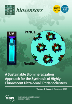
Noble metal nanoclusters (NMNCs) with sizes comparable to the Fermi wavelength of an electron exhibit fluorescence in correlation with the number of atoms in the cluster, which has potential applications in diverse areas, including electroluminescent display, solid-state lighting, and biomedical imaging. However, the conventional synthesis of such fluorescent NMNCs traditionally employs tedious and complex synthesis processes, uses harsh reaction conditions, and utilizes organic solvents that may hinder their biomedical applications. Herein, we propose a facile biomineralization process to produce highly fluorescent Pt-NMNCs in aqueous medium using a multistimulus responsive, biomimetic, intrinsically disordered protein polymer, Rec1-resilin. We demonstrate that Rec1-resilin acts concurrently as the host, reducing agent, and stabilizer of the blue-green fluorescent Pt-NMNCs once they are being [...] Read more.
  • Issues are regarded as officially published after their release is announced to the table of contents alert mailing list.
  • You may sign up for e-mail alerts to receive table of contents of newly released issues.
  • PDF is the official format for papers published in both, html and pdf forms. To view the papers in pdf format, click on the "PDF Full-text" link, and use the free Adobe Reader to open them.
Order results
Result details
Section
Select all
Export citation of selected articles as:
15 pages, 2462 KiB  
Article
Flexible and Transparent Substrates Based on Gold Nanoparticles and TiO2 for in Situ Bioanalysis by Surface-Enhanced Raman Spectroscopy
by Luisa Mandrile, Andrea Mario Giovannozzi, Alessio Sacco, Gianmario Martra and Andrea Mario Rossi
Biosensors 2019, 9(4), 145; https://doi.org/10.3390/bios9040145 - 17 Dec 2019
Cited by 13 | Viewed by 4939
Abstract
Flexible and transparent substrates are emerging as low cost and easy-to-operate support for surface-enhanced Raman spectroscopy (SERS). In particular, in situ SERS detection approach for surface characterization in transmission modality can be efficiently employed for non-invasive analysis of non-planar surfaces. Here we propose [...] Read more.
Flexible and transparent substrates are emerging as low cost and easy-to-operate support for surface-enhanced Raman spectroscopy (SERS). In particular, in situ SERS detection approach for surface characterization in transmission modality can be efficiently employed for non-invasive analysis of non-planar surfaces. Here we propose a new methodology to fabricate a homogenous, transparent, and flexible SERS membrane by the assistance of a thin TiO2 porous layer deposited on the PDMS surface, which supports the uniform loading of gold nanoparticles over large area. The substrate was first characterized for homogeneity, sensitivity and repeatability using a model molecule for SERS, i.e., 7-mercapto-4-methylcoumarin. Satisfactory intra-substrate uniformity and inter-substrates repeatability was achieved, showing an RSD of 10%, and an analytical sensitivity down to 10 nM was determined with an EF of 3.4 × 10⁵ ± 0.4 × 10⁵. Furthermore, SERS detection of pyrimethanil (PMT), a commonly employed pesticide in crops for human consumption, was performed in situ, exploiting the optical transparency of the device, using both model surfaces and non-flat bio-samples. PMT contamination at the phytochemical concentration levels corresponding to commonly used infield doses was successfully detected on the surface of the yellow Ficus benjiamina leaves, supporting the use of this substrate for food safety in-field application. Full article
(This article belongs to the Special Issue Plasmonic and Photonic Biosensors)
Show Figures

Graphical abstract

11 pages, 1738 KiB  
Article
Carbon Nanotube-Based Electrochemical Biosensor for Label-Free Protein Detection
by Jesslyn Janssen, Mike Lambeta, Paul White and Ahmad Byagowi
Biosensors 2019, 9(4), 144; https://doi.org/10.3390/bios9040144 - 17 Dec 2019
Cited by 20 | Viewed by 5409
Abstract
There is a growing need for biosensors that are capable of efficiently and rapidly quantifying protein biomarkers, both in the biological research and clinical setting. While accurate methods for protein quantification exist, the current assays involve sophisticated techniques, take long to administer and [...] Read more.
There is a growing need for biosensors that are capable of efficiently and rapidly quantifying protein biomarkers, both in the biological research and clinical setting. While accurate methods for protein quantification exist, the current assays involve sophisticated techniques, take long to administer and often require highly trained personnel for execution and analysis. Herein, we explore the development of a label-free biosensor for the detection and quantification of a standard protein. The developed biosensors comprise carbon nanotubes (CNTs), a specific antibody and cellulose filtration paper. The change in electrical resistance of the CNT-based biosensor system was used to sense a standard protein, bovine serum albumin (BSA) as a proof-of-concept. The developed biosensors were found to have a limit of detection of 2.89 ng/mL, which is comparable to the performance of the typical ELISA method for BSA quantification. Additionally, the newly developed method takes no longer than 10 min to perform, greatly reducing the time of analysis compared to the traditional ELISA technique. Overall, we present a versatile, affordable, simplified and rapid biosensor device capable of providing great benefit to both biological research and clinical diagnostics. Full article
(This article belongs to the Special Issue Bioelectric Sensors)
Show Figures

Figure 1

21 pages, 1785 KiB  
Article
A Critical Comparison between Flow-through and Lateral Flow Immunoassay Formats for Visual and Smartphone-Based Multiplex Allergen Detection
by Georgina M. S. Ross, Gert IJ. Salentijn and Michel W. F. Nielen
Biosensors 2019, 9(4), 143; https://doi.org/10.3390/bios9040143 - 12 Dec 2019
Cited by 47 | Viewed by 7633
Abstract
(1) Background: The lack of globally standardized allergen labeling legislation necessitates consumer-focused multiplexed testing devices. These should be easy to operate, fast, sensitive and robust. (2) Methods: Herein, we describe the development of three different formats for multiplexed food allergen detection, namely active [...] Read more.
(1) Background: The lack of globally standardized allergen labeling legislation necessitates consumer-focused multiplexed testing devices. These should be easy to operate, fast, sensitive and robust. (2) Methods: Herein, we describe the development of three different formats for multiplexed food allergen detection, namely active and passive flow-through assays, and lateral flow immunoassays with different test line configurations. (3) Results: The fastest assay time was 1 min, whereas even the slowest assay was within 10 min. With the passive flow approach, the limits of detection (LOD) of 0.1 and 0.5 ppm for total hazelnut protein (THP) and total peanut protein (TPP) in spiked buffer were reached, or 1 and 5 ppm of THP and TPP spiked into matrix. In comparison, the active flow approach reached LODs of 0.05 ppm for both analytes in buffer and 0.5 and 1 ppm of THP and TPP spiked into matrix. The optimized LFIA configuration reached LODs of 0.1 and 0.5 ppm of THP and TPP spiked into buffer or 0.5 ppm for both analytes spiked into matrix. The optimized LFIA was validated by testing in 20 different blank and spiked matrices. Using device-independent color space for smartphone analysis, two different smartphone models were used for the analysis of optimized assays. Full article
(This article belongs to the Special Issue Spectroscopy-Based Biosensors)
Show Figures

Figure 1

16 pages, 2276 KiB  
Review
Future Trends for In Situ Monitoring of Polycyclic Aromatic Hydrocarbons in Water Sources: The Role of Immunosensing Techniques
by Shifa Felemban, Patricia Vazquez and Eric Moore
Biosensors 2019, 9(4), 142; https://doi.org/10.3390/bios9040142 - 10 Dec 2019
Cited by 15 | Viewed by 6914
Abstract
Polycyclic aromatic hydrocarbons (PAHs) are hazardous environmental pollutants found in water, soil, and air. Exposure to this family of chemicals presents a danger to human health, and as a result, it is imperative to design methods that are able to detect PAHs in [...] Read more.
Polycyclic aromatic hydrocarbons (PAHs) are hazardous environmental pollutants found in water, soil, and air. Exposure to this family of chemicals presents a danger to human health, and as a result, it is imperative to design methods that are able to detect PAHs in the environment, thus improving the quality of drinking water and agricultural soils. This review presents emerging immunoassay techniques used for in situ detection of PAH in water samples and how they compare to common-place techniques. It will discuss their advantages and disadvantages and why it is required to find new solutions to analyze water samples. These techniques are effective in reducing detection times and complexity of measurements. Immunoassay methods presented here are able to provide in situ analysis of PAH concentrations in a water sample, which can be a great complement to existing laboratory techniques due to their real-time screening and portability for immunoassay techniques. The discussion shows in detail the most relevant state-of-the-art surface functionalization techniques used in the field of immunosensors, with the aim to improve PAH detection capabilities. Specifically, three surface functionalization techniques are key approaches to improve the detection of PAHs, namely, substrate surface reaction, layer-by-layer technique, and redox-active probes. These techniques have shown promising improvements in the detection of PAHs in water samples, since they show a wider linear range and high level of sensitivity compared to traditional PAH detection techniques. This review explores the various methods used in the detection of PAH in water environments. It provides extra knowledge to scientists on the possible solutions that can be used to save time and resources. The combination of the solutions presented here shows great promise in the development of portable solutions that will be able to analyze a sample in a matter of minutes on the field. Full article
Show Figures

Figure 1

12 pages, 1383 KiB  
Article
Establishing a Field-Effect Transistor Sensor for the Detection of Mutations in the Tumour Protein 53 Gene (TP53)—An Electrochemical Optimisation Approach
by Lisa Crossley, Bukola Attoye, Vincent Vezza, Ewen Blair, Damion K. Corrigan and Stuart Hannah
Biosensors 2019, 9(4), 141; https://doi.org/10.3390/bios9040141 - 06 Dec 2019
Cited by 9 | Viewed by 6867
Abstract
We present a low-cost, sensitive and specific DNA field-effect transistor sensor for the rapid detection of a common mutation to the tumour protein 53 gene (TP53). The sensor consists of a commercially available, low-cost, field-effect transistor attached in series to a gold electrode [...] Read more.
We present a low-cost, sensitive and specific DNA field-effect transistor sensor for the rapid detection of a common mutation to the tumour protein 53 gene (TP53). The sensor consists of a commercially available, low-cost, field-effect transistor attached in series to a gold electrode sensing pad for DNA hybridisation. The sensor has been predominantly optimised electrochemically, particularly with respect to open-circuit potentiometry as a route towards understanding potential (voltage) changes upon DNA hybridisation using a transistor. The developed sensor responds sensitively to TP53 mutant DNA as low as 100 nM concentration. The sensor responds linearly as a function of DNA target concentration and is able to differentiate between complementary and noncomplementary DNA target sequences. Full article
Show Figures

Figure 1

12 pages, 1844 KiB  
Article
Influence of Glutathione and Ascorbic Acid Treatments during Vinification of Feteasca Regala Variety and Their Antioxidant Effect on Volatile Profile
by George Adrian Cojocaru and Arina Oana Antoce
Biosensors 2019, 9(4), 140; https://doi.org/10.3390/bios9040140 - 05 Dec 2019
Cited by 13 | Viewed by 3926
Abstract
Volatile profiles were determined for white wines of Feteasca regala variety produced from musts in which the antioxidants glutathione and ascorbic acid were added in different proportions before inception of alcoholic fermentation. Treatments with these antioxidants affect some volatile compound evolution and positively [...] Read more.
Volatile profiles were determined for white wines of Feteasca regala variety produced from musts in which the antioxidants glutathione and ascorbic acid were added in different proportions before inception of alcoholic fermentation. Treatments with these antioxidants affect some volatile compound evolution and positively influence the wine volatile profile. After one year of storage in bottles with and without carbon dioxide protection the volatile profiles of the wines were assessed by using a Fast GC Alpha MOS Heracles e-Nose by applying a DFA multivariate statistical method and AroChemBase database for compound identification. The analyses showed that some higher alcohols, such as 2-phenylethanol and 2-methyl-1-butanol were in lower concentrations in wines treated with reduced glutathione, while the main ethyl fatty acid esters, such as ethyl butanoate, ethyl hexanoate, ethyl octanoate, and ethyl decanoate were better preserved when higher concentrations of any of the antioxidants were added in must. On the other hand, it was observed that some other volatile compounds were not affected by these applied treatments. Full article
Show Figures

Figure 1

16 pages, 5568 KiB  
Article
Biochar from Brewers’ Spent Grain: A Green and Low-Cost Smart Material to Modify Screen-Printed Electrodes
by Rocco Cancelliere, Katya Carbone, Mauro Pagano, Ilaria Cacciotti and Laura Micheli
Biosensors 2019, 9(4), 139; https://doi.org/10.3390/bios9040139 - 03 Dec 2019
Cited by 40 | Viewed by 5751
Abstract
In the present study, biochar from brewers’ spent grain was used, for the first time, to develop screen-printed electrodes. After having investigated the dispersion behaviour of biochar in different organic solvents, a biochar-based screen-printed electrode was prepared with the drop-casting technique. In order [...] Read more.
In the present study, biochar from brewers’ spent grain was used, for the first time, to develop screen-printed electrodes. After having investigated the dispersion behaviour of biochar in different organic solvents, a biochar-based screen-printed electrode was prepared with the drop-casting technique. In order to understand the electrochemical potentiality and performances of the biochar/sensor tool, different electroactive species, i.e., ferricyanide, benzoquinone, epinephrine, ascorbic, and uric acids, were used. The results were compared with those of the same electrodes that were modified with commercial graphene, confirming that the proposed electrode showed improved electrochemical behaviour in terms of resolution, peak-to-peak separation, current intensity, and resistance to charge transfer. Furthermore, a tyrosinase biosensor was developed by direct immobilisation of this enzyme on the biochar/screen printed electrode, as an example of the potential of biochar for disposable biosensor development. The efficiently occurred immobilisation of the biochar on the screen printed electrode’s (SPE’s) surface was demonstrated by the observation of the working electrode with a scanning electron microscope. The detection was performed by measuring the current due to the reduction of the corresponding quinone at low potential, equal to −0.310 V for epinephrine. The experimental conditions for the tyrosinase immobilization and the analytical parameters, such as applied potential and pH of buffer, were studied and optimized. Under these conditions, the electrochemical biosensors were characterized. A linear working range of epinephrine was obtained from 0.05 up to 0.5 mM. The detection limit was 2 × 10⁻⁴ mM for the biosensor. Full article
Show Figures

Graphical abstract

16 pages, 3183 KiB  
Article
The Influence of the Parameters of a Gold Nanoparticle Deposition Method on Titanium Dioxide Nanotubes, Their Electrochemical Response, and Protein Adsorption
by Ewa Paradowska, Katarzyna Arkusz and Dorota G. Pijanowska
Biosensors 2019, 9(4), 138; https://doi.org/10.3390/bios9040138 - 20 Nov 2019
Cited by 14 | Viewed by 4895
Abstract
The goal of this research was to find the best conditions to prepare titanium dioxide nanotubes (TNTs) modified with gold nanoparticles (AuNPs). This paper, for the first time, reports on the influence of the parameters of cyclic voltammetry process (CV) -based AuNP deposition, [...] Read more.
The goal of this research was to find the best conditions to prepare titanium dioxide nanotubes (TNTs) modified with gold nanoparticles (AuNPs). This paper, for the first time, reports on the influence of the parameters of cyclic voltammetry process (CV) -based AuNP deposition, i.e., the number of cycles and the concentration of gold salt solution, on corrosion resistance and the capacitance of TNTs. Another innovation was to fabricate AuNPs with well-formed spherical geometry and uniform distribution on TNTs. The AuNPs/TNTs were characterized using scanning electron microscopy, X-ray photoelectron spectroscopy, electrochemical impedance spectroscopy, and open-circuit potential measurement. From the obtained results, the correlation between the deposition process parameters, the AuNP diameters, and the electrical conductivity of the TNTs was found in a range from 14.3 ± 1.8 to 182.3 ± 51.7 nm. The size and amount of the AuNPs could be controlled by the number of deposition cycles and the concentration of the gold salt solution. The modification of TNTs using AuNPs facilitated electron transfer, increased the corrosion resistance, and caused better adsorption properties for bovine serum albumin. Full article
(This article belongs to the Special Issue Nanoparticle-Based Biosensors for Food and Health Monitoring)
Show Figures

Graphical abstract

9 pages, 12270 KiB  
Article
Multiwalled Carbon Nanotubes and the Electrocatalytic Activity of Gluconobacter oxydans as the Basis of a Biosensor
by Yulia Plekhanova, Sergei Tarasov, Aleksandr Bykov, Natalia Prisyazhnaya, Vladimir Kolesov, Vladimir Sigaev, Maria Assunta Signore and Anatoly Reshetilov
Biosensors 2019, 9(4), 137; https://doi.org/10.3390/bios9040137 - 14 Nov 2019
Cited by 13 | Viewed by 4438
Abstract
This paper considers the effect of multiwalled carbon nanotubes (MWCNTs) on the parameters of Gluconobacter oxydans microbial biosensors. MWCNTs were shown not to affect the structural integrity of microbial cells and their respiratory activity. The positive results from using MWCNTs were due to [...] Read more.
This paper considers the effect of multiwalled carbon nanotubes (MWCNTs) on the parameters of Gluconobacter oxydans microbial biosensors. MWCNTs were shown not to affect the structural integrity of microbial cells and their respiratory activity. The positive results from using MWCNTs were due to a decrease in the impedance of the electrode. The total impedance of the system decreased significantly, from 9000 kOhm (G. oxydans/chitosan composite) to 600 kOhm (G. oxydans/MWCNTs/chitosan). Modification of the amperometric biosensor with nanotubes led to an increase in the maximal signal from 65 to 869 nA for glucose and from 181 to 1048 nA for ethanol. The biosensor sensitivity also increased 4- and 5-fold, respectively, for each of the substrates. However, the addition of MWCNTs reduced the affinity of respiratory chain enzymes to their substrates (both sugars and alcohols). Moreover, the minimal detection limits were not reduced despite a sensitivity increase. The use of MWCNTs thus improved only some microbial biosensor parameters. Full article
(This article belongs to the Special Issue Dedication to TUT President Isao Karube: Microbial Biosensors)
Show Figures

Figure 1

19 pages, 10286 KiB  
Article
Bioelectrical Analysis of Various Cancer Cell Types Immobilized in 3D Matrix and Cultured in 3D-Printed Well
by Georgia Paivana, Sophie Mavrikou, Grigoris Kaltsas and Spyridon Kintzios
Biosensors 2019, 9(4), 136; https://doi.org/10.3390/bios9040136 - 14 Nov 2019
Cited by 6 | Viewed by 4752
Abstract
Cancer cell lines are important tools for anticancer drug research and assessment. Impedance measurements can provide valuable information about cell viability in real time. This work presents the proof-of-concept development of a bioelectrical, impedance-based analysis technique applied to four adherent mammalian cancer cells [...] Read more.
Cancer cell lines are important tools for anticancer drug research and assessment. Impedance measurements can provide valuable information about cell viability in real time. This work presents the proof-of-concept development of a bioelectrical, impedance-based analysis technique applied to four adherent mammalian cancer cells lines immobilized in a three-dimensional (3D) calcium alginate hydrogel matrix, thus mimicking in vivo tissue conditions. Cells were treated with cytostatic agent5-fluoruracil (5-FU). The cell lines used in this study were SK-N-SH, HEK293, HeLa, and MCF-7. For each cell culture, three cell population densities were chosen (50,000, 100,000, and 200,000 cells/100 μL). The aim of this study was the extraction of mean impedance values at various frequencies for the assessment of the different behavior of various cancer cells when 5-FU was applied. For comparison purposes, impedance measurements were implemented on untreated immobilized cell lines. The results demonstrated not only the dependence of each cell line impedance value on the frequency, but also the relation of the impedance level to the cell population density for every individual cell line. By establishing a cell line-specific bioelectrical behavior, it is possible to obtain a unique fingerprint for each cancer cell line reaction to a selected anticancer agent. Full article
(This article belongs to the Special Issue Bioelectric Sensors)
Show Figures

Figure 1

11 pages, 2476 KiB  
Article
Construction of a Hydrogel Pectin-Based Triglyceride Optical Biosensor with Immobilized Lipase Enzymes
by Uswatun Hasanah, Nor Diyana Md Sani, Lee Yook Heng, Rinaldi Idroes and Eka Safitri
Biosensors 2019, 9(4), 135; https://doi.org/10.3390/bios9040135 - 13 Nov 2019
Cited by 39 | Viewed by 7353
Abstract
A novel and simple optical biosensor to detect triglycerides (TGs) has been successfully constructed by using pectin hydrogel membrane as the indicator pH and chromoionophore ETH 5294 (CI), with lipase as the catalyst. The enzymatic working system against TGs releasing H+ ions [...] Read more.
A novel and simple optical biosensor to detect triglycerides (TGs) has been successfully constructed by using pectin hydrogel membrane as the indicator pH and chromoionophore ETH 5294 (CI), with lipase as the catalyst. The enzymatic working system against TGs releasing H+ ions will affect the color absorbance of CI. The characterization results show that a TG biosensor has the optimum condition and sensitivity at the phosphate buffer concentration of 50 mM, pH 7, and enzyme loading of 60 μg. The biosensor works at the tripalmitin (TP) concentration range of 100–400 mg/dL. With the sensitivity of 0.001 (∆A/(mg/dL)), the biosensor response reaches stability after five minutes, and the limit of detection (LOD) of the TG optical biosensor is 15 mg/dL. Relative standard deviation (RSD) in a reproducibility test was 2.5%, with a 15-day lifespan. Full article
Show Figures

Figure 1

14 pages, 5322 KiB  
Article
Characterization of an Aptamer Directed against 25-Hydroxyvitamin D for the Development of a Competitive Aptamer-Based Assay
by Marc Prante, Torsten Schüling, Bernhard Roth, Kort Bremer and Johanna Walter
Biosensors 2019, 9(4), 134; https://doi.org/10.3390/bios9040134 - 13 Nov 2019
Cited by 24 | Viewed by 6809
Abstract
Detection of the small molecule 25-hydroxyvitamin D (25(OH)D) as the most relevant marker for vitamin D supply suffers from a high variability of results using the current detection methods, such as high-performance liquid chromatography (HPLC) and immunoassays. A new detection approach using a [...] Read more.
Detection of the small molecule 25-hydroxyvitamin D (25(OH)D) as the most relevant marker for vitamin D supply suffers from a high variability of results using the current detection methods, such as high-performance liquid chromatography (HPLC) and immunoassays. A new detection approach using a highly specific aptamer directed against 25(OH)D was established in this study based on the target-induced dissociation (TID) sensing approach. In this work, the aptamer was investigated regarding its structural properties as well as its binding affinity by using microscale thermophoresis (MST). Moreover, complementary oligonucleotides were designed based on the aptamer structure and were evaluated in MST experiments. Binding experiments of immobilized aptamers were conducted in microarray experiments. It could be shown that the aptamer exhibited the usual B-DNA structure and did not form any G-quadruplexes. The design of complementary oligonucleotides for the TID assay identified a putative 25(OH)D binding site within the aptamer. The limit of detection of the established competitive assay was determined to be 5.4 nM, which sets the stage for the development of a biosensor system. Full article
(This article belongs to the Special Issue Microarray for Biomarker Detection and Discovery)
Show Figures

Figure 1

11 pages, 3694 KiB  
Article
Development of Microalgae Biosensor Chip by Incorporating Microarray Oxygen Sensor for Pesticides Sensing
by Md. Abul Kashem, Kazuki Kimoto, Yasunori Iribe and Masayasu Suzuki
Biosensors 2019, 9(4), 133; https://doi.org/10.3390/bios9040133 - 12 Nov 2019
Cited by 11 | Viewed by 5828
Abstract
A microalgae (Pseudokirchneriella subcapitata) biosensor chip for pesticide sensing has been developed by attaching the immobilized microalgae biofilm pon the microarray dye spots (size 100 μm and pitch 200 μm). The dye spots (ruthenium complex) were printed upon SO3-modified [...] Read more.
A microalgae (Pseudokirchneriella subcapitata) biosensor chip for pesticide sensing has been developed by attaching the immobilized microalgae biofilm pon the microarray dye spots (size 100 μm and pitch 200 μm). The dye spots (ruthenium complex) were printed upon SO3-modified glass slides using a polydimethylsiloxane (PDMS) stamp and a microcontact printer (μCP). Emitted fluorescence intensity (FI) variance due to photosynthetic activity (O2 production) of microalgae was monitored by an inverted fluorescent microscope and inhibition of the oxygen generation rate was calculated based on the FI responses both before and after injection of pesticide sample. The calibration curves, as the inhibition of oxygen generation rate (%) due to photosynthetic activity inhibition by the pesticides, depicted that among the 6 tested pesticides, the biosensor showed good sensitivity for 4 pesticides (diuron, simetryn, simazine, and atrazine) but was insensitive for mefenacet and pendimethalin. The detection limits were 1 ppb for diuron and 10 ppb for simetryn, simazine, and atrazine. The simple and low-cost nature of sensing of the developed biosensor sensor chip has apparently created opportunities for regular water quality monitoring, where pesticides are an important concern. Full article
(This article belongs to the Special Issue Dedication to TUT President Isao Karube: Microbial Biosensors)
Show Figures

Figure 1

26 pages, 900 KiB  
Review
Optical Biosensors for Therapeutic Drug Monitoring
by Vivian Garzón, Daniel G. Pinacho, Rosa-Helena Bustos, Gustavo Garzón and Sandra Bustamante
Biosensors 2019, 9(4), 132; https://doi.org/10.3390/bios9040132 - 11 Nov 2019
Cited by 59 | Viewed by 12380
Abstract
Therapeutic drug monitoring (TDM) is a fundamental tool when administering drugs that have a limited dosage or high toxicity, which could endanger the lives of patients. To carry out this monitoring, one can use different biological fluids, including blood, plasma, serum, and urine, [...] Read more.
Therapeutic drug monitoring (TDM) is a fundamental tool when administering drugs that have a limited dosage or high toxicity, which could endanger the lives of patients. To carry out this monitoring, one can use different biological fluids, including blood, plasma, serum, and urine, among others. The help of specialized methodologies for TDM will allow for the pharmacodynamic and pharmacokinetic analysis of drugs and help adjust the dose before or during their administration. Techniques that are more versatile and label free for the rapid quantification of drugs employ biosensors, devices that consist of one element for biological recognition coupled to a signal transducer. Among biosensors are those of the optical biosensor type, which have been used for the quantification of different molecules of clinical interest, such as antibiotics, anticonvulsants, anti-cancer drugs, and heart failure. This review presents an overview of TDM at the global level considering various aspects and clinical applications. In addition, we review the contributions of optical biosensors to TDM. Full article
(This article belongs to the Special Issue Optical Diagnostics with Point-of-Care and Point-of-Need Applications)
Show Figures

Figure 1

34 pages, 8576 KiB  
Article
Fully Automated Microsystem for Unmediated Electrochemical Characterization, Visualization and Monitoring of Bacteria on Solid Media; E. coli K-12: A Case Study
by Cesar A. Hernandez, Valerio Beni and Johann F. Osma
Biosensors 2019, 9(4), 131; https://doi.org/10.3390/bios9040131 - 04 Nov 2019
Cited by 7 | Viewed by 6185
Abstract
In this paper, we present a non-fluidic microsystem for the simultaneous visualization and electrochemical evaluation of confined, growing bacteria on solid media. Using a completely automated platform, real-time monitoring of bacterial and image-based computer characterization of growth were performed. Electrochemical tests, using Escherichia [...] Read more.
In this paper, we present a non-fluidic microsystem for the simultaneous visualization and electrochemical evaluation of confined, growing bacteria on solid media. Using a completely automated platform, real-time monitoring of bacterial and image-based computer characterization of growth were performed. Electrochemical tests, using Escherichia coli K-12 as the model microorganism, revealed the development of a faradaic process at the bacteria–microelectrode interface inside the microsystem, as implied by cyclic voltammetry and electrochemical impedance spectrometry measurements. The electrochemical information was used to determine the moment in which bacteria colonized the electrode-enabled area of the microsystem. This microsystem shows potential advantages for long-term electrochemical monitoring of the extracellular environment of cell culture and has been designed using readily available technologies that can be easily integrated in routine protocols. Complementarily, these methods can help elucidate fundamental questions of the electron transfer of bacterial cultures and are potentially feasible to be integrated into current characterization techniques. Full article
Show Figures

Graphical abstract

29 pages, 1723 KiB  
Review
Review of Electroanalytical-Based Approaches for the Determination of Benzodiazepines
by Kevin C. Honeychurch
Biosensors 2019, 9(4), 130; https://doi.org/10.3390/bios9040130 - 02 Nov 2019
Cited by 18 | Viewed by 6316
Abstract
The benzodiazepine class of drugs are characterised by a readily electrochemically reducible azomethine group. A number are also substituted by other electrochemically active nitro, N-oxide, and carbonyl groups, making them readily accessible to electrochemical determination. Techniques such as polarography, voltammetry, and potentiometry have [...] Read more.
The benzodiazepine class of drugs are characterised by a readily electrochemically reducible azomethine group. A number are also substituted by other electrochemically active nitro, N-oxide, and carbonyl groups, making them readily accessible to electrochemical determination. Techniques such as polarography, voltammetry, and potentiometry have been employed for pharmaceutical and biomedical samples, requiring little sample preparation. This review describes current developments in the design and applications of electrochemical-based approaches for the determination of the benzodiazepine class of drugs form their introduction in the early 1960s to 2019. Throughout this period, state-of-the-art electroanalytical techniques have been reported for their determination. Polarography was first employed focused on mechanistic investigations. Subsequent studies showed the adsorption of many the benzodiazepines at Hg electrodes allowed for the highly sensitive technique of adsorptive stripping voltammetry to be employed. The development and introduction of other working electrode materials such as carbon led to techniques such as voltammetry to become commonly reported, and the modification of these electrodes has now become the most commonly employed approach using molecularly imprinting and nanotechnology. Full article
(This article belongs to the Special Issue The Potential of (bio)sensors for the Forensic Sciences)
Show Figures

Figure 1

12 pages, 1222 KiB  
Article
Fast Active Merging of Microdroplets in Microfluidic Chambers Driven by Photo-Isomerisation of Azobenzene Based Surfactants
by Zain Hayat, Nizar Bchellaoui, Claire Deo, Rémi Métivier, Nicolas Bogliotti, Juan Xie, Malcolm Buckle and Abdel I. El Abed
Biosensors 2019, 9(4), 129; https://doi.org/10.3390/bios9040129 - 01 Nov 2019
Cited by 1 | Viewed by 4645
Abstract
In this work, we report on the development of a newly synthesized photoactive reversible azobenzene derived surfactant polymer, which enables active and fast control of the merging of microdroplets in microfluidic chambers, driven by a pulsed UV laser optical stimulus and the well [...] Read more.
In this work, we report on the development of a newly synthesized photoactive reversible azobenzene derived surfactant polymer, which enables active and fast control of the merging of microdroplets in microfluidic chambers, driven by a pulsed UV laser optical stimulus and the well known cis-trans photo-isomerisation of azobenzene groups. We show for the first time that merging of microdroplets can be achieved optically based on a photo-isomerization process with a high spatio-temporal resolution. Our results show that the physical process lying behind the merging of microdroplets is not driven by a change in surface activity of the droplet stabilizing surfactant under UV illumination (as originally expected), and they suggest an original mechanism for the merging of droplets based on the well-known opto-mechanical motion of azobenzene molecules triggered by light irradiation. Full article
(This article belongs to the Special Issue Microfluidics for Biosensing and Diagnostics)
Show Figures

Figure 1

13 pages, 2375 KiB  
Article
A Sustainable Biomineralization Approach for the Synthesis of Highly Fluorescent Ultra-Small Pt Nanoclusters
by Rajkamal Balu, Robert Knott, Christopher M. Elvin, Anita J. Hill, Namita R. Choudhury and Naba K. Dutta
Biosensors 2019, 9(4), 128; https://doi.org/10.3390/bios9040128 - 29 Oct 2019
Cited by 15 | Viewed by 5056
Abstract
Herein we report the first example of a facile biomineralization process to produce ultra-small-sized highly fluorescent aqueous dispersions of platinum noble metal quantum clusters (Pt-NMQCs) using a multi-stimulus responsive, biomimetic intrinsically disordered protein (IDP), Rec1-resilin. We demonstrate that Rec1-resilin acts concurrently as the [...] Read more.
Herein we report the first example of a facile biomineralization process to produce ultra-small-sized highly fluorescent aqueous dispersions of platinum noble metal quantum clusters (Pt-NMQCs) using a multi-stimulus responsive, biomimetic intrinsically disordered protein (IDP), Rec1-resilin. We demonstrate that Rec1-resilin acts concurrently as the host, reducing agent, and stabilizer of the blue-green fluorescent Pt-NMQCs once they are being formed. The photophysical properties, quantum yield, and fluorescence lifetime measurements of the synthesized Pt-NMQCs were examined using UV-Vis and fluorescence spectroscopy. The oxidation state of the Pt-NMQCs was quantitatively analyzed using X-ray photoelectron spectroscopy. Both a small angle X-ray scattering technique and a modeling approach have been attempted to present a detailed understanding of the structure and conformational dynamics of Rec1-resilin as an IDP during the formation of the Pt-NMQCs. It has been demonstrated that the green fluorescent Pt-NMQCs exhibit a high quantum yield of ~7.0% and a lifetime of ~9.5 ns in aqueous media. The change in photoluminescence properties due to the inter-dot interactions between proximal dots and aggregation of the Pt-NMQCs by evaporation was also measured spectroscopically and discussed. Full article
Show Figures

Graphical abstract

30 pages, 3886 KiB  
Review
What Electrochemical Biosensors Can Do for Forensic Science? Unique Features and Applications
by Paloma Yáñez-Sedeño, Lourdes Agüí, Susana Campuzano and José Manuel Pingarrón
Biosensors 2019, 9(4), 127; https://doi.org/10.3390/bios9040127 - 29 Oct 2019
Cited by 24 | Viewed by 8786
Abstract
This article critically discusses the latest advances in the use of voltammetric, amperometric, potentiometric, and impedimetric biosensors for forensic analysis. Highlighted examples that show the advantages of these tools to develop methods capable of detecting very small concentrations of analytes and provide selective [...] Read more.
This article critically discusses the latest advances in the use of voltammetric, amperometric, potentiometric, and impedimetric biosensors for forensic analysis. Highlighted examples that show the advantages of these tools to develop methods capable of detecting very small concentrations of analytes and provide selective determinations through analytical responses, without significant interferences from other components of the samples, are presented and discussed, thus stressing the great versatility and utility of electrochemical biosensors in this growing research field. To illustrate this, the determination of substances with forensic relevance by using electrochemical biosensors reported in the last five years (2015–2019) are reviewed. The different configurations of enzyme or affinity biosensors used to solve analytical problems related to forensic practice, with special attention to applications in complex samples, are considered. Main prospects, challenges to focus, such as the fabrication of devices for rapid analysis of target analytes directly on-site at the crime scene, or their widespread use and successful applications to complex samples of interest in forensic analysis, and future efforts, are also briefly discussed. Full article
(This article belongs to the Special Issue The Potential of (bio)sensors for the Forensic Sciences)
Show Figures

Figure 1

12 pages, 1433 KiB  
Communication
Detection of Superoxide Alterations Induced by 5-Fluorouracil on HeLa Cells with a Cell-Based Biosensor
by Sophia Mavrikou, Vasileios Tsekouras, Maria-Argyro Karageorgou, Georgia Moschopoulou and Spyridon Kintzios
Biosensors 2019, 9(4), 126; https://doi.org/10.3390/bios9040126 - 16 Oct 2019
Cited by 10 | Viewed by 5105
Abstract
Background: In vitro cell culture monitoring can be used as an indicator of cellular oxidative stress for the assessment of different chemotherapy agents. Methods: A cell-based bioelectric biosensor was used to detect alterations in superoxide levels in the culture medium of HeLa cervical [...] Read more.
Background: In vitro cell culture monitoring can be used as an indicator of cellular oxidative stress for the assessment of different chemotherapy agents. Methods: A cell-based bioelectric biosensor was used to detect alterations in superoxide levels in the culture medium of HeLa cervical cancer cells after treatment with the chemotherapeutic agent 5-fluorouracil (5-FU). The cytotoxic effects of 5-fluorouracil on HeLa cells were assessed by the MTT proliferation assay, whereas oxidative damage and induction of apoptosis were measured fluorometrically by the mitochondria-targeted MitoSOX™ Red and caspase-3 activation assays, respectively. Results: The results of this study indicate that 5-FU differentially affects superoxide production and caspase-3 activation when applied in cytotoxic concentrations against HeLa cells, while superoxide accumulation is in accordance with mitochondrial superoxide levels. Our findings suggest that changes in superoxide concentration could be detected with the biosensor in a non-invasive and rapid manner, thus allowing a reliable estimation of oxidative damage due to cell apoptosis. Conclusions: These findings may be useful for facilitating future high throughput screening of different chemotherapeutic drugs with a cytotoxic principle based on free radical production. Full article
(This article belongs to the Special Issue Bioelectric Sensors)
Show Figures

Graphical abstract

11 pages, 2900 KiB  
Article
Tetrahydrocannabinol Sensing in Complex Biofluid with Portable Raman Spectrometer Using Diatomaceous SERS Substrates
by Kundan Sivashanmugan, Yong Zhao and Alan X. Wang
Biosensors 2019, 9(4), 125; https://doi.org/10.3390/bios9040125 - 14 Oct 2019
Cited by 24 | Viewed by 6252
Abstract
Using thin-layer chromatography in tandem with surface-enhanced Raman spectroscopy (TLC-SERS) and tetrahydrocannabinol (THC) sensing in complex biological fluids is successfully conducted with a portable Raman spectrometer. Both THC and THC metabolites are detected from the biofluid of marijuana-users as biomarkers for identifying cannabis [...] Read more.
Using thin-layer chromatography in tandem with surface-enhanced Raman spectroscopy (TLC-SERS) and tetrahydrocannabinol (THC) sensing in complex biological fluids is successfully conducted with a portable Raman spectrometer. Both THC and THC metabolites are detected from the biofluid of marijuana-users as biomarkers for identifying cannabis exposure. In this article, ultra-sensitive SERS substrates based on diatomaceous earth integrated with gold nanoparticles (Au NPs) were employed to detect trace levels of cannabis biomarkers in saliva. Strong characteristic THC and THC metabolite SERS peaks at 1601 and 1681 cm−1 were obtained despite the moderate interference of biological molecules native to saliva. Urine samples were also analyzed, but they required TLC separation of THC from the urine sample to eliminate the strong influence of urea and other organic molecules. TLC separation of THC from the urine was performed by porous microfluidic channel devices using diatomaceous earth as the stationary phase. The experimental results showed clear separation between urea and THC, and strong THC SERS characteristic peaks. Principal component analysis (PCA) was used to analyze the SERS spectra collected from various THC samples. The spectra in the principal component space were well clustered for each sample type and share very similar scores in the main principal component (PC1), which can serve as the benchmark for THC sensing from complex SERS spectra. Therefore, we proved that portable Raman spectrometers can enable an on-site sensing capability using diatomaceous SERS substrates to detect THC in real biological solutions. This portable THC sensing technology will play pivotal roles in forensic analysis, medical diagnosis, and public health. Full article
(This article belongs to the Special Issue Plasmonic and Photonic Biosensors)
Show Figures

Figure 1

15 pages, 1210 KiB  
Article
Development of Aptamer-Based TID Assays Using Thermophoresis and Microarrays
by Tracy Kurth, Sandra Witt, Svenja Bolten, Janice-Joy Waniek, Carlotta Kortmann, Antonina Lavrentieva, Thomas Scheper and Johanna-Gabriela Walter
Biosensors 2019, 9(4), 124; https://doi.org/10.3390/bios9040124 - 14 Oct 2019
Cited by 9 | Viewed by 5159
Abstract
Aptamers are single-stranded oligonucleotides which can be used as alternative recognition elements for protein detection, because aptamers bind their targets with a high affinity similar to antibodies. Due to the target-induced conformational changes of aptamers, these oligonucleotides can be applied in various biosensing [...] Read more.
Aptamers are single-stranded oligonucleotides which can be used as alternative recognition elements for protein detection, because aptamers bind their targets with a high affinity similar to antibodies. Due to the target-induced conformational changes of aptamers, these oligonucleotides can be applied in various biosensing platforms. In this work, aptamers directed against the vascular endothelial growth factor (VEGF) were used as a model system. VEGF plays a key role in physiological angiogenesis and vasculogenesis. Furthermore, VEGF is involved in the development and growth of cancer and other diseases like age-related macular degeneration, rheumatoid arthritis, diabetes mellitus, and neurodegenerative disorders. Detecting the protein biomarker VEGF is therefore of great importance for medical research and diagnostics. In this research, VEGF-binding aptamers were investigated for the systematic development of a target-induced dissociation (TID) assay utilizing thermophoresis and microarrays. The established aptamer-microarray allowed for the detection of 0.1 nM of VEGF. Furthermore, the systematic development of the TID method using the VEGF model protein could help to develop further TID assays for the detection of various protein biomarkers. Full article
Show Figures

Figure 1

26 pages, 2962 KiB  
Article
Detection of Emerald Ash Borer Infestations in Living Green Ash by Noninvasive Electronic-Nose Analysis of Wood Volatiles
by A. Dan Wilson, Lisa B. Forse, Benjamin A. Babst and Mohammad M. Bataineh
Biosensors 2019, 9(4), 123; https://doi.org/10.3390/bios9040123 - 13 Oct 2019
Cited by 13 | Viewed by 5045
Abstract
The emerald ash borer (EAB) has been the most destructive and costly nonnative insect to threaten the health of ash (Fraxinus) species in North America for at least the past 25 years. The development of methods for detecting visually-hidden EAB galleries [...] Read more.
The emerald ash borer (EAB) has been the most destructive and costly nonnative insect to threaten the health of ash (Fraxinus) species in North America for at least the past 25 years. The development of methods for detecting visually-hidden EAB galleries at early stages of infestation would provide a useful tool to more effectively facilitate the planning and implementation of targeted EAB pest-suppression and management activities. We tested the efficacy of using a dual-technology electronic-nose (e-nose)/gas chromatograph device as a means for detection of EAB infestations in green ash trees in different EAB-decline classes by analysis of VOC emissions in sapwood. We found significant differences in VOC profiles for trees from the four decline classes. The VOC composition, quantities, and types of volatile metabolites present in headspace volatiles varied considerably across sample types, and resulted in distinct e-nose smellprint patterns that were characteristic of each unique chemical composition. In addition, specific VOC metabolites were identified as potential healthy and EAB-infestation biomarkers, indicative of the health states of individual trees. Few significant differences in major bark phenolic compounds were found between ash decline classes using LC-MS. The e-nose was effective in discriminating between uninfested and EAB-infested trees based on sapwood VOC emissions. Full article
(This article belongs to the Special Issue Noninvasive Early Disease Diagnosis)
Show Figures

Figure 1

17 pages, 1866 KiB  
Review
FRET Microscopy in Yeast
by Michal Skruzny, Emma Pohl and Marc Abella
Biosensors 2019, 9(4), 122; https://doi.org/10.3390/bios9040122 - 11 Oct 2019
Cited by 15 | Viewed by 11592
Abstract
Förster resonance energy transfer (FRET) microscopy is a powerful fluorescence microscopy method to study the nanoscale organization of multiprotein assemblies in vivo. Moreover, many biochemical and biophysical processes can be followed by employing sophisticated FRET biosensors directly in living cells. Here, we summarize [...] Read more.
Förster resonance energy transfer (FRET) microscopy is a powerful fluorescence microscopy method to study the nanoscale organization of multiprotein assemblies in vivo. Moreover, many biochemical and biophysical processes can be followed by employing sophisticated FRET biosensors directly in living cells. Here, we summarize existing FRET experiments and biosensors applied in yeasts Saccharomyces cerevisiae and Schizosaccharomyces pombe, two important models of fundamental biomedical research and efficient platforms for analyses of bioactive molecules. We aim to provide a practical guide on suitable FRET techniques, fluorescent proteins, and experimental setups available for successful FRET experiments in yeasts. Full article
(This article belongs to the Special Issue FRET-Based Biosensors)
Show Figures

Figure 1

19 pages, 4606 KiB  
Review
Polarization Induced Electro-Functionalization of Pore Walls: A Contactless Technology
by Aurélie Bouchet-Spinelli, Emeline Descamps, Jie Liu, Abdulghani Ismail, Pascale Pham, François Chatelain, Thierry Leïchlé, Loïc Leroy, Patrice Noël Marche, Camille Raillon, André Roget, Yoann Roupioz, Neso Sojic, Arnaud Buhot, Vincent Haguet, Thierry Livache and Pascal Mailley
Biosensors 2019, 9(4), 121; https://doi.org/10.3390/bios9040121 - 11 Oct 2019
Cited by 5 | Viewed by 5216
Abstract
This review summarizes recent advances in micro- and nanopore technologies with a focus on the functionalization of pores using a promising method named contactless electro-functionalization (CLEF). CLEF enables the localized grafting of electroactive entities onto the inner wall of a micro- or nano-sized [...] Read more.
This review summarizes recent advances in micro- and nanopore technologies with a focus on the functionalization of pores using a promising method named contactless electro-functionalization (CLEF). CLEF enables the localized grafting of electroactive entities onto the inner wall of a micro- or nano-sized pore in a solid-state silicon/silicon oxide membrane. A voltage or electrical current applied across the pore induces the surface functionalization by electroactive entities exclusively on the inside pore wall, which is a significant improvement over existing methods. CLEF’s mechanism is based on the polarization of a sandwich-like silicon/silicon oxide membrane, creating electronic pathways between the core silicon and the electrolyte. Correlation between numerical simulations and experiments have validated this hypothesis. CLEF-induced micro- and nanopores functionalized with antibodies or oligonucleotides were successfully used for the detection and identification of cells and are promising sensitive biosensors. This technology could soon be successfully applied to planar configurations of pores, such as restrictions in microfluidic channels. Full article
Show Figures

Figure 1

12 pages, 4782 KiB  
Article
P-N Junction-Based Si Biochips with Ring Electrodes for Novel Biosensing Applications
by Mahdi Kiani, Nan Du, Manja Vogel, Johannes Raff, Uwe Hübner, Ilona Skorupa, Danilo Bürger, Stefan E. Schulz, Oliver G. Schmidt and Heidemarie Schmidt
Biosensors 2019, 9(4), 120; https://doi.org/10.3390/bios9040120 - 11 Oct 2019
Cited by 6 | Viewed by 4906
Abstract
In this work, we report on the impedance of p-n junction-based Si biochips with gold ring top electrodes and unstructured platinum bottom electrodes which allows for counting target biomaterial in a liquid-filled ring top electrode region. The systematic experiments on p-n junction-based Si [...] Read more.
In this work, we report on the impedance of p-n junction-based Si biochips with gold ring top electrodes and unstructured platinum bottom electrodes which allows for counting target biomaterial in a liquid-filled ring top electrode region. The systematic experiments on p-n junction-based Si biochips fabricated by two different sets of implantation parameters (i.e. biochips PS5 and BS5) are studied, and the comparable significant change of impedance characteristics in the biochips in dependence on the number of bacteria suspension, i.e., Lysinibacillus sphaericus JG-A12, in Deionized water with an optical density at 600 nm from OD600 = 4–16 in the electrode ring region is demonstrated. Furthermore, with the help of the newly developed two-phase electrode structure, the modeled capacitance and resistance parameters of the electrical equivalent circuit describing the p-n junction-based biochips depend linearly on the number of bacteria in the ring top electrode region, which successfully proves the potential performance of p-n junction-based Si biochips in observing the bacterial suspension. The proposed p-n junction-based biochips reveal perspective applications in medicine and biology for diagnosis, monitoring, management, and treatment of diseases. Full article
(This article belongs to the Special Issue Bioelectric Sensors)
Show Figures

Figure 1

11 pages, 4186 KiB  
Article
A Novel Photoplethysmography Sensor for Vital Signs Monitoring from the Human Trachea
by James M. May, Justin P. Phillips, Tracey Fitchat, Shankar Ramaswamy, Saowarat Snidvongs and Panayiotis A. Kyriacou
Biosensors 2019, 9(4), 119; https://doi.org/10.3390/bios9040119 - 02 Oct 2019
Cited by 10 | Viewed by 5699
Abstract
Current pulse oximeter sensors can be challenged in working accurately and continuously in situations of reduced periphery perfusion, especially among anaesthetised patients. A novel tracheal photoplethysmography (PPG) sensor has been developed in an effort to address the limitations of current pulse oximeters. The [...] Read more.
Current pulse oximeter sensors can be challenged in working accurately and continuously in situations of reduced periphery perfusion, especially among anaesthetised patients. A novel tracheal photoplethysmography (PPG) sensor has been developed in an effort to address the limitations of current pulse oximeters. The sensor has been designed to estimate oxygen saturation (SpO2) and pulse rate, and has been manufactured on a flexible printed circuit board (PCB) that can adhere to a standard endotracheal (ET) tube. A pilot clinical trial was carried out as a feasibility study on 10 anaesthetised patients. Good quality PPGs from the trachea were acquired at red and infrared wavelengths in all patients. The mean SpO2 reading for the ET tube was 97.1% (SD 1.0%) vs. the clinical monitor at 98.7% (SD 0.7%). The mean pulse rate for the ET sensor was 65.4 bpm (SD 10.0 bpm) vs. the clinical monitor at 64.7 bpm (SD 9.9 bpm). This study supports the hypothesis that the human trachea could be a suitable monitoring site of SpO2 and other physiological parameters, at times where the periphery circulation might be compromised. Full article
(This article belongs to the Special Issue Optical Diagnostics with Point-of-Care and Point-of-Need Applications)
Show Figures

Figure 1

27 pages, 3048 KiB  
Review
A Bottom-Up Approach for Developing Aptasensors for Abused Drugs: Biosensors in Forensics
by Eda Celikbas, Simge Balaban, Serap Evran, Hakan Coskunol and Suna Timur
Biosensors 2019, 9(4), 118; https://doi.org/10.3390/bios9040118 - 01 Oct 2019
Cited by 17 | Viewed by 8180
Abstract
Aptamer-based point-of-care (POC) diagnostics platforms may be of substantial benefit in forensic analysis as they provide rapid, sensitive, user-friendly, and selective analysis tools for detection. Aptasensors have not yet been adapted commercially. However, the significance of the applications of aptasensors in the literature [...] Read more.
Aptamer-based point-of-care (POC) diagnostics platforms may be of substantial benefit in forensic analysis as they provide rapid, sensitive, user-friendly, and selective analysis tools for detection. Aptasensors have not yet been adapted commercially. However, the significance of the applications of aptasensors in the literature exceeded their potential. Herein, in this review, a bottom-up approach is followed to describe the aptasensor development and application procedure, starting from the synthesis of the corresponding aptamer sequence for the selected analyte to creating a smart surface for the sensitive detection of the molecule of interest. Optical and electrochemical biosensing platforms, which are designed with aptamers as recognition molecules, detecting abused drugs are critically reviewed, and existing and possible applications of different designs are discussed. Several potential disciplines in which aptamer-based biosensing technology can be of greatest value, including forensic drug analysis and biological evidence, are then highlighted to encourage researchers to focus on developing aptasensors in these specific areas. Full article
(This article belongs to the Special Issue The Potential of (bio)sensors for the Forensic Sciences)
Show Figures

Graphical abstract

29 pages, 2215 KiB  
Review
Advances in Directly Amplifying Nucleic Acids from Complex Samples
by Faye M. Walker and Kuangwen Hsieh
Biosensors 2019, 9(4), 117; https://doi.org/10.3390/bios9040117 - 30 Sep 2019
Cited by 27 | Viewed by 7701
Abstract
Advances in nucleic acid amplification technologies have revolutionized diagnostics for systemic, inherited, and infectious diseases. Current assays and platforms, however, often require lengthy experimental procedures and multiple instruments to remove contaminants and inhibitors from clinically-relevant, complex samples. This requirement of sample preparation has [...] Read more.
Advances in nucleic acid amplification technologies have revolutionized diagnostics for systemic, inherited, and infectious diseases. Current assays and platforms, however, often require lengthy experimental procedures and multiple instruments to remove contaminants and inhibitors from clinically-relevant, complex samples. This requirement of sample preparation has been a bottleneck for using nucleic acid amplification tests (NAATs) at the point of care (POC), though advances in “lab-on-chip” platforms that integrate sample preparation and NAATs have made great strides in this space. Alternatively, direct NAATs—techniques that minimize or even bypass sample preparation—present promising strategies for developing POC diagnostic tools for analyzing real-world samples. In this review, we discuss the current status of direct NAATs. Specifically, we surveyed potential testing systems published from 1989 to 2017, and analyzed their performances in terms of robustness, sensitivity, clinical relevance, and suitability for POC diagnostics. We introduce bubble plots to facilitate our analysis, as bubble plots enable effective visualization of the performances of these direct NAATs. Through our review, we hope to initiate an in-depth examination of direct NAATs and their potential for realizing POC diagnostics, and ultimately transformative technologies that can further enhance healthcare. Full article
Show Figures

Figure 1

14 pages, 3007 KiB  
Article
Transgenic Plant Detection Using an AuNPs Based SPR Biosensor
by Bartosz F. Grześkowiak, Karol Tuśnio, Anna Woźniak, Marlena Szalata, Daniel Lipiński, Stefan Jurga and Ryszard Słomski
Biosensors 2019, 9(4), 116; https://doi.org/10.3390/bios9040116 - 30 Sep 2019
Cited by 7 | Viewed by 6901
Abstract
The intensive development and commercialization of genetically modified plants observed over the last decade has led to the development of transgenic detection methods that are rapid and sensitive. Among the strategies used for the detection/monitoring of genetically modified organisms (GMOs), surface plasmon resonance [...] Read more.
The intensive development and commercialization of genetically modified plants observed over the last decade has led to the development of transgenic detection methods that are rapid and sensitive. Among the strategies used for the detection/monitoring of genetically modified organisms (GMOs), surface plasmon resonance (SPR) meets the necessary criteria. This optical technique measures the changes in the refractive index in the vicinity of thin metal layers (i.e., gold) in response to biomolecular interactions occurring at a flat metal‒solution interface. Additionally, it allows the application of functionalized gold nanoparticles (AuNPs) in SPR research to enhance the signal intensity. In the present study, an SPR method, enhanced by the application of AuNPs, was developed to detect transgenic tobacco plants carrying a Streptococcus mutans antigen. The basis for the detection of the target DNA was the hybridization between the genomic DNA isolated from the leaves, stems, and roots of the transgenic tobacco and the biotinylated oligonucleotide probes immobilized onto a streptavidin (SA) sensor chip. SA-functionalized AuNPs coated with a second type of biotinylated probe were applied to increase the sensitivity of the detection method. Analysis of the results indicated that the constructed SPR-based sensor chip can potentially recognize complementary standard fragments (nonamplified genomic DNA) at concentrations as low as 1 pM. Thus, nonamplified transgenic DNA was detected using a label-free and real-time AuNPs-enhanced SPR biosensing method. This unique approach could be used to detect GMOs with high efficiency, even at a low detection limit, high repeatability, and with less time and a lower cost needed for each analysis. Full article
Show Figures

Figure 1

Previous Issue
Next Issue
Back to TopTop