Next Issue
Volume 9, September
Previous Issue
Volume 9, March


Biosensors, Volume 9, Issue 2 (June 2019) – 35 articles

Cover Story (view full-size image): 
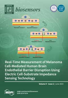
Melanoma is an aggressive cancer with a high propensity to metastasize to the brain. However, this requires the disruption of junctional complexes present at the blood–brain barrier (BBB), which restricts the passage of blood-borne pathogens into the brain, protecting it from injury. Brain endothelial cells are a key component of the BBB and form the first line of defence against circulating molecules (or melanoma cells). Therefore, we investigated the barrier integrity of brain endothelial cells when testing melanoma cell invasion and migration. The research aims were to investigate the ability of human melanoma cells to affect barrier strength of human brain-derived endothelial cells using a sensitive, real-time, and label-free measure of electrical resistance. The research hypothesis was that melanoma cells would disrupt the blood–endothelial barrier. View this paper.
  • Issues are regarded as officially published after their release is announced to the table of contents alert mailing list.
  • You may sign up for e-mail alerts to receive table of contents of newly released issues.
  • PDF is the official format for papers published in both, html and pdf forms. To view the papers in pdf format, click on the "PDF Full-text" link, and use the free Adobe Reader to open them.
Order results
Result details
Section
Select all
Export citation of selected articles as:
10 pages, 1360 KiB  
Article
Biological Effects of C60 Fullerene Revealed with Bacterial Biosensor—Toxic or Rather Antioxidant?
by Sergey Emelyantsev, Evgeniya Prazdnova, Vladimir Chistyakov and Igor Alperovich
Biosensors 2019, 9(2), 81; https://doi.org/10.3390/bios9020081 - 21 Jun 2019
Cited by 24 | Viewed by 6159
Abstract
Nanoparticles have been attracting growing interest for both their antioxidant and toxic effects. Their exact action on cells strongly depends on many factors, including experimental conditions, preparation, and solvents used, which have contributed to the confusion regarding their safety and possible health benefits. [...] Read more.
Nanoparticles have been attracting growing interest for both their antioxidant and toxic effects. Their exact action on cells strongly depends on many factors, including experimental conditions, preparation, and solvents used, which have contributed to the confusion regarding their safety and possible health benefits. In order to clarify the biological effects of the most abundant fullerene C60, its impact on the Escherichia coli model has been studied. The main question was if C60 would have any antioxidant influence on the cell and, if yes, whether and to which extent it would be concentration-dependent. An oxidative stress induced by adding hydrogen peroxide was measured with an E. coli MG1655 pKatG-lux strain sensor, with its time evolution being recorded in the presence of fullerene C60 suspensions of different concentrations. Optimal conditions for the fullerene C60 solubilization in TWEEN 80 2% aqueous solution, together with resulting aggregate sizes, were determined. Results obtained for the bacterial model can be extrapolated on eukaryote mitochondria. The ability of C60 to penetrate through biological membranes, conduct protons, and interact with free radicals is likely responsible for its protective effect detected for E. coli. Thus, fullerene can be considered as a mitochondria-targeted antioxidant, worth further researching as a prospective component of novel medications. Full article
Show Figures

Figure 1

14 pages, 1161 KiB  
Review
Droplets for Sampling and Transport of Chemical Signals in Biosensing: A Review
by Shilun Feng, Elham Shirani and David W. Inglis
Biosensors 2019, 9(2), 80; https://doi.org/10.3390/bios9020080 - 20 Jun 2019
Cited by 20 | Viewed by 6618
Abstract
The chemical, temporal, and spatial resolution of chemical signals that are sampled and transported with continuous flow is limited because of Taylor dispersion. Droplets have been used to solve this problem by digitizing chemical signals into discrete segments that can be transported for [...] Read more.
The chemical, temporal, and spatial resolution of chemical signals that are sampled and transported with continuous flow is limited because of Taylor dispersion. Droplets have been used to solve this problem by digitizing chemical signals into discrete segments that can be transported for a long distance or a long time without loss of chemical, temporal or spatial precision. In this review, we describe Taylor dispersion, sampling theory, and Laplace pressure, and give examples of sampling probes that have used droplets to sample or/and transport fluid from a continuous medium, such as cell culture or nerve tissue, for external analysis. The examples are categorized, as follows: (1) Aqueous-phase sampling with downstream droplet formation; (2) preformed droplets for sampling; and (3) droplets formed near the analyte source. Finally, strategies for downstream sample recovery for conventional analysis are described. Full article
(This article belongs to the Special Issue Microfluidics for Biosensing and Diagnostics)
Show Figures

Figure 1

12 pages, 2354 KiB  
Article
Noncompetitive Chromogenic Lateral-Flow Immunoassay for Simultaneous Detection of Microcystins and Nodularin
by Sultana Akter, Teemu Kustila, Janne Leivo, Gangatharan Muralitharan, Markus Vehniäinen and Urpo Lamminmäki
Biosensors 2019, 9(2), 79; https://doi.org/10.3390/bios9020079 - 18 Jun 2019
Cited by 21 | Viewed by 6271
Abstract
Cyanobacterial blooms cause local and global health issues by contaminating surface waters. Microcystins and nodularins are cyclic cyanobacterial peptide toxins comprising numerous natural variants. Most of them are potent hepatotoxins, tumor promoters, and at least microcystin-LR is possibly carcinogenic. In drinking water, the [...] Read more.
Cyanobacterial blooms cause local and global health issues by contaminating surface waters. Microcystins and nodularins are cyclic cyanobacterial peptide toxins comprising numerous natural variants. Most of them are potent hepatotoxins, tumor promoters, and at least microcystin-LR is possibly carcinogenic. In drinking water, the World Health Organization (WHO) recommended the provisional guideline value of 1 µg/L for microcystin-LR. For water used for recreational activity, the guidance values for microcystin concentration varies mostly between 4–25 µg/L in different countries. Current immunoassays or lateral flow strips for microcystin/nodularin are based on indirect competitive method, which are generally more prone to sample interference and sometimes hard to interpret compared to two-site immunoassays. Simple, sensitive, and easy to interpret user-friendly methods for first line screening of microcystin/nodularin near water sources are needed for assessment of water quality and safety. We describe the development of a two-site sandwich format lateral-flow assay for the rapid detection of microcystins and nodularin-R. A unique antibody fragment capable of broadly recognizing immunocomplexes consisting of a capture antibody bound to microcystins/nodularin-R was used to develop the simple lateral flow immunoassay. The assay can visually detect the major hepatotoxins (microcystin-LR, -dmLR, -RR, -dmRR, -YR, -LY, -LF -LW, and nodularin-R) at and below the concentration of 4 µg/L. The signal is directly proportional to the concentration of the respective toxin, and the use of alkaline phosphatase activity offers a cost efficient alternative by eliminating the need of toxin conjugates or other labeling system. The easy to interpret assay has the potential to serve as a microcystins/nodularin screening tool for those involved in water quality monitoring such as municipal authorities, researchers, as well as general public concerned of bathing water quality. Full article
(This article belongs to the Special Issue Enzyme-linked Immunoassay)
Show Figures

Graphical abstract

39 pages, 11301 KiB  
Review
Silver-Based Plasmonic Nanoparticles for and Their Use in Biosensing
by Alexis Loiseau, Victoire Asila, Gabriel Boitel-Aullen, Mylan Lam, Michèle Salmain and Souhir Boujday
Biosensors 2019, 9(2), 78; https://doi.org/10.3390/bios9020078 - 10 Jun 2019
Cited by 278 | Viewed by 20218
Abstract
The localized surface plasmon resonance (LSPR) property of metallic nanoparticles is widely exploited for chemical and biological sensing. Selective biosensing of molecules using functionalized nanoparticles has become a major research interdisciplinary area between chemistry, biology and material science. Noble metals, especially gold (Au) [...] Read more.
The localized surface plasmon resonance (LSPR) property of metallic nanoparticles is widely exploited for chemical and biological sensing. Selective biosensing of molecules using functionalized nanoparticles has become a major research interdisciplinary area between chemistry, biology and material science. Noble metals, especially gold (Au) and silver (Ag) nanoparticles, exhibit unique and tunable plasmonic properties; the control over these metal nanostructures size and shape allows manipulating their LSPR and their response to the local environment. In this review, we will focus on Ag-based nanoparticles, a metal that has probably played the most important role in the development of the latest plasmonic applications, owing to its unique properties. We will first browse the methods for AgNPs synthesis allowing for controlled size, uniformity and shape. Ag-based biosensing is often performed with coated particles; therefore, in a second part, we will explore various coating strategies (organics, polymers, and inorganics) and their influence on coated-AgNPs properties. The third part will be devoted to the combination of gold and silver for plasmonic biosensing, in particular the use of mixed Ag and AuNPs, i.e., AgAu alloys or Ag-Au core@shell nanoparticles will be outlined. In the last part, selected examples of Ag and AgAu-based plasmonic biosensors will be presented. Full article
(This article belongs to the Special Issue Spectroscopy-Based Biosensors)
Show Figures

Figure 1

17 pages, 10031 KiB  
Article
Towards CMOS Integrated Microfluidics Using Dielectrophoretic Immobilization
by Honeyeh Matbaechi Ettehad, Rahul Kumar Yadav, Subhajit Guha and Christian Wenger
Biosensors 2019, 9(2), 77; https://doi.org/10.3390/bios9020077 - 05 Jun 2019
Cited by 10 | Viewed by 5944
Abstract
Dielectrophoresis (DEP) is a nondestructive and noninvasive method which is favorable for point-of-care medical diagnostic tests. This technique exhibits prominent relevance in a wide range of medical applications wherein the miniaturized platform for manipulation (immobilization, separation or rotation), and detection of biological particles [...] Read more.
Dielectrophoresis (DEP) is a nondestructive and noninvasive method which is favorable for point-of-care medical diagnostic tests. This technique exhibits prominent relevance in a wide range of medical applications wherein the miniaturized platform for manipulation (immobilization, separation or rotation), and detection of biological particles (cells or molecules) can be conducted. DEP can be performed using advanced planar technologies, such as complementary metal-oxide-semiconductor (CMOS) through interdigitated capacitive biosensors. The dielectrophoretically immobilization of micron and submicron size particles using interdigitated electrode (IDE) arrays is studied by finite element simulations. The CMOS compatible IDEs have been placed into the silicon microfluidic channel. A rigorous study of the DEP force actuation, the IDE’s geometrical structure, and the fluid dynamics are crucial for enabling the complete platform for CMOS integrated microfluidics and detection of micron and submicron-sized particle ranges. The design of the IDEs is performed by robust finite element analyses to avoid time-consuming and costly fabrication processes. To analyze the preliminary microfluidic test vehicle, simulations were first performed with non-biological particles. To produce DEP force, an AC field in the range of 1 to 5 V (peak-to-peak) is applied to the IDE. The impact of the effective external and internal properties, such as actuating DEP frequency and voltage, fluid flow velocity, and IDE’s geometrical parameters are investigated. The IDE based system will be used to immobilize and sense particles simultaneously while flowing through the microfluidic channel. The sensed particles will be detected using the capacitive sensing feature of the biosensor. The sensing and detecting of the particles are not in the scope of this paper and will be described in details elsewhere. However, to provide a complete overview of this system, the working principles of the sensor, the readout detection circuit, and the integration process of the silicon microfluidic channel are briefly discussed. Full article
(This article belongs to the Special Issue Microfluidics for Biosensing and Diagnostics)
Show Figures

Figure 1

35 pages, 21221 KiB  
Review
In Vivo Biosensing Using Resonance Energy Transfer
by Shashi Bhuckory, Joshua C. Kays and Allison M. Dennis
Biosensors 2019, 9(2), 76; https://doi.org/10.3390/bios9020076 - 03 Jun 2019
Cited by 31 | Viewed by 11110
Abstract
Solution-phase and intracellular biosensing has substantially enhanced our understanding of molecular processes foundational to biology and pathology. Optical methods are favored because of the low cost of probes and instrumentation. While chromatographic methods are helpful, fluorescent biosensing further increases sensitivity and can be [...] Read more.
Solution-phase and intracellular biosensing has substantially enhanced our understanding of molecular processes foundational to biology and pathology. Optical methods are favored because of the low cost of probes and instrumentation. While chromatographic methods are helpful, fluorescent biosensing further increases sensitivity and can be more effective in complex media. Resonance energy transfer (RET)-based sensors have been developed to use fluorescence, bioluminescence, or chemiluminescence (FRET, BRET, or CRET, respectively) as an energy donor, yielding changes in emission spectra, lifetime, or intensity in response to a molecular or environmental change. These methods hold great promise for expanding our understanding of molecular processes not just in solution and in vitro studies, but also in vivo, generating information about complex activities in a natural, organismal setting. In this review, we focus on dyes, fluorescent proteins, and nanoparticles used as energy transfer-based optical transducers in vivo in mice; there are examples of optical sensing using FRET, BRET, and in this mammalian model system. After a description of the energy transfer mechanisms and their contribution to in vivo imaging, we give a short perspective of RET-based in vivo sensors and the importance of imaging in the infrared for reduced tissue autofluorescence and improved sensitivity. Full article
(This article belongs to the Special Issue FRET-Based Biosensors)
Show Figures

Figure 1

19 pages, 918 KiB  
Review
Evolution of Electronic Noses from Research Objects to Engineered Environmental Odour Monitoring Systems: A Review of Standardization Approaches
by Domenico Cipriano and Laura Capelli
Biosensors 2019, 9(2), 75; https://doi.org/10.3390/bios9020075 - 31 May 2019
Cited by 32 | Viewed by 7689
Abstract
Since electronic noses are used more and more for air quality monitoring purposes, and in some countries are starting to have a legal value, there is a need for standardization and programs for the quality verification of instruments. Such quality programs have the [...] Read more.
Since electronic noses are used more and more for air quality monitoring purposes, and in some countries are starting to have a legal value, there is a need for standardization and programs for the quality verification of instruments. Such quality programs have the aim to guarantee the main characteristics of the instrument for both the final user and local authorities, let the user establish a suitable maintenance procedure and give information on measurement uncertainty. One critical aspect when dealing with electronic noses for environmental odour monitoring is that environmental odours are complex mixtures that are not repeatable nor reproducible, giving that they are not suitable for quality verifications. This paper aims to review and discuss the different approaches that can be adopted in order to perform quality checks on electronic noses (e-noses) used for environmental odour monitoring, thereby referring to existing technical standards, such as the Dutch NTA 9055:2012, the new German VDI 3518-3:2018, and the Italian UNI 1605848 project, which directly refer to electronic noses. Moreover, also the European technical standards that are prescriptive for automatic measuring systems (AMSs) are taken into consideration (i.e., EN 14181:2014 and EN 15267:2009), and their possible applicability to electronic noses is investigated. Finally, the pros and cons of the different approaches are presented and discussed in the conclusions section. Full article
Show Figures

Figure 1

9 pages, 2507 KiB  
Article
Graphene Oxide-Based Nanostructured DNA Sensor
by Aditya Balaji, Songlin Yang, Jeslyn Wang and Jin Zhang
Biosensors 2019, 9(2), 74; https://doi.org/10.3390/bios9020074 - 30 May 2019
Cited by 30 | Viewed by 7255
Abstract
Quick detection of DNA sequence is vital for many fields, especially, early-stage diagnosis. Here, we develop a graphene oxide-based fluorescence quenching sensor to quickly and accurately detect small amounts of a single strand of DNA. In this paper, fluorescent magnetic nanoparticles (FMNPs) modified [...] Read more.
Quick detection of DNA sequence is vital for many fields, especially, early-stage diagnosis. Here, we develop a graphene oxide-based fluorescence quenching sensor to quickly and accurately detect small amounts of a single strand of DNA. In this paper, fluorescent magnetic nanoparticles (FMNPs) modified with target DNA sequence (DNA-t) were bound onto the modified graphene oxide acting as the fluorescence quenching element. FMNPs are made of iron oxide (Fe3O4) core and fluorescent silica (SiO2) shell. The average particle size of FMNPs was 74 ± 6 nm and the average thickness of the silica shell, estimated from TEM results, was 30 ± 4 nm. The photoluminescence and magnetic properties of FMNPs have been investigated. Target oligonucleotide (DNA-t) was conjugated onto FMNPs through glutaraldehyde crosslinking. Meanwhile, graphene oxide (GO) nanosheets were produced by a modified Hummers method. A complementary oligonucleotide (DNA-c) was designed to interact with GO. In the presence of GO-modified with DNA-c, the fluorescence intensity of FMNPs modified with DNA-t was quenched through a FRET quenching mechanism. Our study indicates that FMNPs can not only act as a FRET donor, but also enhance the sensor accuracy by magnetically separating the sensing system from free DNA and non-hybridized GO. Results indicate that this sensing system is ideal to detect small amounts of DNA-t with limitation detection at 0.12 µM. Full article
(This article belongs to the Special Issue FRET-Based Biosensors)
Show Figures

Figure 1

14 pages, 2120 KiB  
Article
Acoustofluidic Micromixing Enabled Hybrid Integrated Colorimetric Sensing, for Rapid Point-of-Care Measurement of Salivary Potassium
by Vikram Surendran, Thomas Chiulli, Swetha Manoharan, Stephen Knisley, Muthukumaran Packirisamy and Arvind Chandrasekaran
Biosensors 2019, 9(2), 73; https://doi.org/10.3390/bios9020073 - 28 May 2019
Cited by 8 | Viewed by 6383
Abstract
The integration of microfluidics with advanced biosensor technologies offers tremendous advantages such as smaller sample volume requirement and precise handling of samples and reagents, for developing affordable point-of-care testing methodologies that could be used in hospitals for monitoring patients. However, the success and [...] Read more.
The integration of microfluidics with advanced biosensor technologies offers tremendous advantages such as smaller sample volume requirement and precise handling of samples and reagents, for developing affordable point-of-care testing methodologies that could be used in hospitals for monitoring patients. However, the success and popularity of point-of-care diagnosis lies with the generation of instantaneous and reliable results through in situ tests conducted in a painless, non-invasive manner. This work presents the development of a simple, hybrid integrated optical microfluidic biosensor for rapid detection of analytes in test samples. The proposed biosensor works on the principle of colorimetric optical absorption, wherein samples mixed with suitable chromogenic substrates induce a color change dependent upon the analyte concentration that could then be detected by the absorbance of light in its path length. This optical detection scheme has been hybrid integrated with an acoustofluidic micromixing unit to enable uniform mixing of fluids within the device. As a proof-of-concept, we have demonstrated the real-time application of our biosensor format for the detection of potassium in whole saliva samples. The results show that our lab-on-a-chip technology could provide a useful strategy in biomedical diagnoses for rapid analyte detection towards clinical point-of-care testing applications. Full article
(This article belongs to the Special Issue Microfluidics for Biosensing and Diagnostics)
Show Figures

Figure 1

15 pages, 4305 KiB  
Article
Detection of Breast Cancer Cells Using Acoustics Aptasensor Specific to HER2 Receptors
by Alexandra Poturnayová, Ľudmila Dzubinová, Monika Buríková, Jozef Bízik and Tibor Hianik
Biosensors 2019, 9(2), 72; https://doi.org/10.3390/bios9020072 - 27 May 2019
Cited by 26 | Viewed by 6723
Abstract
Detection of the breast cancer cells is important for early diagnosis of the cancer. We applied thickness shear mode acoustics method (TSM) for detection of SK-BR-3 breast cancer cells using DNA aptamers specific to HER2 positive membrane receptors. The biotinylated aptamers were immobilized [...] Read more.
Detection of the breast cancer cells is important for early diagnosis of the cancer. We applied thickness shear mode acoustics method (TSM) for detection of SK-BR-3 breast cancer cells using DNA aptamers specific to HER2 positive membrane receptors. The biotinylated aptamers were immobilized at the neutravidin layer chemisorbed at gold surface of TSM transducer. Addition of the cells resulted in decrease of resonant frequency, fs, and in increase of motional resistance, Rm. Using gold nanoparticles (AuNPs), modified by aptamers it was possible improving the limit of detection (LOD) that reached 550 cells/mL, while without amplification the sensitivity of the detection of SK-BR-3 cells was 1574 cells/mL. HER2 negative cell line MDA-MB-231 did not resulted in significant changes of fs. The viability studies demonstrated that cells are stable at experimental conditions used during at least 8 h. AuNPs were not toxic on the cells up to concentration of 1 μg/mL. Full article
(This article belongs to the Special Issue Feature Papers: State-of-the-Art Biosensors Technology 2018)
Show Figures

Figure 1

12 pages, 2421 KiB  
Article
Perfusion Changes at the Forehead Measured by Photoplethysmography during a Head-Down Tilt Protocol
by Tomas Ysehak Abay, Kamran Shafqat and Panayiotis A. Kyriacou
Biosensors 2019, 9(2), 71; https://doi.org/10.3390/bios9020071 - 27 May 2019
Cited by 15 | Viewed by 7097
Abstract
Photoplethysmography (PPG) signals from the forehead can be used in pulse oximetry as they are less affected by vasoconstriction compared to fingers. However, the increase in venous blood caused by the positioning of the patient can deteriorate the signals and cause erroneous estimations [...] Read more.
Photoplethysmography (PPG) signals from the forehead can be used in pulse oximetry as they are less affected by vasoconstriction compared to fingers. However, the increase in venous blood caused by the positioning of the patient can deteriorate the signals and cause erroneous estimations of the arterial oxygen saturation. To date, there is no method to measure this venous presence under the PPG sensor. This study investigates the feasibility of using PPG signals from the forehead in an effort to estimate relative changes in haemoglobin concentrations that could reveal these posture-induced changes. Two identical reflectance PPG sensors were placed on two different positions on the forehead (above the eyebrow and on top of a large vein) in 16 healthy volunteers during a head-down tilt protocol. Relative changes in oxygenated ( Δ HbO 2 ), reduced ( Δ HHb) and total ( Δ tHb) haemoglobin were estimated from the PPG signals and the trends were compared with reference Near Infrared Spectroscopy (NIRS) measurements. Also, the signals from the two PPG sensors were analysed in order to reveal any difference due to the positioning of the sensor. Δ HbO 2 , Δ HHb and Δ tHb estimated from the forehead PPGs trended well with the same parameters from the reference NIRS. However, placing the sensor over a large vasculature reduces trending against NIRS, introduces biases as well as increases the variability of the changes in Δ HHb. Forehead PPG signals can be used to measure perfusion changes to reveal venous pooling induced by the positioning of the subject. Placing the sensor above the eyebrow and away from large vasculature avoids biases and large variability in the measurements. Full article
(This article belongs to the Special Issue In Vivo Physiological Monitoring)
Show Figures

Figure 1

12 pages, 1549 KiB  
Article
Multiplex Label Free Characterization of Cancer Cell Lines Using Surface Plasmon Resonance Imaging
by Ivan Stojanović, Carolina F. Ruivo, Thomas J. G. van der Velden, Richard B. M. Schasfoort and Leon W. M. M. Terstappen
Biosensors 2019, 9(2), 70; https://doi.org/10.3390/bios9020070 - 27 May 2019
Cited by 8 | Viewed by 5520
Abstract
Rapid multiplex cell surface marker analysis can expedite investigations in which large number of antigens need to be analyzed. Simultaneous analysis of multiple surface antigens at the same level of sensitivity is however limited in the current golden standard analysis method, flow cytometry. [...] Read more.
Rapid multiplex cell surface marker analysis can expedite investigations in which large number of antigens need to be analyzed. Simultaneous analysis of multiple surface antigens at the same level of sensitivity is however limited in the current golden standard analysis method, flow cytometry. In this paper we introduce a surface plasmon resonance imaging (SPRi)-based technique for 44-plex parameter analysis using a single sample, in less than 20 min. We analyzed the expression on cells from five different cancer cell lines by SPRi on a 44-plex antibody array including 4 negative controls and compared the output with flow cytometry. The combined correlation of the markers that showed expression by flow cytometry was 0.76. The results demonstrate as a proof of principle that SPRi can be applied for rapid semi-quantitative multiplex cell surface marker analysis. Full article
Show Figures

Figure 1

12 pages, 1329 KiB  
Article
Faecal Scent as a Novel Non-Invasive Biomarker to Discriminate between Coeliac Disease and Refractory Coeliac Disease: A Proof of Principle Study
by Maxine D. Rouvroye, Alfian Wicaksono, Sofie Bosch, Edo Savelkoul, James A. Covington, Hanneke Beaumont, Chris J. Mulder, Gerd Bouma, Tim G.J. de Meij and Nanne K.H. de Boer
Biosensors 2019, 9(2), 69; https://doi.org/10.3390/bios9020069 - 27 May 2019
Cited by 17 | Viewed by 6104
Abstract
Currently, the gold standard for diagnosis of coeliac disease (CD) is based on serology and gastroduodenoscopy with histology of duodenal mucosal biopsies. The aim of this study was to evaluate the potential of faecal volatile organic compounds (VOCs) analysis as a novel, non-invasive [...] Read more.
Currently, the gold standard for diagnosis of coeliac disease (CD) is based on serology and gastroduodenoscopy with histology of duodenal mucosal biopsies. The aim of this study was to evaluate the potential of faecal volatile organic compounds (VOCs) analysis as a novel, non-invasive tool to discriminate between CD in remission in patients on a gluten-free diet (GFD), refractory coeliac disease (RCD) and controls without CD. Patients with an established diagnosis of CD on a GFD, RCD and healthy controls (HC) were instructed to collect a faecal sample. All subjects completed questionnaires on clinical symptoms, lifestyle and dietary information. Faecal VOCs were measured using gas chromatography-ion mobility spectrometry. A total of 13 CD, 7 RCD and 10 HC were included. A significant difference in VOC profiles between CD and RCD patients (area under the curve (AUC) ± 95% CI: 0.91 (0.79–1) p = 0.000) and between CD and HC (AUC ± 95% CI: 0.71 (0.51–0.91) p = 0.0254) was observed. We found no significant differences between faecal VOC patterns of HC and RCD. Based on faecal VOCs, CD could be discriminated from RCD and HC. This implies that faecal VOC analysis may hold potential as a novel non-invasive biomarker for RCD. Future studies should encompass a larger cohort to further investigate and validate this prior to application in clinical practice. Full article
(This article belongs to the Special Issue Noninvasive Early Disease Diagnosis)
Show Figures

Figure 1

13 pages, 3076 KiB  
Article
Casein-Conjugated Gold Nanoparticles for Amperometric Detection of Leishmania infantum
by Mohamed Fethi Diouani, Oussama Ouerghi, Kamel Belgacem, Maher Sayhi, Radu Ionescu and Dhafer Laouini
Biosensors 2019, 9(2), 68; https://doi.org/10.3390/bios9020068 - 27 May 2019
Cited by 14 | Viewed by 7586
Abstract
Sensitive and reliable approaches targeting the detection of Leishmania are critical for effective early diagnosis and treatment of leishmaniasis. In this frame, this paper describes a rapid quantification assay to detect Leishmania parasites based on the combination of the electrocatalytic ability of gold [...] Read more.
Sensitive and reliable approaches targeting the detection of Leishmania are critical for effective early diagnosis and treatment of leishmaniasis. In this frame, this paper describes a rapid quantification assay to detect Leishmania parasites based on the combination of the electrocatalytic ability of gold nanoparticles (AuNPs) to act as a catalyst for the hydrogen formation reaction along with the specificity of the interaction between casein and the major surface protease of the Leishmania parasite, GP63. First, pure and casein-modified AuNPs were prepared and characterized by scanning electron microscopy and ultraviolet–visible spectroscopy. Then, casein-conjugated AuNPs were incubated with Leishsmania parasites in solution; the formed complex was collected by centrifugation, treated by acidic solution, and the pelleted AuNPs were placed on screen-printed carbon electrodes (SPCEs) and chronoamperometric measurements were carried out. Our results suggest that it is possible to detect Leishmania parasites, with a limit less than 1 parasite/mL. A linear response over a wide concentration interval, ranging from 2 × 10⁻² to 2 × 10⁵ parasites/mL, was achieved. Additionally, a pretreatment of Leishmania parasites with Amphotericin B, diminished their interaction with casein. This findings and methodology are very useful for drug efficacy assessment. Full article
(This article belongs to the Special Issue Nanoparticle-Based Biosensors for Food and Health Monitoring)
Show Figures

Figure 1

12 pages, 2579 KiB  
Article
Analytical Solution of the Time-Dependent Microfluidic Poiseuille Flow in Rectangular Channel Cross-Sections and Its Numerical Implementation in Microsoft Excel
by Patrick Risch, Dorothea Helmer, Frederik Kotz and Bastian E. Rapp
Biosensors 2019, 9(2), 67; https://doi.org/10.3390/bios9020067 - 24 May 2019
Cited by 3 | Viewed by 5742
Abstract
We recently demonstrated that the Navier–Stokes equation for pressure-driven laminar (Poiseuille) flow can be solved in any channel cross-section using a finite difference scheme implemented in a spreadsheet analysis tool such as Microsoft Excel. We also showed that implementing different boundary conditions (slip, [...] Read more.
We recently demonstrated that the Navier–Stokes equation for pressure-driven laminar (Poiseuille) flow can be solved in any channel cross-section using a finite difference scheme implemented in a spreadsheet analysis tool such as Microsoft Excel. We also showed that implementing different boundary conditions (slip, no-slip) is straight-forward. The results obtained in such calculations only deviated by a few percent from the (exact) analytical solution. In this paper we demonstrate that these approaches extend to cases where time-dependency is of importance, e.g., during initiation or after removal of the driving pressure. As will be shown, the developed spread-sheet can be used conveniently for almost any cross-section for which analytical solutions are close-to-impossible to obtain. We believe that providing researchers with convenient tools to derive solutions to complex flow problems in a fast and intuitive way will significantly enhance the understanding of the flow conditions as well as mass and heat transfer kinetics in microfluidic systems. Full article
(This article belongs to the Special Issue Microfluidics for Biosensing and Diagnostics)
Show Figures

Figure 1

13 pages, 1709 KiB  
Article
Rapid Profiling of Soybean Aromatic Compounds Using Electronic Nose
by Ramasamy Ravi, Ali Taheri, Durga Khandekar and Reneth Millas
Biosensors 2019, 9(2), 66; https://doi.org/10.3390/bios9020066 - 24 May 2019
Cited by 34 | Viewed by 6422
Abstract
Soybean (Glycine max (L.)) is the world’s most important seed legume, which contributes to 25% of global edible oil, and about two-thirds of the world’s protein concentrate for livestock feeding. One of the factors that limit soybean’s utilization as a major source [...] Read more.
Soybean (Glycine max (L.)) is the world’s most important seed legume, which contributes to 25% of global edible oil, and about two-thirds of the world’s protein concentrate for livestock feeding. One of the factors that limit soybean’s utilization as a major source of protein for humans is its characteristic soy flavor. This off-flavor can be attributed to the presence of various chemicals such as phenols, aldehydes, ketones, furans, alcohols, and amines. In addition, these flavor compounds interact with protein and cause the formation of new off-flavors. Hence, studying the chemical profile of soybean seeds is an important step in understanding how different chemical classes interact and contribute to the overall flavor profile of the crop. In our study, we utilized the HERCALES Fast Gas Chromatography (GC) electronic nose for identification and characterization of different volatile compounds in five high-yielding soybean varieties, and studied their association with off-flavors. With aroma profiling and chemical characterization, we aim to determine the quantity and quality of volatile compounds in these soybean varieties and understand their effect on the flavor profiles. The study could help to understand soybean flavor characteristics, which in turn could increase soybean use and enhance profitability. Full article
Show Figures

Figure 1

22 pages, 3067 KiB  
Review
An Alternative Medical Diagnosis Method: Biosensors for Virus Detection
by Yeşeren Saylan, Özgecan Erdem, Serhat Ünal and Adil Denizli
Biosensors 2019, 9(2), 65; https://doi.org/10.3390/bios9020065 - 21 May 2019
Cited by 196 | Viewed by 15689
Abstract
Infectious diseases still pose an omnipresent threat to global and public health, especially in many countries and rural areas of cities. Underlying reasons of such serious maladies can be summarized as the paucity of appropriate analysis methods and subsequent treatment strategies due to [...] Read more.
Infectious diseases still pose an omnipresent threat to global and public health, especially in many countries and rural areas of cities. Underlying reasons of such serious maladies can be summarized as the paucity of appropriate analysis methods and subsequent treatment strategies due to the limited access of centralized and equipped health care facilities for diagnosis. Biosensors hold great impact to turn our current analytical methods into diagnostic strategies by restructuring their sensing module for the detection of biomolecules, especially nano-sized objects such as protein biomarkers and viruses. Unquestionably, current sensing platforms require continuous updates to address growing challenges in the diagnosis of viruses as viruses change quickly and spread largely from person-to-person, indicating the urgency of early diagnosis. Some of the challenges can be classified in biological barriers (specificity, low number of targets, and biological matrices) and technological limitations (detection limit, linear dynamic range, stability, and reliability), as well as economical aspects that limit their implementation into resource-scarce settings. In this review, the principle and types of biosensors and their applications in the diagnosis of distinct infectious diseases were comprehensively explained. The deployment of current biosensors into resource-scarce settings is further discussed for virus detection by elaborating the pros and cons of existing methods as a conclusion and future perspective. Full article
Show Figures

Figure 1

14 pages, 2080 KiB  
Article
The Application of a Nanomaterial Optical Fiber Biosensor Assay for Identification of Brucella Nomenspecies
by Kelly McCutcheon, Aloka B. Bandara, Ziwei Zuo, James R. Heflin and Thomas J. Inzana
Biosensors 2019, 9(2), 64; https://doi.org/10.3390/bios9020064 - 21 May 2019
Cited by 9 | Viewed by 5432
Abstract
Bacteria in the genus Brucella are the cause of brucellosis in humans and many domestic and wild animals. A rapid and culture-free detection assay to detect Brucella in clinical samples would be highly valuable. Nanomaterial optical fiber biosensors (NOFS) are capable of recognizing [...] Read more.
Bacteria in the genus Brucella are the cause of brucellosis in humans and many domestic and wild animals. A rapid and culture-free detection assay to detect Brucella in clinical samples would be highly valuable. Nanomaterial optical fiber biosensors (NOFS) are capable of recognizing DNA hybridization events or other analyte interactions with high specificity and sensitivity. Therefore, a NOFS assay was developed to detect Brucella DNA from cultures and in tissue samples from infected mice. An ionic self-assembled multilayer (ISAM) film was coupled to a long-period grating optical fiber, and a nucleotide probe complementary to the Brucella IS711 region and modified with biotin was bound to the ISAM by covalent conjugation. When the ISAM/probe duplex was exposed to lysate containing ≥100 killed cells of Brucella, or liver or spleen tissue extracts from Brucella-infected mice, substantial attenuation of light transmission occurred, whereas exposure of the complexed fiber to non-Brucella gram-negative bacteria or control tissue samples resulted in negligible attenuation of light transmission. Oligonucleotide probes specific for B. abortus, B. melitensis, and B. suis could also be used to detect and differentiate these three nomenspecies. In summary, the NOFS biosensor assay detected three nomenspecies of Brucella without the use of polymerase chain reaction within 30 min and could specifically detect low numbers of this bacterium in clinical samples. Full article
(This article belongs to the Special Issue Spectroscopy-Based Biosensors)
Show Figures

Graphical abstract

9 pages, 421 KiB  
Article
Immobilized Luminescent Bacteria for the Detection of Mycotoxins under Discrete and Flow-Through Conditions
by Olga Senko, Nikolay Stepanov, Olga Maslova, Rashid Akhundov, Anvar Ismailov and Elena Efremenko
Biosensors 2019, 9(2), 63; https://doi.org/10.3390/bios9020063 - 20 May 2019
Cited by 15 | Viewed by 5660
Abstract
A biosensitive element in the form of bacterial Photobacterium phosphoreum cells immobilized in poly(vinyl alcohol) cryogel was tested for the determination of different mycotoxins under discrete and flow-through analysis conditions. The immobilized bioluminescent cells made it possible to quantify the presence of Ochratoxin [...] Read more.
A biosensitive element in the form of bacterial Photobacterium phosphoreum cells immobilized in poly(vinyl alcohol) cryogel was tested for the determination of different mycotoxins under discrete and flow-through analysis conditions. The immobilized bioluminescent cells made it possible to quantify the presence of Ochratoxin A, Sterigmatocystin, Zearalenone, and Deoxynivalenon in aqueous media in a wide range of their concentrations (0.017–56 mg/L, 0.010–33 mg/L, 0.009–14 mg/L, and 0.026–177 mg/L, respectively) via measuring the quenching of cell luminescence. The flow conditions allowed the analysis sensitivity to be improved by an order of magnitude in terms of detected concentrations. Using the immobilized luminescent bacterial cells, we have shown the possibility of evaluating the efficiency of the mycotoxins’ hydrolysis under the action of enzymes. In this way, a 94 ± 4.5% efficiency of Zearalenone hydrolysis with hexahistidine-containing organophosphorus hydrolase for 1h-long treatment of the mycotoxin solution (100 mg/L) was shown. Full article
(This article belongs to the Special Issue Cell-based Biosensors)
Show Figures

Graphical abstract

15 pages, 6735 KiB  
Article
Biosensors to Monitor Water Quality Utilizing Insect Odorant-Binding Proteins as Detector Elements
by Spiros D. Dimitratos, Allison S. Hommel, Kenneth D. Konrad, Lauren M. Simpson, Jessica J. Wu-Woods and Daniel F. Woods
Biosensors 2019, 9(2), 62; https://doi.org/10.3390/bios9020062 - 14 May 2019
Cited by 20 | Viewed by 6555
Abstract
In the developing world, the identification of clean, potable water continues to pose a pervasive challenge, and waterborne diseases due to fecal contamination of water supplies significantly threaten public health. The ability to efficiently monitor local water supplies is key to water safety, [...] Read more.
In the developing world, the identification of clean, potable water continues to pose a pervasive challenge, and waterborne diseases due to fecal contamination of water supplies significantly threaten public health. The ability to efficiently monitor local water supplies is key to water safety, yet no low-cost, reliable method exists to detect contamination quickly. We developed an in vitro assay utilizing an odorant-binding protein (OBP), AgamOBP1, from the mosquito, Anopheles gambiae, to test for the presence of a characteristic metabolite, indole, from harmful coliform bacteria. We demonstrated that recombinantly expressed AgamOBP1 binds indole with high sensitivity. Our proof-of-concept assay is fluorescence-based and demonstrates the usefulness of insect OBPs as detector elements in novel biosensors that rapidly detect the presence of bacterial metabolic markers, and thus of coliform bacteria. We further demonstrated that rAgamOBP1 is suitable for use in portable, inexpensive “dipstick” biosensors that improve upon lateral flow technology since insect OBPs are robust, easily obtainable via recombinant expression, and resist detector “fouling.” Moreover, due to their wide diversity and ligand selectivity, insect chemosensory proteins have other biosensor applications for various analytes. The techniques presented here therefore represent platform technologies applicable to various future devices. Full article
Show Figures

Figure 1

27 pages, 3417 KiB  
Review
Electrochemical Immunosensors for Antibiotic Detection
by Aleksandra Pollap and Jolanta Kochana
Biosensors 2019, 9(2), 61; https://doi.org/10.3390/bios9020061 - 01 May 2019
Cited by 86 | Viewed by 11477
Abstract
Antibiotics are an important class of drugs destined for treatment of bacterial diseases. Misuses and overuses of antibiotics observed over the last decade have led to global problems of bacterial resistance against antibiotics (ABR). One of the crucial actions taken towards limiting the [...] Read more.
Antibiotics are an important class of drugs destined for treatment of bacterial diseases. Misuses and overuses of antibiotics observed over the last decade have led to global problems of bacterial resistance against antibiotics (ABR). One of the crucial actions taken towards limiting the spread of antibiotics and controlling this dangerous phenomenon is the sensitive and accurate determination of antibiotics residues in body fluids, food products, and animals, as well as monitoring their presence in the environment. Immunosensors, a group of biosensors, can be considered an attractive tool because of their simplicity, rapid action, low-cost analysis, and especially, the unique selectivity arising from harnessing the antigen–antibody interaction that is the basis of immunosensor functioning. Herein, we present the recent achievements in the field of electrochemical immunosensors designed to determination of antibiotics. Full article
(This article belongs to the Special Issue Electrochemical Immunosensor)
Show Figures

Figure 1

8 pages, 2084 KiB  
Article
Preparation and Characterization of a Pectin Membrane-Based Optical pH Sensor for Fish Freshness Monitoring
by Uswatun Hasanah, Mita Setyowati, Rustam Efendi, Muslem Muslem, Nor Diyana Md Sani, Eka Safitri, Lee Yook Heng and Rinaldi Idroes
Biosensors 2019, 9(2), 60; https://doi.org/10.3390/bios9020060 - 26 Apr 2019
Cited by 41 | Viewed by 8613
Abstract
In a simple and instant procedure for detecting fish freshness, a hydrogel and hydrophilic pectin matrix membrane was used successfully as an optical pH sensor by immobilizing the chromoionophore ETH 5294 (CI), which is very selective and sensitive for the membrane. The Pe/CI [...] Read more.
In a simple and instant procedure for detecting fish freshness, a hydrogel and hydrophilic pectin matrix membrane was used successfully as an optical pH sensor by immobilizing the chromoionophore ETH 5294 (CI), which is very selective and sensitive for the membrane. The Pe/CI optical pH sensor exhibited excellent linearity between pH 5 and pH 9, with a sensor response time of 5 min and reproducibility of 1.49% relative standard deviation (RSD). The sensor showed response stability for 15 days and a response reduction of 8.6%. The sensor’s capability was demonstrated by the detection of fish freshness for 17 days at 4 °C. Full article
Show Figures

Figure 1

19 pages, 2923 KiB  
Article
Which Gait Parameters and Walking Patterns Show the Significant Differences Between Parkinson’s Disease and Healthy Participants?
by Sana M Keloth, Rekha Viswanathan, Beth Jelfs, Sridhar Arjunan, Sanjay Raghav and Dinesh Kumar
Biosensors 2019, 9(2), 59; https://doi.org/10.3390/bios9020059 - 25 Apr 2019
Cited by 12 | Viewed by 6324
Abstract
This study investigated the difference in the gait of patients with Parkinson’s disease (PD), age-matched controls and young controls during three walking patterns. Experiments were conducted with 24 PD, 24 age-matched controls and 24 young controls, and four gait intervals were measured using [...] Read more.
This study investigated the difference in the gait of patients with Parkinson’s disease (PD), age-matched controls and young controls during three walking patterns. Experiments were conducted with 24 PD, 24 age-matched controls and 24 young controls, and four gait intervals were measured using inertial measurement units (IMU). Group differences between the mean and variance of the gait parameters (stride interval, stance interval, swing interval and double support interval) for the three groups were calculated and statistical significance was tested. The results showed that the variance in each of the four gait parameters of PD patients was significantly higher compared with the controls, irrespective of the three walking patterns. This study showed that the variance of any of the gait interval parameters obtained using IMU during any of the walking patterns could be used to differentiate between the gait of PD and control people. Full article
Show Figures

Figure 1

13 pages, 4590 KiB  
Article
Research on Non-Contact Monitoring System for Human Physiological Signal and Body Movement
by Qiancheng Liang, Lisheng Xu, Nan Bao, Lin Qi, Jingjing Shi, Yicheng Yang and Yudong Yao
Biosensors 2019, 9(2), 58; https://doi.org/10.3390/bios9020058 - 19 Apr 2019
Cited by 17 | Viewed by 6689
Abstract
With the rapid increase in the development of miniaturized sensors and embedded devices for vital signs monitoring, personal physiological signal monitoring devices are becoming popular. However, physiological monitoring devices which are worn on the body normally affect the daily activities of people. This [...] Read more.
With the rapid increase in the development of miniaturized sensors and embedded devices for vital signs monitoring, personal physiological signal monitoring devices are becoming popular. However, physiological monitoring devices which are worn on the body normally affect the daily activities of people. This problem can be avoided by using a non-contact measuring device like the Doppler radar system, which is more convenient, is private compared to video monitoring, infrared monitoring and other non-contact methods. Additionally real-time physiological monitoring with the Doppler radar system can also obtain signal changes caused by motion changes. As a result, the Doppler radar system not only obtains the information of respiratory and cardiac signals, but also obtains information about body movement. The relevant RF technology could eliminate some interference from body motion with a small amplitude. However, the motion recognition method can also be used to classify related body motion signals. In this paper, a vital sign and body movement monitoring system worked at 2.4 GHz was proposed. It can measure various physiological signs of the human body in a non-contact manner. The accuracy of the non-contact physiological signal monitoring system was analyzed. First, the working distance of the system was tested. Then, the algorithm of mining collective motion signal was classified, and the accuracy was 88%, which could be further improved in the system. In addition, the mean absolute error values of heart rate and respiratory rate were 0.8 beats/min and 3.5 beats/min, respectively, and the reliability of the system was verified by comparing the respiratory waveforms with the contact equipment at different distances. Full article
(This article belongs to the Special Issue In Vivo Physiological Monitoring)
Show Figures

Figure 1

99 pages, 20711 KiB  
Review
A Review on Surface-Enhanced Raman Scattering
by Roberto Pilot, Raffaella Signorini, Christian Durante, Laura Orian, Manjari Bhamidipati and Laura Fabris
Biosensors 2019, 9(2), 57; https://doi.org/10.3390/bios9020057 - 17 Apr 2019
Cited by 545 | Viewed by 34882
Abstract
Surface-enhanced Raman scattering (SERS) has become a powerful tool in chemical, material and life sciences, owing to its intrinsic features (i.e., fingerprint recognition capabilities and high sensitivity) and to the technological advancements that have lowered the cost of the instruments and improved their [...] Read more.
Surface-enhanced Raman scattering (SERS) has become a powerful tool in chemical, material and life sciences, owing to its intrinsic features (i.e., fingerprint recognition capabilities and high sensitivity) and to the technological advancements that have lowered the cost of the instruments and improved their sensitivity and user-friendliness. We provide an overview of the most significant aspects of SERS. First, the phenomena at the basis of the SERS amplification are described. Then, the measurement of the enhancement and the key factors that determine it (the materials, the hot spots, and the analyte-surface distance) are discussed. A section is dedicated to the analysis of the relevant factors for the choice of the excitation wavelength in a SERS experiment. Several types of substrates and fabrication methods are illustrated, along with some examples of the coupling of SERS with separation and capturing techniques. Finally, a representative selection of applications in the biomedical field, with direct and indirect protocols, is provided. We intentionally avoided using a highly technical language and, whenever possible, intuitive explanations of the involved phenomena are provided, in order to make this review suitable to scientists with different degrees of specialization in this field. Full article
Show Figures

Graphical abstract

17 pages, 7173 KiB  
Article
Real-Time Measurement of Melanoma Cell-Mediated Human Brain Endothelial Barrier Disruption Using Electric Cell-Substrate Impedance Sensing Technology
by Akshata Anchan, Panagiota Kalogirou-Baldwin, Rebecca Johnson, Dan T Kho, Wayne Joseph, James Hucklesby, Graeme J Finlay, Simon J O’Carroll, Catherine E Angel and E Scott Graham
Biosensors 2019, 9(2), 56; https://doi.org/10.3390/bios9020056 - 15 Apr 2019
Cited by 21 | Viewed by 7333
Abstract
Electric cell-substrate impedance sensing (ECIS) is an impedance-based method for monitoring changes in cell behaviour in real-time. In this paper, we highlight the importance of ECIS in measuring the kinetics of human melanoma cell invasion across human brain endothelium. ECIS data can be [...] Read more.
Electric cell-substrate impedance sensing (ECIS) is an impedance-based method for monitoring changes in cell behaviour in real-time. In this paper, we highlight the importance of ECIS in measuring the kinetics of human melanoma cell invasion across human brain endothelium. ECIS data can be mathematically modelled to assess which component of the endothelial paracellular and basolateral barriers is being affected and when. Our results reveal that a range of human melanoma cells can mediate disruption of human brain endothelium, primarily involving the paracellular route, as demonstrated by ECIS. The sensitivity of ECIS also reveals that the paracellular barrier weakens within 30–60 min of the melanoma cells being added to the apical face of the endothelial cells. Imaging reveals pronounced localisation of the melanoma cells at the paracellular junctions consistent with paracellular migration. Time-lapse imaging further reveals junctional opening and disruption of the endothelial monolayer by the invasive melanoma cells all within several hours. We suggest that the ability of ECIS to resolve changes to barrier integrity in real time, and to determine the route of migration, provides a powerful tool for future studies investigating the key molecules involved in the invasive process of cancer cells. Full article
Show Figures

Figure 1

15 pages, 3065 KiB  
Article
Breath Analysis Using eNose and Ion Mobility Technology to Diagnose Inflammatory Bowel Disease—A Pilot Study
by Akira Tiele, Alfian Wicaksono, Jiten Kansara, Ramesh P. Arasaradnam and James A. Covington
Biosensors 2019, 9(2), 55; https://doi.org/10.3390/bios9020055 - 12 Apr 2019
Cited by 43 | Viewed by 8766
Abstract
Early diagnosis of inflammatory bowel disease (IBD), including Crohn’s disease (CD) and ulcerative colitis (UC), remains a clinical challenge with current tests being invasive and costly. The analysis of volatile organic compounds (VOCs) in exhaled breath and biomarkers in stool (faecal calprotectin (FCP)) [...] Read more.
Early diagnosis of inflammatory bowel disease (IBD), including Crohn’s disease (CD) and ulcerative colitis (UC), remains a clinical challenge with current tests being invasive and costly. The analysis of volatile organic compounds (VOCs) in exhaled breath and biomarkers in stool (faecal calprotectin (FCP)) show increasing potential as non-invasive diagnostic tools. The aim of this pilot study is to evaluate the efficacy of breath analysis and determine if FCP can be used as an additional non-invasive parameter to supplement breath results, for the diagnosis of IBD. Thirty-nine subjects were recruited (14 CD, 16 UC, 9 controls). Breath samples were analysed using an in-house built electronic nose (Wolf eNose) and commercial gas chromatograph–ion mobility spectrometer (G.A.S. BreathSpec GC-IMS). Both technologies could consistently separate IBD and controls [AUC ± 95%, sensitivity, specificity], eNose: [0.81, 0.67, 0.89]; GC-IMS: [0.93, 0.87, 0.89]. Furthermore, we could separate CD from UC, eNose: [0.88, 0.71, 0.88]; GC-IMS: [0.71, 0.86, 0.62]. Including FCP did not improve distinction between CD vs. UC; eNose: [0.74, 1.00, 0.56], but rather, improved separation of CD vs. controls and UC vs. controls; eNose: [0.77, 0.55, 1.00] and [0.72, 0.89, 0.67] without FCP, [0.81, 0.73, 0.78] and [0.90, 1.00, 0.78] with FCP, respectively. These results confirm the utility of breath analysis to distinguish between IBD-related diagnostic groups. FCP does not add significant diagnostic value to breath analysis within this study. Full article
(This article belongs to the Special Issue Noninvasive Early Disease Diagnosis)
Show Figures

Graphical abstract

14 pages, 2547 KiB  
Article
Ligand-Based Stability Changes in Duplex DNA Measured with a Microscale Electrochemical Platform
by Sarah M. Robinson, Zuliang Shen, Jon R. Askim, Christopher B. Montgomery, Herman O. Sintim and Steve Semancik
Biosensors 2019, 9(2), 54; https://doi.org/10.3390/bios9020054 - 12 Apr 2019
Cited by 6 | Viewed by 5698
Abstract
Development of technologies for rapid screening of DNA secondary structure thermal stability and the effects on stability for binding of small molecule drugs is important to the drug discovery process. In this report, we describe the capabilities of an electrochemical, microdevice-based approach for [...] Read more.
Development of technologies for rapid screening of DNA secondary structure thermal stability and the effects on stability for binding of small molecule drugs is important to the drug discovery process. In this report, we describe the capabilities of an electrochemical, microdevice-based approach for determining the melting temperatures (Tm) of electrode-bound duplex DNA structures. We also highlight new features of the technology that are compatible with array development and adaptation for high-throughput screening. As a foundational study to exhibit device performance and capabilities, melting-curve analyses were performed on 12-mer DNA duplexes in the presence/absence of two binding ligands: diminazene aceturate (DMZ) and proflavine. By measuring electrochemical current as a function of temperature, our measurement platform has the ability to determine the effect of binding ligands on Tm values with high signal-to-noise ratios and good reproducibility. We also demonstrate that heating our three-electrode cell with either an embedded microheater or a thermoelectric module produces similar results. The ΔTm values we report show the stabilizing ability of DMZ and proflavine when bound to duplex DNA structures. These initial proof-of-concept studies highlight the operating characteristics of the microdevice platform and the potential for future application toward other immobilized samples. Full article
Show Figures

Graphical abstract

13 pages, 2853 KiB  
Article
Robust SERS Platforms Based on Annealed Gold Nanostructures Formed on Ultrafine Glass Substrates for Various (Bio)Applications
by Lan Zhou, Simone Poggesi, Giuliocesare Casari Bariani, Rakesh Mittapalli, Pierre-Michel Adam, Marisa Manzano and Rodica Elena Ionescu
Biosensors 2019, 9(2), 53; https://doi.org/10.3390/bios9020053 - 10 Apr 2019
Cited by 14 | Viewed by 6790
Abstract
In this study, stable gold nanoparticles (AuNPs) are fabricated for the first time on commercial ultrafine glass coverslips coated with gold thin layers (2 nm, 4 nm, 6 nm, and 8 nm) at 25 °C and annealed at high temperatures (350 °C, 450 [...] Read more.
In this study, stable gold nanoparticles (AuNPs) are fabricated for the first time on commercial ultrafine glass coverslips coated with gold thin layers (2 nm, 4 nm, 6 nm, and 8 nm) at 25 °C and annealed at high temperatures (350 °C, 450 °C, and 550 °C) on a hot plate for different periods of time. Such gold nanostructured coverslips were systematically tested via surface enhanced Raman spectroscopy (SERS) to identify their spectral performances in the presence of different concentrations of a model molecule, namely 1,2-bis-(4-pyridyl)-ethene (BPE). By using these SERS platforms, it is possible to detect BPE traces (10−12 M) in aqueous solutions in 120 s. The stability of SERS spectra over five weeks of thiol-DNA probe (2 µL) deposited on gold nano-structured coverslip is also reported. Full article
(This article belongs to the Special Issue Spectroscopy-Based Biosensors)
Show Figures

Figure 1

14 pages, 1174 KiB  
Article
Specific and Generic Immunorecognition of Glycopeptide Antibiotics Promoted by Unique and Multiple Orientations of Hapten
by Maksim A. Burkin, Inna A. Galvidis and Sergei A. Eremin
Biosensors 2019, 9(2), 52; https://doi.org/10.3390/bios9020052 - 04 Apr 2019
Cited by 7 | Viewed by 5015
Abstract
Conjugation chemistry does not always provide adequate spatial orientation of hapten in immunogens for the best presentation of generic or individual epitopes. In the present study, the influence of unique and multiple orientations of immunizing hapten on the immune response repertoire was compared [...] Read more.
Conjugation chemistry does not always provide adequate spatial orientation of hapten in immunogens for the best presentation of generic or individual epitopes. In the present study, the influence of unique and multiple orientations of immunizing hapten on the immune response repertoire was compared to select generic recognition system. The glycopeptides, teicoplanin (TPL) and ristomycin (RSM), were conjugated to BSA to produce immunogens with unique and multiple orientations of haptens. Polyclonal antibodies generated against TPL conjugated through a single site were of uniform specificity and demonstrated selective TPL recognition, regardless of the coating conjugates design. The sensitivity (IC50) of 4 enzyme-linked immunosorbent assays (ELISAs) for TPL varied little within the 3.5–7.4 ng/mL, with a dynamic range of 0.2–100 ng/mL. RSM was coupled to BSA through several glycoside sites that evoked a wider repertoire of response. This first described anti-RSM antibody was selective for RSM in homologous hapten-coated ELISAs with IC50 values in the range 4.2–35 ng/mL. Among the heterologous antigens, periodate-oxidized TPL conjugated to gelatine was selected as the best binder of generic anti-RSM fraction. The developed ELISA showed group recognition of glycopeptides RSM, TPL, eremomycin, and vancomycin with cross-reactivity of 37–100% and a 10–10,000 ng/mL dynamic range. Thus, multiple presentations of immunizing hapten help expand the repertoire of immune responses and opportunities for the selection of the required fine-specificity agent. Full article
(This article belongs to the Special Issue Enzyme-linked Immunoassay)
Show Figures

Figure 1

Previous Issue
Next Issue
Back to TopTop