Next Issue
Volume 8, September
Previous Issue
Volume 8, March


Biosensors, Volume 8, Issue 2 (June 2018) – 30 articles

Cover Story (view full-size image): 
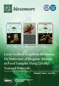
In foods, high levels of biogenic amines (BA) are commonly used as an indicator of food safety and food quality. A large number of fermented foods are consumed in developing countries or marine areas, where access to laboratory techniques, resources, and certified facilities is limited. Thus, often, detection of BA is not regularly performed. In this study, a low-cost, disposable nanobiosensor was developed for measuring BA in food using locally sourced materials that can be obtained from markets in any major city. A comparative study was conducted between the biosensor and a similar device fabricated with analytical-grade materials. View the paper here.
  • Issues are regarded as officially published after their release is announced to the table of contents alert mailing list.
  • You may sign up for e-mail alerts to receive table of contents of newly released issues.
  • PDF is the official format for papers published in both, html and pdf forms. To view the papers in pdf format, click on the "PDF Full-text" link, and use the free Adobe Reader to open them.
Order results
Result details
Section
Select all
Export citation of selected articles as:
22 pages, 7173 KiB  
Article
Design and Parameter Study of Integrated Microfluidic Platform for CTC Isolation and Enquiry; A Numerical Approach
by Amir Shamloo, Saba Ahmad and Maede Momeni
Biosensors 2018, 8(2), 56; https://doi.org/10.3390/bios8020056 - 18 Jun 2018
Cited by 11 | Viewed by 4599
Abstract
Being the second cause of mortality across the globe, there is now a persistent effort to establish new cancer medication and therapies. Any accomplishment in treating cancers entails the existence of accurate identification systems empowering the early diagnosis. Recent studies indicate CTCs’ potential [...] Read more.
Being the second cause of mortality across the globe, there is now a persistent effort to establish new cancer medication and therapies. Any accomplishment in treating cancers entails the existence of accurate identification systems empowering the early diagnosis. Recent studies indicate CTCs’ potential in cancer prognosis as well as therapy monitoring. The chief shortcoming with CTCs is that they are exceedingly rare cells in their clinically relevant concentration. Here, we simulated a microfluidic construct devised for immunomagnetic separation of the particles of interest from the background cells. This separation unit is integrated with a mixer subunit. The mixer is envisioned for mixing the CTC enriched stream with lysis buffer to extract the biological material of the cell. Some modification was proposed on mixing geometry improving the efficacy of the functional unit. A valuation of engaged forces was made and some forces were neglected due to their order of magnitude. The position of the magnet was also optimized by doing parametric study. For the mixer unit, the effect of applied voltage and frequency on mixing index was studied to find the optimal voltage and frequency which provides better mixing. Above-mentioned studies were done on isolated units and the effect of each functional unit on the other is not studied. As the final step, an integrated microfluidic platform composed of both functional subunits was simulated simultaneously. To ensure the independence of results from the grid, grid studies were also performed. The studies carried out on the construct reveal its potential for diagnostic application. Full article
Show Figures

Figure 1

12 pages, 1351 KiB  
Review
Biosensors for Non-Invasive Detection of Celiac Disease Biomarkers in Body Fluids
by Tibor Pasinszki and Melinda Krebsz
Biosensors 2018, 8(2), 55; https://doi.org/10.3390/bios8020055 - 16 Jun 2018
Cited by 13 | Viewed by 6600
Abstract
Celiac disease is a chronic gluten-initiated autoimmune disorder that predominantly damages the mucosa of the small intestine in genetically-susceptible individuals. It affects a large and increasing number of the world’s population. The diagnosis of this disease and monitoring the response of patients to [...] Read more.
Celiac disease is a chronic gluten-initiated autoimmune disorder that predominantly damages the mucosa of the small intestine in genetically-susceptible individuals. It affects a large and increasing number of the world’s population. The diagnosis of this disease and monitoring the response of patients to the therapy, which is currently a life-long gluten-free diet, require the application of reliable, rapid, sensitive, selective, simple, and cost-effective analytical tools. Celiac disease biomarker detection in full blood, serum, or plasma offers a non-invasive way to do this and is well-suited to being the first step of diagnosis. Biosensors provide a novel and alternative way to perform conventional techniques in biomarker sensing, in which electrode material and architecture play important roles in achieving sensitive, selective, and stable detection. There are many opportunities to build and modify biosensor platforms using various materials and detection methods, and the aim of the present review is to summarize developments in this field. Full article
(This article belongs to the Special Issue Biomarkers)
Show Figures

Figure 1

62 pages, 1877 KiB  
Review
Aptamer-Based Biosensors for Antibiotic Detection: A Review
by Asol Mehlhorn, Parvaneh Rahimi and Yvonne Joseph
Biosensors 2018, 8(2), 54; https://doi.org/10.3390/bios8020054 - 11 Jun 2018
Cited by 185 | Viewed by 19236
Abstract
Antibiotic resistance and, accordingly, their pollution because of uncontrolled usage has emerged as a serious problem in recent years. Hence, there is an increased demand to develop robust, easy, and sensitive methods for rapid evaluation of antibiotics and their residues. Among different analytical [...] Read more.
Antibiotic resistance and, accordingly, their pollution because of uncontrolled usage has emerged as a serious problem in recent years. Hence, there is an increased demand to develop robust, easy, and sensitive methods for rapid evaluation of antibiotics and their residues. Among different analytical methods, the aptamer-based biosensors (aptasensors) have attracted considerable attention because of good selectivity, specificity, and sensitivity. This review gives an overview about recently-developed aptasensors for antibiotic detection. The use of various aptamer assays to determine different groups of antibiotics, like β-lactams, aminoglycosides, anthracyclines, chloramphenicol, (fluoro)quinolones, lincosamide, tetracyclines, and sulfonamides are presented in this paper. Full article
Show Figures

Figure 1

10 pages, 2223 KiB  
Article
Determination of the Electrochemical Area of Screen-Printed Electrochemical Sensing Platforms
by Alejandro García-Miranda Ferrari, Christopher W. Foster, Peter J. Kelly, Dale A. C. Brownson and Craig E. Banks
Biosensors 2018, 8(2), 53; https://doi.org/10.3390/bios8020053 - 08 Jun 2018
Cited by 284 | Viewed by 13381
Abstract
Screen-printed electrochemical sensing platforms, due to their scales of economy and high reproducibility, can provide a useful approach to translate laboratory-based electrochemistry into the field. An important factor when utilising screen-printed electrodes (SPEs) is the determination of their real electrochemical surface area, which [...] Read more.
Screen-printed electrochemical sensing platforms, due to their scales of economy and high reproducibility, can provide a useful approach to translate laboratory-based electrochemistry into the field. An important factor when utilising screen-printed electrodes (SPEs) is the determination of their real electrochemical surface area, which allows for the benchmarking of these SPEs and is an important parameter in quality control. In this paper, we consider the use of cyclic voltammetry and chronocoulometry to allow for the determination of the real electrochemical area of screen-printed electrochemical sensing platforms, highlighting to experimentalists the various parameters that need to be diligently considered and controlled in order to obtain useful measurements of the real electroactive area. Full article
(This article belongs to the Special Issue Electrochemical (Bio)sensors for Environmental and Food Analyses)
Show Figures

Graphical abstract

21 pages, 13240 KiB  
Review
Label-Free Bioanalyte Detection from Nanometer to Micrometer Dimensions—Molecular Imprinting and QCMs
by Adnan Mujahid, Ghulam Mustafa and Franz L. Dickert
Biosensors 2018, 8(2), 52; https://doi.org/10.3390/bios8020052 - 01 Jun 2018
Cited by 21 | Viewed by 6591
Abstract
Modern diagnostic tools and immunoassay protocols urges direct analyte recognition based on its intrinsic behavior without using any labeling indicator. This not only improves the detection reliability, but also reduces sample preparation time and complexity involved during labeling step. Label-free biosensor devices are [...] Read more.
Modern diagnostic tools and immunoassay protocols urges direct analyte recognition based on its intrinsic behavior without using any labeling indicator. This not only improves the detection reliability, but also reduces sample preparation time and complexity involved during labeling step. Label-free biosensor devices are capable of monitoring analyte physiochemical properties such as binding sensitivity and selectivity, affinity constants and other dynamics of molecular recognition. The interface of a typical biosensor could range from natural antibodies to synthetic receptors for example molecular imprinted polymers (MIPs). The foremost advantages of using MIPs are their high binding selectivity comparable to natural antibodies, straightforward synthesis in short time, high thermal/chemical stability and compatibility with different transducers. Quartz crystal microbalance (QCM) resonators are leading acoustic devices that are extensively used for mass-sensitive measurements. Highlight features of QCM devices include low cost fabrication, room temperature operation, and most importantly ability to monitor extremely low mass shifts, thus potentially a universal transducer. The combination of MIPs with quartz QCM has turned out as a prominent sensing system for label-free recognition of diverse bioanalytes. In this article, we shall encompass the potential applications of MIP-QCM sensors exclusively label-free recognition of bacteria and virus species as representative micro and nanosized bioanalytes. Full article
(This article belongs to the Special Issue Label-free Biosensing)
Show Figures

Figure 1

10 pages, 2162 KiB  
Article
New Carrier Made from Glass Nanofibres for the Colorimetric Biosensor of Cholinesterase Inhibitors
by Lukáš Matějovský and Vladimír Pitschmann
Biosensors 2018, 8(2), 51; https://doi.org/10.3390/bios8020051 - 30 May 2018
Cited by 23 | Viewed by 5602
Abstract
Cholinesterase inhibitors are widely used as pesticides in agriculture, but also form a group of organophosphates known as nerve chemical warfare agents. This calls for close attention regarding their detection, including the use of various biosensors. One such biosensor made in the Czech [...] Read more.
Cholinesterase inhibitors are widely used as pesticides in agriculture, but also form a group of organophosphates known as nerve chemical warfare agents. This calls for close attention regarding their detection, including the use of various biosensors. One such biosensor made in the Czech Republic is the Detehit, which is based on a cholinesterase reaction that is assessed using a colour indicator—the Ellman’s reagent—which is anchored on cellulose filter paper together with the substrate. With the use of this biosensor, detection is simple, quick, and sensitive. However, its disadvantage is that a less pronounced yellow discoloration occurs, especially under difficult light conditions. As a possible solution, a new indicator/substrate carrier has been designed. It is made of glass nanofibres, so the physical characteristics of the carrier positively influence reaction conditions, and as a result improve the colour response of the biosensor. The authors present and discuss some of the results of the study of this carrier under various experimental conditions. These findings have been used for the development of a modified Detehit biosensor. Full article
(This article belongs to the Special Issue Micro and Nanoscale Biosensors)
Show Figures

Figure 1

23 pages, 1807 KiB  
Article
Limits to the Evaluation of the Accuracy of Continuous Glucose Monitoring Systems by Clinical Trials
by Patrick Schrangl, Florian Reiterer, Lutz Heinemann, Guido Freckmann and Luigi Del Re
Biosensors 2018, 8(2), 50; https://doi.org/10.3390/bios8020050 - 18 May 2018
Cited by 35 | Viewed by 6781
Abstract
Systems for continuous glucose monitoring (CGM) are evolving quickly, and the data obtained are expected to become the basis for clinical decisions for many patients with diabetes in the near future. However, this requires that their analytical accuracy is sufficient. This accuracy is [...] Read more.
Systems for continuous glucose monitoring (CGM) are evolving quickly, and the data obtained are expected to become the basis for clinical decisions for many patients with diabetes in the near future. However, this requires that their analytical accuracy is sufficient. This accuracy is usually determined with clinical studies by comparing the data obtained by the given CGM system with blood glucose (BG) point measurements made with a so-called reference method. The latter is assumed to indicate the correct value of the target quantity. Unfortunately, due to the nature of the clinical trials and the approach used, such a comparison is subject to several effects which may lead to misleading results. While some reasons for the differences between the values obtained with CGM and BG point measurements are relatively well-known (e.g., measurement in different body compartments), others related to the clinical study protocols are less visible, but also quite important. In this review, we present a general picture of the topic as well as tools which allow to correct or at least to estimate the uncertainty of measures of CGM system performance. Full article
(This article belongs to the Special Issue Continuous Glucose Monitoring)
Show Figures

Figure 1

11 pages, 993 KiB  
Article
Consistency of Continuous Ambulatory Interstitial Glucose Monitoring Sensors
by Pei T. Wu, David E. Segovia, Cathy C. Lee and Kim-Lien Nguyen
Biosensors 2018, 8(2), 49; https://doi.org/10.3390/bios8020049 - 16 May 2018
Cited by 3 | Viewed by 4755
Abstract
Aims: The abdominal region is the most common location for continuous glucose monitor (CGM) sensor insertion. However, a paucity of post-marketing data is available to demonstrate intra-individual consistency of CGM readings at different abdominal insertion sites. Methods: Healthy adults (fasting glucose (FG) < [...] Read more.
Aims: The abdominal region is the most common location for continuous glucose monitor (CGM) sensor insertion. However, a paucity of post-marketing data is available to demonstrate intra-individual consistency of CGM readings at different abdominal insertion sites. Methods: Healthy adults (fasting glucose (FG) < 5.5 mmol/L; BMI < 30 kg/m2) were recruited and a CGM sensor was placed on each side of the abdomen. Postprandial and continuous 48-h interstitial glucose levels were analyzed. Results: There was no significant difference in the 3-h postprandial glucose (PPG) level derived from the left versus right CGM, which remained non-significant after adjusting for waist circumference or FG. Among the glucose levels recorded over 48-h, values on the left site were greater in 3.6% of the data points (p < 0.05). After adjusting for waist circumference, only 0.5% of the glucose values remained significantly greater on the left (p < 0.05). When adjusted for FG, similar results were observed. For both PPG and 48-h readings, the mean absolute relative difference was not significant between the two abdominal sites. Conclusions: CGM-derived glucose measures were highly consistent between the left and right abdomen during both the postprandial and post-absorptive periods. Full article
(This article belongs to the Special Issue Continuous Glucose Monitoring)
Show Figures

Figure 1

11 pages, 1009 KiB  
Article
Breathing Pattern Interpretation as an Alternative and Effective Voice Communication Solution
by Yasmin Elsahar, Kaddour Bouazza-Marouf, David Kerr, Atul Gaur, Vipul Kaushik and Sijung Hu
Biosensors 2018, 8(2), 48; https://doi.org/10.3390/bios8020048 - 15 May 2018
Cited by 6 | Viewed by 5421
Abstract
Augmentative and alternative communication (AAC) systems tend to rely on the interpretation of purposeful gestures for interaction. Existing AAC methods could be cumbersome and limit the solutions in terms of versatility. The study aims to interpret breathing patterns (BPs) to converse with the [...] Read more.
Augmentative and alternative communication (AAC) systems tend to rely on the interpretation of purposeful gestures for interaction. Existing AAC methods could be cumbersome and limit the solutions in terms of versatility. The study aims to interpret breathing patterns (BPs) to converse with the outside world by means of a unidirectional microphone and researches breathing-pattern interpretation (BPI) to encode messages in an interactive manner with minimal training. We present BP processing work with (1) output synthesized machine-spoken words (SMSW) along with single-channel Weiner filtering (WF) for signal de-noising, and (2) k-nearest neighbor (k-NN) classification of BPs associated with embedded dynamic time warping (DTW). An approved protocol to collect analogue modulated BP sets belonging to 4 distinct classes with 10 training BPs per class and 5 live BPs per class was implemented with 23 healthy subjects. An 86% accuracy of k-NN classification was obtained with decreasing error rates of 17%, 14%, and 11% for the live classifications of classes 2, 3, and 4, respectively. The results express a systematic reliability of 89% with increased familiarity. The outcomes from the current AAC setup recommend a durable engineering solution directly beneficial to the sufferers. Full article
(This article belongs to the Special Issue Smart Biomedical Sensors)
Show Figures

Figure 1

9 pages, 2386 KiB  
Article
Development of a Polyphenol Oxidase Biosensor from Jenipapo Fruit Extract (Genipa americana L.) and Determination of Phenolic Compounds in Textile Industrial Effluents
by Rafael Souza Antunes, Denes Ferraz, Luane Ferreira Garcia, Douglas Vieira Thomaz, Rafael Luque, Germán Sanz Lobón, Eric De Souza Gil and Flávio Marques Lopes
Biosensors 2018, 8(2), 47; https://doi.org/10.3390/bios8020047 - 15 May 2018
Cited by 17 | Viewed by 6951
Abstract
In this work, an innovative polyphenol oxidase biosensor was developed from Jenipapo (Genipa americana L.) fruit and used to assess phenolic compounds in industrial effluent samples obtained from a textile industry located in Jaraguá-GO, Brasil. The biosensor was prepared and optimized according [...] Read more.
In this work, an innovative polyphenol oxidase biosensor was developed from Jenipapo (Genipa americana L.) fruit and used to assess phenolic compounds in industrial effluent samples obtained from a textile industry located in Jaraguá-GO, Brasil. The biosensor was prepared and optimized according to: the proportion of crude vegetal extract, pH and overall voltammetric parameters for differential pulse voltammetry. The calibration curve presented a linear interval from 10 to 310 µM (r2 = 0.9982) and a limit of detection of 7 µM. Biosensor stability was evaluated throughout 15 days, and it exhibited 88.22% of the initial response. The amount of catechol standard recovered post analysis varied between 87.50% and 96.00%. Moreover, the biosensor was able to detect phenolic compounds in a real sample, and the results were in accordance with standard spectrophotometric assays. Therefore, the innovatively-designed biosensor hereby proposed is a promising tool for phenolic compound detection and quantification when environmental contaminants are concerned. Full article
(This article belongs to the Special Issue Biosensors for Environmental Applications)
Show Figures

Graphical abstract

25 pages, 3315 KiB  
Review
In Vitro and In Vivo SERS Biosensing for Disease Diagnosis
by T. Joshua Moore, Amber S. Moody, Taylor D. Payne, Grace M. Sarabia, Alyssa R. Daniel and Bhavya Sharma
Biosensors 2018, 8(2), 46; https://doi.org/10.3390/bios8020046 - 11 May 2018
Cited by 114 | Viewed by 13999
Abstract
For many disease states, positive outcomes are directly linked to early diagnosis, where therapeutic intervention would be most effective. Recently, trends in disease diagnosis have focused on the development of label-free sensing techniques that are sensitive to low analyte concentrations found in the [...] Read more.
For many disease states, positive outcomes are directly linked to early diagnosis, where therapeutic intervention would be most effective. Recently, trends in disease diagnosis have focused on the development of label-free sensing techniques that are sensitive to low analyte concentrations found in the physiological environment. Surface-enhanced Raman spectroscopy (SERS) is a powerful vibrational spectroscopy that allows for label-free, highly sensitive, and selective detection of analytes through the amplification of localized electric fields on the surface of a plasmonic material when excited with monochromatic light. This results in enhancement of the Raman scattering signal, which allows for the detection of low concentration analytes, giving rise to the use of SERS as a diagnostic tool for disease. Here, we present a review of recent developments in the field of in vivo and in vitro SERS biosensing for a range of disease states including neurological disease, diabetes, cardiovascular disease, cancer, and viral disease. Full article
(This article belongs to the Special Issue SERS-Based Sensors: Design and Biomedical Applications)
Show Figures

Figure 1

12 pages, 1447 KiB  
Article
Droplet Velocity Measurement Based on Dielectric Layer Thickness Variation Using Digital Microfluidic Devices
by Siti Noor Idora Syafinaz Zulkepli, Nor Hisham Hamid and Vineeta Shukla
Biosensors 2018, 8(2), 45; https://doi.org/10.3390/bios8020045 - 08 May 2018
Cited by 9 | Viewed by 5833
Abstract
In recent years, the number of interdisciplinary research works related to the development of miniaturized systems with integrated chemical and biological analyses is increasing. Digital microfluidic biochips (DMFBs) are one kind of miniaturized systems designed for conducting inexpensive, fast, convenient and reliable biochemical [...] Read more.
In recent years, the number of interdisciplinary research works related to the development of miniaturized systems with integrated chemical and biological analyses is increasing. Digital microfluidic biochips (DMFBs) are one kind of miniaturized systems designed for conducting inexpensive, fast, convenient and reliable biochemical assay procedures focusing on basic scientific research and medical diagnostics. The role of a dielectric layer in the digital microfluidic biochips is prominent as it helps in actuating microliter droplets based on the electrowetting-on-dielectric (EWOD) technique. The advantages of using three different material layers of dielectric such as parafilm, polytetrafluoroethylene (PTFE) and ethylene tetrafluoroethylene (ETFE) were reported in the current work. A simple fabrication process of a digital microfluidic device was performed and good results were obtained. The threshold of the actuation voltage was determined for all dielectric materials of varying thicknesses. Additionally, the OpenDrop device was tested by utilizing a single-plate system to transport microliter droplets for a bioassay operation. With the newly proposed fabrication methods, these dielectric materials showed changes in contact angle and droplet velocity when the actuation voltage was applied. The threshold actuation voltage for the dielectric layers of 10–13 μm was 190 V for the open plate DMFBs. Full article
(This article belongs to the Special Issue Micro and Nanoscale Biosensors)
Show Figures

Figure 1

11 pages, 7715 KiB  
Article
Sensor Access to the Cellular Microenvironment Using the Sensing Cell Culture Flask
by Jochen Kieninger, Yaara Tamari, Barbara Enderle, Gerhard Jobst, Joe A. Sandvik, Erik O. Pettersen and Gerald A. Urban
Biosensors 2018, 8(2), 44; https://doi.org/10.3390/bios8020044 - 26 Apr 2018
Cited by 32 | Viewed by 8070
Abstract
The Sensing Cell Culture Flask (SCCF) is a cell culture monitoring system accessing the cellular microenvironment in 2D cell culture using electrochemical microsensors. The system is based on microfabricated sensor chips embedded in standard cell culture flasks. Ideally, the sensor chips could be [...] Read more.
The Sensing Cell Culture Flask (SCCF) is a cell culture monitoring system accessing the cellular microenvironment in 2D cell culture using electrochemical microsensors. The system is based on microfabricated sensor chips embedded in standard cell culture flasks. Ideally, the sensor chips could be equipped with any electrochemical sensor. Its transparency allows optical inspection of the cells during measurement. The surface of the sensor chip is in-plane with the flask surface allowing undisturbed cell growth on the sensor chip. A custom developed rack system allows easy usage of multiple flasks in parallel within an incubator. The presented data demonstrates the application of the SCCF with brain tumor (T98G) and breast cancer (T-47D) cells. Amperometric oxygen sensors were used to monitor cellular respiration with different incubation conditions. Cellular acidification was accessed with potentiometric pH sensors using electrodeposited iridium oxide films. The system itself provides the foundation for electrochemical monitoring systems in 3D cell culture. Full article
(This article belongs to the Special Issue Micro and Nanoscale Biosensors)
Show Figures

Figure 1

6 pages, 208 KiB  
Commentary
Continuous Glucose Monitoring in Resource-Constrained Settings for Hypoglycaemia Detection: Looking at the Problem from the Other Side of the Coin
by Rubao Bila, Rosauro Varo, Lola Madrid, Antonio Sitoe and Quique Bassat
Biosensors 2018, 8(2), 43; https://doi.org/10.3390/bios8020043 - 25 Apr 2018
Cited by 6 | Viewed by 4757
Abstract
The appearance, over a decade ago, of continuous glucose monitoring (CGM) devices has triggered a patient-centred revolution in the control and management of diabetes mellitus and other metabolic conditions, improving the patient’s glycaemic control and quality of life. Such devices, the use of [...] Read more.
The appearance, over a decade ago, of continuous glucose monitoring (CGM) devices has triggered a patient-centred revolution in the control and management of diabetes mellitus and other metabolic conditions, improving the patient’s glycaemic control and quality of life. Such devices, the use of which remains typically restricted to high-income countries on account of their elevated costs, at present show very limited implantation in resource-constrained settings, where many other urgent health priorities beyond diabetes prevention and management still need to be resolved. In this commentary, we argue that such devices could have an additional utility in low-income settings, whereby they could be selectively used among severely ill children admitted to hospital for closer monitoring of paediatric hypoglycaemia, a life-threatening condition often complicating severe cases of malaria, malnutrition, and other common paediatric conditions. Full article
(This article belongs to the Special Issue Continuous Glucose Monitoring)
19 pages, 4073 KiB  
Article
Laser Scribed Graphene Biosensor for Detection of Biogenic Amines in Food Samples Using Locally Sourced Materials
by Diana C. Vanegas, Laksmi Patiño, Connie Mendez, Daniela Alves de Oliveira, Alba M. Torres, Carmen L. Gomes and Eric S. McLamore
Biosensors 2018, 8(2), 42; https://doi.org/10.3390/bios8020042 - 24 Apr 2018
Cited by 92 | Viewed by 11350
Abstract
In foods, high levels of biogenic amines (BA) are the result of microbial metabolism that could be affected by temperatures and storage conditions. Thus, the level of BA is commonly used as an indicator of food safety and quality. This manuscript outlines the [...] Read more.
In foods, high levels of biogenic amines (BA) are the result of microbial metabolism that could be affected by temperatures and storage conditions. Thus, the level of BA is commonly used as an indicator of food safety and quality. This manuscript outlines the development of laser scribed graphene electrodes, with locally sourced materials, for reagent-free food safety biosensing. To fabricate the biosensors, the graphene surface was functionalized with copper microparticles and diamine oxidase, purchased from a local supermarket; and then compared to biosensors fabricated with analytical grade materials. The amperometric biosensor exhibits good electrochemical performance, with an average histamine sensitivity of 23.3 µA/mM, a lower detection limit of 11.6 µM, and a response time of 7.3 s, showing similar performance to biosensors constructed from analytical grade materials. We demonstrated the application of the biosensor by testing total BA concentration in fish paste samples subjected to fermentation with lactic acid bacteria. Biogenic amines concentrations prior to lactic acid fermentation were below the detection limit of the biosensor, while concentration after fermentation was 19.24 ± 8.21 mg histamine/kg, confirming that the sensor was selective in a complex food matrix. The low-cost, rapid, and accurate device is a promising tool for biogenic amine estimation in food samples, particularly in situations where standard laboratory techniques are unavailable, or are cost prohibitive. This biosensor can be used for screening food samples, potentially limiting food waste, while reducing chances of foodborne outbreaks. Full article
(This article belongs to the Special Issue Food Safety and Quality Monitoring)
Show Figures

Figure 1

13 pages, 11714 KiB  
Article
Olfactory Dysfunction as a Global Biomarker for Sniffing out Alzheimer’s Disease: A Meta-Analysis
by Alisha M. Kotecha, Angelo D. C. Corrêa, Kim M. Fisher and Jo V. Rushworth
Biosensors 2018, 8(2), 41; https://doi.org/10.3390/bios8020041 - 13 Apr 2018
Cited by 54 | Viewed by 14588
Abstract
Cases of Alzheimer’s disease (AD) are rising exponentially due to increasing global life expectancy. There are approximately 50 million sufferers worldwide, with prevalence rising most rapidly in low-income countries such as Africa and Asia. There is currently no definite diagnosis of AD until [...] Read more.
Cases of Alzheimer’s disease (AD) are rising exponentially due to increasing global life expectancy. There are approximately 50 million sufferers worldwide, with prevalence rising most rapidly in low-income countries such as Africa and Asia. There is currently no definite diagnosis of AD until after death, thus an early biomarker for AD is urgently required in order to administer timelier and more effective interventions. Olfactory dysfunction (problems with the sense of smell) is one of the earliest, preclinical symptoms observed in AD. Olfaction is a promising early biomarker for use worldwide as it is easy, cheap to measure, and not reliant on specialist clinicians or laboratory analysis. We carried out a meta-analysis to determine the credibility of olfaction in diagnosing AD in the preclinical stages, by comparing olfaction in healthy controls against AD patients and patients with mild cognitive impairment (MCI). Data from 10 articles were subjected to two comparative meta-analyses. In the case of AD, the results illustrated that the overall magnitude of effect size was more apparent, d = −1.63, 95% CI [−1.95, −1.31], in comparison to that of MCI, d = −0.81, 95% CI [−1.08, −0.55]. This shows that olfaction worsens progressively as patients progress from MCI to AD, highlighting the potential for olfactory dysfunction to identify AD in the preclinical stages prior to MCI. Full article
(This article belongs to the Special Issue Biomarkers)
Show Figures

Graphical abstract

25 pages, 2251 KiB  
Article
S-Layer Protein-Based Biosensors
by Bernhard Schuster
Biosensors 2018, 8(2), 40; https://doi.org/10.3390/bios8020040 - 11 Apr 2018
Cited by 32 | Viewed by 9343
Abstract
The present paper highlights the application of bacterial surface (S-) layer proteins as versatile components for the fabrication of biosensors. One technologically relevant feature of S-layer proteins is their ability to self-assemble on many surfaces and interfaces to form a crystalline two-dimensional (2D) [...] Read more.
The present paper highlights the application of bacterial surface (S-) layer proteins as versatile components for the fabrication of biosensors. One technologically relevant feature of S-layer proteins is their ability to self-assemble on many surfaces and interfaces to form a crystalline two-dimensional (2D) protein lattice. The S-layer lattice on the surface of a biosensor becomes part of the interface architecture linking the bioreceptor to the transducer interface, which may cause signal amplification. The S-layer lattice as ultrathin, highly porous structure with functional groups in a well-defined special distribution and orientation and an overall anti-fouling characteristics can significantly raise the limit in terms of variety and the ease of bioreceptor immobilization, compactness of bioreceptor molecule arrangement, sensitivity, specificity, and detection limit for many types of biosensors. The present paper discusses and summarizes examples for the successful implementation of S-layer lattices on biosensor surfaces in order to give a comprehensive overview on the application potential of these bioinspired S-layer protein-based biosensors. Full article
(This article belongs to the Special Issue Micro and Nanoscale Biosensors)
Show Figures

Figure 1

12 pages, 7677 KiB  
Article
Metabolism-Driven High-Throughput Cancer Identification with GLUT5-Specific Molecular Probes
by Srinivas Kannan, Vagarshak V. Begoyan, Joseph R. Fedie, Shuai Xia, Łukasz J. Weseliński, Marina Tanasova and Smitha Rao
Biosensors 2018, 8(2), 39; https://doi.org/10.3390/bios8020039 - 10 Apr 2018
Cited by 18 | Viewed by 8700
Abstract
Point-of-care applications rely on biomedical sensors to enable rapid detection with high sensitivity and selectivity. Despite advances in sensor development, there are challenges in cancer diagnostics. Detection of biomarkers, cell receptors, circulating tumor cells, gene identification, and fluorescent tagging are time-consuming due to [...] Read more.
Point-of-care applications rely on biomedical sensors to enable rapid detection with high sensitivity and selectivity. Despite advances in sensor development, there are challenges in cancer diagnostics. Detection of biomarkers, cell receptors, circulating tumor cells, gene identification, and fluorescent tagging are time-consuming due to the sample preparation and response time involved. Here, we present a novel approach to target the enhanced metabolism in breast cancers for rapid detection using fluorescent imaging. Fluorescent analogs of fructose target the fructose-specific transporter GLUT5 in breast cancers and have limited to no response from normal cells. These analogs demonstrate a marked difference in adenocarcinoma and premalignant cells leading to a novel detection approach. The vastly different uptake kinetics of the analogs yields two unique signatures for each cell type. We used normal breast cells MCF10A, adenocarcinoma cells MCF7, and premalignant cells MCF10AneoT, with hepatocellular carcinoma cells HepG2 as the negative control. Our data indicated that MCF10AneoT and MCF7 cells had an observable difference in response to only one of the analogs. The response, observed as fluorescence intensity, leads to a two-point assessment of the cells in any sample. Since the treatment time is 10 min, there is potential for use in rapid on-site high-throughput diagnostics. Full article
Show Figures

Graphical abstract

11 pages, 12839 KiB  
Article
Raman Spectroscopic and Microscopic Analysis for Monitoring Renal Osteodystrophy Signatures
by John D. Ciubuc, Marian Manciu, Avudaiappan Maran, Michael J. Yaszemski, Emma M. Sundin, Kevin E. Bennet and Felicia S. Manciu
Biosensors 2018, 8(2), 38; https://doi.org/10.3390/bios8020038 - 08 Apr 2018
Cited by 9 | Viewed by 5037
Abstract
Defining the pathogenesis of renal osteodystrophy (ROD) and its treatment efficacy are difficult, since many factors potentially affect bone quality. In this study, confocal Raman microscopy and parallel statistical analysis were used to identify differences in bone composition between healthy and ROD bone [...] Read more.
Defining the pathogenesis of renal osteodystrophy (ROD) and its treatment efficacy are difficult, since many factors potentially affect bone quality. In this study, confocal Raman microscopy and parallel statistical analysis were used to identify differences in bone composition between healthy and ROD bone tissues through direct visualization of three main compositional parametric ratios, namely, calcium content, mineral-to-matrix, and carbonate-to-matrix. Besides the substantially lower values found in ROD specimens for these representative ratios, an obvious accumulation of phenylalanine is Raman spectroscopically observed for the first time in ROD samples and reported here. Thus, elevated phenylalanine could also be considered as an indicator of the disease. Since the image results are based on tens of thousands of spectra per sample, not only are the average ratios statistically significantly different for normal and ROD bone, but the method is clearly powerful in distinguishing between the two types of samples. Furthermore, the statistical outcomes demonstrate that only a relatively small number of spectra need to be recorded in order to classify the samples. This work thus opens the possibility of future development of in vivo Raman sensors for assessment of bone structure, remodeling, and mineralization, where different biomarkers are simultaneously detected with unprecedented accuracy. Full article
(This article belongs to the Special Issue Biomarkers)
Show Figures

Graphical abstract

9 pages, 29054 KiB  
Article
Functionalized Thick Film Impedance Sensors for Use in In Vitro Cell Culture
by Heike Bartsch, Martin Baca, Uta Fernekorn, Jens Müller, Andreas Schober and Hartmut Witte
Biosensors 2018, 8(2), 37; https://doi.org/10.3390/bios8020037 - 05 Apr 2018
Cited by 11 | Viewed by 5955
Abstract
Multi-electrode arrays find application in electrophysiological recordings. The quality of the captured signals depends on the interfacial contact between electrogenic cells and the electronic system. Therefore, it requires reliable low-impedance electrodes. Low-temperature cofired ceramic technology offers a suitable platform for rapid prototyping of [...] Read more.
Multi-electrode arrays find application in electrophysiological recordings. The quality of the captured signals depends on the interfacial contact between electrogenic cells and the electronic system. Therefore, it requires reliable low-impedance electrodes. Low-temperature cofired ceramic technology offers a suitable platform for rapid prototyping of biological reactors and can provide both stable fluid supply and integrated bio-hardware interfaces for recordings in electrogenic cell cultures. The 3D assembly of thick film gold electrodes in in vitro bio-reactors has been demonstrated for neuronal recordings. However, especially when dimensions become small, their performance varies strongly. This work investigates the influence of different coatings on thick film gold electrodes with regard to their influence on impedance behavior. PEDOT:PSS layer, titanium oxynitride and laminin coatings are deposited on LTCC gold electrodes using different 2D and 3D MEA chip designs. Their impedance characteristics are compared and discussed. Titanium oxynitride layers emerged as suitable functionalization. Small 86-µm-electrodes have a serial resistance Rs of 32 kOhm and serial capacitance Cs of 4.1 pF at 1 kHz. Thick film gold electrodes with such coatings are thus qualified for signal recording in 3-dimensional in vitro cell cultures. Full article
Show Figures

Graphical abstract

11 pages, 6352 KiB  
Article
The Use of a Polyphenoloxidase Biosensor Obtained from the Fruit of Jurubeba (Solanum paniculatum L.) in the Determination of Paracetamol and Other Phenolic Drugs
by Rafael Souza Antunes, Luane Ferreira Garcia, Vernon Sydwill Somerset, Eric De Souza Gil and Flavio Marques Lopes
Biosensors 2018, 8(2), 36; https://doi.org/10.3390/bios8020036 - 02 Apr 2018
Cited by 9 | Viewed by 6568
Abstract
The vegetable kingdom is a wide source of a diverse variety of enzymes with broad biotechnological applications. Among the main classes of plant enzymes, the polyphenol oxidases, which convert phenolic compounds to the related quinones, have been successfully used for biosensor development. The [...] Read more.
The vegetable kingdom is a wide source of a diverse variety of enzymes with broad biotechnological applications. Among the main classes of plant enzymes, the polyphenol oxidases, which convert phenolic compounds to the related quinones, have been successfully used for biosensor development. The oxidation products from such enzymes can be electrochemically reduced, and the sensing is easily achieved by amperometric transducers. In this work, the polyphenoloxidases were extracted from jurubeba (Solanum paniculatum L.) fruits, and the extract was used to construct a carbon paste-based biosensor for pharmaceutical analysis and applications. The assay optimization was performed using a 0.1 mM catechol probe, taking into account the amount of enzymatic extract (50 or 200 μL) and the optimum pH (3.0 to 9.0) as well as some electrochemical differential pulse voltammetric (DPV) parameters (e.g., pulse amplitude, pulse range, pulse width, scan rate). Under optimized conditions, the biosensor was evaluated for the quantitative determination of acetaminophen, acetylsalicylic acid, methyldopa, and ascorbic acid. The best performance was obtained for acetaminophen, which responded linearly in the range between 5 and 245 μM (R = 0.9994), presenting a limit of detection of 3 μM and suitable repeatability ranging between 1.52% and 1.74% relative standard deviation (RSD). Full article
Show Figures

Graphical abstract

34 pages, 25743 KiB  
Review
Sensors Based on Bio and Biomimetic Receptors in Medical Diagnostic, Environment, and Food Analysis
by Alisa N. Kozitsina, Tatiana S. Svalova, Natalia N. Malysheva, Andrei V. Okhokhonin, Marina B. Vidrevich and Khiena Z. Brainina
Biosensors 2018, 8(2), 35; https://doi.org/10.3390/bios8020035 - 01 Apr 2018
Cited by 94 | Viewed by 16356
Abstract
Analytical chemistry is now developing mainly in two areas: automation and the creation of complexes that allow, on the one hand, for simultaneously analyzing a large number of samples without the participation of an operator, and on the other, the development of portable [...] Read more.
Analytical chemistry is now developing mainly in two areas: automation and the creation of complexes that allow, on the one hand, for simultaneously analyzing a large number of samples without the participation of an operator, and on the other, the development of portable miniature devices for personalized medicine and the monitoring of a human habitat. The sensor devices, the great majority of which are biosensors and chemical sensors, perform the role of the latter. That last line is considered in the proposed review. Attention is paid to transducers, receptors, techniques of immobilization of the receptor layer on the transducer surface, processes of signal generation and detection, and methods for increasing sensitivity and accuracy. The features of sensors based on synthetic receptors and additional components (aptamers, molecular imprinted polymers, biomimetics) are discussed. Examples of bio- and chemical sensors’ application are given. Miniaturization paths, new power supply means, and wearable and printed sensors are described. Progress in this area opens a revolutionary era in the development of methods of on-site and in-situ monitoring, that is, paving the way from the “test-tube to the smartphone”. Full article
(This article belongs to the Special Issue Electrochemical (Bio)sensors for Environmental and Food Analyses)
Show Figures

Figure 1

11 pages, 13265 KiB  
Article
Label-Free Electrochemical Immunoassay for C-Reactive Protein
by Madasamy Thangamuthu, Christian Santschi and Olivier J. F. Martin
Biosensors 2018, 8(2), 34; https://doi.org/10.3390/bios8020034 - 30 Mar 2018
Cited by 52 | Viewed by 9661
Abstract
C-reactive protein (CRP) is one of the most expressed proteins in blood during acute phase inflammation, and its minute level increase has also been recognized for the clinical diagnosis of cardio vascular diseases. Unfortunately, the available commercial immunoassays are labour intensive, require large [...] Read more.
C-reactive protein (CRP) is one of the most expressed proteins in blood during acute phase inflammation, and its minute level increase has also been recognized for the clinical diagnosis of cardio vascular diseases. Unfortunately, the available commercial immunoassays are labour intensive, require large sample volumes, and have practical limitations, such as low stability and high production costs. Hence, we have developed a simple, cost effective, and label-free electrochemical immunoassay for the measurement of CRP in a drop of serum sample using an immunosensor strip made up of a screen printed carbon electrode (SPE) modified with anti-CRP functionalized gold nanoparticles (AuNPs). The measurement relies on the decrease of the oxidation current of the redox indicator Fe3+/Fe2+, resulting from the immunoreaction between CRP and anti-CRP. Under optimal conditions, the present immunoassay measures CRP in a linear range from 0.4–200 nM (0.047–23.6 µg mL−1), with a detection limit of 0.15 nM (17 ng mL−1, S/N = 3) and sensitivity of 90.7 nA nM−1, in addition to a good reproducibility and storage stability. The analytical applicability of the presented immunoassay is verified by CRP measurements in human blood serum samples. This work provides the basis for a low-priced, safe, and easy-to-use point-of-care immunosensor assay to measure CRP at clinically relevant concentrations. Full article
(This article belongs to the Special Issue Immunoanalytical Tool with Electrochemical and Optical Detection)
Show Figures

Figure 1

14 pages, 6526 KiB  
Article
Smart Garment Fabrics to Enable Non-Contact Opto-Physiological Monitoring
by Dmitry Iakovlev, Sijung Hu, Harnani Hassan, Vincent Dwyer, Roya Ashayer-Soltani, Chris Hunt and Jinsong Shen
Biosensors 2018, 8(2), 33; https://doi.org/10.3390/bios8020033 - 29 Mar 2018
Cited by 5 | Viewed by 7054
Abstract
Imaging photoplethysmography (iPPG) is an emerging technology used to assess microcirculation and cardiovascular signs by collecting backscattered light from illuminated tissue using optical imaging sensors. The aim of this study was to study how effective smart garment fabrics could be capturing physiological signs [...] Read more.
Imaging photoplethysmography (iPPG) is an emerging technology used to assess microcirculation and cardiovascular signs by collecting backscattered light from illuminated tissue using optical imaging sensors. The aim of this study was to study how effective smart garment fabrics could be capturing physiological signs in a non-contact mode. The present work demonstrates a feasible approach of, instead of using conventional high-power illumination sources, integrating a grid of surface-mounted light emitting diodes (LEDs) into cotton fabric to spotlight the region of interest (ROI). The green and the red LEDs (525 and 660 nm) placed on a small cotton substrate were used to locally illuminate palm skin in a dual-wavelength iPPG setup, where the backscattered light is transmitted to a remote image sensor through the garment fabric. The results show that the illuminations from both wavelength LEDs can be used to extract heart rate (HR) reaching an accuracy of 90% compared to a contact PPG probe. Stretching the fabric over the skin surface alters the morphology of iPPG signals, demonstrating a significantly higher pulsatile amplitude in both channels of green and red illuminations. The skin compression by the fabric could be potentially utilised to enhance the penetration of illumination into cutaneous microvascular beds. The outcome could lead a new avenue of non-contact opto-physiological monitoring and assessment with functional garment fabrics. Full article
Show Figures

Figure 1

11 pages, 7963 KiB  
Article
Development of a β-Lactoglobulin Sensor Based on SPR for Milk Allergens Detection
by Jon Ashley, Roberta D’Aurelio, Monika Piekarska, Jeff Temblay, Mike Pleasants, Linda Trinh, Thomas L. Rodgers and Ibtisam E. Tothill
Biosensors 2018, 8(2), 32; https://doi.org/10.3390/bios8020032 - 27 Mar 2018
Cited by 55 | Viewed by 9193
Abstract
A sensitive and label-free surface plasmon resonance (SPR) based sensor was developed in this work for the detection of milk allergens. β-lactoglobulin (BLG) protein was used as the biomarker for cow milk detection. This is to be used directly in final rinse samples [...] Read more.
A sensitive and label-free surface plasmon resonance (SPR) based sensor was developed in this work for the detection of milk allergens. β-lactoglobulin (BLG) protein was used as the biomarker for cow milk detection. This is to be used directly in final rinse samples of cleaning in-place (CIP) systems of food manufacturers. The affinity assay was optimised and characterised before a standard curve was performed in pure buffer conditions, giving a detection limit of 0.164 µg mL−1 as a direct binding assay. The detection limit can be further enhanced through the use of a sandwich assay and amplification with nanomaterials. However, this was not required here, as the detection limit achieved exceeded the required allergen detection levels of 2 µg mL−1 for β-lactoglobulin. The binding affinities of the polyclonal antibody for BLG, expressed by the dissociation constant (KD), were equal to 2.59 × 10⁻⁹ M. The developed SPR-based sensor offers several advantages in terms of label-free detection, real-time measurements, potential on-line system and superior sensitivity when compared to ELISA-based techniques. The method is novel for this application and could be applied to wider food allergen risk management decision(s) in food manufacturing. Full article
Show Figures

Graphical abstract

20 pages, 11236 KiB  
Review
Review of Stratum Corneum Impedance Measurement in Non-Invasive Penetration Application
by Fei Lu, Chenshuo Wang, Rongjian Zhao, Lidong Du, Zhen Fang, Xiuhua Guo and Zhan Zhao
Biosensors 2018, 8(2), 31; https://doi.org/10.3390/bios8020031 - 26 Mar 2018
Cited by 84 | Viewed by 22546
Abstract
Due to advances in telemedicine, mobile medical care, wearable health monitoring, and electronic skin, great efforts have been directed to non-invasive monitoring and treatment of disease. These processes generally involve disease detection from interstitial fluid (ISF) instead of blood, and transdermal drug delivery. [...] Read more.
Due to advances in telemedicine, mobile medical care, wearable health monitoring, and electronic skin, great efforts have been directed to non-invasive monitoring and treatment of disease. These processes generally involve disease detection from interstitial fluid (ISF) instead of blood, and transdermal drug delivery. However, the quantitative extraction of ISF and the level of drug absorption are greatly affected by the individual’s skin permeability, which is closely related to the properties of the stratum corneum (SC). Therefore, measurement of SC impedance has been proposed as an appropriate way for assessing individual skin differences. In order to figure out the current status and research direction of human SC impedance detection, investigations regarding skin impedance measurement have been reviewed in this paper. Future directions are concluded after a review of impedance models, electrodes, measurement methods and systems, and their applications in treatment. It is believed that a well-matched skin impedance model and measurement method will be established for clinical and point-of care applications in the near future. Full article
Show Figures

Figure 1

19 pages, 2312 KiB  
Article
Coverage of Emotion Recognition for Common Wearable Biosensors
by Terence K.L. Hui and R. Simon Sherratt
Biosensors 2018, 8(2), 30; https://doi.org/10.3390/bios8020030 - 24 Mar 2018
Cited by 53 | Viewed by 11867
Abstract
The present research proposes a novel emotion recognition framework for the computer prediction of human emotions using common wearable biosensors. Emotional perception promotes specific patterns of biological responses in the human body, and this can be sensed and used to predict emotions using [...] Read more.
The present research proposes a novel emotion recognition framework for the computer prediction of human emotions using common wearable biosensors. Emotional perception promotes specific patterns of biological responses in the human body, and this can be sensed and used to predict emotions using only biomedical measurements. Based on theoretical and empirical psychophysiological research, the foundation of autonomic specificity facilitates the establishment of a strong background for recognising human emotions using machine learning on physiological patterning. However, a systematic way of choosing the physiological data covering the elicited emotional responses for recognising the target emotions is not obvious. The current study demonstrates through experimental measurements the coverage of emotion recognition using common off-the-shelf wearable biosensors based on the synchronisation between audiovisual stimuli and the corresponding physiological responses. The work forms the basis of validating the hypothesis for emotional state recognition in the literature and presents coverage of the use of common wearable biosensors coupled with a novel preprocessing algorithm to demonstrate the practical prediction of the emotional states of wearers. Full article
(This article belongs to the Special Issue Latest Wearable Biosensors)
Show Figures

Figure 1

21 pages, 28654 KiB  
Review
Electrochemical Biosensors: A Solution to Pollution Detection with Reference to Environmental Contaminants
by Gustavo Hernandez-Vargas, Juan Eduardo Sosa-Hernández, Sara Saldarriaga-Hernandez, Angel M. Villalba-Rodríguez, Roberto Parra-Saldivar and Hafiz M. N. Iqbal
Biosensors 2018, 8(2), 29; https://doi.org/10.3390/bios8020029 - 24 Mar 2018
Cited by 140 | Viewed by 25173
Abstract
The increasing environmental pollution with particular reference to emerging contaminants, toxic heavy elements, and other hazardous agents is a serious concern worldwide. Considering this global issue, there is an urgent need to design and develop strategic measuring techniques with higher efficacy and precision [...] Read more.
The increasing environmental pollution with particular reference to emerging contaminants, toxic heavy elements, and other hazardous agents is a serious concern worldwide. Considering this global issue, there is an urgent need to design and develop strategic measuring techniques with higher efficacy and precision to detect a broader spectrum of numerous contaminants. The development of precise instruments can further help in real-time and in-process monitoring of the generation and release of environmental pollutants from different industrial sectors. Moreover, real-time monitoring can also reduce the excessive consumption of several harsh chemicals and reagents with an added advantage of on-site determination of contaminant composition prior to discharge into the environment. With key scientific advances, electrochemical biosensors have gained considerable attention to solve this problem. Electrochemical biosensors can be an excellent fit as an analytical tool for monitoring programs to implement legislation. Herein, we reviewed the current trends in the use of electrochemical biosensors as novel tools to detect various contaminant types including toxic heavy elements. A particular emphasis was given to screen-printed electrodes, nanowire sensors, and paper-based biosensors and their role in the pollution detection processes. Towards the end, the work is wrapped up with concluding remarks and future perspectives. In summary, electrochemical biosensors and related areas such as bioelectronics, and (bio)-nanotechnology seem to be growing areas that will have a marked influence on the development of new bio-sensing strategies in future studies. Full article
(This article belongs to the Special Issue Electrochemical (Bio)sensors for Environmental and Food Analyses)
Show Figures

Figure 1

13 pages, 6793 KiB  
Review
Electrochemical Aptasensors for Food and Environmental Safeguarding: A Review
by Geetesh Kumar Mishra, Vinay Sharma and Rupesh K. Mishra
Biosensors 2018, 8(2), 28; https://doi.org/10.3390/bios8020028 - 23 Mar 2018
Cited by 90 | Viewed by 9820
Abstract
Food and environmental monitoring is one of the most important aspects of dealing with recent threats to human well-being and ecosystems. In this framework, electrochemical aptamer-based sensors are resilient due to their ability to resolve food and environmental contamination. An aptamer-based sensor is [...] Read more.
Food and environmental monitoring is one of the most important aspects of dealing with recent threats to human well-being and ecosystems. In this framework, electrochemical aptamer-based sensors are resilient due to their ability to resolve food and environmental contamination. An aptamer-based sensor is a compact analytical device combining an aptamer as the bio-sensing element integrated on the transducer surface. Aptamers display many advantages as biorecognition elements in sensor development when compared to affinity-based (antibodies) sensors. Aptasensors are small, chemically unchanging, and inexpensive. Moreover, they offer extraordinary elasticity and expediency in the design of their assemblies, which has led to innovative sensors that show tremendous sensitivity and selectivity. This review will emphasize recent food and environmental safeguarding using aptasensors; there are good prospects for their performance as a supplement to classical techniques. Full article
(This article belongs to the Special Issue Electrochemical (Bio)sensors for Environmental and Food Analyses)
Show Figures

Figure 1

28 pages, 14807 KiB  
Review
Advances in Enzyme-Based Biosensors for Pesticide Detection
by Bogdan Bucur, Florentina-Daniela Munteanu, Jean-Louis Marty and Alina Vasilescu
Biosensors 2018, 8(2), 27; https://doi.org/10.3390/bios8020027 - 22 Mar 2018
Cited by 107 | Viewed by 16074
Abstract
The intensive use of toxic and remanent pesticides in agriculture has prompted research into novel performant, yet cost-effective and fast analytical tools to control the pesticide residue levels in the environment and food. In this context, biosensors based on enzyme inhibition have been [...] Read more.
The intensive use of toxic and remanent pesticides in agriculture has prompted research into novel performant, yet cost-effective and fast analytical tools to control the pesticide residue levels in the environment and food. In this context, biosensors based on enzyme inhibition have been proposed as adequate analytical devices with the added advantage of using the toxicity of pesticides for detection purposes, being more “biologically relevant” than standard chromatographic methods. This review proposes an overview of recent advances in the development of biosensors exploiting the inhibition of cholinesterases, photosynthetic system II, alkaline phosphatase, cytochrome P450A1, peroxidase, tyrosinase, laccase, urease, and aldehyde dehydrogenase. While various strategies have been employed to detect pesticides from different classes (organophosphates, carbamates, dithiocarbamates, triazines, phenylureas, diazines, or phenols), the number of practical applications and the variety of environmental and food samples tested remains limited. Recent advances focus on enhancing the sensitivity and selectivity by using nanomaterials in the sensor assembly and novel mutant enzymes in array-type sensor formats in combination with chemometric methods for data analysis. The progress in the development of solar cells enriched the possibilities for efficient wiring of photosynthetic enzymes on different surfaces, opening new avenues for development of biosensors for photosynthesis-inhibiting herbicides. Full article
(This article belongs to the Special Issue Electrochemical (Bio)sensors for Environmental and Food Analyses)
Show Figures

Figure 1

Previous Issue
Next Issue
Back to TopTop