Article

Cobalt and Iron Phthalocyanine Derivatives: Effect of Substituents on the Structure of Thin Films and Their Sensor Response to Nitric Oxide

1
Nikolaev Institute of Inorganic Chemistry SB RAS, Novosibirsk 630090, Russia
2
School of Chemical Engineering, University of Science and Technology Liaoning, Anshan 114051, China
3
International Research Center of Spectroscopy and Quantum Chemistry, Siberian Federal University, Krasnoyarsk 660074, Russia
*
Author to whom correspondence should be addressed.
Biosensors 2023, 13(4), 484; https://doi.org/10.3390/bios13040484
Submission received: 8 March 2023 / Revised: 7 April 2023 / Accepted: 16 April 2023 / Published: 17 April 2023
(This article belongs to the Special Issue Advanced Thin Film Sensors for Clinical Diagnosis)

Abstract

:
In this work, we study the effect of substituents in cobalt(II) and iron(II) phthalocyanines (CoPcR4 and FePcR4 with R = H, F, Cl, tBu) on the structural features of their films, and their chemi-resistive sensor response to a low concentration of nitric oxide. For the correct interpretation of diffractograms of phthalocyanine films, structures of CoPcCl4 and FePcCl4 single crystals were determined for the first time. Films were tested as active layers for the determination of low concentrations of NO (10–1000 ppb). It was found that the best sensor response to NO was observed for the films of chlorinated derivatives MPcCl4 (M = Co, Fe), while the lowest response was in the case of MPc(tBu)4 films. FePcCl4 films exhibited the maximal response to NO, with a calculated limit of detection (LOD) of 3 ppb; the response and recovery times determined at 30 ppb of NO were 30 s and 80 s, respectively. The LOD of a CoPcCl4 film was 7 ppb. However, iron phthalocyanine films had low stability and their sensitivity to NO decreased rapidly over time, while the response of cobalt phthalocyanine films remained stable for at least several months. In order to explain the obtained regularities, quantum chemical calculations of the binding parameters between NO and phthalocyanine molecules were carried out. It was shown that the binding of NO to the side atoms of phthalocyanines occurred through van der Waals forces, and the values of the binding energies were in direct correlation with the values of the sensor response to NO.

1. Introduction

Gas sensors play an important role in industry and agriculture in monitoring the composition of the surrounding atmosphere and determining the freshness of food. Another important area of their application is medical diagnostics. In a number of works, it has been shown that some diseases can be determined by the composition of exhaled air. For example, gases and vapors such as ammonia, nitric oxide, acetone and aldehydes can serve as important biomarkers of certain diseases [1]. For instance, an increased concentration of NO of more than 25 ppb in the exhaled air may indicate the presence of inflammatory processes in the respiratory tract [2,3,4].
The search for new materials for the creation of gas sensors with high sensitivity to low concentrations of analytes (up to the ppb level), good reproducibility, and selectivity is an urgent task of modern materials science. Semiconductor active layers, such as oxides, nitrides of transition metals, and carbon-containing materials, are widely used as sensor materials [5]. Despite a large number of studies, most modern semiconductor sensors have insufficient sensitivity to gaseous NO [6]. For this reason, the search for new materials with high sensitivity to gaseous NO is a very important task.
Metal phthalocyanines (MPc) are successfully used as sensing layers due to the possibility of varying their resistive, electrochemical and electrocatalytic properties across a wide range by changing the composition (e.g., substituents in macro-ring and central metal ions) or structural characteristics, and due to their high thermal and chemical stability compared to other organic compounds [7,8]. The advantages of MPc-based sensors also include their fast response times, good selectivity and the possibility of obtaining films on flexible substrates [9]. In comparison with sensors based on semiconductor oxides, MPc-based sensors demonstrate low time along with reversibility of the sensor response even at room temperature without additional heating.
Literature analysis shows that only a few papers describe the determination of nitric oxide in a gaseous medium using metal phthalocyanines and porphyrins [10,11,12,13,14]. However, a large number of works are devoted to the determination of NO metabolites (viz. NO22−, NO32−) in aqueous media by various electrochemical methods [15,16,17,18,19].
It is known that the sensor properties of metal phthalocyanines depend on their molecular structure. Previously, we studied the effect of the central metal, as well as the type of substituents and their position in the phthalocyanine ring, on the sensor response of MPc layers. For instance, Klyamer et al. [7,20] demonstrated that the central metals in MPcFx (x = 4, 16) had a significant effect on the magnitude of the sensor response to ammonia, which decreased in the following series of metals: VO ~ Zn > Pb > Cu. It was also shown in our previous work [21] that the position and type of halogen (F or Cl) substituents affect the chemi-resistive sensor response to NH3. For instance, the sensor response of Cl-substituted zinc phthalocyanines was higher than that of their F-substituted analogues, and zinc phthalocyanines bearing halogen substituents in peripheral positions exhibited a higher value of response to NH3 compared to their analogues with substituents in non-peripheral positions. Knoben et al. [14] prepared monolayers of porphyrins 2H-PP, Co-PP, Fe-PP, and Zn-PP and studied their sensor characteristics. It was found that Zn-PP had the largest and fastest response to NO, and the ppb level of NO could be determined under ambient conditions. Nguyen et al. [22,23] used DFT calculation to study the interaction of nitric oxide (NO) with MPcs and various central metals, namely M = Mn, Fe, Co, Ni, Cu, Zn. It was shown that, among these phthalocyanines, FePc and CoPc had maximal energies for binding with NO. Similar to gaseous sensors, Ndebele and Nyokong showed in their work [19] that glassy carbon electrode modified with cobalt phthalocyanine derivatives displayed the best electrocatalytic activity for nitrite detection among other MPcs (M = Co, Cu, Mn, Ni). Considering these properties of cobalt and iron phthalocyanines, as well as the low time and the reversibility of the sensor response of phthalocyanine-based sensors at room temperature, these compounds were chosen as the subject of this study.
In this work, films of cobalt(II) and iron(II) phthalocyanines (CoPcR4 and FePcR4 with R = H, F, Cl, tBu) were studied as active layers of chemi-resistive sensors for the detection of low concentration of nitric oxide. The concentration of NO was analyzed in the range from 10 to 1000 ppb; however, the main attention was paid to determining the concentration of nitric oxide at the level of tens of ppb, since such concentrations are of interest for biological and medical applications. For example, in the clinical analysis of exhaled air, a NO level < 25 ppb is considered normal, 25–50 ppb is intermediate, and > 50 ppb is high [24,25]. The comparative analysis of films of phthalocyanines bearing various substituents allowed collection of the active layers with the best sensor sensitivity, detection limits, response and recovery times. MPcR4 films were deposited by thermal evaporation in a vacuum and their structure and morphology were investigated by X-ray diffraction (XRD) and microscopy methods. DFT calculations of the binding parameters between NO and CoPcR4 molecules were carried out to study the nature of the interaction and the regularities of the sensor response.

2. Materials and Methods

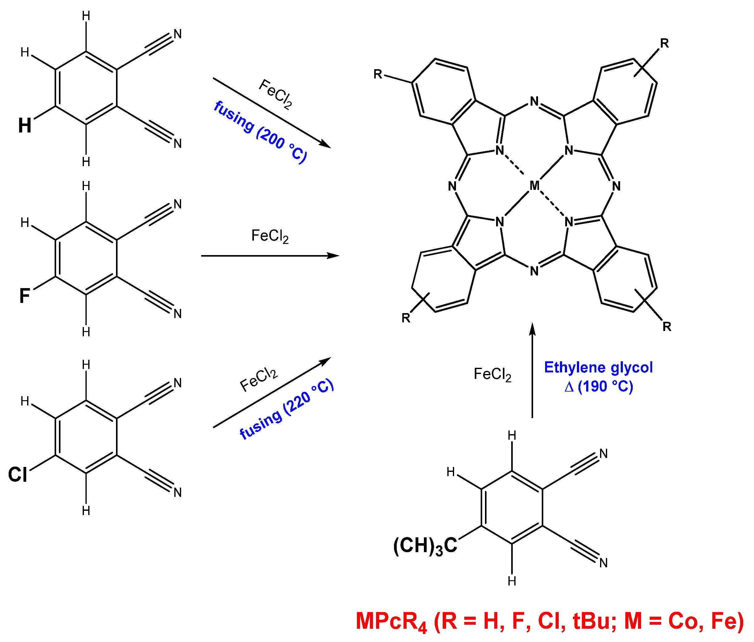
2.1. Synthesis of CoPcR4 and FePcR4 and Preparation of Their Films

In this work, cobalt and iron phthalocyanine derivatives with four different substituents (Figure 1) were synthesized. Unsubstituted (MPc) and tetra-fluoro-substituted (MPcF4) iron and cobalt phthalocyanines were synthesized by the method of template synthesis (200 °C, 2 h) from metal chloride and phthalonitrile or 4-fluorophthalonitrile, respectively, according to the synthetic pathway described in the previous works [7,26].
CoPc: C32H16N8Co. Anal. Calc: C 67.3; H 2.8; N 19.6. Found: C 67.4; H 2.7; N 19.7. IR spectrum (KBr; ω, cm−1): 1609, 1591, 1522, 1468, 1425, 1333, 1288, 1165, 1121, 1088, 1001, 951, 912, 874, 779, 754, 571, 517, 434.
FePc: C32H16N8Fe. Anal. Calc: C 67.6; H 2.8; N 19.7. Found: C 67.2; H 2.8; N 19.6. IR spectrum (KBr; ω, cm−1): 1609, 1589, 1513, 1466, 1422, 1332, 1289, 1164, 1119, 1085, 1002, 949, 910, 804, 781 756, 573, 516, 436.
CoPcF4: C32H12F4N8Co. Anal. Calc: C 59.7; H 1.9; N 17.4, F 11.8. Found: C 60.1; H 2.0; N 17.5, F 12.0. IR spectrum (KBr; ω, cm−1): 1616, 1603, 1528, 1479, 1408, 1333, 1264, 1213, 1167, 1113, 1092, 1055, 955, 872, 820, 779, 750, 640, 515, 436.
FePcF4: C32H12F4N8Fe. Anal. Calc: C 60.0; H 1.9; N 17.5, F 11.9. Found: C 60.1; H 2.0; N 17.7, F 12.0. IR spectrum (KBr; ω, cm−1): 1616, 1600, 1518, 1485, 1407, 1334, 1264, 1167, 1113, 1087, 1053, 954, 945, 871, 750, 732, 642, 515, 436.
Tetra-chloro-substituted phthalocyanines (MPcCl4, M = Co, Fe) were synthesized by heating of a mixture of 4-chlorophthalonitrile (1 g, 4 mol) and iron(II) or cobalt(II) chloride (0.3 g, 1.5 mol) in a glass tube at 220 °C for 2 h. Then, the mixture was cooled to room temperature and crushed into powder.
CoPcCl4: C32H12Cl4N8Co. Anal. Calc: C 54.2; H 1.7; N 15.8. Found: C 54.4; H 1.7; N 15.9. IR spectrum (KBr; ω, cm−1): 1605, 1524, 1501, 1450, 1398, 1342, 1256, 1199, 1186, 1144, 1097, 1084, 1063, 966, 932, 883, 820, 775, 766, 750, 694, 636, 528, 430.
FePcCl4: C32H12Cl4N8Fe. Anal. Calc: C 54.4; H 1.7; N 15.9. Found: C 54.4; H 1.6; N 15.8. IR spectrum (KBr; ω, cm−1): 1605, 1516, 1456, 1396, 1329, 1308, 1256, 1202, 1185, 1142, 1076, 1045, 962, 928, 895, 885, 824, 775, 748, 694, 669, 624, 526, 432.
Tetra-tert-butyl phthalocyanines (MPc(tBu)4, M = Co, Fe) were synthesized according to the synthetic pathway previously described in [27]. The ground mixture of 4-tert-butylphthalonitrile (0.3 g, 4 mol) and iron(II) chloride (0.18 g, 1.5 mol) were refluxed in 3 mL of ethylene glycol at 190 °C for 5 h in argon atmosphere. Then, the mixture was cooled to room temperature and poured into water–ethanol solution (V(ethanol):V (water) = 1:1, 50 mL). The resulting precipitate was filtered and washed several times with the same ethanol–water mixture.
CoPc(tBu)4: C48H48N8Co. Anal. Calc: C 72.4; H 6.1; N 14.1. Found: C 72.2; H 6.1; N 14.2. IR spectrum (KBr; ω, cm−1): 1603, 1573, 1521, 1463, 1408, 1396, 1369, 1350, 1331, 1281, 1259, 1203, 1192, 1119, 1093, 1024, 927, 914, 895, 831, 756, 692, 663, 609, 536, 517, 434.
FePc(tBu)4: C48H48N8Fe. Anal. Calc: C 72.7; H 6.1; N 14.1. Found: C 72.7; H 6.0; N 14.2. IR spectrum (KBr; ω, cm−1): 1603, 1572, 1522, 1464, 1396, 1369, 1352, 1333, 1283, 1259, 1204, 1192, 1126, 1084, 1026, 928, 914, 897, 833, 750, 692, 663, 563, 517, 418.
The complexes obtained after synthesis were purified by double gradient sublimation in a (10−5 Torr, 400–450 °C). The structure of the MPcR4 phthalocyanines was verified using X-ray diffraction analysis, elemental analysis and FT-IR spectroscopy. Single crystals of MPcCl4 were obtained in the process of sublimation of the initial products.
Note that, in the case of tetrasubstituted complexes of metal phthalocyanines, substituents can be introduced into both peripheral and non-peripheral positions of the phthalocyanine ring. In this paper, only phthalocyanine derivatives with substituents in the peripheral positions of the phthalocyanine ring were considered, due to the fact that they have a significantly higher sensor response toward electron donor gases compared to non-peripherally substituted examples [21,28].
Thin films were obtained by organic molecular beam deposition on glass substrates or glass slides with pre-deposited Pt interdigitated electrodes (IDE). The substrate temperature was about 60 °C. The nominal thickness of the films was about 75–90 nm. The films’ thickness was controlled by a quartz crystal microbalance and verified by spectral ellipsometry, as described in our previous work [29].

2.2. Characterization of Metal Phthalocyanines and Their Films

Structures of single crystal of MPcR4 were determined using a single-crystal diffractometer Bruker D8 VENTURE with the following characteristics: MoKα λ = 0.71073 Å Incoatec IμS 3.0 microfocus source, PHOTON III C14 CPAD detector, 3-circle goniometer). The diffractometer was equipped with an open-flow nitrogen cooler (Oxford cryo-systems Cryostream 800 plus), which allowed maintenance of a sample temperature at 150(1)K. To collect data, several standard ω scans were performed with frames 0.5° wide. APEX3 V2018.7-2 (SAINT 8.38A, SADABS-2016/2) was used for data collection and reduction, unit cell refinement and absorption correction [30]. In order to solve and refine structure, Fhkl datasets were processed in Olex2 v.1.5 [31] using SHELXT 2018/2 [32] and SHELXL 2018/3 [33]. Diffraction patterns of MPcR4 films and powders were obtained using a Bruker D8 Advance powder diffractometer (Cu-anode sealed tube, 40 mA @ 40 kV, LYNXEYE XE-T compound silicon strip detector) in the Bragg-Brentano geometry. The scan step was 0.01023° and the acquisition time was 2 s/step.
Atomic force microscopy (AFM) images of MPcR4 films were obtained using a Ntegra Prima II (NT-MDT, Russia) microscope in semi-contact mode. The HA_NC tip with Au reflective side (TipsNano, Estonia) had a length of 124 μm, a width of 34 μm and a thickness of 1.85 μm. The force constant was 3.5 N/m, while the resonance frequency was 140 kHz. Nova SPM software was utilized to calculate roughness parameters according to the standards ISO 4287-1, ISO 4287 and ASME B46. Scanning electron microscopy (SEM) images were obtained on a scanning electron microscopes JEOL 6700F.

2.3. Study of the Sensor Properties of MPc Films

To test the chemi-resistive sensor response, films were deposited onto commercial platinum IDE (Dropsens, Spain). The IDE parameters were as follows: the number of digits was 125 × 2, while the gap between digits was 10 μm. Phthalocyanine films deposited onto IDE were placed in a gas flow cell and kept for 30 min in an argon stream until their resistance reached a steady value. Argon was chosen as carrier and diluent gas due to the fact that nitric oxide (NO) is highly reactive and easily oxidizes to nitrogen dioxide (NO2) in air. The required gas flow was regulated using mass flow regulators. The resistance of MPcR4 films was measured using a Keithley 236 electrometer (constant DC voltage = 10 V). Injection of NO was carried out at the constant air flow rate of 1000 mL/min and the exposure time was fixed at 15 s.
The sensor response was investigated in dynamic mode with constant argon purging. Static mode was used to measure response and recovery times. In this mode, the cell was first purged with argon, then, when a constant resistance value was reached, the argon supply was turned off and a mixture of gases containing the required concentration of NO was introduced into the cell. After saturation of the sensor layer, argon purging was resumed.

2.4. Quantum-Chemical Calculations

Quantum-chemical calculations of the interaction of the NO molecule with phthalocyanines in the form of MPcR4 monomers and 2MPcR4 dimers (M = Co, Fe; X = H, F, and Cl) were performed with the help of the ORCA software package [34,35], using the DFT BP86-D3/def2-SVP method [36,37,38,39,40], RI approximation [41,42,43,44,45,46], and the corresponding auxiliary basis set Def2/J [47]. The introduction of four substituents (one in each benzene ring) leads to the formation of four isomers that differ in the mutual arrangement of substituents, which, in the case of a planar macrocycle, corresponds to the D2h, C2v, C4h, and Cs point symmetry groups of the substituted molecule. In this situation, the molecule has several nonequivalent adsorption centers for gas molecules that bind through the side atoms of the macrocycle. At the same time, considering separately the interaction of an NH3 molecule with CoPcF4 and VOPcF4 through four bridge nitrogen atoms, we showed that the parameters of this interaction were very close in all four cases [28]. In this regard, in order to simplify calculations by reducing the number of possible nonequivalent adsorption centers for the NO molecule, MPcR4 molecules with C4h symmetry (Figure 2) were considered in this work, although symmetry constraints were not used in the calculation process. In this case, four different places were identified for the formation of a bond between nitric oxide and side atoms of macrocycles—three around the benzene ring and one opposite the bridge nitrogen atom. Further, aggregates of monomeric phthalocyanines with the NO molecule will be designated as MPcR4/NO-m, where m is 1–4 depending on the site of adsorption of NO. Note that, in the case of unsubstituted MPcs, the aggregates MPc/NO-2 and MPc/NO-4 are equivalent, so only the first of them will be considered. Aggregates of dimeric phthalocyanines will be designated as 2MPcR4/NO.
Since the considered compounds have an open electron shell, the calculations were performed according to the spin-unrestricted Kohn-Sham (UKS) theory. In this case, the spin multiplicity was equal to one for 2CoPcR4 dimers, two for NO molecule, CoPcR4 monomers, and 2CoPcR4/NO aggregates, three for FePcR4 monomers and CoPcR4/NO-m aggregates, four for FePcR4/NO-m aggregates, five for FePcR4 dimers, and six for 2FePcR4/NO aggregates. Preliminary calculations have shown that these spin states were energetically more favorable.
In the process of calculation, the geometry of all considered compounds was first optimized, followed by the calculation of their vibrational spectra, to make sure that there were no imaginary frequencies, since this, along with the minimum of the total energy, is the criterion for achieving an equilibrium state. Then the binding energy Eb of the NO molecule with monomeric or dimeric phthalocyanines was calculated from the difference in the total energies of both aggregate parts and the aggregate itself
E b = E NO + E ( 2 ) MPcR 4 E ( 2 ) MPcR 4 / NO Δ E BSSE
where ΔEBSSE is the correction to the binding energy, taking into account the basis set superposition error (BSSE), which is estimated as follows:
Δ E BSSE = E ( 2 ) MPcR 4 * + E NO * E ( 2 ) MPcR 4 * * + E NO * *
Here, the asterisk in the superscript means that the corresponding total energies were calculated for MPcR4 (2MPcR4) and NO compounds separated from the equilibrium structure of the aggregate, without subsequent optimization of their geometric structure. An asterisk in the lower index indicates that, instead of the atoms of the second fragment of the entire aggregate, points described by the corresponding sets of atomic orbitals were considered.
The next step was a topological analysis of the electron density ρ(r) distribution in the MPcR4/NO-m and 2MPcR4/NO aggregates, performed in the framework of the QTAIM theory [48,49,50] using the AIMAll software package [51]. For this purpose, the electronic wave functions of these structures were calculated using a similar method and the cc-pVTZ basis set of atomic orbitals [52] instead of def2-SVP for greater accuracy. As a result, the values of the electron density, its Laplacian ∇2ρ(r), and the electronic energy density h(r) at bond critical points (BCPs) characterizing the interaction of the NO molecule with the side atoms of phthalocyanines were obtained.

3. Results and Discussion

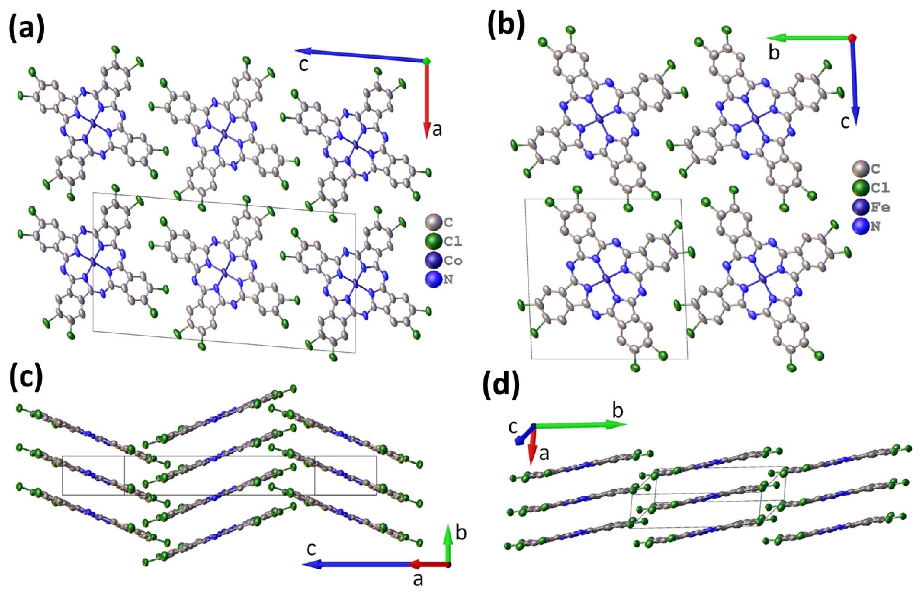
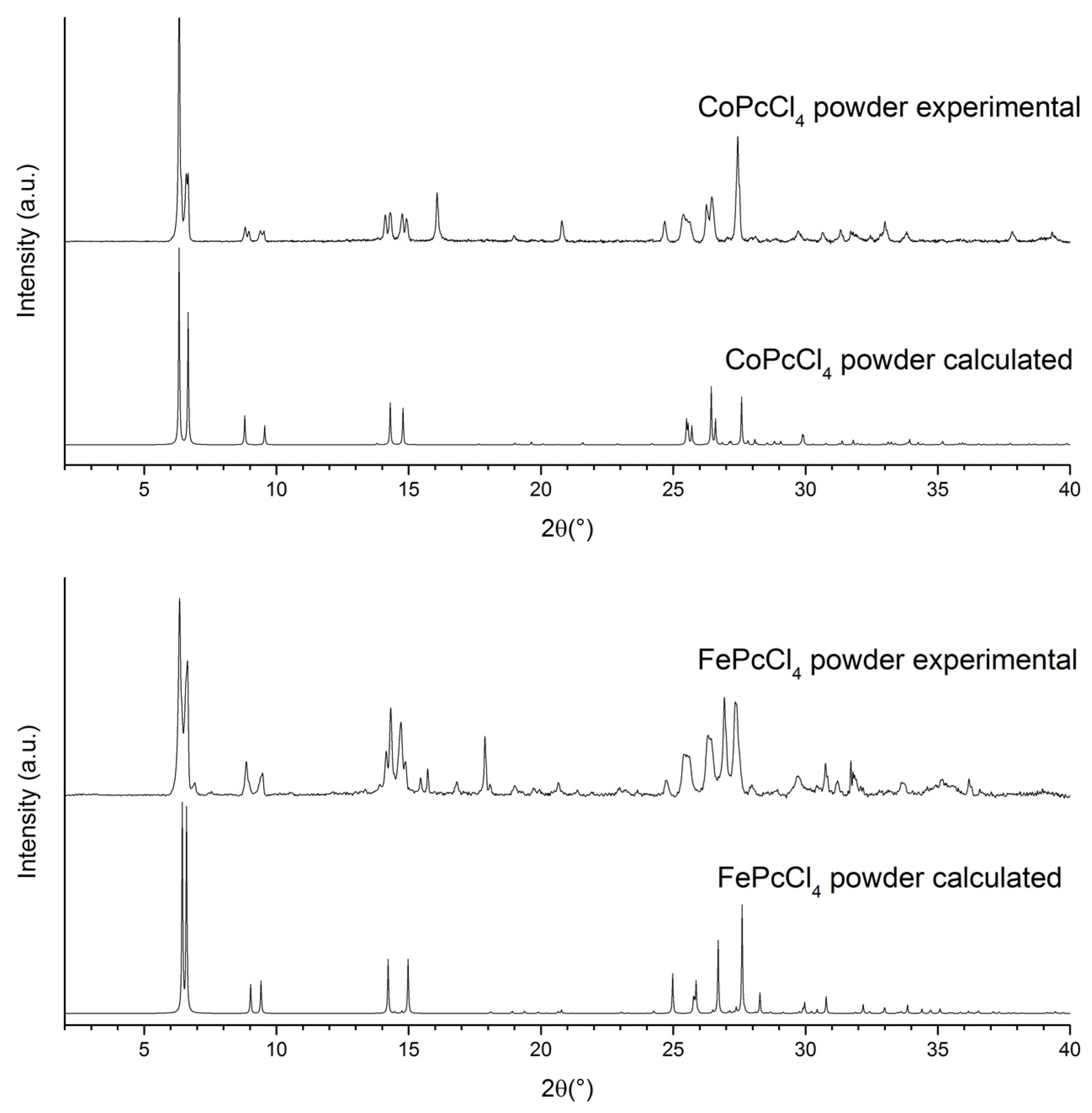
3.1. Single Crystal Structure of CoPcCl4 and FePcCl4

In order to correctly interpret XRD patterns of phthalocyanine films, it is necessary to have data on the structure of their single crystals. The structures of CoPc, FePc, CoPcF4, and FePcF4 single crystals have already been determined in previous works [53,54,55,56]. Single crystals of CoPcCl4 and FePcCl4 were grown by sublimation in a vacuum and their structures were determined for the first time.
CoPcCl4 crystallizes in a P21/c space group with Z = 2 and is isostructural to the previously reported CuPcCl4 [57]. CoPcCl4 molecules are packed into stacks (Figure 3a), which form a “herringbone” pattern when viewed from the side (Figure 3b). The detailed refinement statistics and unit cell parameters are given in Table 1. CoPcCl4 molecules are relatively flat; the maximum deviation from the mean squared plane is less than 0.1 Å for any atom in the molecule (except hydrogen). The packing angle (angle between the line through the central metal atoms and the normal to the least-squares plane, through all atoms in the phthalocyanine molecule except hydrogen) is 21.17° for CoPcCl4. The angle between molecules in adjacent stacks is 42.34°. The distance between neighboring molecules in the stack is 3.376 Å, while the distance between neighboring Co atoms is 3.620 Å. For comparison, the packing angle in CuPcCl4 is 21.67°, while the distance between neighboring molecules is 3.381 Å and the distance between Cu atoms is 3.638 Å. For unsubstituted β-CoPc [58], the distance between molecules in the stack is 3.320 Å, the stacking angle is 45.93°, and the distance between neighboring Co atoms is 4.773 Å, while for α-CoPc [55] and tetra-fluorinated CoPcF4 [54], these values are 3.425 Å/24.16°/3.754 Å and 3.322 Å/24.58°/3.653 Å, respectively. In general, if the CoPcCl4 packaging is similar to the “herringbone” packing motif of β-CoPc, then the arrangement of CoPcCl4 molecules within the stack is more is more like the arrangement in α-CoPc and CoPcF4.
Since a mixture of four regio-isomers is formed during the synthesis of MPcCl4-p, the CoPcCl4 structure contains two symmetrically independent chlorine atoms, each of which is disordered over two positions, with a total occupancy equal to 1. In CoPcCl4, the chlorine atom occupancy ratios are 0.514(3)/0.486(3) and 0.531(3)/0.469(3). The fact that these ratios are close to 0.5/0.5 indicates that there are no preferred positions for Cl atoms.
FePcCl4 crystallizes in P-1 space group with Z = 1 and is isostructural to FePcF4 [53], as well as to α-polymorphs of unsubstituted MPcs, e.g., α-CuPc [59] (Table 1). FePcCl4 molecules are packed into uniform stacks, with molecules in adjacent stacks parallel to each other (Figure 3). FePcCl4 molecules are less flat than CoPcCl4, with the maximum deviation from mean squared plane equal to 0.125 Å. The packing angle for FePcCl4 molecules is 19.63°, the distance between adjacent molecules is 3.388Å and the distance between neighboring Fe atoms is 3.597 Å. For comparison, the packing angle in FePcF4 is 24.06°, the distance between molecules is 3.332 Å and the distance between Fe atoms in neighboring molecules is 3.649 Å, while for unsubstituted β-FePc [60] these values are 3.301 Å/46.33°/4.781 Å. The FePcCl4 molecule contains two symmetrically independent chlorine atoms, each of which is disordered over two positions with occupancy ratios of 0.484(3)/0.516(3) and 0.443(3)/0.557(3).
Figure 4 shows experimental powder diffraction patterns of CoPcCl4 and FePcCl4 in the 2θ range of 2–40° in comparison with those calculated from their single-crystal data. It is clear that both experimental diffraction patterns do not completely coincide with the corresponding calculated patterns and contain one additional crystal phase in a comparable quantity (this is especially noticeable in the range from 8° to 15° of the diffraction patterns in Figure 4). For example, the calculated diffraction pattern of CoPcCl4 contains only four diffraction peaks in the 2θ range of 5–10° (6.31°, 6.67°, 8.80°, 9.56°), while the experimental diffraction pattern shows four additional diffraction peaks in the same region (6.41°, 6.60°, 8.96°, 9.40°), which cannot be explained using the CoPcCl4 single-crystal data.
Despite all our efforts, we were unable to find single crystals for the second phase for either CoPcCl4 or FePcCl4. However, it should be noted that additional four diffraction peaks in the region of 5–10° on the CoPcCl4 diffraction pattern coincide very well with the first four peaks calculated from the FePcCl4 single-crystal data (6.43°, 6.60°, 9.00°, 9.42°). The same, but in reverse order, is true for the experimental powder pattern of FePcCl4. This is a very strong argument in favor of the fact that the second crystal phase of CoPcCl4 is isostructural with the first crystal phase of FePcCl4 and vice versa, and that the bulk powders of CoPcCl4 and FePcCl4 contain both monoclinic and triclinic polymorphs.

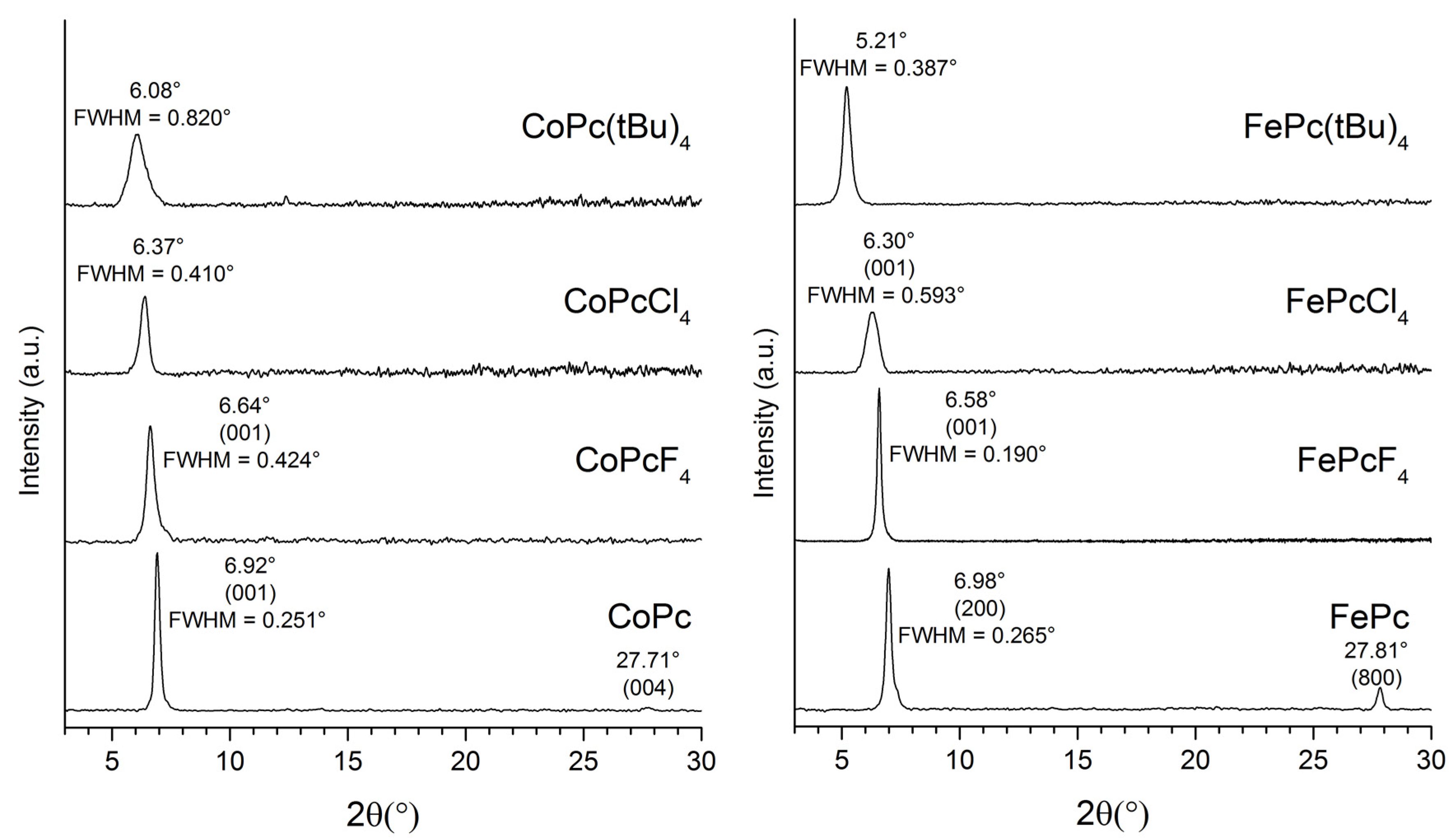
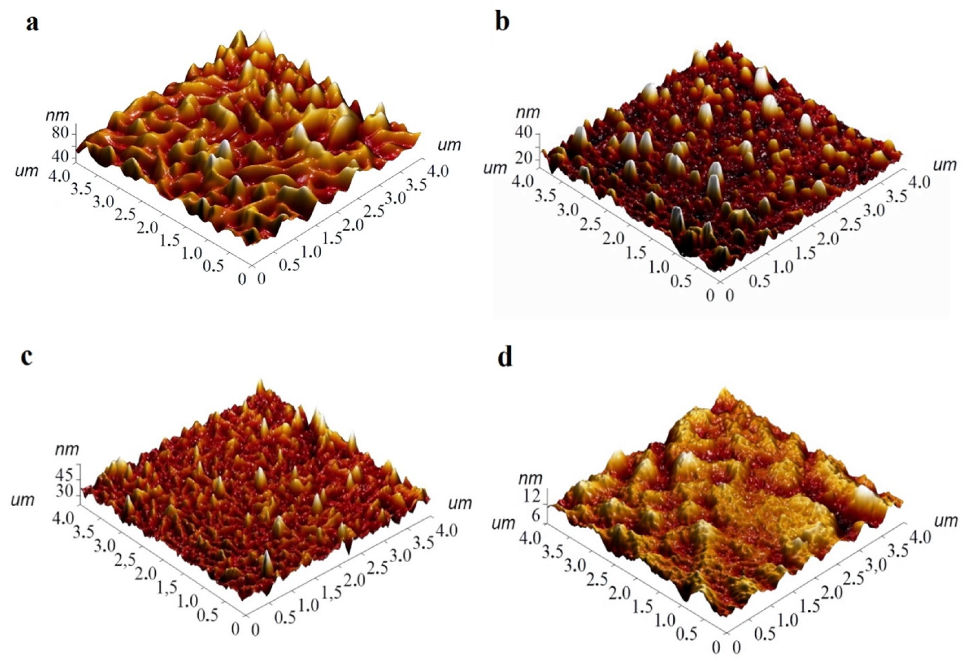
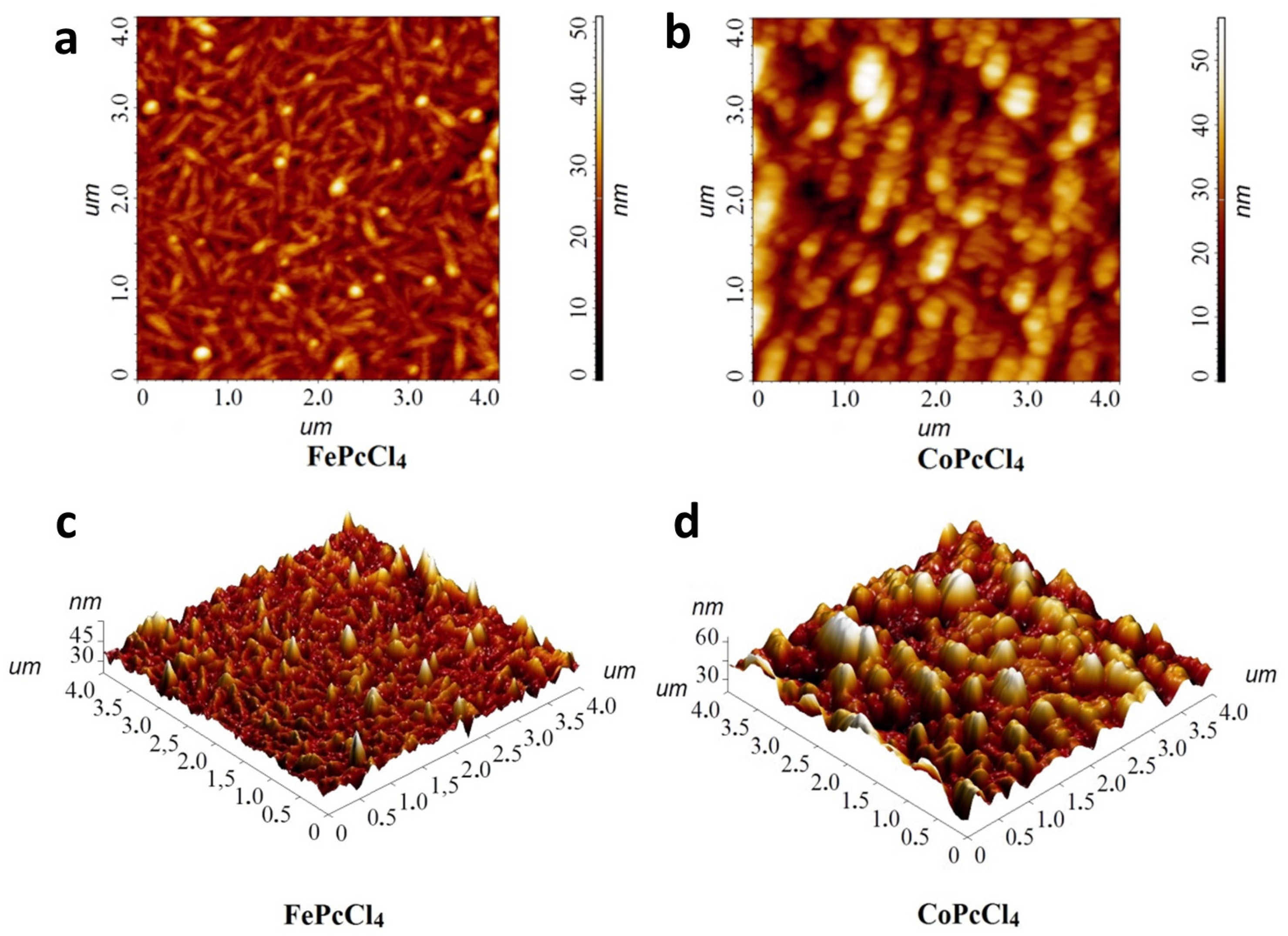
3.2. Thin Films of CoPcR4 and FePcR4 (R = H, F, Cl, tBu)

The films of CoPcR4 and FePcR4 were deposited by a PVD method. The composition of films coincides with that of powders, which is confirmed by Raman spectroscopy. The Raman spectra of thin films and powders of CoPcCl4 and FePcCl4 are shown in Figure S1 (Supporting Information). Raman spectra of other investigated phthalocyanines were studied in previous works [26,53].
XRD patterns of CoPcR4 and FePcR4 thin films (R = H, F, Cl, tBu) are shown in Figure 5. One strong diffraction peak is observed in all diffraction patterns, which indicates a strong preferred orientation of phthalocyanine crystallites relative to the substrate surface in these films. A strong peak at 6.92° on the XRD pattern of CoPc film coincides well with the (001) peak of α-CoPc at 6.91° [55]. An additional weak diffraction peak is visible at 27.71°, which corresponds to the (004) plane. A strong peak at 6.98° on the XRD pattern of FePc can be attributed to the (200) peak of α-FePc at 6.94° [56], while a diffraction peak at 27.83° corresponds to the (800) peak of α-FePc. The XRD patterns of both CoPcF4 and FePcF4 films have single strong diffraction peaks, which correspond to the (001) plane of the respective crystal phases (6.62° for CoPcF4 and 6.64° for FePcF4, as calculated from single-crystal data) [53,54].
The relatively wide diffraction peak on the CoPcCl4 thin film XRD pattern may coincide with the first peak of either the monoclinic or triclinic phase. The same is partially true for the FePcCl4 thin film; however, judging by the position of the peak, it most likely refers to the (100) peak of the monoclinic phase. Finally, XRD patterns of CoPc(tBu)4 and FePc(tBu)4 films also have one wide diffraction peak. Since there is no data for CoPc(tBu)4 and FePc(tBu)4 single crystals, and tert-butyl substituted phthalocyanines usually tend to be amorphous, we cannot say whether CoPc(tBu)4 and FePc(tBu)4 films have a preferred orientation or not. The only conclusion, judging by the difference in the positions of the peaks on the corresponding XRD patterns, is that CoPc(tBu)4 and FePc(tBu)4 films have completely different styles of molecular packaging.
Knowing the FWHM values of the observed diffraction peaks, the coherent scattering region size for each thin film can be estimated using the Scherrer equation. Taking into account the instrumental peak broadening of 0.05° (measured using SRM-660a LaB6 powder), the following values were obtained: 44 nm for CoPc, 24 nm for CoPcF4-p, 25 nm for CoPcCl4-p, and 11.5 nm for CoPc(tBu)4. The values for FePcR4 films were 41, 63, 16.3, and 26 nm, respectively.
The morphology of the films was studied by AFM. Figure 6 shows 3D AFM images for FePcR4 films as an example. 2D AFM images are given in the Supporting Information (Figure S2). SEM images are also presented in Figure S3. All investigated films have different surface morphology. For example, the surface of a FePc film consists of elongated crystallites, with length reaching 0.8 μm. The root mean square (RMS) roughness value of a FePc film is 9.5 nm. The film of FePcF4 consists of the clearly distinguishable smaller roundish grains combined in bigger aggregates and has RMS roughness of 5.0 nm. The morphology of the FePcCl4 film differs significantly from that of the films of FePcF4; the film is formed by thin elongated crystallites, the size of which reaches 0.5 μm. Its RMS roughness value is 4.5 nm. The film of FePc(tBu)4 has no clearly visible crystallites, consists of big aggregates, and its RMS roughness value is 3.8 nm.

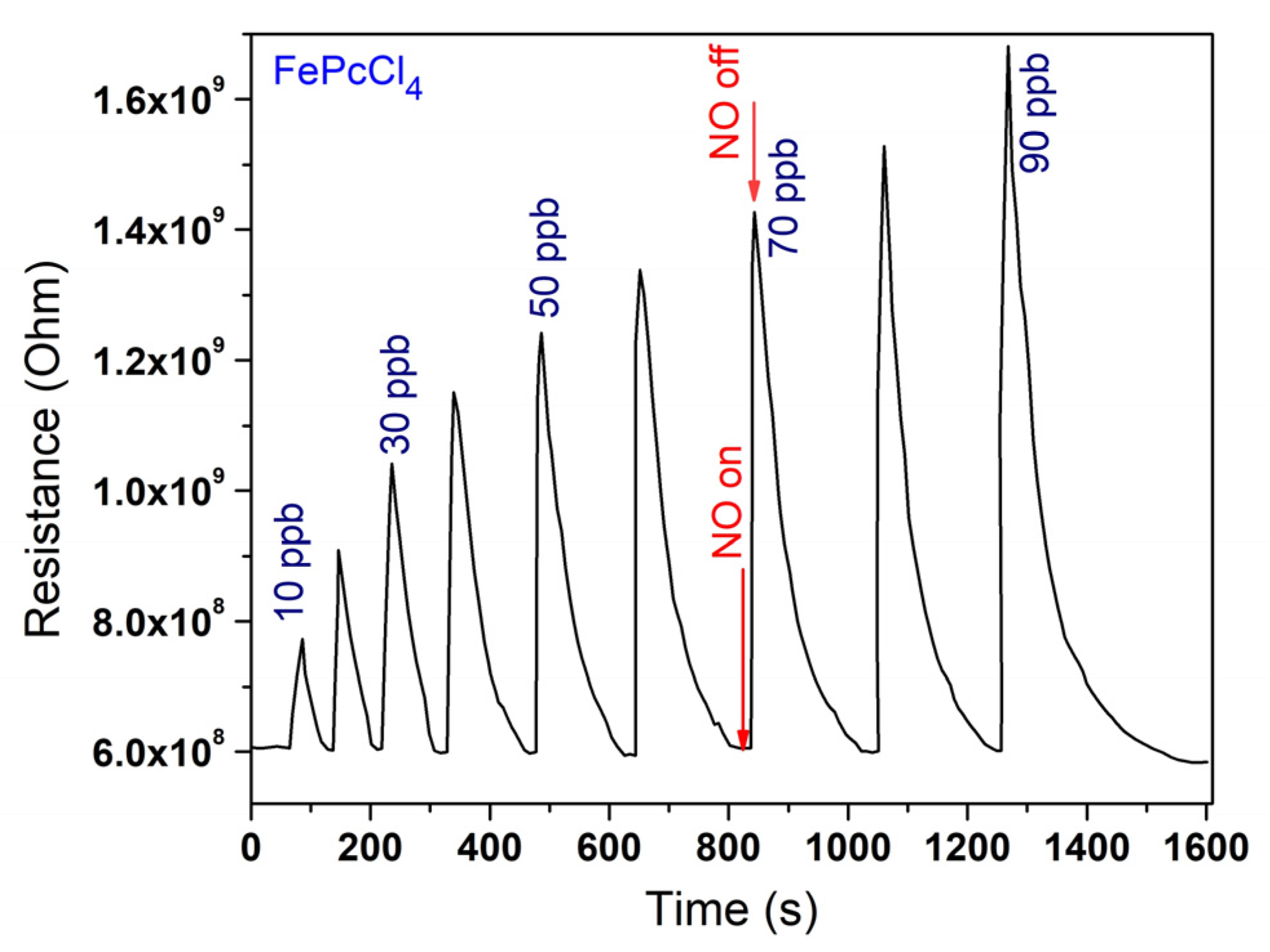
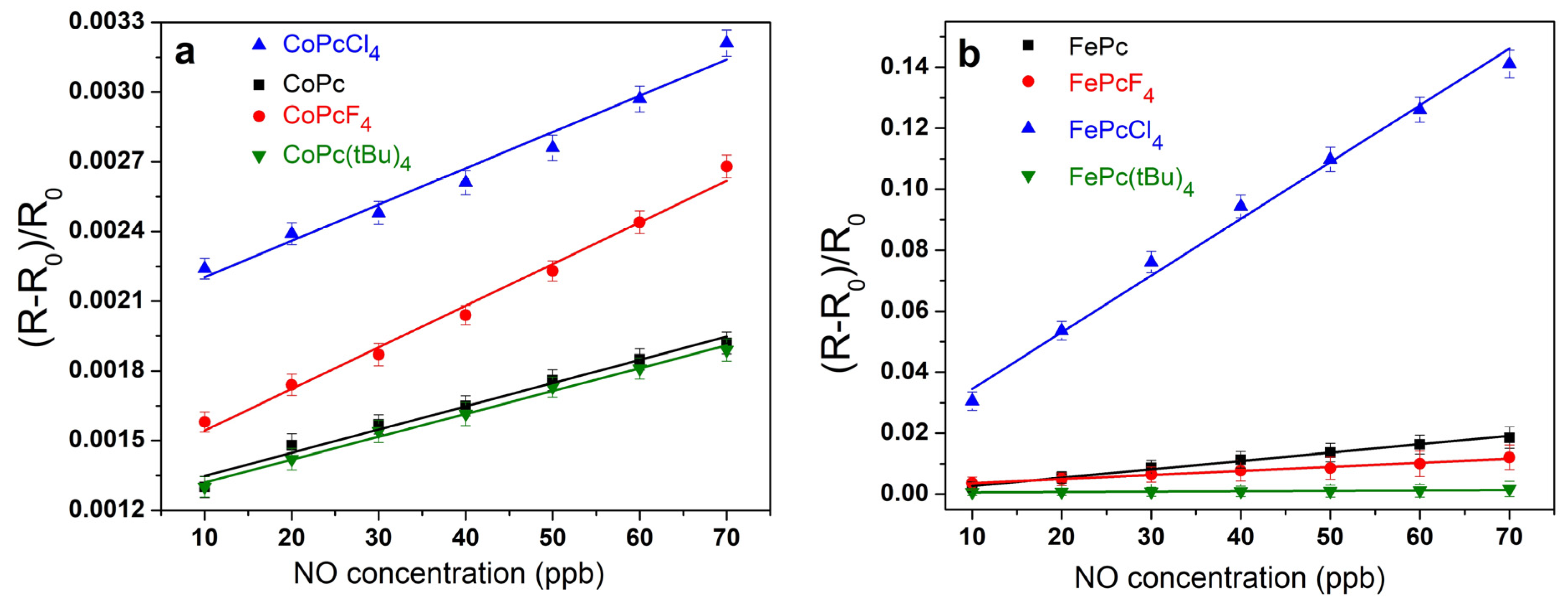
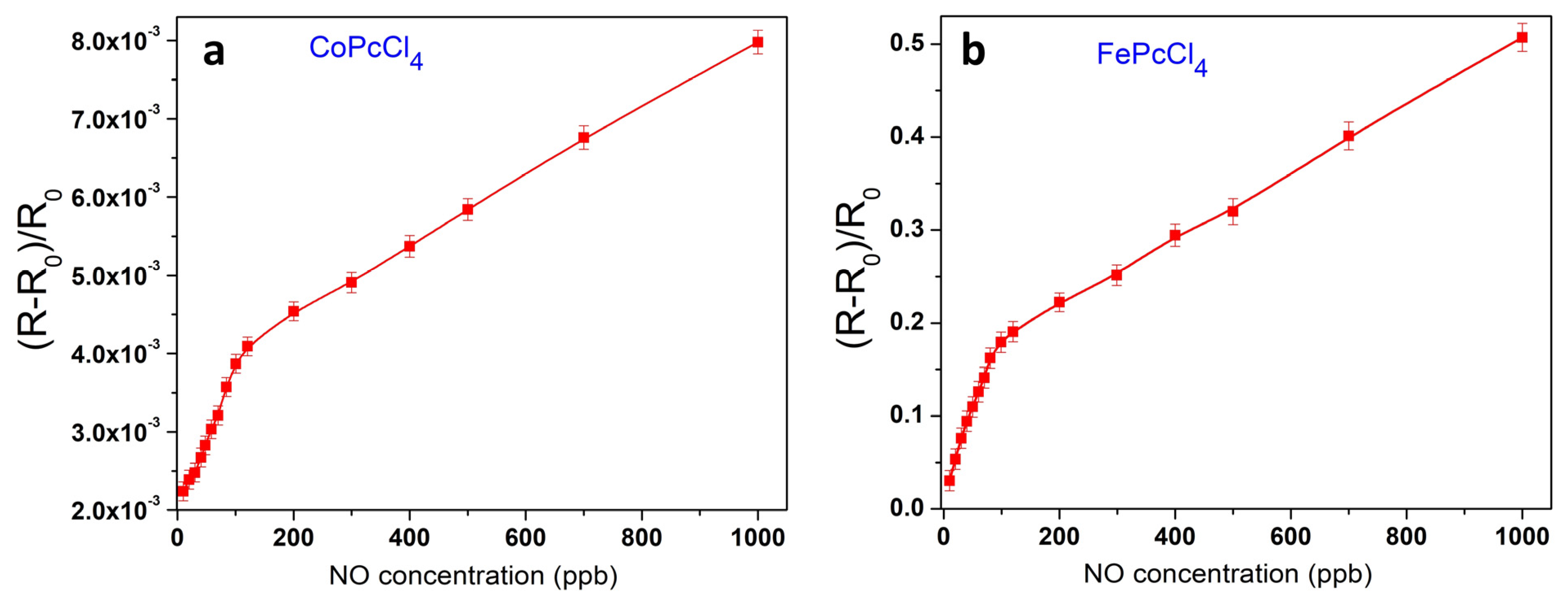
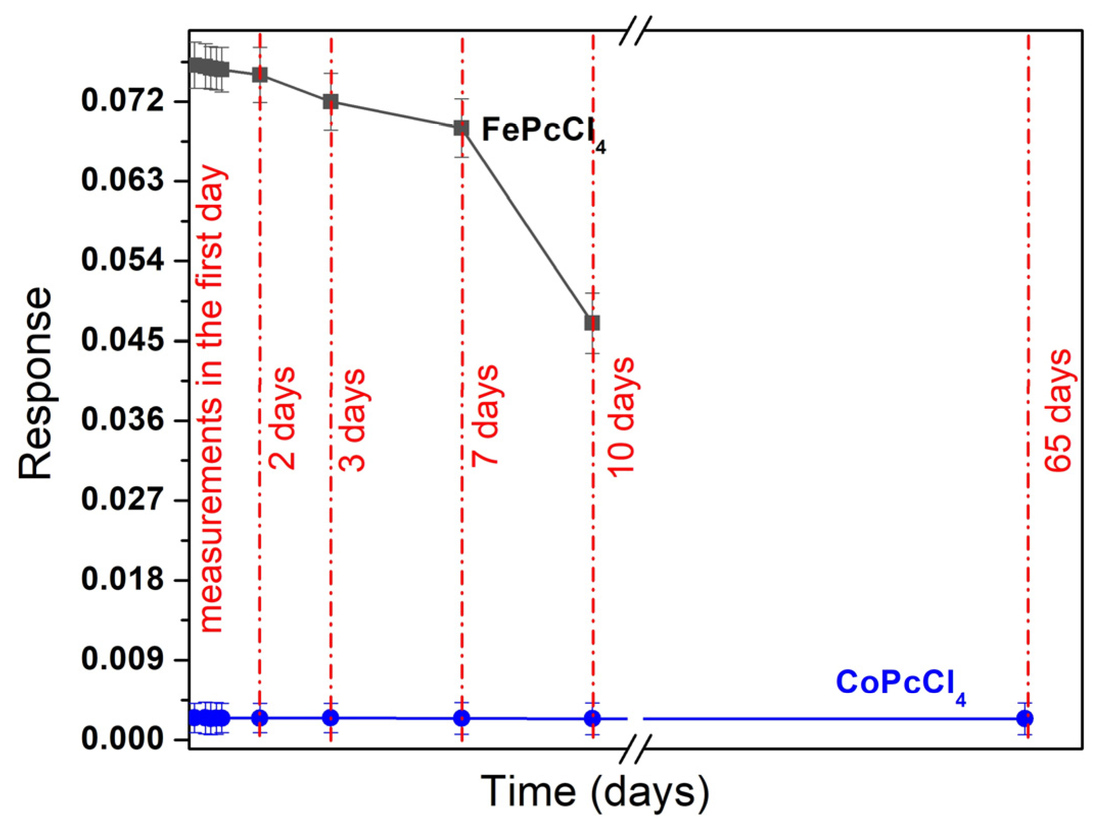
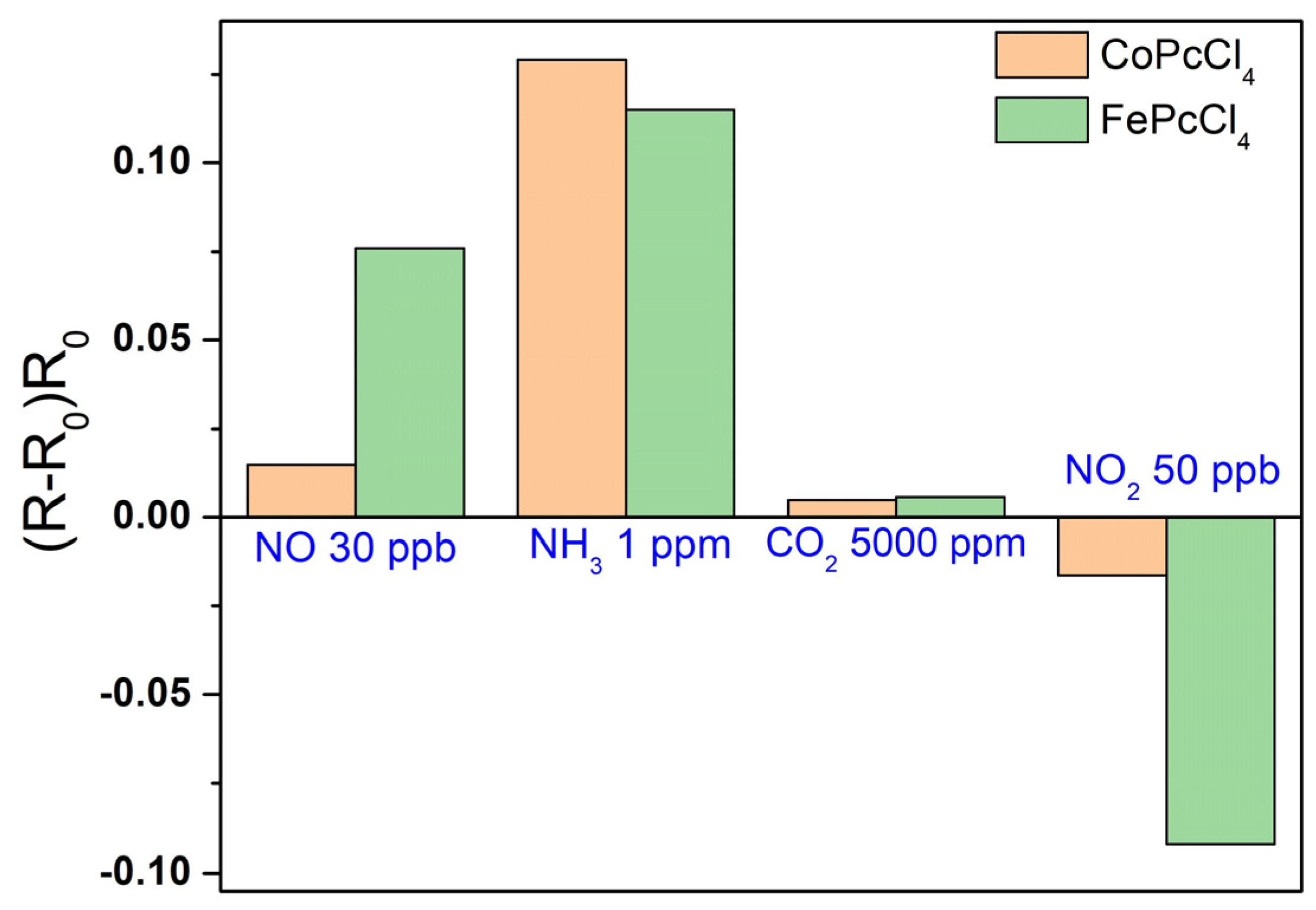
3.3. Sensor Properties of CoPcR4 and FePcR4 (R = H, F, Cl, tBu) Films

The films of CoPcR4 and FePcR4 films were deposited on glass substrates with IDE Pt electrodes for the investigation of the sensor response to nitric oxide. Figure 7 shows a typical sensor response of FePcCl4 film as an example. Similarly to the case of the sensor response of MPcF4 films to ammonia [28], when NO was introduced into the cell, a sharp increase in resistance was observed, and after purging with argon, the resistance returned to its original value. With an increase in NO concentration, the change in resistance increased.
The sensor response was defined as Sresp = (R − R0)/R0, where R0 is the initial resistance of the film in argon atmosphere and R is the resistance of the phthalocyanine film at a certain NO concentration. The influence of different types of substituents in MPcR4 on the chemi-resistive response to nitric oxide (10–70 ppb) was studied to select the material with the best sensor characteristics. The dependences of the sensor response on NO concentration are shown in Figure 8 for all investigated MPcR4 films.
Comparison of the sensor response of iron and cobalt phthalocyanine films with various substituents showed that the best sensor response was observed for chlorinated derivatives MPcCl4 (M = Co, Fe), while the lowest response was for MPc(tBu)4 films. For example, the sensor response of a CoPcF4 film to 30 ppb NO was 1.2 times higher than that of a CoPc film, while the response of CoPcCl4 to NO was 1.6 times higher than that (Figure 8a). In the case of iron phthalocyanines derivatives, sensor response of a FePcCl4 film to 30 ppb of ammonia was more than 11 times higher than for FePcF4 and FePc films and about 90 higher than in the case of Fe(tBu)4 (Figure 8b).
The dependence of the sensor response of CoPcCl4 and FePcCl4 films, which have the best sensor characteristics, on NO concentration was studied in a wider concentration range from 10 to 1000 ppb (Figure 9). The curves have two linear ranges: from 10 to 90 ppb and from 100 to 1000 ppb. In the whole range, a reversible sensor response was observed.
The average sensor responses to 30 ppb of nitric oxide, as well as the response and recovery time of all investigated films, are summarized in Table 2.
The calculated limits of detection (LOD) of NO, defined as 3σ/m, where σ is the standard deviation of the sensor response to 10 ppb NO and m is the slope of the corresponding calibration plot (Figure 8) in the linear region (10–90 ppb), are also given in Table 2. The response of iron phthalocyanine films, with the exception of FePc(tBu)4, is several times higher than the response of cobalt phthalocyanine films. Among the investigated sensors, FePcCl4 film exhibited the maximal response to NO, with response and recovery times (determined at 30 ppb of NO) of 30 s and 80 s, respectively. Its calculated detection limit was 3 ppb. At the same time, the LOD of a CoPcCl4 film was 7 ppb. In addition to the molecular structure of the complex, the sensor response is influenced by the morphology and structural features of thin films (Figure 10). CoPcCl4 films are formed by rounded crystals, the size of which reaches 100 nm. The morphology of the FePcCl4 film differs significantly from the morphology of FePcF4 films; the film is formed by thin elongated crystallites, the size of which reaches 0.6 μm.
Further tests of the repeatability and stability of the films showed that iron phthalocyanine films had low stability and their sensor response dropped rapidly. The sensor response of a fresh FePcCl4 film to 30 ppb of nitric oxide and the same film after 2, 3, 7, and 10 days is shown in Figure 11. The change in the sensor response within 2 days did not exceed the measurement error, but after 7 days the response decreased dramatically. At the same time, all cobalt phthalocyanine films remained stable, at least for several months.
The sensor response of FePcCl4 and CoPcCl4 films to NO was also compared with that to ammonia (NH3), carbon dioxide (CO2) and nitrogen dioxide (NO2) (Figure 12), which shows that small concentrations of NO (at ppb level) can be detected in the presence of CO2, but ammonia at concentrations of the ppm level can interfere with the determination of nitric oxide.
One more interfering gas is nitrogen dioxide (NO2), which is a strong electron acceptor. In contrast to the sensor response to NO, the introduction of NO2 to the flow cell leads to a decrease in the resistance of FePcCl4 and CoPcCl4 layers. It is also necessary to mention that determination of NO can be performed only in a strictly controlled inert atmosphere due to its low stability. One of the approaches for determining NO in air is the quantitative oxidation of NO to NO2, followed by a study of the chemi-resistive sensor response to NO2 [61].
Thus, active layers based on chloro-substituted phthalocyanines of cobalt and iron have a sufficiently high sensitivity to NO, fast reversible response and low recovery time at room temperature, and their characteristics are comparable, or in some parameters even exceed, the characteristics of sensors based on other materials [62,63,64,65]. Most chemi-resistive sensors based on semiconductor oxides operate only at elevated temperatures. For example, sensors based on iron oxide nanorods exhibited reversible sensor response to NO in the concentration range from 0.5 ppm to 2.75 ppm at 250 °C [62]. Su and Li [63] reported a chemi-resistive gas sensor made of composite films with the complex structure Fe2O3/MWCNTs/WO3 modified with noble metals, which demonstrated a reversible sensor response to NO at room temperature, but the minimal detected concentration of NO was 100 ppb.

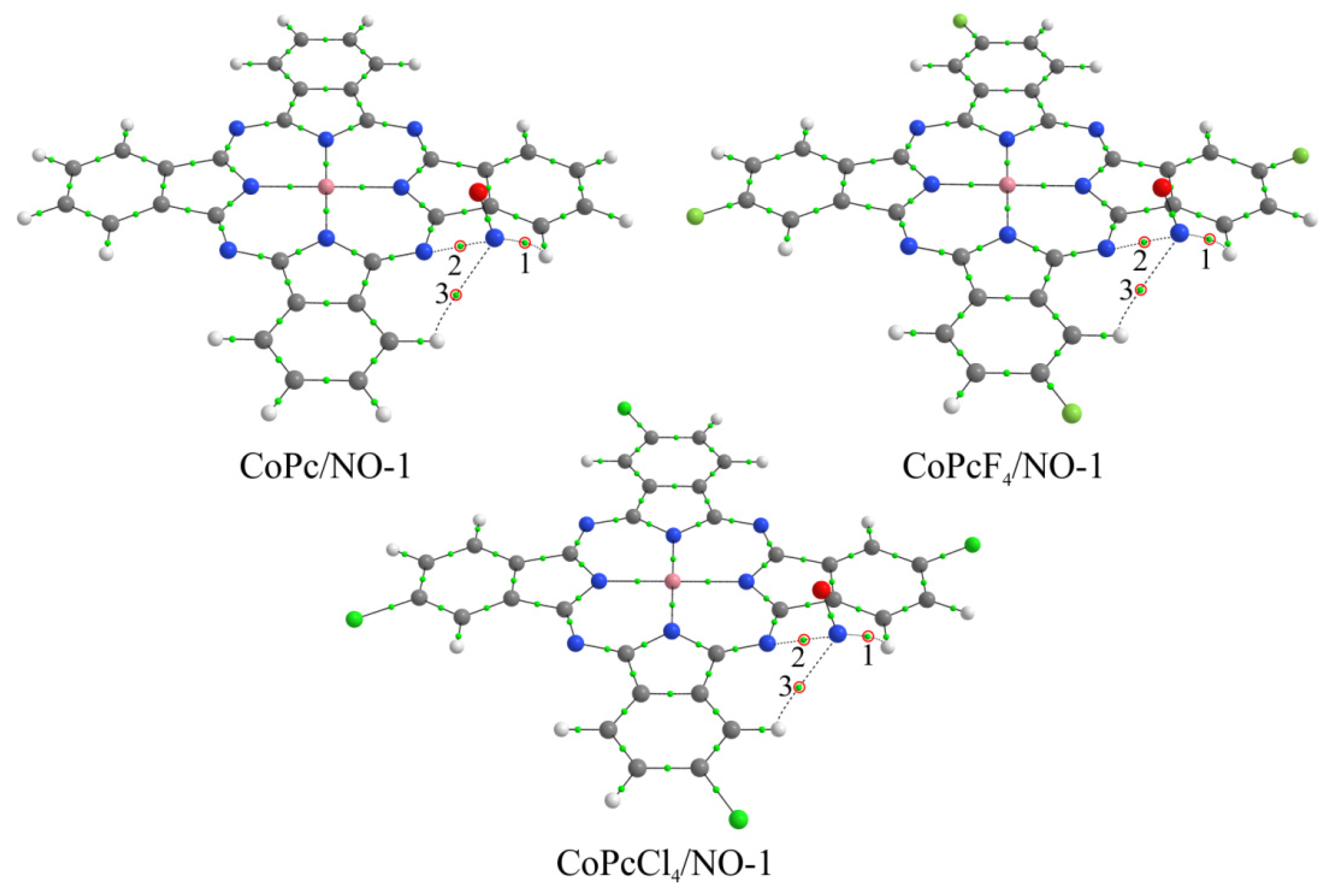
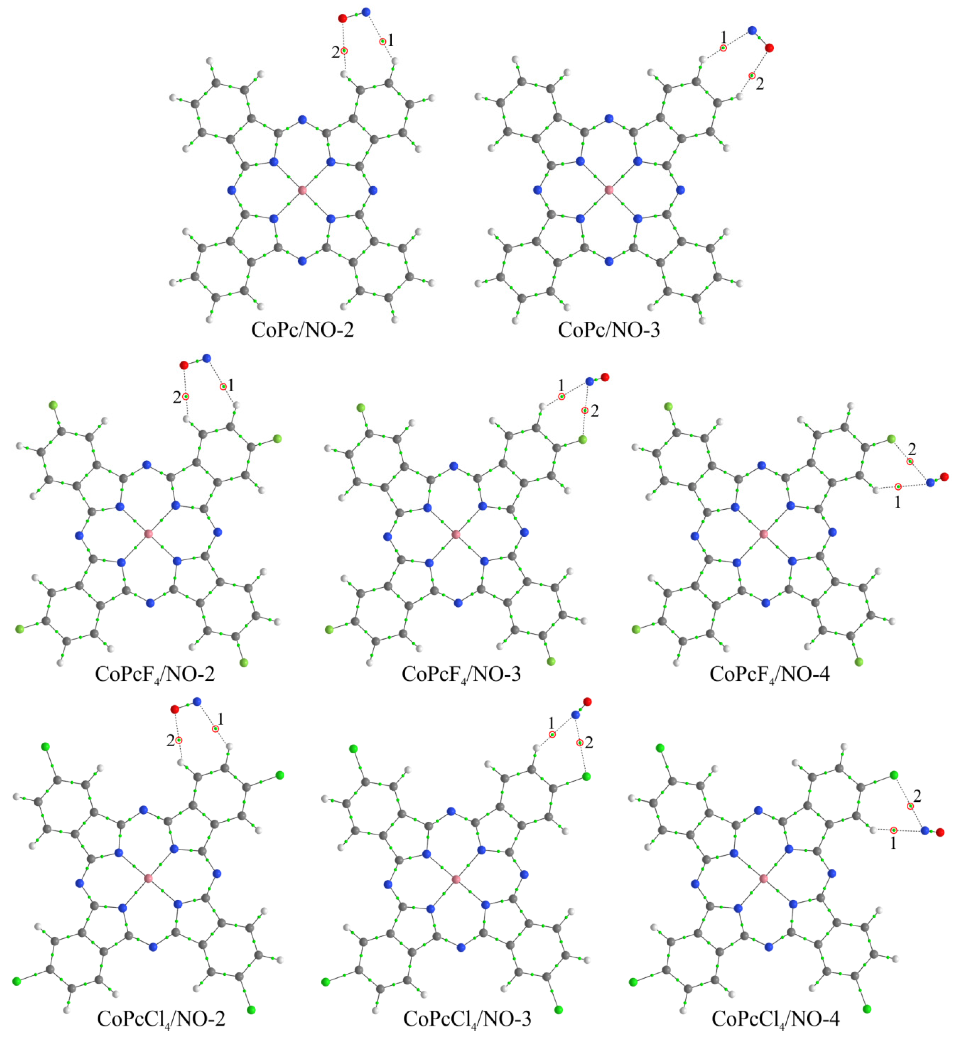
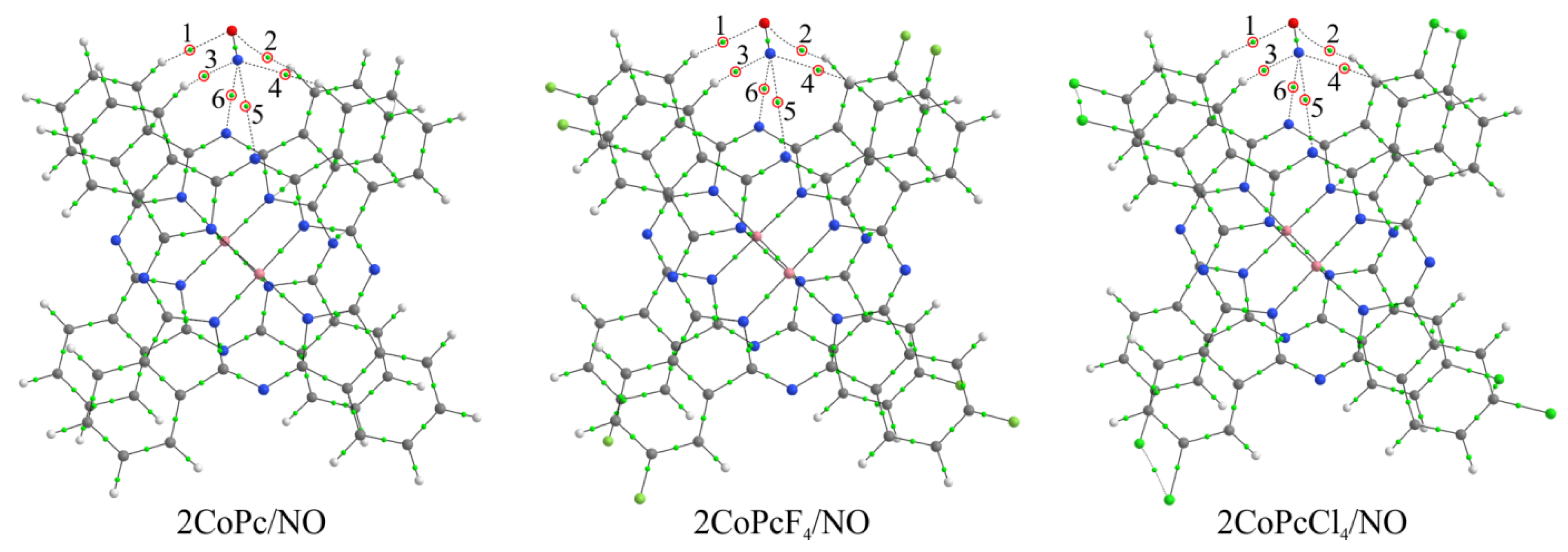
3.4. Quantum-Chemical Modeling of the Interaction between NO and MPcR4 Molecules

The most common accepted interpretation of the mechanism of chemi-resistive sensor response to NO is that the effect of gaseous NO on metal phthalocyanine films, which usually behave like p-type semiconductors, leads to a change in their conductivity due to depletion of positively charged holes by electrons donated by the NO [22]. At the same time, the place of the adsorption of gas molecules on the surface of phthalocyanine films remains a subject of discussion.
To study the nature of interaction between NO and phthalocyanine molecules and to explain the effect of R in MPcR4 phthalocyanines on the sensor response of their films to nitric oxide, quantum-chemical modeling of the NO molecule interaction with MPcR4 (R = H, F, Cl) molecules was carried out. It is necessary to mention that the calculation for MPc(tBu)4 was not performed because, firstly, the sensor response is much less than that of MPcR4 with R = H, Cl, F. Secondly, molecules in films of MPcR4 (R = H, F, Cl) are packed in stacks, and their dimers, considered in the process of quantum chemical calculations, are fragments of these stacks. The structure of MPc(tBu)4 films differs from others and, as XRD analysis shows, the films are less crystalline and possibly disordered due to the steric effects of tert-butyl groups. This does not allow us to consider the same calculation models for them as for MPcR4 (R = H, F, Cl) films.
Despite the fact that strong binding should be observed in the case of interaction with a central metal atom [22], when the binding energies reach 1.5 eV for CoPc and 1.9 eV for FePc, depending on the orientation of the nitric oxide molecule, we did not consider this method of coordination for two reasons [28]. First, due to the π-π interaction, the typical distance between two phthalocyanine molecules in a stack is too small for any gas molecule to penetrate between them. This distance is about 3.4 Å [66], which roughly corresponds to the van der Waals diameter of a carbon atom. Second, the indicated values of binding energy of the NO molecule with metal atoms are large enough for the desorption of nitric oxide under normal conditions [21]. Therefore, the sensor response must be irreversible, which contradicts the experimental data presented above. Moreover, Chia et al. [67] demonstrated by in-situ X-ray absorption spectroscopy (XAS) and EXAFS that NO2 interacts with CuPc at the pyrrole moiety of the Pc macrocycle, rather than on the metal center. In this regard, the simulated interaction was carried out through the side atoms of the macrocycles (Figure 13 and Figure 14), similar to previous studies [21,28].
As a result, it was found that, in the case of monomeric phthalocyanines, the most energetically favorable method of NO molecule binding is its interaction through the bridge nitrogen atom (MPcR4/NO-1 aggregates) (Figure 13), when the Eb values are higher (Table 3). However, in these compounds, nitric oxide is actually located above the plane of the macrocycle, and in the case of a stack, this would mean that the gas molecule is located between two phthalocyanine molecules. However, as has already been mentioned above, the distance between two molecules in the stack is not enough for the location of NO molecules. For this reason, this aggregate is not of interest for further consideration.
The binding energy of nitric oxide at the other three positions (two in the case of MPc/NO) is very low (Table 3). This indicates that these methods of NO molecule coordination cannot have a significant effect on the change in the electrical conductivity of phthalocyanine films, and therefore are also of no interest for further consideration. In general, the low binding energies between NO and phthalocyanine molecule with phthalocyanines in MPcR4/NO-m aggregates are evidence of the van der Waals interaction between them. This is supported by the results of a topological analysis of the electron density distribution in these compounds, which made it possible to establish the corresponding BCPs between the atoms of nitrogen oxide and phthalocyanines (Figure 13 and Figure 14). In particular, it is shown that the values of ∇2ρ(r) and h(r) at these points are positive (Table S1, Supplementary Materials).
This means that the so-called closed-shell interaction is observed here, which is characteristic of ionic, highly polar covalent, hydrogen, and van der Waals bonds [48,49,50]. However, low values of ρ(r), in most cases less than 0.01 a.u., testify in favor of the latter. Previously, it was shown that, for van der Waals bonds, the character values of the electron density are of the order of 10−3 a.u. [48,68]. The exception here is the MPcR4/NO-1 aggregates, in which the ρ(r) values at critical points 2 (Figure 13) between the nitrogen atoms of the NO and phthalocyanine molecules are the largest and exceed 0.01 a.u. (Table S1, Supplementary Materials). Although it was noted above that these aggregates are of no interest for the interpretation of experimental data in view of the impossibility of nitric oxide penetration into the stack between phthalocyanines, it is the method of the NO molecule coordination with the bridging nitrogen atom that is of interest. Such bonding is energetically more favorable and, therefore, should have a greater effect on the electrical conductivity of thin films. In this regard, we considered aggregates consisting of two phthalocyanine molecules linked by π-π-interaction, 2MPcR4/NO (Figure 15). In these, the NO molecule is located above two bridging nitrogen atoms and, due to the strong binding of macrocycles, cannot penetrate between them. This model seems to be more correct and suitable for the interpretation of experimental data.
It was found as a result of the calculations that, in a series of 2MPcCl4/NO, 2MPc/NO, and 2MPcF4/NO aggregates, regardless of the nature of the metal atom, the absolute value of binding energy of the NO molecule with phthalocyanines decreases (Table 3). Moreover, when passing from MPcCl4 to MPc, this value changes more significantly (by 0.08 eV) than when passing from MPc to MPcF4 (by 0.03–0.04 eV). At the same time, the absolute value of the NO molecule binding energy is slightly higher (by 0.01–0.02 eV) in the case of aggregates with iron phthalocyanines than in the case of those with cobalt phthalocyanines. This is in good agreement with experimental results, which show firstly that the sensitivity of films of chloro-substituted phthalocyanines is much higher than films of MPcR4 with other substituents. This slightly differs, although higher, in the case of unsubstituted phthalocyanines compared to fluorine-substituted ones. Secondly, iron phthalocyanines have a stronger sensor response compared to the corresponding cobalt phthalocyanines. However, it is worth noting here that the difference in the NO molecule binding energies in 2FePcR4/NO and 2CoPcR4/NO, which is 0.01–0.02 eV, is quite small when compared with the difference in the case of different substituents. However, the differences in the sensor response values of iron and cobalt phthalocyanines are more significant. This effect, as noted earlier [28], may be due to the fact that, in addition to the binding energy, when performing quantum-chemical calculations, it is also necessary to consider changes in the electronic structure of films during the gas molecule adsorption. This can be realized in the future in the process of performing calculations of phthalocyanine stacks in the form of periodic systems.
The nitric oxide interaction with phthalocyanine dimers is accompanied by the appearance of six bond critical points (Figure 15), two of which are between two hydrogen atoms and an oxygen atom and four between the nitrogen atom of the NO molecule, two hydrogen atoms and two bridging nitrogen atoms of phthalocyanines. The values of ρ(r), ∇2ρ(r), and h(r) are generally similar to those observed in the case of MPcR4/NO-m (Table S2, Supplementary Materials), which indicates the binding of NO to dimers by van der Waals forces. In this case, the strongest interaction is described by BCPs 6 (Figure 15) between the nitrogen atom of the NO molecule and the bridging nitrogen atom of one of the two phthalocyanines. The electron density values at these points are in the range of 0.017–0.020 a.u. (Table S2, Supplementary Materials).
It was previously shown [23,30] that NH3 interacted with metal phthalocyanines through the formation of hydrogen bonds between the ammonia hydrogen atom and the nitrogen bridge of a phthalocyanine. Here we showed that NO interacted through the formation of van der Waals bonds. Stronger hydrogen bonds may indicate a stronger interaction and a stronger change in the electrical conductivity of thin films during the binding of NH3, and, as a consequence, a greater sensor response to ammonia than to nitric oxide, as shown by studies of the sensor response to interfering gases (Figure 12). At the same time, MPcCl4 films have less sensor response to CO2. Similar to NO, CO2 molecule do not have hydrogen atoms and can interact with phthalocyanine molecules only via the formation of van der Waals bonds; however they are not polar. For this reason, the bonds appear to be weaker than in the case of NO.

4. Conclusions

In this work, the effect of substituents in cobalt(II) and iron(II) phthalocyanines (CoPcR4 and FePcR4 with R = H, F, Cl, tBu) on the structural features of their films and their chemi-resistive sensor response to low concentration of nitric oxide were studied. For the correct interpretation of XRD patterns of phthalocyanine films, structures of CoPcCl4 and FePcCl4 single crystals were determined for the first time. It was shown that CoPcCl4 molecules packed with a “herringbone” pattern, which is similar to β-CoPc, but with the packing angle closer to α-CoPc. FePcCl4 was isostructural to FePcF4, with molecules packed in uniform stacks. Both CoPcCl4 and FePcCl4 exhibited polymorphism, which was different from their tetra-fluorinated analogs. All studied cobalt(II) and iron(II) phthalocyanines formed oriented polycrystalline films with various degrees of crystallinity when deposited onto glass substrate.
All films were tested as active layers for the determination of a low concentration of nitric oxide. Comparison of the chemi-resistive sensor response of iron and cobalt phthalocyanine films with various substituents showed that the best sensor response was observed for chlorinated derivatives MPcCl4 (M = Co, Fe), while the lowest response was for MPc(tBu)4 films. FePcCl4 films exhibited the maximal response to NO with the detection limit of 3 ppb; the response and recovery times determined at 30 ppb of NO were 30 s and 80 s, respectively. The LOD of a CoPcCl4 film was 7 ppb. However, iron phthalocyanine films had lower stability and their sensitivity to NO decreased rapidly over time, while the response of cobalt phthalocyanine films remained stable for at least several months.
In order to explain the obtained regularities, quantum chemical calculations of the binding parameters between NO and phthalocyanine molecules were carried out. It was shown that the binding of NO to the side atoms of phthalocyanines occurred through van der Waals forces, and the values of the binding energies were in direct correlation with the values of the sensor response to NO.
It should be noted that NO is instable in air and easily oxides to NO2, which is one of the interfering gases in the process of NO detection. One of the approaches for determining NO in air is the quantitative oxidation of NO to NO2, followed by a study of the chemi-resistive sensor response to NO2. Thus, further research can be directed to the study of correlations between the response of phthalocyanine films to NO and the response to NO2, which is obtained by oxidation of NO.

Supplementary Materials

The following supporting information can be downloaded at: https://www.mdpi.com/article/10.3390/bios13040484/s1, Figure S1. Raman spectra of CoPcCl4 (a) and FePcCl4 (b) films and powders. Differences in the ratio of intensities of some bands may be due to preferential orientation of thin films relative to the substrate surface; Figure S2. 2D AFM images of FePc (a), FePcF4 (b), FePcCl4 (c), and FePc(tBu)4 (d) films; Figure S3. SEM images of FePc (a), FePcF4 (b), FePcCl4 (c), and FePc(tBu)4 (d) films; Table S1: Topological parameters at bond critical points and binding energy of the NO molecule with cobalt and iron phthalocyanines; Table S2: Topological parameters at bond critical points and binding energy of NO molecule with cobalt and iron phthalocyanine dimers.

Author Contributions

Conceptualization, S.D., X.L. and T.B.; methodology, D.K. and T.B.; validation, S.D., D.K. and P.K.; formal analysis, A.S., D.K., P.P. and T.B.; investigation, W.S., S.D., D.K., A.S., P.P. and P.K.; resources, S.D. and T.B.; data curation, S.D., P.P. and D.K.; writing—original draft preparation, A.S., W.S., P.K., T.B. and D.K.; writing—review and editing, S.D., T.B., X.L., P.P. and D.K.; visualization, S.D., P.K., T.B., A.S. and D.K.; supervision, S.D. and T.B.; project administration, S.D.; funding acquisition, S.D. All authors have read and agreed to the published version of the manuscript.

Funding

This research was funded by the Russian Science Foundation (grant 21-73-10142).

Institutional Review Board Statement

Not applicable.

Informed Consent Statement

Not applicable.

Data Availability Statement

Not applicable.

Acknowledgments

The authors acknowledge the Russian Ministry of Education and Science (project 121031700314-5) for the access to literature search databases and CCDC database.

Conflicts of Interest

The authors declare no conflict of interest. The funders had no role in the design of the study; in the collection, analyses, or interpretation of data; in the writing of the manuscript, or in the decision to publish the results.

References

  1. Vasilescu, A.; Hrinczenko, B.; Swain, G.M.; Peteu, S.F. Exhaled breath biomarker sensing. Biosens. Bioelectron. 2021, 182, 113193. [Google Scholar] [CrossRef] [PubMed]
  2. Pisi, R.; Aiello, M.; Tzani, P.; Marangio, E.; Olivieri, D.; Chetta, A. Measurement of fractional exhaled nitric oxide by a new portable device: Comparison with the standard technique. J. Asthma 2010, 47, 805–809. [Google Scholar] [CrossRef]
  3. Lim, K.G. Nitric oxide measurement in chronic cough. Lung 2010, 188, 20–23. [Google Scholar] [CrossRef] [PubMed]
  4. Das, S.; Pal, M. Review—Non-Invasive Monitoring of Human Health by Exhaled Breath Analysis: A Comprehensive Review. J. Electrochem. Soc. 2020, 167, 037562. [Google Scholar] [CrossRef]
  5. Dey, A. Semiconductor metal oxide gas sensors: A review. Mater. Sci. Eng. B 2018, 229, 206–217. [Google Scholar] [CrossRef]
  6. Birajdar, S.N.; Adhyapak, P.V. Palladium-decorated vanadium pentoxide as NOx gas sensor. Ceram. Int. 2020, 46, 27381–27393. [Google Scholar] [CrossRef]
  7. Klyamer, D.; Sukhikh, A.; Gromilov, S.; Krasnov, P.; Basova, T. Fluorinated metal phthalocyanines: Interplay between fluorination degree, films orientation, and ammonia sensing properties. Sensors 2018, 18, 2141. [Google Scholar] [CrossRef]
  8. Kumawat, L.K.; Mergu, N.; Singh, A.K.; Gupta, V.K. A novel optical sensor for copper ions based on phthalocyanine tetrasulfonic acid. Sens. Actuators B Chem. 2015, 212, 389–394. [Google Scholar] [CrossRef]
  9. Yang, R.D.; Gredig, T.; Colesniuc, C.N.; Park, J.; Schuller, I.K.; Trogler, W.C.; Kummel, A.C. Ultrathin organic transistors for chemical sensing. Appl. Phys. Lett. 2007, 90, 263506. [Google Scholar] [CrossRef]
  10. Bouvet, M.; Gaudillat, P.; Suisse, J.M. Phthalocyanine-based hybrid materials for chemosensing. J. Porphyr. Phthalocyanines 2013, 17, 913–919. [Google Scholar] [CrossRef]
  11. Giancane, G.; Valli, L. State of art in porphyrin Langmuir–Blodgett films as chemical sensors. Adv. Colloid Interface Sci. 2012, 171–172, 17–35. [Google Scholar] [CrossRef]
  12. Gounden, D.; Nombona, N.; van Zyl, W.E. Recent advances in phthalocyanines for chemical sensor, non-linear optics (NLO) and energy storage applications. Coord. Chem. Rev. 2020, 420, 213359. [Google Scholar] [CrossRef]
  13. Klyamer, D.; Shutilov, R.; Basova, T. Recent Advances in Phthalocyanine and Porphyrin-Based Materials as Active Layers for Nitric Oxide Chemical Sensors. Sensors 2022, 22, 895. [Google Scholar] [CrossRef] [PubMed]
  14. Knoben, W.; Crego-Calama, M.; Brongersma, S.H. Comparison of nitric oxide binding to different pure and mixed protoporphyrin IX monolayers. Sens. Actuators B Chem. 2012, 166–167, 349–356. [Google Scholar] [CrossRef]
  15. Sudarvizhi, A.; Pandian, K.; Oluwafemi, O.S.; Gopinath, S.C.B. Amperometry detection of nitrite in food samples using tetrasulfonated copper phthalocyanine modified glassy carbon electrode. Sens. Actuators B Chem. 2018, 272, 151–159. [Google Scholar] [CrossRef]
  16. Wang, M.; Zhu, L.; Zhang, S.; Lou, Y.; Zhao, S.; Tan, Q.; He, L.; Du, M. A copper(II) phthalocyanine-based metallo-covalent organic framework decorated with silver nanoparticle for sensitively detecting nitric oxide released from cancer cells. Sens. Actuators B Chem. 2021, 338, 129826. [Google Scholar] [CrossRef]
  17. Cui, L.; Pu, T.; Liu, Y.; He, X. Layer-by-layer construction of graphene/cobalt phthalocyanine composite film on activated GCE for application as a nitrite sensor. Electrochim. Acta 2013, 88, 559–564. [Google Scholar] [CrossRef]
  18. Sudhakara, S.M.; Devendrachari, M.C.; Khan, F.; Thippeshappa, S.; Kotresh, H.M.N. Highly sensitive and selective detection of nitrite by polyaniline linked tetra amino cobalt (II) phthalocyanine surface functionalized ZnO hybrid electrocatalyst. Surf. Interfaces 2023, 36, 102565. [Google Scholar] [CrossRef]
  19. Ndebele, N.; Nyokong, T. Electrocatalytic behaviour of chalcone substituted Co, Cu, Mn and Ni phthalocyanines towards the detection of nitrite. J. Electroanal. Chem. 2022, 926, 116951. [Google Scholar] [CrossRef]
  20. Klyamer, D.; Bonegardt, D.; Basova, T. Fluoro-Substituted Metal Phthalocyanines for Active Layers of Chemical Sensors. Chemosensors 2021, 9, 133. [Google Scholar] [CrossRef]
  21. Bonegardt, D.; Klyamer, D.; Sukhikh, A.; Krasnov, P.; Popovetskiy, P.; Basova, T. Fluorination vs. Chlorination: Effect on the Sensor Response of Tetrasubstituted Zinc Phthalocyanine Films to Ammonia. Chemosensors 2021, 9, 137. [Google Scholar] [CrossRef]
  22. Nguyen, T.Q.; Escaño, M.C.S.; Kasai, H. Nitric oxide adsorption effects on metal phthalocyanines. J. Phys. Chem. B 2010, 114, 10017–10021. [Google Scholar] [CrossRef]
  23. Nguyen, T.Q.; Padama, A.A.B.; Escano, M.C.S.; Kasai, H. Theoretical Study on The Adsorption of NO on Metal Macrocycles, Metal=Mn,Fe,Co,Ni,Cu,Zn. ECS Trans. 2013, 45, 91–100. [Google Scholar] [CrossRef]
  24. Grob, N.M.; Dweik, R.A. Exhaled Nitric Oxide in Asthma: From Diagnosis, to Monitoring, to Screening: Are We There Yet? Chest 2008, 133, 837–839. [Google Scholar] [CrossRef]
  25. Dweik, R.A.; Boggs, P.B.; Erzurum, S.C.; Irvin, C.G.; Leigh, M.W.; Lundberg, J.O.; Olin, A.-C.; Plummer, A.L.; Taylor, D.R. Interpretation of Exhaled Nitric Oxide Levels (FE NO) for Clinical Applications. Am. J. Respir. Crit. Care Med. 2011, 184, 602–615. [Google Scholar] [CrossRef]
  26. Klyamer, D.D.; Sukhikh, A.S.; Krasnov, P.O.; Gromilov, S.A.; Morozova, N.B.; Basova, T.V. Thin films of tetrafluorosubstituted cobalt phthalocyanine: Structure and sensor properties. Appl. Surf. Sci. 2016, 372, 79–86. [Google Scholar] [CrossRef]
  27. Şahin, Z.; Meunier-Prest, R.; Dumoulin, F.; Işci, Ü.; Bouvet, M. Alkylthio-tetrasubstituted μ-Nitrido Diiron Phthalocyanines: Spectroelectrochemistry, Electrical Properties, and Heterojunctions for Ammonia Sensing. Inorg. Chem. 2020, 59, 1057–1067. [Google Scholar] [CrossRef]
  28. Klyamer, D.; Bonegardt, D.; Krasnov, P.; Sukhikh, A.; Popovetskiy, P.; Basova, T. Tetrafluorosubstituted Metal Phthalocyanines: Study of the Effect of the Position of Fluorine Substituents on the Chemiresistive Sensor Response to Ammonia. Chemosensors 2022, 10, 515. [Google Scholar] [CrossRef]
  29. Klyamer, D.D.; Sukhikh, A.S.; Gromilov, S.A.; Kruchinin, V.N.; Spesivtsev, E.V.; Hassan, A.K.; Basova, T.V. Influence of fluorosubstitution on the structure of zinc phthalocyanine thin films. Macroheterocycles 2018, 11, 304–311. [Google Scholar] [CrossRef]
  30. Bruker AXS Inc. Bruker Advanced X-ray Solutions; Bruker AXS Inc.: Madison, WI, USA, 2004. [Google Scholar]
  31. Dolomanov, O.V.; Bourhis, L.J.; Gildea, R.J.; Howard, J.A.K.; Puschmann, H. OLEX2: A complete structure solution, refinement and analysis program. J. Appl. Crystallogr. 2009, 42, 339–341. [Google Scholar] [CrossRef]
  32. Sheldrick, G.M. SHELXT—Integrated space-group and crystal-structure determination. Acta Crystallogr. Sect. A Found. Crystallogr. 2015, 71, 3–8. [Google Scholar] [CrossRef] [PubMed]
  33. Sheldrick, G.M. Crystal structure refinement with SHELXL. Acta Crystallogr. Sect. C Struct. Chem. 2015, 71, 3–8. [Google Scholar] [CrossRef] [PubMed]
  34. Neese, F. The ORCA program system. Wiley Interdiscip. Rev. Comput. Mol. Sci. 2012, 2, 73–78. [Google Scholar] [CrossRef]
  35. Neese, F. Software update: The ORCA program system, version 4.0. Wiley Interdiscip. Rev. Comput. Mol. Sci. 2018, 8, 1–6. [Google Scholar] [CrossRef]
  36. Becke, A.D. Density-functional exchange-energy approximation with correct asymptotic behavior. Phys. Rev. A 1988, 38, 3098–3100. [Google Scholar] [CrossRef]
  37. Perdew, J.P. Density-functional approximation for the correlation energy of the inhomogeneous electron gas. Phys. Rev. B 1986, 33, 8822–8824. [Google Scholar] [CrossRef]
  38. Weigend, F.; Ahlrichs, R. Balanced basis sets of split valence, triple zeta valence and quadruple zeta valence quality for H to Rn: Design and assessment of accuracy. Phys. Chem. Chem. Phys. 2005, 7, 3297–3305. [Google Scholar] [CrossRef]
  39. Grimme, S.; Ehrlich, S.; Goerigk, L. Effect of the damping function in dispersion corrected density functional theory. J. Comput. Chem. 2011, 32, 1456–1465. [Google Scholar] [CrossRef]
  40. Grimme, S.; Antony, J.; Ehrlich, S.; Krieg, H. A consistent and accurate ab initio parametrization of density functional dispersion correction (DFT-D) for the 94 elements H-Pu. J. Chem. Phys. 2010, 132, 154104. [Google Scholar] [CrossRef]
  41. Baerends, E.J.; Ellis, D.E.; Ros, P. Self-consistent molecular Hartree-Fock-Slater calculations I. The computational procedure. Chem. Phys. 1973, 2, 41–51. [Google Scholar] [CrossRef]
  42. Dunlap, B.I.; Connolly, J.W.D.; Sabin, J.R. On some approximations in applications of Xα theory. J. Chem. Phys. 1979, 71, 3396–3402. [Google Scholar] [CrossRef]
  43. Van Alsenoy, C. Ab initio calculations on large molecules: The multiplicative integral approximation. J. Comput. Chem. 1988, 9, 620–626. [Google Scholar] [CrossRef]
  44. Kendall, R.A.; Früchtl, H.A. The impact of the resolution of the identity approximate integral method on modern ab initio algorithm development. Theor. Chem. Acc. 1997, 97, 158–163. [Google Scholar] [CrossRef]
  45. Eichkorn, K.; Treutler, O.; Öhm, H.; Häser, M.; Ahlrichs, R. Auxiliary basis sets to approximate Coulomb potentials (Chem. Phys. Lett. 240 (1995) 283) (PII:0009-2614(95)00621-4). Chem. Phys. Lett. 1995, 242, 652–660. [Google Scholar] [CrossRef]
  46. Eichkorn, K.; Weigend, F.; Treutler, O.; Ahlrichs, R. Auxiliary basis sets for main row atoms and transition metals and their use to approximate Coulomb potentials. Theor. Chem. Acc. 1997, 97, 119–124. [Google Scholar] [CrossRef]
  47. Weigend, F. Accurate Coulomb-fitting basis sets for H to Rn. Phys. Chem. Chem. Phys. 2006, 8, 1057–1065. [Google Scholar] [CrossRef] [PubMed]
  48. Bader, R.F.W.; Essén, H. The characterization of atomic interactions. J. Chem. Phys. 1984, 80, 1943–1960. [Google Scholar] [CrossRef]
  49. Bader, R.F.W. A quantum theory of molecular structure and its applications. Chem. Rev. 1991, 91, 893–928. [Google Scholar] [CrossRef]
  50. Bader, R.F.W. Atom in Molecules: A Quantum Theory; Oxford University Press: Oxford, UK, 1994. [Google Scholar]
  51. Todd, A. Keith AIMAll, version 19.10.12; TK Gristmill Software: Overland Park, KS, USA, 2019. Available online: aim.tkgristmill.com (accessed on 1 March 2023).
  52. Dunning, T.H. Gaussian basis sets for use in correlated molecular calculations. I. The atoms boron through neon and hydrogen. J. Chem. Phys. 1989, 90, 1007–1023. [Google Scholar] [CrossRef]
  53. Klyamer, D.D.; Basova, T.V.; Krasnov, P.O.; Sukhikh, A.S. Effect of fluorosubstitution and central metals on the molecular structure and vibrational spectra of metal phthalocyanines. J. Mol. Struct. 2019, 1189, 73–80. [Google Scholar] [CrossRef]
  54. Klyamer, D.D.; Sukhikh, A.S.; Trubin, S.V.; Gromilov, S.A.; Morozova, N.B.; Basova, T.V.; Hassan, A.K. Tetrafluorosubstituted Metal Phthalocyanines: Interplay between Saturated Vapor Pressure and Crystal Structure. Cryst. Growth Des. 2020, 20, 1016–1024. [Google Scholar] [CrossRef]
  55. Ballirano, P.; Caminiti, R.; Ercolani, C.; Maras, A.; Orrù, M.A. X-ray powder diffraction structure reinvestigation of the α and β forms of cobalt phthalocyanine and kinetics of the α→β phase transition. J. Am. Chem. Soc. 1998, 120, 12798–12807. [Google Scholar] [CrossRef]
  56. Ercolani, C.; Neri, C.; Porta, P. Synthesis and x-ray data of a stable in air crystalline modification of chromium(II) phthalocyanine (Cr-α-Pc). Inorg. Chim. Acta 1967, 1, 415–418. [Google Scholar] [CrossRef]
  57. Sukhikh, A.; Bonegardt, D.; Klyamer, D.; Krasnov, P.; Basova, T. Chlorosubstituted copper phthalocyanines: Spectral study and structure of thin films. Molecules 2020, 25, 1620. [Google Scholar] [CrossRef]
  58. Mason, R.; Williams, G.A.; Fielding, P.E. Structural chemistry of phthalocyaninato-cobalt(II) and -manganese(II). J. Chem. Soc. Dalt. Trans. 1979, 4, 676–683. [Google Scholar] [CrossRef]
  59. Erk, P. CCDC 112723: Experimental Crystal Structure Determination; CCDC: Cambridge, UK, 2004. [Google Scholar]
  60. Konarev, D.V.; Ishikawa, M.; Khasanov, S.S.; Otsuka, A.; Yamochi, H.; Saito, G.; Lyubovskaya, R.N. Synthesis, structural and magnetic properties of ternary complexes of (Me4P+)·{[Fe(I)Pc(-2)]-} triptycene and (Me4P+)·{[Fe(I)Pc(-2)]-}·(N,N,N′,N′-tetrabenzyl-p-phenylenediamine)0.5 with iron(I) phthalocyanine anions. Inorg. Chem. 2013, 52, 3851–3859. [Google Scholar] [CrossRef]
  61. Miki, H.; Matsubara, F.; Nakashima, S.; Ochi, S.; Nakagawa, K.; Matsuguchi, M.; Sadaoka, Y. A fractional exhaled nitric oxide sensor based on optical absorption of cobalt tetraphenylporphyrin derivatives. Sens. Actuators B Chem. 2016, 231, 458–468. [Google Scholar] [CrossRef]
  62. Jha, R.K.; Nanda, A.; Avasthi, P.; Arya, N.; Yadav, A.; Balakrishnan, V.; Bhat, N. Scalable Approach to Develop High Performance Chemiresistive Nitric Oxide Sensor. IEEE Trans. Nanotechnol. 2022, 21, 177–184. [Google Scholar] [CrossRef]
  63. Su, P.G.; Li, M.C. Recognition of binary mixture of NO2 and NO gases using a chemiresistive sensors array combined with principal component analysis. Sens. Actuators A Phys. 2021, 331, 112980. [Google Scholar] [CrossRef]
  64. Ho, K.-C.; Tsou, Y.-H. Chemiresistor-type NO gas sensor based on nickel phthalocyanine thin films. Sens. Actuators B Chem. 2001, 77, 253–259. [Google Scholar] [CrossRef]
  65. Meng, Z.; Aykanat, A.; Mirica, K.A. Welding Metallophthalocyanines into Bimetallic Molecular Meshes for Ultrasensitive, Low-Power Chemiresistive Detection of Gases. J. Am. Chem. Soc. 2019, 141, 2046–2053. [Google Scholar] [CrossRef] [PubMed]
  66. Sukhikh, A.; Bonegardt, D.; Klyamer, D.; Basova, T. Effect of non-peripheral fluorosubstitution on the structure of metal phthalocyanines and their films. Dye. Pigment. 2021, 192, 109442. [Google Scholar] [CrossRef]
  67. Chia, L.S.; Du, Y.H.; Palale, S.; Lee, P.S. Interaction of Copper Phthalocyanine with Nitrogen Dioxide and Ammonia Investigation Using X-ray Absorption Spectroscopy and Chemiresistive Gas Measurements. ACS Omega 2019, 4, 10388–10395. [Google Scholar] [CrossRef] [PubMed]
  68. Kumar, P.S.V.; Raghavendra, V.; Subramanian, V. Bader’s Theory of Atoms in Molecules (AIM) and its Applications to Chemical Bonding. J. Chem. Sci. 2016, 128, 1527–1536. [Google Scholar] [CrossRef]
Figure 1. Synthetic route of MPcR4 (R = H, F, Cl, tBu; M = Co, Fe).
Biosensors 13 00484 g001
Figure 2. The structural formula of MPcR4 monomeric phthalocyanines with C4h symmetry. Numbers in circles denote nonequivalent attachment sites of the NO molecule.
Biosensors 13 00484 g002
Figure 3. Molecular packing diagrams for CoPcCl4 (a,b) and FePcCl4 (c,d).
Biosensors 13 00484 g003
Figure 4. Experimental and calculated powder diffraction patterns of CoPcCl4 and FePcCl4 (CuKα, λ = 1.54187 Å).
Biosensors 13 00484 g004
Figure 5. XRD patterns of CoPcR4 and FePcR4 (R = H, F, Cl, tBu) films.
Biosensors 13 00484 g005
Figure 6. 3D AFM images of FePc (a), FePcF4 (b), FePcCl4 (c), and FePc(tBu)4 (d) films.
Biosensors 13 00484 g006
Figure 7. Typical sensor response of a FePcCl4 film to NO (10–90 ppb).
Biosensors 13 00484 g007
Figure 8. Dependence of the sensor response of CoPcR4 (a) and FePcR4 (b) (R = H, F, Cl, tBu) films on NO concentration (10–70 ppm).
Biosensors 13 00484 g008
Figure 9. Dependence of the sensor response of CoPcCl4 (a) and FePcCl4 (b) films on NO concentration (10–1000 ppb).
Biosensors 13 00484 g009
Figure 10. 2D and 3D AFM images of FePcCl4 (a,c) and CoPcCl4 films (b,d).
Biosensors 13 00484 g010
Figure 11. Change of the sensor response of FePcCl4 and CoPcCl4 films to NO (30 ppb) over time.
Biosensors 13 00484 g011
Figure 12. Sensor response of FePcCl4 and CoPcCl4 films to NO (30 ppb), NH3 (3 ppm), and CO2 (5000 ppm) and NO2 (50 ppb).
Biosensors 13 00484 g012
Figure 13. Structure of CoPcR4/NO-1 aggregates, where R = H, F, and Cl, along with bond critical points. Red circles and numbers 1–3 indicate BCPs characterizing the NO molecule interaction with phthalocyanine atoms.
Biosensors 13 00484 g013
Figure 14. Structure of CoPcR4/NO-m aggregates, where R = H, F, and Cl, m = 2–4, and bond critical points in them. Red circles and numbers 1 and 2 indicate BCPs characterizing the NO molecule interaction with phthalocyanine atoms.
Biosensors 13 00484 g014
Figure 15. Structure of 2CoPcR4/NO aggregates, where R = H, F, and Cl, and bond critical points in them. Red circles and numbers 1–6 indicate BCPs characterizing the NO molecule interaction with phthalocyanine atoms.
Biosensors 13 00484 g015
Table 1. Unit cell parameters and refinement statistics for CoPcCl4 and FePcCl4.
CompoundCoPcCl4FePcCl4
Empirical formulaC32H12Cl4CoN8C32H12Cl4FeN8
Formula weight709.23706.15
Temperature/K150150
Crystal systemmonoclinictriclinic
Space groupP21/cP-1
a/Å14.0520(16)3.5971(3)
b/Å3.6200(4)13.5180(13)
c/Å26.611(3)13.7541(14)
α/°9092.487(4)
β/°94.725(5)90.116(3)
γ/°9097.517(3)
Volume/Å31349.0(3)662.41(11)
Z21
ρcalcg/cm31.7461.770
μ/mm-11.0751.017
F(000)710.0354.0
Crystal size/mm30.12 × 0.02 × 0.020.04 × 0.02 × 0.005
RadiationMoKα (λ = 0.71073)MoKα (λ = 0.71073)
2Θ range for data collection/°4.4 to 51.374.152 to 51.472
Index ranges−17 ≤ h ≤ 17, 0 ≤ k ≤ 4, 0 ≤ l ≤ 32−4 ≤ h ≤ 4, −16 ≤ k ≤ 16, −16 ≤ l ≤ 16
Reflections collected112207812
Independent reflections2932 (Rint = 0.0636, Rsigma = 0.0640)2537 (Rint = 0.1003, Rsigma = 0.1331)
Data/restraints/parameters2932/0/2262537/0/225
Goodness-of-fit on F21.0300.951
Final R indexes (I >= 2σ (I))R1 = 0.0511, wR2 = 0.0983R1 = 0.0571,
wR2 = 0.0984
Final R indexes (all data)R1 = 0.1023, wR2 = 0.1168R1 = 0.1565,
wR2 = 0.1267
Largest diff. peak/hole/e Å−30.26/−0.310.26/−0.27
CCDC deposition №22315832231584
Table 2. Sensor characteristics of MPcR4 (M = Co, Fe; R = H, F, Cl, tBu) films to 30 ppb of NO.
Sensing LayerSensor Response
to 30 ppb of NO
Response/Recovery Time, sCalculated LOD,
ppb
CoPcCl40.002515/1207
CoPcF40.001920/1408.5
CoPc0.001640/1809
CoPc(tBu)40.001520/909
FePcCl40.07630/803
FePcF40.006515/2905
FePc0.008745/2654
FePc(tBu)40.000815/10510
Table 3. The NO molecule Eb values (eV) in MPcR4/NO-m and 2MPcR4/NO aggregates.
AggregateM = CoM = Fe
m = 1m = 2m = 3m = 4m = 1m = 2m = 3m = 4
MPc/NO-m−0.098−0.025−0.022−0.102−0.025−0.022
MPcF4/NO-m−0.099−0.028−0.019−0.020−0.101−0.029−0.016−0.021
MPcCl4/NO-m−0.099−0.030−0.042−0.045−0.101−0.030−0.043−0.046
2MPc/NO−0.093−0.095
2MPcF4/NO−0.090−0.091
2MPcCl4/NO−0.101−0.103
Disclaimer/Publisher’s Note: The statements, opinions and data contained in all publications are solely those of the individual author(s) and contributor(s) and not of MDPI and/or the editor(s). MDPI and/or the editor(s) disclaim responsibility for any injury to people or property resulting from any ideas, methods, instructions or products referred to in the content.

