Stimulus-Responsive DNA Hydrogel Biosensors for Food Safety Detection


Abstract
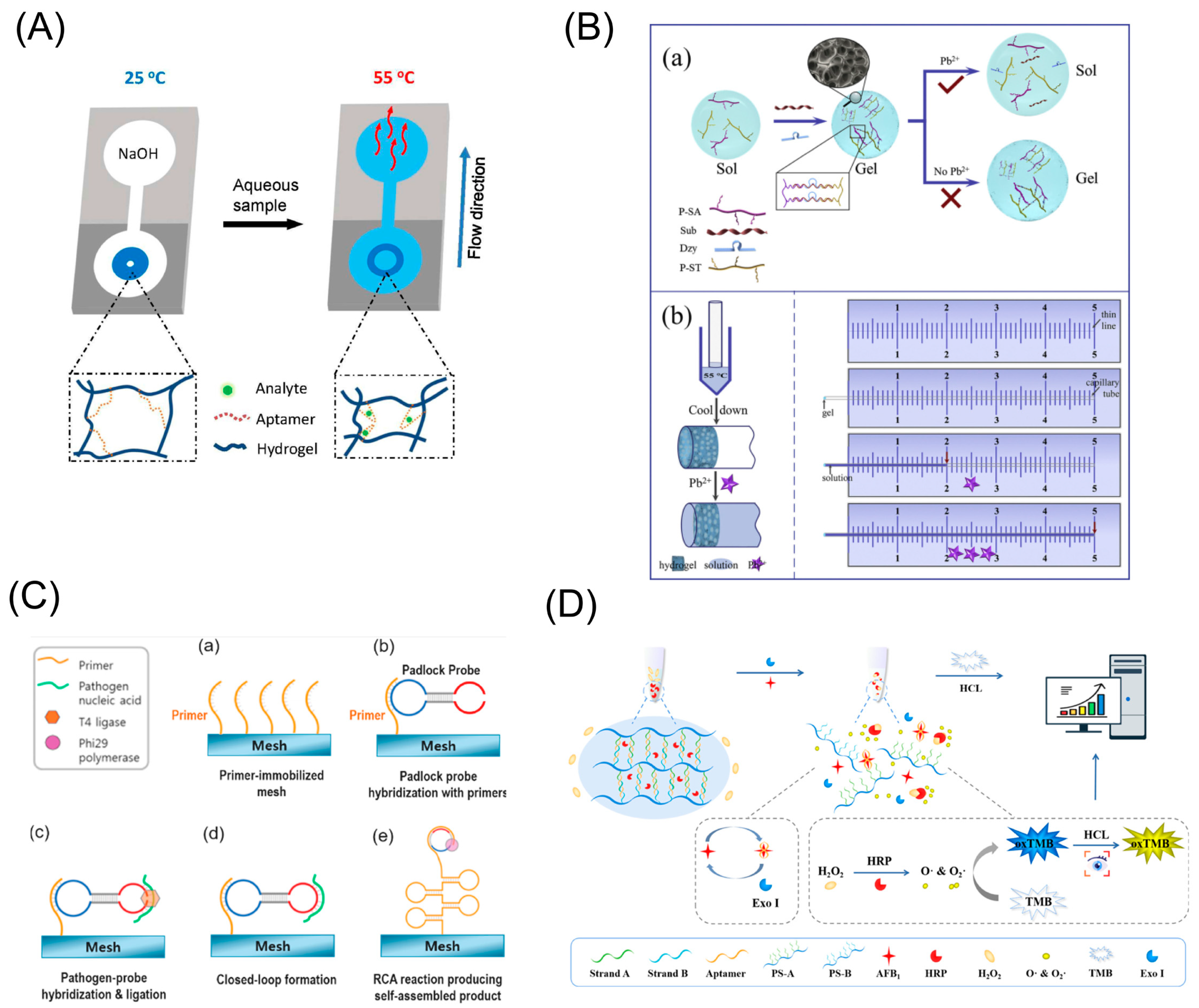
:1. Introduction
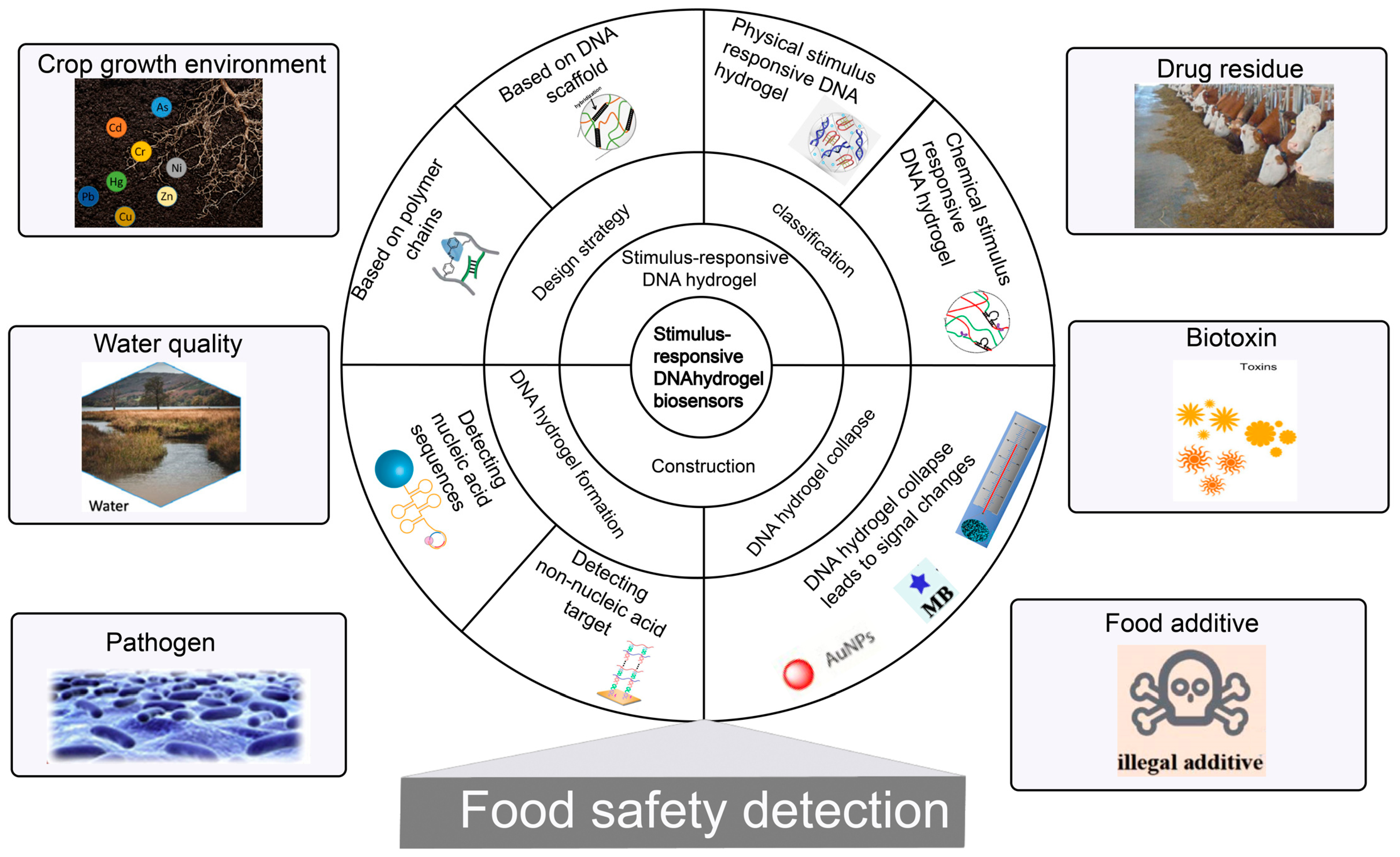
2. Preparation and Classification of Stimulus-Responsive DNA Hydrogel
2.1. Design Strategies of Stimulus-Responsive DNA Hydrogels
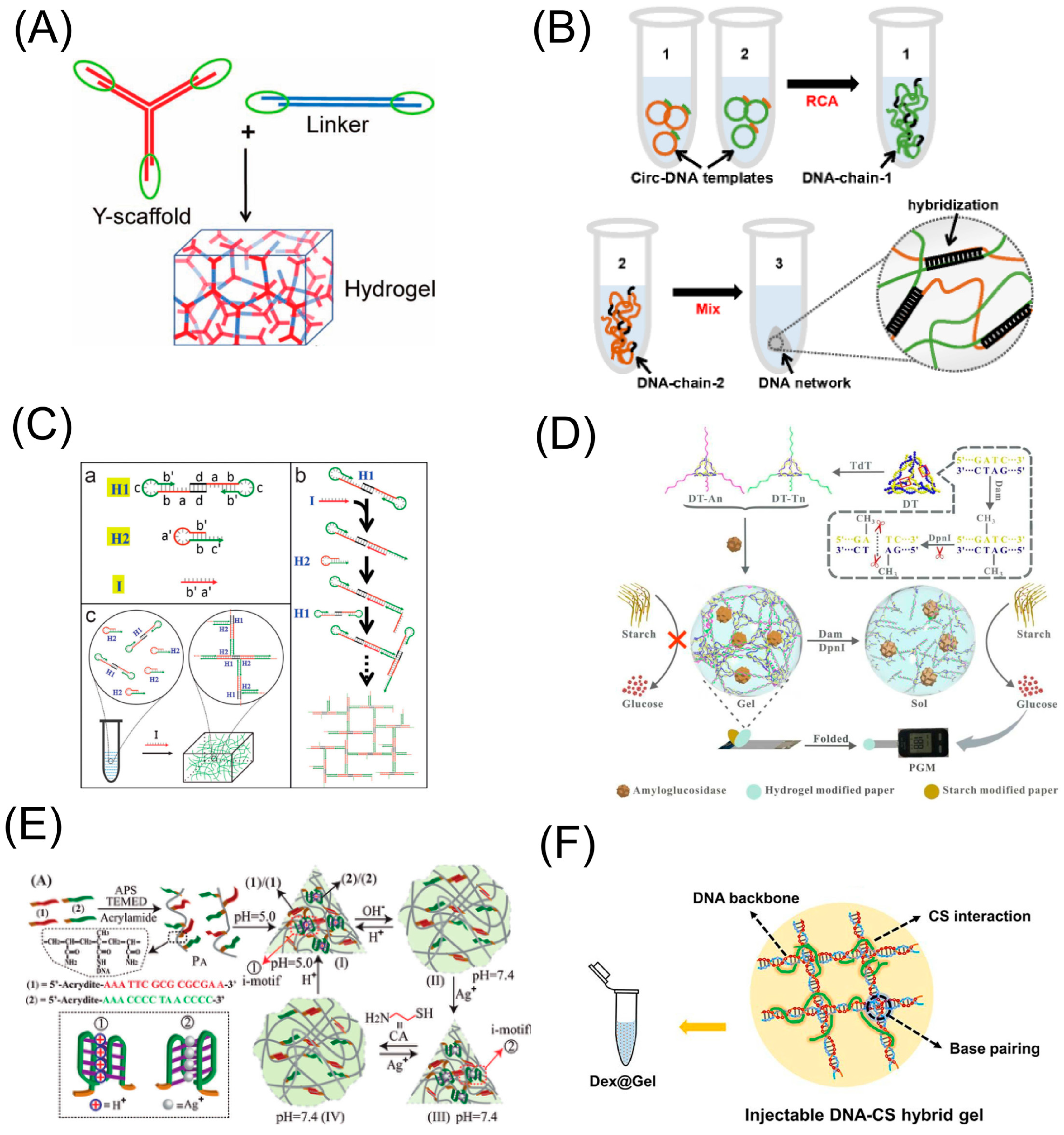
2.1.1. DNA Hydrogel Based on DNA Scaffold
2.1.2. DNA Hydrogel Based on Other Polymer Chain
2.2. Classification of Stimulus-Responsive DNA Hydrogel
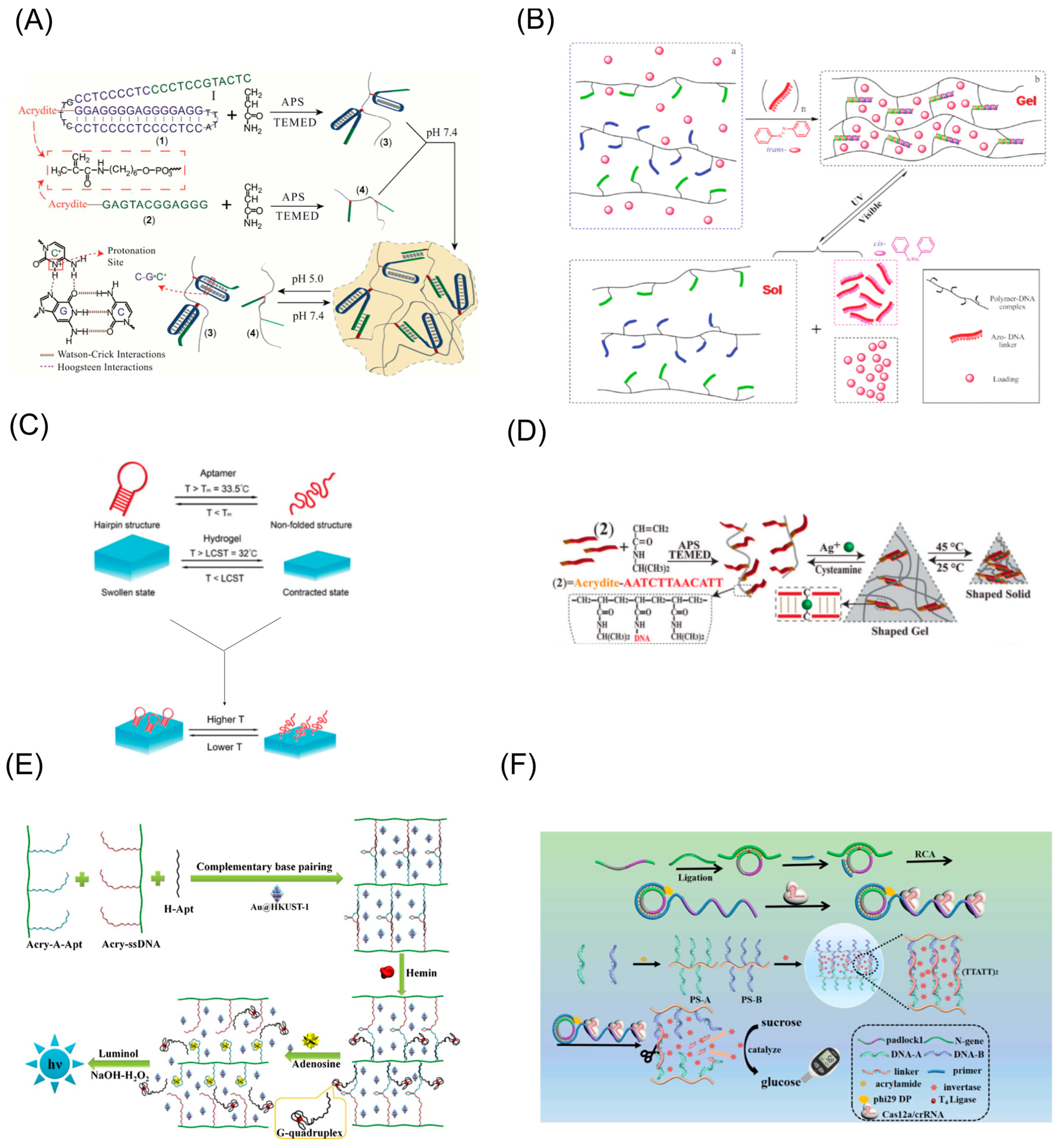
2.2.1. Physical-Stimulus-Responsive DNA Hydrogel
pH-Responsive DNA Hydrogels
Light-Responsive DNA Hydrogel
Temperature-Responsive DNA Hydrogel
2.2.2. Chemical-Stimulus-Responsive DNA Hydrogels
Metal-Ion-Responsive DNA Hydrogel
Biomolecules-Responsive DNA Hydrogel
3. Stimulus-Responsive DNA Hydrogel-Based Biosensor
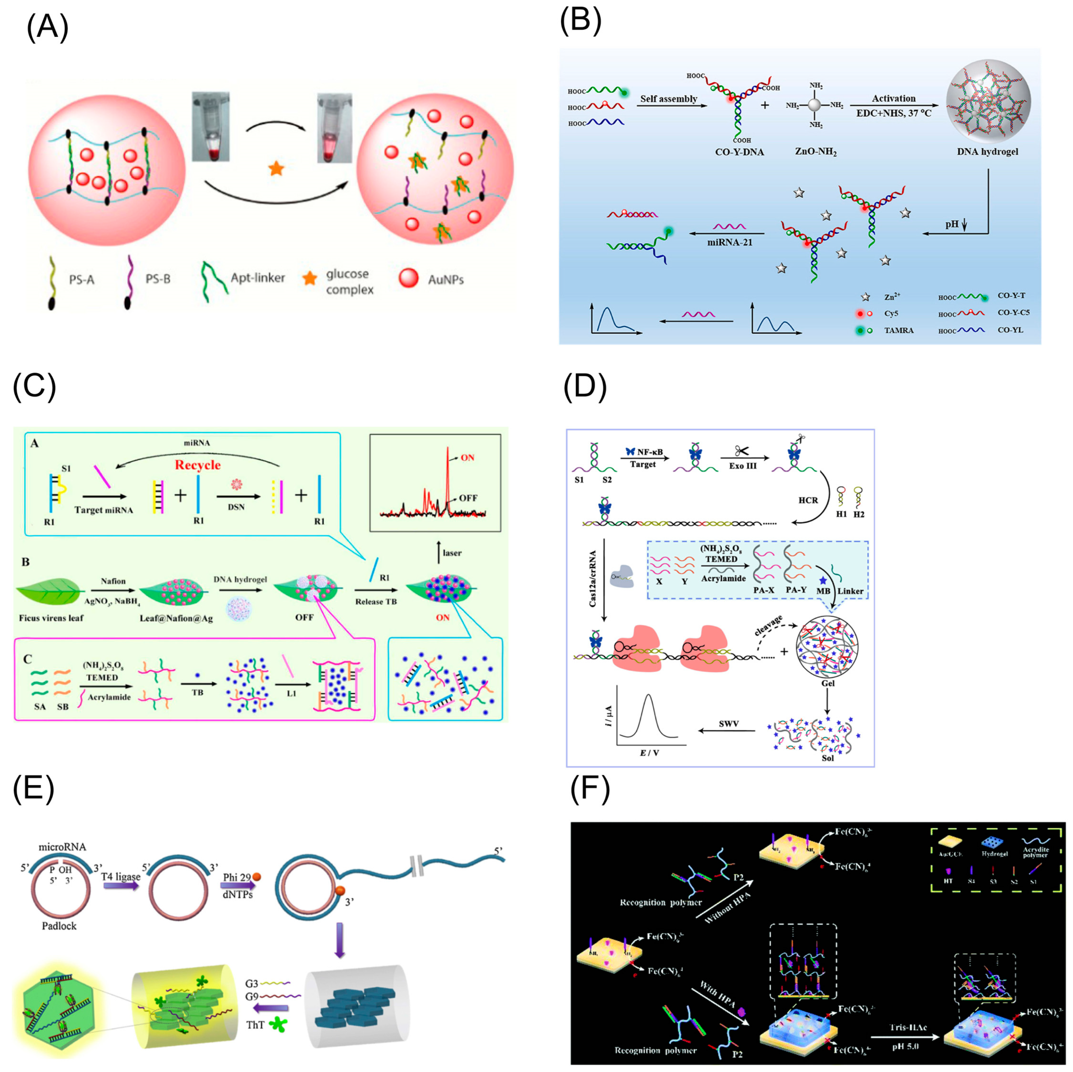
3.1. DNA Hydrogel Collapse Principle
3.2. DNA Hydrogel Formation Principle
4. Stimulus-Responsive DNA Hydrogel Biosensors for Food Safety Detection
4.1. Heavy Metals
4.2. Pathogen Monitoring
4.3. Drug Residues
4.4. Biotoxins
4.5. Food Additives
4.6. Other Food Contaminants
4.7. Interference Suppression Strategies
5. Conclusions and Perspective
Author Contributions
Funding
Institutional Review Board Statement
Informed Consent Statement
Data Availability Statement
Conflicts of Interest
Abbreviations
| AFB1 | Aflatoxin B1 |
| AgNPs | Silver nanoparticles |
| ATP | Adenosine triphosphate |
| AuNCs | Gold nanoclusters |
| AuNPs | Gold nanoparticles |
| AuNRs | Gold nanorods |
| Au@PtNPs | Au@Pt core–shell nanoparticles |
| Azo | Azobenzene |
| BPA | Bisphenol A |
| CMC | Carboxymethyl cellulose |
| COFs | Covalent-organic frameworks |
| CS | chitosan |
| DNA | Deoxyribonucleic acid |
| DTE | Dithienylethene |
| ELISA | Enzyme-linked immunoassay |
| E. coli | Escherichia coli |
| FB1 | Fumonisin B1 |
| GC | Gas chromatography |
| GcNPs | Gap-containing nanoparticles |
| GM | Genetically modified |
| GOD | Glucose oxidase |
| HCR | Hybridization chain reaction |
| HPLC | High-performance liquid chromatography |
| HRP | Horseradish peroxidase |
| LCST | Lower critical solution temperature |
| LOD | Lowest limit of detection |
| MB | Methylene blue |
| MEL | Melamine |
| MiRNA | MicroRNA |
| MOFs | Metal–organic frameworks |
| MRL | Maximum residue limits |
| MS | Mass spectrometry |
| OTA | Ochratoxin A |
| PCR | Polymerase chain reaction |
| PNIPAAm | Propylacrylamide |
| PtNPs | Ptnanoparticles |
| QDs | Quantum dots |
| RCA | Rolling circle amplification |
| SERS | Surface-enhanced Raman scattering |
| TB | Toluidine blue |
| TdT | Terminal deoxynucleotidyl transferase |
| UCST | Upper critical solution temperature |
| V.P | Vibrio parahaemolyticus |
| WHO | World Health Organization |
| ZEN | Zearalenone |
References
- Soares, P.A.; de Seixas, J.R.; Albuquerque, P.B.; Santos, G.R.; Mourao, P.A.; Barros, W., Jr.; Correia, M.T.; Carneiro-da-Cunha, M.G. Development and characterization of a new hydrogel based on galactomannan and kappa-carrageenan. Carbohydr. Polym. 2015, 134, 673–679. [Google Scholar] [CrossRef] [Green Version]
- Tavakoli, J.; Wang, J.; Chuah, C.; Tang, Y. Natural-based Hydrogels: A Journey from Simple to Smart Networks for Medical Examination. Curr. Med. Chem. 2020, 27, 2704–2733. [Google Scholar] [CrossRef]
- Wu, S.; Wu, S.; Zhang, X.; Feng, T.; Wu, L. Chitosan-Based Hydrogels for Bioelectronic Sensing: Recent Advances and Applications in Biomedicine and Food Safety. Biosensors 2023, 13, 93. [Google Scholar] [CrossRef]
- Pardo, Y.A.; Yancey, K.G.; Rosenwasser, D.S.; Bassen, D.M.; Butcher, J.T.; Sabin, J.E.; Ma, M.; Hamada, S.; Luo, D. Interfacing DNA hydrogels with ceramics for biofunctional architectural materials. Mater. Today 2022, 53, 98–105. [Google Scholar] [CrossRef]
- Xu, P.F.; Noh, H.; Lee, J.H.; Domaille, D.W.; Nakatsuka, M.A.; Goodwin, A.P.; Cha, J.N. Imparting the unique properties of DNA into complex material architectures and functions. Mater. Today 2013, 16, 290–296. [Google Scholar] [CrossRef]
- Simon, A.J.; Walls-Smith, L.T.; Plaxco, K.W. Exploiting the conformational-selection mechanism to control the response kinetics of a “smart” DNA hydrogel. Analyst 2018, 143, 2531–2538. [Google Scholar] [CrossRef]
- Wang, D.; Hu, Y.; Liu, P.F.; Luo, D. Bioresponsive DNA Hydrogels: Beyond the Conventional Stimuli Responsiveness. Acc. Chem. Res. 2017, 50, 733–739. [Google Scholar] [CrossRef]
- Mo, F.L.; Jiang, K.; Zhao, D.; Wang, Y.Q.; Song, J.; Tan, W.H. DNA hydrogel-based gene editing and drug delivery systems. Adv. Drug Delivery Rev. 2021, 168, 79–98. [Google Scholar] [CrossRef]
- Beyer, A.; Pollok, S.; Rudloff, A.; Cialla-May, D.; Weber, K.; Popp, J. Fast-Track, One-Step E. coli Detection: A Miniaturized Hydrogel Array Permits Specific Direct PCR and DNA Hybridization while Amplification. Macromol. Biosci. 2016, 16, 1325–1333. [Google Scholar] [CrossRef]
- Jiang, H.L.; Kim, Y.K.; Lee, S.M.; Park, M.R.; Kim, E.M.; Jin, Y.M.; Arote, R.; Jeong, H.J.; Song, S.C.; Cho, M.H.; et al. Galactosylated chitosan-g-PEI/DNA complexes-loaded poly(organophosphazene) hydrogel as a hepatocyte targeting gene delivery system. Arch. Pharm. Res. 2010, 33, 551–556. [Google Scholar] [CrossRef]
- Wang, Y.; Zhu, Y.; Hu, Y.; Zeng, G.; Zhang, Y.; Zhang, C.; Feng, C. How to Construct DNA Hydrogels for Environmental Applications: Advanced Water Treatment and Environmental Analysis. Small 2018, 14, e1703305. [Google Scholar] [CrossRef]
- Li, C.; Faulkner-Jones, A.; Dun, A.R.; Jin, J.; Chen, P.; Xing, Y.; Yang, Z.; Li, Z.; Shu, W.; Liu, D.; et al. Rapid formation of a supramolecular polypeptide-DNA hydrogel for in situ three-dimensional multilayer bioprinting. Angew. Chem. Int. Ed. 2015, 54, 3957–3961. [Google Scholar] [CrossRef]
- Chen, M.; Wang, Y.; Zhang, J.; Peng, Y.; Li, S.; Han, D.; Ren, S.; Qin, K.; Li, S.; Gao, Z. Stimuli-responsive DNA-based hydrogels for biosensing applications. J. Nanobiotechnol. 2022, 20, 40. [Google Scholar] [CrossRef]
- Chu, J.; Chen, C.; Li, X.; Yu, L.; Li, W.; Cheng, M.; Tang, W.; Xiong, Z. A responsive pure DNA hydrogel for label-free detection of lead ion. Anal. Chim. Acta 2021, 1157, 338400. [Google Scholar] [CrossRef]
- Li, J.; Mo, L.T.; Lu, C.H.; Fu, T.; Yang, H.H.; Tan, W.H. Functional nucleic acid-based hydrogels for bioanalytical and biomedical applications. Chem. Soc. Rev. 2016, 45, 1410–1431. [Google Scholar] [CrossRef] [Green Version]
- Ma, Y.; Mao, Y.; An, Y.; Tian, T.; Zhang, H.; Yan, J.; Zhu, Z.; Yang, C.J. Target-responsive DNA hydrogel for non-enzymatic and visual detection of glucose. Analyst 2018, 143, 1679–1684. [Google Scholar] [CrossRef]
- Yao, S.; Xiang, L.; Wang, L.; Gong, H.; Chen, F.; Cai, C. pH-responsive DNA hydrogels with ratiometric fluorescence for accurate detection of miRNA-21. Anal. Chim. Acta 2022, 1207, 339795. [Google Scholar] [CrossRef]
- Jiang, C.; Li, Y.; Wang, H.; Chen, D.; Wen, Y. A portable visual capillary sensor based on functional DNA crosslinked hydrogel for point-of-care detection of lead ion. Sens. Actuators B 2020, 307, 127625. [Google Scholar] [CrossRef]
- Xing, Y.; Cheng, E.; Yang, Y.; Chen, P.; Zhang, T.; Sun, Y.; Yang, Z.; Liu, D. Self-assembled DNA hydrogels with designable thermal and enzymatic responsiveness. Adv. Mater. 2011, 23, 1117–1121. [Google Scholar] [CrossRef]
- Cheng, E.; Xing, Y.; Chen, P.; Yang, Y.; Sun, Y.; Zhou, D.; Xu, L.; Fan, Q.; Liu, D. A pH-triggered, fast-responding DNA hydrogel. Angew. Chem. Int. Ed. 2009, 48, 7660–7663. [Google Scholar] [CrossRef]
- Cheng, L.; He, Y.; Yang, Y.; Chen, J.; He, H.; Liu, Y.; Lin, Z.; Hong, G. Highly reproducible and sensitive electrochemical biosensor for Chlamydia trachomatis detection based on duplex-specific nuclease-assisted target-responsive DNA hydrogels and bovine serum albumin carrier platform. Anal. Chim. Acta 2022, 1197, 339496. [Google Scholar] [CrossRef]
- Iqbal, S.; Ahmed, F.; Xiong, H. Responsive-DNA hydrogel based intelligent materials: Preparation and applications. Chem. Eng. J. 2021, 420, 130384. [Google Scholar] [CrossRef]
- Willner, I. Stimuli-Controlled Hydrogels and Their Applications. Acc. Chem. Res. 2017, 50, 657–658. [Google Scholar] [CrossRef] [Green Version]
- Vazquez-Gonzalez, M.; Willner, I. Stimuli-Responsive Biomolecule-Based Hydrogels and Their Applications. Angew. Chem. Int. Ed. 2020, 59, 15342–15377. [Google Scholar] [CrossRef]
- Wang, C.; Zhang, J. Recent Advances in Stimuli-Responsive DNA-Based Hydrogels. ACS Appl. Bio Mater. 2022, 5, 1934–1953. [Google Scholar] [CrossRef]
- Ferrari, A.G.; Crapnell, R.D.; Banks, C.E. Electroanalytical Overview: Electrochemical Sensing Platforms for Food and Drink Safety. Biosensors 2021, 11, 291. [Google Scholar] [CrossRef]
- Saravanan, A.; Kumar, P.S.; Hemavathy, R.V.; Jeevanantham, S.; Kamalesh, R.; Sneha, S.; Yaashikaa, P.R. Methods of detection of food-borne pathogens: A review. Environ. Chem. Lett. 2020, 19, 189–207. [Google Scholar] [CrossRef]
- Shenashen, M.A.; Emran, M.Y.; El Sabagh, A.; Selim, M.M.; Elmarakbi, A.; El-Safty, S.A. Progress in sensory devices of pesticides, pathogens, coronavirus, and chemical additives and hazards in food assessment: Food safety concerns. Prog. Mater Sci. 2022, 124, 100866. [Google Scholar] [CrossRef]
- Zhang, J.; Huang, H.; Song, G.; Huang, K.; Luo, Y.; Liu, Q.; He, X.; Cheng, N. Intelligent biosensing strategies for rapid detection in food safety: A review. Biosens. Bioelectron. 2022, 202, 114003. [Google Scholar] [CrossRef]
- Kalita, J.J.; Sharma, P.; Bora, U. Recent developments in application of nucleic acid aptamer in food safety. Food Control 2022, 145, 109406. [Google Scholar] [CrossRef]
- Xia, X.; Yang, H.; Cao, J.; Zhang, J.; He, Q.; Deng, R. Isothermal nucleic acid amplification for food safety analysis. TrAC Trends Anal. Chem. 2022, 153, 116641. [Google Scholar] [CrossRef]
- Kotsiri, Z.; Vidic, J.; Vantarakis, A. Applications of biosensors for bacteria and virus detection in food and water-A systematic review. J. Environ. Sci. 2022, 111, 367–379. [Google Scholar] [CrossRef]
- Zhang, Z.; Zhou, J.; Du, X. Electrochemical Biosensors for Detection of Foodborne Pathogens. Micromachines 2019, 10, 222. [Google Scholar] [CrossRef] [Green Version]
- Ribeiro, B.V.; Ferreira, L.F.; Franco, D.L. Advances in biosensor development for the determination of antibiotics in cow’s milk—A review. Talanta Open 2022, 6, 100145. [Google Scholar] [CrossRef]
- Cao, X.; Chen, C.; Zhu, Q. Biosensors based on functional nucleic acids and isothermal amplification techniques. Talanta 2022, 253, 123977. [Google Scholar] [CrossRef]
- Zhao, H.; Lan, X.; Yu, F.; Li, Z.; Yang, J.; Du, L. Comprehensive assessment of heavy metals in soil-crop system based on PMF and evolutionary game theory. Sci. Total Environ. 2022, 849, 157549. [Google Scholar] [CrossRef]
- Zhang, X.; Wu, D.; Zhou, X.; Yu, Y.; Liu, J.; Hu, N.; Wang, H.; Li, G.; Wu, Y. Recent progress in the construction of nanozyme-based biosensors and their applications to food safety assay. TrAC Trends Anal. Chem. 2019, 121, 115668. [Google Scholar] [CrossRef]
- Penagos-Tabares, F.; Sulyok, M.; Faas, J.; Krska, R.; Khiaosa-Ard, R.; Zebeli, Q. Residues of pesticides and veterinary drugs in diets of dairy cattle from conventional and organic farms in Austria. Environ. Pollut. 2023, 316, 120626. [Google Scholar] [CrossRef]
- Um, S.H.; Lee, J.B.; Park, N.; Kwon, S.Y.; Umbach, C.C.; Luo, D. Enzyme-catalysed assembly of DNA hydrogel. Nat. Mater. 2006, 5, 797–801. [Google Scholar] [CrossRef]
- Wang, J.; Chao, J.; Liu, H.; Su, S.; Wang, L.; Huang, W.; Willner, I.; Fan, C. Clamped Hybridization Chain Reactions for the Self-Assembly of Patterned DNA Hydrogels. Angew. Chem. Int. Ed. 2017, 56, 2171–2175. [Google Scholar] [CrossRef]
- Xiang, B.B.; He, K.Y.; Zhu, R.; Liu, Z.L.; Zeng, S.; Huang, Y.; Nie, Z.; Yao, S.Z. Self-Assembled DNA Hydrogel Based on Enzymatically Polymerized DNA for Protein Encapsulation and Enzyme/DNAzyme Hybrid Cascade Reaction. ACS Appl. Mater. Interfaces 2016, 8, 22801–22807. [Google Scholar] [CrossRef]
- Sun, Y.; Li, S.; Chen, R.; Wu, P.; Liang, J. Ultrasensitive and rapid detection of T-2 toxin using a target-responsive DNA hydrogel. Sens. Actuators B 2020, 311, 127912. [Google Scholar] [CrossRef]
- Lu, S.S.; Shen, J.L.; Fan, C.H.; Li, Q.; Yang, X.R. DNA Assembly-Based Stimuli-Responsive Systems. Adv. Sci. 2021, 8, 2100328. [Google Scholar] [CrossRef]
- Ren, J.; Hu, Y.; Lu, C.H.; Guo, W.; Aleman-Garcia, M.A.; Ricci, F.; Willner, I. pH-responsive and switchable triplex-based DNA hydrogels. Chem. Sci. 2015, 6, 4190–4195. [Google Scholar] [CrossRef] [Green Version]
- Guo, W.; Qi, X.J.; Orbach, R.; Lu, C.H.; Freage, L.; Mironi-Harpaz, I.; Seliktar, D.; Yang, H.H.; Willner, I. Reversible Ag(+)-crosslinked DNA hydrogels. Chem. Commun. 2014, 50, 4065–4068. [Google Scholar] [CrossRef]
- Wu, Y.; Wang, D.; Willner, I.; Tian, Y.; Jiang, L. Smart DNA Hydrogel Integrated Nanochannels with High Ion Flux and Adjustable Selective Ionic Transport. Angew. Chem. Int. Ed. 2018, 57, 7790–7794. [Google Scholar] [CrossRef]
- Cangialosi, A.; Yoon, C.; Liu, J.; Huang, Q.; Guo, J.K.; Nguyen, T.D.; Gracias, D.H.; Schulman, R. DNA sequence-directed shape change of photopatterned hydrogels via high-degree swelling. Science 2017, 357, 1126–1129. [Google Scholar] [CrossRef] [Green Version]
- Yao, C.; Tang, H.; Wu, W.; Tang, J.; Guo, W.; Luo, D.; Yang, D. Double Rolling Circle Amplification Generates Physically Cross-Linked DNA Network for Stem Cell Fishing. J. Am. Chem. Soc. 2020, 142, 3422–3429. [Google Scholar] [CrossRef]
- Gao, X.; Li, X.Y.; Sun, X.Z.; Zhang, J.Y.; Zhao, Y.C.; Liu, X.J.; Li, F. DNA Tetrahedra-Cross-linked Hydrogel Functionalized Paper for Onsite Analysis of DNA Methyltransferase Activity Using a Personal Glucose Meter. Anal. Chem. 2020, 92, 4592–4599. [Google Scholar] [CrossRef]
- Yu, X.; Hu, Y.W.; Kahn, J.S.; Cecconello, A.; Willner, I. Orthogonal Dual-Triggered Shape-Memory DNA-Based Hydrogels. Chem. Eur. J. 2016, 22, 14504–14507. [Google Scholar] [CrossRef]
- Chen, F.; He, Y.; Li, Z.; Xu, B.; Ye, Q.; Li, X.; Ma, Z.; Song, W.; Zhang, Y. A novel tunable, highly biocompatible and injectable DNA-chitosan hybrid hydrogel fabricated by electrostatic interaction between chitosan and DNA backbone. Int. J. Pharm. 2021, 606, 120938. [Google Scholar] [CrossRef]
- Ko, O.; Han, S.; Lee, J.B. Selective release of DNA nanostructures from DNA hydrogel. J. Ind. Eng. Chem. 2020, 84, 46–51. [Google Scholar] [CrossRef]
- Tang, J.; Liang, A.; Yao, C.; Yang, D. Assembly of Rolling Circle Amplification-Produced Ultralong Single-Stranded DNA to Construct Biofunctional DNA Materials. Chemistry 2022, 28, e202202673. [Google Scholar] [CrossRef]
- Xu, W.L.; Huang, Y.S.; Zhao, H.R.; Li, P.; Liu, G.Y.; Li, J.; Zhu, C.S.; Tian, L.L. DNA Hydrogel with Tunable pH-Responsive Properties Produced by Rolling Circle Amplification. Chem. Eur. J. 2017, 23, 18276–18281. [Google Scholar] [CrossRef]
- Bi, S.; Yue, S.; Zhang, S. Hybridization chain reaction: A versatile molecular tool for biosensing, bioimaging, and biomedicine. Chem. Soc. Rev. 2017, 46, 4281–4298. [Google Scholar] [CrossRef]
- Li, Z.Y.; Davidson-Rozenfeld, G.; Vazquez-Gonzalez, M.; Fadeev, M.; Zhang, J.J.; Tian, H.; Willner, I. Multi-triggered Supramolecular DNA/Bipyridinium Dithienylethene Hydrogels Driven by Light, Redox, and Chemical Stimuli for Shape-Memory and Self-Healing Applications. J. Am. Chem. Soc. 2018, 140, 17691–17701. [Google Scholar] [CrossRef]
- Alemdaroglu, F.E.; Herrmann, A. DNA meets synthetic polymers—Highly versatile hybrid materials. Org. Biomol. Chem. 2007, 5, 1311–1320. [Google Scholar] [CrossRef] [Green Version]
- Wang, C.; Fadeev, M.; Zhang, J.J.; Vazquez-Gonzalez, M.; Davidson-Rozenfeld, G.; Tian, H.; Willner, I. Shape-memory and self-healing functions of DNA-based carboxymethyl cellulose hydrogels driven by chemical or light triggers. Chem. Sci. 2018, 9, 7145–7152. [Google Scholar] [CrossRef] [Green Version]
- Fu, X.; Chen, T.; Song, Y.; Feng, C.; Chen, H.; Zhang, Q.; Chen, G.; Zhu, X. mRNA Delivery by a pH-Responsive DNA Nano-Hydrogel. Small 2021, 17, e2101224. [Google Scholar] [CrossRef]
- Jeong, J.Y.; Do, J.Y.; Hong, C.A. Target DNA- and pH-responsive DNA hydrogel-based capillary assay for the optical detection of short SARS-CoV-2 cDNA. Mikrochim. Acta 2021, 189, 34. [Google Scholar] [CrossRef]
- Hu, Y.; Gao, S.; Lu, H.; Ying, J.Y. Acid-Resistant and Physiological pH-Responsive DNA Hydrogel Composed of A-Motif and i-Motif toward Oral Insulin Delivery. J. Am. Chem. Soc. 2022, 144, 5461–5470. [Google Scholar] [CrossRef]
- Kang, H.; Liu, H.; Zhang, X.; Yan, J.; Zhu, Z.; Peng, L.; Yang, H.; Kim, Y.; Tan, W. Photoresponsive DNA-cross-linked hydrogels for controllable release and cancer therapy. Langmuir 2011, 27, 399–408. [Google Scholar] [CrossRef] [Green Version]
- Zhang, B.; Wang, C.; Du, Y.; Paxton, R.; He, X. A ‘smart’ aptamer-functionalized continuous label-free cell catch-transport-release system. J. Mater. Chem. B 2021, 9, 7196–7204. [Google Scholar] [CrossRef]
- Guo, W.W.; Lu, C.H.; Qi, X.J.; Orbach, R.; Fadeev, M.; Yang, H.H.; Willner, I. Switchable Bifunctional Stimuli-Triggered Poly-N-Isopropylacrylamide/DNA Hydrogels. Angew. Chem. Int. Ed. 2014, 53, 10134–10138. [Google Scholar] [CrossRef]
- Lin, Y.N.; Wang, X.Y.; Sun, Y.L.; Dai, Y.X.; Sun, W.Y.; Zhu, X.D.; Liu, H.; Han, R.; Gao, D.D.; Luo, C.N. A chemiluminescent biosensor for ultrasensitive detection of adenosine based on target-responsive DNA hydrogel with Au@HKUST-1 encapsulation. Sens. Actuators B 2019, 289, 56–64. [Google Scholar] [CrossRef]
- Ma, W.; Liu, M.; Xie, S.; Liu, B.; Jiang, L.; Zhang, X.; Yuan, X. CRISPR/Cas12a system responsive DNA hydrogel for label-free detection of non-glucose targets with a portable personal glucose meter. Anal. Chim. Acta 2022, 1231, 340439. [Google Scholar] [CrossRef]
- Zhang, Z.; Xie, Z.; Nie, C.; Wu, S. Photo-controlled properties and functions of azobenzene-terminated polymers. Polymer 2022, 256, 125166. [Google Scholar] [CrossRef]
- Simeth, N.A.; de Mendoza, P.; Dubach, V.R.A.; Stuart, M.C.A.; Smith, J.W.; Kudernac, T.; Browne, W.R.; Feringa, B.L. Photoswitchable architecture transformation of a DNA-hybrid assembly at the microscopic and macroscopic scale. Chem. Sci. 2022, 13, 3263–3272. [Google Scholar] [CrossRef]
- Huang, F.; Chen, M.; Zhou, Z.; Duan, R.; Xia, F.; Willner, I. Spatiotemporal patterning of photoresponsive DNA-based hydrogels to tune local cell responses. Nat. Commun. 2021, 12, 2364. [Google Scholar] [CrossRef]
- Zhang, C.-Y.; Zhang, N.-H. Size dependent correlation between structure and apparent stiffness of viral DNA during temperature variation. J. Mech. Phys. Solids 2021, 154, 104501. [Google Scholar] [CrossRef]
- Zhu, X.; Wu, J.; Shao, F.; Hu, X. Reversible Thermal Cycling of DNA Material for Efficient Cellulose Hydrolysis. ACS Appl. Bio Mater. 2018, 1, 1118–1123. [Google Scholar] [CrossRef] [PubMed]
- Wang, G.; Wang, S.; Yan, C.; Bai, G.; Liu, Y. DNA-functionalized gold nanoparticle-based fluorescence polarization for the sensitive detection of silver ions. Colloids Surf. B Biointerfaces 2018, 167, 150–155. [Google Scholar] [CrossRef] [PubMed]
- Xing, X.; Feng, Y.; Yu, Z.; Hidaka, K.; Liu, F.; Ono, A.; Sugiyama, H.; Endo, M. Direct Observation of the Double-Stranded DNA Formation through Metal Ion-Mediated Base Pairing in the Nanoscale Structure. Chemistry 2019, 25, 1446–1450. [Google Scholar] [CrossRef] [PubMed]
- Liu, X.; Zhang, J.J.; Fadeev, M.; Li, Z.Y.; Wulf, V.; Tian, H.; Willner, I. Chemical and photochemical DNA “gears” reversibly control stiffness, shape-memory, self-healing and controlled release properties of polyacrylamide hydrogels. Chem. Sci. 2019, 10, 1008–1016. [Google Scholar] [CrossRef] [PubMed] [Green Version]
- Kahn, J.S.; Trifonov, A.; Cecconello, A.; Guo, W.W.; Fan, C.H.; Willner, I. Integration of Switchable DNA-Based Hydrogels with Surfaces by the Hybridization Chain Reaction. Nano Lett. 2015, 15, 7773–7778. [Google Scholar] [CrossRef]
- Hou, M.; Yin, X.; Jiang, J.H.; He, J.J. DNAzyme-Triggered Sol-Gel-Sol Transition of a Hydrogel Allows Target Cell Enrichment. ACS Appl. Mater. Interfaces 2021, 13, 15031–15039. [Google Scholar] [CrossRef] [PubMed]
- Kahn, J.S.; Hu, Y.W.; Willner, I. Stimuli-Responsive DNA-Based Hydrogels: From Basic Principles to Applications. Acc. Chem. Res. 2017, 50, 680–690. [Google Scholar] [CrossRef]
- He, Y.; Yang, X.; Yuan, R.; Chai, Y.Q. Switchable Target-Responsive 3D DNA Hydrogels As a Signal Amplification Strategy Combining with SERS Technique for Ultrasensitive Detection of miRNA 155. Anal. Chem. 2017, 89, 8538–8544. [Google Scholar] [CrossRef]
- Bae, S.W.; Lee, J.S.; Harms, V.M.; Murphy, W.L. Dynamic, Bioresponsive Hydrogels via Changes in DNA Aptamer Conformation. Macromol. Biosci. 2019, 19, e1800353. [Google Scholar] [CrossRef]
- Hegde, M.; Pai, P.; Gangadhar Shetty, M.; Sundara Babitha, K. Gold nanoparticle based biosensors for rapid pathogen detection: A Review. Environ. Nanotechnol. Monit. Manag. 2022, 18, 100756. [Google Scholar] [CrossRef]
- Jiang, C.; Wang, F.; Zhang, K.; Min, T.; Chen, D.; Wen, Y. Distance-Based Biosensor for Ultrasensitive Detection of Uracil-DNA Glycosylase Using Membrane Filtration of DNA Hydrogel. ACS Sens. 2021, 6, 2395–2402. [Google Scholar] [CrossRef]
- Wei, X.F.; Tian, T.; Jia, S.S.; Zhu, Z.; Ma, Y.L.; Sun, J.J.; Lin, Z.Y.; Yang, C.J. Target-Responsive DNA Hydrogel Mediated “Stop-Flow” Microfluidic Paper-Based Analytic Device for Rapid, Portable and Visual Detection of Multiple Targets. Anal. Chem. 2015, 87, 4275–4282. [Google Scholar] [CrossRef] [PubMed]
- Yanez-Aulestia, A.; Gupta, N.K.; Hernandez, M.; Osorio-Toribio, G.; Sanchez-Gonzalez, E.; Guzman-Vargas, A.; Rivera, J.L.; Ibarra, I.A.; Lima, E. Gold nanoparticles: Current and upcoming biomedical applications in sensing, drug, and gene delivery. Chem. Commun. 2022, 58, 10886–10895. [Google Scholar] [CrossRef] [PubMed]
- Guo, Y.; Zhao, W. In situ formed nanomaterials for colorimetric and fluorescent sensing. Coord. Chem. Rev. 2019, 387, 249–261. [Google Scholar] [CrossRef]
- Zhao, M.; Li, Y.; Ma, X.; Xia, M.; Zhang, Y. Adsorption of cholesterol oxidase and entrapment of horseradish peroxidase in metal-organic frameworks for the colorimetric biosensing of cholesterol. Talanta 2019, 200, 293–299. [Google Scholar] [CrossRef]
- Fang, B.; Xu, S.; Huang, Z.; Wang, S.; Chen, W.; Yuan, M.; Hu, S.; Peng, J.; Lai, W. Glucose oxidase-induced colorimetric immunoassay for qualitative detection of danofloxacin based on iron (Ⅱ) chelation reaction with phenanthroline. Food Chem. 2020, 328, 127099. [Google Scholar] [CrossRef]
- Gao, L.; Chen, L.; Zhang, R.; Yan, X. Nanozymes: Next-generation artificial enzymes. Sci. Sin. Chim. 2022, 52, 1649–1663. [Google Scholar] [CrossRef]
- Liu, X.; Huang, D.; Lai, C.; Qin, L.; Zeng, G.; Xu, P.; Li, B.; Yi, H.; Zhang, M. Peroxidase-Like Activity of Smart Nanomaterials and Their Advanced Application in Colorimetric Glucose Biosensors. Small 2019, 15, e1900133. [Google Scholar] [CrossRef]
- Subhashree, S.; Kumar, P.S. New analytical strategies amplified with carbon-based nanomaterial for sensing food pollutants. Chemosphere 2022, 295, 133847. [Google Scholar] [CrossRef]
- Feng, Y.; Wang, Y.; Ying, Y. Structural design of metal–organic frameworks with tunable colorimetric responses for visual sensing applications. Coord. Chem. Rev. 2021, 446, 214102. [Google Scholar] [CrossRef]
- Lan, L.; Yao, Y.; Ping, J.; Ying, Y. Recent advances in nanomaterial-based biosensors for antibiotics detection. Biosens. Bioelectron. 2017, 91, 504–514. [Google Scholar] [CrossRef]
- Chen, M.; Zhang, J.; Peng, Y.; Bai, J.; Li, S.; Han, D.; Ren, S.; Qin, K.; Zhou, H.; Han, T.; et al. Design and synthesis of DNA hydrogel based on EXPAR and CRISPR/Cas14a for ultrasensitive detection of creatine kinase MB. Biosens. Bioelectron. 2022, 218, 114792. [Google Scholar] [CrossRef]
- Qiu, F.; Gan, X.; Yao, J.; Jiang, B.; Yuan, R.; Xiang, Y. CRISPR/Cas12a-derived sensitive electrochemical biosensing of NF-kappaB p50 based on hybridization chain reaction and DNA hydrogel. Biosens. Bioelectron. 2022, 216, 114665. [Google Scholar] [CrossRef]
- Song, H.; Zhang, Y.; Wang, S.; Huang, K.; Luo, Y.; Zhang, W.; Xu, W. Label-free polygonal-plate fluorescent-hydrogel biosensor for ultrasensitive microRNA detection. Sens. Actuators B 2020, 306, 127554. [Google Scholar] [CrossRef]
- Yang, Z.H.; Zhuo, Y.; Yuan, R.; Chai, Y.Q. Amplified impedimetric aptasensor combining target-induced DNA hydrogel formation with pH-stimulated signal amplification for the heparanase assay. Nanoscale 2017, 9, 2556–2562. [Google Scholar] [CrossRef]
- Han, S.; Dai, R.; Hu, Y.; Han, L. Fluorometric and colorimetric detection of cerium(IV) ion using carbon dots and bathophenanthroline-disulfonate-ferrum(II) complex. Spectrochim. Acta A Mol. Biomol. Spectrosc. 2022, 264, 120295. [Google Scholar] [CrossRef]
- Lan, L.; Yao, Y.; Ping, J.; Ying, Y. Recent Progress in Nanomaterial-Based Optical Aptamer Assay for the Detection of Food Chemical Contaminants. ACS Appl. Mater. Interfaces 2017, 9, 23287–23301. [Google Scholar] [CrossRef]
- Resch-Genger, U.; Grabolle, M.; Cavaliere-Jaricot, S.; Nitschke, R.; Nann, T. Quantum dots versus organic dyes as fluorescent labels. Nat. Methods 2008, 5, 763–775. [Google Scholar] [CrossRef]
- Yang, S.; Li, Y. Fluorescent hybrid silica nanoparticles and their biomedical applications. Wiley Interdiscip. Rev. Nanomed. Nanobiotechnol. 2020, 12, e1603. [Google Scholar] [CrossRef]
- Dong, Y.; Chen, Z.; Hou, M.; Qi, L.; Yan, C.; Lu, X.; Liu, R.; Xu, Y. Mitochondria-targeted aggregation-induced emission active near infrared fluorescent probe for real-time imaging. Spectrochim. Acta A Mol. Biomol. Spectrosc. 2020, 224, 117456. [Google Scholar] [CrossRef]
- Farkkila, S.M.A.; Kiers, E.T.; Jaaniso, R.; Maeorg, U.; Leblanc, R.M.; Treseder, K.K.; Kang, Z.; Tedersoo, L. Fluorescent nanoparticles as tools in ecology and physiology. Biol. Rev. Camb. Philos. Soc. 2021, 96, 2392–2424. [Google Scholar] [CrossRef] [PubMed]
- Chang, W.-H.; Lee, Y.-F.; Liu, Y.-W.; Willner, I.; Liao, W.-C. Stimuli-responsive hydrogel microcapsules for the amplified detection of microRNAs. Nanoscale 2021, 13, 16799–16808. [Google Scholar] [CrossRef] [PubMed]
- Xu, K.; Zhou, R.; Takei, K.; Hong, M. Toward Flexible Surface-Enhanced Raman Scattering (SERS) Sensors for Point-of-Care Diagnostics. Adv. Sci. 2019, 6, 1900925. [Google Scholar] [CrossRef] [PubMed]
- Chen, H.; Das, A.; Bi, L.; Choi, N.; Moon, J.I.; Wu, Y.; Park, S.; Choo, J. Recent advances in surface-enhanced Raman scattering-based microdevices for point-of-care diagnosis of viruses and bacteria. Nanoscale 2020, 12, 21560–21570. [Google Scholar] [CrossRef]
- He, Y.; Yang, X.; Yuan, R.; Chai, Y. A novel ratiometric SERS biosensor with one Raman probe for ultrasensitive microRNA detection based on DNA hydrogel amplification. J. Mater. Chem. B 2019, 7, 2643–2647. [Google Scholar] [CrossRef]
- Na, W.; Nam, D.; Lee, H.; Shin, S. Rapid molecular diagnosis of infectious viruses in microfluidics using DNA hydrogel formation. Biosens. Bioelectron. 2018, 108, 9–13. [Google Scholar] [CrossRef]
- Hong, C.A.; Park, J.C.; Na, H.; Jeon, H.; Nam, Y.S. Short DNA-catalyzed formation of quantum dot-DNA hydrogel for enzyme-free femtomolar specific DNA assay. Biosens. Bioelectron. 2021, 182, 113110. [Google Scholar] [CrossRef]
- Chen, Q.; Tian, R.; Liu, G.; Wen, Y.; Bian, X.; Luan, D.; Wang, H.; Lai, K.; Yan, J. Fishing unfunctionalized SERS tags with DNA hydrogel network generated by ligation-rolling circle amplification for simple and ultrasensitive detection of kanamycin. Biosens. Bioelectron. 2022, 207, 114187. [Google Scholar] [CrossRef]
- Cai, W.; Xie, S.B.; Zhang, J.; Tang, D.Y.; Tang, Y. An electrochemical impedance biosensor for Hg2+ detection based on DNA hydrogel by coupling with DNAzyme-assisted target recycling and hybridization chain reaction. Biosens. Bioelectron. 2017, 98, 466–472. [Google Scholar] [CrossRef]
- Hua, Z.; Yu, T.; Liu, D.; Xianyu, Y. Recent advances in gold nanoparticles-based biosensors for food safety detection. Biosens. Bioelectron. 2021, 179, 113076. [Google Scholar] [CrossRef]
- Cheng, W.; Tang, X.; Zhang, Y.; Wu, D.; Yang, W. Applications of metal-organic framework (MOF)-based sensors for food safety: Enhancing mechanisms and recent advances. Trends Food Sci. Technol. 2021, 112, 268–282. [Google Scholar] [CrossRef]
- Qin, G.; Niu, Z.; Yu, J.; Li, Z.; Ma, J.; Xiang, P. Soil heavy metal pollution and food safety in China: Effects, sources and removing technology. Chemosphere 2021, 267, 129205. [Google Scholar] [CrossRef] [PubMed]
- Lu, Y.; Song, S.; Wang, R.; Liu, Z.; Meng, J.; Sweetman, A.J.; Jenkins, A.; Ferrier, R.C.; Li, H.; Luo, W.; et al. Impacts of soil and water pollution on food safety and health risks in China. Environ. Int. 2015, 77, 5–15. [Google Scholar] [CrossRef] [PubMed] [Green Version]
- Nilghaz, A.; Mousavi, S.M.; Li, M.; Tian, J.; Cao, R.; Wang, X. Paper-based microfluidics for food safety and quality analysis. Trends Food Sci. Technol. 2021, 118, 273–284. [Google Scholar] [CrossRef]
- Lin, X.; Wang, Z.; Jia, X.; Chen, R.; Qin, Y.; Bian, Y.; Sheng, W.; Li, S.; Gao, Z. Stimulus-responsive hydrogels: A potent tool for biosensing in food safety. Trends Food Sci. Technol. 2023, 131, 91–103. [Google Scholar] [CrossRef]
- Ye, Y.; Guo, H.; Sun, X. Recent progress on cell-based biosensors for analysis of food safety and quality control. Biosens. Bioelectron. 2019, 126, 389–404. [Google Scholar] [CrossRef]
- Cheng, W.; Wu, X.; Zhang, Y.; Wu, D.; Meng, L.; Chen, Y.; Tang, X. Recent applications of hydrogels in food safety sensing: Role of hydrogels. Trends Food Sci. Technol. 2022, 129, 244–257. [Google Scholar] [CrossRef]
- Yang, Z.; Chen, L.; McClements, D.J.; Qiu, C.; Li, C.; Zhang, Z.; Miao, M.; Tian, Y.; Zhu, K.; Jin, Z. Stimulus-responsive hydrogels in food science: A review. Food Hydrocoll. 2022, 124, 107218. [Google Scholar] [CrossRef]
- Teng, Y.; Ni, S.; Wang, J.; Zuo, R.; Yang, J. A geochemical survey of trace elements in agricultural and non-agricultural topsoil in Dexing area, China. J. Geochem. Explor. 2010, 104, 118–127. [Google Scholar] [CrossRef]
- Zartman, R.E. Treated Wastewater in Agriculture: Use and Impacts on the Soil Environment and Crops. J. Environ. Qual. 2011, 40, 1995. [Google Scholar] [CrossRef] [Green Version]
- Meng, W.; Wang, Z.; Hu, B.; Wang, Z.; Li, H.; Goodman, R.C. Heavy metals in soil and plants after long-term sewage irrigation at Tianjin China: A case study assessment. Agric. Water Manag. 2016, 171, 153–161. [Google Scholar] [CrossRef]
- Helwa, Y.; Dave, N.; Froidevaux, R.; Samadi, A.; Liu, J.W. Aptamer-Functionalized Hydrogel Microparticles for Fast Visual Detection of Mercury(II) and Adenosine. ACS Appl. Mater. Interfaces 2012, 4, 2228–2233. [Google Scholar] [CrossRef] [PubMed] [Green Version]
- Huang, Y.S.; Ma, Y.L.; Chen, Y.H.; Wu, X.M.; Fang, L.T.; Zhu, Z.; Yang, C.J. Target-Responsive DNAzyme Cross-Linked Hydrogel for Visual Quantitative Detection of Lead. Anal. Chem. 2014, 86, 11434–11439. [Google Scholar] [CrossRef] [PubMed]
- Gao, B.B.; Liu, H.; Gu, Z.Z. An exothermic chip for point-of-care testing using a forehead thermometer as a readout. Lab on a Chip 2016, 16, 525–531. [Google Scholar] [CrossRef] [PubMed]
- Kim, H.S.; Abbas, N.; Shin, S. A rapid diagnosis of SARS-CoV-2 using DNA hydrogel formation on microfluidic pores. Biosens. Bioelectron. 2021, 177, 113005. [Google Scholar] [CrossRef]
- Zheng, M.; Liu, H.; Ye, J.; Ni, B.; Xie, Y.; Wang, S. Target-responsive aptamer-cross-linked hydrogel sensors for the visual quantitative detection of aflatoxin B1 using exonuclease I-Triggered target cyclic amplification. Food Chemistry X 2022, 15, 100395. [Google Scholar] [CrossRef]
- Huang, Y.S.; Fang, L.T.; Zhu, Z.; Ma, Y.L.; Zhou, L.J.; Chen, X.; Xu, D.M.; Yang, C.Y. Design and synthesis of target-responsive hydrogel for portable visual quantitative detection of uranium with a microfluidic distance-based readout device. Biosens. Bioelectron. 2016, 85, 496–502. [Google Scholar] [CrossRef] [Green Version]
- Nnachi, R.C.; Sui, N.; Ke, B.; Luo, Z.; Bhalla, N.; He, D.; Yang, Z. Biosensors for rapid detection of bacterial pathogens in water, food and environment. Environ. Int. 2022, 166, 107357. [Google Scholar] [CrossRef]
- Ali, A.A.; Altemimi, A.B.; Alhelfi, N.; Ibrahim, S.A. Application of Biosensors for Detection of Pathogenic Food Bacteria: A Review. Biosensors 2020, 10, 58. [Google Scholar] [CrossRef] [PubMed]
- Chen, Y.; Qian, C.; Liu, C.; Shen, H.; Wang, Z.; Ping, J.; Wu, J.; Chen, H. Nucleic acid amplification free biosensors for pathogen detection. Biosens. Bioelectron. 2020, 153, 112049. [Google Scholar] [CrossRef]
- Wu, H.; Qian, C.; Wang, R.; Wu, C.; Wang, Z.; Wang, L.; Zhang, M.; Ye, Z.; Zhang, F.; He, J.-S.; et al. Identification of pork in raw meat or cooked meatballs within 20 min using rapid PCR coupled with visual detection. Food Control 2020, 109, 106905. [Google Scholar] [CrossRef]
- Cossettini, A.; Vidic, J.; Maifreni, M.; Marino, M.; Pinamonti, D.; Manzano, M. Rapid detection of Listeria monocytogenes, Salmonella, Campylobacter spp., and Escherichia coli in food using biosensors. Food Control 2022, 137, 108962. [Google Scholar] [CrossRef]
- Tariq, L.; Haagsma, J.; Havelaar, A. Cost of illness and disease burden in The Netherlands due to infections with Shiga toxin-producing Escherichia coli O157. J. Food Prot. 2011, 74, 545–552. [Google Scholar] [CrossRef] [PubMed]
- Zhang, T.; Tao, Q.; Bian, X.-J.; Chen, Q.; Yan, J. Rapid Visualized Detection of Escherichia Coli O157:H7 by DNA Hydrogel Based on Rolling Circle Amplification. Chin. J. Anal. Chem. 2021, 49, 377–386. [Google Scholar] [CrossRef]
- Yu, J.; Xiao, S.; Yu, Z.; Hui, Y.; Li, T.; Wu, D.; Bi, W.; Gan, N.; Jia, Z. On-site and dual-mode detection of live Vibrio parahaemolyticus in waters: A universal pathogen sensing platform based on a smart hydrogel aptasensor imbedded with gold nanoclusters. Sens. Actuators B 2022, 366, 131947. [Google Scholar] [CrossRef]
- Lee, H.Y.; Jeong, H.; Jung, I.Y.; Jang, B.; Seo, Y.C.; Lee, H.; Lee, H. DhITACT: DNA Hydrogel Formation by Isothermal Amplification of Complementary Target in Fluidic Channels. Adv. Mater. 2015, 27, 3513–3517. [Google Scholar] [CrossRef] [PubMed]
- Chen, X.; Xie, Y.X.; Zhang, Y.Z.; Li, C.W.; Xu, W.T. Programmable 3D rigid clathrate hydrogels based on self-assembly of tetrahedral DNA and linker PCR products. Chem. Commun. 2020, 56, 13181–13184. [Google Scholar] [CrossRef]
- Su, W.; Liang, D.; Tan, M. Nucleic acid-based detection for foodborne virus utilizing microfluidic systems. Trends Food Sci. Technol. 2021, 113, 97–109. [Google Scholar] [CrossRef]
- Nam, J.; Jang, W.S.; Kim, J.; Lee, H.; Lim, C.S. Lamb wave-based molecular diagnosis using DNA hydrogel formation by rolling circle amplification (RCA) process. Biosens. Bioelectron. 2019, 142, 111496. [Google Scholar] [CrossRef] [PubMed]
- Wang, R.; Li, Y. Hydrogel based QCM aptasensor for detection of avian influenza virus. Biosens. Bioelectron. 2013, 42, 148–155. [Google Scholar] [CrossRef]
- Xu, L.; Wang, R.; Kelso, L.C.; Ying, Y.; Li, Y. A target-responsive and size-dependent hydrogel aptasensor embedded with QD fluorescent reporters for rapid detection of avian influenza virus H5N1. Sens. Actuators B 2016, 234, 98–108. [Google Scholar] [CrossRef]
- Han, S.; Roy, P.K.; Hossain, M.I.; Byun, K.H.; Choi, C.; Ha, S.D. COVID-19 pandemic crisis and food safety: Implications and inactivation strategies. Trends Food Sci. Technol. 2021, 109, 25–36. [Google Scholar] [CrossRef] [PubMed]
- Yang, T.; Zhang, Z.; Zhao, B.; Hou, R.; Kinchla, A.; Clark, J.M.; He, L. Real-Time and in Situ Monitoring of Pesticide Penetration in Edible Leaves by Surface-Enhanced Raman Scattering Mapping. Anal. Chem. 2016, 88, 5243–5250. [Google Scholar] [CrossRef] [PubMed]
- Yang, L.; Zhao, J.; Wang, C.; Wang, Z.; Xing, C.; Guo, H.; Wang, Y.; Zhao, Z.; Hu, Z.; Cai, Z. Bi/BiVO4/NiFe-LDH heterostructures with enhanced photoelectrochemical performance for streptomycin detection. J. Environ. Sci. 2021, 109, 114–122. [Google Scholar] [CrossRef] [PubMed]
- Wang, Z.; Sun, Y.; Liang, D.; Zeng, Y.; He, S.; Mari, G.M.; Peng, T.; Jiang, H. Highly sensitive chromatographic time-resolved fluoroimmunoassay for rapid onsite detection of streptomycin in milk. J. Dairy Sci. 2020, 103, 8750–8760. [Google Scholar] [CrossRef]
- Du, B.; Wen, F.; Zhang, Y.; Zheng, N.; Li, S.; Li, F.; Wang, J. Presence of tetracyclines, quinolones, lincomycin and streptomycin in milk. Food Control 2019, 100, 171–175. [Google Scholar] [CrossRef]
- Luo, Y.; Tan, X.; Young, D.J.; Chen, Q.; Huang, Y.; Feng, D.; Ai, C.; Mi, Y. A photoelectrochemical aptasensor for the sensitive detection of streptomycin based on a TiO(2)/BiOI/BiOBr heterostructure. Anal. Chim. Acta 2020, 1115, 33–40. [Google Scholar] [CrossRef]
- Wang, X.; Chen, C.; Waterhouse, G.I.N.; Qiao, X.; Xu, Z. Ultra-sensitive detection of streptomycin in foods using a novel SERS switch sensor fabricated by AuNRs array and DNA hydrogel embedded with DNAzyme. Food Chem. 2022, 393, 133413. [Google Scholar] [CrossRef]
- Deng, J.; Liu, Y.; Lin, X.; Lyu, Y.; Qian, P.; Wang, S. A ratiometric fluorescent biosensor based on cascaded amplification strategy for ultrasensitive detection of kanamycin. Sens. Actuators B 2018, 273, 1495–1500. [Google Scholar] [CrossRef]
- Qin, L.; Zeng, G.; Lai, C.; Huang, D.; Zhang, C.; Xu, P.; Hu, T.; Liu, X.; Cheng, M.; Liu, Y.; et al. A visual application of gold nanoparticles: Simple, reliable and sensitive detection of kanamycin based on hydrogen-bonding recognition. Sens. Actuators B 2017, 243, 946–954. [Google Scholar] [CrossRef]
- Tan, B.; Zhao, H.; Du, L.; Gan, X.; Quan, X. A versatile fluorescent biosensor based on target-responsive graphene oxide hydrogel for antibiotic detection. Biosens. Bioelectron. 2016, 83, 267–273. [Google Scholar] [CrossRef] [PubMed]
- Tang, J.; Liu, L.; Gao, S.; Qin, J.; Liu, X.; Tang, D. A portable thermal detection method based on the target responsive hydrogel mediated self-heating of a warming pad. Chem. Commun. 2021, 57, 9862–9865. [Google Scholar] [CrossRef] [PubMed]
- Jigyasa; Rajput, J.K. Nanomaterial-based sensors as potential remedy for detection of biotoxins. Food Control 2022, 135, 108686. [Google Scholar] [CrossRef]
- Shan, H.; Li, X.; Liu, L.; Song, D.; Wang, Z. Recent advances in nanocomposite-based electrochemical aptasensors for the detection of toxins. J. Mater. Chem. B 2020, 8, 5808–5825. [Google Scholar] [CrossRef] [PubMed]
- Ji, B.; Kenaan, A.; Gao, S.; Cheng, J.; Cui, D.; Yang, H.; Wang, J.; Song, J. Label-free detection of biotoxins via a photo-induced force infrared spectrum at the single-molecular level. Analyst 2019, 144, 6108–6117. [Google Scholar] [CrossRef] [PubMed]
- Tang, L.Y.; Huang, Y.Y.; Lin, C.Y.; Qiu, B.; Guo, L.H.; Luo, F.; Lin, Z.Y. Highly sensitive and selective aflatoxin B-1 biosensor based on Exonuclease I-catalyzed target recycling amplification and targeted response aptamer-crosslinked hydrogel using electronic balances as a readout. Talanta 2020, 214, 120862. [Google Scholar] [CrossRef] [PubMed]
- Xie, Y.; Wang, W.; Zhang, S. Purification and identification of an aflatoxin B1 degradation enzyme from Pantoea sp. T6. Toxicon 2019, 157, 35–42. [Google Scholar] [CrossRef]
- Ma, Y.; Mao, Y.; Huang, D.; He, Z.; Yan, J.; Tian, T.; Shi, Y.; Song, Y.; Li, X.; Zhu, Z.; et al. Portable visual quantitative detection of aflatoxin B1 using a target-responsive hydrogel and a distance-readout microfluidic chip. Lab Chip 2016, 16, 3097–3104. [Google Scholar] [CrossRef]
- Zhao, M.M.; Wang, P.L.; Guo, Y.J.; Wang, L.X.; Luo, F.; Qiu, B.; Guo, L.H.; Su, X.O.; Lin, Z.Y.; Chen, G.N. Detection of aflatoxin B-1 in food samples based on target-responsive aptamer-cross-linked hydrogel using a handheld pH meter as readout. Talanta 2018, 176, 34–39. [Google Scholar] [CrossRef]
- Quintela, S.; Villarán, M.C.; López de Armentia, I.; Elejalde, E. Ochratoxin A removal in wine: A review. Food Control 2013, 30, 439–445. [Google Scholar] [CrossRef]
- Jiang, C.; Lan, L.; Yao, Y.; Zhao, F.; Ping, J. Recent progress in application of nanomaterial-enabled biosensors for ochratoxin A detection. TrAC Trends Anal. Chem. 2018, 102, 236–249. [Google Scholar] [CrossRef]
- Liu, R.D.; Huang, Y.S.; Ma, Y.L.; Jia, S.S.; Gao, M.X.; Li, J.X.; Zhang, H.M.; Xu, D.M.; Wu, M.; Chen, Y.; et al. Design and Synthesis of Target-Responsive Aptamer-Cross-linked Hydrogel for Visual Quantitative Detection of Ochratoxin A. ACS Appl. Mater. Interfaces 2015, 7, 6982–6990. [Google Scholar] [CrossRef] [PubMed]
- Hao, L.L.; Wang, W.; Shen, X.Q.; Wang, S.L.; Li, Q.; An, F.L.; Wu, S.J. A Fluorescent DNA Hydrogel Aptasensor Based on the Self-Assembly of Rolling Circle Amplification Products for Sensitive Detection of Ochratoxin A. J. Agric. Food Chem. 2020, 68, 369–375. [Google Scholar] [CrossRef] [PubMed]
- Hao, W.; Ge, Y.; Qu, M.; Wen, Y.; Liang, H.; Li, M.; Chen, C.; Xu, L. A simple rapid portable immunoassay of trace zearalenone in feed ingredients and agricultural food. J. Food Compos. Anal. 2022, 107, 104292. [Google Scholar] [CrossRef]
- Sun, Y.; Qi, S.; Dong, X.; Qin, M.; Zhang, Y.; Wang, Z. Colorimetric aptasensor targeting zearalenone developed based on the hyaluronic Acid-DNA hydrogel and bimetallic MOFzyme. Biosens. Bioelectron. 2022, 212, 114366. [Google Scholar] [CrossRef] [PubMed]
- Sun, Y.; Lv, Y.; Zhang, Y.; Wang, Z. A Stimuli-Responsive Colorimetric Aptasensor Based on the DNA Hydrogel-Coated MOF for Fumonisin B1 Determination in Food Samples. Food Chem. 2022, 403, 134242. [Google Scholar] [CrossRef]
- Wu, L.; Zhang, C.; Long, Y.; Chen, Q.; Zhang, W.; Liu, G. Food additives: From functions to analytical methods. Crit. Rev. Food Sci. Nutr. 2022, 62, 8497–8517. [Google Scholar] [CrossRef]
- Li, J.; Gao, X.; He, Y.; Wang, L.; Wang, Y.; Zeng, L. Elevated emissions of melamine and its derivatives in the indoor environments of typical e-waste recycling facilities and adjacent communities and implications for human exposure. J. Hazard. Mater. 2022, 432, 128652. [Google Scholar] [CrossRef]
- Wang, Z.; Chen, R.; Hou, Y.; Qin, Y.; Li, S.; Yang, S.; Gao, Z. DNA hydrogels combined with microfluidic chips for melamine detection. Anal. Chim. Acta 2022, 1228, 340312. [Google Scholar] [CrossRef]
- Liu, D.; Jia, S.S.; Zhang, H.M.; Ma, Y.L.; Guan, Z.C.; Li, J.X.; Zhu, Z.; Ji, T.H.; Yang, C.J. Integrating Target-Responsive Hydrogel with Pressuremeter Readout Enables Simple, Sensitive, User-Friendly, Quantitative Point-of-Care Testing. ACS Appl. Mater. Interfaces 2017, 9, 22252–22258. [Google Scholar] [CrossRef]
- Li, Y.; Ma, Y.; Jiao, X.; Li, T.; Lv, Z.; Yang, C.J.; Zhang, X.; Wen, Y. Control of capillary behavior through target-responsive hydrogel permeability alteration for sensitive visual quantitative detection. Nat. Commun. 2019, 10, 1036. [Google Scholar] [CrossRef] [Green Version]
- Lim, H.J.; Lee, E.H.; Lee, S.D.; Yoon, Y.; Son, A. Quantitative screening for endocrine-disrupting bisphenol A in consumer and household products using NanoAptamer assay. Chemosphere 2018, 211, 72–80. [Google Scholar] [CrossRef] [PubMed]
- Wang, J.Y.; Guo, Q.Y.; Yao, Z.Y.; Yin, N.; Ren, S.Y.; Li, Y.; Li, S.; Peng, Y.; Bai, J.L.; Ning, B.A.; et al. A low-field nuclear magnetic resonance DNA-hydrogel nanoprobe for bisphenol A determination in drinking water. Mikrochim. Acta 2020, 187, 333. [Google Scholar] [CrossRef] [PubMed]
- Wu, H.; Zhang, X.; Wu, B.; Qian, C.; Zhang, F.; Wang, L.; Ye, Z.; Wu, J. Rapid on-site detection of genetically modified soybean products by real-time loop-mediated isothermal amplification coupled with a designed portable amplifier. Food Chem. 2020, 323, 126819. [Google Scholar] [CrossRef] [PubMed]
- Zeng, H.; Wang, J.; Jia, J.; Wu, G.; Yang, Q.; Liu, X.; Tang, X. Development of a lateral flow test strip for simultaneous detection of BT-Cry1Ab, BT-Cry1Ac and CP4 EPSPS proteins in genetically modified crops. Food Chem. 2021, 335, 127627. [Google Scholar] [CrossRef] [PubMed]
- Cheng, N.; Shang, Y.; Xu, Y.; Zhang, L.; Luo, Y.; Huang, K.; Xu, W. On-site detection of stacked genetically modified soybean based on event-specific TM-LAMP and a DNAzyme-lateral flow biosensor. Biosens. Bioelectron. 2017, 91, 408–416. [Google Scholar] [CrossRef] [PubMed]
- Gryadunov, D.A.; Getman, I.A.; Chizhova, S.I.; Mikhailovich, V.M.; Zasedatelev, A.S.; Romanov, G.A. Identification of plant-derived genetically modified organisms in food and feed using a hydrogel oligonucleotide microchip. Mol. Biol. 2011, 45, 894–903. [Google Scholar] [CrossRef]
- Bucur, B.; Purcarea, C.; Andreescu, S.; Vasilescu, A. Addressing the Selectivity of Enzyme Biosensors: Solutions and Perspectives. Sensors 2021, 21, 38. [Google Scholar] [CrossRef]
- Wang, Q.; Zhao, Y.; Yang, Q.; Du, D.; Yang, H.; Lin, Y. Amperometric sarcosine biosensor with strong anti-interference capabilities based on mesoporous organic-inorganic hybrid materials. Biosens. Bioelectron. 2019, 141, 111431. [Google Scholar] [CrossRef]
- Ayenimo, J.G.; Adeloju, S.B. Amperometric detection of glucose in fruit juices with polypyrrole-based biosensor with an integrated permselective layer for exclusion of interferences. Food Chem. 2017, 229, 127–135. [Google Scholar] [CrossRef]
- Mao, X.X.; Chen, G.F.; Wang, Z.H.; Zhang, Y.G.; Zhu, X.L.; Li, G.X. Surface-immobilized and self-shaped DNA hydrogels and their application in biosensing. Chem. Sci. 2018, 9, 811–818. [Google Scholar] [CrossRef] [PubMed] [Green Version]
- Pi, K.; Liu, J.; Van Cappellen, P. A DNA-based biosensor for aqueous Hg(II): Performance under variable pH, temperature and competing ligand composition. J. Hazard. Mater. 2020, 385, 121572. [Google Scholar] [CrossRef] [PubMed]


| Construction Strategy | Formation of the Scaffold | Advantages | Disadvantages | Refs. |
|---|---|---|---|---|
| DNA hydrogels based on DNA scaffolds | Self-assembly of DNA building blocks | Adjustable mechanical properties, simple steps | High cost and high synthetic conditions | [19,20,39] |
| Rolling circle amplification (RCA) | Simplicity, efficiency, low synthesis conditions | Low mechanical properties | [48,54] | |
| Hybridization chain reaction (HCR) | Adjustable mechanical properties, simple sequence design | Complicated process and high cost | [40] | |
| Terminal deoxynucleotidyl transferase (TdT) | Strong mechanical properties, low cost | - | [49] | |
| DNA hydrogels based on other polymer chains | Polyacrylamide chains | Strong mechanical stability | Low biocompatibility and degradability | [21,50] |
| Carboxymethyl cellulose (CMC) | Adjustable mechanical properties | Complex modification process | [58] | |
| Chitosan (CS) | Convenient DNA ligation | - | [51] |
| Type of Stimulation | Response Factors | Stimuli Factors | Refs. |
|---|---|---|---|
| Physical-stimulus-responsive | i-motif structure | pH | [54] |
| A-motif, C-motif structure | pH | [61] | |
| T-A·T, C-G·C+ structure | pH | [44] | |
| azobenzene | light | [58] | |
| DTE | light | [68] | |
| o-nitrobenzylphosphate ester | light | [69] | |
| DNA material | temperature | [71] | |
| PNIPAAm, aptamer | temperature | [63] | |
| Chemical-stimulus-responsive | C-Ag+-C | Ag+ | [64] |
| G-quadruplexes | K+ | [75] | |
| DNAzyme sequence | Zn2+ | [76] | |
| aptamer | ATP | [79] | |
| aptamer | Adenosine | [65] | |
| Cas-12a response sequence | Cas-12a | [66] |
| Construction Principle | Sensor Type | Characteristics | Refs. |
|---|---|---|---|
| DNA hydrogel collapse principle | Colorimetric | Encapsulated AuNPs, PtNPs/Cu-TCPP | [16,92] |
| Fluorescent | Modified with TAMRA and Cy5, encapsulated QDs | [17,102] | |
| SERS | Encapsulated TB molecule | [78] | |
| Electrochemical | Encapsulated MB | [93] | |
| DNA hydrogel construction principle | Detecting nucleic acid sequences | Trigger nucleic acid amplification | [94] |
| Detecting non-nucleic acid targets | Recognition between aptamer and targets, and trigger nucleic acid amplification | [95] |
| Types of Monitoring | Target Analytes | Response Factor | Sensor Type | Construction Strategy | Food Sample | Analytical Performance | Refs. |
|---|---|---|---|---|---|---|---|
| Heavy metals | Pb2+ | DNAzyme chain | Colorimetric | Gel collapse DNA fragment measurement | Water | Linear range: 0–500 nM LOD: 7.7 nM | [82] |
| Hg2+ | Aptamer | Thermal | Gel collapse NaOH exothermic | - | Linear range: 0.1–10 μM LOD: 0.081 µM | [124] | |
| UO22+ | DNAzyme chain | Colorimetric | Gel collapse Encapsulated AuNPs | Water | Linear range: 0–600 nM LOD: 37 nM | [127] | |
| Pathogen | E. coli O157:H7 | Aptamer | Visualization | Gel construction | Milk | LOD: 4 × 10³ CFU mL−1 | [134] |
| V.P | Aptamer | Colorimetric | Gel collapse Encapsulated AuNCs | Fish products | Linear range: 10–107 CFU mL−1 LOD:10 CFU mL−1 | [135] | |
| AIV H5N1 | Aptamer | Fluorescent | Gel collapse Modified QDs and quencher | - | Linear range: 2−1.2–26 HAU 20 µL−1 LOD: 0.4 HAU | [141] | |
| SARS-CoV-2 | Padlock probe | Colorimetric | Gel construction | - | LOD: ~3 aM in 15 min or 30 aM in 5 min | [125] | |
| Drug residue | Streptomycin | DNAzyme chain | SERS | Gel collapse Encapsulated 4-MB | Milk, honey | Linear range: 0.01–150 nM LOD: 4.85 × 10⁻³ nM | [148] |
| Kanamycin | Aptamer | SERS | Gel construction Encapsulated GcNPs | Milk, honey | Linear range: 1 pg L−1–10 ng L−1 LOD: 2.3 fM | [108] | |
| Oxytetracycline | Aptamer | Fluorescent | Gel collapse | Water | Linear range: 25–1000 μg L−1 LOD: 25 μg L−1 | [151] | |
| Organophosphate pesticides | Aptamer | Thermal | Gel collapse Encapsulated catalase | - | Linear range: 0.0001–10 ng mL−1 LOD: 0.032 pg mL−1 | [152] | |
| Biotoxins | AFB1 | Aptamer | Colorimetric | Gel collapse Encapsulated ptNPs | Beer | Linear range: 0–60 nM LOD:1.77 nM | [158] |
| AFB1 | Aptamer | Colorimetric | Gel collapse Encapsulated urease | Peanut | Linear range: 0.2–20 µM LOD: 0.1 µM | [159] | |
| AFB1 | Aptamer | Colorimetric | Gel collapse Encapsulated HRP | Peanut oil | Linear range: 0–500 nM LOD: 4.93 nM | [126] | |
| OTA | Aptamer | Colorimetric | Gel collapse Encapsulated Au@PtNPs | Beer | LOD: 11.1 nM | [162] | |
| OTA | Aptamer | Fluorescent | Gel construction Modified Cy3 | Beer | Linear range: 0.05–100 ng mL−1 LOD: 0.01 ng mL−1 | [163] | |
| ZEN | Aptamer | Colorimetric | Gel collapse Encapsulated MOFzyme | Corn and soybeans | Linear range: 0.001–200 ng mL−1 LOD: 0.8 pg mL−1 | [165] | |
| FB1 | Aptamer | Colorimetric | Gel collapse Encapsulated MOF | Maize and wheat | Linear range: 0.05–100 ng mL−1 LOD:0.024 ng mL−1 | [166] | |
| Food additive | Melamine | Aptamer | Colorimetric | Gel collapse Encapsulated AuNPs | Milk and infant milk powder | Linear range: 0.2–50 μM LOD: 37 nM | [169] |
| Cocaine | Aptamer | Gas | Gel collapse Encapsulated PtNPs | - | LOD: 2.3 fM | [170] | |
| Cocaine | Aptamer | Colorimetric | Gel construction | - | Linear range: 10 nM–100 μM LOD: 1.17 nM | [171] | |
| Other | BPA | Aptamer | LF-NMR | Gel collapse Encapsulated Fe3O4 SPIONs | Water | Linear range: 10−2–102 ng mL−1 LOD: 0.07 ng mL−1 | [173] |
| GM food | - | Fluorescent | - | Soybean, maize, potato | LOD: 0.5% | [177] |
Disclaimer/Publisher’s Note: The statements, opinions and data contained in all publications are solely those of the individual author(s) and contributor(s) and not of MDPI and/or the editor(s). MDPI and/or the editor(s) disclaim responsibility for any injury to people or property resulting from any ideas, methods, instructions or products referred to in the content. |
© 2023 by the authors. Licensee MDPI, Basel, Switzerland. This article is an open access article distributed under the terms and conditions of the Creative Commons Attribution (CC BY) license (https://creativecommons.org/licenses/by/4.0/).
Share and Cite
Wang, H.; Wang, X.; Lai, K.; Yan, J. Stimulus-Responsive DNA Hydrogel Biosensors for Food Safety Detection. Biosensors 2023, 13, 320. https://doi.org/10.3390/bios13030320
Wang H, Wang X, Lai K, Yan J. Stimulus-Responsive DNA Hydrogel Biosensors for Food Safety Detection. Biosensors. 2023; 13(3):320. https://doi.org/10.3390/bios13030320
Chicago/Turabian StyleWang, Huiyuan, Xinyu Wang, Keqiang Lai, and Juan Yan. 2023. "Stimulus-Responsive DNA Hydrogel Biosensors for Food Safety Detection" Biosensors 13, no. 3: 320. https://doi.org/10.3390/bios13030320