Next Issue
Volume 13, April
Previous Issue
Volume 13, February


Biosensors, Volume 13, Issue 3 (March 2023) – 113 articles

Cover Story (view full-size image): 
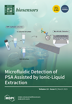
Prostate cancer (PCa) is one of the most prevalent cancer types that affects males worldwide and is among the highest contributors to cancer mortality rates. Therefore, there is an urgent need to find reliable and accurate strategies to facilitate the early diagnosis of PCa. In this paper, a microbead-based microfluidic device is used to detect PSA after its extraction and purification from spiked serum samples using ionic liquid- and polymer-based aqueous biphasic systems (ABS). Different ionic liquids (ILs) and polymers were tested, with the best results being obtained after the application of IL-based ABS-mediated extraction. These results demonstrated that it is possible to detect PSA in non-physiological environments and reinforce the potential of IL systems in microfluidics for future point-of-care (PoC) measurements. View this paper
  • Issues are regarded as officially published after their release is announced to the table of contents alert mailing list.
  • You may sign up for e-mail alerts to receive table of contents of newly released issues.
  • PDF is the official format for papers published in both, html and pdf forms. To view the papers in pdf format, click on the "PDF Full-text" link, and use the free Adobe Reader to open them.
Order results
Result details
Section
Select all
Export citation of selected articles as:
15 pages, 2754 KiB  
Perspective
A Short Review on Miniaturized Biosensors for the Detection of Nucleic Acid Biomarkers
by Madhusudan B. Kulkarni, Narasimha H. Ayachit and Tejraj M. Aminabhavi
Biosensors 2023, 13(3), 412; https://doi.org/10.3390/bios13030412 - 22 Mar 2023
Cited by 7 | Viewed by 2395
Abstract
Even today, most biomarker testing is executed in centralized, dedicated laboratories using bulky instruments, automated analyzers, and increased analysis time and expenses. The development of miniaturized, faster, low-cost microdevices is immensely anticipated for substituting for these conventional laboratory-oriented assays and transferring diagnostic results [...] Read more.
Even today, most biomarker testing is executed in centralized, dedicated laboratories using bulky instruments, automated analyzers, and increased analysis time and expenses. The development of miniaturized, faster, low-cost microdevices is immensely anticipated for substituting for these conventional laboratory-oriented assays and transferring diagnostic results directly onto the patient’s smartphone using a cloud server. Pioneering biosensor-based approaches might make it possible to test biomarkers with reliability in a decentralized setting, but there are still a number of issues and restrictions that must be resolved before the development and use of several biosensors for the proper understanding of the measured biomarkers of numerous bioanalytes such as DNA, RNA, urine, and blood. One of the most promising processes to address some of the issues relating to the growing demand for susceptible, quick, and affordable analysis techniques in medical diagnostics is the creation of biosensors. This article critically discusses a short review of biosensors used for detecting nucleic acid biomarkers, and their use in biomedical prognostics will be addressed while considering several essential characteristics. Full article
(This article belongs to the Special Issue Biosensors for Nucleic Acid Biomarkers)
Show Figures

Figure 1

16 pages, 3256 KiB  
Review
Gold Nanoparticle-Based Plasmonic Biosensors
by Enrico Ferrari
Biosensors 2023, 13(3), 411; https://doi.org/10.3390/bios13030411 - 22 Mar 2023
Cited by 24 | Viewed by 3986
Abstract
One of the emerging technologies in molecular diagnostics of the last two decades is the use of gold nanoparticles (AuNPs) for biosensors. AuNPs can be functionalized with various biomolecules, such as nucleic acids or antibodies, to recognize and bind to specific targets. AuNPs [...] Read more.
One of the emerging technologies in molecular diagnostics of the last two decades is the use of gold nanoparticles (AuNPs) for biosensors. AuNPs can be functionalized with various biomolecules, such as nucleic acids or antibodies, to recognize and bind to specific targets. AuNPs present unique optical properties, such as their distinctive plasmonic band, which confers a bright-red color to AuNP solutions, and their extremely high extinction coefficient, which makes AuNPs detectable by the naked eye even at low concentrations. Ingenious molecular mechanisms triggered by the presence of a target analyte can change the colloidal status of AuNPs from dispersed to aggregated, with a subsequent visible change in color of the solution due to the loss of the characteristic plasmonic band. This review describes how the optical properties of AuNPs have been exploited for the design of plasmonic biosensors that only require the simple mixing of reagents combined with a visual readout and focuses on the molecular mechanisms involved. This review illustrates selected examples of AuNP-based plasmonic biosensors and promising approaches for the point-of-care testing of various analytes, spanning from the viral RNA of SARS-CoV-2 to the molecules that give distinctive flavor and color to aged whisky. Full article
(This article belongs to the Special Issue Advanced Optical Sensing Techniques for Applications in Biomedicine)
Show Figures

Figure 1

17 pages, 5770 KiB  
Review
Multiplex Detection of Infectious Diseases on Microfluidic Platforms
by Fumin Chen, Qinqin Hu, Huimin Li, Yi Xie, Leshan Xiu, Yuqian Zhang, Xiaokui Guo and Kun Yin
Biosensors 2023, 13(3), 410; https://doi.org/10.3390/bios13030410 - 21 Mar 2023
Cited by 6 | Viewed by 3579
Abstract
Infectious diseases contribute significantly to the global disease burden. Sensitive and accurate screening methods are some of the most effective means of identifying sources of infection and controlling infectivity. Conventional detecting strategies such as quantitative polymerase chain reaction (qPCR), DNA sequencing, and mass [...] Read more.
Infectious diseases contribute significantly to the global disease burden. Sensitive and accurate screening methods are some of the most effective means of identifying sources of infection and controlling infectivity. Conventional detecting strategies such as quantitative polymerase chain reaction (qPCR), DNA sequencing, and mass spectrometry typically require bulky equipment and well-trained personnel. Therefore, mass screening of a large population using conventional strategies during pandemic periods often requires additional manpower, resources, and time, which cannot be guaranteed in resource-limited settings. Recently, emerging microfluidic technologies have shown the potential to replace conventional methods in performing point-of-care detection because they are automated, miniaturized, and integrated. By exploiting the spatial separation of detection sites, microfluidic platforms can enable the multiplex detection of infectious diseases to reduce the possibility of misdiagnosis and incomplete diagnosis of infectious diseases with similar symptoms. This review presents the recent advances in microfluidic platforms used for multiplex detection of infectious diseases, including microfluidic immunosensors and microfluidic nucleic acid sensors. As representative microfluidic platforms, lateral flow immunoassay (LFIA) platforms, polymer-based chips, paper-based devices, and droplet-based devices will be discussed in detail. In addition, the current challenges, commercialization, and prospects are proposed to promote the application of microfluidic platforms in infectious disease detection. Full article
(This article belongs to the Special Issue Immunoassays and Biosensing)
Show Figures

Figure 1

12 pages, 6972 KiB  
Article
Highly Stretchable and Robust Electrochemical Sensor Based on 3D Graphene Oxide–CNT Composite for Detecting Ammonium in Sweat
by Yunzhi Hua, Mingxiang Guan, Linzhong Xia, Yu Chen, Junhao Mai, Cong Zhao and Changrui Liao
Biosensors 2023, 13(3), 409; https://doi.org/10.3390/bios13030409 - 21 Mar 2023
Cited by 5 | Viewed by 2078
Abstract
Wearable electrochemical sensors have attracted tremendous attention and have been experiencing rapid growth in recent years. Sweat, one of the most suitable biological fluids for non-invasive monitoring, contains various chemical elements relating abundant information about human health conditions. In this work, a new [...] Read more.
Wearable electrochemical sensors have attracted tremendous attention and have been experiencing rapid growth in recent years. Sweat, one of the most suitable biological fluids for non-invasive monitoring, contains various chemical elements relating abundant information about human health conditions. In this work, a new type of non-invasive and highly stretchable potentiometric sweat sensor was developed based on all-solid-state ion-selective electrode (ISE) coupled with poly(dimethylsiloxane; PDMS) and polyurethane (PU). This highly stretchable composite of PDMS-PU allows the sensor to be robust, with the PDMS providing a flexible backbone and the PU enhancing the adhesion between the electrodes and the substrate. In addition, graphene–carbon nanotube (CNT) network 3D nanomaterials were introduced to modify the ion selective membrane (ISM) in order to increase the charge transfer activity of the ISEs, which also could minimize the formation of water layers on the electrode surface, as such nanomaterials are highly hydrophobic. As a result, the sensor demonstrated a wide detection range of NH4+ from 10−6 M to 10−1 M with high stability and sensitivity—showing a high sensitivity of 59.6 ± 1.5 mV/log [NH4+] and an LOD lower than 10−6 M. Under a strain of 40%, the sensor still showed a sensitivity of 42.7 ± 3.1 mV/log [NH4+]. The proposed highly stretchable and robust electrochemical sweat sensor provides a new choice for wearable-device-based personal daily healthcare management beyond hospital-centric healthcare monitoring. Full article
(This article belongs to the Special Issue Advance in Wearable Biosensors for Healthcare Monitoring)
Show Figures

Figure 1

17 pages, 5038 KiB  
Article
Fluorescent Nanocomposite Hydrogels Based on Conjugated Polymer Nanoparticles as Platforms for Alkaline Phosphatase Detection
by Yolanda Alacid, Rocío Esquembre, Francisco Montilla, María José Martínez-Tomé and C. Reyes Mateo
Biosensors 2023, 13(3), 408; https://doi.org/10.3390/bios13030408 - 21 Mar 2023
Cited by 1 | Viewed by 1917
Abstract
This work describes the development and characterization of fluorescent nanocomposite hydrogels, with high swelling and absorption capacity, and prepared using a green protocol. These fluorescent materials are obtained by incorporating, for the first time, polyfluorenes-based nanoparticles with different emission bands—poly[9,9-dioctylfluorenyl-2,7-diyl] (PFO) and poly[(9,9-di-n-octylfluorenyl-2,7-diyl)-alt-(1,4-benzo-{2,1,3}-thiadiazole)] [...] Read more.
This work describes the development and characterization of fluorescent nanocomposite hydrogels, with high swelling and absorption capacity, and prepared using a green protocol. These fluorescent materials are obtained by incorporating, for the first time, polyfluorenes-based nanoparticles with different emission bands—poly[9,9-dioctylfluorenyl-2,7-diyl] (PFO) and poly[(9,9-di-n-octylfluorenyl-2,7-diyl)-alt-(1,4-benzo-{2,1,3}-thiadiazole)] (F8BT)—into a three-dimensional polymeric network based on polyacrylamide. To this end, two strategies were explored: incorporation of the nanoparticles during the polymerization process (in situ) and embedment after the hydrogel formation (ex situ). The results show that the combination of PFO nanoparticles introduced by the ex situ method provided materials with good storage stability, homogeneity and reproducibility properties, allowing their preservation in the form of xerogel. The fluorescent nanocomposite hydrogels have been tested as a transportable and user-friendly sensing platform. In particular, the ability of these materials to specifically detect the enzyme alkaline phosphatase (ALP) has been evaluated as a proof-of-concept. The sensor was able to quantify the presence of the enzyme in an aqueous sample with a response time of 10 min and LOD of 21 nM. Given these results, we consider that this device shows great potential for quantifying physiological ALP levels as well as enzyme activity in environmental samples. Full article
Show Figures

Figure 1

4 pages, 177 KiB  
Editorial
Biosensors in 2022
by Giovanna Marrazza
Biosensors 2023, 13(3), 407; https://doi.org/10.3390/bios13030407 - 21 Mar 2023
Viewed by 1555
Abstract
Sixty years have passed since Clark and Lyons proposed the concept of using glucose enzyme electrodes to monitor the oxygen that is consumed during an enzyme-catalyzed reaction [...] Full article
(This article belongs to the Special Issue Biosensors in 2022)
16 pages, 5333 KiB  
Article
Clinically Deployable Bioelectronic Sensing Platform for Ultrasensitive Detection of Transferrin in Serum Sample
by Harleen Kaur, Prasanthi Chittineedi, Ravi Shankar Bellala, Venkata Madhavi Bellala, Sandeep Singh, Rohini Kumari, Pranjal Chandra, Santhi Latha Pandrangi and Surinder P. Singh
Biosensors 2023, 13(3), 406; https://doi.org/10.3390/bios13030406 - 20 Mar 2023
Cited by 2 | Viewed by 2037
Abstract
Varying levels of transferrin (Tf) have been associated with different disease conditions and are known to play a crucial role in various malignancies. Regular monitoring of the variations in Tf levels can be useful for managing related diseases, especially for the prognosis of [...] Read more.
Varying levels of transferrin (Tf) have been associated with different disease conditions and are known to play a crucial role in various malignancies. Regular monitoring of the variations in Tf levels can be useful for managing related diseases, especially for the prognosis of certain cancers. We fabricated an immunosensor based on graphene oxide (GO) nanosheets to indirectly detect Tf levels in cancer patients. The GO nanosheets were deposited onto an indium tin oxide (ITO)-coated glass substrate and annealed at 120 °C to obtain reduced GO (rGO) films, followed by the immobilization of an antibody, anti-Tf. The materials and sensor probe used were systematically characterized by UV–Visible spectroscopy (UV–Vis), X-ray diffraction (XRD), atomic force microscopy (AFM), and Fourier transform infrared spectroscopy (FTIR). Cyclic voltammetry (CV), electrochemical impedance spectroscopy (EIS), and differential pulse voltammetry (DPV) were also used for the stepwise sensor probe characterizations and Tf detection in serum samples, respectively. The anti-Tf/rGO/ITO immunosensor DPV output demonstrated an excellent Tf detection capability in the linear range of 0.1 mg mL−1 to 12 mg mL−1 compared to the enzyme-linked immunosorbent assay (ELISA) detection range, with a limit of detection (LOD) of 0.010 ± 0.007 mg mL−1. Furthermore, the results of the fabricated immunosensor were compared with those of the ELISA and autobioanalyzer techniques, showing an outstanding match with < 5% error and demonstrating the immunosensor’s clinical potential. Full article
Show Figures

Figure 1

29 pages, 12462 KiB  
Review
Recent Advancements of LSPR Fiber-Optic Biosensing: Combination Methods, Structure, and Prospects
by Hongxin Zhang, Xue Zhou, Xuegang Li, Pengqi Gong, Yanan Zhang and Yong Zhao
Biosensors 2023, 13(3), 405; https://doi.org/10.3390/bios13030405 - 20 Mar 2023
Cited by 11 | Viewed by 3463
Abstract
Fiber-optic biosensors based on localized surface plasmon resonance (LSPR) have the advantages of great biocompatibility, label-free, strong stability, and real-time monitoring of various analytes. LSPR fiber-optic biosensors have attracted extensive research attention in the fields of environmental science, clinical medicine, disease diagnosis, and [...] Read more.
Fiber-optic biosensors based on localized surface plasmon resonance (LSPR) have the advantages of great biocompatibility, label-free, strong stability, and real-time monitoring of various analytes. LSPR fiber-optic biosensors have attracted extensive research attention in the fields of environmental science, clinical medicine, disease diagnosis, and food safety. The latest development of LSPR fiber-optic biosensors in recent years has focused on the detection of clinical disease markers and the detection of various toxic substances in the environment and the progress of new sensitization mechanisms in LSPR fiber-optic sensors. Therefore, this paper reviews the LSPR fiber-optic sensors from the aspects of working principle, structure, and application fields in biosensors. According to the structure, the sensor can be divided into three categories: traditional ordinary optical fiber, special shape optical fiber, and specialty optical fiber. The advantages and disadvantages of existing and future LSPR fiber-optic biosensors are discussed in detail. Additionally, the prospect of future development of fiber-optic biosensors based on LSPR is addressed. Full article
(This article belongs to the Special Issue Recent Advances in Optical Biosensors)
Show Figures

Figure 1

15 pages, 1367 KiB  
Review
Label-Free Long-Term Methods for Live Cell Imaging of Neurons: New Opportunities
by Zrinko Baričević, Zahra Ayar, Samuel M. Leitao, Miranda Mladinic, Georg E. Fantner and Jelena Ban
Biosensors 2023, 13(3), 404; https://doi.org/10.3390/bios13030404 - 20 Mar 2023
Cited by 1 | Viewed by 1987
Abstract
Time-lapse light microscopy combined with in vitro neuronal cultures has provided a significant contribution to the field of Developmental Neuroscience. The establishment of the neuronal polarity, i.e., formation of axons and dendrites, key structures responsible for inter-neuronal signaling, was described in 1988 by [...] Read more.
Time-lapse light microscopy combined with in vitro neuronal cultures has provided a significant contribution to the field of Developmental Neuroscience. The establishment of the neuronal polarity, i.e., formation of axons and dendrites, key structures responsible for inter-neuronal signaling, was described in 1988 by Dotti, Sullivan and Banker in a milestone paper that continues to be cited 30 years later. In the following decades, numerous fluorescently labeled tags and dyes were developed for live cell imaging, providing tremendous advancements in terms of resolution, acquisition speed and the ability to track specific cell structures. However, long-term recordings with fluorescence-based approaches remain challenging because of light-induced phototoxicity and/or interference of tags with cell physiology (e.g., perturbed cytoskeletal dynamics) resulting in compromised cell viability leading to cell death. Therefore, a label-free approach remains the most desirable method in long-term imaging of living neurons. In this paper we will focus on label-free high-resolution methods that can be successfully used over a prolonged period. We propose novel tools such as scanning ion conductance microscopy (SICM) or digital holography microscopy (DHM) that could provide new insights into live cell dynamics during neuronal development and regeneration after injury. Full article
(This article belongs to the Special Issue Advanced Optical Sensing Techniques for Applications in Biomedicine)
Show Figures

Graphical abstract

11 pages, 3118 KiB  
Article
Photoacoustic Spectral Analysis for Evaluating the Aggressiveness of Prostate Cancer Labeled by Methylene Blue Polyacrylamide Nanoparticles
by Janggun Jo, Eamon Salfi, Jeff Folz, Aaron M. Udager, Evan Keller, Raoul Kopelman, Sri-Rajasekhar Kothapalli, Guan Xu and Xueding Wang
Biosensors 2023, 13(3), 403; https://doi.org/10.3390/bios13030403 - 20 Mar 2023
Cited by 2 | Viewed by 1590
Abstract
Evaluating the aggressiveness of prostate cancer (PCa) is crucial for PCa diagnosis and prognosis. Previously, studies have shown that photoacoustic spectral analysis (PASA) can assess prostate tissue microarchitecture for evaluating the aggressiveness of PCa. In this study, in a transgenic mouse (TRAMP) model [...] Read more.
Evaluating the aggressiveness of prostate cancer (PCa) is crucial for PCa diagnosis and prognosis. Previously, studies have shown that photoacoustic spectral analysis (PASA) can assess prostate tissue microarchitecture for evaluating the aggressiveness of PCa. In this study, in a transgenic mouse (TRAMP) model of PCa, we utilized methylene blue polyacrylamide nanoparticles (MB PAA NPs) to label the cancer cells in prostate in vivo. MB PAA NPs can specifically target proliferating cancer cells as a contrast agent, allowing photoacoustic (PA) imaging to better detect PCa tumors, and also assessing prostate glandular architecture. With the PA signals from the prostates measured simultaneously by a needle hydrophone and a PA and ultrasound (US) dual-imaging system, we conducted PASA and correlated the quantified spectral parameter slopes with the cancer grading from histopathology. The PASA results from 18 mice showed significant differences between normal and cancer, and also between low-score cancer and high-score cancer. This study in the clinically relevant TRAMP model of PCa demonstrated that PA imaging and PASA, powered by MB PAA NPs that can label the PCa microarchitectures in vivo after systemic administration, can detect PCa and, more importantly, evaluate cancer aggressiveness. Full article
(This article belongs to the Section Optical and Photonic Biosensors)
Show Figures

Figure 1

14 pages, 1393 KiB  
Review
Immunosensors for Assay of Toxic Biological Warfare Agents
by Miroslav Pohanka
Biosensors 2023, 13(3), 402; https://doi.org/10.3390/bios13030402 - 20 Mar 2023
Cited by 2 | Viewed by 2757
Abstract
An immunosensor for the assay of toxic biological warfare agents is a biosensor suitable for detecting hazardous substances such as aflatoxin, botulinum toxin, ricin, Shiga toxin, and others. The application of immunosensors is used in outdoor assays, point-of-care tests, as a spare method [...] Read more.
An immunosensor for the assay of toxic biological warfare agents is a biosensor suitable for detecting hazardous substances such as aflatoxin, botulinum toxin, ricin, Shiga toxin, and others. The application of immunosensors is used in outdoor assays, point-of-care tests, as a spare method for more expensive devices, and even in the laboratory as a standard analytical method. Some immunosensors, such as automated flow-through analyzers or lateral flow tests, have been successfully commercialized as tools for toxins assay, but the research is ongoing. New devices are being developed, and the use of advanced materials and assay techniques make immunosensors highly competitive analytical devices in the field of toxic biological warfare agents assay. This review summarizes facts about current applications and new trends of immunosensors regarding recent papers in this area. Full article
(This article belongs to the Special Issue Immunosensors and Immunoassays for the Control of Hazardous Compounds)
Show Figures

Figure 1

15 pages, 5282 KiB  
Article
Dual-Mode Biosensor for Simultaneous and Rapid Detection of Live and Whole Salmonella typhimurium Based on Bioluminescence and Fluorescence Detection
by Zhenli Xu, Bailu Liu, Dengfeng Li, Zhenzhong Yu and Ning Gan
Biosensors 2023, 13(3), 401; https://doi.org/10.3390/bios13030401 - 19 Mar 2023
Cited by 2 | Viewed by 1912
Abstract
Both live and dead Salmonella typhimurium (S.T) are harmful to human health, but there are differences in pathological mechanism, dosage, and security. It is crucial to develop a rapid and simultaneous assay to distinguish and quantify live and dead S.T in foods. Herein, [...] Read more.
Both live and dead Salmonella typhimurium (S.T) are harmful to human health, but there are differences in pathological mechanism, dosage, and security. It is crucial to develop a rapid and simultaneous assay to distinguish and quantify live and dead S.T in foods. Herein, one dual-mode biosensor for simultaneous detection of live and dead S.T was fabricated based on two phage probes, using portable bioluminescence and fluorescent meter as detectors, respectively. Firstly, a magnetic phage capture probe (M-P1) and a phage signal tag (P2-S) labeled with SYTO 13 fluorescent dye were prepared, respectively. Both M-P1 and P2-S can specifically conjugate with S.T to form a magnetic sandwich complex. After magnetic separation, the isolated complex can emit a fluorescent signal under an excited 365 nm laser, which can reflect the total amount of S.T. Afterwards, the lysozyme was added to decompose the captured live S.T, which can release ATP and produce a bioluminescent signal corresponding to the live S.T amount. The dead S.T concentration can be deduced by the difference between total and live examples. The detection limit of 55 CFU/mL for total S.T and 9 CFU/mL for live ones was within 20 min. The assay was successfully employed in milk samples and prospectively for on-site screening of other dead and live bacteria, while changing the phages for the targets. Full article
Show Figures

Figure 1

16 pages, 2491 KiB  
Review
Design of Polymeric Surfaces as Platforms for Streamlined Cancer Diagnostics in Liquid Biopsies
by Faezeh Ghorbanizamani, Hichem Moulahoum, Emine Guler Celik, Figen Zihnioglu, Tutku Beduk, Tuncay Goksel, Kutsal Turhan and Suna Timur
Biosensors 2023, 13(3), 400; https://doi.org/10.3390/bios13030400 - 18 Mar 2023
Cited by 1 | Viewed by 1765
Abstract
Minimally invasive approaches for cancer diagnosis are an integral step in the quest to improve cancer survival. Liquid biopsies such as blood samples are matrices explored to extract valuable information about the tumor and its state through various indicators, such as proteins, peptides, [...] Read more.
Minimally invasive approaches for cancer diagnosis are an integral step in the quest to improve cancer survival. Liquid biopsies such as blood samples are matrices explored to extract valuable information about the tumor and its state through various indicators, such as proteins, peptides, tumor DNA, or circulating tumor cells. Although these markers are scarce, making their isolation and detection in complex matrices challenging, the development in polymer chemistry producing interesting structures, including molecularly imprinted polymers, branched polymers, nanopolymer composites, and hybrids, allowed the development of enhanced platforms with impressive performance for liquid biopsies analysis. This review describes the latest advances and developments in polymer synthesis and their application for minimally invasive cancer diagnosis. The polymer structures improve the operational performances of biosensors through various processes, such as increased affinity for enhanced sensitivity, improved binding, and avoidance of non-specific interactions for enhanced specificity. Furthermore, polymer-based materials can be a tremendous help in signal amplification of usually low-concentrated targets in the sample. The pros and cons of these materials, how the synthesis process affects their performance, and the device applications for liquid biopsies diagnosis will be critically reviewed to show the essentiality of this technology in oncology and clinical biomedicine. Full article
(This article belongs to the Special Issue Emerging Biosensing Technologies for Healthcare Applications)
Show Figures

Figure 1

10 pages, 4044 KiB  
Communication
Functionalization of a Fully Integrated Electrophotonic Silicon Circuit for Biotin Sensing
by Oscar Pérez-Diaz, Denise Estrada-Wiese, Mariano Aceves-Mijares and Alfredo A. González-Fernández
Biosensors 2023, 13(3), 399; https://doi.org/10.3390/bios13030399 - 18 Mar 2023
Viewed by 1419
Abstract
Electrophotonic (EPh) circuits are novel systems where photons and electrons can be controlled simultaneously in the same integrated circuit, attaining the development of innovative sensors for different applications. In this work, we present a complementary metal-oxide-semiconductor (CMOS)-compatible EPh circuit for biotin sensing, in [...] Read more.
Electrophotonic (EPh) circuits are novel systems where photons and electrons can be controlled simultaneously in the same integrated circuit, attaining the development of innovative sensors for different applications. In this work, we present a complementary metal-oxide-semiconductor (CMOS)-compatible EPh circuit for biotin sensing, in which a silicon-based light source is monolithically integrated. The device is composed of an integrated light source, a waveguide, and a p–n photodiode, which are all fabricated in the same chip. The functionalization of the waveguide’s surface was investigated to biotinylate the EPh system for potential biosensing applications. The modified surfaces were characterized by AFM, optical microscopy, and Raman spectroscopy, as well as by photoluminescence measurements. The changes on the waveguide’s surface due to functionalization and biotinylation translated into different photocurrent intensities detected in the photodiode, demonstrating the potential uses of the EPh circuit as a biosensor. Full article
(This article belongs to the Special Issue Recent Advances in Optical Biosensors)
Show Figures

Figure 1

28 pages, 2999 KiB  
Review
Recent Trends in Biosensing and Diagnostic Methods for Novel Cancer Biomarkers
by Jagadeeswara Rao Bommi, Shekher Kummari, Kavitha Lakavath, Reshmi A. Sukumaran, Lakshmi R. Panicker, Jean Louis Marty and Kotagiri Yugender Goud
Biosensors 2023, 13(3), 398; https://doi.org/10.3390/bios13030398 - 18 Mar 2023
Cited by 18 | Viewed by 4132
Abstract
Cancer is one of the major public health issues in the world. It has become the second leading cause of death, with approximately 75% of cancer deaths transpiring in low- or middle-income countries. It causes a heavy global economic cost estimated at more [...] Read more.
Cancer is one of the major public health issues in the world. It has become the second leading cause of death, with approximately 75% of cancer deaths transpiring in low- or middle-income countries. It causes a heavy global economic cost estimated at more than a trillion dollars per year. The most common cancers are breast, colon, rectum, prostate, and lung cancers. Many of these cancers can be treated effectively and cured if detected at the primary stage. Nowadays, around 50% of cancers are detected at late stages, leading to serious health complications and death. Early diagnosis of cancer diseases substantially increases the efficient treatment and high chances of survival. Biosensors are one of the potential screening methodologies useful in the early screening of cancer biomarkers. This review summarizes the recent findings about novel cancer biomarkers and their advantages over traditional biomarkers, and novel biosensing and diagnostic methods for them; thus, this review may be helpful in the early recognition and monitoring of treatment response of various human cancers. Full article
(This article belongs to the Special Issue Biosensing and Diagnosis)
Show Figures

Figure 1

20 pages, 7023 KiB  
Article
Mental Stress Detection Using a Wearable In-Ear Plethysmography
by Hika Barki and Wan-Young Chung
Biosensors 2023, 13(3), 397; https://doi.org/10.3390/bios13030397 - 17 Mar 2023
Cited by 4 | Viewed by 2675
Abstract
This study presents an ear-mounted photoplethysmography (PPG) system that is designed to detect mental stress. Mental stress is a prevalent condition that can negatively impact an individual’s health and well-being. Early detection and treatment of mental stress are crucial for preventing related illnesses [...] Read more.
This study presents an ear-mounted photoplethysmography (PPG) system that is designed to detect mental stress. Mental stress is a prevalent condition that can negatively impact an individual’s health and well-being. Early detection and treatment of mental stress are crucial for preventing related illnesses and maintaining overall wellness. The study used data from 14 participants that were collected in a controlled environment. The participants were subjected to stress-inducing tasks such as the Stroop color–word test and mathematical calculations. The raw PPG signal was then preprocessed and transformed into scalograms using continuous wavelet transform (CWT). A convolutional neural network classifier was then used to classify the transformed signals as stressed or non-stressed. The results of the study show that the PPG system achieved high levels of accuracy (92.04%) and F1-score (90.8%). Furthermore, by adding white Gaussian noise to the raw PPG signals, the results were improved even more, with an accuracy of 96.02% and an F1-score of 95.24%. The proposed ear-mounted device shows great promise as a reliable tool for the early detection and treatment of mental stress, potentially revolutionizing the field of mental health and well-being. Full article
(This article belongs to the Special Issue Advance in Wearable Biosensors for Healthcare Monitoring)
Show Figures

Figure 1

33 pages, 7933 KiB  
Review
Surface Plasmon Resonance (SPR) Sensor for Cancer Biomarker Detection
by Sreyashi Das, Ram Devireddy and Manas Ranjan Gartia
Biosensors 2023, 13(3), 396; https://doi.org/10.3390/bios13030396 - 17 Mar 2023
Cited by 23 | Viewed by 6433
Abstract
A biomarker is a physiological observable marker that acts as a stand-in and, in the best-case scenario, forecasts a clinically significant outcome. Diagnostic biomarkers are more convenient and cost-effective than directly measuring the ultimate clinical outcome. Cancer is among the most prominent global [...] Read more.
A biomarker is a physiological observable marker that acts as a stand-in and, in the best-case scenario, forecasts a clinically significant outcome. Diagnostic biomarkers are more convenient and cost-effective than directly measuring the ultimate clinical outcome. Cancer is among the most prominent global health problems and a major cause of morbidity and death globally. Therefore, cancer biomarker assays that are trustworthy, consistent, precise, and verified are desperately needed. Biomarker-based tumor detection holds a lot of promise for improving disease knowledge at the molecular scale and early detection and surveillance. In contrast to conventional approaches, surface plasmon resonance (SPR) allows for the quick and less invasive screening of a variety of circulating indicators, such as circulating tumor DNA (ctDNA), microRNA (miRNA), circulating tumor cells (CTCs), lipids, and proteins. With several advantages, the SPR technique is a particularly beneficial choice for the point-of-care identification of biomarkers. As a result, it enables the timely detection of tumor markers, which could be used to track cancer development and suppress the relapse of malignant tumors. This review emphasizes advancements in SPR biosensing technologies for cancer detection. Full article
Show Figures

Figure 1

41 pages, 8056 KiB  
Review
Recent Progress in Long-Term Sleep Monitoring Technology
by Jiaju Yin, Jiandong Xu and Tian-Ling Ren
Biosensors 2023, 13(3), 395; https://doi.org/10.3390/bios13030395 - 17 Mar 2023
Cited by 3 | Viewed by 4635
Abstract
Sleep is an essential physiological activity, accounting for about one-third of our lives, which significantly impacts our memory, mood, health, and children’s growth. Especially after the COVID-19 epidemic, sleep health issues have attracted more attention. In recent years, with the development of wearable [...] Read more.
Sleep is an essential physiological activity, accounting for about one-third of our lives, which significantly impacts our memory, mood, health, and children’s growth. Especially after the COVID-19 epidemic, sleep health issues have attracted more attention. In recent years, with the development of wearable electronic devices, there have been more and more studies, products, or solutions related to sleep monitoring. Many mature technologies, such as polysomnography, have been applied to clinical practice. However, it is urgent to develop wearable or non-contacting electronic devices suitable for household continuous sleep monitoring. This paper first introduces the basic knowledge of sleep and the significance of sleep monitoring. Then, according to the types of physiological signals monitored, this paper describes the research progress of bioelectrical signals, biomechanical signals, and biochemical signals used for sleep monitoring. However, it is not ideal to monitor the sleep quality for the whole night based on only one signal. Therefore, this paper reviews the research on multi-signal monitoring and introduces systematic sleep monitoring schemes. Finally, a conclusion and discussion of sleep monitoring are presented to propose potential future directions and prospects for sleep monitoring. Full article
(This article belongs to the Special Issue Biophysical Sensors for Biomedical/Health Monitoring Applications)
Show Figures

Figure 1

16 pages, 5174 KiB  
Article
Bioimpedance Analysis as Early Predictor for Clot Formation Inside a Blood-Perfused Test Chamber: Proof of Concept Using an In Vitro Test-Circuit
by Muhammet Türkmen, Tobias Lauwigi, Tamara Fechter, Fabienne Gries, Anna Fischbach, Thomas Gries, Rolf Rossaint, Christian Bleilevens and Patrick Winnersbach
Biosensors 2023, 13(3), 394; https://doi.org/10.3390/bios13030394 - 17 Mar 2023
Cited by 1 | Viewed by 3679
Abstract
Clot formation inside a membrane oxygenator (MO) due to blood-to-foreign surface interaction represents a frequent complication during extracorporeal membrane oxygenation. Since current standard monitoring methods of coagulation status inside the MO fail to detect clot formation at an early stage, reliable sensors for [...] Read more.
Clot formation inside a membrane oxygenator (MO) due to blood-to-foreign surface interaction represents a frequent complication during extracorporeal membrane oxygenation. Since current standard monitoring methods of coagulation status inside the MO fail to detect clot formation at an early stage, reliable sensors for early clot detection are in demand to reduce associated complications and adverse events. Bioimpedance analysis offers a monitoring concept by integrating sensor fibers into the MO. Herein, the feasibility of clot detection via bioimpedance analysis is evaluated. A custom-made test chamber with integrated titanium fibers acting as sensors was perfused with heparinized human whole blood in an in vitro test circuit until clot formation occurred. The clot detection capability of bioimpedance analysis was directly compared to the pressure difference across the test chamber (ΔP-TC), analogous to the measurement across MOs (ΔP-MO), the clinical gold standard for clot detection. We found that bioimpedance measurement increased significantly 8 min prior to a significant increase in ΔP-TC, indicating fulminant clot formation. Experiments without clot formation resulted in a lack of increase in bioimpedance or ΔP-TC. This study shows that clot detection via bioimpedance analysis under flow conditions in a blood-perfused test chamber is generally feasible, thus paving the way for further investigation. Full article
(This article belongs to the Special Issue Biophysical Sensors for Biomedical/Health Monitoring Applications)
Show Figures

Figure 1

23 pages, 5671 KiB  
Review
Recent Progress of Biomaterials-Based Epidermal Electronics for Healthcare Monitoring and Human–Machine Interaction
by Ningning Han, Xin Yao, Yifan Wang, Wenhao Huang, Mengjuan Niu, Pengcheng Zhu and Yanchao Mao
Biosensors 2023, 13(3), 393; https://doi.org/10.3390/bios13030393 - 17 Mar 2023
Cited by 8 | Viewed by 2617
Abstract
Epidermal electronics offer an important platform for various on-skin applications including electrophysiological signals monitoring and human–machine interactions (HMI), due to their unique advantages of intrinsic softness and conformal interfaces with skin. The widely used nondegradable synthetic materials may produce massive electronic waste to [...] Read more.
Epidermal electronics offer an important platform for various on-skin applications including electrophysiological signals monitoring and human–machine interactions (HMI), due to their unique advantages of intrinsic softness and conformal interfaces with skin. The widely used nondegradable synthetic materials may produce massive electronic waste to the ecosystem and bring safety issues to human skin. However, biomaterials extracted from nature are promising to act as a substitute material for the construction of epidermal electronics, owing to their diverse characteristics of biocompatibility, biodegradability, sustainability, low cost and natural abundance. Therefore, the development of natural biomaterials holds great prospects for advancement of high-performance sustainable epidermal electronics. Here, we review the recent development on different types of biomaterials including proteins and polysaccharides for multifunctional epidermal electronics. Subsequently, the applications of biomaterials-based epidermal electronics in electrophysiological monitoring and HMI are discussed, respectively. Finally, the development situation and future prospects of biomaterials-based epidermal electronics are summarized. We expect that this review can provide some inspirations for the development of future, sustainable, biomaterials-based epidermal electronics. Full article
(This article belongs to the Special Issue Epidermal Electronics and Implantable Devices)
Show Figures

Figure 1

9 pages, 7059 KiB  
Communication
HPTLC-Bioluminescent Bioautography Screening of Herbal Teas for Adulteration with Hypolipidemic Drugs
by Yuting Wang, Xingjun Xi, Liao Wang and Yisheng Chen
Biosensors 2023, 13(3), 392; https://doi.org/10.3390/bios13030392 - 17 Mar 2023
Cited by 1 | Viewed by 1643
Abstract
Teas based on nutraceutical herbs are an effective tool against hyperlipidemia. However, the adulteration with chemical drugs is frequently detected. By coupling bioluminescent bioautography with high performance thin-layer chromatography (HPTLC), we developed a facile method suitable for screening hypolipidemic drugs (ciprofibrate and bezafibrate) [...] Read more.
Teas based on nutraceutical herbs are an effective tool against hyperlipidemia. However, the adulteration with chemical drugs is frequently detected. By coupling bioluminescent bioautography with high performance thin-layer chromatography (HPTLC), we developed a facile method suitable for screening hypolipidemic drugs (ciprofibrate and bezafibrate) adulteration in five different herbal teas (lotus leaf, Apocynum, Ginkgo biloba, Gynostemia and chrysanthemum). First, the sensitivity of a bioluminescent bacteria to the analyte was evaluated on different HPTLC layer materials, revealing that the best performance was achieved on the silica gel layer. On this basis, sample extracts were separated on silica gel plates via a standardized HPTLC procedure, forming a selective detection window for the targeted compound. Then, the separation results were rapidly visualized by the bioluminescence inhibition of bacteria cells within 6 min after dipping. The observed inhibition displayed an acceptable limit of detection (<20 ng/zone or 2 mg/kg) and linearity (R2 ≥ 0.9279) within a wide concentration range (50–1000 ng/zone). Furthermore, the optimized method was performed with artificially adulterated samples and the recovery rates were determined to be within the range of 71% to 91%, bracing its practical reliability. Showing superiorly high simplicity, throughput and specificity, this work demonstrated that the analytical method jointly based on HPTLC and bioautography was an ideal tool for screening bioactive compounds in complex biological matrix. Full article
Show Figures

Figure 1

31 pages, 6807 KiB  
Review
Recent Advances in Recognition Receptors for Electrochemical Biosensing of Mycotoxins—A Review
by Manpreet Kaur, Jyoti Gaba, Komal Singh, Yashika Bhatia, Anoop Singh and Narinder Singh
Biosensors 2023, 13(3), 391; https://doi.org/10.3390/bios13030391 - 17 Mar 2023
Cited by 2 | Viewed by 2268
Abstract
Mycotoxins are naturally occurring toxic secondary metabolites produced by fungi in cereals and foodstuffs during the stages of cultivation and storage. Electrochemical biosensing has emerged as a rapid, efficient, and economical approach for the detection and quantification of mycotoxins in different sample media. [...] Read more.
Mycotoxins are naturally occurring toxic secondary metabolites produced by fungi in cereals and foodstuffs during the stages of cultivation and storage. Electrochemical biosensing has emerged as a rapid, efficient, and economical approach for the detection and quantification of mycotoxins in different sample media. An electrochemical biosensor consists of two main units, a recognition receptor and a signal transducer. Natural or artificial antibodies, aptamers, molecularly imprinted polymers (MIP), peptides, and DNAzymes have been extensively employed as selective recognition receptors for the electrochemical biosensing of mycotoxins. This article affords a detailed discussion of the recent advances and future prospects of various types of recognition receptors exploited in the electrochemical biosensing of mycotoxins. Full article
Show Figures

Figure 1

14 pages, 2366 KiB  
Article
Microfluidic Platform Integrated with Carbon Nanofibers-Decorated Gold Nanoporous Sensing Device for Serum PSA Quantification
by Emiliano Felici, Matías D. Regiart, Sirley V. Pereira, Francisco G. Ortega, Lúcio Angnes, Germán A. Messina and Martín A. Fernández-Baldo
Biosensors 2023, 13(3), 390; https://doi.org/10.3390/bios13030390 - 16 Mar 2023
Cited by 2 | Viewed by 3624
Abstract
Prostate cancer is a disease with a high incidence and mortality rate in men worldwide. Serum prostate-specific antigens (PSA) are the main circulating biomarker for this disease in clinical practices. In this work, we present a portable and reusable microfluidic device for PSA [...] Read more.
Prostate cancer is a disease with a high incidence and mortality rate in men worldwide. Serum prostate-specific antigens (PSA) are the main circulating biomarker for this disease in clinical practices. In this work, we present a portable and reusable microfluidic device for PSA quantification. This device comprises a polymethyl methacrylate microfluidic platform coupled with electrochemical detection. The platinum working microelectrode was positioned in the outflow region of the microchannel and was modified with carbon nanofibers (CNF)-decorated gold nanoporous (GNP) structures by the dynamic hydrogen bubble template method, through the simultaneous electrodeposition of metal precursors in the presence of CNF. CNF/GNP structures exhibit attractive properties, such as a large surface to volume ratio, which increases the antibody’s immobilization capacity and the electroactive area. CNFs/GNP structures were characterized by scanning electron microscopy, energy dispersive spectrometry, and cyclic voltammetry. Anti-PSA antibodies and HRP were employed for the immune-electrochemical reaction. The detection limit for the device was 5 pg mL−1, with a linear range from 0.01 to 50 ng mL−1. The coefficients of variation within and between assays were lower than 4.40%, and 6.15%, respectively. Additionally, its clinical performance was tested in serum from 30 prostate cancer patients. This novel device was a sensitive, selective, portable, and reusable tool for the serological diagnosis and monitoring of prostate cancer. Full article
(This article belongs to the Special Issue Nanomaterial Based Biosensors for Biomedical Applications)
Show Figures

Figure 1

25 pages, 6096 KiB  
Review
The Synergy between Deep Learning and Organs-on-Chips for High-Throughput Drug Screening: A Review
by Manna Dai, Gao Xiao, Ming Shao and Yu Shrike Zhang
Biosensors 2023, 13(3), 389; https://doi.org/10.3390/bios13030389 - 15 Mar 2023
Cited by 3 | Viewed by 3670
Abstract
Organs-on-chips (OoCs) are miniature microfluidic systems that have arguably become a class of advanced in vitro models. Deep learning, as an emerging topic in machine learning, has the ability to extract a hidden statistical relationship from the input data. Recently, these two areas [...] Read more.
Organs-on-chips (OoCs) are miniature microfluidic systems that have arguably become a class of advanced in vitro models. Deep learning, as an emerging topic in machine learning, has the ability to extract a hidden statistical relationship from the input data. Recently, these two areas have become integrated to achieve synergy for accelerating drug screening. This review provides a brief description of the basic concepts of deep learning used in OoCs and exemplifies the successful use cases for different types of OoCs. These microfluidic chips are of potential to be assembled as highly potent human-on-chips with complex physiological or pathological functions. Finally, we discuss the future supply with perspectives and potential challenges in terms of combining OoCs and deep learning for image processing and automation designs. Full article
(This article belongs to the Special Issue Lab on a Chip for High-Throughput Drug Screening)
Show Figures

Figure 1

13 pages, 3048 KiB  
Article
Developing a Fluorescent Inducible System for Free Fucose Quantification in Escherichia coli
by Samantha Nuñez, Maria Barra and Daniel Garrido
Biosensors 2023, 13(3), 388; https://doi.org/10.3390/bios13030388 - 15 Mar 2023
Cited by 1 | Viewed by 1951
Abstract
L-Fucose is a monosaccharide abundant in mammalian glycoconjugates. In humans, fucose can be found in human milk oligosaccharides (HMOs), mucins, and glycoproteins in the intestinal epithelium. The bacterial consumption of fucose and fucosylated HMOs is critical in the gut microbiome assembly of infants, [...] Read more.
L-Fucose is a monosaccharide abundant in mammalian glycoconjugates. In humans, fucose can be found in human milk oligosaccharides (HMOs), mucins, and glycoproteins in the intestinal epithelium. The bacterial consumption of fucose and fucosylated HMOs is critical in the gut microbiome assembly of infants, dominated by Bifidobacterium. Fucose metabolism is important for the production of short-chain fatty acids and is involved in cross-feeding microbial interactions. Methods for assessing fucose concentrations in complex media are lacking. Here we designed and developed a molecular quantification method of free fucose using fluorescent Escherichia coli. For this, low- and high-copy plasmids were evaluated with and without the transcription factor fucR and its respective fucose-inducible promoter controlling the reporter gene sfGFP. E. coli BL21 transformed with a high copy plasmid containing pFuc and fucR displayed a high resolution across increasing fucose concentrations and high fluorescence/OD values after 18 h. The molecular circuit was specific against other monosaccharides and showed a linear response in the 0–45 mM range. Adjusting data to the Hill equation suggested non-cooperative, simple regulation of FucR to its promoter. Finally, the biosensor was tested on different concentrations of free fucose and the supernatant of Bifidobacterium bifidum JCM 1254 supplemented with 2-fucosyl lactose, indicating the applicability of the method in detecting free fucose. In conclusion, a bacterial biosensor of fucose was validated with good sensitivity and precision. A biological method for quantifying fucose could be useful for nutraceutical and microbiological applications, as well as molecular diagnostics. Full article
(This article belongs to the Special Issue Nano/Micro Biosensors for Biomedical Applications)
Show Figures

Figure 1

33 pages, 5539 KiB  
Review
Biomarker Detection in Early Diagnosis of Cancer: Recent Achievements in Point-of-Care Devices Based on Paper Microfluidics
by Bilge Asci Erkocyigit, Ozge Ozufuklar, Aysenur Yardim, Emine Guler Celik and Suna Timur
Biosensors 2023, 13(3), 387; https://doi.org/10.3390/bios13030387 - 15 Mar 2023
Cited by 14 | Viewed by 5162
Abstract
Microfluidics is very crucial in lab-on-a-chip systems for carrying out operations in a large-scale laboratory environment on a single chip. Microfluidic systems are miniaturized devices in which the fluid behavior and control can be manipulated on a small platform, with surface forces on [...] Read more.
Microfluidics is very crucial in lab-on-a-chip systems for carrying out operations in a large-scale laboratory environment on a single chip. Microfluidic systems are miniaturized devices in which the fluid behavior and control can be manipulated on a small platform, with surface forces on the platform being greater than volumetric forces depending on the test method used. In recent years, paper-based microfluidic analytical devices (μPADs) have been developed to be used in point-of-care (POC) technologies. μPADs have numerous advantages, including ease of use, low cost, capillary action liquid transfer without the need for power, the ability to store reagents in active form in the fiber network, and the capability to perform multiple tests using various measurement techniques. These benefits are critical in the advancement of paper-based microfluidics in the fields of disease diagnosis, drug application, and environment and food safety. Cancer is one of the most critical diseases for early detection all around the world. Detecting cancer-specific biomarkers provides significant data for both early diagnosis and controlling the disease progression. μPADs for cancer biomarker detection hold great promise for improving cure rates, quality of life, and minimizing treatment costs. Although various types of bioanalytical platforms are available for the detection of cancer biomarkers, there are limited studies and critical reviews on paper-based microfluidic platforms in the literature. Hence, this article aims to draw attention to these gaps in the literature as well as the features that future platforms should have. Full article
(This article belongs to the Special Issue Recent Trends in Paper-Based Microfluidic Devices)
Show Figures

Figure 1

18 pages, 11325 KiB  
Article
An Amperometric Acetylcholine Biosensor Based on Co-Immobilization of Enzyme Nanoparticles onto Nanocomposite
by Jyoti Ahlawat, Minakshi Sharma and Chandra Shekhar Pundir
Biosensors 2023, 13(3), 386; https://doi.org/10.3390/bios13030386 - 15 Mar 2023
Cited by 4 | Viewed by 1847
Abstract
An electrochemical biosensor was fabricated using nanoparticles of acetylcholinesterase (AChE) and choline oxidase (ChO)/Pt nanoparticles (PtNPs)/porous graphene oxide nanosheet (GONS) composite. A pencil graphite electrode (PGE) was used for the electrodeposition of nanocomposite and the determination of acetylcholine (ACh), a neurotransmitter. Various techniques [...] Read more.
An electrochemical biosensor was fabricated using nanoparticles of acetylcholinesterase (AChE) and choline oxidase (ChO)/Pt nanoparticles (PtNPs)/porous graphene oxide nanosheet (GONS) composite. A pencil graphite electrode (PGE) was used for the electrodeposition of nanocomposite and the determination of acetylcholine (ACh), a neurotransmitter. Various techniques such as scanning electron microscopy (SEM), transmission electron microscopy (TEM), X-ray diffraction (XRD), Fourier-transform infrared (FTIR) spectra and cyclic voltammetry (CV) were used for characterization. This biosensor (AChENPs-ChONPs/GONS/PtNPs/PGE) indicated a very short response time (3 s), a lower limit of detection (0.001 µM), good linearity (0.001–200 µM), longer storage stability (6 months) and better reproducibility. The percent analytical recoveries of added acetylcholine in serum (5.0 and 10 µM) were found to be 97.6 ± 0.7 and 96.5 ± 0.3 for the present biosensor. The coefficients of variation were obtained to be 8% and 3.25%, correspondingly. The biosensor was applied to measure the ACh amount in the serum of healthy individuals and patients with Alzheimer’s disease. The number of interferents had no effect on the biosensor at their physiological concentrations. Full article
(This article belongs to the Special Issue Electrochemical Biosensors: From Design to Applications)
Show Figures

Figure 1

8 pages, 249 KiB  
Editorial
Plasmonic Sensors: A New Frontier in Nanotechnology
by Samir Kumar and Sungkyu Seo
Biosensors 2023, 13(3), 385; https://doi.org/10.3390/bios13030385 - 15 Mar 2023
Cited by 3 | Viewed by 2026
Abstract
Plasmonics is the study of surface plasmons formed by the interaction of incident light with electrons to form a surface-bound electromagnetic wave [...] Full article
(This article belongs to the Special Issue Plasmonic Sensors: A New Frontier in Nanotechnology)
18 pages, 2804 KiB  
Review
Nanomaterials-Based Electrochemical Δ9-THC and CBD Sensors for Chronic Pain
by Dadbeh Pazuki, Raja Ghosh and Matiar M. R. Howlader
Biosensors 2023, 13(3), 384; https://doi.org/10.3390/bios13030384 - 14 Mar 2023
Viewed by 2290
Abstract
Chronic pain is now included in the designation of chronic diseases, such as cancer, diabetes, and cardiovascular disease, which can impair quality of life and are major causes of death and disability worldwide. Pain can be treated using cannabinoids such as Δ9 [...] Read more.
Chronic pain is now included in the designation of chronic diseases, such as cancer, diabetes, and cardiovascular disease, which can impair quality of life and are major causes of death and disability worldwide. Pain can be treated using cannabinoids such as Δ9-tetrahydrocannabinol (Δ9-THC) and cannabidiol (CBD) due to their wide range of therapeutic benefits, particularly as sedatives, analgesics, neuroprotective agents, or anti-cancer medicines. While little is known about the pharmacokinetics of these compounds, there is increasing interest in the scientific understanding of the benefits and clinical applications of cannabinoids. In this review, we study the use of nanomaterial-based electrochemical sensing for detecting Δ9-THC and CBD. We investigate how nanomaterials can be functionalized to obtain highly sensitive and selective electrochemical sensors for detecting Δ9-THC and CBD. Additionally, we discuss the impacts of sensor pretreatment at fixed potentials and physiochemical parameters of the sensing medium, such as pH, on the electrochemical performance of Δ9-THC and CBD sensors. We believe this review will serve as a guideline for developing Δ9-THC and CBD electrochemical sensors for point-of-care applications. Full article
Show Figures

Graphical abstract

13 pages, 2250 KiB  
Article
Cell-Based Sensors for the Detection of EGF and EGF-Stimulated Ca2+ Signaling
by Euiyeon Lee, Keshab Lal Shrestha, Seonhye Kang, Neethu Ramakrishnan and Youngeun Kwon
Biosensors 2023, 13(3), 383; https://doi.org/10.3390/bios13030383 - 14 Mar 2023
Cited by 1 | Viewed by 3385
Abstract
Epidermal growth factor (EGF)-mediated activation of EGF receptors (EGFRs) has become an important target in drug development due to the implication of EGFR-mediated cellular signaling in cancer development. While various in vitro approaches are developed for monitoring EGF-EGFR interactions, they have several limitations. [...] Read more.
Epidermal growth factor (EGF)-mediated activation of EGF receptors (EGFRs) has become an important target in drug development due to the implication of EGFR-mediated cellular signaling in cancer development. While various in vitro approaches are developed for monitoring EGF-EGFR interactions, they have several limitations. Herein, we describe a live cell-based sensor system that can be used to monitor the interaction of EGF and EGFR as well as the subsequent signaling events. The design of the EGF-detecting sensor cells is based on the split-intein-mediated conditional protein trans-cleavage reaction (CPC). CPC is triggered by the presence of the target (EGF) to activate a signal peptide that translocates the fluorescent cargo to the target cellular location (mitochondria). The developed sensor cell demonstrated excellent sensitivity with a fast response time. It was also successfully used to detect an agonist and antagonist of EGFR (transforming growth factor-α and Cetuximab, respectively), demonstrating excellent specificity and capability of screening the analytes based on their function. The usage of sensor cells was then expanded from merely detecting the presence of target to monitoring the target-mediated signaling cascade, by exploiting previously developed Ca2+-detecting sensor cells. These sensor cells provide a useful platform for monitoring EGF-EGFR interaction, for screening EGFR effectors, and for studying downstream cellular signaling cascades. Full article
(This article belongs to the Section Biosensor and Bioelectronic Devices)
Show Figures

Figure 1

Previous Issue
Next Issue
Back to TopTop