Progress in Fluorescence Biosensing and Food Safety towards Point-of-Detection (PoD) System


Abstract
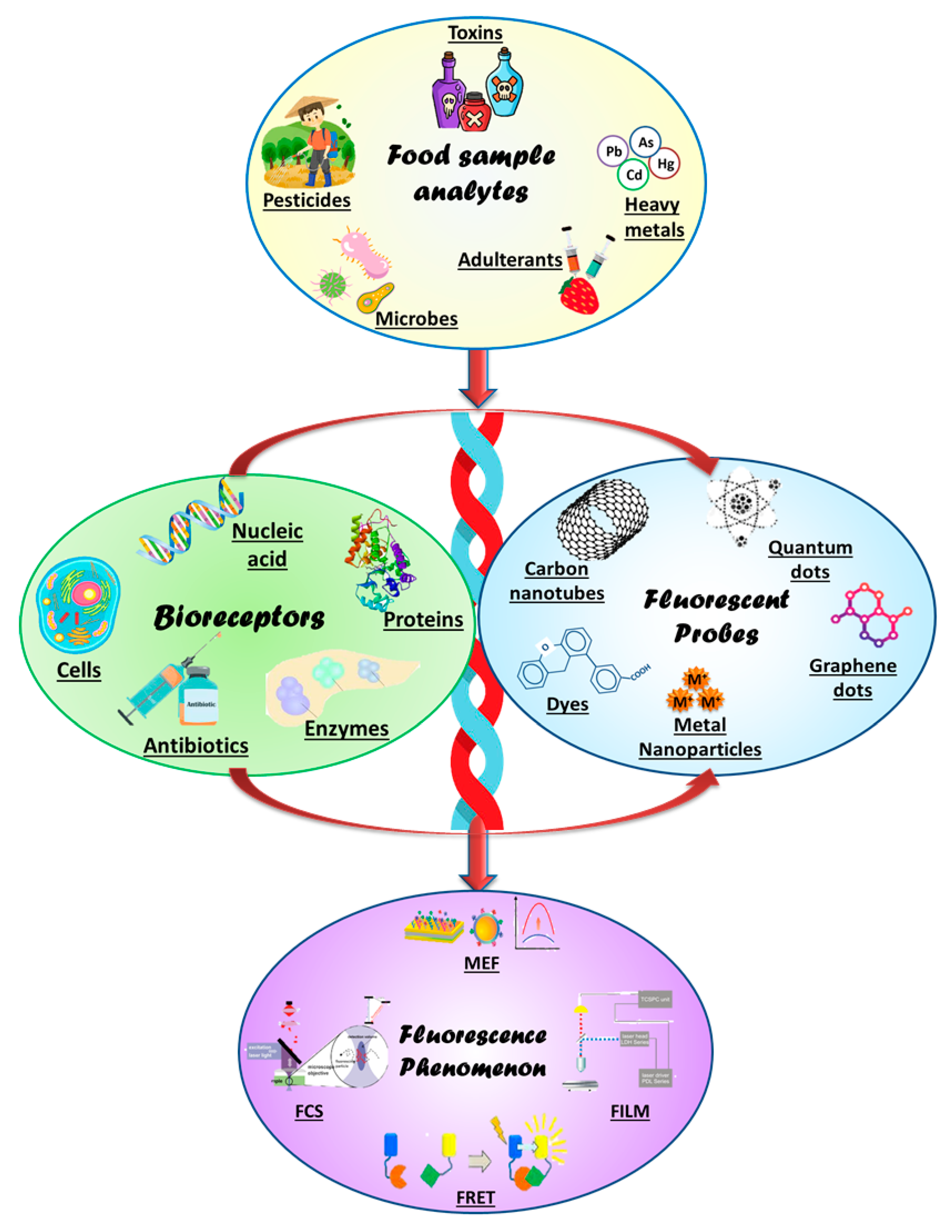
:1. Introduction
2. Fundamental Aspects of Fluorescence Biosensing
3. Fluorescence Biosensing Materials
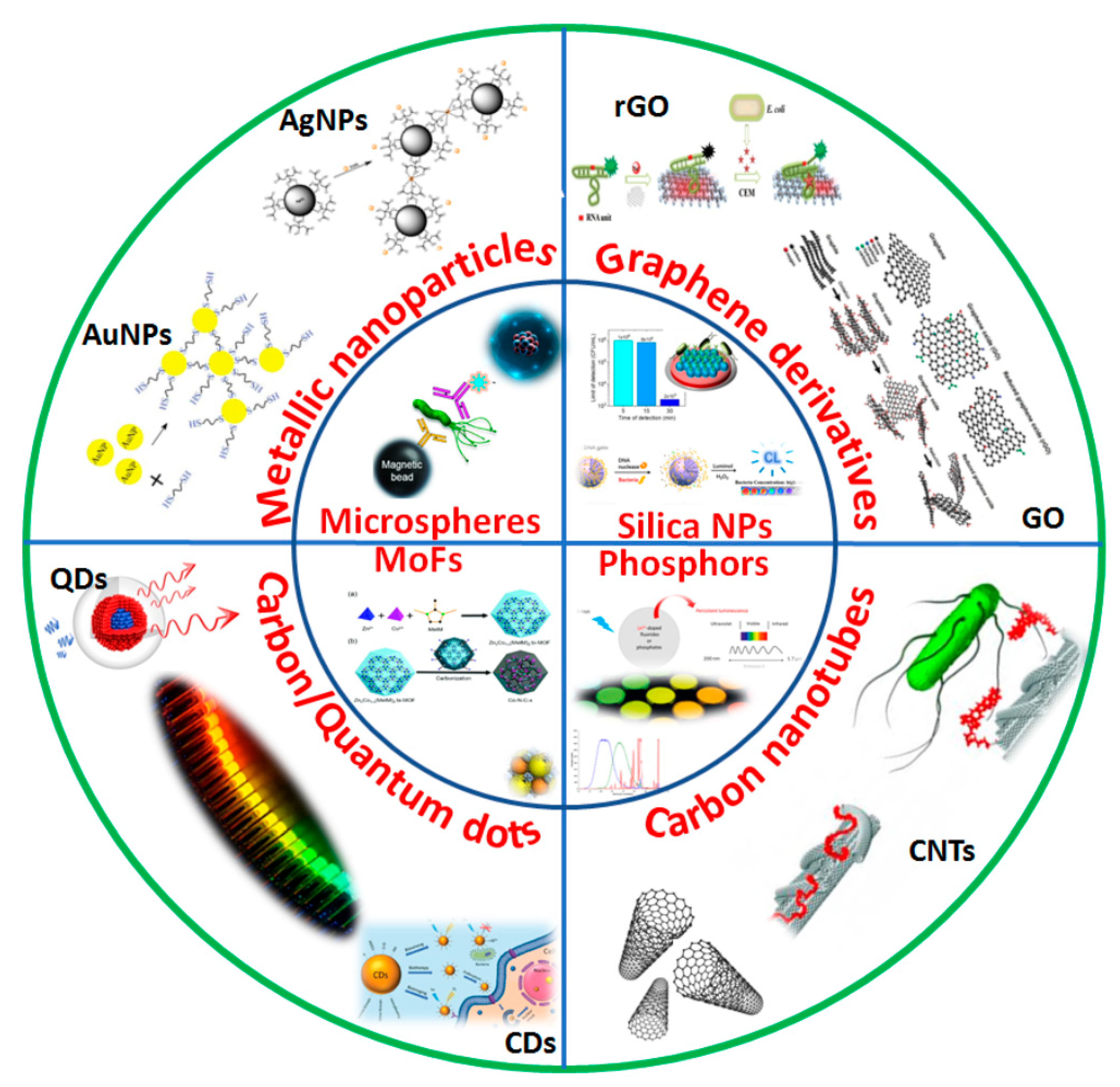
3.1. Nanomaterials

| Nanomaterial | Analyte | Biorecognition Element | LoD | Ref. |
|---|---|---|---|---|
| Metal Nanoparticles | ||||
| Gold nanoparticles | Salmonella typhimurium | DNA aptamer | 36 CFU/mL | [33] |
| Gold nanoparticles | Dipicolinic acid | Eu3+ ion/gold nanocluster | 0.8 μM | [34] |
| Gold nanoparticles | Histamine | Gold nanoparticles | 2.04 nM | [35] |
| Silver nanoparticles | Melamine | Polyethyleneimine–silver nanobioprobe | 132 nM | [36] |
| Silver nanoparticles | Staphylococcal enterotoxin A | DNA aptamer | 0.3393 ng/mL | [37] |
| Silver nanoparticles | Fe+3 ions | Vitamin B12-functionalized biological silver nanoparticles (FAgNPs) | 2 mg/L | [38] |
| Copper nanoparticles | Zearalenone | Antibodies | 16.0 μg/kg | [39] |
| Platinum nanoparticles | Hypoxanthine | Platinum nanoparticles | 2.88 μM | [40] |
| Tungsten nanoparticles | Maltose and sucrose | Fenugreek β-amylase functionalized tungsten disulfide nanoparticles | 0.052 and 0.096 mM | [41] |
| Palladium nanoparticles | Tetracyclines | Graphene quantum dots/palladium nanoparticles | 45 ng/mL | [42] |
| Carbon-based nanomaterials | ||||
| Carbon nanotubes | Escherichia coli O157:H7 | Carbonyl iron powder/MWCNT-DNA aptamer | 3.15 × 10² cfu/mL | [43] |
| Carbon nanotubes | Patulin mycotoxin | Carboxyfluorescein dye MWCNTs–DNA aptamer | 0.13 μg/L | [44] |
| Carbon nanohorns | Fipronil | FAM–aptamer with oxidized single-walled carbon nanohorns | 3 nM | [45] |
| Carbon dots | Tartrazine | Fluorescent carbon dots | 12.4 nM | [46] |
| Carbon dots | Tetracyclines and Al3+ | Fluorescent carbon dots | 0.057–0.23 μM and 0.091 μM | [47] |
| Carbon dots | Ascorbic acid | Carbon Dots/Fe3+ composite | 3.11 μmol·L-1 μmol/L | [48] |
| Quantum dots | Acrylamide | DNA aptamer | 2.41 × 10⁻⁸ M | [49] |
| Quantum dots | Histamine | Carbon quantum dots with peptide | 13 μg/kg | [50] |
| Quantum dots | Biogenic amines | Carbon dots/yellow fluorescent CdTe quantum dots | 1.259-5.428 μM | [51] |
| Graphene oxide | β-lactoglobulin | DNA aptamer | 96.91 μg/L | [32] |
| Graphene quantum dots | Formaldehyde | Graphene quantum dots | 0.0515 μg/mL | [52] |
| Graphene oxide | Zearalenone and ochratoxin A | Cy3 aptamer and Alexa Fluor 488 aptamer | 1.797 ng/mL and 1.484 ng/mL | [53] |
| Other nanomaterials | ||||
| Silica nanoparticles | Thiram | Mesoporous silica with gold nanoparticles | 0.19 ng/mL | [15] |
| Silica nanoparticles | Aflatoxin B1 | DNA aptamer | 0.13 ng/mL | [54] |
| Up-conversion nanoparticles | Staphylococcus aureus | Aptamer-functionalized gold nanoparticles | 10.7 CFU/mL | [55] |
| Up-conversion nanoparticles | Histamine | Up-conversion nanoparticles | 7.34 mg/L | [56] |
| Metal organic frameworks (MOF) | Acrylamide | 6-carboxyfluorescein-labeled aptamer (FAM-ssDNA) | 1.9 nM | [57] |
| Metal organic frameworks | Tetracycline antibiotics | Luminescent MOF | 0.28–0.30 μM | [58] |
| Metal organic frameworks | Ethanolamine | Zeolitic imidazolate framework-8/FAM-aptamer | 17.86 pM | [59] |
| Phosphors | Zearalenone in cereals | Black phosphorus–gold nanocomposite | 2 μg/kg | [60] |
3.2. Nucleic-Acid-Based Molecular Markers
3.3. Antibodies
3.4. Proteins/Enzymes
4. Integration of Fluorescence Biosensing for Microbe Detection
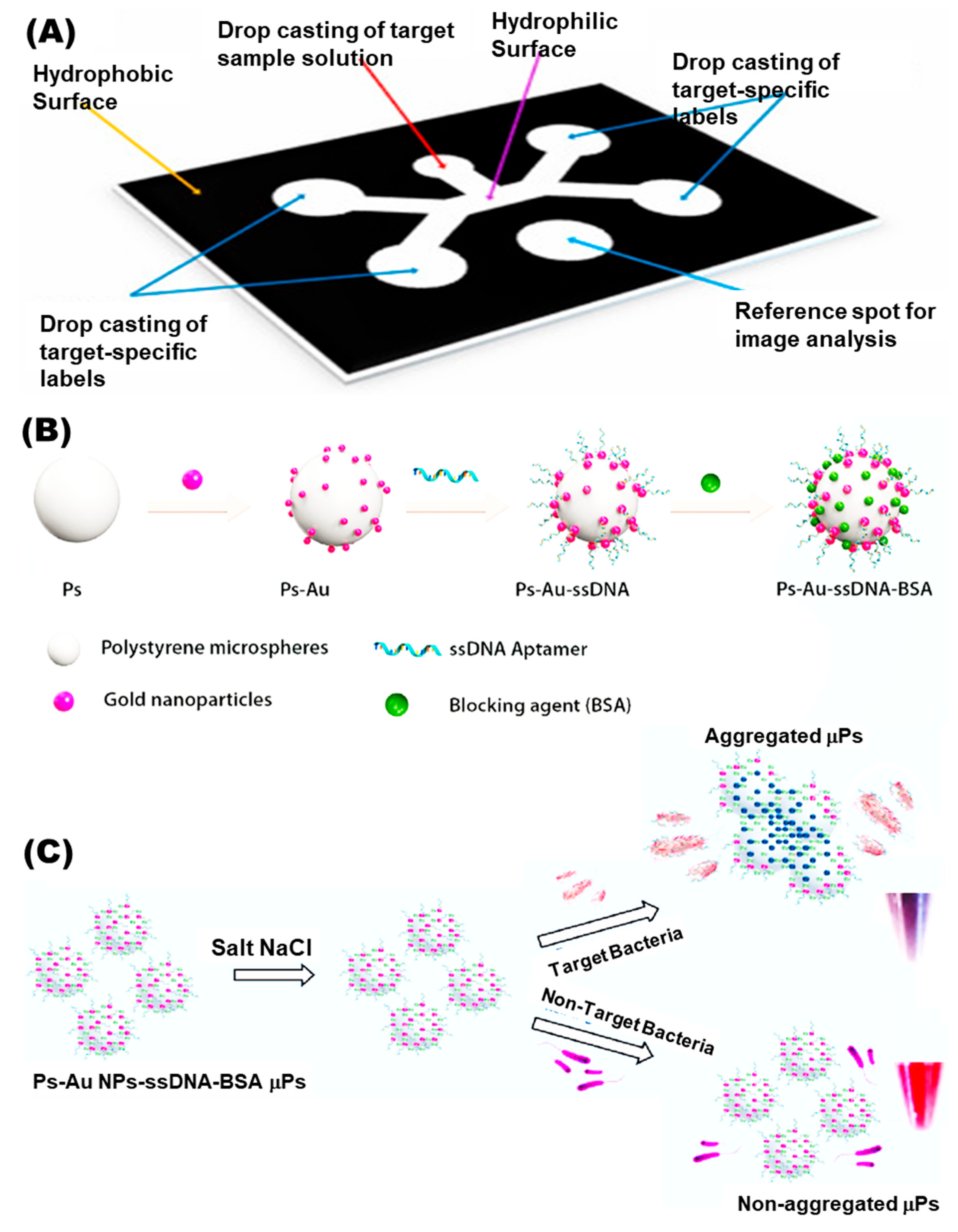
4.1. Microarrays
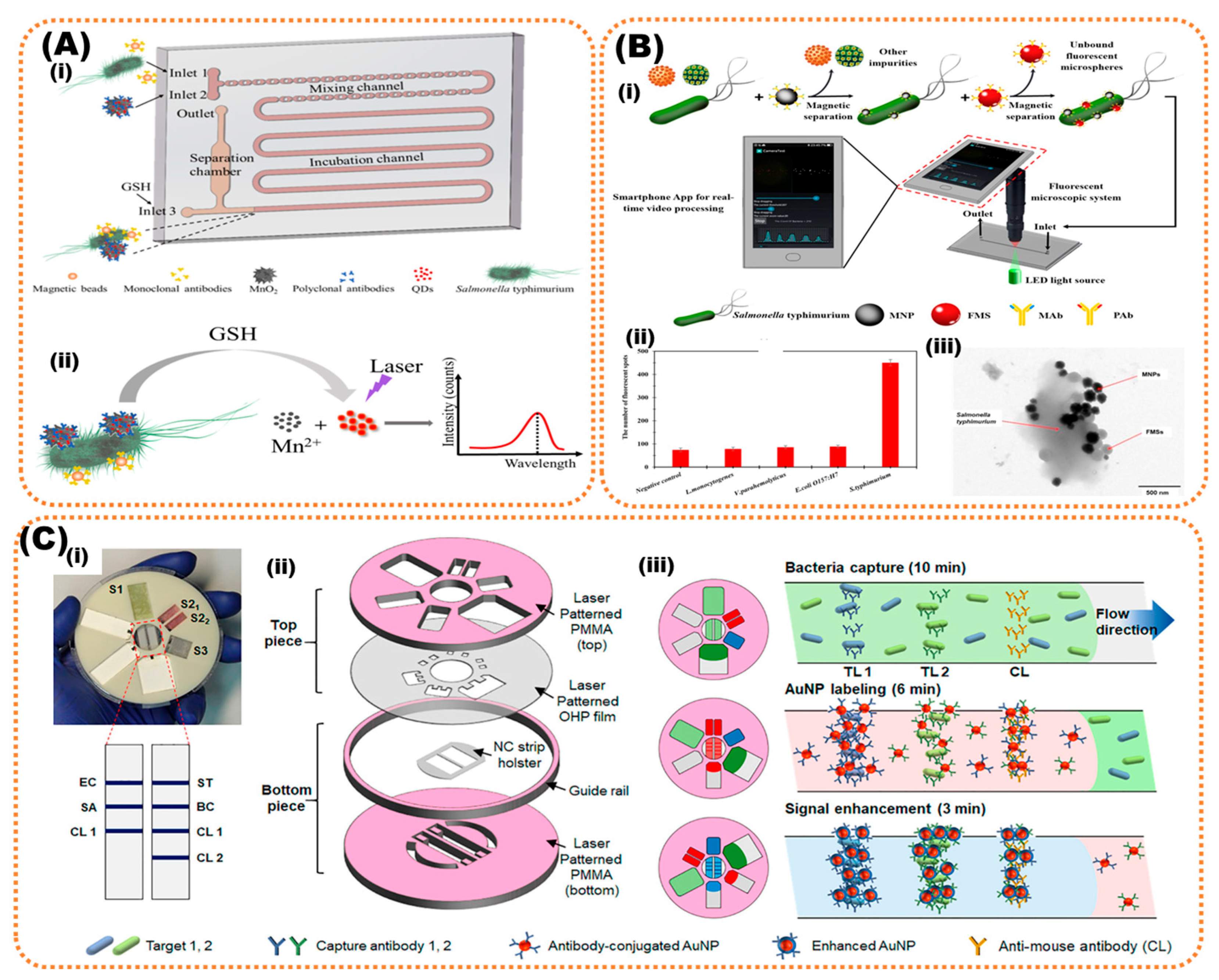
4.2. Microfluidic Devices
5. Future Outlook
Author Contributions
Funding
Institutional Review Board Statement
Informed Consent Statement
Data Availability Statement
Acknowledgments
Conflicts of Interest
References
- Carstens, C.K.; Salazar, J.K.; Darkoh, C. Multistate Outbreaks of Foodborne Illness in the United States Associated with Fresh Produce from 2010 to 2017. Front. Microbiol. 2019, 10, 2667. [Google Scholar] [CrossRef]
- Shonhiwa, A.M.; Ntshoe, G.; Essel, V.; Thomas, J.; McCarthy, K. A Review of Foodborne Diseases Outbreaks Reported to the Outbreak Response Unit, National Institute for Communicable Diseases, South Africa, 2013–2017. Int. J. Infect. Dis. 2019, 79, 73. [Google Scholar] [CrossRef]
- Van der Fels-Klerx, H.J.; Van Asselt, E.D.; Raley, M.; Poulsen, M.; Korsgaard, H.; Bredsdorff, L.; Nauta, M.; D’agostino, M.; Coles, D.; Marvin, H.J.P. Critical Review of Methods for Risk Ranking of Food-Related Hazards, Based on Risks for Human Health. Crit. Rev. Food Sci. Nutr. 2018, 58, 178–193. [Google Scholar] [CrossRef]
- Law, J.W.-F.; Ab Mutalib, N.-S.; Chan, K.-G.; Lee, L.-H. Rapid Methods for the Detection of Foodborne Bacterial Pathogens: Principles, Applications, Advantages and Limitations. Front. Microbiol. 2015, 5, 770. [Google Scholar] [CrossRef]
- Campbell, V.R.; Carson, M.S.; Lao, A.; Maran, K.; Yang, E.J.; Kamei, D.T. Point-of-Need Diagnostics for Foodborne Pathogen Screening. SLAS Technol. 2021, 26, 55–79. [Google Scholar] [CrossRef]
- Kant, K.; Ngo, T.A.; Matteucci, M.; Wolff, A. Fabrication of 3D Microstructure Array on Chip for Rapid Pathogen Detection. Sensors Actuators B Chem. 2019, 281, 774–782. [Google Scholar] [CrossRef]
- Choi, J.R.; Yong, K.W.; Choi, J.Y.; Cowie, A.C. Emerging Point-of-Care Technologies for Food Safety Analysis. Sensors 2019, 19, 817. [Google Scholar] [CrossRef]
- Vinayaka, A.C.; Ngo, T.A.; Kant, K.; Engelsmann, P.; Dave, V.P.; Shahbazi, M.-A.; Wolff, A.; Bang, D.D. Rapid Detection of Salmonella Enterica in Food Samples by a Novel Approach with Combination of Sample Concentration and Direct PCR. Biosens. Bioelectron. 2019, 129, 224–230. [Google Scholar] [CrossRef]
- Mandal, P.K.; Biswas, A.K.; Choi, K.; Pal, U.K. Methods for Rapid Detection of Foodborne Pathogens: An Overview. Am. J. Food Technol. 2011, 6, 87–102. [Google Scholar] [CrossRef]
- Valderrama, W.B.; Dudley, E.G.; Doores, S.; Cutter, C.N. Commercially Available Rapid Methods for Detection of Selected Food-Borne Pathogens. Crit. Rev. Food Sci. Nutr. 2016, 56, 1519–1531. [Google Scholar]
- Choi, J.R.; Yong, K.W.; Tang, R.; Gong, Y.; Wen, T.; Li, F.; Pingguan-Murphy, B.; Bai, D.; Xu, F. Advances and Challenges of Fully Integrated Paper-Based Point-of-Care Nucleic Acid Testing. TrAC Trends Anal. Chem. 2017, 93, 37–50. [Google Scholar] [CrossRef]
- Lakowicz, J.R. Fluorescence Sensing. In Principles of Fluorescence Spectroscopy; Springer: Berlin/Heidelberg, Germany, 1999; pp. 531–572. [Google Scholar]
- Ding, X.; Yin, K.; Li, Z.; Pandian, V.; Smyth, J.A.; Helal, Z.; Liu, C. Cleavable Hairpin Beacon-Enhanced Fluorescence Detection of Nucleic Acid Isothermal Amplification and Smartphone-Based Readout. Sci. Rep. 2020, 10, 18819. [Google Scholar] [CrossRef] [PubMed]
- Hu, Q.; Pan, Y.; Gong, X.; Rao, S.; Xiao, L.; Liu, L.; Yang, Z. A Sensitivity Enhanced Fluorescence Method for the Detection of Ferrocyanide Ions in Foodstuffs Using Carbon Nanoparticles as Sensing Agents. Food Chem. 2020, 308, 125590. [Google Scholar] [CrossRef] [PubMed]
- Ren, H.X.; Qian, Z.J.; Li, M.; Peng, C.F.; Wang, Z.P.; Wei, X.L.; Xu, J.G. Mesoporous Silica-Loaded Gold Nanocluster with Enhanced Fluorescence and Ratiometric Fluorescent Detection of Thiram in Foods. Mikrochim. Acta 2021, 188, 363. [Google Scholar] [CrossRef]
- Zhang, Y.; Tang, H.; Chen, W.; Zhang, J. Nanomaterials Used in Fluorescence Polarization Based Biosensors. Int. J. Mol. Sci. 2022, 23, 8625. [Google Scholar] [CrossRef]
- Zhong, W. Nanomaterials in Fluorescence-Based Biosensing. Anal. Bioanal. Chem. 2009, 394, 47–59. [Google Scholar] [CrossRef]
- Sapsford, K.E.; Berti, L.; Medintz, I.L. Materials for Fluorescence Resonance Energy Transfer Analysis: Beyond Traditional Donor–Acceptor Combinations. Angew. Chemie Int. Ed. 2006, 45, 4562–4589. [Google Scholar] [CrossRef]
- El-Nour, K.M.A.; Salam, E.T.A.; Soliman, H.M.; Orabi, A.S. Gold Nanoparticles as a Direct and Rapid Sensor for Sensitive Analytical Detection of Biogenic Amines. Nanoscale Res. Lett. 2017, 12, 231. [Google Scholar] [CrossRef]
- Kato, R.; Uesugi, M.; Komatsu, Y.; Okamoto, F.; Tanaka, T.; Kitawaki, F.; Yano, T.A. Highly Stable Polymer Coating on Silver Nanoparticles for Efficient Plasmonic Enhancement of Fluorescence. ACS Omega 2022, 7, 4286–4292. [Google Scholar] [CrossRef]
- Cui, D.; Pan, B.; Zhang, H.; Gao, F.; Wu, R.; Wang, J.; He, R.; Asahi, T. Self-Assembly of Quantum Dots and Carbon Nanotubes for Ultrasensitive DNA and Antigen Detection. Anal. Chem. 2008, 80, 7996–8001. [Google Scholar] [CrossRef]
- Chen, H.; Zuo, X.; Su, S.; Tang, Z.; Wu, A.; Song, S.; Zhang, D.; Fan, C. An Electrochemical Sensor for Pesticide Assays Based on Carbon Nanotube-Enhanced Acetycholinesterase Activity. Analyst 2008, 133, 1182–1186. [Google Scholar] [CrossRef]
- Alivisatos, P. The Use of Nanocrystals in Biological Detection. Nat. Biotechnol. 2003, 22, 47–52. [Google Scholar] [CrossRef]
- Reshma, V.G.; Mohanan, P.`V. Quantum Dots: Applications and Safety Consequences. J. Lumin. 2019, 205, 287–298. [Google Scholar] [CrossRef]
- Li, J.; Zhu, J.J. Quantum Dots for Fluorescent Biosensing and Bio-Imaging Applications. Analyst 2013, 138, 2506–2515. [Google Scholar] [CrossRef]
- Cardoso Dos Santos, M.; Algar, W.R.; Medintz, I.L.; Hildebrandt, N. Quantum Dots for Förster Resonance Energy Transfer (FRET). TrAC—Trends Anal. Chem. 2020, 125, 115819. [Google Scholar] [CrossRef]
- Xiong, J.; Zhang, H.; Qin, L.; Zhang, S.; Cao, J.; Jiang, H. Magnetic Fluorescent Quantum Dots Nanocomposites in Food Contaminants Analysis: Current Challenges and Opportunities. Int. J. Mol. Sci. 2022, 23, 4088. [Google Scholar] [CrossRef]
- Mohamadi, E.; Moghaddasi, M.; Farahbakhsh, A.; Kazemi, A. A Quantum-Dot-Based Fluoroassay for Detection of Food-Borne Pathogens. J. Photochem. Photobiol. B Biol. 2017, 174, 291–297. [Google Scholar] [CrossRef]
- Sun, F.; Zhang, J.; Yang, Q.; Wu, W. Quantum Dot Biosensor Combined with Antibody and Aptamer for Tracing Food-Borne Pathogens. Food Qual. Saf. 2021, 5, fyab019. [Google Scholar] [CrossRef]
- Yin, P.T.; Shah, S.; Chhowalla, M.; Lee, K.B. Design, Synthesis, and Characterization of Graphene-Nanoparticle Hybrid Materials for Bioapplications. Chem. Rev. 2015, 115, 2483–2531. [Google Scholar] [CrossRef]
- Zheng, P.; Wu, N. Fluorescence and Sensing Applications of Graphene Oxide and Graphene Quantum Dots: A Review. Chem. Asian J. 2017, 12, 2343. [Google Scholar] [CrossRef]
- Yang, J.; Zhang, Y.; Lu, Y. A Fluorescence Detection Method for the Determination of β-Lactoglobulin in Foods. Anal. Methods 2022, 14, 1872–1879. [Google Scholar] [CrossRef] [PubMed]
- Chen, M.; Pan, L.; Tu, K. A Fluorescence Biosensor for Salmonella Typhimurium Detection in Food Based on the Nano-Self-Assembly of Alendronic Acid Modified Upconversion and Gold Nanoparticles. Anal. Methods 2021, 13, 2415–2423. [Google Scholar] [CrossRef] [PubMed]
- Li, X.; Luo, J.; Jiang, X.; Yang, M.; Rasooly, A. Gold Nanocluster-Europium(III) Ratiometric Fluorescence Assay for Dipicolinic Acid. Mikrochim. Acta 2021, 188, 26. [Google Scholar] [CrossRef] [PubMed]
- Bi, J.; Tian, C.; Zhang, G.L.; Hao, H.; Hou, H.M. Detection of Histamine Based on Gold Nanoparticles with Dual Sensor System of Colorimetric and Fluorescence. Foods 2020, 9, 316. [Google Scholar] [CrossRef]
- Li, Z.; Li, Y.; Li, L.; Wang, T. Aquamarine Blue Emitting Silver Nanoparticles as Fluorescent Sensor for Melamine Detection. Spectrochim. Acta Part A Mol. Biomol. Spectrosc. 2019, 217, 51–59. [Google Scholar] [CrossRef]
- Zhang, X.; Khan, I.M.; Ji, H.; Wang, Z.; Tian, H.; Cao, W.; Mi, W. A Label-Free Fluorescent Aptasensor for Detection of Staphylococcal Enterotoxin A Based on Aptamer-Functionalized Silver Nanoclusters. Polymers 2020, 12, 152. [Google Scholar] [CrossRef]
- Harke, S.S.; Patil, R.V.; Dar, M.A.; Pandit, S.R.; Pawar, K.D. Functionalization of Biogenic Silver Nanoparticles with Vitamin B12 for the Detection of Iron in Food Samples. Food Chem. Adv. 2022, 1, 100017. [Google Scholar] [CrossRef]
- Xuan, Z.; Wu, Y.; Liu, H.; Li, L.; Ye, J.; Wang, S. Copper Oxide Nanoparticle-Based Immunosensor for Zearalenone Analysis by Combining Automated Sample Pre-Processing and High-Throughput Terminal Detection. Sensors 2021, 21, 6538. [Google Scholar] [CrossRef]
- Chen, J.; Lu, Y.; Yan, F.; Wu, Y.; Huang, D.; Weng, Z. A Fluorescent Biosensor Based on Catalytic Activity of Platinum Nanoparticles for Freshness Evaluation of Aquatic Products. Food Chem. 2020, 310, 125922. [Google Scholar] [CrossRef]
- Agrawal, D.C.; Yadav, A.; Singh, V.K.; Srivastava, A.; Kayastha, A.M. Immobilization of Fenugreek β-Amylase onto Functionalized Tungsten Disulfide Nanoparticles Using Response Surface Methodology: Its Characterization and Interaction with Maltose and Sucrose. Colloids Surf. B Biointerfaces 2020, 185, 110600. [Google Scholar] [CrossRef]
- Ahmed, S.R.; Kumar, S.; Ortega, G.A.; Srinivasan, S.; Rajabzadeh, A.R. Target Specific Aptamer-Induced Self-Assembly of Fluorescent Graphene Quantum Dots on Palladium Nanoparticles for Sensitive Detection of Tetracycline in Raw Milk. Food Chem. 2021, 346, 128893. [Google Scholar] [CrossRef]
- Yang, T.; Yang, X.; Guo, X.; Fu, S.; Zheng, J.; Chen, S.; Qin, X.; Wang, Z.; Zhang, D.; Man, C.; et al. A Novel Fluorometric Aptasensor Based on Carbon Nanocomposite for Sensitive Detection of Escherichia coli O157:H7 in Milk. J. Dairy Sci. 2020, 103, 7879–7889. [Google Scholar] [CrossRef]
- Khan, R.; Sherazi, T.A.; Catanante, G.; Rasheed, S.; Marty, J.L.; Hayat, A. Switchable Fluorescence Sensor toward PAT via CA-MWCNTs Quenched Aptamer-Tagged Carboxyfluorescein. Food Chem. 2020, 312, 126048. [Google Scholar] [CrossRef]
- Zhang, J.; Feng, T.; Zhang, J.; Liang, N.; Zhao, L. Fluorescence Assay for the Sensitive Detection of Fipronil Based on an “on–off” Oxidized SWCNH/Aptamer Sensor. Anal. Methods 2021, 13, 3282–3291. [Google Scholar] [CrossRef]
- Liu, L.; Sun, H.; Xiao, L.; Yang, Z.-q.; Han, J.; Gong, X.; Hu, Q. Development of a Highly Sensitive Fluorescence Method for Tartrazine Determination in Food Matrices Based on Carbon Dots. Anal. Bioanal. Chem. 2021, 413, 1485–1492. [Google Scholar] [CrossRef]
- Jia, J.; Lu, W.; Cui, S.; Dong, C.; Shuang, S. Preparation of Yellow-Emitting Carbon Dots and Their Bifunctional Detection of Tetracyclines and Al3+ in Food and Living Cells. Mikrochim. Acta 2021, 188, 418. [Google Scholar] [CrossRef]
- Wang, T.; Luo, H.; Jing, X.; Yang, J.; Huo, M.; Wang, Y. Synthesis of Fluorescent Carbon Dots and Their Application in Ascorbic Acid Detection. Molecules 2021, 26, 1246. [Google Scholar] [CrossRef]
- Wei, Q.; Zhang, P.; Liu, T.; Pu, H.; Sun, D.W. A Fluorescence Biosensor Based on Single-Stranded DNA and Carbon Quantum Dots for Acrylamide Detection. Food Chem. 2021, 356, 129668. [Google Scholar] [CrossRef]
- Shi, R.; Feng, S.; Park, C.Y.; Park, K.Y.; Song, J.; Park, J.P.; Chun, H.S.; Park, T.J. Fluorescence Detection of Histamine Based on Specific Binding Bioreceptors and Carbon Quantum Dots. Biosens. Bioelectron. 2020, 167, 112519. [Google Scholar] [CrossRef]
- Yan, J.; Fu, Q.; Zhang, S.; Liu, Y.; Shi, X.; Hou, J.; Duan, J.; Ai, S. A Sensitive Ratiometric Fluorescent Sensor Based on Carbon Dots and CdTe Quantum Dots for Visual Detection of Biogenic Amines in Food Samples. Spectrochim. Acta Part A Mol. Biomol. Spectrosc. 2022, 282, 121706. [Google Scholar] [CrossRef]
- Zhang, Y.; Qi, J.; Li, M.; Gao, D.; Xing, C. Fluorescence Probe Based on Graphene Quantum Dots for Selective, Sensitive and Visualized Detection of Formaldehyde in Food. Sustainability 2021, 13, 5273. [Google Scholar] [CrossRef]
- Wang, Q.; Yang, Q.; Wu, W. Graphene-Based Steganographic Aptasensor for Information Computing and Monitoring Toxins of Biofilm in Food. Front. Microbiol. 2020, 10, 3139. [Google Scholar] [CrossRef] [PubMed]
- Tan, H.; Ma, L.; Guo, T.; Zhou, H.; Chen, L.; Zhang, Y.; Dai, H.; Yu, Y. A Novel Fluorescence Aptasensor Based on Mesoporous Silica Nanoparticles for Selective and Sensitive Detection of Aflatoxin B1. Anal. Chim. Acta 2019, 1068, 87–95. [Google Scholar] [CrossRef] [PubMed]
- Ouyang, Q.; Yang, Y.; Ali, S.; Wang, L.; Li, H.; Chen, Q. Upconversion Nanoparticles-Based FRET System for Sensitive Detection of Staphylococcus Aureus. Spectrochim. Acta Part A Mol. Biomol. Spectrosc. 2021, 255, 119734. [Google Scholar] [CrossRef] [PubMed]
- Cao, Y.; Wang, X.; Feng, T.; Li, Z.; Xue, C.; Xu, J. Ratiometric Fluorescent Nanosystem Based on Upconversion Nanoparticles for Histamine Determination in Seafood. Food Chem. 2022, 390, 133194. [Google Scholar] [CrossRef]
- Gan, Z.; Zhang, W.; Arslan, M.; Hu, X.; Zhang, X.; Li, Z.; Shi, J.; Zou, X. Ratiometric Fluorescent Metal-Organic Framework Biosensor for Ultrasensitive Detection of Acrylamide. J. Agric. Food Chem. 2022, 70, 10065–10074. [Google Scholar] [CrossRef]
- Liu, Q.; Ning, D.; Li, W.J.; Du, X.M.; Wang, Q.; Li, Y.; Ruan, W.J. Metal-Organic Framework-Based Fluorescent Sensing of Tetracycline-Type Antibiotics Applicable to Environmental and Food Analysis. Analyst 2019, 144, 1916–1922. [Google Scholar] [CrossRef]
- Khoshbin, Z.; Zamanian, J.; Davoodian, N.; Mohammad Danesh, N.; Ramezani, M.; Alibolandi, M.; Abnous, K.; Mohammad Taghdisi, S. A Simple and Ultrasensitive Metal-Organic Framework-Based Aptasensor for Fluorescence Detection of Ethanolamine. Spectrochim. Acta. A. Mol. Biomol. Spectrosc. 2022, 267, 120488. [Google Scholar] [CrossRef]
- Li, S.; Zhang, F.; Wang, J.; Wen, W.; Wang, S. Black Phosphorus-Au Nanocomposite-Based Fluorescence Immunochromatographic Sensor for High-Sensitive Detection of Zearalenone in Cereals. Nanophotonics 2020, 9, 2397–2406. [Google Scholar] [CrossRef]
- He, Y.; Li, Y. Fluorescent Markers. In Encyclopedia of Systems Biology; Springer: New York, NY, USA, 2013; pp. 746–748. [Google Scholar] [CrossRef]
- Nutiu, R.; Billen, L.P.; Li, Y. Fluorescence-Signaling Nucleic Acid-Based Sensors. In Nucleic Acid Switches and Sensors; Springer: Boston, MA, USA, 2006; pp. 49–72. [Google Scholar] [CrossRef]
- Podder, A.; Lee, H.J.; Kim, B.H. Fluorescent Nucleic Acid Systems for Biosensors. Bull. Chem. Soc. Jpn. 2020, 94, 1010–1035. [Google Scholar] [CrossRef]
- Tyagi, S.; Kramer, F.R. Molecular Beacons: Probes That Fluoresce upon Hybridization. Nat. Biotechnol. 1996, 14, 303–308. [Google Scholar] [CrossRef]
- Burris, K.P.; Wu, T.C.; Vasudev, M.; Stroscio, M.A.; Millwood, R.J.; Stewart, C.N. Mega-Nano Detection of Foodborne Pathogens and Transgenes Using Molecular Beacon and Semiconductor Quantum Dot Technologies. IEEE Trans. Nanobiosci. 2013, 12, 233–238. [Google Scholar] [CrossRef]
- Vidic, J.; Vizzini, P.; Manzano, M.; Kavanaugh, D.; Ramarao, N.; Zivkovic, M.; Radonic, V.; Knezevic, N.; Giouroudi, I.; Gadjanski, I. Point-of-Need DNA Testing for Detection of Foodborne Pathogenic Bacteria. Sensors 2019, 19, 1100. [Google Scholar] [CrossRef]
- Hadjinicolaou, A.V.; Demetriou, V.L.; Emmanuel, M.A.; Kakoyiannis, C.K.; Kostrikis, L.G. Molecular Beacon-Based Real-Time PCR Detection of Primary Isolates of Salmonella Typhimurium and Salmonella Enteritidis in Environmental and Clinical Samples. BMC Microbiol. 2009, 9, 97. [Google Scholar] [CrossRef]
- Hu, Q.; Lyu, D.Y.; Shi, X.; Jiang, Y.; Lin, Y.; Li, Y.; Qiu, Y.; He, L.; Zhang, R.; Li, Q. A Modified Molecular Beacons-Based Multiplex Real-Time PCR Assay for Simultaneous Detection of Eight Foodborne Pathogens in a Single Reaction and Its Application. Foodborne Pathog. Dis. 2014, 11, 207–214. [Google Scholar] [CrossRef]
- Su, X.; Xiao, X.; Zhang, C.; Zhao, M. Nucleic Acid Fluorescent Probes for Biological Sensing. Appl. Spectrosc. 2012, 66, 1249–1261. [Google Scholar] [CrossRef]
- Ellington, A.D.; Szostak, J.W. In Vitro Selection of RNA Molecules That Bind Specific Ligands. Nature 1990, 346, 818–822. [Google Scholar] [CrossRef]
- Tuerk, C.; Gold, L. Systematic Evolution of Ligands by Exponential Enrichment: RNA Ligands to Bacteriophage T4 DNA Polymerase. Science 1990, 249, 505–510. [Google Scholar] [CrossRef]
- Hong, L.; Pan, M.; Xie, X.; Liu, K.; Yang, J.; Wang, S.; Wang, S. Aptamer-Based Fluorescent Biosensor for the Rapid and Sensitive Detection of Allergens in Food Matrices. Foods 2021, 10, 2598. [Google Scholar] [CrossRef]
- Wang, L.; Chi, E.Z.; Zhao, X.H.; Zhang, Q. A Simple and Rapid “Signal On” Fluorescent Sensor for Detecting Mercury (II) Based on the Molecular Beacon Aptamer. Foods 2022, 11, 1847. [Google Scholar] [CrossRef]
- Abe, R.; Ohashi, H.; Iijima, I.; Ihara, M.; Takagi, H.; Hohsaka, T.; Ueda, H. “quenchbodies”: Quench-Based Antibody Probes That Show Antigen-Dependent Fluorescence. J. Am. Chem. Soc. 2011, 133, 17386–17394. [Google Scholar] [CrossRef] [PubMed]
- Ueda, H.; Dong, J. From Fluorescence Polarization to Quenchbody: Recent Progress in Fluorescent Reagentless Biosensors Based on Antibody and Other Binding Proteins. Biochim. Biophys. Acta Proteins Proteom. 2014, 1844, 1951–1959. [Google Scholar] [CrossRef] [PubMed]
- Dong, J.; Ueda, H. Recent Advances in Quenchbody, a Fluorescent Immunosensor. Sensors 2021, 21, 1223. [Google Scholar] [CrossRef] [PubMed]
- Huang, A.; Li, W.; Shi, S.; Yao, T. Quantitative Fluorescence Quenching on Antibody-Conjugated Graphene Oxide as a Platform for Protein Sensing. Sci. Rep. 2017, 7, 40772. [Google Scholar] [CrossRef] [PubMed]
- Capo, A.; D’Auria, S.; Lacroix, M. A Fluorescence Immunoassay for a Rapid Detection of Listeria Monocytogenes on Working Surfaces. Sci. Rep. 2020, 10, 21729. [Google Scholar] [CrossRef]
- Nishi, K.; Isobe, S.I.; Zhu, Y.; Kiyama, R. Fluorescence-Based Bioassays for the Detection and Evaluation of Food Materials. Sensors 2015, 15, 25831. [Google Scholar] [CrossRef]
- Wu, J.; Lu, Y.; Ren, N.; Jia, M.; Wang, R.; Zhang, J. DNAzyme-Functionalized R-Phycoerythrin as a Cost-Effective and Environment-Friendly Fluorescent Biosensor for Aqueous Pb2+ Detection. Sensors 2019, 19, 2732. [Google Scholar] [CrossRef]
- Gensberger, E.T.; Kostić, T. Green Fluorescent Protein Labeling of Food Pathogens Yersinia Enterocolitica and Yersinia Pseudotuberculosis. J. Microbiol. Methods 2017, 132, 21–26. [Google Scholar] [CrossRef]
- Futra, D.; Heng, L.Y.; Ahmad, A.; Surif, S.; Ling, T.L. An Optical Biosensor from Green Fluorescent Escherichia coli for the Evaluation of Single and Combined Heavy Metal Toxicities. Sensors 2015, 15, 12668–12681. [Google Scholar] [CrossRef] [Green Version]
- Li, B.; Yu, Q.; Duan, Y. Fluorescent Labels in Biosensors for Pathogen Detection. Crit. Rev. Biotechnol. 2015, 35, 82–93. [Google Scholar] [CrossRef]
- Huang, F.; Zhang, Y.; Lin, J.; Liu, Y. Biosensors Coupled with Signal Amplification Technology for the Detection of Pathogenic Bacteria: A Review. Biosensors 2021, 11, 190. [Google Scholar] [CrossRef] [PubMed]
- Poltronieri, P.; Mezzolla, V.; Primiceri, E.; Maruccio, G. Biosensors for the Detection of Food Pathogens. Foods 2014, 3, 511. [Google Scholar] [CrossRef] [PubMed]
- Sarengaowa; Hu, W.; Feng, K.; Jiang, A.; Xiu, Z.; Lao, Y.; Li, Y.; Long, Y. An in Situ-Synthesized Gene Chip for the Detection of Food-Borne Pathogens on Fresh-Cut Cantaloupe and Lettuce. Front. Microbiol. 2020, 10, 3089. [Google Scholar] [CrossRef] [PubMed]
- Hormsombut, T.; Rijiravanich, P.; Surareungchai, W.; Kalasin, S. Highly Sensitive and Selective Antibody Microarrays Based on a Cy5-Antibody Complexes Coupling ES-Biochip for E. Coli and Salmonella Detection. RSC Adv. 2022, 12, 24760–24768. [Google Scholar] [CrossRef] [PubMed]
- Luka, G.; Ahmadi, A.; Najjaran, H.; Alocilja, E.; Derosa, M.; Wolthers, K.; Malki, A.; Aziz, H.; Althani, A.; Hoorfar, M. Microfluidics Integrated Biosensors: A Leading Technology towards Lab-on-a-Chip and Sensing Applications. Sensors 2015, 15, 30011–30031. [Google Scholar] [CrossRef]
- Bahavarnia, F.; Hasanzadeh, M.; Sadighbayan, D.; Seidi, F. Recent Progress and Challenges on the Microfluidic Assay of Pathogenic Bacteria Using Biosensor Technology. Biomimetics 2022, 7, 175. [Google Scholar] [CrossRef]
- Nawrot, W.; Drzozga, K.; Baluta, S.; Cabaj, J.; Malecha, K. A Fluorescent Biosensors for Detection Vital Body Fluids’ Agents. Sensors 2018, 18, 2357. [Google Scholar] [CrossRef]
- Azinheiro, S.; Kant, K.; Shahbazi, M.A.; Garrido-Maestu, A.; Prado, M.; Dieguez, L. A Smart Microfluidic Platform for Rapid Multiplexed Detection of Foodborne Pathogens. Food Control 2020, 114, 107242. [Google Scholar] [CrossRef]
- Kant, K.; Shahbazi, M.A.; Dave, V.P.; Ngo, T.A.; Chidambara, V.A.; Than, L.Q.; Bang, D.D.; Wolff, A. Microfluidic Devices for Sample Preparation and Rapid Detection of Foodborne Pathogens. Biotechnol. Adv. 2018, 36, 1003–1024. [Google Scholar] [CrossRef] [Green Version]
- Mi, F.; Hu, C.; Wang, Y.; Wang, L.; Peng, F.; Geng, P.F.; Guan, M. Recent Advancements in Microfluidic Chip Biosensor Detection of Foodborne Pathogenic Bacteria: A Review. Anal. Bioanal. Chem. 2022, 414, 2883–2902. [Google Scholar] [CrossRef]
- Hao, L.; Xue, L.; Huang, F.; Cai, G.; Qi, W.; Zhang, M.; Han, Q.; Wang, Z.; Lin, J. A Microfluidic Biosensor Based on Magnetic Nanoparticle Separation, Quantum Dots Labeling and MnO2 Nanoflower Amplification for Rapid and Sensitive Detection of Salmonella Typhimurium. Micromachines 2020, 11, 281. [Google Scholar] [CrossRef]
- Wang, S.; Zheng, L.; Cai, G.; Liu, N.; Liao, M.; Li, Y.; Zhang, X.; Lin, J. A Microfluidic Biosensor for Online and Sensitive Detection of Salmonella Typhimurium Using Fluorescence Labeling and Smartphone Video Processing. Biosens. Bioelectron. 2019, 140, 111333. [Google Scholar] [CrossRef]
- Shin, J.H.; Hong, J.; Go, H.; Park, J.; Kong, M.; Ryu, S.; Kim, K.P.; Roh, E.; Park, J.K. Multiplexed Detection of Foodborne Pathogens from Contaminated Lettuces Using a Handheld Multistep Lateral Flow Assay Device. J. Agric. Food Chem. 2018, 66, 290–297. [Google Scholar] [CrossRef]
- Fu, L.M.; Wang, Y.N. Detection Methods and Applications of Microfluidic Paper-Based Analytical Devices. TrAC Trends Anal. Chem. 2018, 107, 196–211. [Google Scholar] [CrossRef]
- Wang, J.; Davidson, J.L.; Kaur, S.; Dextre, A.A.; Ranjbaran, M.; Kamel, M.S.; Athalye, S.M.; Verma, M.S. Paper-Based Biosensors for the Detection of Nucleic Acids from Pathogens. Biosensors 2022, 12, 1094. [Google Scholar] [CrossRef]
- Ngom, B.; Guo, Y.; Wang, X.; Bi, D. Development and Application of Lateral Flow Test Strip Technology for Detection of Infectious Agents and Chemical Contaminants: A Review. Anal. Bioanal. Chem. 2010, 397, 1113–1135. [Google Scholar] [CrossRef]
- Zhao, X.; Lin, C.W.; Wang, J.; Oh, D.H. Advances in Rapid Detection Methods for Foodborne Pathogens. J. Microbiol. Biotechnol. 2014, 24, 297–312. [Google Scholar] [CrossRef]
- Somvanshi, S.B.; Ulloa, A.M.; Zhao, M.; Liang, Q.; Barui, A.K.; Lucas, A.; Jadhav, K.M.; Allebach, J.P.; Stanciu, L.A. Microfluidic Paper-Based Aptasensor Devices for Multiplexed Detection of Pathogenic Bacteria. Biosens. Bioelectron. 2022, 207, 114214. [Google Scholar] [CrossRef]


Disclaimer/Publisher’s Note: The statements, opinions and data contained in all publications are solely those of the individual author(s) and contributor(s) and not of MDPI and/or the editor(s). MDPI and/or the editor(s) disclaim responsibility for any injury to people or property resulting from any ideas, methods, instructions or products referred to in the content. |
© 2023 by the authors. Licensee MDPI, Basel, Switzerland. This article is an open access article distributed under the terms and conditions of the Creative Commons Attribution (CC BY) license (https://creativecommons.org/licenses/by/4.0/).
Share and Cite
Kakkar, S.; Gupta, P.; Kumar, N.; Kant, K. Progress in Fluorescence Biosensing and Food Safety towards Point-of-Detection (PoD) System. Biosensors 2023, 13, 249. https://doi.org/10.3390/bios13020249
Kakkar S, Gupta P, Kumar N, Kant K. Progress in Fluorescence Biosensing and Food Safety towards Point-of-Detection (PoD) System. Biosensors. 2023; 13(2):249. https://doi.org/10.3390/bios13020249
Chicago/Turabian StyleKakkar, Saloni, Payal Gupta, Navin Kumar, and Krishna Kant. 2023. "Progress in Fluorescence Biosensing and Food Safety towards Point-of-Detection (PoD) System" Biosensors 13, no. 2: 249. https://doi.org/10.3390/bios13020249