Electrochemical Biosensors as a Novel Platform in the Identification of Listeriosis Infection

 ,
,
Abstract
:1. Introduction
2. Electrochemical Biosensors and L.M. Diagnosis
2.1. Amperometric Biosensor
2.2. Potentiometric Biosensors
2.3. Impedimetric Biosensors
3. Type of Bioreceptor
3.1. Antibody-Based Bioreceptor (Immunosensors)
3.2. Cytosensor
3.3. Enzymatic Biosensors
3.4. Nucleic Acid-Based Bioreceptor
3.4.1. Aptasensors
3.4.2. Genosensors
3.4.3. CRISPR/Cas-Based Electrochemical Biosensor
3.5. Bacteriophages as Bioreceptors
4. Category of Working Electrodes
4.1. Interdigitated Electrode
4.2. Screen-Printed Electrodes
4.3. Glassy Carbon Electrode (GCE)
4.4. Gold Electrode (AuE)
5. Method of Signal Amplification
5.1. Enhancement of Sensitivity Using Labeling or Capturing Methods
5.1.1. Enzymes as a Label
5.1.2. Electroactive Agents as a Label
5.1.3. Nanomaterials as a Label
Conjugation of Biomaterials with Au
Carbon-Based Nanomaterial
5.2. DNA-Based Signal Amplification Method
6. Conclusions
Author Contributions
Funding
Institutional Review Board Statement
Informed Consent Statement
Data Availability Statement
Acknowledgments
Conflicts of Interest
References
- Kumar, H.; Rani, N. Enzyme-based electrochemical biosensors for food safety: A review. Nanobiosensors Dis. Diagn. 2016, 2016, 29–39. [Google Scholar] [CrossRef]
- Pourakbari, R.; Shadjou, N.; Yousefi, H.; Isildak, I.; Yousefi, M.; Rashidi, M.-R.; Khalilzadeh, B. Recent progress in nanomaterial-based electrochemical biosensors for pathogenic bacteria. Microchim. Acta 2019, 186, 820. [Google Scholar] [CrossRef]
- Tauxe, R.V. Emerging foodborne pathogens. Int. J. Food Microbiol. 2002, 78, 31–41. [Google Scholar] [CrossRef]
- Sidhu, R.K.; Cavallaro, N.D.; Pola, C.C.; Danyluk, M.D.; McLamore, E.S.; Gomes, C.L. Planar Interdigitated Aptasensor for Flow-Through Detection of Listeria spp. in Hydroponic Lettuce Growth Media. Sensors 2020, 20, 5773. [Google Scholar] [CrossRef] [PubMed]
- Wang, C.; Liu, M.; Wang, Z.; Li, S.; Deng, Y.; He, N. Point-of-care diagnostics for infectious diseases: From methods to devices. Nano Today 2021, 37, 101092. [Google Scholar] [PubMed]
- Gomez-Sjoberg, R.; Morisette, D.T.; Bashir, R. Impedance microbiology-on-a-chip: Microfluidic bioprocessor for rapid detection of bacterial metabolism. J. Microelectromech. Syst. 2005, 14, 829–838. [Google Scholar] [CrossRef]
- Liu, A.; Shen, L.; Zeng, Z.; Sun, M.; Liu, Y.; Liu, S.; Li, C.; Wang, X. A Minireview of the Methods for Listeria monocytogenes Detection. Food Anal. Methods 2018, 11, 215–223. [Google Scholar]
- Hills, K.D.; Oliveira, D.A.; Cavallaro, N.D.; Gomes, C.L.; McLamore, E.S. Actuation of chitosan-aptamer nanobrush borders for pathogen sensing. Analyst 2018, 143, 1650–1661. [Google Scholar] [CrossRef] [PubMed]
- Mishra, A.; Tyagi, M.; Pilloton, R.; Jain, S.; Narang, J. Evolving techniques for the detection of Listeria monocytogenes: Underlining the electrochemical approach. Int. J. Environ. Anal. Chem. 2020, 100, 507–523. [Google Scholar] [CrossRef]
- Martínez-Periñán, E.; García-Mendiola, T.; Enebral-Romero, E.; Del Caño, R.; Vera-Hidalgo, M.; Sulleiro, M.V.; Navío, C.; Pariente, F.; Pérez, E.M.; Lorenzo, E. A MoS2 platform and thionine-carbon nanodots for sensitive and selective detection of pathogens. Biosens. Bioelectron. 2021, 189, 113375. [Google Scholar] [CrossRef] [PubMed]
- Ding, J.; Lei, J.; Ma, X.; Gong, J.; Qin, W. Potentiometric Aptasensing of Listeria monocytogenes Using Protamine as an Indicator. Anal. Chem. 2014, 86, 9412–9416. [Google Scholar] [CrossRef]
- Churchill, R.L.T.; Lee, H.; Hall, J.C. Detection of Listeria monocytogenes and the toxin listeriolysin O in food. J. Microbiol. Methods 2006, 64, 141–170. [Google Scholar]
- Castellazzi, M.L.; Marchisio, P.; Bosis, S. Listeria monocytogenes meningitis in immunocompetent and healthy children: A case report and a review of the literature. Ital. J. Pediatr. 2018, 44, 152. [Google Scholar] [CrossRef]
- Wang, L.; Zhao, P.; Si, X.; Li, J.; Dai, X.; Zhang, K.; Gao, S.; Dong, J. Rapid and Specific Detection of Listeria monocytogenes With an Isothermal Amplification and Lateral Flow Strip Combined Method That Eliminates False-Positive Signals From Primer–Dimers. Front. Microbiol. 2020, 10, 2959. [Google Scholar] [CrossRef] [PubMed]
- Ledlod, S.; Bunroddith, K.; Areekit, S.; Santiwatanakul, S.; Chansiri, K. Development of a duplex lateral flow dipstick test for the detection and differentiation of Listeria spp. and Listeria monocytogenes in meat products based on loop-mediated isothermal amplification. J. Chromatogr. B 2020, 1139, 121834. [Google Scholar] [CrossRef] [PubMed]
- Afnan Uda, M.N.; Jambek, A.B.; Hashim, U.; Uda, M.N.A.; Bahrin, M.A.F. Aluminium Interdigitated Electrode Based Biosensor for Specific ssDNA Target Listeria Detection. IOP Conf. Ser. Mater. Sci. Eng. 2020, 743, 012032. [Google Scholar] [CrossRef]
- Dezhakam, E.; Khalilzadeh, B.; Mahdipour, M.; Isildak, I.; Yousefi, H.; Ahmadi, M.; Naseri, A.; Rahbarghazi, R. Electrochemical biosensors in exosome analysis; a short journey to the present and future trends in early-stage evaluation of cancers. Biosens. Bioelectron. 2022, 222, 114980. [Google Scholar] [CrossRef]
- Nasrollahpour, H.; Khalilzadeh, B.; Naseri, A.; Yousefi, H.; Erk, N.; Rahbarghazi, R. Electrochemical biosensors for stem cell analysis; applications in diagnostics, differentiation and follow-up. TrAC Trends Anal. Chem. 2022, 156, 116696. [Google Scholar]
- Scott, A.; Pandey, R.; Saxena, S.; Osman, E.; Li, Y.; Soleymani, L. A Smartphone Operated Electrochemical Reader and Actuator that Streamlines the Operation of Electrochemical Biosensors. ECS Sens. Plus 2022, 1, 014601. [Google Scholar] [CrossRef]
- Chenaghlou, S.; Khataee, A.; Jalili, R.; Rashidi, M.-R.; Khalilzadeh, B.; Joo, S.W. Gold nanostar-enhanced electrochemiluminescence immunosensor for highly sensitive detection of cancer stem cells using CD133 membrane biomarker. Bioelectrochemistry 2021, 137, 107633. [Google Scholar] [CrossRef] [PubMed]
- Isildak, I.; Navaeipour, F.; Afsharan, H.; Kanberoglu, G.S.; Agir, I.; Ozer, T.; Annabi, N.; Totu, E.E.; Khalilzadeh, B. Electrochemiluminescence methods using CdS quantum dots in aptamer-based thrombin biosensors: A comparative study. Microchim. Acta 2020, 187, 25. [Google Scholar] [CrossRef] [PubMed]
- Khalilzadeh, B.; Rashidi, M.; Soleimanian, A.; Tajalli, H.; Kanberoglu, G.S.; Baradaran, B.; Rashidi, M.-R. Development of a reliable microRNA based electrochemical genosensor for monitoring of miR-146a, as key regulatory agent of neurodegenerative disease. Int. J. Biol. Macromol. 2019, 134, 695–703. [Google Scholar] [CrossRef]
- Mansouri, M.; Khalilzadeh, B.; Barzegari, A.; Shoeibi, S.; Isildak, S.; Bargahi, N.; Omidi, Y.; Dastmalchi, S.; Rashidi, M.-R. Design a highly specific sequence for electrochemical evaluation of meat adulteration in cooked sausages. Biosens. Bioelectron. 2020, 150, 111916. [Google Scholar] [CrossRef]
- Nasrollahpour, H.; Isildak, I.; Rashidi, M.-R.; Hashemi, E.A.; Naseri, A.; Khalilzadeh, B. Ultrasensitive bioassaying of HER-2 protein for diagnosis of breast cancer using reduced graphene oxide/chitosan as nanobiocompatible platform. Cancer Nanotechnol. 2021, 12, 10. [Google Scholar] [CrossRef]
- Nasrollahpour, H.; Mahdipour, M.; Isildak, I.; Rashidi, M.-R.; Naseri, A.; Khalilzadeh, B. A highly sensitive electrochemiluminescence cytosensor for detection of SKBR-3 cells as metastatic breast cancer cell line: A constructive phase in early and precise diagnosis. Biosens. Bioelectron. 2021, 178, 113023. [Google Scholar] [CrossRef] [PubMed]
- Karimzadeh, Z.; Hasanzadeh, M.; Isildak, I.; Khalilzadeh, B. Multiplex bioassaying of cancer proteins and biomacromolecules: Nanotechnological, structural and technical perspectives. Int. J. Biol. Macromol. 2020, 165, 3020–3039. [Google Scholar]
- Nasrollahpour, H.; Khalilzadeh, B.; Naseri, A.; Sillanpää, M.; Chia, C.H. Homogeneous Electrochemiluminescence in the Sensors Game: What Have We Learned from Past Experiments? Anal. Chem. 2021, 94, 349–365. [Google Scholar] [CrossRef]
- Balayan, S.; Chauhan, N.; Chandra, R.; Kuchhal, N.K.; Jain, U. Recent advances in developing biosensing based platforms for neonatal sepsis. Biosens. Bioelectron. 2020, 169, 112552. [Google Scholar]
- Yakovleva, M.; Bhand, S.; Danielsson, B. The enzyme thermistor—A realistic biosensor concept. A critical review. Anal. Chim. Acta 2013, 766, 1–12. [Google Scholar] [CrossRef] [PubMed]
- Li, F.; Ye, Q.; Chen, M.; Zhou, B.; Zhang, J.; Pang, R.; Xue, L.; Wang, J.; Zeng, H.; Wu, S. An ultrasensitive CRISPR/Cas12a based electrochemical biosensor for Listeria monocytogenes detection. Biosens. Bioelectron. 2021, 179, 113073. [Google Scholar] [CrossRef]
- Manzoori, J.L.; Amjadi, M.; Soleymani, J.; Tamizi, E.; Rezamand, A.; Jouyban, A. Determination of deferiprone in urine and serum using a terbium-sensitized luminescence method. Luminescence 2012, 27, 268–273. [Google Scholar] [CrossRef]
- Shafaei, S.; Akbari Nakhjavani, S.; Kanberoglu, G.S.; Khalilzadeh, B.; Mohammad-Rezaei, R. Electrodeposition of Cerium Oxide Nanoparticles on the Graphenized Carbon Ceramic Electrode (GCCE) for the Sensitive Determination of Isoprenaline in Human Serum by Differential Pulse Voltammetry (DPV). Anal. Lett. 2022, 55, 2418–2435. [Google Scholar] [CrossRef]
- Soleymani, J.; Hasanzadeh, M.; Somi, M.H.; Jouyban, A. The role of nanomaterials on the cancer cells sensing based on folate receptor: Analytical approach. TrAC Trends Anal. Chem. 2020, 125, 115834. [Google Scholar]
- Rozenblum, G.T.; Pollitzer, I.G.; Radrizzani, M. Challenges in Electrochemical Aptasensors and Current Sensing Architectures Using Flat Gold Surfaces. Chemosensors 2019, 7, 57. [Google Scholar] [CrossRef]
- Radhakrishnan, R.; Poltronieri, P. Label Free Biosensor Methods in Detection of Food Pathogens and Listeria monocytogenes. Preprints 2017, 2017100094. [Google Scholar] [CrossRef]
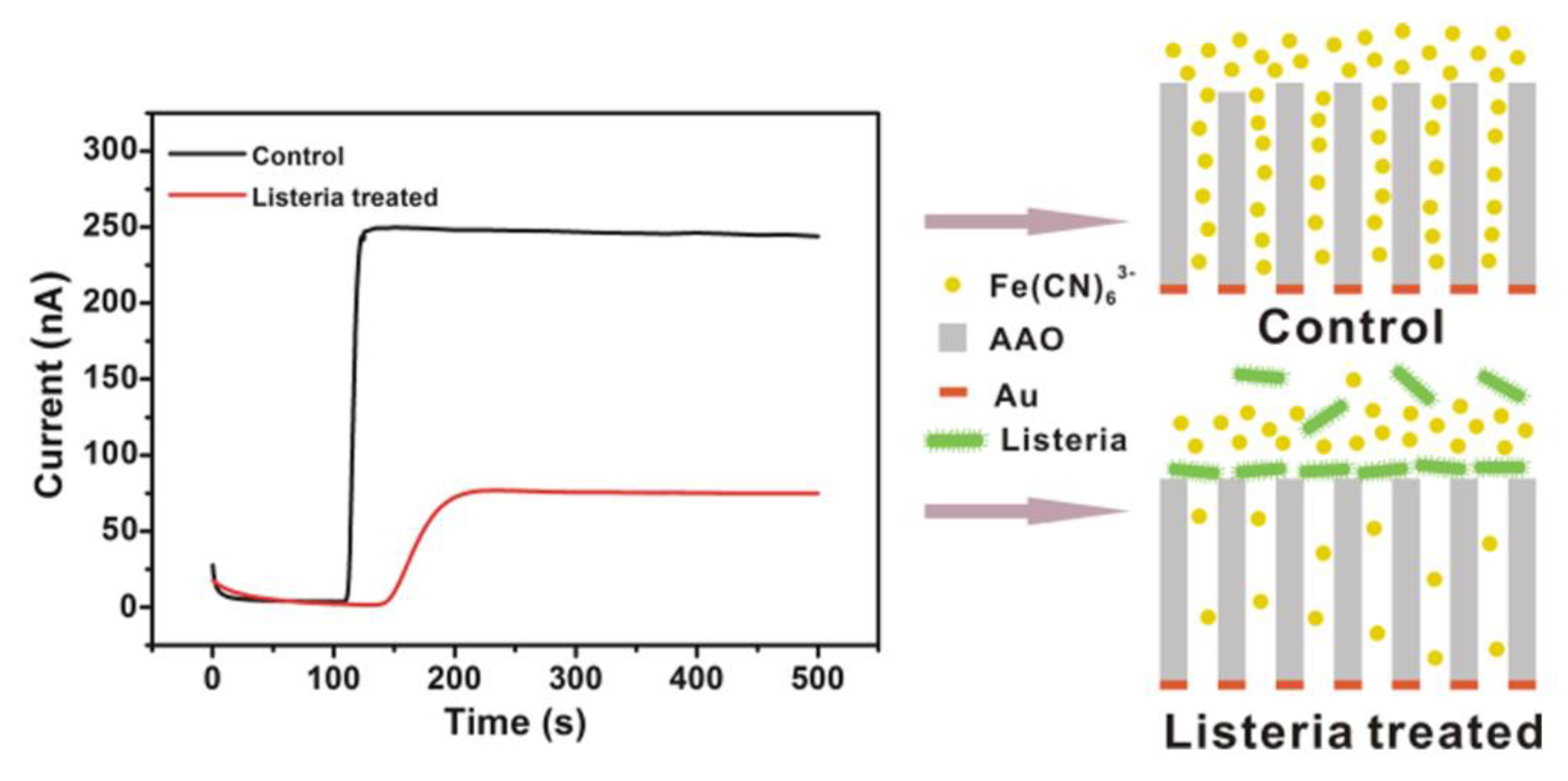
- Zhou, C.-X.; Mo, R.-J.; Chen, Z.-M.; Wang, J.; Shen, G.-Z.; Li, Y.-P.; Quan, Q.-G.; Liu, Y.; Li, C. Quantitative Label-Free Listeria Analysis Based On Aptamer Modified Nanoporous Sensor. ACS Sens. 2016, 1, 965–969. [Google Scholar] [CrossRef]
- Scholz, F. Voltammetric techniques of analysis: The essentials. ChemTexts 2015, 1, 17. [Google Scholar] [CrossRef]
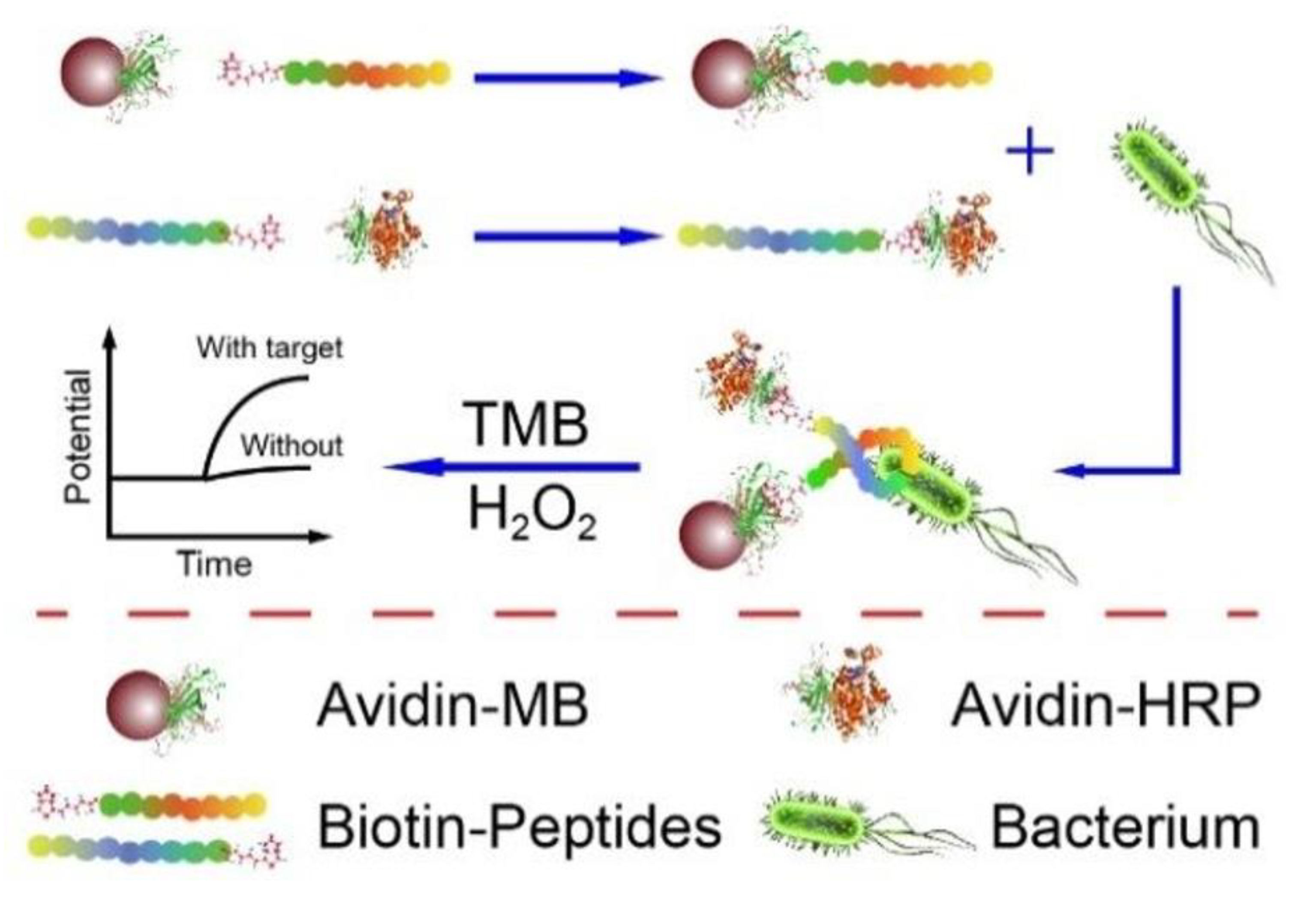
- Lv, E.; Ding, J.; Qin, W. Potentiometric Detection of Listeria monocytogenes via a Short Antimicrobial Peptide Pair-Based Sandwich Assay. Anal. Chem. 2018, 90, 13600–13606. [Google Scholar] [CrossRef]
- Kashish; Soni, D.K.; Mishra, S.K.; Prakash, R.; Dubey, S.K. Label-free impedimetric detection of Listeria monocytogenes based on poly-5-carboxy indole modified ssDNA probe. J. Biotechnol. 2015, 200, 70–76. [Google Scholar] [CrossRef] [PubMed]
- Zihni Onur, U.; Hilmiye Deniz Ertuğrul, U.; Ferhan Girgin, S. Nucleic Acids for Electrochemical Biosensor Technology. In Biosensors; Luis Jesús, V.-G., Ana Leticia, I., Eds.; IntechOpen: Rijeka, Croatia, 2020. [Google Scholar]
- Zhao, Q.; Lu, D.; Zhang, G.; Zhang, D.; Shi, X. Recent improvements in enzyme-linked immunosorbent assays based on nanomaterials. Talanta 2021, 223, 121722. [Google Scholar] [PubMed]
- Chen, Q.; Lin, J.; Gan, C.; Wang, Y.; Wang, D.; Xiong, Y.; Lai, W.; Li, Y.; Wang, M. A sensitive impedance biosensor based on immunomagnetic separation and urease catalysis for rapid detection of Listeria monocytogenes using an immobilization-free interdigitated array microelectrode. Biosens. Bioelectron. 2015, 74, 504–511. [Google Scholar] [CrossRef] [PubMed]
- Hadjilouka, A.; Loizou, K.; Apostolou, T.; Dougiakis, L.; Inglezakis, A.; Tsaltas, D. A Cell-Based Biosensor System for Listeria monocytogenes Detection in Food. Proceedings 2020, 60, 49. [Google Scholar]
- Chen, P.-H.; Lin, C.; Guo, K.-H.; Yeh, Y.-C. Development of a pigment-based whole-cell biosensor for the analysis of environmental copper. RSC Adv. 2017, 7, 29302–29305. [Google Scholar] [CrossRef]
- Silva, N.F.D.; Neves, M.M.P.S.; Magalhães, J.M.C.S.; Freire, C.; Delerue-Matos, C. Electrochemical immunosensor towards invasion-associated protein p60: An alternative strategy for Listeria monocytogenes screening in food. Talanta 2020, 216, 120976. [Google Scholar] [CrossRef]
- Lu, Y.; Liu, Y.; Zhao, Y.; Li, W.; Qiu, L.; Li, L. A Novel and Disposable Enzyme-Labeled Amperometric Immunosensor Based on MWCNT Fibers for Listeria monocytogenes Detection. J. Nanomater. 2016, 2016, 3895920. [Google Scholar] [CrossRef]
- Minett, A.I.; Barisci, J.N.; Wallace, G.G. Coupling conducting polymers and mediated electrochemical responses for the detection of Listeria. Anal. Chim. Acta 2003, 475, 37–45. [Google Scholar] [CrossRef]
- Susmel, S.; Guilbault, G.; O’Sullivan, C. Demonstration of labeless detection of food pathogens using electrochemical redox probe and screen printed gold electrodes. Biosens. Bioelectron. 2003, 18, 881–889. [Google Scholar] [CrossRef]
- Wang, D.; Chen, Q.; Huo, H.; Bai, S.; Cai, G.; Lai, W.; Lin, J. Efficient separation and quantitative detection of Listeria monocytogenes based on screen-printed interdigitated electrode, urease and magnetic nanoparticles. Food Control 2017, 73, 555–561. [Google Scholar] [CrossRef]
- Kanayeva, D.A.; Wang, R.; Rhoads, D.; Erf, G.F.; Slavik, M.F.; Tung, S.; Li, Y. Efficient separation and sensitive detection of Listeria monocytogenes using an impedance immunosensor based on magnetic nanoparticles, a microfluidic chip, and an interdigitated microelectrode. J. Food Prot. 2012, 75, 1951–1959. [Google Scholar] [CrossRef]
- Hadjilouka, A.; Loizou, K.; Apostolou, T.; Dougiakis, L.; Inglezakis, A.; Tsaltas, D. Newly developed system for the robust detection of Listeria monocytogenes based on a bioelectric cell biosensor. Biosensors 2020, 10, 178. [Google Scholar] [CrossRef]
- Cheng, C.; Peng, Y.; Bai, J.; Zhang, X.; Liu, Y.; Fan, X.; Ning, B.; Gao, Z. Rapid detection of Listeria monocytogenes in milk by self-assembled electrochemical immunosensor. Sens. Actuators B Chem. 2014, 190, 900–906. [Google Scholar] [CrossRef]
- Tully, E.; Higson, S.P.; O’Kennedy, R. The development of a ‘labeless’ immunosensor for the detection of Listeria monocytogenes cell surface protein, Internalin B. Biosens. Bioelectron. 2008, 23, 906–912. [Google Scholar] [CrossRef]
- Wang, R.; Ruan, C.; Kanayeva, D.; Lassiter, K.; Li, Y. TiO2 nanowire bundle microelectrode based impedance immunosensor for rapid and sensitive detection of Listeria monocytogenes. Nano Lett. 2008, 8, 2625–2631. [Google Scholar] [CrossRef]
- Radhakrishnan, R.; Jahne, M.; Rogers, S.; Suni, I.I. Detection of Listeria monocytogenes by electrochemical impedance spectroscopy. Electroanalysis 2013, 25, 2231–2237. [Google Scholar] [CrossRef]
- Sannigrahi, S.; Arumugasamy, S.K.; Mathiyarasu, J.; Suthindhiran, K. Development of magnetosomes-based biosensor for the detection of Listeria monocytogenes from food sample. IET Nanobiotechnol. 2020, 14, 839–850. [Google Scholar] [CrossRef] [PubMed]
- Davis, D.; Guo, X.; Musavi, L.; Lin, C.-S.; Chen, S.-H.; Wu, V.C.H. Gold Nanoparticle-Modified Carbon Electrode Biosensor for the Detection of Listeria monocytogenes. Ind. Biotechnol. 2013, 9, 31–36. [Google Scholar] [CrossRef]
- Tolba, M.; Ahmed, M.U.; Tlili, C.; Eichenseher, F.; Loessner, M.J.; Zourob, M. A bacteriophage endolysin-based electrochemical impedance biosensor for the rapid detection of Listeria cells. Analyst 2012, 137, 5749–5756. [Google Scholar] [CrossRef] [PubMed]
- Wu, Q.; Zhang, Y.; Yang, Q.; Yuan, N.; Zhang, W. Review of Electrochemical DNA Biosensors for Detecting Food Borne Pathogens. Sensors 2019, 19, 4916. [Google Scholar]
- Lung Khung, Y.; Narducci, D. Synergizing nucleic acid aptamers with 1-dimensional nanostructures as label-free field-effect transistor biosensors. Biosens. Bioelectron. 2013, 50, 278–293. [Google Scholar] [PubMed]
- Arshavsky-Graham, S.; Heuer, C.; Jiang, X.; Segal, E. Aptasensors versus immunosensors—Which will prevail? Eng. Life Sci. 2022, 22, 319–333. [Google Scholar] [CrossRef] [PubMed]
- Xue, Y.-Q.; Yang, X.; Sun, X.-L.; Han, Z.-Y.; Sun, J.; He, H. Reversible Structural Transformation of CuI–TbIII Heterometallic MOFs with Highly Efficient Detection Capability toward Penicillin. Inorg. Chem. 2021, 60, 11081–11089. [Google Scholar] [CrossRef] [PubMed]
- Tasbasi, B.B.; Guner, B.C.; Sudagidan, M.; Ucak, S.; Kavruk, M.; Ozalp, V.C. Label-free lateral flow assay for Listeria monocytogenes by aptamer-gated release of signal molecules. Anal. Biochem. 2019, 587, 113449. [Google Scholar] [CrossRef]
- Oliveira, D.A.; McLamore, E.S.; Gomes, C.L. Rapid and label-free Listeria monocytogenes detection based on stimuli-responsive alginate-platinum thiomer nanobrushes. Sci. Rep. 2022, 12, 21413. [Google Scholar] [CrossRef] [PubMed]
- Chen, W.; Cui, L.; Li, C.; Su, Y.; Tang, Y.; Xu, W. A novel aptamer biosensor using ZnO-3DNGH for sensitive and selective detection of Listeria monocytogenes. Microchem. J. 2022, 179, 107414. [Google Scholar] [CrossRef]
- Kharrati-koopaee, H.; Vahideh, R.; Esmailizadeh, A.; Sabahi, F. DNA Biosensors Techniques and Their Applications in Food Safety, Environmental Protection and Biomedical Research: A mini-review. J. Cell Dev. Biol. 2020, 3, 28–35. [Google Scholar]
- El Goumi, Y. Electrochemical Genosensors: Definition and Fields of Application. Int. J. Biosens. Bioelectron. 2017, 3, 353–355. [Google Scholar]
- Tichoniuk, M.; Ligaj, M.; Filipiak, M. Application of DNA Hybridization Biosensor as a Screening Method for the Detection of Genetically Modified Food Components. Sensors 2008, 8, 2118–2135. [Google Scholar] [CrossRef]
- Kashish; Gupta, S.; Dubey, S.K.; Prakash, R. Genosensor based on a nanostructured, platinum-modified glassy carbon electrode for Listeria detection. Anal. Methods 2015, 7, 2616–2622. [Google Scholar] [CrossRef]
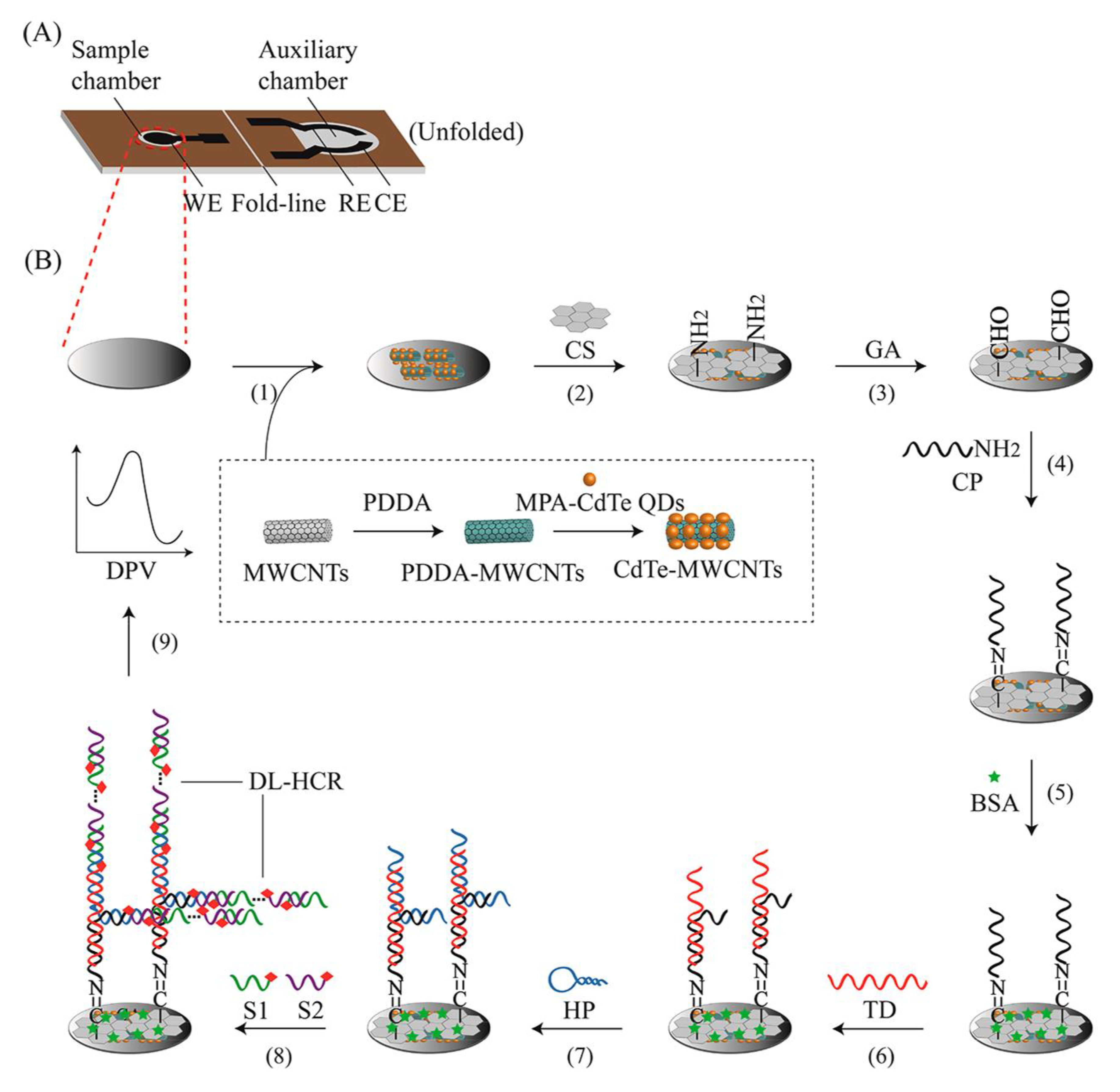
- Jiang, J.; Wu, H.; Su, Y.; Liang, Y.; Shu, B.; Zhang, C. Electrochemical Cloth-Based DNA Sensors (ECDSs): A New Class of Electrochemical Gene Sensors. Anal. Chem. 2020, 92, 7708–7716. [Google Scholar] [CrossRef]
- Liébana, S.; Brandão, D.; Cortés, P.; Campoy, S.; Alegret, S.; Pividori, M.I. Electrochemical genosensing of Salmonella, Listeria and Escherichia coli on silica magnetic particles. Anal. Chim. Acta 2016, 904, 1–9. [Google Scholar] [CrossRef]
- Bifulco, L.; Ingianni, A.; Pompei, R. An internalin a probe-based genosensor for Listeria monocytogenes detection and differentiation. BioMed Res. Int. 2013, 2013, 640163. [Google Scholar] [CrossRef] [PubMed]
- Wu, L.; Liu, Q.; Wu, Z.; Lu, Z. Detection of hlyA Gene of Listeria monocytogenes with Electrochemical DNA Biosensor. In Proceedings of the 2008 2nd International Conference on Bioinformatics and Biomedical Engineering, Shanghai, China, 16–18 May 2008; pp. 375–378. [Google Scholar]
- Laschi, S.; Palchetti, I.; Marrazza, G.; Mascini, M. Development of disposable low density screen-printed electrode arrays for simultaneous electrochemical measurements of the hybridisation reaction. J. Electroanal. Chem. 2006, 593, 211–218. [Google Scholar] [CrossRef]
- Sun, W.; Qi, X.; Zhang, Y.; Yang, H.; Gao, H.; Chen, Y.; Sun, Z. Electrochemical DNA biosensor for the detection of Listeria monocytogenes with dendritic nanogold and electrochemical reduced graphene modified carbon ionic liquid electrode. Electrochim. Acta 2012, 85, 145–151. [Google Scholar] [CrossRef]
- Niu, X.; Zheng, W.; Yin, C.; Weng, W.; Li, G.; Sun, W.; Men, Y. Electrochemical DNA biosensor based on gold nanoparticles and partially reduced graphene oxide modified electrode for the detection of Listeria monocytogenes hly gene sequence. J. Electroanal. Chem. 2017, 806, 116–122. [Google Scholar] [CrossRef]
- Gao, H.W.; Qin, P.; Lin, C.; Shang, Z.M.; Sun, W. Electrochemical DNA biosensor for the detection of Listeria monocytogenes using toluidine blue as a hybridization indicator. J. Iran. Chem. Soc. 2010, 7, 119–127. [Google Scholar] [CrossRef]
- Yan, L.; Zhao, W.; Wen, Z.; Li, X.; Niu, X.; Huang, Y.; Sun, W. Electrochemical DNA Sensor for hly gene of Listeria monocytogenes by three-dimensional graphene and gold nanocomposite modified electrode. Int. J. Electrochem. 2017, 12, 4086–4095. [Google Scholar] [CrossRef]
- Saini, K.; Kaushal, A.; Gupta, S.; Kumar, D. PlcA-based nanofabricated electrochemical DNA biosensor for the detection of Listeria monocytogenes in raw milk samples. 3 Biotech 2020, 10, 327. [Google Scholar] [CrossRef] [PubMed]
- Kavanagh, P.; Leech, D. Redox polymer and probe DNA tethered to gold electrodes for enzyme-amplified amperometric detection of DNA hybridization. Anal. Chem. 2006, 78, 2710–2716. [Google Scholar] [CrossRef]
- Wang, X.; Niu, S.; Wei, M.; Liu, S.; Liu, R.; Shi, C.; Ma, C. Ultrasensitive electrochemical DNA biosensor based on a tetrahedral structure and proximity-dependent surface hybridization. Analyst 2020, 145, 150–156. [Google Scholar] [CrossRef]
- Koonin, E.V.; Makarova, K.S. Origins and evolution of CRISPR-Cas systems. Philos. Trans. R. Soc. B 2019, 374, 20180087. [Google Scholar] [CrossRef] [Green Version]
- Manghwar, H.; Lindsey, K.; Zhang, X.; Jin, S. CRISPR/Cas system: Recent advances and future prospects for genome editing. Trends Plant Sci. 2019, 24, 1102–1125. [Google Scholar] [CrossRef]
- Principi, N.; Silvestri, E.; Esposito, S. Advantages and limitations of bacteriophages for the treatment of bacterial infections. Front. Pharmacol. 2019, 10, 513. [Google Scholar] [CrossRef] [PubMed]
- Pourakbari, R.; Yousefi, M.; Khalilzadeh, B.; Irani-nezhad, M.H.; Khataee, A.; Aghebati-Maleki, L.; Soleimanian, A.; Kamrani, A.; Chakari-Khiavi, F.; Abolhasan, R. Early stage evaluation of colon cancer using tungsten disulfide quantum dots and bacteriophage nano-biocomposite as an efficient electrochemical platform. Cancer Nanotechnol. 2022, 13, 7. [Google Scholar] [CrossRef]
- Zolti, O.; Suganthan, B.; Maynard, R.; Asadi, H.; Locklin, J.; Ramasamy, R.P. Electrochemical Biosensor for Rapid Detection of Listeria monocytogenes. J. Electrochem. Soc. 2022, 169, 067510. [Google Scholar]
- Rajapaksha, A.; Hashim, U.; Natasha, N.Z.; Uda, M.N.; Vijayakumaran, T.; Fernando, C.A. Gold nano-particle based Al interdigitated electrode electrical biosensor for specific ssDNA target detection. In Proceedings of the 2017 IEEE Regional Symposium on Micro and Nanoelectronics (RSM), Batu Ferringhi, Malaysia, 23–25 August 2017; pp. 191–194. [Google Scholar] [CrossRef]
- Sidhu, R.; Rong, Y.; Vanegas, D.C.; Claussen, J.; McLamore, E.S.; Gomes, C. Impedance biosensor for the rapid detection of Listeria spp. based on aptamer functionalized Pt-interdigitated microelectrodes array. SPIE 2016, 9863, 77–84. [Google Scholar]
- Wang, X.; Zhang, Z.; Wu, G.; Xu, C.; Wu, J.; Zhang, X.; Liu, J. Applications of electrochemical biosensors based on functional antibody-modified screen-printed electrodes: A review. Anal. Methods 2022, 14, 7–16. [Google Scholar] [CrossRef] [PubMed]
- Poltronieri, P.; Primiceri, E.; Radhakrishnan, R. EIS-Based Biosensors in Foodborne Pathogen Detection with a Special Focus on Listeria monocytogenes. In Foodborne Bacterial Pathogens: Methods and Protocols; Bridier, A., Ed.; Springer: New York, NY, USA, 2019; pp. 87–101. [Google Scholar] [CrossRef]
- Chen, Q.; Wang, D.; Cai, G.; Xiong, Y.; Li, Y.; Wang, M.; Huo, H.; Lin, J. Fast and sensitive detection of foodborne pathogen using electrochemical impedance analysis, urease catalysis and microfluidics. Biosens. Bioelectron. 2016, 86, 770–776. [Google Scholar] [CrossRef] [PubMed]
- Wang, Q.; Wang, J.; Huang, Y.; Du, Y.; Zhang, Y.; Cui, Y.; Kong, D.-M. Development of the DNA-based biosensors for high performance in detection of molecular biomarkers: More rapid, sensitive, and universal. Biosens. Bioelectron. 2022, 197, 113739. [Google Scholar] [CrossRef]
- Lettieri, S.; Avitabile, A.; Della Ventura, B.; Funari, R.; Ambrosio, A.; Maddalena, P.; Valadan, M.; Velotta, R.; Altucci, C. Nano-and femtosecond UV laser pulses to immobilize biomolecules onto surfaces with preferential orientation. Appl. Phys. A 2014, 117, 185–190. [Google Scholar] [CrossRef]
- Radhakrishnan, R.; Poltronieri, P. Fluorescence-Free Biosensor Methods in Detection of Food Pathogens with a Special Focus on Listeria monocytogenes. Biosensors 2017, 7, 63. [Google Scholar] [CrossRef] [Green Version]
- Hajdukiewicz, J.; Boland, S.; Kavanagh, P.; Leech, D. An enzyme-amplified amperometric DNA hybridisation assay using DNA immobilised in a carboxymethylated dextran film anchored to a graphite surface. Biosens. Bioelectron. 2010, 25, 1037–1042. [Google Scholar] [CrossRef] [PubMed]
- Aliakbarinodehi, N.; Stradolini, F.; Nakhjavani, S.A.; Tzouvadaki, I.; Taurino, I.; De Micheli, G.; Carrara, S. Performance of carbon nano-scale allotropes in detecting midazolam and paracetamol in undiluted human serum. IEEE Sens. J. 2018, 18, 5073–5081. [Google Scholar] [CrossRef]
- Rasouliyan, F.; Eskandani, M.; Jaymand, M.; Nakhjavani, S.A.; Farahzadi, R.; Vandghanooni, S.; Eskandani, M. Preparation, physicochemical characterization, and anti-proliferative properties of Lawsone-loaded solid lipid nanoparticles. Chem. Phys. Lipids 2021, 239, 105123. [Google Scholar] [CrossRef] [PubMed]
- Same, S.; Nakhjavani, S.A.; Samee, G.; Davaran, S. Halloysite clay nanotube in regenerative medicine for tissue and wound healing. Ceram. Int. 2022, 48, 31065–31079. [Google Scholar]
- Vandghanooni, S.; Rasoulian, F.; Eskandani, M.; Akbari Nakhjavani, S.; Eskandani, M. Acriflavine-loaded solid lipid nanoparticles: Preparation, physicochemical characterization, and anti-proliferative properties. Pharm. Dev. Technol. 2021, 26, 934–942. [Google Scholar] [CrossRef] [PubMed]
- Wu, Z.; Huang, C.; Dong, Y.; Zhao, B.; Chen, Y. Gold core @ platinum shell nanozyme-mediated magnetic relaxation switching DNA sensor for the detection of Listeria monocytogenes in chicken samples. Food Control 2022, 137, 108916. [Google Scholar] [CrossRef]
- El Aamri, M.; Yammouri, G.; Mohammadi, H.; Amine, A.; Korri-Youssoufi, H. Electrochemical biosensors for detection of microRNA as a cancer biomarker: Pros and cons. Biosensors 2020, 10, 186. [Google Scholar] [CrossRef]
- Zhang, T.; Chen, Y.; Huang, W.; Wang, Y.; Hu, X. A novel AuNPs-doped COFs composite as electrochemical probe for chlorogenic acid detection with enhanced sensitivity and stability. Sens. Actuators B Chem. 2018, 276, 362–369. [Google Scholar] [CrossRef]
- Xu, M.; Wang, L.; Xie, Y.; Song, Y.; Wang, L. Ratiometric electrochemical sensing and biosensing based on multiple redox-active state COFDHTA-TTA. Sens. Actuators B Chem. 2019, 281, 1009–1015. [Google Scholar] [CrossRef]
- Li, H.-K.; Ye, H.-L.; Zhao, X.-X.; Sun, X.-L.; Zhu, Q.-Q.; Han, Z.-Y.; Yuan, R.; He, H. Artful union of a zirconium-porphyrin MOF/GO composite for fabricating an aptamer-based electrochemical sensor with superb detecting performance. Chin. Chem. Lett. 2021, 32, 2851–2855. [Google Scholar] [CrossRef]
- Zhang, H.-W.; Zhu, Q.-Q.; Yuan, R.; He, H. Crystal engineering of MOF@ COF core-shell composites for ultra-sensitively electrochemical detection. Sens. Actuators B Chem. 2021, 329, 129144. [Google Scholar] [CrossRef]
- Sheikhzadeh, E.; Beni, V.; Zourob, M. Nanomaterial application in bio/sensors for the detection of infectious diseases. Talanta 2021, 230, 122026. [Google Scholar] [PubMed]
- Pumera, M.; Sanchez, S.; Ichinose, I.; Tang, J. Electrochemical nanobiosensors. Sens. Actuators B Chem. 2007, 123, 1195–1205. [Google Scholar] [CrossRef]
- Anik, U.; Timur, S.; Dursun, Z. Metal organic frameworks in electrochemical and optical sensing platforms: A review. Microchim. Acta 2019, 186, 196. [Google Scholar]
- Muniandy, S.; Teh, S.J.; Thong, K.L.; Thiha, A.; Dinshaw, I.J.; Lai, C.W.; Ibrahim, F.; Leo, B.F. Carbon nanomaterial-based electrochemical biosensors for foodborne bacterial detection. Crit. Rev. Anal. Chem. 2019, 49, 510–533. [Google Scholar] [CrossRef]
- Yu, W.; Sisi, L.; Haiyan, Y.; Jie, L. Progress in the functional modification of graphene/graphene oxide: A review. RSC Adv. 2020, 10, 15328–15345. [Google Scholar] [CrossRef] [PubMed]
- Kaur, J.; Vergara, A.; Rossi, M.; Gravagnuolo, A.M.; Valadan, M.; Corrado, F.; Conte, M.; Gesuele, F.; Giardina, P.; Altucci, C. Electrostatically driven scalable synthesis of mos 2–graphene hybrid films assisted by hydrophobins. Rsc Adv. 2017, 7, 50166–50175. [Google Scholar] [CrossRef]
- Vanegas, D.C.; Rong, Y.; Schwalb, N.; Hills, K.D.; Gomes, C.; McLamore, E.S. Rapid detection of Listeria spp. using an internalin A aptasensor based on carbon-metal nanohybrid structures. SPIE 2015, 9487, 14–20. [Google Scholar]
- Balaji Viswanath, K.; Suganya, K.; Krishnamoorthy, G.; Marudhamuthu, M.; Tamil Selvan, S.; Vasantha, V.S. Enzyme-Free Multiplex Detection of Foodborne Pathogens Using Au Nanoparticles-Decorated Multiwalled Carbon Nanotubes. ACS Food Sci. Technol. 2021, 1, 1236–1246. [Google Scholar] [CrossRef]
- Mishra, A.; Pilloton, R.; Jain, S.; Roy, S.; Khanuja, M.; Mathur, A.; Narang, J. Paper-Based Electrodes Conjugated with Tungsten Disulfide Nanostructure and Aptamer for Impedimetric Detection of Listeria monocytogenes. Biosensors 2022, 12, 88. [Google Scholar] [CrossRef] [PubMed]
- Santovito, E.; Greco, D.; D’Ascanio, V.; Sanzani, S.; Avantaggiato, G. Development of a DNA-based biosensor for the fast and sensitive detection of ochratoxin A in urine. Anal. Chim. Acta 2020, 1133, 20–29. [Google Scholar] [CrossRef] [PubMed]
- Goodarzi, M.; Shahhosseiny, M.H.; Bayat, M.; Hashemi, S.J.; Ghahri, M. Comparison between molecular methods (PCR vs LAMP) to detect Candida albicans in bronchoalveolar lavage samples of suspected tuberculosis patients. Microbiol. Res. 2018, 8, 7306. [Google Scholar] [CrossRef] [Green Version]


| Capture | Technique | Plan of Action | Sample | Interfering Agent | LOD | DT * | Ref |
|---|---|---|---|---|---|---|---|
| Gold nanoparticle—polyclonal antibody | Impedometry | Free-microelectrode | Listeria cells | Other bacteria | 300 CFU/mL | NR | [42] |
| HRP-tagged anti-Listeria monocytogenes antibody | Amperometry | Functionalized multi-walled carbon nanotube | Milk | Other bacteria | 1.07 × 10² CFU/mL | NR | [46] |
| Antibody against Listeria monocytogenes | Amperometry | Antibody-conjugated polypyrrole on platinum electrode | Listeria cells (alcohol-killed cells) | E. coli and salmonella | 105 cells/mL | 30 min | [47] |
| Antibody against Listeria monocytogenes | Impedometry | Modified screen-printed gold electrode with SAMs | Listeria-containing solution | mouse IgG | Not reported | NR | [48] |
| Gold nanoparticle –polyclonal antibody | Impedometry | Screen-printed circle-shaped interdigitated gold electrodes (three-finger) | Spiked sample (lettuce) | E. coli O157:H7 | 1.6 × 10³ CFU/mL | NR | [49] |
| Gold NPs-polyclonal anti-Listeria monocytogenes antibody conjugate | Impedometry | Microfluidic chip containing interdigitated gold microelectrodes | Lettuce, milk, ground beef | NR | 104 and 105 CFU/mL | 3 h | [50] |
| Anti-Listeria monocytogenes P60 monoclonal antibody | Amperometry | Modified screen-printed carbon electrodes with anti-P60 protein | Spiked milk | NR | 1.5 ng/mL | <3 h | [45] |
| Anti-Listeria monocytogenes p60 antibody | Potentiometry | Eight gold screen-printed electrodes modified by membrane-modified African monkey cells with AntiP60 | Cultured Listeria bacteria | NR | 102 CFU/mL | 3 min | [51] |
| Anti-Listeria monoclonal antibody | Chronoamperometry | Functionalized gold electrode with mercaptopropionic acid thiol-SAM | Spiked milk | NR | 103 to 106 CFU/mL, | NR | [52] |
| Biotinylated anti-internalin B polyclonal antibody | Impedometry | Electrical polymerized neutravidin-polyaniline on planar screen-printed carbon electrodes | The recombinant internalin F3 fragment | anti-IgG antibody | 4.1 pg/mL | NR | [53] |
| Anti-Listeria monocytogenes monoclonal antibody | Impedometry | Modified gold microelectrode with TiO2-nanowires | Cultured bacteria | NR | 102 CFU/mL | 1 h | [54] |
| Anti-Listeria monocytogenes monoclonal antibody | Impedometry | Modified Au electrode with 11-mercaptoundecanoic acid as a SAM | Filtered tomato extract | NR | 4 CFU/mL | NR | [55] |
| Anti-listeriolysin O antibody | Impedometry | Magnetosomes-Anti-LLO modified screen-printing carbon electrode | Contaminated milk and water | NR | 101 Cfu/mL | 30-min | [56] |
| Anti-Listeria monocytogenes antibody | Amperometry | Functionalized screen-printed carbon electrode (SPCE) with gold nanoparticles | Blueberry samples | E coli O157:H7 and Salmonella Typhimurium | 2 log CFU/g | NR | [57] |
| Capture | Technique | Plan of Action | Sample | Interfering Agent | LOD | Detection Time | Ref |
|---|---|---|---|---|---|---|---|
| Antibodies (polyclonal IgG) and a DNA aptamer (47-mer) | Impedometry | Chitosan nanobrushes on graphene/nanoplatinum electrodes | Foods | Other Gram-positive bacteria | Immunosensor (15.6 CFU mL−1) Aptasensor (9.1 CFU mL−1 | 17 min | [8] |
| 47-mer aptamer against internalin A | Potentiometry | The polycation-responsive membrane | Spiked coastal seawater | High concentration of Listeria (2000 CFU mL−1) | 10 CFU mL−1 | 0.67 h | [11] |
| 24 oligonucleotides of hlyA gene ssDNA | Impedometry | Electropolymerized 5-carboxyindole on gold electrode | Gene extraction product from Listeria | NR | 2.34 × 10⁻¹³ M | NR | [39] |
| 24-mer single-strand DNA probe complementary to hlyA gene | Impedometry | Platinum nanoparticles dispersed in the chitosan field | Milk samples | NR | 1 × 10⁻¹² M to 1 × 10⁻⁴ M | NR | [69] |
| 59-base inlA gene probe | Potentiometry | Screen-printed gold electrodes decorated with a mercaptan-activated self-assembled monolayer | Culture of Listeria monocytogenes | Non-pathogenic Listeria species DNA | NR | 50–60 min | [72] |
| 30-mer carboxylated probe | Amperometry | Silanization aluminum interdigitated electrode | Target DNA | NR | 1 fM to 1 µM | 1 h | [16] |
| Thiolated 49-mer Listeria hlyA gene probe | Impedometry | Gold electrodes activated by mercaptan, N-hydroxysulfosuccinimide (NHS), and N-(3-dimethylamion)propyl-N’-ethyl carbodiimide hydrochloride (EDC) | PCR product (Hly gene fragments) | NR | Not reported | NR | [73] |
| 18 thiol-treated oligonucleotides inlA Listeria toxin probe | Potentiometry | Screen-printed electrode using carbon and gold-based ink modified with the thiolated probe | Listeria inlA amplicon | Non-specific gene fragments | 1.5 to 3.6 µM | Less than 1 h | [74] |
| Amino-treated Listeria hly ssDNA probe | Potentiometry | Carbon ionic liquid electrode decorated by reduced graphene and gold nanostructures | PCR product of fish-isolated Listeria gene | Mismatch sequences | 2.9 × 10⁻¹³ mol/L | NR | [75] |
| Amino- 5′- end ssDNA | Potentiometry | Carbon ionic liquid electrode modified by Au nanoparticles and partially-reduced graphene oxide (p-RGO) | Synthetic DNA oligonucleotide | Mismatch sequences | 3.17 × 10⁻¹⁴ mol/L | NR | [76] |
| Listeria actA gene ssDNA | Potentiometry | Modified gold electrode with mercaptoacetic acid | PCR product | Not reported | NR | NR | [77] |
| Aminated Hly gene sequences as ssDNA probe | Potentiometry | Self-assembled mercaptoacetic acid (MAA) monolayer on carbon ionic liquid electrode functionalized with three-dimensional graphene rods (3DGR) and gold (Au) nanostructures | PCR product of hly Listeria gene amplification | Mismatched sequences | 3.3 × 10⁻¹⁵ mol/L | NR | [78] |
| 217-mer-biotinated-PCR amplicon of prfA gene | Amperometry | Silica magnetic particles | Streptavidin– horseradish peroxidase | NR | 0.13 ng/mL | 3 h | [71] |
| Aminated 20-mer plcA gene sequence | Impedometry | Carbon nanofiber modified gold nanoparticles | Raw milk | Other food-borne bacteria | 82 fg/6 µL | 30 min | [79] |
| Aminated ssDNA (ssrA gene) | Amperometry | Decorated gold electrode with cysteamine as a self-assembled monolayer and (osmium-based) redox polymer | Biotinylated ssDNA | NR | 1.4 fmol | 1 h | [80] |
| Tetrahedral structure of DNA probe | Amperometry | Bare gold electrode | Listeria monocytogenes extraction genome | NR | 0.2 fM | ~2 h | [81] |
| Working Electrode | Modified Method | LOD | Detection Time | Ref |
|---|---|---|---|---|
| Aluminum interdigitated with 1 µm gap-width (Al IDE) | Silanization of the electrode by APTES and immobilization of ssDNA with gold nanomaterials | 10 fM to 10 µM | NR | [87] |
| 4 and 8 screen-printed electrodes using carbon and gold-based ink | NR | 1.5 to 3.6 µM | Less than 1 h | [74] |
| Carbon ionic liquid electrode | NR | 2.9 × 10⁻¹³ mol/L | NR | [75] |
| Gold electrode | Use of conducting polymer (5-carboxyindole) | 2.34 × 10⁻¹³ M | NR | [39] |
| Gold electrode | methylene blue tagged ssDNA | 0.68 aM | 45 min | [30] |
| Screen-printed gold electrodes | Thioctic acid, mercaptopropionic acid, and mercaptoundecanoic acid self-assembled monolayers | Not reported | NR | [48] |
| Gold electrode | NR | NR | NR | [77] |
| Microfluidic/microelectrode conjugated system | Magnetic nanoparticle, monoclonal anti-Listeria | 1.6 × 10² CFU/mL | ~1 h | [91] |
| Screen-printed carbon electrode (SPCE) strips | Anti-Listeria conjugated gold nanoparticle | 2 log CFU/g | NR | [57] |
| Screen-printed interdigitated gold electrode | Polyclonal antibody and urease | 1.6 × 10 3 CFU/mL | NR | [49] |
| Screen-printed interdigitated gold electrode | Anti-P60 Listeria protein, engineered membrane cells | 102 CFU/ mL | 3 min | [51] |
| Planar screen-printed carbon electrodes | Electrical polymerized neutravidin–polyaniline | 4.1 pg/ml | NR | [53] |
| Screen-printed carbon electrode | Conjugation of Magnetosomes and anti-LLO | 101 CFU/ml | 30 min | [56] |
| Screen-printed gold electrode | Endolysine, self assembled monolayer | 1.1 × 10⁴ and 105 CFU/mL | NR | [58] |
| Capture | Technique | Sample | Reporter | LOD | Ref |
|---|---|---|---|---|---|
| 20-mer aminated ssDNA(ssrA gene) of Listeria | Amperometry | Solution of target DNA | Glucose oxidase | 0.2 nmol | [95] |
| Amino-treated Listeria hly ssDNA probe | Potentiometry | PCR product of deteriorated fish-isolated Listeria gene | Methylene blue | 2.9 × 10⁻¹³ mol/L | [75] |
| Amino- 5′-end ssDNA | Potentiometry | Synthetic DNA oligonucleotides | Methylene blue | 3.17 × 10⁻¹⁴ mol/L | [76] |
| Synthetic probe of Listeria actA gene | Potentiometry | PCR product of actA gene | Toluidine blue (TB) | NR | [77] |
| Amino-treated Listeria hly ssDNA probe | Potentiometry | PCR product of hly Listeria gene amplification | Methylene blue | 3.3 × 10⁻¹⁵ mol/L | [78] |
| Gold nanoparticle—polyclonal antibody | Impedometry | Listeria cells | Urease | 300 CFU/mL | [42] |
| Amino-treated Listeria ssrA gene-probe | Amperometry | Biotinylated DNA strand | Glucose oxidase | 1.4 fmol | [80] |
| Tetrahedral structure of DNA probe | Potentiometry | Listeria monocytogenes extraction genome | Ferrocene (Fc) | 0.2 fM | [81] |
| Anti-Listeria antibody | Amperometry | Blueberry samples | Horseradish peroxidase | 2 log CFU/g | [57] |
| Anti-Listeria antibody | Amperometry | Listeria cells | Toluidine blue (TB) | 105 cells/mL | [47] |
| Anti-p60 antibody | Amperometry | Spiked milk | Alkaline phosphatase | 1.5 ng mL−1 | [45] |
| ssDNA | Amperometry | Extracted genome | Methylene blue | 0.68 aM | [30] |
| Capture | Technique | Applied Nanomaterial | LOD | Detection Time | Ref |
|---|---|---|---|---|---|
| InlA aptamer | Impedimetry | Graphene/nanoplatinum on Pt/Ir electrodes | 100 CFU/mL | 3 h | [112] |
| Amino-treated Listeria hly ssDNA probe | Potentiometry | Reduced graphene/nanogold on carbon ionic liquid electrode | 2.9 × 10⁻¹³ mol/L | NR | [75] |
| Amino- 5′- end ssDNA | Potentiometry | Gold nanoparticle/partially reduced graphene oxide on carbon ionic liquid electrode | 3.17 × 10⁻¹⁴ mol/L | NR | [76] |
| HRP-tagged anti-Listeria antibody | Amperometry | Multi-walled carbon nanotube | 1.07 × 10² cfu/mL | NR | [46] |
| Anti-Listeria antibody(monoclonal) | Impdometry | TiO2-Nanowires | 102 CFU/mL | 1 h | [54] |
| P100 bacteriophage | Impdometry | Modified carbon nanotubes with quarternized polyethyleneimine | 8.4 CFU/mL | [86] | |
| InlA aptamer | Impdometry | PH-sensitive–electroactive ALG-thiomer/Pt nanobrush | (5 CFU mL−1) | 17 min | [64] |
| 47-mer aminated aptamer | Impdometry | WS2 nanoparticles | 10 CFU/mL | NR | [114] |
| Capture | Technique | Signal Amplification Method | LOD | Ref |
| Amino-treated ssDNA | Amperometry | Double linear hybridization chain reaction (DL-HCR) | 1.64 × 10⁴ CFU/mL and 11 CFU/mL | [70] |
Disclaimer/Publisher’s Note: The statements, opinions and data contained in all publications are solely those of the individual author(s) and contributor(s) and not of MDPI and/or the editor(s). MDPI and/or the editor(s) disclaim responsibility for any injury to people or property resulting from any ideas, methods, instructions or products referred to in the content. |
© 2023 by the authors. Licensee MDPI, Basel, Switzerland. This article is an open access article distributed under the terms and conditions of the Creative Commons Attribution (CC BY) license (https://creativecommons.org/licenses/by/4.0/).
Share and Cite
Mehrannia, L.; Khalilzadeh, B.; Rahbarghazi, R.; Milani, M.; Saydan Kanberoglu, G.; Yousefi, H.; Erk, N. Electrochemical Biosensors as a Novel Platform in the Identification of Listeriosis Infection. Biosensors 2023, 13, 216. https://doi.org/10.3390/bios13020216
Mehrannia L, Khalilzadeh B, Rahbarghazi R, Milani M, Saydan Kanberoglu G, Yousefi H, Erk N. Electrochemical Biosensors as a Novel Platform in the Identification of Listeriosis Infection. Biosensors. 2023; 13(2):216. https://doi.org/10.3390/bios13020216
Chicago/Turabian StyleMehrannia, Leila, Balal Khalilzadeh, Reza Rahbarghazi, Morteza Milani, Gulsah Saydan Kanberoglu, Hadi Yousefi, and Nevin Erk. 2023. "Electrochemical Biosensors as a Novel Platform in the Identification of Listeriosis Infection" Biosensors 13, no. 2: 216. https://doi.org/10.3390/bios13020216