Article

A Fluorescent Biosensor for Streptavidin Detection Based on Double-Hairpin DNA-Templated Copper Nanoparticles

1
Department of Hepatobiliary and Pancreatic Surgery, The Third Xiangya Hospital of Central South University, Changsha 410013, China
2
School of Life Sciences, Central South University, Changsha 410017, China
3
The Third Affiliated Hospital of Southern Medical University, Guangzhou 510640, China
*
Authors to whom correspondence should be addressed.
Biosensors 2023, 13(2), 168; https://doi.org/10.3390/bios13020168
Submission received: 5 December 2022 / Revised: 15 January 2023 / Accepted: 17 January 2023 / Published: 20 January 2023
(This article belongs to the Special Issue New Biosensors and Nanosensors)

Abstract

:
In this paper, we developed a sensitive, label-free and facile fluorescent strategy for detecting streptavidin (SA) based on double-hairpin DNA-templated copper nanoparticles (CuNPs) and terminal protection of small molecule-linked DNA. Herein, a special DNA hairpin probe was designed and synthesized, which contained two poly T single-stranded loops and a nick point in the middle of the stem. Inspired by the concept of the terminal protection interaction, the specific binding of SA to the biotinylated DNA probe can prevent the exonuclease degradation and keep the integrity of DNA probe, which can be used for synthesizing fluorescent CuNPs as a template. Conversely, the DNA probe would be digested by exonucleases and therefore, would fail to form CuNPs without SA. After systematic optimization, the detection range of SA concentration is from 0.5 to 150 nM with a low detection limit of 0.09 nM. Additionally, the proposed method was also successfully applied in the biological samples. Finally, the proposed method is sensitive, effective and simple, and can be potentially applied for predicting diseases and discovering new drugs.

1. Introduction

In recent years, the interactions between proteins and small molecules have been extensively studied for developing molecular diagnostics and discovering new drugs [1,2]. The detection method of DNA and proteins is based on the principle that the strong connections between proteins and small molecules at the terminal of single-stranded DNA (ssDNA) can prevent the degradation of ssDNA by exonuclease (Exo) [3,4]. This phenomenon, named terminal protection interaction, is convenient, inexpensive and easy to operate when used in biological samples [5,6]. The small molecule/protein interactions have been extensively exploited in protein assay in recent years [7,8].
Streptavidin (SA) obtained from Streptomyces avidinii has shown an extraordinarily high affinity for biotin [9]. SA is used in many aspects, such as biochemical sensors and nano biotechnology [8]. Its conjugation with enzyme or fluorescein is widely used in immunological detection assays. Due to the good biological resistance against extreme conditions, such as proteolytic enzymes, detergents (e.g., SDS), denaturants (e.g., guanidinium chloride) and extreme temperatures or pH, the SA-biotin complex is widely used in the fields of molecular biology [10]. The strong interactions between SA and biotin can be applied for biomedical diagnosis (predicting diseases and chemical genetics), target drug screening and molecular therapeutics. Therefore, it is crucial to develop a sensitive and facile strategy for SA detection [11,12].
Traditionally, different assays are available for the detection of SA, such as the protein-fragment complementation assay [13], near-field scanning optical microscopy [14], kinetic capillary electrophoresis [15], and surface plasmon resonance [16]. These traditional detection methods are usually highly sensitive and require a small amount of sample, but they also have some disadvantages, e.g., expensive instruments and a time-consuming and cumbersome assay, which limit their practical applications [17]. Focsan and Campu et al. reported an efficient and simple technique for detecting biotin-SA interactions with plasmonic nanobiosensors. Despite its high specificity, the limit of detection is still unsatisfactory in SA determination [18]. Khan et al. developed a highly sensitive SA detection method using liquid crystal, and the requirement for a special instrument is the major disadvantage for its application [19]. Therefore, a low-cost, highly sensitive and facile method needs to be developed for detecting SA.
DNA-templated fluorescent metal nanoparticles are new fluorescent probes with several advantages, such as facile synthetic process, good biological compatibility, outstanding optical properties and powerful signals [20,21,22]. Compared with other metal nanoparticles (i.e., silver nanoparticles and gold nanoparticles), copper nanoparticles (CuNPs) are extensively used in various bioanalysis due to their distinctive properties, such as faster synthesis, less toxicity, low-cost and lager MegaStokes shift [23,24,25]. As of now, CuNPs are used for developing various reporter systems for label-free sensing [26,27,28,29,30,31]. Wang et al. reported an efficient technique for microRNA detection using CuNPs based on rolling circle replication [32].
In this study, these distinct features of fluorescent CuNPs have been exploited for developing novel streptavidin detective sensors, based on double-hairpin DNA-templated CuNPs and terminal protection of small molecule-linked DNA. In this paper, a special DNA hairpin probe, which contained two poly T single-stranded loops and a nick point in the middle of the stem, was designed and synthesized for fluorescent CuNPs’ formation. In the absence of SA, Tn can be degraded by Exo I and III to become mononucleotide, which failed to act as a template in the synthesis of fluorescent CuNPs. Conversely, the strong interaction between streptavidin and biotin can protect the hairpin DNA even in the presence of Exo III and Exo I. Therefore, the reaction system would generate very high fluorescence intensity with the CuNPs, and the concentrations of SA can be associated with the fluorescence signal. Furthermore, the proposed method would be beneficial in the further research on developing protein detection methods, but also can be utilized as biosensors.

2. Materials and Methods

2.1. Materials and Reagents

Exonuclease I (Exo I), Exonuclease III (Exo III) were purchased from New England Biolabs Inc. (Ipswich, MA, USA). Streptavidin, human serum albumin (HSA), bovine serum albumin (BSA), lysozyme (Lyz), Immunoglobulin G (IgG) were bought from Sigma-Aldrich (St. Louis, MO, USA). The tris base, sodium chloride (NaCl), magnesium chloride (MgCl2), copper sulfate (CuSO4), sodium ascorbate (Vc) were bought from Sinopharm Chemical Reagent Co., Ltd. (Shanghai, China). The DNA probe was obtained from Shanghai Sangon Biotech Co. Ltd. (Shanghai, China). The sequence is as follows. The special DNA probe (Tn): 5′-TATATAGCTTTTTTTTTTTTTTTTTTTTTAGCTATATATATATA GCTTTTTTTTTTTTTTTTTTTTTAGCTATATA-biotin-3′.

2.2. Apparatus

The fluorescence intensity was determined by an F-2700 fluorescence spectrophotometer (Hitachi Ltd, Hitachi, Japan). The fluorescence emission spectra were recorded in the range of 530–650 nm with the excitation wavelength set at 340 nm. The excitation slit and emission slit were both set to 10 nm. The photomultiplier tube voltage was 700 V.

2.3. Optimization of the Experimental Conditions

For obtaining the best reaction systems, the reaction conditions, such as the concentrations of Tn, CuSO4, Vc and enzyme, and their reaction times were optimized. For optimizing Tn concentration, different concentrations of Tn were mixed with 500 nM SA in Tris-HCl buffer at 37 °C for 30 min. Then, 100 U/mL of Exo I and Exo III was fed into the reaction system and incubated for 30 min at 37 °C. Then, 100 μM of CuSO4 and 1 mM of Vc were added and incubated for 10 min at room temperature, utilizing the variation of fluorescence intensity to identify the optimal Tn concentration. The rest of the reaction conditions were optimized in a similar way.

2.4. Detection of SA

For quantifying the concentration of SA, different concentrations of SA were mixed with 200 nM of Tn in reaction buffer at 37 °C for 30 min, and then 30 U/mL of Exo I and 50 U/mL of Exo III were added and incubated for 30 min at 37 °C. Next, 80 μM of CuSO4 and 1 mM of Vc were added into the reaction system for 10 min at room temperature. An F-2700 fluorescence spectrophotometer was used to measure the fluorescence intensity. For verifying the application of this assay, different concentrations of SA in 1% human serum were measured.

2.5. Selectivity

SA was substituted by 150 nM of different proteins (i.e., BSA, HSA, Lyz, IgG and blank) for investigating the selectivity of the proposed method. The rest of the experimental steps were same in the above-mentioned method. At last, the fluorescence intensity can be measured at 600 nm with the excitation wavelength of 340 nm.

3. Results

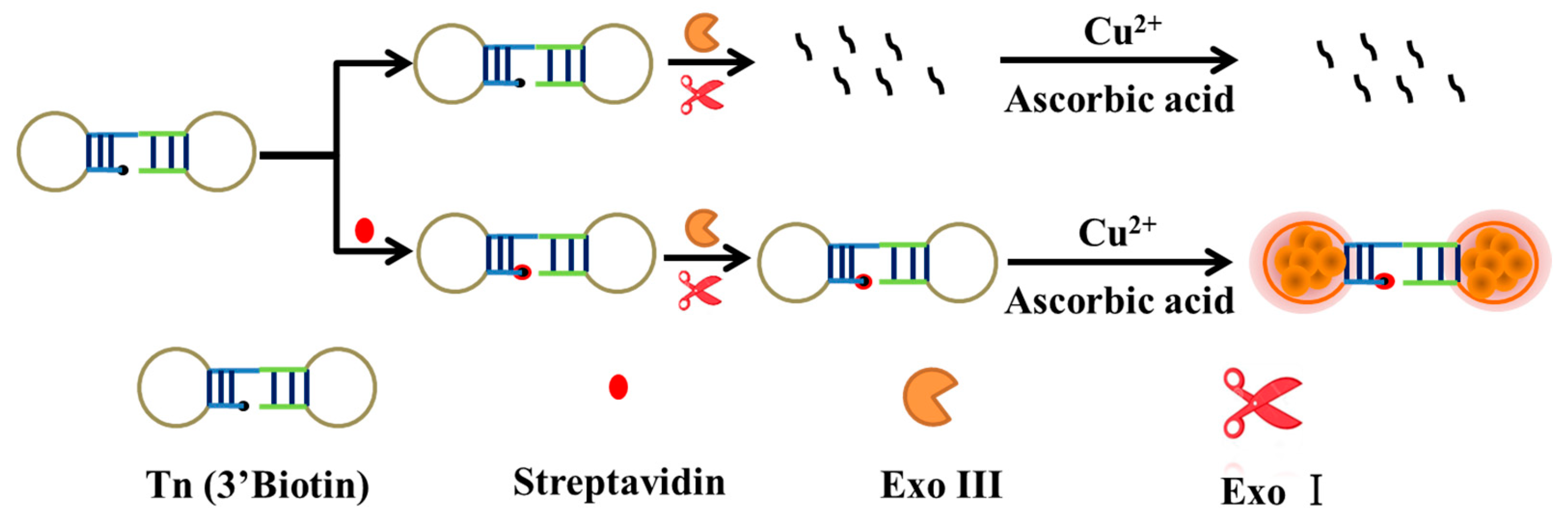
3.1. Principle of the SA Detection

The key part of the fluorescent biosensor is the formation of the SA-biotin complex, which can prevent degradation of the DNA probe (Tn) by the Exo I and Exo III with its strong steric hindrance [33]. At first, a special 3′-biotin-modified DNA probe was designed, which contained two poly T loops after forming the hairpin structure. In the absence of SA, Tn can be easily degraded into mononucleotide by exonucleases (Exo III for the double-stranded stem and Exo I for the remaining single-stranded loop) [34]. Therefore, no CuNPs were formed, and fluorescence did not generate. Conversely, in the presence of SA, due to the terminal protection of the SA-biotin complex, Tn retained the intact hairpin structure after adding Exo III and Exo I. Tn could be used as a template for CuNPs’ formation after adding CuSO4 and Vc, generating a high fluorescence intensity [35,36]. Therefore, the SA concentrations can be easily identified from the fluorescence change in the solutions (Scheme 1).

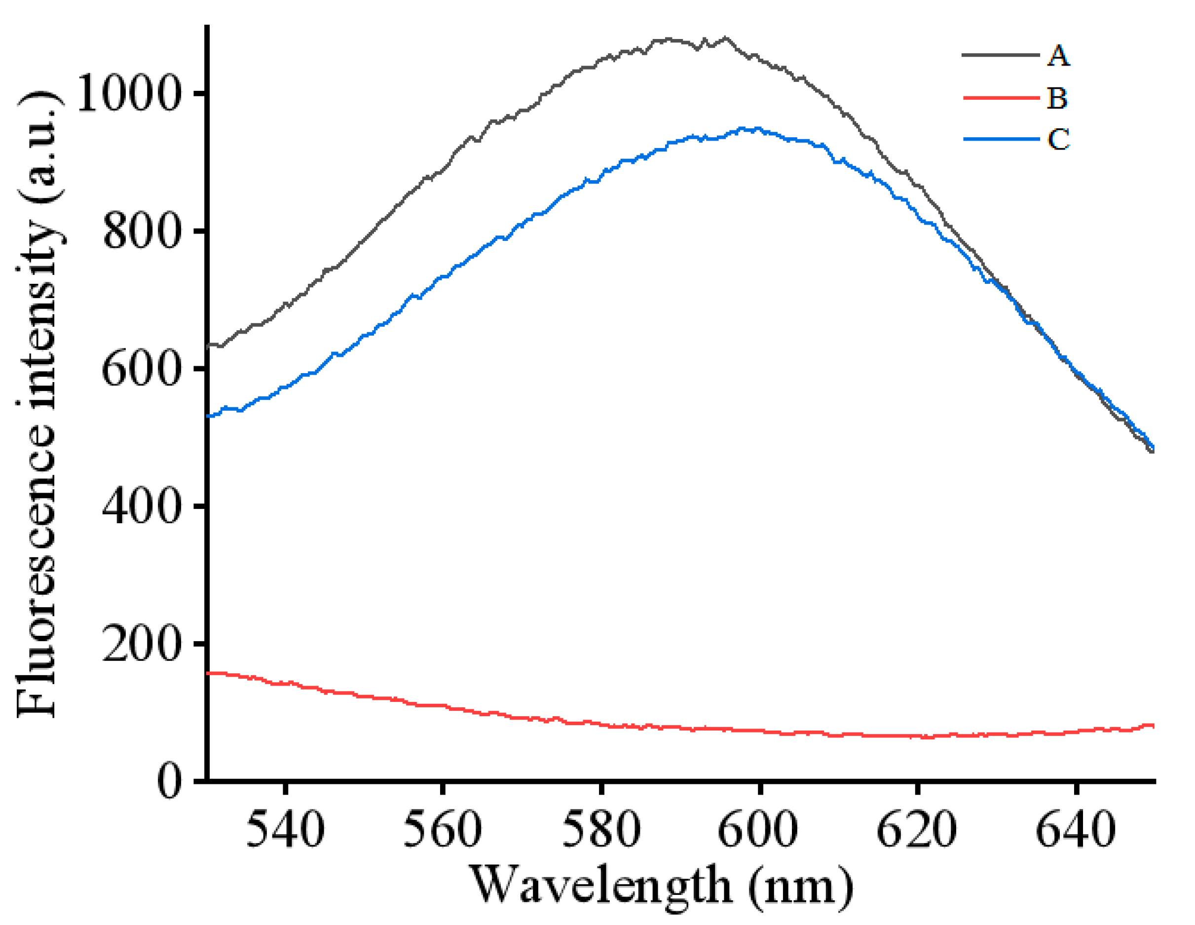
3.2. Feasibility Assessment of the SA Detection Assay

In order to evaluate the feasibility of the fluorescent biosensor, the fluorescence spectra were obtained under different conditions (Figure 1). A high fluorescence intensity was detected when the Tn probe was mixed with CuSO4 and Vc, indicating that CuNPs were synthesized successfully (Figure 1A). After the addition of Exo III and Exo I, the fluorescence intensity was diminished considerably due to the degradation of the enzyme (Figure 1B). As expected, when SA was added into the mixture before the enzyme, the fluorescence intensity increased dramatically (Figure 1C). The results suggested that the SA-biotin complex can prevent the catalysis of exonuclease [37]. Therefore, the proposed method can be used for the detection of SA.

3.3. Optimization of the Detection Strategy

The reaction conditions were optimized to obtain the best detection performance. (a) concentration of Tn; (b) concentration of Cu2+; (c) concentration of Vc; (d) concentration of Exo I; (e) concentration of Exo III; (f) reaction time of enzyme; (g) reaction time of SA. The best results of reaction conditions are as follows: (a) optimal Tn concentration: 200 nM (Figure S1 in Supplementary Materials); (b) optimal Cu2+ concentration: 80 μM (Figure S2); (c) optimal Vc concentration: 1 mM (Figure S3); (d) optimal concentration of Exo I: 30 U/mL (Figure S4); (e) optimal concentration of Exo III: 50 U/mL (Figure S5); (f) optimal reaction time of enzyme: 30 min (Figure S6); (g) optimal reaction time of SA: 30 min (Figure S7).

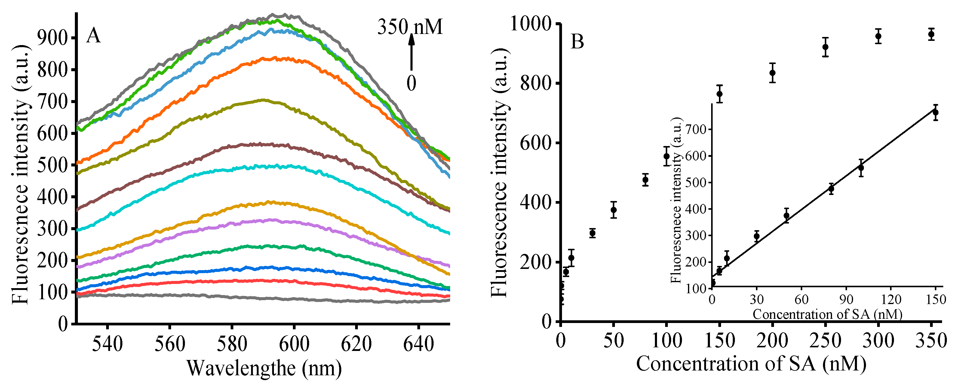
3.4. Performances of the Proposed Strategy

Under the optimal reaction conditions, the fluorescence intensities at different concentrations of SA (0, 0.5, 5, 10, 30, 50, 80, 100, 150, 200, 250, 300, 350 nM) were recorded. The fluorescence intensity increased with the enhancement of SA concentrations (in Figure 2A). Figure 2B shows a good linear relationship in the concentrations from 0.5 to 150 nM. Moreover, the regression equation was Y = 4.0902X + 153.58 with an R2 of 0.9927 (where Y was the fluorescence intensity at 600 nm and X was the SA concentration, respectively). The LOD (detectable lowest concentration) was 0.09 nM (S/N = 3), which shows equivalent or better detection capacity than the previous works in the literature (Table 1). Compared with the previous work, this method is simple and rapid [32]. Therefore, this method has a wide detection range and a very low detection limit, which is a very promising SA detection method.

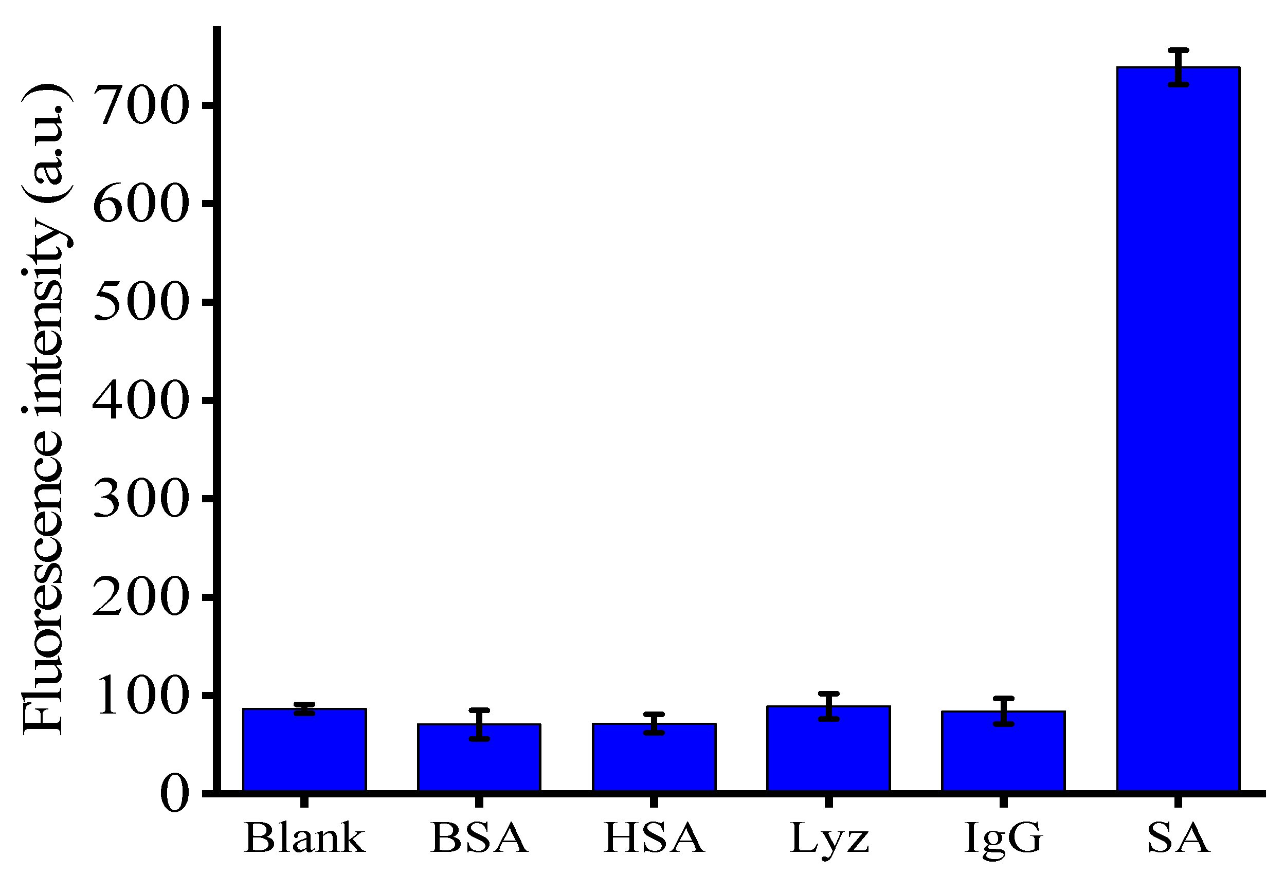
3.5. Selectivity of the Method

We tested 150 nM of several proteins, such as BSA, HAS, IgG, Lyz and blank, by the proposed assay under the optimized concentrations to investigate the selectivity [38]. The fluorescence intensity of other protein samples, which is comparable to the blank one, is obviously different from the sample adding SA (in Figure 3). The reason is that there is a high specificity between SA and biotin, which can protect DNA from exonuclease degradation through the terminal protection interaction. Thus, only the group with SA has a high fluorescence signal. These results indicate that the proposed assay showed good selectivity for the quantitative determination of SA.

3.6. Practical Applicability of the Detection Strategy

In order to further verify the practical application value of this biosensor, we applied the fabricated sensing platform to the detection of SA in 1% human serum [39]. The human serum samples were provided by us. Three concentrations of SA (20, 60, 100 nM) were determined by the proposed assay in 1% human serum diluted by adding reaction buffer. We evaluated the recovery rates for different concentrations of SA (Table 2), such as 99% for 20 nM, 98.5% for 60 nM and 100.6% for 100 nM. Therefore, the proposed strategy can be successfully applied in biological systems for SA detection.

4. Conclusions

In summary, a simple and sensitive fluorescent method, based on the small-molecule-linked DNA terminal protection strategy and double-hairpin DNA-templated CuNPs, was successfully developed for SA determination. The fluorescent biosensor utilizes the specific binding between SA and biotin to prevent the degradation of enzymes and protect the formation of CuNPs, and then affect the change of fluorescence signal. This method exhibits a linear range from 0.5 to 150 nM with a low detection limit of 0.09 nM (S/N = 3). Overall, the detection assay has a wide linear range, low detection limit, and good specificity. In addition, the successful application of the method in the human serum demonstrates its practical use in biological systems. Thus, the proposed method is sensitive, label-free and facile, and can be potentially applied in biological systems.

Supplementary Materials

The following supporting information can be downloaded at: https://www.mdpi.com/article/10.3390/bios13020168/s1, Figure S1: Optimization of the concentration of Tn; Figure S2: Optimization of the concentration of Cu2+; Figure S3: Optimization of the concentration of Vc; Figure S4: Optimization of the concentration of Exo I; Figure S5: Optimization of the concentration of Exo III; Figure S6: Optimization of the reaction time of enzyme; Figure S7: Optimization of the reaction time of SA.

Author Contributions

Conceptualization, C.M. and X.Y.; investigation, Q.X. and M.C.; writing—original draft preparation, Q.X., M.C. and W.N.; writing—review and editing, F.X. and C.M.; supervision, C.M. and X.Y.; funding acquisition, X.Y. All authors have read and agreed to the published version of the manuscript.

Funding

This research was funded by the Research Innovation Program for Graduates of Central South University (2018zzts384, 2019zzts453).

Institutional Review Board Statement

The study was approved by the Ethics Committee of Central South University (protocol code 2020-1-11; date of approval 4 February 2020).

Informed Consent Statement

Informed consent was obtained from all subjects involved in the study.

Data Availability Statement

Not applicable.

Conflicts of Interest

The authors declare no conflict of interest.

References

  1. Lee, C.Y.; Kim, H.Y.; Kim, S.; Park, K.S.; Park, H.G. A simple and sensitive detection of small molecule-protein interactions based on terminal protection-mediated exponential strand displacement amplification. Analyst 2018, 143, 2023–2028. [Google Scholar] [CrossRef]
  2. Wu, Y.; Huang, J.; Yang, X.; Yang, Y.; Quan, K.; Xie, N.; Li, J.; Ma, C.; Wang, K. Gold nanoparticle loaded split DNAzyme probe for amplified miRNA detection in living cells. Anal. Chem. 2017, 89, 8377–8383. [Google Scholar] [CrossRef] [PubMed]
  3. Bai, D.M.; Ji, D.Y.; Shang, J.H.; Hu, Y.L.; Gao, J.; Lin, Z.; Ge, J.; Li, Z.H. A rapid biosensor for highly sensitive protein detection based on G-quadruplex-Thioflavin T complex and terminal protection of small molecule-linked DNA. Sens. Actuators B Chem. 2017, 252, 1146–1152. [Google Scholar] [CrossRef]
  4. Chen, M.; Li, W.; Ma, C.; Wu, K.; He, H.; Wang, K. Fluorometric determination of the activity of uracil-DNA glycosylase by using on graphene oxide and exonuclease I assisted signal amplification. Microchim. Acta 2019, 186, 110. [Google Scholar] [CrossRef]
  5. Wang, Q.; Jiang, B.Y.; Xu, J.; Xie, J.Q.; Xiang, Y.; Yuan, R.; Chai, Y.Q. Amplified terminal protection assay of small molecule/protein interactions via a highly characteristic solid-state Ag/AgCl process. Biosens. Bioelectron. 2013, 43, 19–24. [Google Scholar] [CrossRef]
  6. Liu, Q.; Sun, X.; Liu, M.; Jin, Y.; Li, B. G-triplex molecular beacon-based fluorescence biosensor for sensitive detection of small molecule-protein interaction via exonuclease III-assisted recycling amplification. Sens. Actuators B Chem. 2020, 310, 127804. [Google Scholar] [CrossRef]
  7. Zhang, Z.; Xia, X.; Xiang, X.; Huang, F.; Han, L. Conjugated cationic polymer-assisted amplified fluorescent biosensor for protein detection via terminal protection of small molecule-linked DNA and graphene oxide. Sens. Actuators B Chem. 2017, 249, 8–13. [Google Scholar] [CrossRef]
  8. Ouyang, P.; Fang, C.; Han, J.; Zhang, J.; Yang, Y.; Qing, Y.; Chen, Y.; Shang, W.; Du, J. A DNA Electrochemical Sensor via Terminal Protection of Small-Molecule-Linked DNA for Highly Sensitive Protein Detection. Biosensors 2021, 11, 451. [Google Scholar] [CrossRef]
  9. He, Y.; Jiao, B.N. Detection of biotin-streptavidin interactions via terminal protection of small molecule linked DNA and the formation of fluorescent DNA-templated silver nanoclusters. Microchim. Acta 2016, 183, 3183–3189. [Google Scholar] [CrossRef]
  10. Xu, F.F.; Yang, T.; Chen, Y. Quantification of microRNA by DNA-peptide probe and liquid chromatography-tandem mass spectrometry-based quasi-targeted proteomics. Anal. Chem. 2016, 88, 754–763. [Google Scholar] [CrossRef]
  11. Chen, M.J.; Deng, Z.Y.; Ma, C.B.; Zhao, H.; Wu, K.F.; Wang, K.M. A sensitive fluorescence method for the detection of streptavidin based on target-induced DNA machine amplification. Anal. Methods. 2018, 10, 1870–1874. [Google Scholar] [CrossRef]
  12. Cao, J.; Wang, W.; Bo, B.; Mao, X.; Wang, K.; Zhu, X. A dual-signal strategy for the solid detection of both small molecules and proteins based on magnetic separation and highly fluorescent copper nanoclusters. Biosens. Bioelectron. 2017, 90, 534–541. [Google Scholar] [CrossRef]
  13. Zhuang, Y.D.; Chiang, P.Y.; Wang, C.W.; Tan, K.T. Environment-sensitive fluorescent turn-on probes targeting hydrophobic ligand-binding domains for selective protein detection. Angew. Chem. Int. Ed. 2013, 52, 8124–8128. [Google Scholar] [CrossRef] [PubMed]
  14. Sun, Q.; Qian, J.H.; Tian, H.Y.; Duan, L.P.; Zhang, W.B. Rational design of biotinylated probes: Fluorescent turn-on detection of (strept)avidin and bioimaging in cancer cells. Chem. Commun. 2014, 50, 8518–8521. [Google Scholar] [CrossRef] [PubMed]
  15. Drabovich, A.P.; Berezovski, M.V.; Musheev, M.U.; Krylov, S.N. Selection of smart small-molecule ligands: The proof of principle. Anal. Chem. 2009, 81, 490–494. [Google Scholar] [CrossRef] [PubMed]
  16. Seto, H.; Yamashita, C.; Kamba, S.; Kondo, T.; Hasegawa, M.; Matsuno, M.; Ogawa, Y.C.; Hoshino, Y.; Miura, Y. Biotinylation of silicon and nickel surfaces and detection of streptavidin as biosensor. Langmuir 2009, 29, 9457–9463. [Google Scholar] [CrossRef] [PubMed]
  17. Ban, F.F.; Shi, H.; Feng, C.; Mao, X.X.; Yin, Y.M.; Zhu, X.L. A one-pot strategy for the detection of proteins based on sterically and allosterically tunable hybridization chain reaction. Biosens. Bioelectron. 2016, 86, 219–224. [Google Scholar] [CrossRef]
  18. Focsan, M.; Campu, A.; Craciun, A.M.; Potara, M.; Leordean, C.; Maniu, D.; Astilean, S. A simple and efficient design to improve the detection of biotin-streptavidin interaction with plasmonic nanobiosensors. Biosens. Bioelectron. 2016, 86, 728–735. [Google Scholar] [CrossRef]
  19. Khan, M.; Park, S.Y. Specific detection of avidin-biotin binding using liquid crystal droplets. Colloids Surf. B Biointerfaces 2015, 127, 241–246. [Google Scholar] [CrossRef]
  20. Liu, H.S.; Ma, C.B.; Wang, J.; Wang, K.M.; Wu, K.F. A turn-on fluorescent method for determination of the activity of alkaline phosphatase based on dsDNA-templated copper nanoparticles and exonuclease based amplification. Microchim. Acta 2017, 184, 2483–2488. [Google Scholar] [CrossRef]
  21. Zhang, X.; Liu, Q.; Jin, Y.; Li, B. Determination of the activity of T4 polynucleotide kinase phosphatase by exploiting the sequence-dependent fluorescence of DNA-templated copper nanoclusters. Microchim. Acta 2019, 186, 3. [Google Scholar] [CrossRef]
  22. Han, Y.; Zhang, F.; Gong, H.; Cai, C. Double G-quadruplexes in a copper nanoparticle based fluorescent probe for determination of HIV genes. Microchim. Acta 2019, 186, 30. [Google Scholar] [CrossRef] [PubMed]
  23. Qing, Z.; He, X.; He, D.; Wang, K.; Xu, F.; Qing, T.; Yang, X. Poly(thymine)-Templated Selective Formation of Fluorescent Copper Nanoparticles. Angew. Chem. Int. Ed. 2013, 52, 9719–9722. [Google Scholar] [CrossRef]
  24. Qing, T.; Long, C.; Wang, X.; Zhang, K.; Zhang, P.; Feng, B. Detection of micrococcal nuclease for identifying Staphylococcus aureus based on DNA templated fluorescent copper nanoclusters. Microchim. Acta 2019, 186, 248. [Google Scholar] [CrossRef] [PubMed]
  25. Chen, M.; Ma, C.; Yan, Y. Label-free fluorescence method for actin detection based on DNA-templated silver nanoclusters. Anal. Methods 2019, 11, 4348–4353. [Google Scholar] [CrossRef]
  26. Cai, Q.; Ge, J.; Xu, H.; Zhang, L.; Hu, Y.; Huang, Z.; Li, Z. A label-free aptasensor for highly sensitive ATP detection by using exonuclease I and oligonucleotide-templated fluorescent copper nanoparticles. Anal. Methods 2017, 9, 2710–2714. [Google Scholar] [CrossRef]
  27. Ge, J.; Dong, Z.; Zhang, L.; Cai, Q.; Bai, D.; Li, Z. Label-free biosensor based on dsDNA-templated copper nanoparticles for highly sensitive and selective detection of NAD+. RSC Adv. 2016, 6, 91077–91082. [Google Scholar] [CrossRef]
  28. Chen, M.; Khusbu, F.; Ma, C.; Wu, K.; Zhao, H.; Chen, H.; Wang, K. A sensitive detection method of carcinoembryonic antigen based on dsDNA-templated copper nanoparticles. New J. Chem. 2018, 42, 13702–13707. [Google Scholar] [CrossRef]
  29. Zhang, L.; Cai, Q.; Li, J.; Ge, J.; Wang, J.; Dong, Z.; Li, Z. A label-free method for detecting biothiols based on poly(thymine)-templated copper nanoparticles. Biosens. Bioelectron. 2015, 69, 77–82. [Google Scholar] [CrossRef] [PubMed]
  30. Kim, S.; Kim, J.; Kwon, W.; Hwang, S.; Cha, B.; Kim, J.; Seung, S.; Ki, S. Synthesis of DNA-templated copper nanoparticles with enhanced fluorescence stability for cellular imaging. Microchim. Acta 2019, 186, 479. [Google Scholar] [CrossRef]
  31. Chen, W.; Dai, L.; Liu, Z.; Yang, W.; Zhao, C.; Li, Y.; Chen, Y. Hairpin-shaped DNA templated copper nanopartides for fluorescence detection of adenosine triphosphate based on ligation-mediated exonuclease cleavage. Anal. Sci. 2017, 33, 203–207. [Google Scholar]
  32. Xu, F.; Shi, H.; He, X.; Wang, K.; He, D.; Guo, Q.; Qing, Z.; Yan, L.; Ye, X.; Li, D.; et al. Concatemeric dsDNA-templated copper nanoparticles strategy with improved sensitivity and stability based on rolling circle replication and its application in microRNA Detection. Anal. Chem. 2014, 86, 6976–6982. [Google Scholar] [CrossRef]
  33. He, Y.; Jiao, B.N. DNA covalently linked to graphene oxide for biotin-streptavidin interaction assay. Talanta 2017, 63, 140–145. [Google Scholar] [CrossRef] [PubMed]
  34. Qing, T.P.; He, X.X.; He, D.G.; Ye, X.S.; Shangguan, J.F.; Liu, J.Q.; Yuan, B.Y.; Wang, K.M. Dumbbell DNA-templated CuNPs as a nano-fluorescent probe for detection of enzymes involved in ligase-mediated DNA repair. Biosens. Bioelectron. 2017, 94, 456–463. [Google Scholar] [CrossRef] [PubMed]
  35. Yin, J.X.; Liu, F.; Fan, T.T.; Ren, Y.; Jiang, Y.Y. Rapid detection of methyltransferases utilizing dumbbell DNA-templated copper nanoparticles. Sens. Actuators B Chem. 2018, 276, 499–506. [Google Scholar] [CrossRef]
  36. He, Y.; Tian, F.; Zhou, J.; Jiao, B. A fluorescent aptasensor for ochratoxin A detection based on enzymatically generated copper nanoparticles with a polythymine scaffold. Microchim. Acta 2019, 186, 199. [Google Scholar] [CrossRef] [PubMed]
  37. Chen, X.X.; Lin, C.S.; Chen, Y.Y.; Luo, F.; Wang, Y.R.; Chen, X. Terminal protection of a small molecule-linked loop DNA probe for turn-on label-free fluorescence detection of proteins. Biosens. Bioelectron. 2016, 83, 97–101. [Google Scholar] [CrossRef]
  38. Wang, H.B.; Zhang, H.D.; Chen, Y.; Liu, Y.M. A fluorescent biosensor for protein detection based on poly(thymine)-templated copper nanoparticles and terminal protection of small molecule-linked DNA. Biosens. Bioelectron. 2015, 74, 581–586. [Google Scholar] [CrossRef]
  39. Xiang, X.; Shi, J.B.; Huang, F.G.; Zheng, M.M.; Deng, Q.C.; Xu, J.Q. MoS2 nanosheet-based fluorescent biosensor for protein detection via terminal protection of small-molecule-linked DNA and exonuclease III-aided DNA recycling amplification. Biosens. Bioelectron. 2015, 74, 227–232. [Google Scholar] [CrossRef]
Scheme 1. Schematic of streptavidin detection based on double-hairpin DNA-templated copper nanoparticles and terminal protection of small molecule-linked DNA.
Biosensors 13 00168 sch001
Figure 1. The feasibility of the proposed method. (A) Tn + CuSO4 + Vc; (B) Tn + Exo III + Exo I+ CuSO4 + Vc; (C) Tn + SA + Exo III + Exo I+ CuSO4 + Vc. Concentrations of Tn, SA, Exo III, Exo I, CuSO4 and Vc were 200 nM, 500 nM, 100 U/mL, 100 U/mL, 100 μM and 1 mM, respectively.
Biosensors 13 00168 g001
Figure 2. (A) Fluorescence emission spectra in the response to different concentrations of SA (0, 0.5, 5, 10, 30, 50, 80, 100, 150, 200, 250, 300, 350 nM) under optimal concentration. (B) Fluorescence intensity versus SA concentration at 600 nm and the linear relationship between fluorescence intensities and low concentrations of SA (inset); error bars were estimated from three replicate measurements.
Biosensors 13 00168 g002
Figure 3. Fluorescence intensity of reaction system in the presence of SA, BSA, HSA, Lyz and IgG. Error bars were estimated from three replicated measurements.
Biosensors 13 00168 g003
Table 1. Comparison of different methods for the determination of SA.
MethodMaterialDetection Range (nM)LOD (nM)Reference
SPRGold nanoparticle 518
ElectrochemistryGold electrode0.5–50000.028
FluorescenceSYBR Green I0.01–0.10.021
FluorescenceAgNCs6–6002.69
FluorescenceSYBR Green I5–2000.410
FluorescenceThT0.1–170.0711
FluorescenceCuNCs1–2000.4712
FluorescencePyrene4–10001.0717
FluorescenceFAM0.15–120.0833
FluorescenceCuNPs0.5–1500.09This work
Table 2. Recovery experiments of SA in diluted human serum using this method (n = 3).
SampleAdded (nM)Found (nM)Recovery (%)RSD (%)
12019.8 ± 0.4997.71
26059.1 ± 1.598.59.31
3100100.6 ± 2.1100.66.05
Disclaimer/Publisher’s Note: The statements, opinions and data contained in all publications are solely those of the individual author(s) and contributor(s) and not of MDPI and/or the editor(s). MDPI and/or the editor(s) disclaim responsibility for any injury to people or property resulting from any ideas, methods, instructions or products referred to in the content.

