Conjugated Aggregation-Induced Fluorescent Materials for Biofluorescent Probes: A Review

Abstract
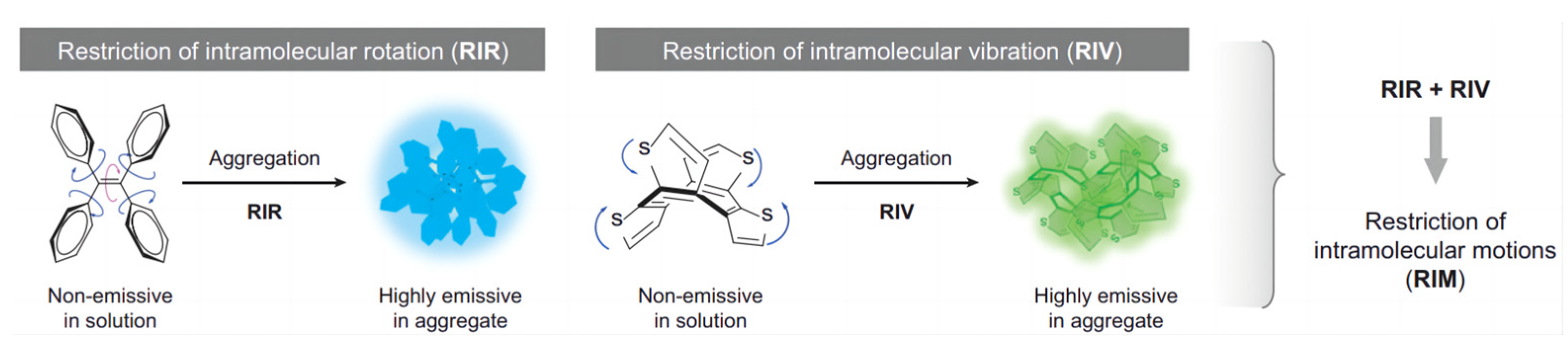
:1. Introduction
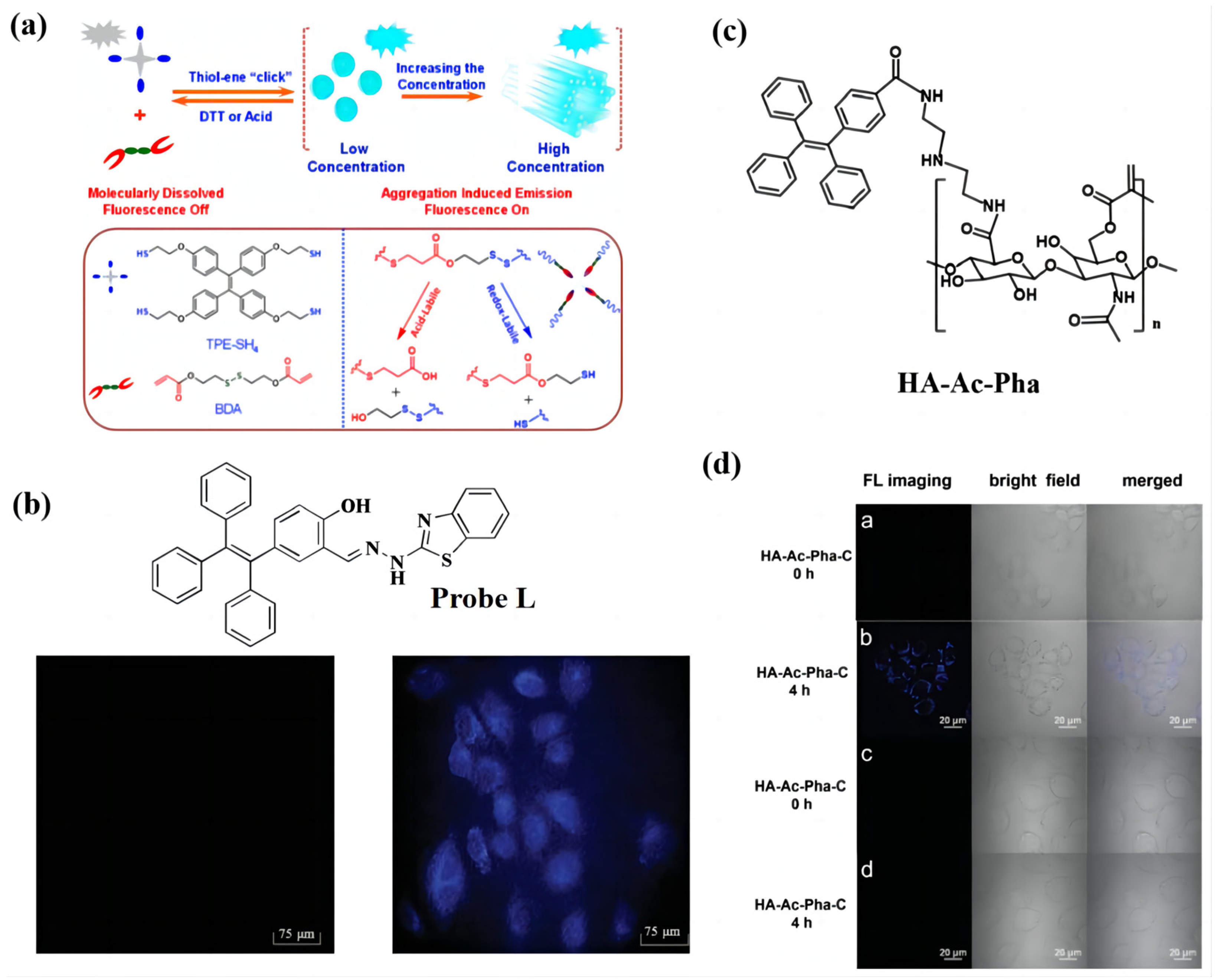
2. AIE Material Design Strategy Based on the Functional Groups

3. Hydrogen Bonding and D-A System Strategy
4. Conclusions and Outlook
Funding
Institutional Review Board Statement
Informed Consent Statement
Data Availability Statement
Conflicts of Interest
References
- Luo, J.; Xie, Z.; Lam, J.W.Y.; Cheng, L.; Tang, B.Z.; Chen, H.; Qiu, C.; Kwok, H.S.; Zhan, X.; Liu, Y.; et al. Aggregation-Induced Emission of 1-Methyl-1,2,3,4,5-Pentaphenylsilole. Chem. Commun. 2001, 18, 1740–1741. [Google Scholar] [CrossRef] [PubMed]
- Zhao, Z.; Zhang, H.; Lam, J.W.Y.; Tang, B.Z. Aggregation-Induced Emission: New Vistas at the Aggregate Level. Angew. Chem. Int. Ed. 2020, 59, 9888–9907. [Google Scholar] [CrossRef] [PubMed]
- Ren, P.; Zhang, Y.; Zhang, H.; Zhang, X.; Li, W.; Yang, W. Synthesis and Characterization of Highly Soluble 9,10-Diphenyl-Substituted Poly(2,6-Anthracenevinylene). Polymer 2009, 50, 4801–4806. [Google Scholar] [CrossRef]
- Qi, J.; Duan, X.; Cai, Y.; Jia, S.; Chen, C.; Zhao, Z.; Li, Y.; Peng, H.-Q.; Kwok, R.T.K.; Lam, J.W.Y.; et al. Simultaneously Boosting the Conjugation, Brightness and Solubility of Organic Fluorophores by Using AIEgens. Chem. Sci. 2020, 11, 8438–8447. [Google Scholar] [CrossRef] [PubMed]
- Wang, D.; Zhou, X.; Ma, C.; Liu, M.; Huang, H.; Zhang, X.; Wei, Y. An Amphiphilic Fluorogen with Aggregation-Induced Emission Characteristic for Highly Sensitive and Selective Detection of Cu2+ in Aqueous Solution and Biological System. Arab. J. Chem. 2021, 14, 10335. [Google Scholar] [CrossRef]
- Zang, T.; Xie, Y.; Su, S.; Liu, F.; Chen, Q.; Jing, J.; Zhang, R.; Niu, G.; Zhang, X. In Vitro Light-Up Visualization of a Subunit-Specific Enzyme by an AIE Probe via Restriction of Single Molecular Motion. Angew. Chem. Int. Ed. 2020, 59, 10003–10007. [Google Scholar] [CrossRef]
- Zhang, R.; Zhang, C.-J.; Feng, G.; Hu, F.; Wang, J.; Liu, B. Specific Light-Up Probe with Aggregation-Induced Emission for Facile Detection of Chymase. Anal. Chem. 2016, 88, 9111–9911. [Google Scholar] [CrossRef]
- Liang, J.; Feng, G.; Kwok, R.T.K.; Ding, D.; Tang, B.; Liu, B. AIEgen Based Light-up Probes for Live Cell Imaging. Sci. China Chem. 2016, 59, 53–56. [Google Scholar] [CrossRef]
- Xu, C.; Zou, H.; Zhao, Z.; Zhang, P.; Kwok, R.T.K.; Lam, J.W.Y.; Sung, H.H.Y.; Williams, I.D.; Tang, B.Z. A New Strategy toward “Simple” Water-Soluble AIE Probes for Hypoxia Detection. Adv. Funct. Mater. 2019, 29, 1903278. [Google Scholar] [CrossRef]
- Guan, W.; Lu, J.; Zhou, W.; Lu, C. Aggregation-Induced Emission Molecules in Layered Matrices for Two-Color Luminescence Films. Chem. Commun. 2014, 50, 11895–11898. [Google Scholar] [CrossRef]
- Zhang, J.; Ma, S.; Fang, H.; Xu, B.; Sun, H.; Chan, I.; Tian, W. Insights into the Origin of Aggregation Enhanced Emission of 9,10-Distyrylanthracene Derivatives. Mater. Chem. Front. 2017, 1, 1422–1429. [Google Scholar] [CrossRef]
- Wang, Z.; Ma, K.; Xu, B.; Li, X.; Tian, W. A Highly Sensitive “Turn-on” Fluorescent Probe for Bovine Serum Albumin Protein Detection and Quantification Based on AIE-Active Distyrylanthracene Derivative. Sci. China Chem. 2013, 56, 1234–1238. [Google Scholar] [CrossRef]
- Zhang, X.; Wang, Y.-X.; Zhao, J.; Duan, P.; Chen, Y.; Chen, L. Structural Insights Into 9-Styrylanthracene-Based Luminophores: Geometry Control Versus Mechanofluorochromism and Sensing Properties. Chem. Asian J. 2017, 12, 830–834. [Google Scholar] [CrossRef]
- Zhao, J.; Chi, Z.; Yang, Z.; Mao, Z.; Zhang, Y.; Ubba, E.; Chi, Z. Recent Progress in the Mechanofluorochromism of Distyrylanthracene Derivatives with Aggregation-Induced Emission. Mater. Chem. Front. 2018, 2, 1595–1608. [Google Scholar] [CrossRef]
- Barot, Y.B.; Anand, V.; Mishra, R. Di-Triphenylamine-Based AIE Active Schiff Base for Highly Sensitive and Selective Fluorescence Sensing of Cu2+ and Fe3+. J. Photochem. Photobiol. A Chem. 2022, 426, 113785. [Google Scholar] [CrossRef]
- Liu, M.-X.; Ma, L.-L.; Liu, X.-Y.; Liu, J.-Y.; Lu, Z.-L.; Liu, R.; He, L. Combination of [12]AneN 3 and Triphenylamine-Benzylideneimidazolone as Nonviral Gene Vectors with Two-Photon and AIE Properties. ACS Appl. Mater. Interfaces 2019, 11, 42975–42987. [Google Scholar] [CrossRef] [PubMed]
- Liu, M.; Cao, J.; Tu, Y.; Huang, C.; Zhang, M.; Zheng, J. An Ultra-Sensitive near-Infrared Fluorescent Probe Based on Triphenylamine with High Selectivity Detecting the Keratin. Anal. Biochem. 2022, 646, 114638. [Google Scholar] [CrossRef]
- Xiang, X.; Zhan, Y.; Yang, W.; Jin, F. Aggregation-Induced Emission Enhancement and Mechanofluorochromism Based on Dicyanovinyl Derivatives Decorated Carbazole or Triphenylamine Units: Effects of Electronic Structures and Spatial Conformations. Dye. Pigment. 2022, 206, 110670. [Google Scholar] [CrossRef]
- Mathivanan, M.; Murugesapandian, B. Substitution Dependent Multi-Color AIE Behavior and ESIPT Active A-D-A Type Triphenylamine Supported Bis Unsymmetrical Azine Derivatives and Their Antibacterial Activity. Dye. Pigment. 2022, 203, 110367. [Google Scholar] [CrossRef]
- Wang, X.; Wang, Y.; Zhan, Y.; Yang, P.; Zhang, X.; Xu, Y. Piezofluorochromism of Triphenylamine-Based Triphenylacrylonitrile Derivative with Intramolecular Charge Transfer and Aggregation-Induced Emission Characteristics. Tetrahedron Lett. 2018, 59, 2057–2061. [Google Scholar] [CrossRef]
- Tang, B.Z.; Zhan, X.; Yu, G.; Sze Lee, P.P.; Liu, Y.; Zhu, D. Efficient Blue Emission from Siloles. J. Mater. Chem. 2001, 11, 2974–2978. [Google Scholar] [CrossRef]
- Chen, J.; Xu, B.; Ouyang, X.; Tang, B.Z.; Cao, Y. Aggregation-Induced Emission of Cis, Cis -1,2,3,4-Tetraphenylbutadiene from Restricted Intramolecular Rotation. J. Phys. Chem. A 2004, 108, 7522–7526. [Google Scholar] [CrossRef]
- Yamaguchi, S.; Tamao, K. Cross-Coupling Reactions in the Chemistry of Silole-Containing p-Conjugated Oligomers and Polymers. J. Organomet. Chem. 2002, 653, 223–228. [Google Scholar] [CrossRef]
- Li, Q.; Li, Z. The Strong Light-Emission Materials in the Aggregated State: What Happens from a Single Molecule to the Collective Group. Adv. Sci. 2017, 4, 1600484. [Google Scholar] [CrossRef]
- Chen, B.; Feng, G.; He, B.; Goh, C.; Xu, S.; Ramos-Ortiz, G.; Aparicio-Ixta, L.; Zhou, J.; Ng, L.; Zhao, Z.; et al. Silole-Based Red Fluorescent Organic Dots for Bright Two-Photon Fluorescence In Vitro Cell and In Vivo Blood Vessel Imaging. Small 2016, 12, 782–792. [Google Scholar] [CrossRef]
- Verbitskiy, E.V.; Rusinov, G.L.; Chupakhin, O.N.; Charushin, V.N. Design of Fluorescent Sensors Based on Azaheterocyclic Push-Pull Systems towards Nitroaromatic Explosives and Related Compounds: A Review. Dye. Pigment. 2020, 180, 10841. [Google Scholar] [CrossRef]
- Verbitskiy, E.V.; Baranova, A.A.; Lugovik, K.I.; Khokhlov, K.O.; Chuvashov, R.D.; Dinastiya, E.M.; Rusinov, G.L.; Chupakhin, O.N.; Charushin, V.N. Linear and V-Shaped Push–Pull Systems on a Base of Pyrimidine Scaffold with a Pyrene-Donative Fragment for Detection of Nitroaromatic Compounds. J. Iran. Chem. Soc. 2018, 15, 787–797. [Google Scholar] [CrossRef]
- Chen, Y. Recent Advances in Excimer-Based Fluorescence Probes for Biological Applications. Molecules 2022, 27, 8628. [Google Scholar] [CrossRef]
- Islam, K.; Narjinari, H.; Kumar, A. Polycyclic Aromatic Hydrocarbons Bearing Polyethynyl Bridges: Synthesis, Photophysical Properties, and Their Applications. Asian J. Org. Chem. 2021, 10, 1544–1566. [Google Scholar] [CrossRef]
- Wu, D.; Sedgwick, A.C.; Gunnlaugsson, T.; Akkaya, E.U.; Yoon, J.; James, T.D. Fluorescent Chemosensors: The Past, Present and Future. Chem. Soc. Rev. 2017, 46, 7105–7123. [Google Scholar] [CrossRef]
- Zhang, J.; Yan, Z.; Wang, S.; She, M.; Zhang, Z.; Cai, W.; Liu, P.; Li, J. Water Soluble Chemosensor for Ca2+ Based on Aggregation-Induced Emission Characteristics and Its Fluorescence Imaging in Living Cells. Dye. Pigment. 2018, 150, 112–120. [Google Scholar] [CrossRef]
- Zhang, W.; Luo, Y.; Zhao, J.; Lin, W.-H.; Ni, X.-L.; Tao, Z.; Xiao, X.; Xiao, C.-D. TQ[14]-Based AIE Supramolecular Network Polymers as Potential Bioimaging Agents for the Detection of Fe3+ in Live HeLa Cells. Sens. Actuators B Chem. 2022, 354, 131189. [Google Scholar] [CrossRef]
- Xu, J.; Xiong, J.; Qin, Y.; Li, Z.; Pan, C.; Huo, Y.; Zhang, H. A Novel Quinolinyl-Tetraphenylethene-Based Fluorescence “Turn-on” Sensor for Zn2+ with a Large Stokes Shift and Its Applications for Portable Test Strips and Biological Imaging. Mater. Chem. Front. 2020, 4, 3338–3348. [Google Scholar] [CrossRef]
- Zeng, C.; Ouyang, J.; Sun, L.; Zeng, Z.; Tan, Y.; Zeng, F.; Wu, S. An Activatable Probe for Detection and Therapy of Food-Additive-Related Hepatic Injury via NIR-II Fluorescence/Optoacoustic Imaging and Biomarker-Triggered Drug Release. Anal. Chim. Acta 2022, 1208, 339831. [Google Scholar] [CrossRef] [PubMed]
- Yang, S.; Zhou, F.; Yao, X.; Liu, W.; Zhu, W.; Qian, X.; Liu, Y. A Novel Near-Infrared Fluorescent Probe to in Vivo Evaluate the Release Efficiency of H2S Prodrug. Sens. Actuators B Chem. 2021, 339, 129881. [Google Scholar] [CrossRef]
- Wang, R.; Zhou, L.; Wang, W.; Li, X.; Zhang, F. In Vivo Gastrointestinal Drug-Release Monitoring through Second near-Infrared Window Fluorescent Bioimaging with Orally Delivered Microcarriers. Nat. Commun. 2017, 8, 1470. [Google Scholar] [CrossRef] [Green Version]
- He, X.; Wang, X.; Zhang, L.; Fang, G.; Liu, J.; Wang, S. Sensing and Intracellular Imaging of Zn2+ Based on Affinity Peptide Using an Aggregation Induced Emission Fluorescence “Switch-on” Probe. Sens. Actuators B Chem. 2018, 271, 289–299. [Google Scholar] [CrossRef]
- Zhou, H.; Mei, J.; Chen, Y.-A.; Chen, C.-L.; Chen, W.; Zhang, Z.; Su, J.; Chou, P.-T.; Tian, H. Phenazine-Based Ratiometric Hg2+ Probes with Well-Resolved Dual Emissions: A New Sensing Mechanism by Vibration-Induced Emission (VIE). Small 2016, 12, 6542–6546. [Google Scholar] [CrossRef]
- Li, Q.; Li, Z. Molecular Packing: Another Key Point for the Performance of Organic and Polymeric Optoelectronic Materials. Acc. Chem. Res. 2020, 53, 962–973. [Google Scholar] [CrossRef]
- Huang, X.; Zhang, R.; Chen, C.; Kwok, R.T.K.; Tang, B.Z. Wash-Free Detection and Bioimaging by AIEgens. Mater. Chem. Front. 2021, 5, 723–774. [Google Scholar] [CrossRef]
- Tang, F.; Liu, J.-Y.; Wu, C.-Y.; Liang, Y.-X.; Lu, Z.-L.; Ding, A.-X.; Xu, M.-D. Two-Photon Near-Infrared AIE Luminogens as Multifunctional Gene Carriers for Cancer Theranostics. ACS Appl. Mater. Interfaces 2021, 13, 23384–23395. [Google Scholar] [CrossRef] [PubMed]
- Yu, M. On the Application of Incentive Mechanism in Human Resource Management. RERR 2020, 1, 1. [Google Scholar] [CrossRef] [Green Version]
- Asad, M.; Imran Anwar, M.; Abbas, A.; Younas, A.; Hussain, S.; Gao, R.; Li, L.-K.; Shahid, M.; Khan, S. AIE Based Luminescent Porous Materials as Cutting-Edge Tool for Environmental Monitoring: State of the Art Advances and Perspectives. Coord. Chem. Rev. 2022, 463, 214539. [Google Scholar] [CrossRef]
- Kang, X.; Li, Y.; Yin, S.; Li, W.; Qi, J. Reactive Species-Activatable AIEgens for Biomedical Applications. Biosensors 2022, 12, 646. [Google Scholar] [CrossRef]
- Panigrahi, A.; Are, V.N.; Jain, S.; Nayak, D.; Giri, S.; Sarma, T.K. Cationic Organic Nanoaggregates as AIE Luminogens for Wash-Free Imaging of Bacteria and Broad-Spectrum Antimicrobial Application. ACS Appl. Mater. Interfaces 2020, 12, 5389–5402. [Google Scholar] [CrossRef] [PubMed]
- Sun, F.; Liu, J.; Huang, Y.; Zhu, X.; Liu, Y.; Zhang, L.; Yan, J. A Quinoline Derived D-A-D Type Fluorescent Probe for Sensing Tetrameric Transthyretin. Bioorganic Med. Chem. Lett. 2021, 52, 128408. [Google Scholar] [CrossRef]
- Enbanathan, S.; Iyer, S.K. A Novel Phenanthridine and Terpyridine Based D-π-A Fluorescent Probe for the Ratiometric Detection of Cd2+ in Environmental Water Samples and Living Cells. Ecotoxicol. Environ. Saf. 2022, 247, 114272. [Google Scholar] [CrossRef]
- Xu, H.; Wang, A.; Qin, L.; Mo, M.; Zhou, Y.; Lü, C.; Zou, L. D-A-D Based Reversible Fluorescent Probe for Selective Detection and Cell Imaging of Copper Ion. Chem. Phys. 2022, 560, 111571. [Google Scholar] [CrossRef]
- Zhou, Y.; Liu, M.-M.; Li, J.-Y.; Ye, M.-A.; Yao, C. Fluorescence Turn-on Detection of Fluoride Using HPQ-Silyl Ether Reactive Probes and Its in Vivo Application. Dye. Pigment. 2018, 158, 277–284. [Google Scholar] [CrossRef]
- Zhu, L.; Liu, H.-W.; Yang, Y.; Hu, X.-X.; Li, K.; Xu, S.; Li, J.-B.; Ke, G.; Zhang, X.-B. Near-Infrared Fluorescent Furin Probe for Revealing the Role of Furin in Cellular Carcinogenesis and Specific Cancer Imaging. Anal. Chem. 2019, 91, 9682–9968. [Google Scholar] [CrossRef]
- Zhang, E.; Hou, X.; Zhang, Z.; Zhang, Y.; Wang, J.; Yang, H.; You, J.; Ju, P. A Novel Biomass-Based Reusable AIE Material: AIE Properties and Potential Applications in Amine/Ammonia Vapor Sensing and Information Storage. J. Mater. Chem. C 2019, 7, 8404–8411. [Google Scholar] [CrossRef]
- Ouyang, J.; Sun, L.; Zeng, F.; Wu, S. Biomarker-Activatable Probes Based on Smart AIEgens for Fluorescence and Optoacoustic Imaging. Coord. Chem. Rev. 2022, 458, 214438. [Google Scholar] [CrossRef]
- Ma, J.; Gu, Y.; Ma, D.; Lu, W.; Qiu, J. Insights into AIE Materials: A Focus on Biomedical Applications of Fluorescence. Front. Chem. 2022, 10, 985578. [Google Scholar] [CrossRef] [PubMed]
- Cai, X.; Liu, B. Aggregation-Induced Emission: Recent Advances in Materials and Biomedical Applications. Angew. Chem. Int. Ed. 2020, 59, 9868–9886. [Google Scholar] [CrossRef] [PubMed]
- Yan, X.; Zhang, M.; Wei, P.; Zheng, B.; Chi, X.; Ji, X.; Huang, F. PH-Responsive Assembly and Disassembly of a Supramolecular Cryptand-Based Pseudorotaxane Driven by π–π Stacking Interaction. Chem. Commun. 2011, 47, 984. [Google Scholar] [CrossRef] [PubMed]
- Tong, H.; Hong, Y.; Dong, Y.; Häußler, M.; Lam, J.W.Y.; Li, Z.; Guo, Z.; Guo, Z.; Tang, B.Z. Fluorescent “Light-up” Bioprobes Based on Tetraphenylethylene Derivatives with Aggregation-Induced Emission Characteristics. Chem. Commun. 2006, 35, 3705–3707. [Google Scholar] [CrossRef] [Green Version]
- Guan, J.; Wei, R.; Prlj, A.; Peng, J.; Lin, K.; Liu, J.; Han, H.; Corminboeuf, C.; Zhao, D.; Yu, Z.; et al. Direct Observation of Aggregation-Induced Emission Mechanism. Angew. Chem. Int. Ed. 2020, 59, 14903–14909. [Google Scholar] [CrossRef]
- Shi, S.; Zhang, G. Click-Formed Polymer Gels with Aggregation-Induced Emission and Dual Stimuli-Responsive Behaviors. Chin. J. Chem. Phys. 2021, 34, 365–372. [Google Scholar] [CrossRef]
- Xing, Y.; Li, D.; Dong, B.; Wang, X.; Wu, C.; Ding, L.; Zhou, S.; Fan, J.; Song, B. Water-Soluble and Highly Emissive near-Infrared Nano-Probes by Co-Assembly of Ionic Amphiphiles: Towards Application in Cell Imaging. New J. Chem. 2019, 43, 8059–8806. [Google Scholar] [CrossRef]
- Wang, J.; Lu, L.; Wang, C.; Wang, M.; Ju, J.; Zhu, J.; Sun, T. An AIE and PET Fluorescent Probe for Effective Zn(ii) Detection and Imaging in Living Cells. New J. Chem. 2020, 44, 15426–15431. [Google Scholar] [CrossRef]
- Li, B.; Chen, T.; Wang, Z.; Guo, Z.; Peña, J.; Zeng, L.; Xing, J. A Novel Cross-Linked Nanoparticle with Aggregation-Induced Emission Properties for Cancer Cell Imaging. J. Mater. Chem. B 2020, 8, 2431–2437. [Google Scholar] [CrossRef]
- Kim, S.; Zheng, Q.; He, G.S.; Bharali, D.J.; Pudavar, H.E.; Baev, A.; Prasad, P.N. Aggregation-Enhanced Fluorescence and Two-Photon Absorption in Nanoaggregates of a 9,10-Bis[4′-(4″-Aminostyryl)Styryl]Anthracene Derivative. Adv. Funct. Mater. 2006, 16, 2317–2323. [Google Scholar] [CrossRef]
- Ma, K.; Wang, H.; Li, H.; Xu, B.; Tian, W. Label-Free Detection for SNP Using AIE Probes and Carbon Nanotubes. Sens. Actuators B Chem. 2017, 253, 92–99. [Google Scholar] [CrossRef]
- Wang, H.; Ma, K.; Xu, B.; Tian, W. Tunable Supramolecular Interactions of Aggregation-Induced Emission Probe and Graphene Oxide with Biomolecules: An Approach toward Ultrasensitive Label-Free and “Turn-On” DNA Sensing. Small 2016, 12, 6613–6622. [Google Scholar] [CrossRef] [PubMed]
- Ma, K.; Wang, H.; Li, X.; Xu, B.; Tian, W. Turn-on Sensing for Ag+ Based on AIE-Active Fluorescent Probe and Cytosine-Rich DNA. Anal. Bioanal. Chem. 2015, 407, 2625–2630. [Google Scholar] [CrossRef] [PubMed]
- Li, H.; Zhang, X.; Zhang, X.; Yang, B.; Yang, Y.; Huang, Z.; Wei, Y. Biocompatible Fluorescent Polymeric Nanoparticles Based on AIE Dye and Phospholipid Monomers. RSC Adv. 2014, 4, 21588. [Google Scholar] [CrossRef]
- Lu, H.; Zhao, X.; Tian, W.; Wang, Q.; Shi, J. Pluronic F127–Folic Acid Encapsulated Nanoparticles with Aggregation-Induced Emission Characteristics for Targeted Cellular Imaging. RSC Adv. 2014, 4, 18460. [Google Scholar] [CrossRef]
- Han, W.; Du, Y.; Song, M.; Sun, K.; Xu, B.; Yan, F.; Tian, W. Fluorescent Nanorods Based on 9,10-Distyrylanthracene (DSA) Derivatives for Efficient and Long-Term Bioimaging. J. Mater. Chem. B 2020, 8, 9544–9554. [Google Scholar] [CrossRef]
- Wang, L.; Pan, X.; Tang, S.; Wang, Y.; Shi, H.; Wang, H.; Liu, W.; Chen, Z. An Intelligent Photosensitizer That Selectively Kills Gram-Positive Pathogenic Cocci While Preventing Harm to Beneficial Bacilli. Dye. Pigment. 2022, 201, 11019. [Google Scholar] [CrossRef]
- Zhang, Y.; Zhuang, W.; Chen, J.; Li, C.; Li, S.; Chen, M. Aggregation-Induced Emission Fluorescent Probes for Lipid Droplets-Specific Bioimaging of Cells and Atherosclerosis Plaques. Spectrochim. Acta Part A Mol. Biomol. Spectrosc. 2023, 286, 12201. [Google Scholar] [CrossRef]
- Zhang, X.; Chen, Y.; Li, C.; Xue, Z.; Wu, H.; Li, J.; Ou, H.; Shen, J.; Ding, D. Root Canal Disinfection Using Highly Effective Aggregation-Induced Emission Photosensitizer. ACS Appl. Bio. Mater. 2021, 4, 3796–3804. [Google Scholar] [CrossRef] [PubMed]
- Gao, H.; Kam, C.; Chou, T.Y.; Wu, M.-Y.; Zhao, X.; Chen, S. A Simple yet Effective AIE-Based Fluorescent Nano-Thermometer for Temperature Mapping in Living Cells Using Fluorescence Lifetime Imaging Microscopy. Nanoscale Horiz. 2020, 5, 488–494. [Google Scholar] [CrossRef] [PubMed] [Green Version]
- Wu, W.; Shen, M.; Liu, X.; Shen, L.; Ke, X.; Li, W. Highly Sensitive Fluorescence-Linked Immunosorbent Assay Based on Aggregation-Induced Emission Luminogens Incorporated Nanobeads. Biosens. Bioelectron. 2020, 150, 111912. [Google Scholar] [CrossRef]
- Angerani, S.; Lindberg, E.; Klena, N.; Bleck, C.K.E.; Aumeier, C.; Winssinger, N. Kinesin-1 Activity Recorded in Living Cells with a Precipitating Dye. Nat. Commun. 2021, 12, 1463. [Google Scholar] [CrossRef]
- Li, K.; Lyu, Y.; Huang, Y.; Xu, S.; Liu, H.-W.; Chen, L.; Ren, T.-B.; Xiong, M.; Huan, S.; Yuan, L.; et al. A de Novo Strategy to Develop NIR Precipitating Fluorochrome for Long-Term in Situ Cell Membrane Bioimaging. Proc. Natl. Acad. Sci. USA 2021, 118, e201803311. [Google Scholar] [CrossRef]
- Zhao, Z.; Bi, X.; Mao, W.; Xu, X. A Novel HPQ-Based Turn-on Fluorescent Probe for Detection of Fluoride Ions in Living Cells. Tetrahedron Lett. 2017, 58, 4129–4132. [Google Scholar] [CrossRef]
- Chen, L.; Wu, D.; Lim, C.S.; Kim, D.; Nam, S.-J.; Lee, W.; Kim, G.; Kim, H.M.; Yoon, J. A Two-Photon Fluorescent Probe for Specific Detection of Hydrogen Sulfide Based on a Familiar ESIPT Fluorophore Bearing AIE Characteristics. Chem. Commun. 2017, 53, 4791–4794. [Google Scholar] [CrossRef] [PubMed]
- Gao, Y.; He, Z.; He, X.; Zhang, H.; Weng, J.; Yang, X.; Meng, F.; Luo, L.; Tang, B.Z. Dual-Color Emissive AIEgen for Specific and Label-Free Double-Stranded DNA Recognition and Single-Nucleotide Polymorphisms Detection. J. Am. Chem. Soc. 2019, 141, 20097–22010. [Google Scholar] [CrossRef]
- Wang, F.; Yu, S.; Xu, Z.; Li, L.; Dang, Y.; Xu, X.; Luo, Y.; Cheng, Z.; Yu, H.; Zhang, W.; et al. Acid-Promoted D-A-D Type Far-Red Fluorescent Probe with High Photostability for Lysosomal Nitric Oxide Imaging. Anal. Chem. 2018, 90, 7953–7962. [Google Scholar] [CrossRef]
- Liu, S.; Zhu, Y.; Wu, P.; Xiong, H. Highly Sensitive D–A–D-Type Near-Infrared Fluorescent Probe for Nitric Oxide Real-Time Imaging in Inflammatory Bowel Disease. Anal. Chem. 2021, 93, 4975–4983. [Google Scholar] [CrossRef]
- Chen, H.; Cheng, Z.; Zhou, X.; Wang, R.; Yu, F. Emergence of Surface-Enhanced Raman Scattering Probes in Near-Infrared Windows for Biosensing and Bioimaging. Anal. Chem. 2022, 94, 143–164. [Google Scholar] [CrossRef] [PubMed]
- Wang, Y.; Niu, H.; Wang, K.; Wang, G.; Liu, J.; James, T.D.; Zhang, H. MtDNA-Specific Ultrasensitive Near-Infrared Fluorescent Probe Enables the Differentiation of Healthy and Apoptotic Cells. Anal. Chem. 2022, 94, 7510–7519. [Google Scholar] [CrossRef] [PubMed]
- Antaris, A.L.; Chen, H.; Cheng, K.; Sun, Y.; Hong, G.; Qu, C.; Diao, S.; Deng, Z.; Hu, X.; Zhang, B.; et al. A Small-Molecule Dye for NIR-II Imaging. Nat. Mater 2016, 15, 235–242. [Google Scholar] [CrossRef] [PubMed]


| Probe | Cell-Imaging or Detection | Emission Wavelength (λem) (nm) | Excitation Wavelength (λex) (nm) | Ref. |
|---|---|---|---|---|
| TPE-SH4 | Dithiothreitol/Trifluoroacetic acid | 478 | 390 | [58] |
| PBI-TPE-11 | HeLa cells imaging | 660 | 575 | [59] |
| Probe L | Zn2+ in HeLa cells | 484 | 416 | [60] |
| HA-Ac-Pha | cancer cells | 460 | 312 | [61] |
| NDSA | A549 cells imaging | 554 | 456 | [68] |
| CNDSA | A549 cells imaging | 542 | 453 | [68] |
| TTM | lipid droplets | 684.2 | 575.2 | [70] |
| MeO-TTM | lipid droplets | 719.2 | 684.2 | [70] |
| HPS/butter/DSPE-PEG-biotin | fluorescent thermometer | 490 | 370 | [72] |
| Probe | Detection or Cell-Imaging | Emission Wavelength (λem) (nm) | Excitation Wavelength (λex) (nm) | Ref. |
|---|---|---|---|---|
| HYPQG | imaging of γ-glutamyltranspeptidase activity on the cell membrane | 584–676 | 488 | [75] |
| Probe 1 | Hydrogen sulfide | 540 | 350 | [77] |
| TPBT | specifically recognize double-stranded DNA (dsDNA) | 537 | 450 | [78] |
| MBTD | lysosomal nitric oxide (NO) | 565 | 365 | [79] |
| LS-NO | NO in Inflammatory bowel disease | 760/804 | 650 | [80] |
| YON | Mitochondrial DNA | 640 | 435 | [82] |
Disclaimer/Publisher’s Note: The statements, opinions and data contained in all publications are solely those of the individual author(s) and contributor(s) and not of MDPI and/or the editor(s). MDPI and/or the editor(s) disclaim responsibility for any injury to people or property resulting from any ideas, methods, instructions or products referred to in the content. |
© 2023 by the authors. Licensee MDPI, Basel, Switzerland. This article is an open access article distributed under the terms and conditions of the Creative Commons Attribution (CC BY) license (https://creativecommons.org/licenses/by/4.0/).
Share and Cite
Wang, Z.; Ma, J.; Li, C.; Zhang, H. Conjugated Aggregation-Induced Fluorescent Materials for Biofluorescent Probes: A Review. Biosensors 2023, 13, 159. https://doi.org/10.3390/bios13020159
Wang Z, Ma J, Li C, Zhang H. Conjugated Aggregation-Induced Fluorescent Materials for Biofluorescent Probes: A Review. Biosensors. 2023; 13(2):159. https://doi.org/10.3390/bios13020159
Chicago/Turabian StyleWang, Zheng, Ji Ma, Changlin Li, and Haichang Zhang. 2023. "Conjugated Aggregation-Induced Fluorescent Materials for Biofluorescent Probes: A Review" Biosensors 13, no. 2: 159. https://doi.org/10.3390/bios13020159