Article

Peptide Nanosheet-Inspired Biomimetic Synthesis of CuS Nanoparticles on Ti3C2 Nanosheets for Electrochemical Biosensing of Hydrogen Peroxide

1
College of Chemistry and Chemical Engineering, Qingdao University, Qingdao 266071, China
2
Institute of Biomedical Engineering, College of Life Science, Qingdao University, Qingdao 266071, China
*
Authors to whom correspondence should be addressed.
Biosensors 2023, 13(1), 14; https://doi.org/10.3390/bios13010014
Submission received: 28 November 2022 / Revised: 12 December 2022 / Accepted: 20 December 2022 / Published: 22 December 2022
(This article belongs to the Special Issue Functional Nanomaterials for Biosensing)

Abstract

:
Hydrogen peroxide (H2O2) is one of the intermediates or final products of biological metabolism and participates in many important biological processes of life activities. The detection of H2O2 is of great significance in clinical disease monitoring, environmental protection, and bioanalysis. In this study, Ti3C2-based nanohybrids are prepared by the biological modification and self-assembled peptide nanosheets (PNSs)-based biomimetic synthesis of copper sulfide nanoparticles (CuS NPs), which show potential application in the fabrication of low-cost and high-performance electrochemical H2O2 biosensors. The synthesized CuS-PNSs/Ti3C2 nanohybrids exhibit excellent electrochemical performance towards H2O2, in which CuS NPs can catalyze the decomposition of H2O2 and realize the transformation from a chemical signal to an electrical signal to achieve the purpose of H2O2 detection. The prepared CuS-PNSs/Ti3C2-based electrochemical biosensor platform exhibits a wide detection range (5 μM–15 mM) and a low detection limit (0.226 μM). In addition, it reveals good selectivity and stability and can realize the monitoring of H2O2 in a complex environment. The successful biomimetic synthesis of CuS-PNSs/Ti3C2 hybrid nanomaterials provides a green and friendly strategy for the design and synthesis of functional nanomaterials and also provides a new inspiration for the construction of highly effective electrochemical biosensors for practical detection of H2O2 in various environments.

1. Introduction

As a very important biomolecule that is related to biological reaction processes, hydrogen peroxide (H2O2) is of great importance in food science, industrial production, clinical medicine, and biological life activities. In particular, H2O2 is a very important substance involved in the metabolism of living organisms, but excessive accumulation of H2O2 can cause irreparable damage to cells and living organisms [1,2]. Therefore, the level of H2O2 has become an important parameter to determine whether the cells are normal or not, and the detection of H2O2 has become a key factor in the early diagnosis of some diseases. Among various methods for the detection of H2O2, electrochemical detection has the characteristics of being a simple operation and having a low detection limit, high sensitivity, and good selectivity; therefore, it has great potential for the detection of H2O2 with high sensing performance [3,4,5,6]. In order to fabricate high-performance electrochemical H2O2 biosensors, it is necessary to find active materials with good electrochemical properties.
The easy surface modification, large specific surface area, and unique layered structure of two-dimensional (2D) materials have tremendous advantages for the design and fabrication of electrochemical sensors and biosensors [7,8,9,10,11]. As one of the emerging 2D materials, MXene is mainly composed of layered transition metal carbides or nitrides. MXene has shown powerful capabilities in catalysis, biosensing, capacitors, and electrochemistry by virtue of its large specific surface area, potential surface modifiability, good biocompatibility, excellent electrical conductivity, and high electron transfer effects [12,13,14]. Compared with graphene, which opens the door for 2D materials, Ti3C2 MXene materials with faster electron mobility are endowed with enhanced electrical conductivity, and studies have been conducted using Ti3C2 nanohybrids for various electrochemical sensors. For example, Ti3C2 has been modified with platinum nanoparticles (Pt NPs) for electrochemical detection of H2O2. The Ti3C2/Pt NPs-modified glassy carbon electrode (GCE) exhibited a stronger redox capability and also revealed a lower detection limit and better stability for the detection of H2O2 [15]. Therefore, it is important to take advantage of the electrochemical properties of Ti3C2 in combination with other materials to increase the abundance of Ti3C2 surface groups, in order to further improve the biocompatibility and electrochemical properties of Ti3C2 nanosheets [16].
Peptide is a kind of widely used biomolecule with a tailored self-assembly function, which consists of amino acids that are arranged in a certain sequence. The amino and carboxyl groups at the head and tail (or on the side chains) of peptides are able to complex and coordinate with metals or metal oxides, thus enhancing the biocompatibility of metals. It has been demonstrated that metal ions play a very important role in the self-assembly process of some peptides [17,18]. For instance, Vello et al. studied the reciprocal recognition of amyloid-β peptide (Aβ) by Zn(II) and Cu(II) ions and found that Aβ fibrils encapsulated with Cu(II) were able to generate reactive oxygen species, which can lead to cell death in the presence of H2O2 and reducing agents [19]. Additionally, the charge carried by the peptide changes with the pH of the solution system. The pH of the peptide in electroneutrality is called its isoelectric point, and this property enables it to adsorb metal ions through electrostatic interactions under the modulation of the external environment. For instance, in the work of Liu et al., a peptide with the isoelectric point of 9.94 was used to adsorb Ag+ in a neutral environment, and then Na2S was used for the in-situ reduction of Ag+ on the peptides to form Ag2S NPs for photothermal therapy of tumors [20]. Peptide-based nanomaterials with good morphology and functionality can be easily obtained by designing the peptide sequence and controlling the self-assembly conditions, which can be further used as a bridge between materials to construct hybrid nanomaterials with unique structures and desirable properties [21,22,23,24].
In the conjugation process of peptides with 2D materials, the cross-linking agents are usually required to achieve the binding. Glutaraldehyde (GA) is a widely used biological cross-linking agent whose aldehyde groups can react with -OH or -NH4 groups to achieve the cross-linking. In the work of Ou et al., the binding of GO with cellulose was achieved by GA-based linking, where both GO and cellulose surfaces are rich in the presence of -OH groups, and where GA binds to the -OH groups on the cellulose surface at one end and to the -OH group on the GO surface at the other end [25]. Besides reacting with the -OH group, GA is also capable of cross-linking with the -NH4 group. The surface of Ti3C2 that is synthesized by the etching method is rich in the -OH group, which can react with the aldehyde group of GA, while the aldehyde group at the other end of GA can react with the -OH group or the -NH4 group of peptide molecules to realize the combination. Therefore, GA-modified Ti3C2 will widen the distance between Ti3C2 nanosheets and narrow the force between nanosheets, making Ti3C2 have better dispersion and stability.
In this work, GA is used as a cross-linking agent to connect Ti3C2 with self-assembled peptide nanosheets (PNSs) to enrich the Ti3C2 surface groups. The peptide with the sequence KLVFFAK is selected to modify the Ti3C2 surface for this aim. The peptide sequence KLVFFAK is derived from a partial fragment of β-amyloid and is able to form nanosheets of moderate thickness and size through π–π interactions [26,27,28]. By modulating the self-assembly of KLVFFAK peptides, smaller size PNSs are prepared and modified onto the Ti3C2 surface by GA-based cross-linking, which leads to an increase in the type and number of groups on the Ti3C2 surface. At the same time, the isoelectric point of the KLVFFAK peptide is 10.6, and the PNSs formed by the peptide self-assembly under neutral and acidic environments have a negative charge, which enhances the electronegativity of Ti3C2 after the modification and facilitates the subsequent binding with metal ions. CuS nanoparticles (NPs) were selected to improve the catalytic performance of PNSs/Ti3C2 hybrid materials. As a common metal, copper ion has good electrical conductivity and catalytic performance. It has been widely used in electrochemical sensors, carbon enrichment products, and hydrogen evolution reactions [29,30]. Additionally, sulfides are important semiconductor materials due to their metal-like properties for chemical sensing, and it should be noted that there is an additional advantage of cost-effectiveness of copper sulfide due to its Earth abundant property [31,32].
As shown in Scheme 1, the synthesized Ti3C2 nanosheets are cross-linked with self-assembled PNSs by GA linking to form the PNSs/Ti3C2 nanohybrids with abundant negative charges on the surface. Then, the adsorption of Cu2+ is achieved by electrostatic interaction between PNSs and Cu2+, and the in situ biomimetic synthesis of CuS NPs on the prepared PNSs/Ti3C2 nanohybrids is carried out by adding Na2S as the reaction agent, which is a green and friendly biomimetic synthesis method for the CuS-PNSs/Ti3C2 nanohybrids. Further, the created CuS-PNSs/Ti3C2 nanohybrids are modified onto the surface of GCE to construct the electrochemical H2O2 sensor platform. The fabricated biosensor platform exhibits good stability and high sensitivity, with a detection range of 5 μM–15 mM and a minimum detection limit of 0.226 μM. As an economic, highly catalytic, and electroactive material for detecting H2O2, the CuS-PNSs/Ti3C2-based electrochemical sensor platform has the advantages of excellent sensitivity, extremely high interference immunity, and long-term stability, showing potential applications for high-performance determination of H2O2 in biological and natural environments.

2. Materials and Methods

2.1. Materials and Reagents

A peptide with the sequence of KLVFFAK was bought from SynPeptide Biotechnology Co., Ltd. (Nanjing, China). Copper chloride, sodium sulfide, ethylene diamine tetracetic acid (EDTA), dimethyl sulfoxide (DMSO), Nafion, lysine (Lys), L-ascorbic acid (AA), and uric acid (UA) were obtained from Macklin Biochemical Co., Ltd. (Shanghai, PR China). Ethanol, hydrochloric acid, and sodium hydroxide (96%) were provided by Shanghai Test Laboratory Equipment Co., Ltd. (Shanghai, China). Lithium fluoride (LiF), titanium aluminum carbide (Ti3AlC2), sodium bicarbonate, and trifluoroacetic acid (TFA) were bought from Shanghai Yien Chemical Technology Co., Ltd. (Shanghai, China).

2.2. Synthesis of Ti3C2 Nanosheets

In order to obtain Ti3C2 monolayered nanosheets, the methods of HF etching of Ti3AlC2 and DMSO intercalation of multilayer Ti3C2 nanosheets were used to synthesize the Ti3C2 monolayer nanosheets on the basis of a previously reported method [33,34]. In brief, 1 mg LiF was dissolved in 20 mL HCl (12 M) for 10 min with magnetic stirring. Next, 1 mg Ti3AlC2 was added and magnetically stirred at 35 °C in a water bath for 24 h. Then, the black Ti3C2 solution was first washed with HCl (1 M) to remove the unreacted LiF and other impurities before being washed by deionized water at 8000 rpm rotating speed for 5 min until the final pH of the solution reached 6.0–7.0. Monolayer Ti3C2 nanosheets were obtained by sonicating the obtained solution for 30 min, and the middle part of the supernatant was collected for morphological characterizations. Clay-like multilayer Ti3C2 on the bottom of the centrifuge tubes was added into DMSO to intercalate under magnetic stirring overnight. To remove DMSO, a dialysis operation was applied. The sodium ascorbate solution was used as the exchange solvent to exchange DMSO for 24 h. Finally, freeze-drying was used to obtain the monolayer Ti3C2 pounds for further use.

2.3. Tailoring the Self-Assembly of Peptides into PNSs

PNSs are created by the self-assembly method by controlling the reaction condition, and the creation of PNSs in the whole study followed the following procedure: 20 mg of peptide (KLVFFAK) was dissolved in 10 mL 0.1% TFA and ethanol solution with a volume ratio of 1:9. The obtained peptide solution with a concentration of 2 mg mL−1 was then incubated at 47 °C in a water bath for 2 h to obtain PNSs.

2.4. Synthesis of CuS-PNSs/Ti3C2 Nanohybrids

The synthesis of the CuS-PNSs/Ti3C2 nanohybrids followed the following procedure: 10 mg Ti3C2 pounds was dissolved in 5 mL sodium ascorbate solution. To further modify the surface of the Ti3C2 nanosheets, 0.1% GA was added into the Ti3C2 solution with a volume ratio of 1:10 to obtain the GA-modified Ti3C2 under stirring for 12 h. After removing the uncombined GA through centrifuging, 2 mg mL−1 GA-modified Ti3C2 solution was mixed with prepared PNSs in the same volume under stirring for 12 h to obtain the PNSs/Ti3C2 nanohybrids. After 12 h reaction, a centrifuge was applied to remove the PNSs that did not combine with Ti3C2 nanosheets. After that, CuCl2 (10 mM) solution was added into PNSs/Ti3C2 solution with the volume ratio of 1:5 under stirring for 12 h, and Na2S was used to react with Cu2+ with the same volume to obtain the CuS-PNSs/Ti3C2 nanohybrids.

2.5. Electrochemical Detection of H2O2

GCEs (4.0 mm in diameter) were first polished with 0.3 mm and 0.05 mm alumina powder and then ultrasonically cleaned in ultrapure water and ethanol solution. The materials for the modification of GCEs were prepared by mixing 50 µL Nafion solution with 1 mL of PNSs, PNFs/Ti3C2, and CuS-PNSs/Ti3C2 solutions, respectively. After that, 10 mL of the modification solution was added dropwise onto the surface of GCE to obtain the Ti3C2/GCE, PNSs/Ti3C2/GCE, and CuS-PNSs/Ti3C2/GCE for subsequent electrochemical tests.

2.6. Characterization Techniques

All atomic force microscope (AFM) samples were prepared by dropping 10 mL of sample solution onto freshly cleaved mica substrates and air-dried for characterization. AFM measurements were performed in air using the FM-Nanoview 6800 AFM (FSM-Precision, Suzhou FSM Precision Instrument Co., Ltd., Jiangshu, China) using the tapping mode. Tap300Al-G (300 kHz, 40 N m−1) silicon probes were used for capturing AFM images. The tapping mode images were recorded and analyzed with Gwyddion software (Version 2.57). A transmission electron microscope (TEM, Tecnai G2 F20, FEI Co; Tokyo, Japan) was used to observe the structure and morphology of various nanocomposites. A scanning electron microscope (SEM, Regulus 8100, Hitachi, Japan) was used to observe the microstructure of the peptide-based nanocomposites. X-ray spectroscopy (XPS) characterization of the samples was performed on a PHI 5000 VersaProbe III spectrometer (UlVAC-PHI Company, Tokyo, Japan). All the electrochemical experiments were carried out at room temperature using an electrochemical workstation (CHI660E, Shanghai Chenhua, China) using a traditional three-electrode system, in which the working electrode was the modified GCE, the auxiliary electrode was platinum wire, and the reference electrode was a saturated calomel electrode.

3. Results

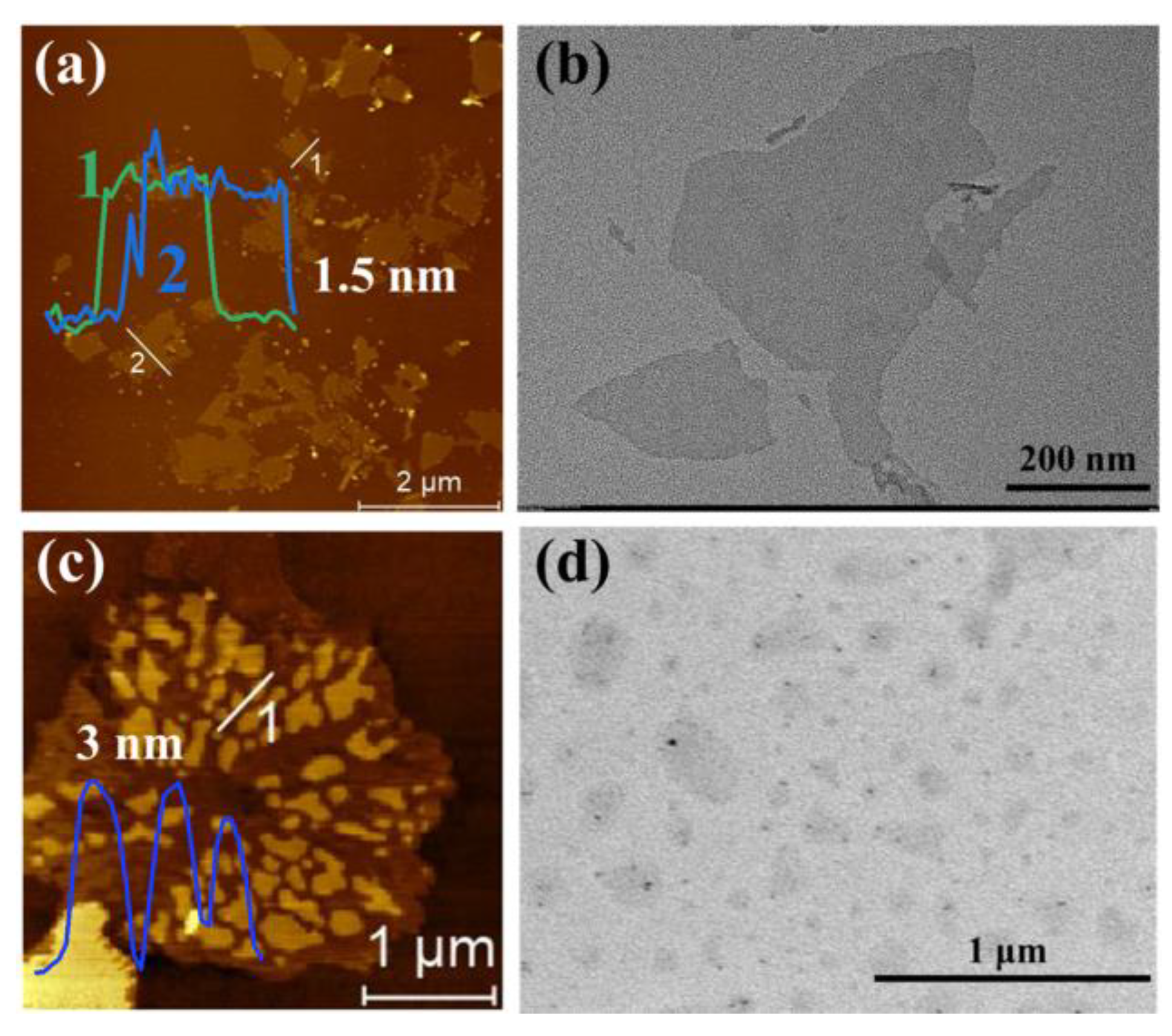
3.1. Characterizations of Ti3C2 and PNSs

The AFM technique was first used to characterize the prepared Ti3C2 monolayered nanosheets and self-assembled PNSs. It can be seen from Figure 1a that the Ti3C2 nanosheets were successfully prepared by HF etching for 24 h in a 37 °C water bath. In the obtained AFM image, the height of the Ti3C2 nanosheets is measured to be about 1.5 nm, which reveals good height distribution and agrees well with the height data previously reported for the Ti3C2 monolayer. In the corresponding TEM image (Figure 1b), the Ti3C2 nanosheets can also be seen clearly, and the size of the Ti3C2 nanosheets is between 200 nm and 1 µm, which is similar to the size that is shown in the presented AFM image. Therefore, we suggest that both AFM and TEM images prove that the Ti3C2 monolayered nanosheets with good distribution and uniform size were synthesized successfully.
In previous studies, it has been reported that the peptide with the sequence KLVFFAK could self-assemble into 2D peptide nanoribbons (PNRs) with a width of 200 nm and a length of more than 1 µm [35,36]. If the PNRs are used to modify Ti3C2 nanosheets, the size of the PNRs is bigger than that of the Ti3C2 nanosheets, which may cause the aggregation of Ti3C2 monolayers on the PNRs. Due to the aggregation of layers, the rate of electron movement of Ti3C2 multilayers is slower than that of Ti3C2 monolayers, which would significantly reduce the performance of electrochemical properties of peptide-based materials. Based on those studies, we tailored the self-assembly conditions of peptide molecules and synthesized PNSs with modernity size, which then can be modified onto the surface of Ti3C2 nanosheets for the formation of stable nanohybrids. As shown in Figure 1c, the formed self-assembled PNSs present a small size with a height of about 3 nm. The corresponding TEM image that is shown in Figure 1d indicates that the size of the formed PNSs is about 100–300 nm, which is much smaller than that of the Ti3C2 nanosheets, and could be suitable for the synthesis of Ti3C2 monolayer-based hybrid nanomaterials through GA linking.
In previous study, it has been found that the peptide with the KLVFFAK sequence can form nanostructures through the π–π interaction. Dai et al. found that the addition of inorganic salts would induce KLVFFAK to form large and thin nanobelts, and the size of nanobelts would increase with an increase in the salt concentration [37]. In the simulation study of Liang et al., the VFFA, KFFA, and FFFA sequences could self-assemble to form nanosheet structures, but VFFA, as the core of the KLVFFAK sequence, was less capable of forming large sheets than the other two sequences, and the VFFA sequence tended to form small nanosheet structures [38]. In the self-assembly system of this study, TFA can better dissolve the peptide powder, improve the dispersion of peptides in solution, and reduce the aggregation of polypeptide monomers, which makes it possible to form small-size nanostructures. At the same time, ethanol provides a more polar environment for the self-assembly of peptides, which tends to break the ring to form large-size nanosheets, thus facilitating the formation of small-size nanosheets.

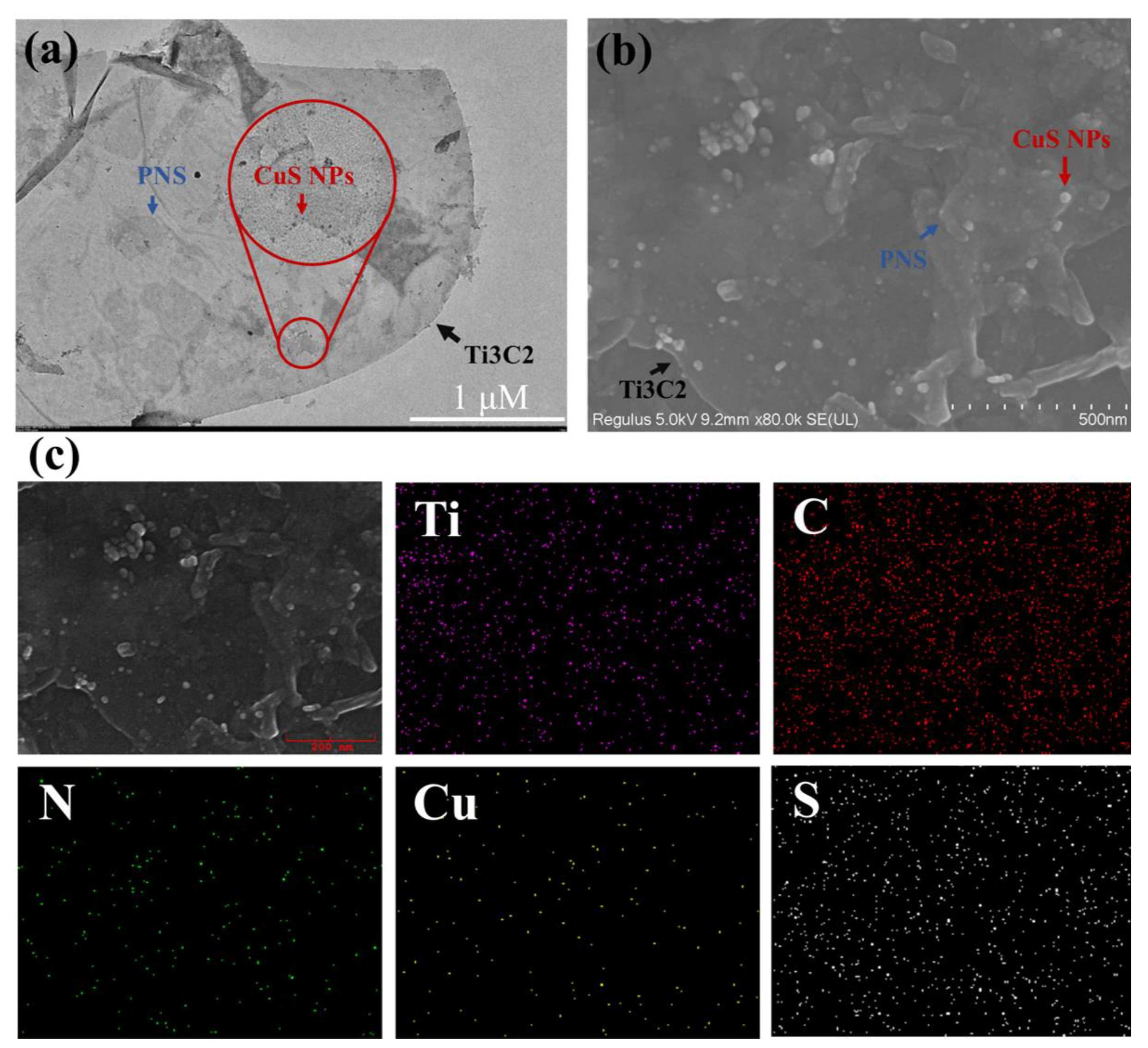
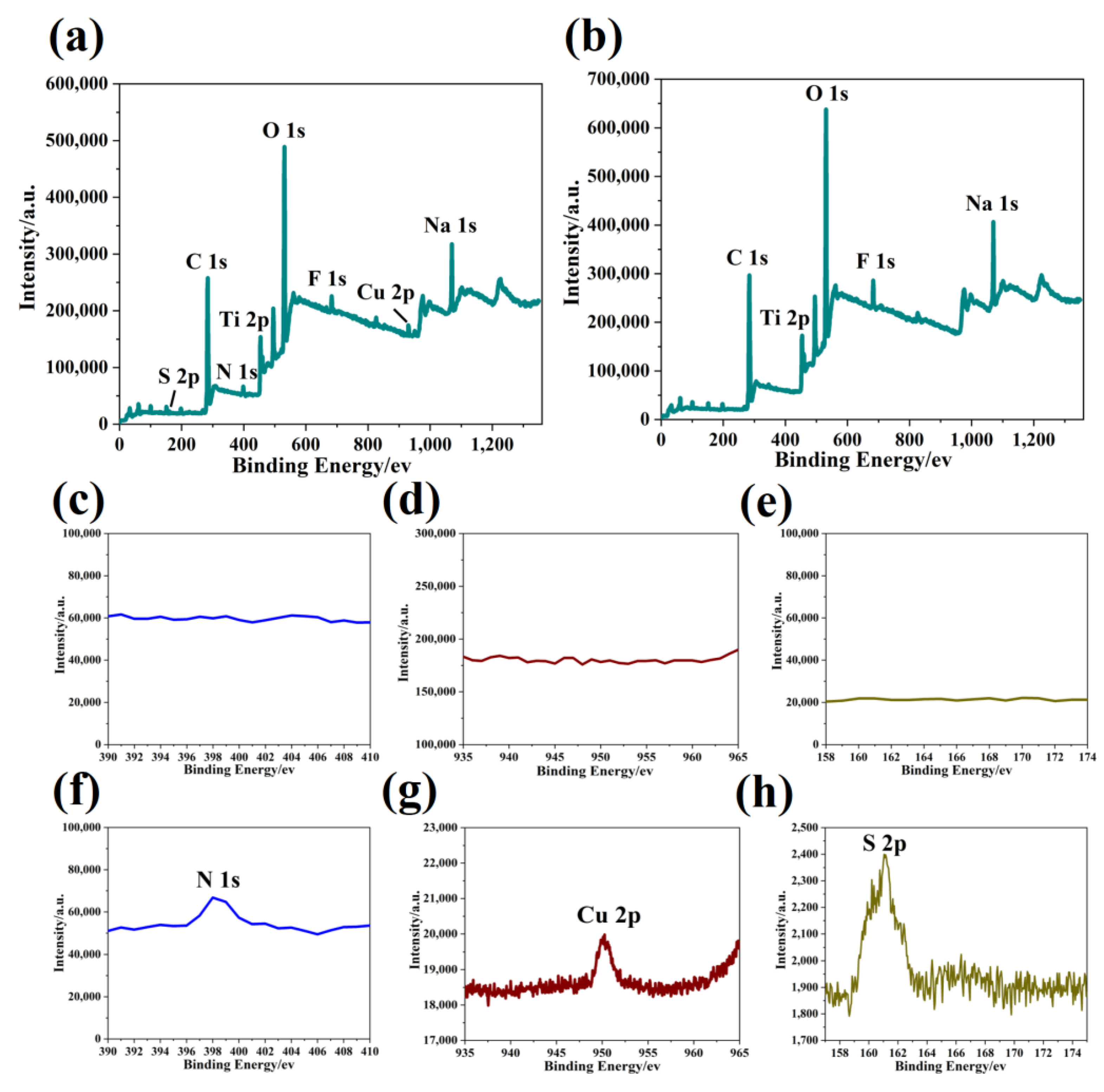
3.2. Characterizations of CuS-PNSs/Ti3C2 Nanohybrids

The prepared PNSs and GA-modified Ti3C2 monolayered nanosheets were mixed together with the same volume at room temperature for 12 h under stirring to obtain the PNSs/Ti3C2 nanohybrids. Next, CuCl2 (10 mM) was added into the PNSs/Ti3C2 solution, and Cu2+ would be bound onto PNSs under the action of electrostatic adsorption between them. After 12 h stirring at room temperature, Na2S was added to react with Cu2+ for in-situ biomimetic synthesis of the CuS-PNSs/Ti3C2 nanohybrids.
Figure 2a provides the TEM image of the created CuS-PNSs/Ti3C2 nanohybrids, from which it can be seen that PNSs are conjugated onto the Ti3C2 monolayers, and a lot of CuS NPs are loaded onto the surface of the PNSs/Ti3C2 nanohybrids. Both SEM and elemental mapping were utilized to analyze the prepared CuS-PNSs/Ti3C2 nanohybrids. The corresponding SEM image in Figure 2b proves successful synthesis of CuS-PNSs/Ti3C2 nanohybrids, in which well-distributed CuS NPs are loaded on the surface of the PNSs/Ti3C2 nanohybrids. The formed PNSs and Ti3C2 nanosheets have the same appearance and there are fewer features to distinguish them; as a result of this, elemental mapping was applied to characterize the elemental distribution of the CuS-PNS/Ti3C2 nanohybrids. It can be observed that, besides Ti and C, the existence of N, Cu, and S provide strong evidence of the successful synthesis of the CuS-PNSs/Ti3C2 nanohybrids (Figure 2c).
In order to identify the formation of CuS NPs and the combination of CuS NPs with PNSs and Ti3C2 into CuS-PNSs/Ti3C2, XPS characterization was used to measure the formed nanohybrids. The XPS spectrum of CuS-PNS/Ti3C2 clearly shows the existence of the peak of Ti2p, C1s, N1s, Cu2p, and S2p, as indicated in Figure 3a. Compared with this spectrum, the XPS survey spectrum of the prepared Ti3C2 nanosheets (Figure 3b) does not show the peak of N, Cu, and S, which further proves the synthesis of CuS-PNSs/Ti3C2. The fine XPS spectra shown in Figure 3c–e present the characteristic peaks of Ti3C2, in which the spectrum curves are flat without any clear characteristic peaks. However, after the formation of CuS-PNSs/Ti3C2, characteristic peaks are revealed at 398.3, 930.36, and 161.18 eV, which correspond to the typical peaks of N1s, Cu2p, and S2p, respectively (Figure 3f–h). In the process of Ti3C2 synthesis, HF etching and sodium ascorbate protection result in the binding of F and Na+ onto the surface of the Ti3C2 monolayer, which results in the appearance of the peaks of F1s and Na1s in the XPS spectra (Figure 3a,b).
Based on the above AFM, TEM, SEM, and XPS analysis, it can be concluded that the CuS-PNSs/Ti3C2 nanohybrids have been synthesized successfully via the biomimetic synthesis method.

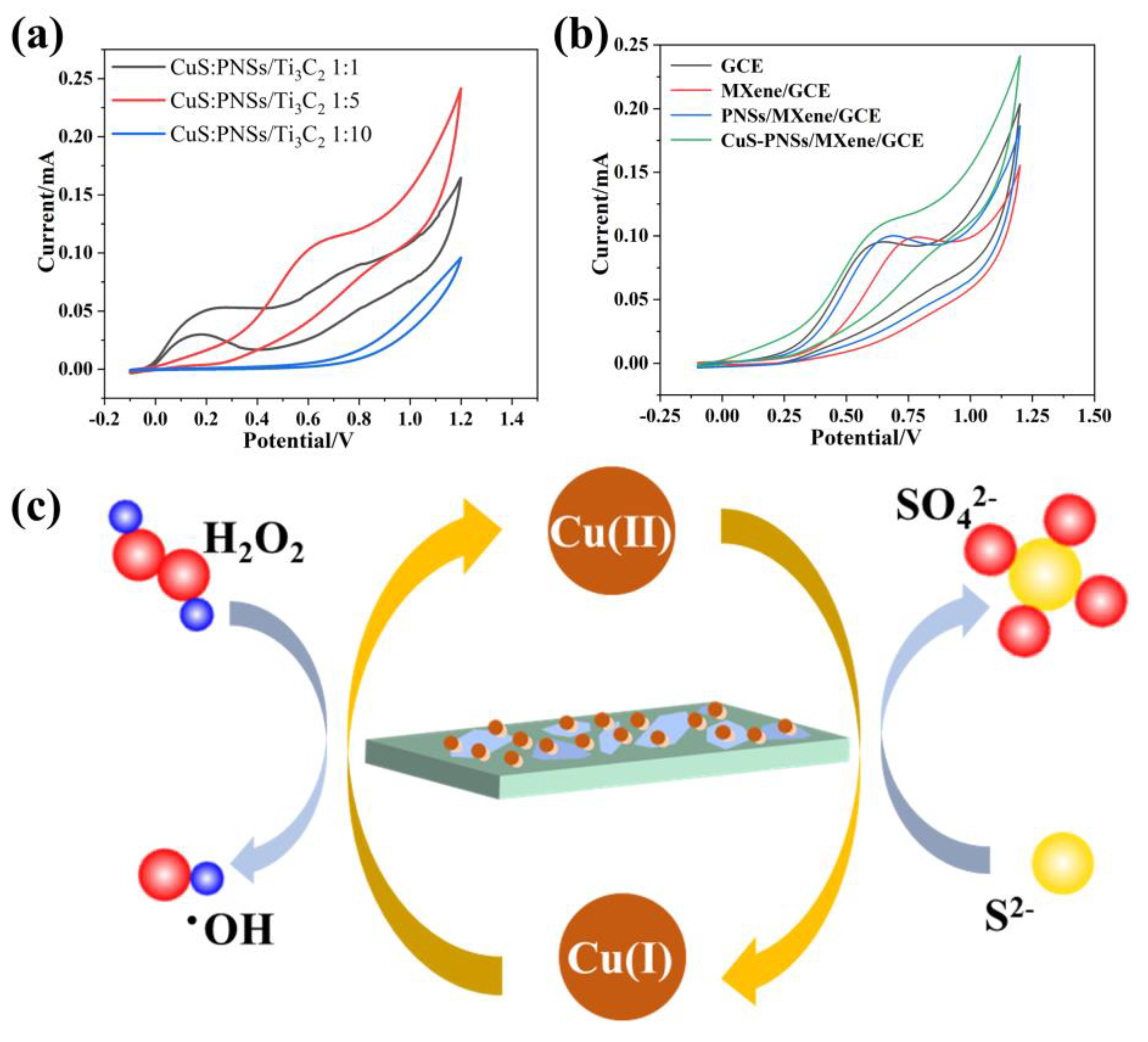
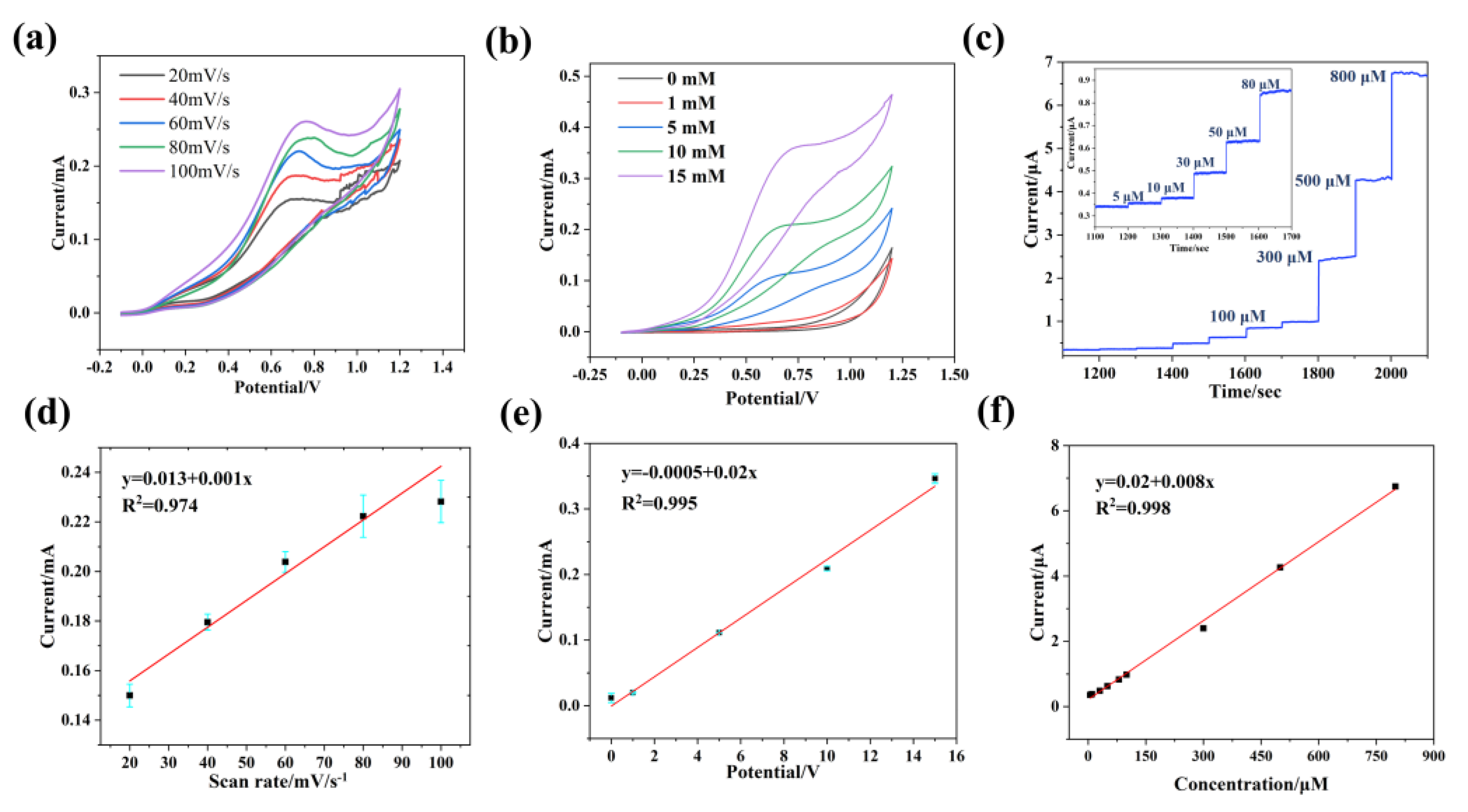
3.3. CuS-PNSs/Ti3C2 Nanohybrid-Based Electrochemical Detection of H2O2

Electrochemical tests of the synthesized CuS-PNS/Ti3C2 nanohybrids were carried out in 0.1 M NaOH electrolyte for H2O2 sensing. First, the electrochemical tests of CuS-PNSs/Ti3C2 hybrids containing different ratios of CuS NPs were carried out. When the concentration of H2O2 was 5 mM, the ratio of CuS to PNSs/Ti3C2 was 1:1, 1:5, and 1:10, and they were conducted with cyclic voltammograms (CV) tests. As can be seen from the CV curves, the hybrid material with a ratio of 1:5 has a good and stable current response, as shown in Figure 4a. Next, different materials were modified onto the surface of GCEs to prepare functional electrodes for subsequent CV tests.
Figure 4b shows the CV curves of the prepared GCE, Ti3C2/GCE, PNSs/Ti3C2/GCE, and CuS-PNSs/Ti3C2/GCE, respectively. It was found that the fabricated CuS-PNSs/Ti3C2/GCE revealed the strongest current signal in the four GCEs with the potential of 0.4–0.8 V. Due to the contribution of the addition of CuS NPs, the electrochemical capabilities of the CuS-PNSs/Ti3C2 nanohybrid were improved significantly. Meanwhile, the addition of PNSs enriched the functional groups on the Ti3C2 monolayer surface and provided more combined points for the formation of CuS NPs, which is more beneficial to catalyze the decomposition of H2O2 to generate corresponding current responses.
As a 2D material with fast electron transfer rate, MXene has good electrical conductivity, which lays a foundation for the electrochemical performance of the CuS-PNSs/Ti3C2 nanohybrid. The addition of PNSs improves the richness of the surface groups of Ti3C2, modifies the surface with only hydroxyl and oxygen, further improves the binding force of hybrid materials to metals, and provides abundant sites for the nucleation and growth of CuS NPs. After the addition of CuS NPs, the catalytic performance of the CuS-PNSs/Ti3C2 nanohybrid was greatly improved, which promoted the detection of H2O2. When it comes to the electrochemical capabilities of CuS-PNSs/Ti3C2, CuS NPs play an important role in electrochemical reactions. As indicated in Figure 4c, Cu(II) is first reduced by S2− into Cu(I), which meanwhile can reduce H2O2 to generate electrical signals. Combined with Figure 4a, it can be found that there is a weak current response between 0–0.25 V in the CV curve of the CuS-PNSs/MXene/GCE. We suggest that the weak current response here is attributed to the fact that, in the process of H2O2 dripping, S2− in CuS NPs is first oxidized to SO42−, and then the whole reduction process of H2O2 takes place.
To further test the electrochemical capability of the CuS-PNSs/Ti3C2 nanohybrids, the CV test of the different concentrations of H2O2 (0–15 mM) was carried out. First, different scan rates were applied to the CuS-PNSs/Ti3C2/GCE to determine the best scan rate. As it can be seen in Figure 5a, with the increase in scan rate, the current response increases significantly. Figure 5d shows the linear fitting of 20–100 mV s−1, which presents the great relationship of 20–80 mV s−1. Due to the obtained result, the scan rate of 50 mV s−1 was chosen for a further CV test. From Figure 5b, it can be found that with the increase in the concentration of H2O2, the current response increases gradually. Figure 5e shows the linear fitting of 0–15 mM concentration H2O2, which presents a great relationship. In order to explore the detection limit of the fabricated H2O2 electrochemical biosensor, an I–T test was applied for the CuS-PNSs/Ti3C2/GCE. The current response peak, found from the CV curve in Figure 5a, is 0.67 V; therefore, 0.67 V was chosen as the potential for the I–T test. It can be seen from the current response for the continuous addition of H2O2 in Figure 5c, when the H2O2 concentration reaches 5 µM in the solution, that an obvious current signal is generated. More importantly, with the increase in the H2O2 concentration, the I–T curve generates current responses of different intensities. The linear relationship between the current and H2O2 concentration has a good fit, and the linear correlation coefficient R2 is 0.997, as shown in Figure 5f. After the calculation, the lowest detection limit of the CuS-PNSs/Ti3C2/GCE-based electrochemical biosensor exhibits a detection limit of 0.226 µM towards H2O2, and the linear detection range is in the concentration range of 5–800 µM.

3.4. Selectivity and Stability of CuS-PNS/Ti3C2 Electrochemical Platform

In actual samples, there are some other substances in the solution besides H2O2. The nanohybrid-based electrochemical biosensors need to have good selectivity and be able to detect H2O2 sensitively among many interfering substances. Therefore, the selectivity of the CuS-PNSs/Ti3C2-based electrochemical biosensor was tested.
As shown in Figure 6a, when lysine, UA, and AA were added successively during the test, the I–T curve does not reveal an obvious current response; but when H2O2 is added again, the current intensity shows a very obvious current response. Therefore, it can be concluded that the CuS-PNSs/Ti3C2 hybrid material-based electrochemical biosensor has good anti-interference ability. Furthermore, to test the stability of the material, CuS-PNSs/Ti3C2/GCE was stored at −4 °C for 7 days, and the electrochemical detection ability of CuS-PNSs/Ti3C2/GCE for H2O2 was tested once every day. After seven days of testing, it was found that the electrochemical performance of the material was basically the same (Figure 6b), which indicates that the CuS-PNSs/Ti3C2/GCE has good stability and can still maintain an extremely high detection performance after long-term storage. At the same time, the actual sample was tested. H2O2 was added to the milk to prepare different concentrations of H2O2 milk solution to simulate the actual sample. In Figure 6c, it can be seen that the current response at 0.6–0.8 V increases with the increase in H2O2 concentration in milk, indicating that the CuS-PNSs/Ti3C2-based electrochemical biosensor also has good detection performance in reagent samples. In Figure 6d, it can be seen that the CuS-PNSs/Ti3C2-based electrochemical biosensor has a good linear relationship with the detection of real samples, and it can be utilized as a reliable electrochemical detection platform for determining H2O2.

4. Discussion

In this study, monolayered Ti3C2 MXene nanosheets with good morphology and good structure were synthesized by HF etching and DMSO intercalation. At the same time, in view of the fact that the edge of Ti3C2 is easy to oxidize, DMSO is replaced by sodium ascorbate by dialysis to protect Ti3C2, which slows down the rate of oxidation and prolongs the shelf life, addressing the problem that Ti3C2 is easily oxidized and decomposed into titanium dioxide in air. At the same time, in order to increase the richness and biocompatibility of Ti3C2 surface functional groups, PNSs were used to modify and functionalize the Ti3C2 surface. Through the design of the peptide sequence and the regulation of its self-assembly conditions, the PNSs with good morphology, moderate size, and negative charge in neutral condition were obtained. In order to realize the strong combination between Ti3C2 and PNS, GA was used as the bridge between them. The aldehyde group at one end of GA binds with the -OH group on the surface of Ti3C2, and the other end binds with the -NH4 group of PNSs to realize the recombination between them. Finally, to further improve the electrochemical performance of the materials, Na2S was used for in situ chemical reaction with Cu2+ to one-step synthesize CuS NPs without using additional reagent and waste production, meanwhile greatly improving the chemical properties of the CuS-PNSs/Ti3C2 nanohybrid materials.
Ti3C2 with large specific surface area can be loaded with PNSs on a large scale, which provides abundant binding sites for the nucleation and growth of CuS NPs. The one-step synthesis of CuS NPs on the surface of PNSs/Ti3C2 by direct reaction of sodium sulfide to prepare CuS-PNSs/Ti3C2 hybrid materials is a green, convenient, and environment-friendly biomimetic synthesis method. By using biomolecules as bridges and in-situ chemical reaction to synthesize sulphide NPs, the combination of metal sulfides and 2D materials is realized, which provides a new strategy and idea for the development of functional biosensors with good stability and selectivity. It has a broad prospect in biological detection, clinical application and environmental monitoring.
Based on the above AFM, TEM, SEM, and XPS analysis, it is confirmed that the CuS−PNSs/Ti3C2 nanohybrids have been synthesized successfully. At the same time, the biosensor platform has good electrochemical performance, anti−interference, and stability. Compared with another H2O2 detection platforms in Table 1, the CuS−PNSs/Ti3C2−based electrochemical biosensor platform in this study has a lower detection limit and a wider detection range of H2O2 concentration [39,40,41]. Therefore, in view of the wide detection range, low detection limit, and high sensitivity of the CuS−PNS/Ti3C2−based electrochemical sensors, it is suggested that it can be applied in the detection of H2O2 in a real environment, clinical detection, environmental protection, and biomedicine.
In the CuS−PNSs/Ti3C2 nanohybrids, the Ti3C2 nanosheets loaded with peptides provide binding sites for the formation of CuS NPs. At the same time, the existence of peptides promotes the dispersion of CuS NPs and prevents the performance degradation caused by the aggregation of NPs. In addition, for the synthesis of CuS NPs, a green and environment−friendly biomimetic synthesis method was adopted, and CuS NPs were formed in situ at the Cu2+ binding sites directly by using the self−assembled PNSs as templates. Compared with the traditional methods for CuS synthesis, such as the microwave−assisted solvothermal, chemical coprecipitation, and sol−gel synthesis, biomimetic synthesis in this current study is greener and more environmentally friendly, and there is no extra solvent and no additional waste liquid [42,43,44,45,46]. Furthermore, biomimetic CuS NPs can increase their biocompatibility to a certain extent and have more advantages for biological applications.

5. Conclusions

In summary, we report the peptide-medicated biomimetic synthesis of CuS NPs in hybrid 2D materials for the preparation of biosensors for the electrochemical detection of H2O2. The combination of Ti3C2 and PNSs and the modification of CuS NPs were realized by biological cross-linking and biomimetic synthesis, and CuS-PNSs/Ti3C2 nanohybrids were successfully prepared. The obtained results shows that CuS-PNSs/Ti3C2 nanohybrids have good sensitivity, high selectivity, and stability for the detection of H2O2. It is expected the design and synthesis of peptide nanomaterials for the binding with 2D materials and subsequent biomimetic synthesis functional nanomaterials could greatly enhance the properties and functions of hybrid nanomaterials and promote their application in various fields.

Author Contributions

L.G. and G.W. supervised the project; D.Z. carried out the experiments; D.Z., H.K., G.Y., P.H. and X.L. carried out the data analysis; D.Z. and G.W. wrote the manuscript; L.G. and G.W. did the review and final editing. All authors have read and agreed to the published version of the manuscript.

Funding

The authors thank the financial support from the National Natural Science Foundation of China (No. 51873225), the Taishan Scholars Program of Shandong Province (No. tsqn201909104), and the High-Grade Talents Plan of Qingdao University.

Institutional Review Board Statement

Not applicable.

Informed Consent Statement

Not applicable.

Data Availability Statement

Not applicable.

Acknowledgments

The authors appreciate the support from Youyin Xu and Rongqiu Mu (Qingdao University) for their valuable assistant and discussion of the data analysis.

Conflicts of Interest

The authors declare no conflict of interest.

References

  1. Tapeinos, C.; Larranaga, A.; Sarasua, J.R.; Pandit, A. Functionalised collagen spheres reduce H2O2 mediated apoptosis by scavenging overexpressed ROS. Nanomed. Nanotechnol. Biol. Med. 2018, 14, 2397–2405. [Google Scholar] [CrossRef]
  2. Chen, L.F.; Xing, S.H.; Lei, Y.L.; Chen, Q.S.; Zou, Z.; Quan, K.; Qing, Z.H.; Liu, J.W.; Yang, R.H. A Glucose-Powered Activatable Nanozyme Breaking pH and H2O2 Limitations for Treating Diabetic Infections. Angew. Chem. Int. Ed. 2021, 60, 23534–23539. [Google Scholar] [CrossRef] [PubMed]
  3. Yang, X.Y.; Cheng, X.W.; Song, H.Y.; Ma, J.H.; Pan, P.P.; Elzatahry, A.A.; Su, J.C.; Deng, Y.H. 3D Interconnected Mesoporous Alumina with Loaded Hemoglobin as a Highly Active Electrochemical Biosensor for H2O2. Adv. Healthc. Mater. 2018, 7, 1800149. [Google Scholar] [CrossRef] [PubMed]
  4. Seven, F.; Golcez, T.; Sen, M. Nanoporous carbon-fiber microelectrodes for sensitive detection of H2O2 and dopamine. J. Electroanal. Chem. 2020, 864, 114104. [Google Scholar] [CrossRef]
  5. Ahmed, S.R.; Cirone, J.; Chen, A.C. Fluorescent Fe3O4 Quantum Dots for H2O2 Detection. ACS Appl. Nano Mater. 2019, 2, 2076–2085. [Google Scholar] [CrossRef]
  6. Tantawi, O.; Baalbaki, A.; El Asmar, R.; Ghauch, A. A rapid and economical method for the quantification of hydrogen peroxide (H2O2) using a modified HPLC apparatus. Sci. Total Environ. 2019, 654, 107–117. [Google Scholar] [CrossRef] [PubMed]
  7. Mao, W.T.; Ding, Y.M.; Li, M.L.; Ma, C.; Gong, H.X.; Pan, J.L.; Zhang, S.J.; Qian, Y.T.; Bao, K.Y. Synthesis and electrochemical characterization of 2D SnS2/RGO as anode material in sodium-ion batteries. J. Alloys Compd. 2021, 855, 157209. [Google Scholar] [CrossRef]
  8. Munteanu, R.E.; Moreno, P.S.; Bramini, M.; Gáspár, S. 2D materials in electrochemical sensors for in vitro or in vivo use. Anal. Bioanal. Chem. 2021, 413, 701–725. [Google Scholar] [CrossRef]
  9. Tapia, M.A.; Gusmão, R.; Serrano, N.; Sofer, Z.; Ariño, C.; Díaz-Cruz, J.M.; Esteban, M. Phosphorene and other layered pnictogens as a new source of 2D materials for electrochemical sensors. TrAC Trends Anal. Chem. 2021, 139, 116249. [Google Scholar] [CrossRef]
  10. Zhu, D.Z.; Liu, B.; Wei, G. Two-Dimensional Material-Based Colorimetric Biosensors: A Review. Biosensors 2021, 11, 259. [Google Scholar] [CrossRef]
  11. Zhu, D.Z.; He, P.; Kong, H.; Yang, G.Z.; Luan, X.; Wei, G. Biomimetic graphene-supported ultrafine platinum nanowires for colorimetric and electrochemical detection of hydrogen peroxide. J. Mater. Chem. B 2022, 10, 9216–9225. [Google Scholar] [CrossRef]
  12. Fu, B.; Sun, J.X.; Wang, C.; Shang, C.; Xu, L.J.; Li, J.B.; Zhang, H. MXenes: MXenes: Synthesis, Optical Properties, and Applications in Ultrafast Photonics. Small 2021, 17, 2170048. [Google Scholar] [CrossRef]
  13. Huang, H.; Dong, C.H.; Feng, W.; Wang, Y.; Huang, B.C.; Chen, Y. Biomedical engineering of two-dimensional MXenes. Adv. Drug Deliver. Rev. 2022, 184, 114178. [Google Scholar] [CrossRef] [PubMed]
  14. Shin, H.; Eom, W.; Lee, K.H.; Jeong, W.; Kang, D.J.; Han, T.H. Highly Electroconductive and Mechanically Strong Ti3C2Tx MXene Fibers Using a Deformable MXene Gel. ACS Nano 2021, 15, 3320–3329. [Google Scholar] [CrossRef] [PubMed]
  15. Lorencova, L.; Bertok, T.; Filip, J.; Jerigova, M.; Velic, D.; Kasak, P.; Mahmoud, K.A.; Tkac, J. Highly stable Ti3C2Tx (MXene)/Pt nanoparticles-modified glassy carbon electrode for H2O2 and small molecules sensing applications. Sens. Actuators B Chem. 2018, 263, 360–368. [Google Scholar] [CrossRef]
  16. Zhang, C.F. Interfacial assembly of two-dimensional MXenes. J. Energy Chem. 2021, 60, 417–434. [Google Scholar] [CrossRef]
  17. Vallee, A.; Humblot, V.; Pradier, C.M. Peptide Interactions with Metal and Oxide Surfaces. Acc. Chem. Res. 2010, 43, 1297–1306. [Google Scholar] [CrossRef]
  18. Zou, R.F.; Wang, Q.; Wu, J.C.; Wu, J.X.; Schmuck, C.; Tian, H. Peptide self-assembly triggered by metal ions. Chem. Soc. Rev. 2015, 44, 5200–5219. [Google Scholar] [CrossRef]
  19. Tõugu, V.; Tiiman, A.; Palumaa, P. Interactions of Zn(Ⅱ) and Cu(Ⅱ) ions with Alzheimer’s amyloid-beta peptide. Metal ion binding, contribution to fibrillization and toxicity. Metallomics 2011, 3, 250–261. [Google Scholar] [CrossRef]
  20. Liu, B.; Yao, J.L.; Xing, J.; Yang, M.; Zhu, D.Z.; Ren, W.Z.; Xiang, L.C.; Wang, Y.; Wu, A.G.; Wei, G. Design, biomimetic synthesis, and tumor photothermal therapy of peptide-based two-dimensional photothermal conversion nanomaterials. Mol. Syst. Des. Eng. 2022, 7, 1549–1560. [Google Scholar] [CrossRef]
  21. Yang, G.Z.; He, P.; Zhu, D.Z.; Wan, K.M.; Kong, H.; Luan, X.; Fang, L.; Wang, Y.; Wei, G. Functional regulation of polymer aerogels by graphene doping and peptide nanofiber-induced biomineralization as sustainable adsorbents of contaminants. Environ. Sci. Nano 2022, 9, 4497–4507. [Google Scholar] [CrossRef]
  22. Gu, S.T.; Shi, X.M.; Zhang, D.; Fan, G.C.; Luo, X.L. Peptide-Based Photocathodic Biosensors: Integrating a Recognition Peptide with an Antifouling Peptide. Anal. Chem. 2021, 93, 2706–2712. [Google Scholar] [CrossRef] [PubMed]
  23. Kong, H.; Liu, B.; Yang, G.Z.; Chen, Y.; Wei, G. Tailoring Peptide Self-Assembly and Formation of 2D Nanoribbons on Mica and HOPG Surface. Materials 2022, 15, 310. [Google Scholar] [CrossRef] [PubMed]
  24. He, P.; Yang, G.; Zhu, D.; Kong, H.; Corrales-Ureña, Y.R.; Colombi Ciacchi, L.; Wei, G. Biomolecule-mimetic nanomaterials for photothermal and photodynamic therapy of cancers: Bridging nanobiotechnology and biomedicine. J. Nanobiotechnol. 2022, 20, 483. [Google Scholar] [CrossRef]
  25. Mohammed, S.; Hegab, H.M.; Ou, R.W. Nanofiltration performance of glutaraldehyde crosslinked graphene oxide-cellulose nanofiber membrane. Chem. Eng. Res. Des. 2022, 183, 1–12. [Google Scholar] [CrossRef]
  26. Zhang, W.H.; Yang, P. 2D bio-nanostructures fabricated by supramolecular self-assembly of protein, peptide, or peptoid. Adv. Compos. Hybrid Mater. 2019, 2, 201–213. [Google Scholar] [CrossRef]
  27. Magnotti, E.; Conticello, V. Two-Dimensional Peptide and Protein Assemblies. Protein Eng. Nanostruct. 2016, 940, 29–60. [Google Scholar]
  28. Jia, B.H.; Sun, Y.; Yang, L.J.; Yu, Y.; Fan, H.R.; Ma, G. A structural model of the hierarchical assembly of an amyloid nanosheet by an infrared probe technique. Phys. Chem. Chem. Phys. 2018, 20, 27261–27271. [Google Scholar] [CrossRef]
  29. Saxena, A.; Liyanage, W.; Masud, J.; Kapila, S.; Nath, M. Selective electroreduction of CO2 to carbon-rich products with a simple binary copper selenide electrocatalyst. J. Mater. Chem. A 2021, 9, 7150–7161. [Google Scholar] [CrossRef]
  30. Masud, J.; Liyanage, W.P.R.; Cao, X.; Saxena, A.; Nath, M. Copper Selenides as High-Efficiency Electrocatalysts for Oxygen Evolution Reaction. ACS Appl. Energy Mater. 2018, 1, 4075–4083. [Google Scholar] [CrossRef]
  31. Jin, W.; Fu, Y.Q.; Cai, W.Q. In situ growth of CuS decorated graphene oxide-multiwalled carbon nanotubes for ultrasensitive H2O2 detection in alkaline solution. New J. Chem. 2019, 43, 3309–3316. [Google Scholar] [CrossRef]
  32. Li, X.Y.; Du, X.Z. Molybdenum disulfide nanosheets supported Au-Pd bimetallic nanoparticles for non-enzymatic electrochemical sensing of hydrogen peroxide and glucose. Sens. Actuators B Chem. 2017, 239, 536–543. [Google Scholar] [CrossRef]
  33. Lipatov, A.; Alhabeb, M.; Lukatskaya, M.R.; Boson, A.; Gogotsi, Y.; Sinitskii, A. Effect of Synthesis on Quality, Electronic Properties and Environmental Stability of Individual Monolayer Ti3C2 MXene Flakes. Adv. Electron. Mater. 2016, 2, 1600255. [Google Scholar] [CrossRef] [Green Version]
  34. Mashtalir, O.; Naguib, M.; Mochalin, V.N.; Dall’Agnese, Y.; Heon, M.; Barsoum, M.W.; Gogotsi, Y. Intercalation and delamination of layered carbides and carbonitrides. Nat. Commun. 2013, 4, 1716. [Google Scholar] [CrossRef] [PubMed] [Green Version]
  35. Liu, L.; Klausen, L.H.; Dong, M.D. Two-dimensional peptide based functional nanomaterials. Nano Today 2018, 23, 40–58. [Google Scholar] [CrossRef]
  36. Xu, L.; Xu, S.; Xiang, T.Y.; Liu, H.; Chen, L.W.; Jiang, B.P.; Yao, J.H.; Zhu, H.L.; Hu, R.F.; Chen, Z.P. Multifunctional building elements for the construction of peptide drug conjugates. Eng. Regen. 2022, 3, 92–109. [Google Scholar] [CrossRef]
  37. Dai, B.; Li, D.; Xi, W.; Luo, F.; Zhang, X.; Zou, M.; Cao, M.; Hu, J.; Wang, W.Y.; Wei, G.H.; et al. Tunable assembly of amyloid-forming peptides into nanosheets as a retrovirus carrier. Proc. Natl. Acad. Sci. USA 2015, 112, 2996–3001. [Google Scholar] [CrossRef] [Green Version]
  38. Liang, L.J.; Wang, L.W.; Shen, J.W. The self-assembly mechanism of tetra-peptides from the motif of beta-amyloid peptides: A combined coarse-grained and all-atom molecular dynamics simulation. RSC Adv. 2016, 6, 100072–100078. [Google Scholar] [CrossRef]
  39. Nethravathi, C.; Nath, R.R.; Rajamathi, J.T.; Rajamathi, M. Microwave-Assisted Synthesis of Porous Aggregates of CuS Nanoparticles for Sunlight Photocatalysis. ACS Omega 2019, 4, 4825–4831. [Google Scholar] [CrossRef] [Green Version]
  40. Pejjai, B.; Reddivari, M.; Kotte, T.R.R. Phase controllable synthesis of CuS nanoparticles by chemical co-precipitation method: Effect of copper precursors on the properties of CuS. Mater. Chem. Phys. 2020, 239, 122030. [Google Scholar] [CrossRef]
  41. Riyaz, S.; Parveen, A.; Azam, A. Microstructural and optical properties of CuS nanoparticles prepared by sol–gel route. Perspect. Sci. 2016, 8, 632–635. [Google Scholar] [CrossRef]
  42. Lu, K.C.; Wang, J.K.; Lin, D.H.; Chen, X.; Yin, S.Y.; Chen, G.S. Construction of a novel electrochemical biosensor based on a mesoporous silica/oriented graphene oxide planar electrode for detecting hydrogen peroxide. Anal. Methods 2020, 12, 2661–2667. [Google Scholar] [CrossRef] [PubMed]
  43. Salazar, P.; Fernandez, I.; Rodriguez, M.C.; Hernandez-Creus, A.; Gonzalez-Mora, J.L. One-step green synthesis of silver nanoparticle-modified reduced graphene oxide nanocomposite for H2O2 sensing applications. J. Electroanal. Chem. 2019, 855, 113638. [Google Scholar] [CrossRef]
  44. Devaraj, M.; Rajendran, S.; Jebaranjitham, J.N.; Ranjithkumar, D.; Sathiyaraj, M.; Manokaran, J.; Sundaravadivel, E.; Santhanalakshmi, J.; Ponce, L.C. Horseradish Peroxidase-Immobilized Graphene Oxide-Chitosan Gold Nanocomposites as Highly Sensitive Electrochemical Biosensor for Detection of Hydrogen Peroxide. J. Electrochem. Soc. 2020, 167, 147517. [Google Scholar] [CrossRef]
  45. Cheng, D.; Li, P.P.; Zhu, X.H.; Liu, M.L.; Zhang, Y.Y.; Liu, Y. Enzyme-free Electrochemical Detection of Hydrogen Peroxide Based on the Three-Dimensional Flower-like Cu-based Metal Organic Frameworks and MXene Nanosheets(dagger). Chin. J. Chem. 2021, 39, 2181–2187. [Google Scholar] [CrossRef]
  46. Zhou, K.W.; Li, Y.; Zhuang, S.J.; Ren, J.; Tang, F.; Mu, J.L.; Wang, P. A novel electrochemical sensor based on CuO-CeO2/MXene nanocomposite for quantitative and continuous detection of H2O2. J. Electroanal. Chem. 2022, 921, 116655. [Google Scholar] [CrossRef]
Scheme 1. Schematic presentation of the biomimetic synthesis of CuS-PNSs/Ti3C2 and the fabrication of electrochemical H2O2 biosensors: (A) preparation of Ti3C2 nanosheets; (B) preparation of CuS-PNSs/Ti3C2 and fabrication of electrochemical H2O2 biosensors.
Biosensors 13 00014 sch001
Figure 1. Morphological characterizations of Ti3C2 nanosheets and self-assembled PNSs: (a) AFM image of Ti3C2 nanosheets, the number and color line in green and bule corresponding to the thickness of 1 and 2 samples respectively; (b) TEM image of Ti3C2 nanosheets; (c) AFM height image of PNSs; (d) TEM image of PNSs.
Biosensors 13 00014 g001
Figure 2. Characterizations of CuS-PNSs/Ti3C2 nanohybrids: (a) TEM image; (b) SEM image; (c) SEM-based elemental mapping.
Biosensors 13 00014 g002
Figure 3. XPS characterization of CuS-PNS/Ti3C2 nanohybrids: (a) XPS survey spectrum of CuS-PNSs/Ti3C2; (b) XPS spectrum of Ti3C2; (ce) XPS spectra of N, Cu, and S in Ti3C2; (fh) XPS spectra of N, Cu, and S in the CuS-PNSs/Ti3C2 nanohybrids.
Biosensors 13 00014 g003
Figure 4. Electrochemical tests of different GCEs. (a) CV curves of different ratios of CuS and PNSs/Ti3C2 in the H2O2 concentration of 5mM; (b) CV curves of GCE, Ti3C2/GCE, PNSs/Ti3C2/GCE, and CuS-PNSs/Ti3C2/GCE; (c) electrochemical oxidization–reduction reaction mechanism by CuS NPs.
Biosensors 13 00014 g004
Figure 5. Electrochemical tests of CuS-PNSs/Ti3C2/GCE in H2O2 detection: (a) current response of CuS-PNSs/Ti3C2/GCE toward H2O2 with different scan rates of 20–100 mV s−1; (b) current response of CuS-PNSs/Ti3C2/GCE toward H2O2 with different concentrations of 0–15 mM; (c) I–T response of different concentrations of H2O2; (d) linear calibration of CV response with different scan rates of 20–100 mV s−1; (e) linear calibration of CV response with H2O2 concentration from 0 to 15 mM; (f) linear calibration of I–T response with H2O2 concentration from 5–800 µM.
Biosensors 13 00014 g005
Figure 6. Anti-interference ability and stability of CuS-PNSs/Ti3C2-based electrochemical platform. (a) Amperometric responses upon successive additions of H2O2, Lys, UA, AA, and H2O2; (b) stability of CuS-PNSs/Ti3C2 electrochemical platform for 7 days; (c) current response of CuS-PNSs/Ti3C2/GCE toward H2O2 with different concentrations of 0–15 mM in milk; (d) linear calibration of CV response with H2O2 concentration from 0 to 15 mM in milk.
Biosensors 13 00014 g006
Table 1. Comparison of the sensing performances of various electrochemical H2O2 sensors.
MaterialsLinear Range
[mM]
Limit of Detection [µM]Ref.
CMF/Gox/HRP@MS0.0001–0.23510[42]
rGO/AgNPs0.002–200.73[43]
HRP/Au/ERGO-CHIT/GCE0.01–6.314[44]
Cu-MOF/MXene0.001–6.120.35[45]
CuO-CeO2/MXene0.005–0.11.67[46]
CuS-PNS/Ti3C20.005–150.226This work
Disclaimer/Publisher’s Note: The statements, opinions and data contained in all publications are solely those of the individual author(s) and contributor(s) and not of MDPI and/or the editor(s). MDPI and/or the editor(s) disclaim responsibility for any injury to people or property resulting from any ideas, methods, instructions or products referred to in the content.

