CRISPR/Cas12a-Assisted Dual Visualized Detection of SARS-CoV-2 on Frozen Shrimps

Abstract
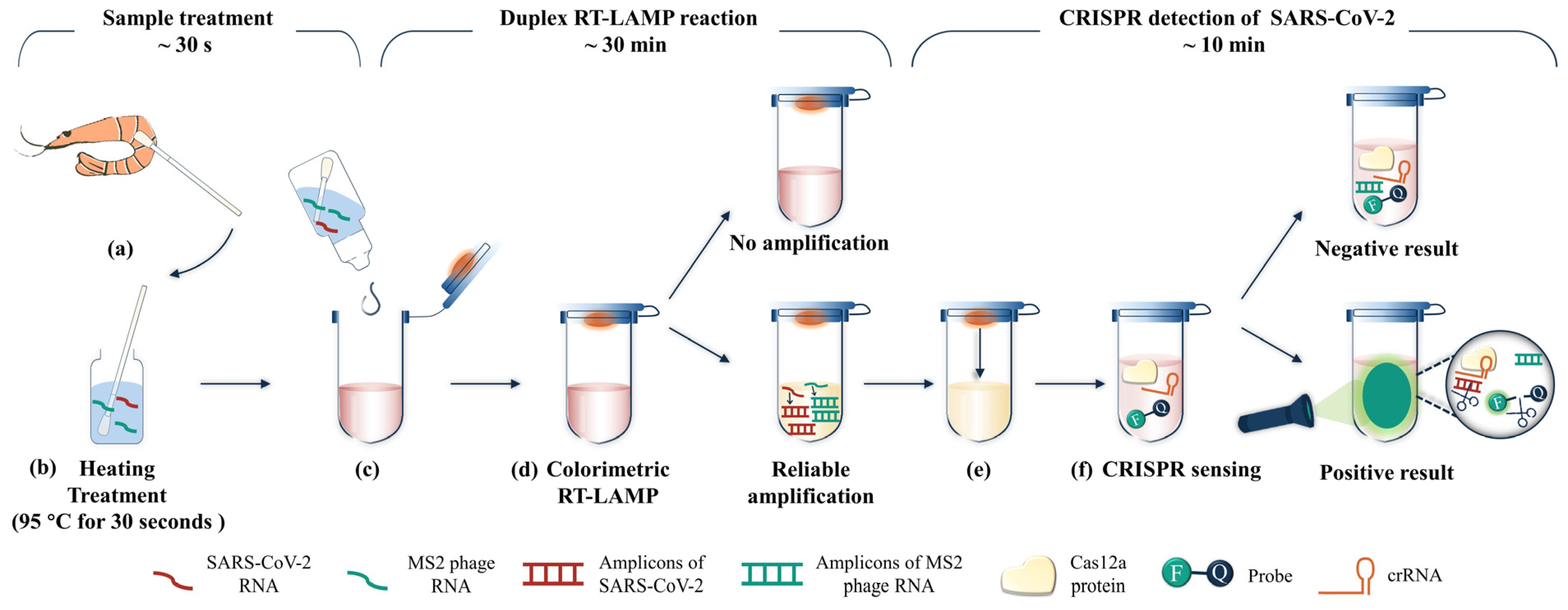
:1. Introduction
2. Materials and Methods
2.1. Materials and Chemicals
2.2. Sample Collection and Treatment
2.3. RT-LAMP, Uracil-RT-LAMP, and Colorimetric RT-LAMP Assay
2.4. Endpoint-Specific Detection with CRISPR
3. Results and Discussion
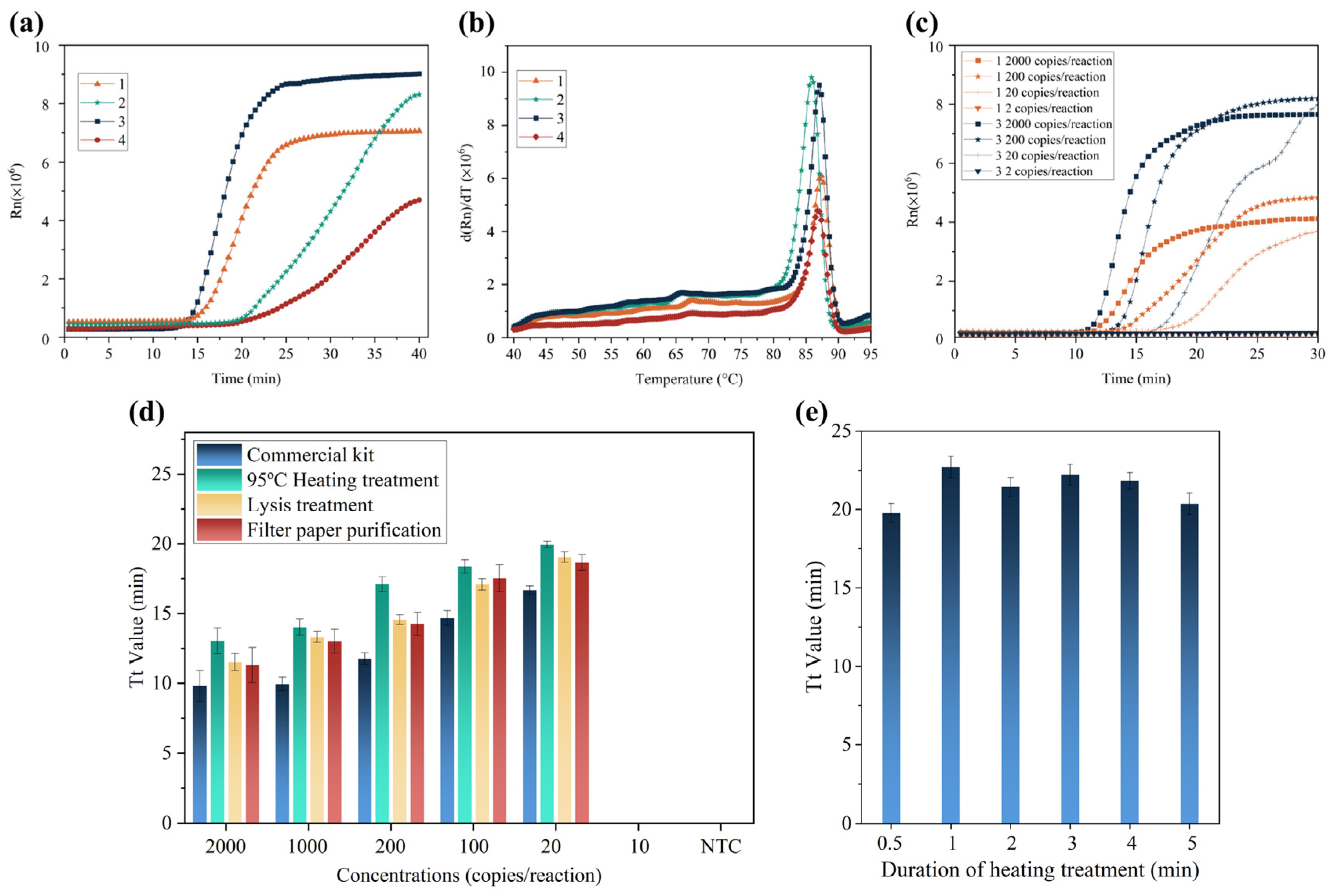
3.1. Primer Design and Sample Treatment
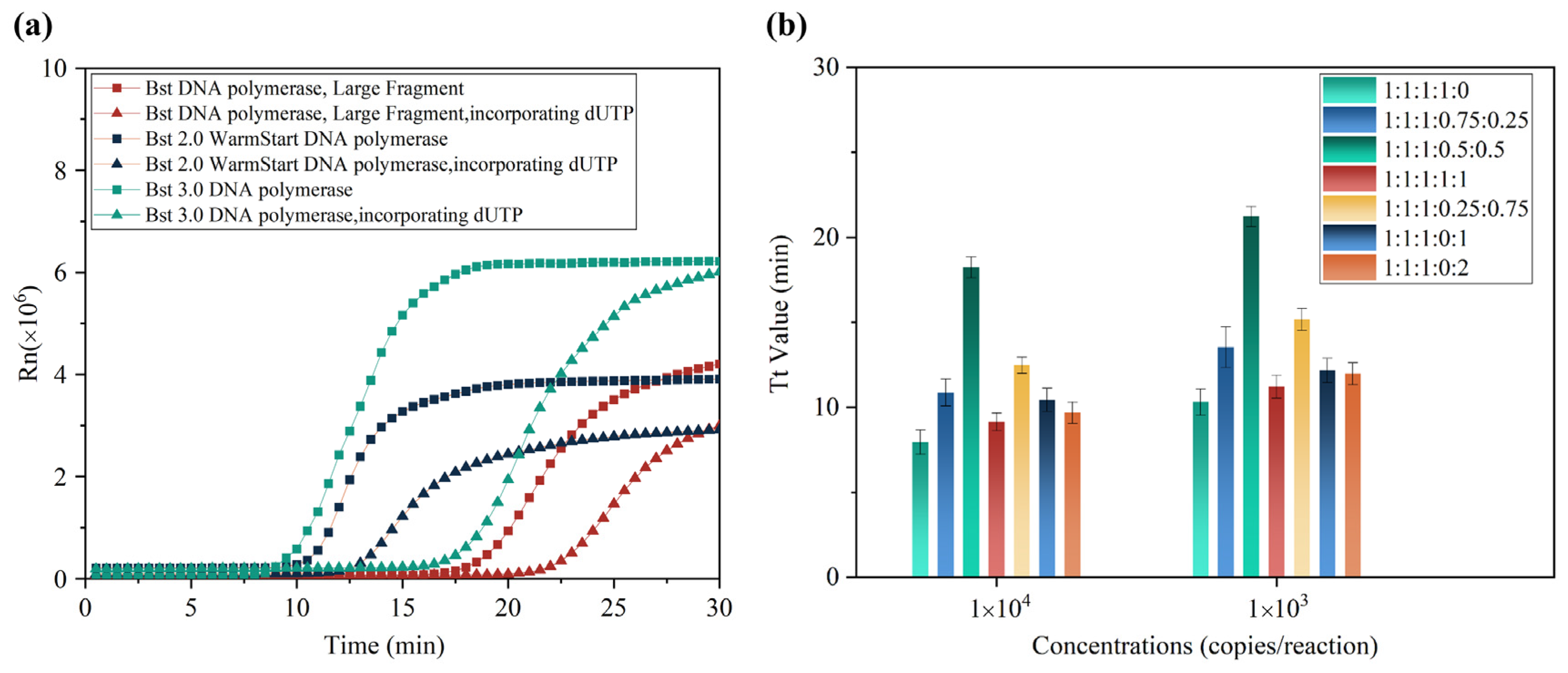
3.2. Optimization of Uracil-Mediated RT-LAMP System
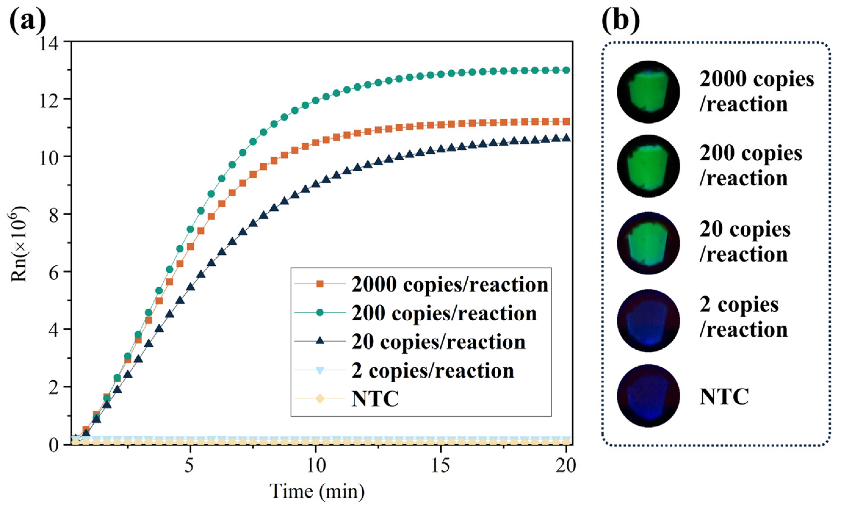
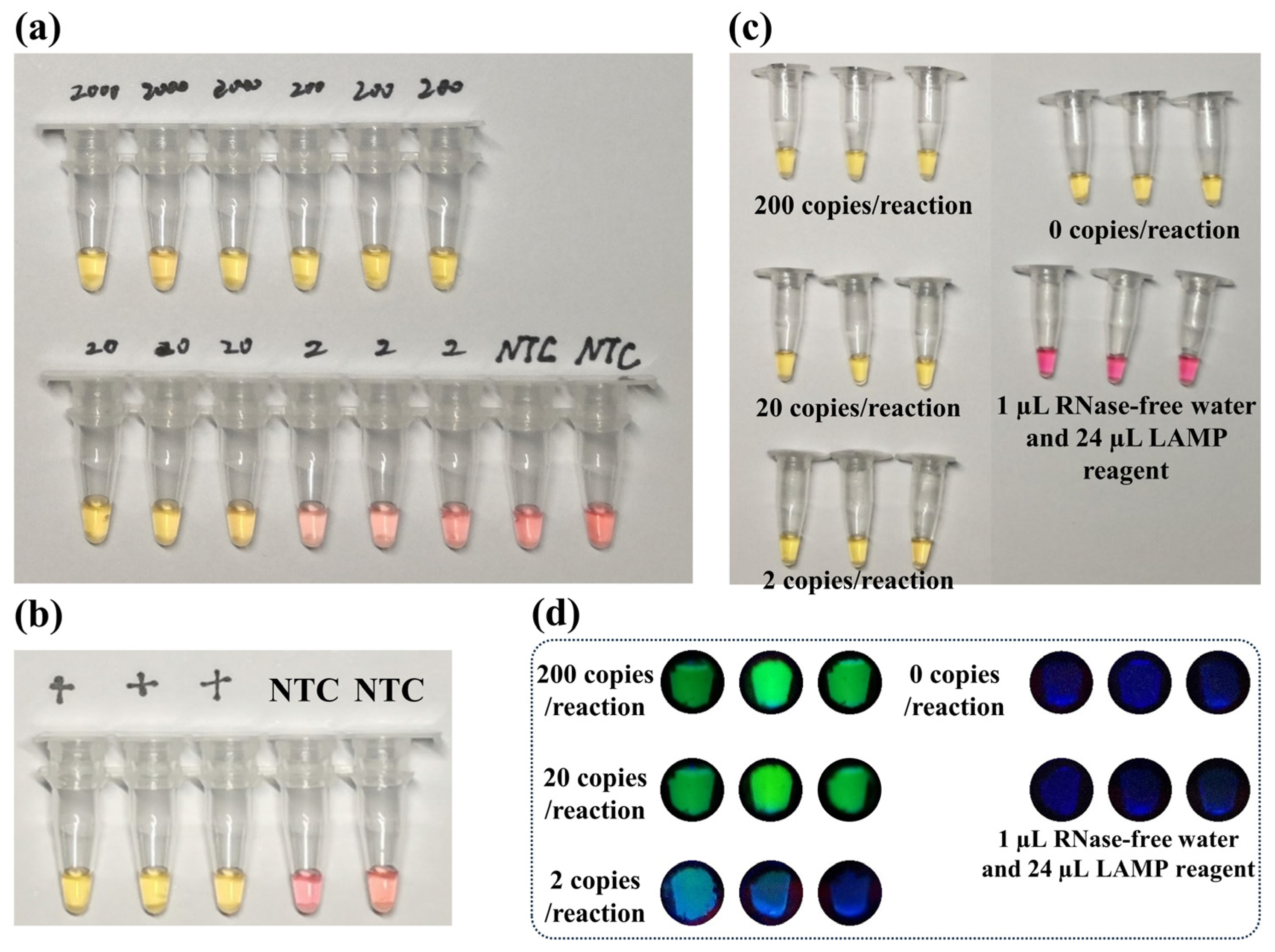
3.3. Development of UDG-LAMP-CRISPR Detection
3.4. Detection of SARS-CoV-2 with MS2 Phage as Internal Control
4. Conclusions
Supplementary Materials
Author Contributions
Funding
Institutional Review Board Statement
Informed Consent Statement
Data Availability Statement
Conflicts of Interest
References
- Zhu, N.; Zhang, D.; Wang, W.; Li, X.; Yang, B.; Song, J.; Zhao, X.; Huang, B.; Shi, W.; Lu, R.; et al. China Novel Coronavirus, T. Research, A Novel Coronavirus from Patients with Pneumonia in China, 2019. NEJM 2020, 382, 727–733. [Google Scholar] [CrossRef]
- Won, J.; Lee, S.; Park, M.; Kim, T.; Park, M.; Choi, B.; Kim, D.; Chang, H.; Kim, V.; Lee, C. Development of a Laboratory-safe and Low-cost Detection Protocol for SARS-CoV-2 of the Coronavirus Disease 2019 (COVID-19). Exp. Neurobiol. 2020, 29, 107–119. [Google Scholar] [CrossRef]
- Feng, Y.; Marchal, T.; Sperry, T.; Yi, H. Influence of wind and relative humidity on the social distancing effectiveness to prevent COVID-19 airborne transmission: A numerical study. J. Aerosol Sci. 2020, 147, 105585. [Google Scholar] [CrossRef] [PubMed]
- Rizou, M.; Galanakis, I.; Aldawoud, T.; Galanakis, C. Safety of foods, food supply chain and environment within the COVID-19 pandemic. Trends Food Sci. Tech. 2020, 102, 293–299. [Google Scholar] [CrossRef]
- Zhang, W.; He, H.; Zhu, L.; Liu, G.; Wu, L. Food Safety in Post-COVID-19 Pandemic: Challenges and Countermeasures. Biosensors 2021, 11, 71. [Google Scholar] [CrossRef]
- Dhakal, J.; Jia, M.; Joyce, J.; Moore, G.; Ovissipour, R.; Bertke, A. Survival of Severe Acute Respiratory Syndrome Coronavirus 2 (SARS-CoV-2) and Herpes Simplex Virus 1 (HSV-1) on Foods Stored at Refrigerated Temperature. Foods 2021, 10, 1005. [Google Scholar] [CrossRef]
- Jia, M.; Taylor, T.; Senger, S.; Ovissipour, R.; Bertke, A. SARS-CoV-2 Remains Infectious on Refrigerated Deli Food, Meats, and Fresh Produce for up to 21 Days. Foods 2022, 11, 286. [Google Scholar] [CrossRef] [PubMed]
- Jung, S.; Kim, D.; Ahn, H.; Go, H.; Wang, Z.; Yeo, D.; Woo, S.; Seo, Y.; Hossain, M.; Choi, I.; et al. Stability and inactivation of SARS-CoV-2 on food contact surfaces. Food Control. 2023, 143, 109306. [Google Scholar] [CrossRef]
- Donahue, M.; Sreenivasan, N.; Stover, D.; Rajasingham, A.; Watson, J.; Bealle, A.; Ritchison, N.; Safranek, T.; Waltenburg, M.; Buss, B.; et al. Notes from the Field: Characteristics of Meat Processing Facility Workers with Confirmed SARS-CoV-2 Infection—Nebraska, April-May 2020. MMWR Morb. Mortal. Wkly. Rep. 2020, 69, 1020–1022. [Google Scholar] [CrossRef] [PubMed]
- Qun, Y.; Zengqiang, K.; Fachun, J.; Zhongjie, L.; Lijie, Z.; Huihui, L.; Xiang, Z.; Dianmin, K.; Ruqin, G.; Jie, L. A Nosocomial COVID-19 Outbreak Initiated by an Infected Dockworker at Qingdao City Port—Shandong Province, China, October, 2020. China CDC Wkly. 2020, 2, 838–840. [Google Scholar] [CrossRef]
- Han, J.; Zhang, X.; He, S.; Jia, P. Can the coronavirus disease be transmitted from food? A review of evidence, risks, policies and knowledge gaps. Environ. Chem. Lett. 2021, 19, 5–16. [Google Scholar] [CrossRef]
- Yekta, R.; Vahid-Dastjerdi, L.; Norouzbeigi, S.; Mortazavian, A. Food products as potential carriers of SARS-CoV-2. Food Control 2021, 123, 107754. [Google Scholar] [CrossRef] [PubMed]
- Tomita, N.; Mori, Y.; Kanda, H.; Notomi, T. Loop-mediated isothermal amplification (LAMP) of gene sequences and simple visual detection of products. Nat. Protoc. 2008, 3, 877–882. [Google Scholar] [CrossRef]
- Li, J.; Hu, X.; Wang, X.; Yang, J.; Zhang, L.; Deng, Q.; Zhang, X.; Wang, Z.; Hou, T.; Li, S. A novel One-pot rapid diagnostic technology for COVID-19. Anal. Chim. Acta 2021, 1154, 338310. [Google Scholar] [CrossRef]
- Lu, R.; Wu, X.; Wan, Z.; Li, Y.; Jin, X.; Zhang, C. A Novel Reverse Transcription Loop-Mediated Isothermal Amplification Method for Rapid Detection of SARS-CoV-2. Int. J. Mol. Sci. 2020, 21, 2826. [Google Scholar] [CrossRef] [PubMed] [Green Version]
- Park, G.-S.; Ku, K.; Baek, S.-H.; Kim, S.-J.; Kim, S.; Kim, B.-T.; Maeng, J.-S. Development of Reverse Transcription Loop-Mediated Isothermal Amplification Assays Targeting Severe Acute Respiratory Syndrome Coronavirus 2 (SARS-CoV-2). JMD 2020, 22, 729–735. [Google Scholar] [CrossRef] [PubMed]
- Chen, J.; Ma, E.; Harrington, L.; Da Costa, M.; Tian, X.; Palefsky, J.; Doudna, J. CRISPR-Cas12a target binding unleashes indiscriminate single-stranded DNase activity. Science 2018, 360, 436–439. [Google Scholar] [CrossRef] [Green Version]
- Qian, C.; Wang, R.; Wu, H.; Zhang, F.; Wu, J.; Wang, L. Uracil-Mediated New Photospacer-Adjacent Motif of Cas12a To Realize Visualized DNA Detection at the Single-Copy Level Free from Contamination. Anal. Chem. 2019, 91, 11362–11366. [Google Scholar] [CrossRef]
- Qian, S.; Chen, Y.; Peng, C.; Wang, X.; Wu, H.; Che, Y.; Wang, H.; Xu, J.; Wu, J. Dipstick-based rapid nucleic acids purification and CRISPR/Cas12a-mediated isothermal amplification for visual detection of African swine fever virus. Talanta 2022, 242, 123294. [Google Scholar] [CrossRef]
- Jiang, X.; Loeb, J.; Manzanas, C.; Lednicky, J.; Fan, Z. Valve-Enabled Sample Preparation and RNA Amplification in a Coffee Mug for Zika Virus Detection. ANGEW Chem. Int. Edit. 2018, 57, 17211–17214. [Google Scholar] [CrossRef]
- Wang, C.; Horby, P.; Hayden, F.; Gao, G. A novel coronavirus outbreak of global health concern. Lancet 2020, 395, 470–473. [Google Scholar] [CrossRef] [PubMed] [Green Version]
- Huang, W.; Lim, B.; Hsu, C.; Xiong, D.; Wu, W.; Yu, Y.; Jia, H.; Wang, Y.; Zeng, Y.; Ji, M.; et al. RT-LAMP for rapid diagnosis of coronavirus SARS-CoV-2. Microb. Biotechnol. 2020, 13, 950–961. [Google Scholar] [CrossRef] [PubMed] [Green Version]
- Kim, D.; Lee, J.; Yang, J.; Kim, J.; Kim, V.; Chang, H. The Architecture of SARS-CoV-2 Transcriptome. Cell 2020, 181, 914–921. [Google Scholar] [CrossRef] [PubMed]
- Cui, J.; Li, F.; Shi, Z.-L. Origin and evolution of pathogenic coronaviruses. Nat. Rev. Microbiol. 2019, 17, 181–192. [Google Scholar] [CrossRef] [PubMed] [Green Version]
- Hsieh, K.; Mage, P.; Csordas, A.; Eisenstein, M.; Soh, H.T. Simultaneous elimination of carryover contamination and detection of DNA with uracil-DNA-glycosylase-supplemented loop-mediated isothermal amplification (UDG-LAMP). Chem. Commun. 2014, 50, 3747–3749. [Google Scholar] [CrossRef]
- Cheng, M.; Xiong, E.; Tian, T.; Zhu, D.; Ju, H.-q.; Zhou, X. A CRISPR-driven colorimetric code platform for highly accurate telomerase activity assay. Biosens. Bioelectron. 2021, 172, 112749. [Google Scholar] [CrossRef]
- Bhatt, A.; Fatima, Z.; Ruwali, M.; Misra, C.; Rangu, S.; Rath, D.; Rattan, A.; Hameed, S. CLEVER assay: A visual and rapid RNA extraction-free detection of SARS-CoV-2 based on CRISPR-Cas integrated RT-LAMP technology. J. Appl. Microbiol. 2022, 133, 410–421. [Google Scholar] [CrossRef]
- Misra, C.; Rangu, S.; Phulsundar, R.; Bindal, G.; Singh, M.; Shashidhar, R.; Saha, T.; Rao, A.; Rath, D. An improved, simple and field-deployable CRISPR-Cas12a assay for the detection of SARS-CoV-2. J. Appl. Microbiol. 2022, 133, 2668–2677. [Google Scholar] [CrossRef]
- Yin, K.; Ding, X.; Li, Z.; Sfeir, M.; Ballesteros, E.; Liu, C. Autonomous lab-on-paper for multiplexed, CRISPR-based diagnostics of SARS-CoV-2. Lab Chip 2021, 21, 2730–2737. [Google Scholar] [CrossRef]
- Zhang, F.; Wu, J.; Wang, R.; Wang, L.; Ying, Y. Portable pH-inspired electrochemical detection of DNA amplification. Chem. Commun. 2014, 50, 8416–8419. [Google Scholar] [CrossRef]
- Wang, R.; Xiao, X.; Chen, Y.; Wu, J.; Qian, W.; Wang, L.; Liu, Y.; Ji, F.; Wu, J. A loop-mediated, isothermal amplification-based method for visual detection of Vibrio parahaemolyticus within only 1 h, from shrimp sampling to results. Anal. Methods 2017, 9, 1695–1701. [Google Scholar]
- Oscorbin, I.P.; Shevelev, G.Y.; Pronyaeva, K.A.; Stepanov, A.A.; Shamovskaya, D.V.; Mishukova, O.V.; Pyshnyi, D.V.; Filipenko, M.L. Detection of SARS-CoV-2 RNA by a Multiplex Reverse-Transcription Loop-Mediated Isothermal Amplification Coupled with Melting Curves Analysis. Int. J. Mol. Sci. 2021, 22, 5743. [Google Scholar]


Disclaimer/Publisher’s Note: The statements, opinions and data contained in all publications are solely those of the individual author(s) and contributor(s) and not of MDPI and/or the editor(s). MDPI and/or the editor(s) disclaim responsibility for any injury to people or property resulting from any ideas, methods, instructions or products referred to in the content. |
© 2023 by the authors. Licensee MDPI, Basel, Switzerland. This article is an open access article distributed under the terms and conditions of the Creative Commons Attribution (CC BY) license (https://creativecommons.org/licenses/by/4.0/).
Share and Cite
Qian, S.; Chen, Y.; Wang, X.; Wang, T.; Che, Y.; Wu, J.; Ye, Z.; Xu, J. CRISPR/Cas12a-Assisted Dual Visualized Detection of SARS-CoV-2 on Frozen Shrimps. Biosensors 2023, 13, 138. https://doi.org/10.3390/bios13010138
Qian S, Chen Y, Wang X, Wang T, Che Y, Wu J, Ye Z, Xu J. CRISPR/Cas12a-Assisted Dual Visualized Detection of SARS-CoV-2 on Frozen Shrimps. Biosensors. 2023; 13(1):138. https://doi.org/10.3390/bios13010138
Chicago/Turabian StyleQian, Siwenjie, Yanju Chen, Xiaofu Wang, Tingzhang Wang, Yang Che, Jian Wu, Zhangying Ye, and Junfeng Xu. 2023. "CRISPR/Cas12a-Assisted Dual Visualized Detection of SARS-CoV-2 on Frozen Shrimps" Biosensors 13, no. 1: 138. https://doi.org/10.3390/bios13010138