In Situ Deposition of Gold Nanoparticles and L-Cysteine on Screen-Printed Carbon Electrode for Rapid Electrochemical Determination of As(III) in Water and Tea

Abstract
:1. Introduction
2. Experimental Section
2.1. Reagents and Chemicals
2.2. Apparatus
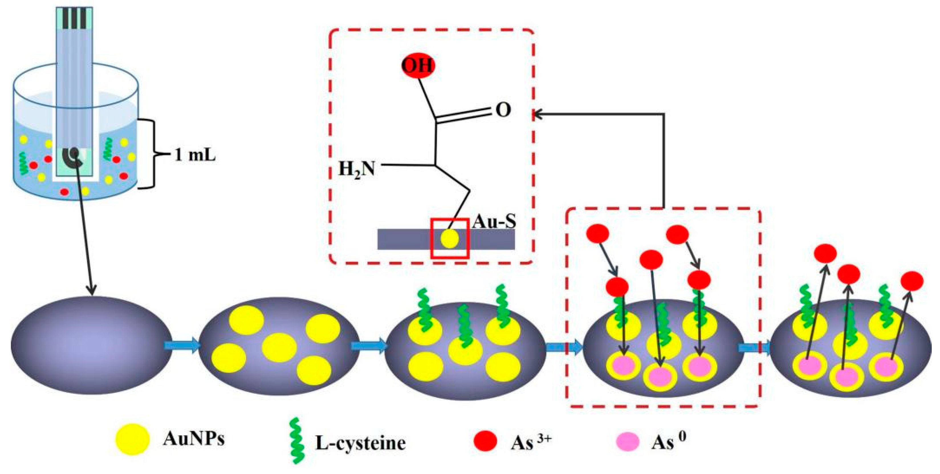
2.3. Methods
2.4. Recovery Studies
3. Results and Discussion
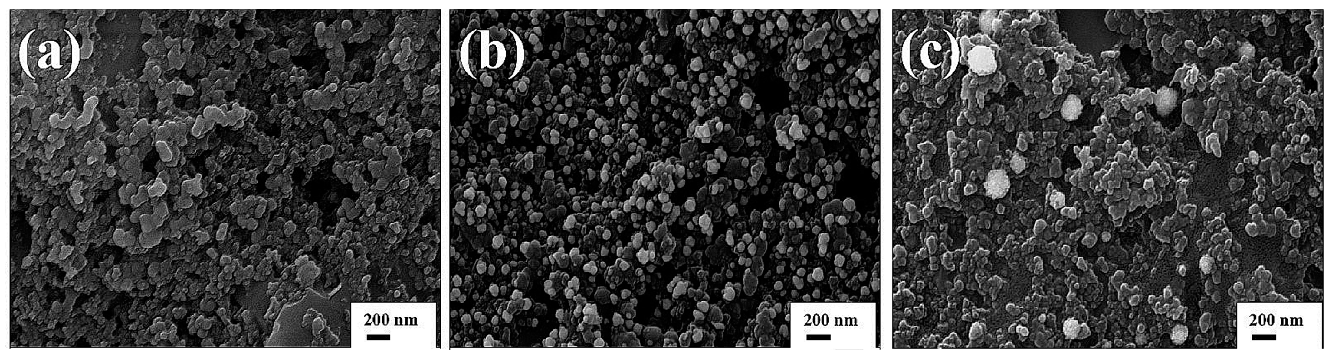
3.1. Morphological Characterization of SPCEs
3.2. Electrochemical Characterization of Electrodes
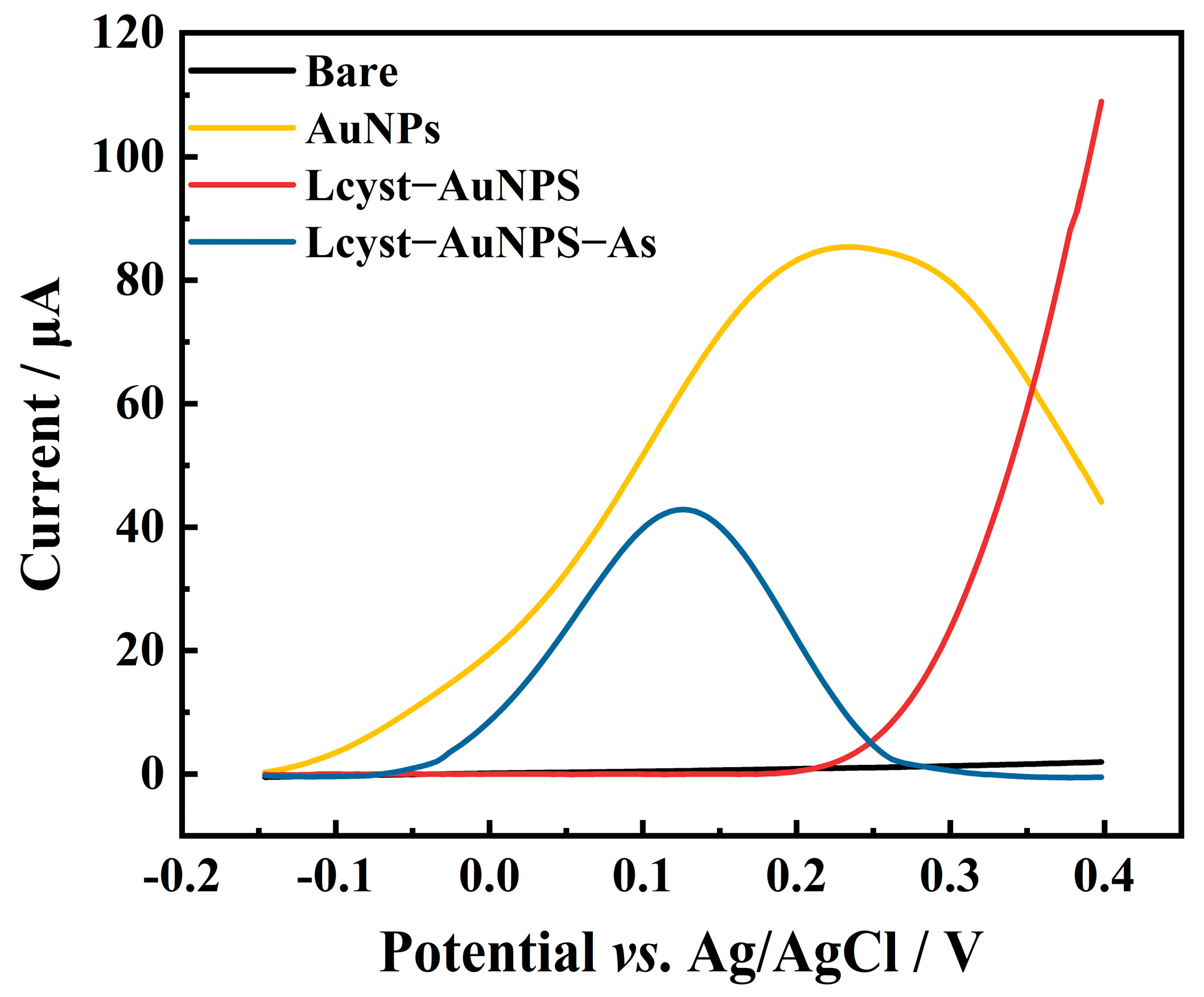
3.2.1. Cyclic Voltammetry Characterization of Gold Nanoparticles on SPCEs
3.2.2. Electrode Performance Evaluation with Cyclic Voltammetry
3.2.3. Measurement of Effective Surface Area of Electrodes
3.3. Electrochemical Behavior of Arsenic on Different Electrodes
3.4. Optimization of Experimental Parameters
3.4.1. Electrolyte
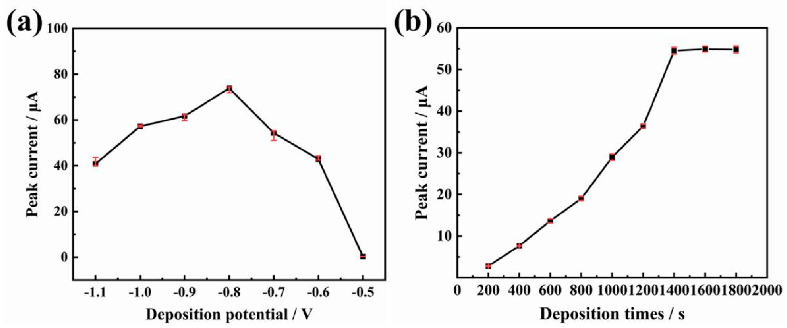
3.4.2. Enrichment Potential and Enrichment Time
3.4.3. Stirring Speed
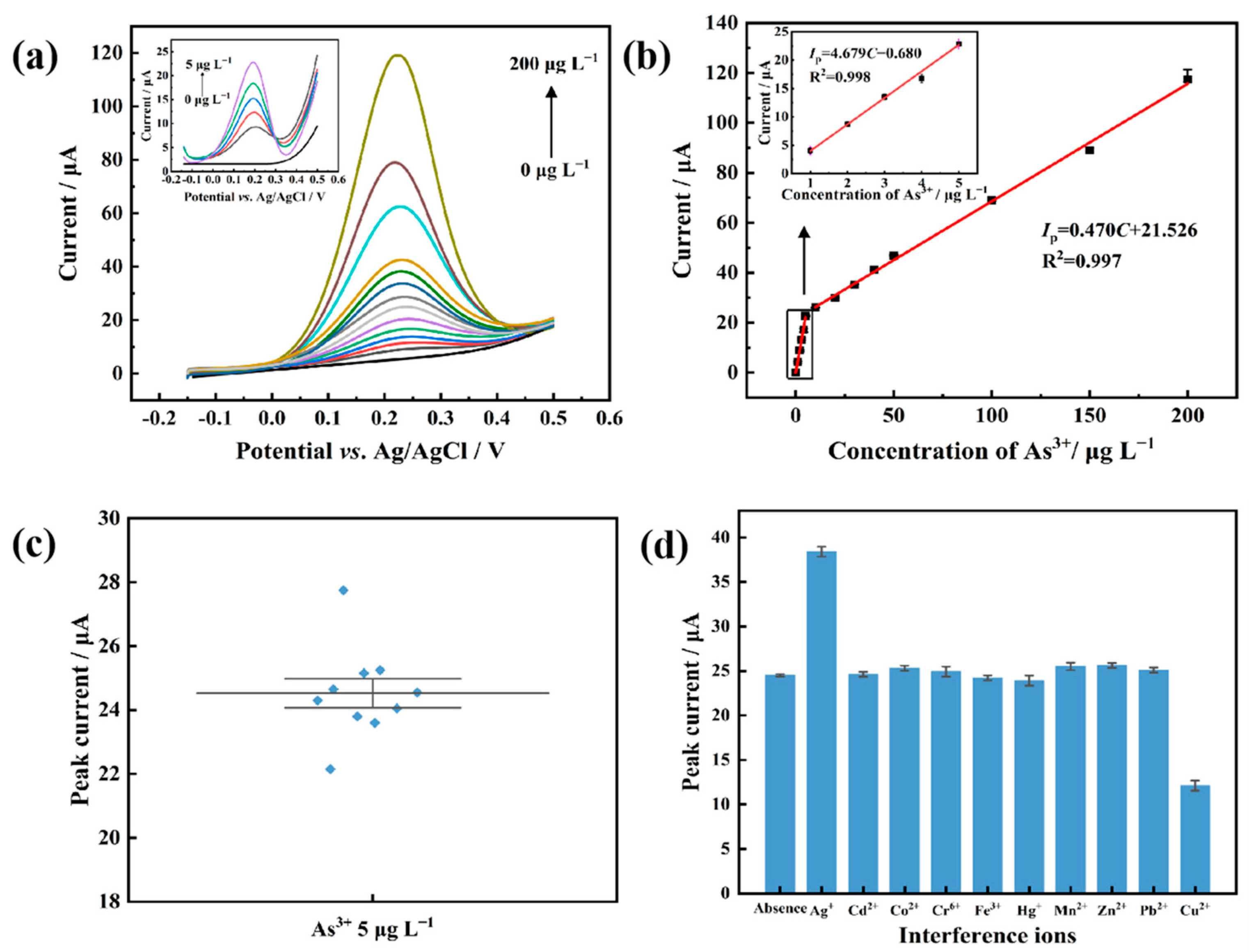
3.5. Electrochemical Determination of As(III)
3.6. Recovery Studies
4. Conclusions
Supplementary Materials
Author Contributions
Funding
Institutional Review Board Statement
Informed Consent Statement
Data Availability Statement
Conflicts of Interest
References
- He, Q.; Silliman, B. Climate change, human impacts, and coastal ecosystems in the Anthropocene. Curr. Biol. 2019, 29, R1021–R1035. [Google Scholar] [CrossRef]
- Gianì, F.; Masto, R.; Trovato, M.A.; Malandrino, P.; Russo, M.; Pellegriti, G.; Vigneri, P.; Vigneri, R. Heavy metals in the environment and thyroid cancer. Cancers 2021, 13, 4052. [Google Scholar] [CrossRef]
- Cai, L.; Ying, D.; Liang, X.; Zhu, M.; Lin, X.; Xu, Q.; Cai, Z.; Xu, X.; Zhang, L. A novel cationic polyelectrolyte microsphere for ultrafast and ultra-efficient removal of heavy metal ions and dyes. Chem. Eng. J. 2021, 410, 128404. [Google Scholar] [CrossRef]
- Upadhyay, M.K.; Shukla, A.; Yadav, P.; Srivastava, S. A review of arsenic in crops, vegetables, animals and food products. Food Chem. 2019, 276, 608–618. [Google Scholar] [CrossRef]
- Zheng, S.; Wang, Q.; Yuan, Y.; Sun, W. Human health risk assessment of heavy metals in soil and food crops in the Pearl River Delta urban agglomeration of China. Food Chem. 2020, 316, 126213. [Google Scholar] [CrossRef]
- Shankar, S.; Shanker, U. Arsenic contamination of groundwater: A review of sources, prevalence, health risks, and strategies for mitigation. Sci. World J. 2014, 2014, 1–18. [Google Scholar] [CrossRef]
- Zhang, X.; Yang, H.; Sun, R.; Cui, M.; Sun, N.; Zhang, S. Evaluation and analysis of heavy metals in iron and steel industrial area. Environ. Dev. Sustain. 2021, 24, 10997–11010. [Google Scholar] [CrossRef]
- World Health Organization. Guidelines for Drinking-Water Quality: First Addendum to the Fourth Edition; World Health Organization: Geneva, Switzerland, 2017. [Google Scholar]
- Gudlavalleti, R.H.; Bose, S.C.; Verma, S.K.; Khatri, P.; Scaria, J.; Dhewa, S.; Chaubey, V.K. A novel fluorometric bio-sensing-based arsenic detection system for groundwater. IEEE Sens. J. 2017, 17, 5391–5398. [Google Scholar] [CrossRef]
- Omeje, K.O.; Ezema, B.O.; Okonkwo, F.; Onyishi, N.C.; Ozioko, J.; Rasaq, W.A.; Sardo, G.; Okpala, C.O.R. Quantification of Heavy Metals and Pesticide Residues in Widely Consumed Nigerian Food Crops Using Atomic Absorption Spectroscopy (AAS) and Gas Chromatography (GC). Toxins 2021, 13, 870. [Google Scholar] [CrossRef]
- Balcaen, L.; Bolea-Fernandez, E.; Resano, M.; Vanhaecke, F. Inductively coupled plasma–Tandem mass spectrometry (ICP-MS/MS): A powerful and universal tool for the interference-free determination of (ultra) trace elements—A tutorial review. Anal. Chim. Acta 2015, 894, 7–19. [Google Scholar] [CrossRef]
- Zou, H.; Zhou, C.; Li, Y.; Yang, X.; Wen, J.; Song, S.; Li, C.; Sun, C. Speciation analysis of arsenic in edible mushrooms by high-performance liquid chromatography hyphenated to inductively coupled plasma mass spectrometry. Food Chem. 2020, 327, 127033. [Google Scholar] [CrossRef] [PubMed]
- Krishnan, S. Electrochemical Sensors for Large and Small Molecules in Biofluids. J. Electrochem. Soc. 2020, 167, 167505. [Google Scholar] [CrossRef]
- Pereira, D.F.; Santana, E.; Spinelli, A. Electrochemical paper-based analytical devices containing magnetite nanoparticles for the determination of vitamins B2 and B6. Microchem. J. 2022, 179, 107588. [Google Scholar] [CrossRef]
- Zhou, M.-H.; Tian, W.; Zhang, J.-Q.; Chen, X.; Wu, Y.-X.; Wang, S.-X. A rapid on-site analysis method for the simultaneous extraction and determination of Pb2+ and Cd2+ in cereals. RSC Adv. 2019, 9, 32839–32847. [Google Scholar] [CrossRef] [PubMed] [Green Version]
- Buffa, A.; Mandler, D. Arsenic (III) detection in water by flow-through carbon nanotube membrane decorated by gold nanoparticles. Electrochim. Acta 2019, 318, 496–503. [Google Scholar] [CrossRef]
- Sanllorente-Méndez, S.; Domínguez-Renedo, O.; Arcos-Martínez, M. Determination of arsenic (III) using platinum nanoparticle-modified screen-printed carbon-based electrodes. Electroanal. Int. J. Devoted Fundam. Pract. Asp. Electroanal. 2009, 21, 635–639. [Google Scholar] [CrossRef]
- Núñez, C.; Triviño, J.J.; Segura, R.; Arancibia, V. Development of a fast and sensitive method for the determination of As(III) at trace levels in urine by differential pulse anodic voltammetry using a simple graphene screen–printed electrode. Microchem. J. 2020, 159, 105393. [Google Scholar] [CrossRef]
- Torres-Rivero, K.; Pérez-Ràfols, C.; Bastos-Arrieta, J.; Florido, A.; Martí, V.; Serrano, N. Direct As (V) determination using screen-printed electrodes modified with silver nanoparticles. Nanomaterials 2020, 10, 1280. [Google Scholar] [CrossRef]
- Sivasothy, T.; Ndifor-Angwafor, N.; Marken, F. Voltammetric characteristics of hydrous Fe (III) oxide embedded into Nafion and immobilised onto a screen-printed carbon electrode: Binding of arsenate versus phosphate. J. Solid State Electrochem. 2018, 22, 3059–3067. [Google Scholar] [CrossRef] [Green Version]
- Nellaiappan, S.; KPillai, C.; Kumar, A. Flow-injection analysis coupled with electrochemical detection of poisonous inorganic arsenic (III) species using a gold nanoparticle/carbon nanofiber/chitosan chemically modified carbon screen printed electrode in neutral pH solution. Anal. Methods 2018, 10, 799–808. [Google Scholar] [CrossRef]
- Zhang, Y.; Li, D.; Compton, R. Arsenic (III) Detection with Underpotential Deposition and Anodic Stripping Voltammetry. ChemElectroChem 2021, 8, 3707–3715. [Google Scholar] [CrossRef]
- Zhang, W.; Chen, Z.; Guan, Y.; Liu, C.; Zheng, K.; Zou, X. Aptamer-functionalized screen-printed electrode coupled with graphene oxide and methylene blue nanocomposite as enhanced signal label for total arsenic determination in shellfish. Sens. Actuators B Chem. 2021, 335, 129383. [Google Scholar] [CrossRef]
- Sullivan, C.; Lu, D.; Senecal, A.; Kurup, P. Voltammetric detection of arsenic (III) using gold nanoparticles modified carbon screen printed electrodes: Application for facile and rapid analysis in commercial apple juice. Food Chem. 2021, 352, 129327. [Google Scholar] [CrossRef]
- Ismail, S.; Yusof, N.A.; Abdullah, J.; Rahman, S.F.A. Electrochemical detection of arsenite using a silica nanoparticles-modified screen-printed carbon electrode. Materials 2020, 13, 3168. [Google Scholar] [CrossRef]
- Núñez, C.; Triviño, J.J.; Arancibia, V. A electrochemical biosensor for As(III) detection based on the catalytic activity of Alcaligenes faecalis immobilized on a gold nanoparticle–modified screen–printed carbon electrode. Talanta 2021, 223, 121702. [Google Scholar] [CrossRef]
- Chen, L.; Zhou, N.; Li, J.; Chen, Z.; Liao, C.; Chen, J. Synergy of glutathione, dithiothreitol and N-acetyl-L-cysteine self-assembled monolayers for electrochemical assay: Sensitive determination of arsenic (III) in environmental and drinking water. Analyst 2011, 136, 4526–4532. [Google Scholar] [CrossRef]
- Kumar AK, S.; Zhang, Y.; Li, D.; Compton, R.G. A mini-review: How reliable is the drop casting technique? Electrochem. Commun. 2020, 121, 106867. [Google Scholar] [CrossRef]
- Newair, E.F.; Kilmartin, P.; Garcia, F. Square wave voltammetric analysis of polyphenol content and antioxidant capacity of red wines using glassy carbon and disposable carbon nanotubes modified screen-printed electrodes. Eur. Food Res. Technol. 2018, 244, 1225–1237. [Google Scholar] [CrossRef]
- Jijana, A.N.; Mphuthi, N.; Shumbula, P.; Vilakazi, S.; Sikhwivhilu, L. The Ultra-sensitive Electrochemical Detection of As(III) in Ground Water Using Disposable L-cysteine/Lipoic Acid Functionalised Gold Nanoparticle Modified Screen-Printed Electrodes. Electrocatalysis 2021, 12, 310–325. [Google Scholar] [CrossRef]
- Lin, T.-H.; Lin, C.-W.; Liu, H.-H.; Sheu, J.-T.; Hung, W.-H. Potential-controlled electrodeposition of gold dendrites in the presence of cysteine. Chem. Commun. 2011, 47, 2044–2046. [Google Scholar] [CrossRef]
- El-Deab, M.S.; Sotomura, T.; Ohsaka, T. Size and crystallographic orientation controls of gold nanoparticles electrodeposited on GC electrodes. J. Electrochem. Soc. 2004, 152, C1. [Google Scholar] [CrossRef]
- Zaki, M.H.M.; Mohd, Y.; Chin, L. Surface properties of nanostructured gold coatings electrodeposited at different potentials. Int. J. Electrochem. Sci 2020, 15, 11401–11415. [Google Scholar] [CrossRef]
- Arihara, K.; Ariga, T.; Takashima, N.; Arihara, K.; Okajima, T.; Kitamura, F.; Tokuda, K.; Ohsaka, T. Multiple voltammetric waves for reductive desorption of cysteine and 4-mercaptobenzoic acid monolayers self-assembled on gold substrates. Phys. Chem. Chem. Phys. 2003, 5, 3758–3761. [Google Scholar] [CrossRef]
- Wu, T.; Zhu, Y.; Song, L.; Chen, Y.; Huang, Y.; Tang, J.; Ma, X.; Wang, H.; Zhang, J.; Lin, D.; et al. Three-dimensional gold nanowires with high specific surface area for simultaneous detection of heavy metal ions. Anal. Methods 2022, 14, 859–868. [Google Scholar] [CrossRef]
- Lin, D.; Harris, K.D.; Chan, N.W.; Jemere, A.B. Nanostructured indium tin oxide electrodes immobilized with toll-like receptor proteins for label-free electrochemical detection of pathogen markers. Sens. Actuators B Chem. 2018, 257, 324–330. [Google Scholar] [CrossRef]
- Yin, S.; Wang, J.; Li, Y.; Wu, T.; Song, L.; Zhu, Y.; Chen, Y.; Cheng, K.; Zhang, J.; Ma, X.; et al. Macroscopically Oriented Magnetic Core-regularized Nanomaterials for Glucose Biosensors Assisted by Self-sacrificial Label. Electroanalysis 2021, 33, 2216–2225. [Google Scholar] [CrossRef]
- Dong, X.-X.; Li, M.-Y.; Feng, N.-N.; Sun, Y.-M.; Yang, C.; Xu, Z.-L. A nanoporous MgO based nonenzymatic electrochemical sensor for rapid screening of hydrogen peroxide in milk. RSC Adv. 2015, 5, 86485–86489. [Google Scholar] [CrossRef]
- Marcucci, K.; Zamboni, R.; d’Ulivo, A. Studies in hydride generation atomic fluorescence determination of selenium and tellurium. Part 2—Effect of thiourea and thiols. Spectrochim. Acta Part B At. Spectrosc. 2001, 56, 393–407. [Google Scholar] [CrossRef]


| Electrode | Method | Linear Range (ppb) | LOD (ppb) | Applications | Ref. |
|---|---|---|---|---|---|
| PtNPs/SPCE 1 | CV | 5.68 | Tap water | [15] | |
| GO/SPCE 2 | DPV 3 | 0.1~50 | 0.92 | Tap water and urine | [16] |
| Ag-NP-SPCNFEs 4 | DPASV | 1.9~25.1 | 1.9 | Drinking water | [17] |
| CNF-CHIT-AuNPs/SPCE 5 | CV | 100~1000 | 11.4 | Tap water | [19] |
| Pt/GCE 6 | LSV | 4~77 | 4 | Tap water | [20] |
| GO-MB/Aptamer-AuNPs/SPCE | DPV | 0.4~1000 | 0.2 | Shellfish | [21] |
| AuNPs/SPCE | SWV 7 | 16.73 | Apple juice | [22] | |
| SiNPs/SPCE 8 | LSASV | 5~30 | 6.2 | Tap water | [23] |
| AF-AuNPs/SPCE 9 | CV | 6~200 | 6 | River water | [24] |
| AuNPs-L-Cyst/SPCE | LSV | 1~200 | 0.91 | Tap water and tea leaf | This work |
| Sample | Added (μg L−1) | Found (μg L−1) | Recovery/% | RSD/% | Found by ICP-MA (μg L−1) | |Error| | t 1 | F2 |
|---|---|---|---|---|---|---|---|---|
| Tap water 1 | 0 | 0 | ||||||
| 10 | 12.3 | 105.4 | 3.18 | 11.9 | 3.4% | 0.13 | 0.35 | |
| 50 | 48.2 | 98.8 | 2.15 | 49.4 | 2.4% | 0 | 0.19 | |
| Tap water 2 | 0 | 0 | ||||||
| 10 | 10.6 | 101.2 | 4.02 | 10.2 | 3.8% | 0.56 | 0.03 | |
| 50 | 51.4 | 99.3 | 1.99 | 50.8 | 1.1% | 0 | 0.12 | |
| Tea leaf 1 | 0 | 0 | 2.3 | |||||
| 10 | 11.3 | 97.6 | 4.57 | 11.9 | 5.0% | 0.09 | 0.23 | |
| 50 | 52.6 | 102.6 | 3.06 | 52.7 | 0.18% | 0.76 | 0.29 | |
| Tea leaf 2 | 0 | 0 | ||||||
| 10 | 10.8 | 98.1 | 4.88 | 10.5 | 2.8% | 0.94 | 0.10 | |
| 50 | 51.1 | 97.7 | 3.71 | 51.7 | 1.2% | 0 | 0.44 | |
| Tea leaf 3 | 0 | 0 | ||||||
| 10 | 8.8 | 96.2 | 1.32 | 9.1 | 3.2% | 0.05 | 0.08 | |
| 50 | 47.2 | 93.8 | 2.94 | 48.6 | 2.9% | 0 | 0.43 |
Disclaimer/Publisher’s Note: The statements, opinions and data contained in all publications are solely those of the individual author(s) and contributor(s) and not of MDPI and/or the editor(s). MDPI and/or the editor(s) disclaim responsibility for any injury to people or property resulting from any ideas, methods, instructions or products referred to in the content. |
© 2023 by the authors. Licensee MDPI, Basel, Switzerland. This article is an open access article distributed under the terms and conditions of the Creative Commons Attribution (CC BY) license (https://creativecommons.org/licenses/by/4.0/).
Share and Cite
Wang, W.; Yi, Z.; Liang, Q.; Zhen, J.; Wang, R.; Li, M.; Zeng, L.; Li, Y. In Situ Deposition of Gold Nanoparticles and L-Cysteine on Screen-Printed Carbon Electrode for Rapid Electrochemical Determination of As(III) in Water and Tea. Biosensors 2023, 13, 130. https://doi.org/10.3390/bios13010130
Wang W, Yi Z, Liang Q, Zhen J, Wang R, Li M, Zeng L, Li Y. In Situ Deposition of Gold Nanoparticles and L-Cysteine on Screen-Printed Carbon Electrode for Rapid Electrochemical Determination of As(III) in Water and Tea. Biosensors. 2023; 13(1):130. https://doi.org/10.3390/bios13010130
Chicago/Turabian StyleWang, Wenjing, Zhijian Yi, Qiongxin Liang, Junjie Zhen, Rui Wang, Mei Li, Lingwen Zeng, and Yongfang Li. 2023. "In Situ Deposition of Gold Nanoparticles and L-Cysteine on Screen-Printed Carbon Electrode for Rapid Electrochemical Determination of As(III) in Water and Tea" Biosensors 13, no. 1: 130. https://doi.org/10.3390/bios13010130