Recent Advances in Electrochemical Immunosensors with Nanomaterial Assistance for Signal Amplification


Abstract
:
1. Introduction
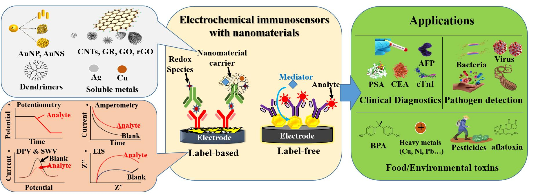
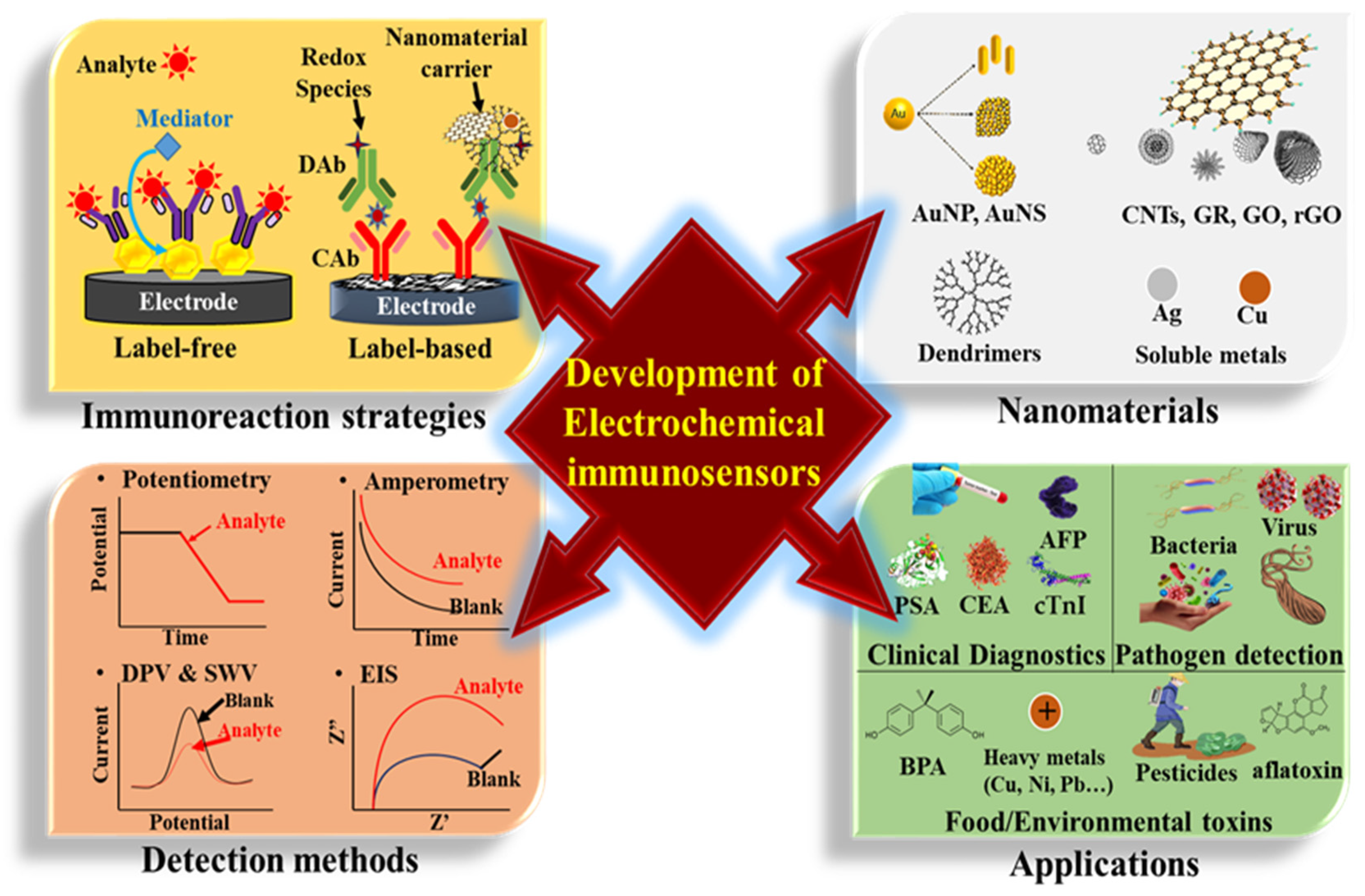
2. Immunoreacting Strategies and Electrochemical Detection
2.1. Label-Free Electrochemical Immunosensor
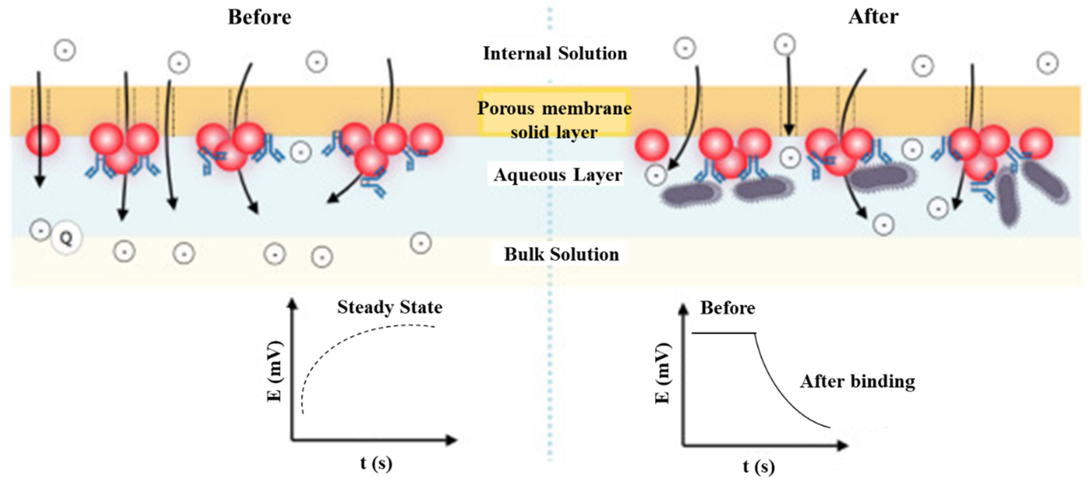
2.1.1. Potentiometry-Based Immunosensors
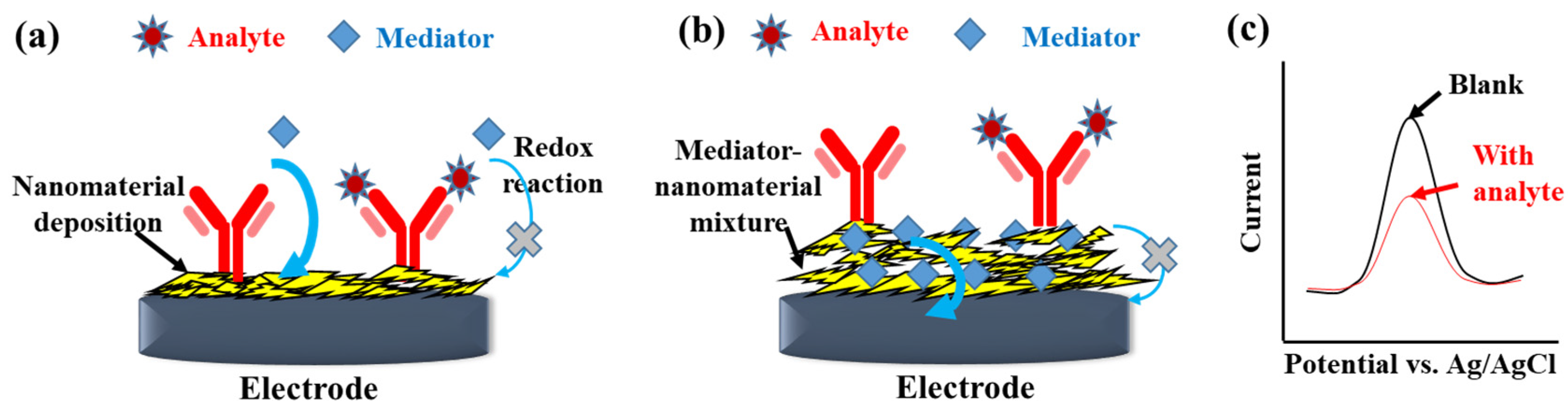
2.1.2. Voltammetry-Based Immunosensors
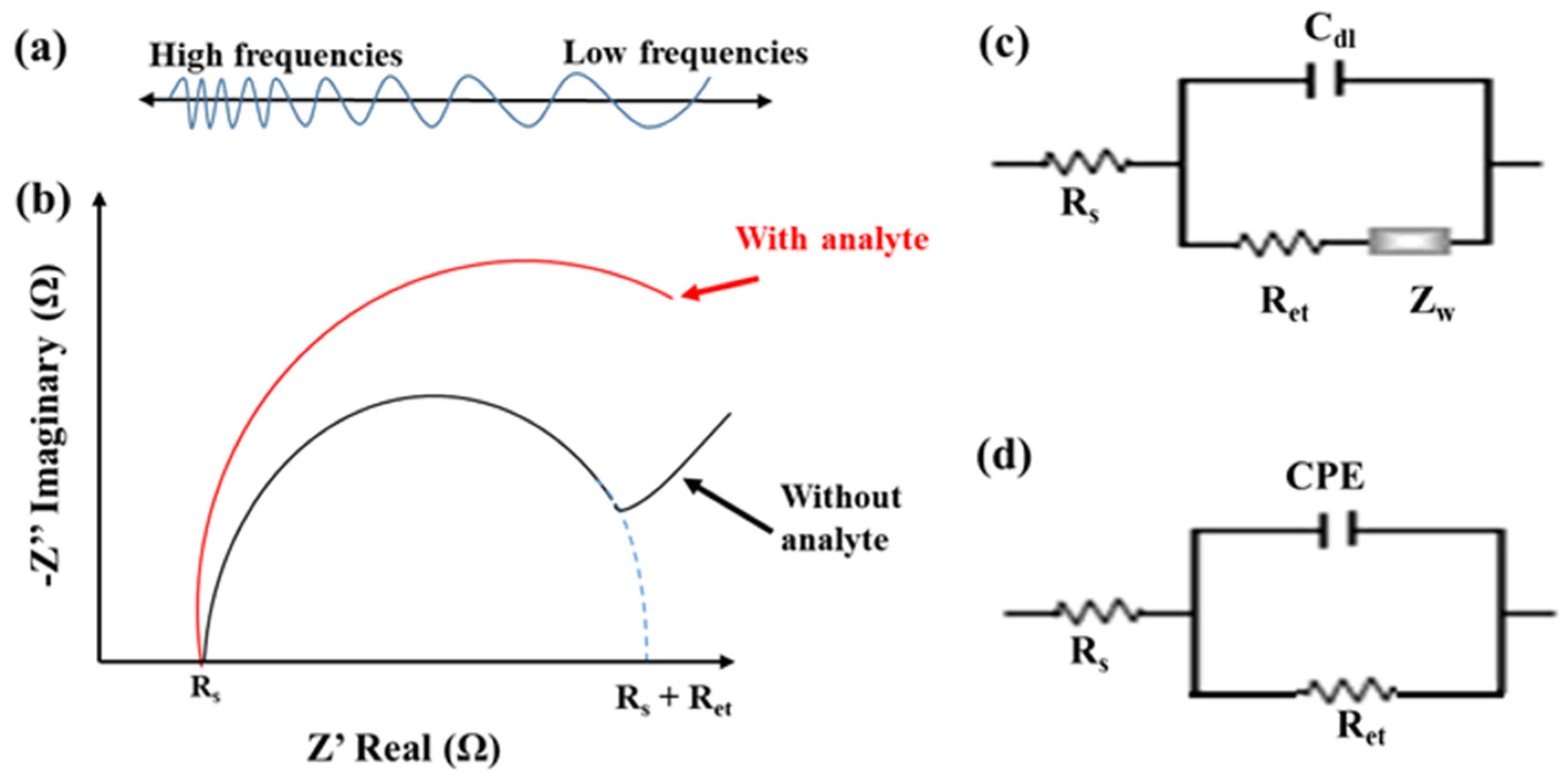
2.1.3. EIS-Based Immunosensors
2.2. Label-Based Electrochemical Immunosensor
3. Nanomaterials for Electrode Modification
3.1. Using AuNPs/AuNSs

| Preparation Methods | Electrodes | Sensing Strategies/Techniques | LOD | LR | Refs. |
|---|---|---|---|---|---|
| Electrodeposition | Anti-prolactin/AuNPs/carbon paste electrode | Sandwich reaction with HRP-DAb and prolactin/DPV | 12.5 mIU/L | 25.0–2000.0 mIU/L | [42] |
| Electrodeposition | Anti-NSE/AuNPs-MoS2-rGO/GCE 1 | Sandwich reaction with DAb/ CoFe2O4-Ag, and NSE by SWV | 3 fg/mL | 0.01–1.00 pg/mL | [60] |
| Electrodeposition | Anti-HSA/AuNPs/PpPD/PEDOT-PSS-Fc /SPCEs 2 | Label-free/DPV | 0.54 fg/mL | 1–10 ng/mL | [61] |
| Electroless plating | Anti-PHB2/PA/AuNS/AuE | Sandwich reaction with HRP-Dab and PHB2/SWV | 40 pg/mL | 0–10 ng/mL | [52] |
| Electrostatic adsorption | Anti-CA153/PPy-AuNPs-luminol/ITO 3 | Label-free/EIS & electrochemiluminescence | 5.8 × 10⁻⁴ U/mL | 0.001–700 U/mL | [62] |
| Multi-layer electrostatic adsorption | Anti-PSA-GSH-AuNPs/PEI/PVS/PEI/MUA/AuE 4 | Label-free/EIS | 0.17 ng/mL | 0.1–20 ng/mL | [63] |
3.2. Using CNTs
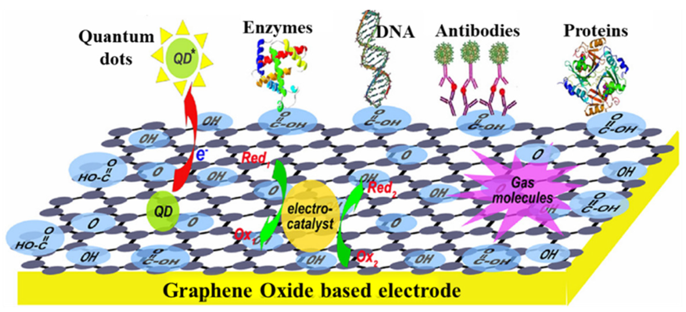
3.3. Using GR/RGO
4. Nanomaterials Used as Labels
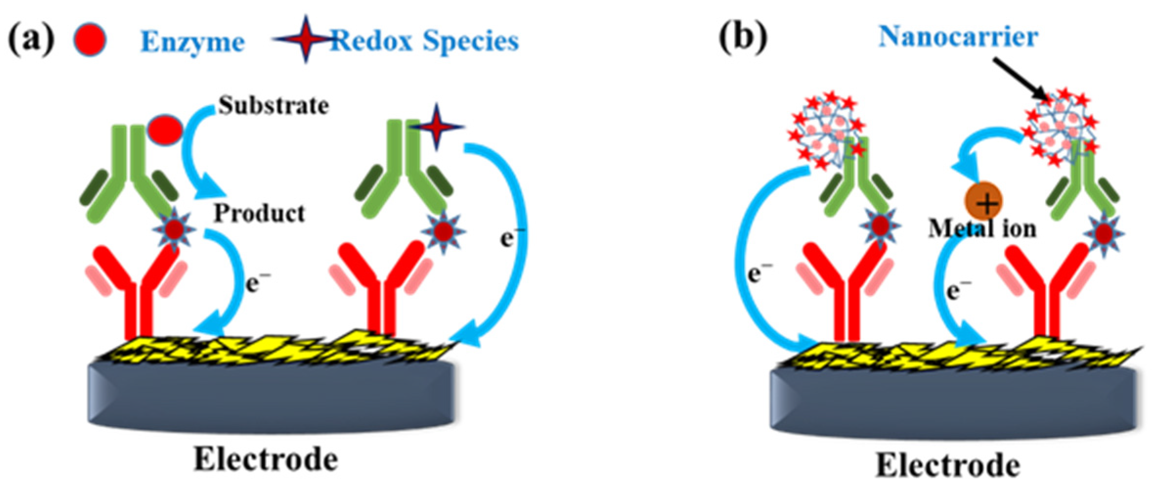
4.1. Nanomaterial Carriers with Enzymatic/Electroactive Catalyzer
4.2. Nanomaterial Carriers with Electroactive Species for Direct Redox Reaction
4.3. Nanomaterial Carriers with Soluble NPs
| Label-Based Nanomaterials | Analyte/Electrodes | Sensing Strategies/Techniques | LOD | LR | Refs. |
|---|---|---|---|---|---|
| Anti-HFA 1 DAb & HRP/COOH-MWCNT | HFA/CAb-biotin/streptavidin/SPCE | H2O2/HRP/hydroquinone/amperometry | 16 pg/mL | 20–2000 pg/mL | [96] |
| Anti-CA125 DAb/AuNP-LaOx 2 | CA125/CAb/CS-AuNP/MWCNT-GO/GCE | H2O2/LaOx/lactic acid/amperometry | 2 mU/mL | 0.01–100 U/mL | [97] |
| Anti-CEA DAb/MoS2 NFs/Au@AgPt YNCs 3 | CEA/CAb/MoS2/Au@AgPt YNCs/GCE | Enhanced H2O2 reduction via AgPt/amperometry | 3.09 fg/mL | 1 × 10⁵ –100 ng/mL | [98] |
| Anti-CEA DAb/GR sheet-Fe3O4/Au@Ag/Ni2+ | CEA/CAb/AuNPs/GCE | Enhanced H2O2 reduction via Ni2+/amperometry | 69.7 fg/mL | 1 × 10⁻⁴–100 ng/mL | [99] |
| Anti-CEA DAb/Au@SiO2/Cu2O | CEA/CAb/Ag/g-C3N4 4 /GCE | Enhanced H2O2 reduction via Cu2O/amperometry | 3.8 fg/mL | 1 × 10⁻⁵−80 ng/mL | [100] |
| Anti-PSA DAb/Au@Ag-Cu2O | PSA/CAb/Au@N-GQDs/GCE | Enhanced H2O2 reduction via Cu2O/amperometry | 3 fg/mL | 1 × 10⁻⁵−100 ng/mL | [101] |
| Anti-cTnI DAb/N,S-cGO/L-lys/AuNR@Pt MBs/Thi 5 | cTnI/CAb/AuNR@PDA/GCE | Direct reduction of Thi/ amperometry and DPV | 16.7 fg/mL | 5 × 10⁻⁵–250 ng/mL | [102] |
| Anti-CEA DAb/Ag@CeO2 core-shell-Au NPs | CEA/CAb/AuNPs/GCE | Ag-CeO2 direct redox/CV & EIS | 32 fg/mL | 1 × 10⁻⁴–5 ng/mL | [48] |
| Anti-CA125 DAb-TB/Suc-CS@MNP 6 | CA125/CAb/PAMAM 7 /AuNP-3D RGO-MWCNT | Direct reduction of TB/SWV | 6 μU/mL | 0.0005–75 U/mL | [103] |
| Carbaryl hapten@CuNP-CS | Carbaryl/CAb/AuNP/GCE | Direct oxidation of CuNPs after immuno-competition/linear sweep ASV | 0.05 ng/mL | 0.5–20.0 ng/mL | [104] |
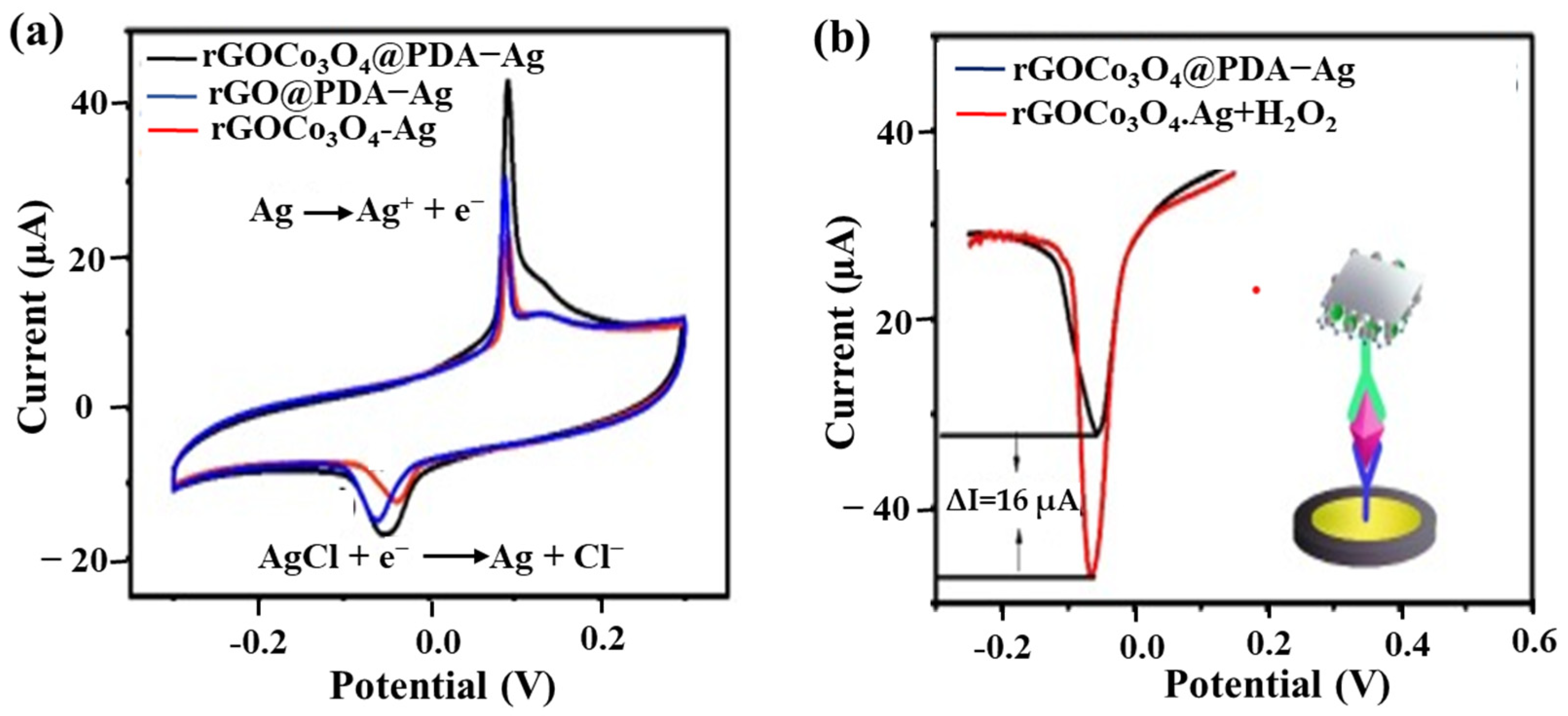
| Anti-CEA DAb/RGO/Co3O4-Ag@ PDA | CEA/CAb/AuNP/GCEs | Ag-Ag+ redox with H2O2 enhancement/DPV | 0.17 pg/mL | 0.0005–80 ng/mL | [49] |
| CdS nanocrystals/phage | Molinate/14D7 CAb/polynitroaniline/GCE | CdS-Cd2+ with HCl dissolution/square wave ASV | 34 pg/mL | 0.1–10 ng/mL | [105] |
| Anti-casein biotin-CAb/Streptavidin/CdSe/ZnS QDs | Bovine casein/Bovine casein/Sb2O5-SnO2/SPCEs | CdSe-Cd2+ after immuno-competition with HCl dissolution/ASV | 0.07 % (v/v) | 0.1–10% (v/v) Cow’s milk in ewe/goat’s cheese | [106] |
| anti-HE4 CAb/CdSe/ZnS QDs | HE4 8 /DAb/MBs/Hg/SPCEs | CdSe-Cd2+ after immuno-competition with HCl dissolution/ASV | 2 pM | 20–40 nM | [107] |
| Anit-FABP DAb/CdS- ZnO-MWCNTs | FABP/CAb/CD-GS/GCE 9 | CdS-Cd2+ with HNO3 dissolution/ASV | 0.3 fg/mL | 1.3–130 ng/mL | [108] |
5. Other Trends and Challenges in Electrochemical Immunosensors
- (1)
- Although GCE is durable and frequently used for immunosensor construction, SPCEs, possessing the benefits of low cost and ease of massive production, present great promise to develop disposable immunosensors. The nanocarrier can be dripped on the SPCEs as substrate for antibody immobilization. Wei et al. [110] synthesized RGO/Prussian blue/core-shell Au@PtNPs as a nanocarrier for drop-coating SPCEs. The anti-hepatitis B antibody could be directly adsorbed on the nanocarrier surface, and the Prussian blue served as an electron-transfer mediator. After immunoreaction with the hepatitis B surface antigen, the label-free LOD measured by DPV was 80 pg/mL. Furthermore, Malla et al. [111] fixed a magnet to the backside of SPCEs for adsorbing HRP-CAb-modified MBs conjugated with parathyroid hormone antigen and then performed the catalysis of H2O2 and hydroquinone with SWV detection to obtain an LOD of 11.56 pg/mL. The drip-coating fixation or the magnetic adsorption of CAb-modified nanocomposite carriers on SPCEs can simplify the preparation of SPCE-based disposable immunosensors. Although SPCEs are prevailing in the development of disposable point-of-care testing strips, the activation or the peroxidation procedures of the SPCE surface still take up much time before use. Oxygen plasma treatment is an alternative for mass production. Subsequently, a sealing package for long-term storage is essential after plasma treatment.
- (2)
- Preventing the effect of non-specific adsorption on label-free electrochemical immunosensors from versatile molecules of actual samples is an essential issue. Antifouling materials, such as poly(ethylene glycol) and zwitterionic polymers [112], block the electrode surface to reduce non-specific adsorption. Wang and Hui electrodeposited polyaniline nanowires on a GCE to produce a highly rough surface, and photopolymerized zwitterionic poly(carboxybetaine methacrylate) (polyCBMA) on the polyaniline nanowire to obtain a hierarchical structure. After chemical activation, the anti-CEA antibody was covalently immobilized on polyCBMA without extra surface blocking. The DPV-based immunosensors presented an ultralow LOD of 3.05 fg/mL and an impressive antifouling ability from cow’s milk, saliva, bovine fetal serum, and human serum [113]. The modification technique of antibody and polyCBMA supplies promising potential for the antifouling treatment of immunosensors.
- (3)
- Multiplexed detection in clinical diagnosis, agricultural pesticide/herbicide residue, and environmental toxins has considerable importance due to their excellent analytical efficiency compared with parallel single-analyte assays. Two kinds of multiplexed detecting strategies have been developed. One is to use different multi-detectors placed on the same substrate. Serafín et al. [114] separated immobilized anti-tau protein (tau) CAb and anti-TAR DNA-binding protein 43 (TDP-43) CAb on the two 3D-Au-PAMAM-modified working electrodes of the SPCEs. After sandwich immunoreaction, the HRP-conjugated DAb can quantify the tau and TDP-43 in raw plasma samples by catalyzing the H2O2/hydroquinone reaction with amperometric detection. Furthermore, Salahandish et al. [115] developed dual-immunosensors for the label-free detection of SARS-CoV-2 nucleocapsid protein by EIS. The other multiplexed technique uses biorecognition molecules, tagging different electroactive mediators on the identical sensing interface. Shen et al. [116] tagged anthraquinone on the VEGF-aptamer, methyl blue on the IFN-γ aptamer, and ferrocene on the TNF-α-aptamer to achieve multiplex detection, respectively. The three kinds of aptamers were biotinylated to immobilize them on the single streptavidin/GO/AuE. Then, SWV was performed to obtain the redox signal of anthraquinone, methyl blue, and ferrocene at −0.45 V, −0.26 V, and 0.25 V before and after the label-free immunoreaction. Compared to the single electrode immobilized by multi-CAbs, the multi-electrodes with different CAb immobilization are easier to control the density of CAbs to obtain better sensing properties.
- (4)
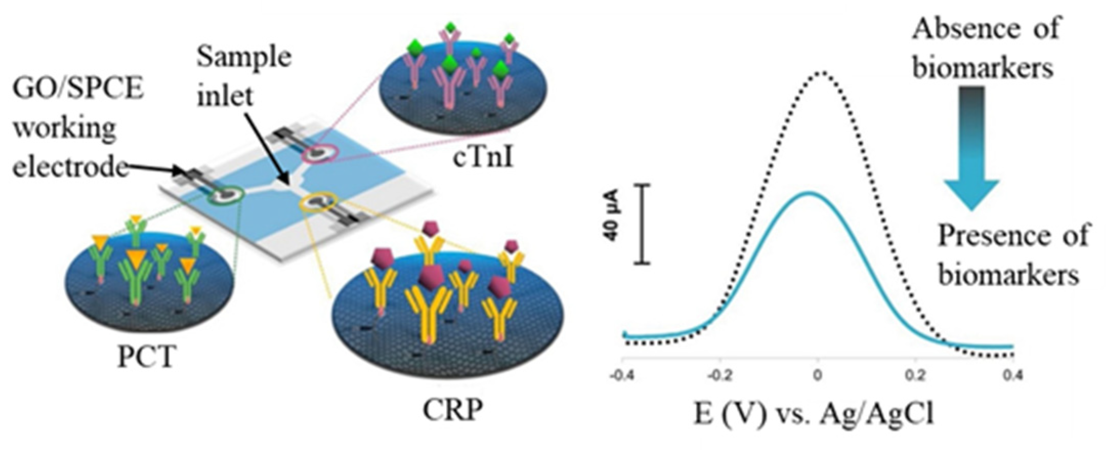
- The immunosensors integrating fluidic transportation can promote immunoreaction efficiency in shorter immunoreaction time and reduce detecting procedures. Lin et al. [41] fabricated an impedimetric affinity sensing chip integrated with an AC electrokinetic flow vortex. The protein A-antibody affinity time can reach the plateau in 8 min with AC electrokinetic flow. The corresponding EIS-Ret value of the affinity plateau was 2.26 times larger than that obtained in an unstirred solution. Furthermore, the paper-based immunoassay becomes an exciting alternative for constructing disposable, low-cost, and eco-friendly analytical devices due to flexibility, lightness, capillary-driven flow, and affordability in an austere environment. Shu et al. [117] utilized a paper-based electrochemical immunosensing device for the label-free detection of AFP. The Ni-Co MOF nanosheets were modified with CNT and streptavidin and then coated onto a GR-printed working electrode for biotinylated CAb immobilization. After immunoreaction with samples conducted through vertical flow, the H2O2/hydroquinone mixture was dripped to produce a DPV signal via Ni-Co MOF catalysis. Furthermore, Boonkaew et al. [118] constructed triple three-electrode SPCEs in triple channels in an identical substrate to form multiplexed electrochemical paper-based analytical devices (ePADs), as shown in Figure 10. Three kinds of antibodies were respectively immobilized on the different GO/SPCEs to capture C-reactive protein (CRP), cardiac troponin I (cTnI), and procalcitonin (PCT) of the cardiovascular disease biomarkers. After immunoreaction, the redox solution was dripped into the central inlet and conducted to the sensing region via lateral flow for DPV detection. The multiplexed ePADs can detect C-reactive protein, cTnI, and PCT with corresponding LODs of 0.38 ng/mL, 0.16 pg/mL, and 0.27 pg/mL, respectively. The design and fabrication of ePADs have promising potential in constructing a multiplexed point-of-care testing device.
6. Conclusions
Author Contributions
Funding
Institutional Review Board Statement
Informed Consent Statement
Data Availability Statement
Conflicts of Interest
References
- Ravina; Kumar, D.; Prasad, M.; Mohan, H. Biological Recognition Elements. In Electrochemical Sensors; Elsevier: Amsterdam, The Netherlands, 2022; pp. 213–239. [Google Scholar] [CrossRef]
- Zheng, Y.; Li, J.; Zhou, B.; Ian, H.; Shao, H. Advanced Sensitivity Amplification Strategies for Voltammetric Immunosensors of Tumor Marker: State of the Art. Biosens. Bioelectron. 2021, 178, 113021. [Google Scholar] [CrossRef] [PubMed]
- Koo, K.M.; Soda, N.; Shiddiky, M.J.A. Magnetic Nanomaterial–Based Electrochemical Biosensors for the Detection of Diverse Circulating Cancer Biomarkers. Curr. Opin. Electrochem. 2021, 25, 100645. [Google Scholar] [CrossRef]
- Farzin, L.; Shamsipur, M. Recent Advances in Design of Electrochemical Affinity Biosensors for Low Level Detection of Cancer Protein Biomarkers Using Nanomaterial-Assisted Signal Enhancement Strategies. J. Pharm. Biomed. Anal. 2018, 147, 185–210. [Google Scholar] [CrossRef] [PubMed]
- Li, Z.; Zhang, J.; Huang, Y.; Zhai, J.; Liao, G.; Wang, Z.; Ning, C. Development of Electroactive Materials-Based Immunosensor towards Early-Stage Cancer Detection. Coord. Chem. Rev. 2022, 471, 214723. [Google Scholar] [CrossRef]
- Filik, H.; Avan, A.A. Electrochemical Immunosensors for the Detection of Cytokine Tumor Necrosis Factor Alpha: A Review. Talanta 2020, 211, 120758. [Google Scholar] [CrossRef] [PubMed]
- Skládal, P. Advances in Electrochemical Immunosensors for Pathogens. Curr. Opin. Electrochem. 2019, 14, 66–70. [Google Scholar] [CrossRef]
- Mokhtarzadeh, A.; Eivazzadeh-Keihan, R.; Pashazadeh, P.; Hejazi, M.; Gharaatifar, N.; Hasanzadeh, M.; Baradaran, B.; dela Guardia, M. Nanomaterial-Based Biosensors for Detection of Pathogenic Virus. TrAC Trends Anal. Chem. 2017, 97, 445–457. [Google Scholar] [CrossRef]
- Brazaca, L.C.; dos Santos, P.L.; de Oliveira, P.R.; Rocha, D.P.; Stefano, J.S.; Kalinke, C.; Abarza Muñoz, R.A.; Bonacin, J.A.; Janegitz, B.C.; Carrilho, E. Biosensing Strategies for the Electrochemical Detection of Viruses and Viral Diseases—A Review. Anal. Chim. Acta 2021, 1159, 338384. [Google Scholar] [CrossRef]
- Kalambate, P.K.; Noiphung, J.; Rodthongkum, N.; Larpant, N.; Thirabowonkitphithan, P.; Rojanarata, T.; Hasan, M.; Huang, Y.; Laiwattanapaisal, W. Nanomaterials-Based Electrochemical Sensors and Biosensors for the Detection of Non-Steroidal Anti-Inflammatory Drugs. TrAC Trends Anal. Chem. 2021, 143, 116403. [Google Scholar] [CrossRef]
- Kumar, V.; Vaid, K.; Bansal, S.A.; Kim, K.H. Nanomaterial-Based Immunosensors for Ultrasensitive Detection of Pesticides/Herbicides: Current Status and Perspectives. Biosens. Bioelectron. 2020, 165, 112382. [Google Scholar] [CrossRef]
- Lan, L.; Yao, Y.; Ping, J.; Ying, Y. Recent Advances in Nanomaterial-Based Biosensors for Antibiotics Detection. Biosens. Bioelectron. 2017, 91, 504–514. [Google Scholar] [CrossRef] [PubMed]
- Chen, Q.; Meng, M.; Li, W.; Xiong, Y.; Fang, Y.; Lin, Q. Emerging Biosensors to Detect Aflatoxin M 1 in Milk and Dairy Products. Food Chem. 2023, 398, 133848. [Google Scholar] [CrossRef] [PubMed]
- Cesewski, E.; Johnson, B.N. Electrochemical Biosensors for Pathogen Detection. Biosens. Bioelectron. 2020, 159, 112214. [Google Scholar] [CrossRef] [PubMed]
- Moon, J.M.; Thapliyal, N.; Hussain, K.K.; Goyal, R.N.; Shim, Y.B. Conducting Polymer-Based Electrochemical Biosensors for Neurotransmitters: A Review. Biosens. Bioelectron. 2018, 102, 540–552. [Google Scholar] [CrossRef] [PubMed]
- Reta, N.; Saint, C.P.; Michelmore, A.; Prieto-Simon, B.; Voelcker, N.H. Nanostructured Electrochemical Biosensors for Label-Free Detection of Water- and Food-Borne Pathogens. ACS Appl. Mater. Interfaces 2018, 10, 6055–6072. [Google Scholar] [CrossRef]
- Fahmy, H.M.; Abu Serea, E.S.; Salah-Eldin, R.E.; Al-Hafiry, S.A.; Ali, M.K.; Shalan, A.E.; Lanceros-Méndez, S. Recent Progress in Graphene- and Related Carbon-Nanomaterial-Based Electrochemical Biosensors for Early Disease Detection. ACS Biomater. Sci. Eng. 2022, 8, 964–1000. [Google Scholar] [CrossRef]
- Bhattacharyya, I.M.; Cohen, S.; Shalabny, A.; Bashouti, M.; Akavayov, B.; Shalev, G. Specific and Label-Free Immunosensing of Protein-Protein Interactions with Silicon-Based ImmunoFETs. Biosens. Bioelectron. 2019, 132, 143–161. [Google Scholar] [CrossRef]
- Poghossian, A.; Schöning, M.J. Recent Progress in Silicon-Based Biologically Sensitive Field-Effect Devices. Curr. Opin. Electrochem. 2021, 29, 100811. [Google Scholar] [CrossRef]
- Sedki, M.; Shen, Y.; Mulchandani, A. Nano-FET-Enabled Biosensors: Materials Perspective and Recent Advances in North America. Biosens. Bioelectron. 2021, 176, 112941. [Google Scholar] [CrossRef]
- Rashid, R.B.; Ji, X.; Rivnay, J. Organic Electrochemical Transistors in Bioelectronic Circuits. Biosens. Bioelectron. 2021, 190, 113461. [Google Scholar] [CrossRef]
- Silva, N.F.D.; Almeida, C.M.R.; Magalhães, J.M.C.S.; Gonçalves, M.P.; Freire, C.; Delerue-Matos, C. Development of a Disposable Paper-Based Potentiometric Immunosensor for Real-Time Detection of a Foodborne Pathogen. Biosens. Bioelectron. 2019, 141, 111317. [Google Scholar] [CrossRef]
- Silva, N.F.D.; Magalhães, J.M.C.S.; Barroso, M.F.; Oliva-Teles, T.; Freire, C.; Delerue-Matos, C. In Situ Formation of Gold Nanoparticles in Polymer Inclusion Membrane: Application as Platform in a Label-Free Potentiometric Immunosensor for Salmonella typhimurium Detection. Talanta 2019, 194, 134–142. [Google Scholar] [CrossRef] [PubMed]
- Iftikhar, T.; Aziz, A.; Ashraf, G.; Xu, Y.; Li, G.; Zhang, T.; Asif, M.; Xiao, F.; Liu, H. Engineering MOFs Derived Metal Oxide Nanohybrids: Towards Electrochemical Sensing of Catechol in Tea Samples. Food Chem. 2022, 395, 133642. [Google Scholar] [CrossRef] [PubMed]
- Lan, Q.; Ren, C.; Lambert, A.; Zhang, G.; Li, J.; Cheng, Q.; Hu, X.; Yang, Z. Platinum Nanoparticle-Decorated Graphene Oxide@Polystyrene Nanospheres for Label-Free Electrochemical Immunosensing of Tumor Markers. ACS Sustain. Chem. Eng. 2020, 8, 4392–4399. [Google Scholar] [CrossRef]
- Wang, B.; Xu, Y.T.; Lv, J.L.; Xue, T.Y.; Ren, S.W.; Cao, J.T.; Liu, Y.M.; Zhao, W.W. Ru(NH 3) 63+/Ru(NH 3) 62+-Mediated Redox Cycling: Toward Enhanced Triple Signal Amplification for Photoelectrochemical Immunoassay. Anal. Chem. 2019, 91, 3768–3772. [Google Scholar] [CrossRef] [PubMed] [Green Version]
- Lopes, L.C.; Santos, A.; Bueno, P.R. An Outlook on Electrochemical Approaches for Molecular Diagnostics Assays and Discussions on the Limitations of Miniaturized Technologies for Point-of-Care Devices. Sens. Actuators Rep. 2022, 4, 100087. [Google Scholar] [CrossRef]
- Yaiwong, P.; Semakul, N.; Bamrungsap, S.; Jakmunee, J.; Ounnunkad, K. Electrochemical Detection of Matrix Metalloproteinase-7 Using an Immunoassay on a Methylene Blue/2D MoS2/Graphene Oxide Electrode. Bioelectrochemistry 2021, 142, 107944. [Google Scholar] [CrossRef]
- Dong, H.; Zhao, Q.; Li, J.; Xiang, Y.; Liu, H.; Guo, Y.; Yang, Q.; Sun, X. Broad-Spectrum Electrochemical Immunosensor Based on One-Step Electrodeposition of AuNP–Abs and Prussian Blue Nanocomposite for Organophosphorus Pesticide Detection. Bioprocess Biosyst. Eng. 2021, 44, 585–594. [Google Scholar] [CrossRef]
- Zhao, Q.; Wu, Y.; Shi, X.; Dong, H.; Liu, H.; Zheng, Y.; Yang, Q.; Sun, X.; Guo, Y.; Zhao, S. Rapid Quantitative Detection of Capsaicinoids in Serum Based on an Electrochemical Immunosensor with a Dual-Signal Amplification Strategy. J. Solid State Electrochem. 2021, 25, 671–681. [Google Scholar] [CrossRef]
- Yang, Q.; Wang, Y.; Liu, X.; Liu, H.; Bao, H.; Wang, J.; Zeng, H. A Label-Free Immunosensor Based on Gold Nanoparticles/Thionine for Sensitive Detection of PAT Protein in Genetically Modified Crops. Front. Chem. 2021, 9, 770584. [Google Scholar] [CrossRef]
- Farzin, L.; Sadjadi, S.; Shamsipur, M.; Sheibani, S. An Immunosensing Device Based on Inhibition of Mediator’s Faradaic Process for Early Diagnosis of Prostate Cancer Using Bifunctional Nanoplatform Reinforced by Carbon Nanotube. J. Pharm. Biomed. Anal. 2019, 172, 259–267. [Google Scholar] [CrossRef] [PubMed]
- Choosang, J.; Khumngern, S.; Thavarungkul, P.; Kanatharana, P.; Numnuam, A. An Ultrasensitive Label-Free Electrochemical Immunosensor Based on 3D Porous Chitosan–Graphene–Ionic Liquid–Ferrocene Nanocomposite Cryogel Decorated with Gold Nanoparticles for Prostate-Specific Antigen. Talanta 2021, 224, 121787. [Google Scholar] [CrossRef] [PubMed]
- Verma, S.; Singh, A.; Shukla, A.; Kaswan, J.; Arora, K.; Ramirez-Vick, J.; Singh, P.; Singh, S.P. Anti-IL8/AuNPs-RGO/ITO as an Immunosensing Platform for Noninvasive Electrochemical Detection of Oral Cancer. ACS Appl. Mater. Interfaces 2017, 9, 27462–27474. [Google Scholar] [CrossRef] [PubMed]
- Zheng, Y.; Ma, Z. Dual-Reaction Triggered Sensitivity Amplification for Ultrasensitive Peptide-Cleavage Based Electrochemical Detection of Matrix Metalloproteinase-7. Biosens. Bioelectron. 2018, 108, 46–52. [Google Scholar] [CrossRef]
- Leva-Bueno, J.; Peyman, S.A.; Millner, P.A. A Review on Impedimetric Immunosensors for Pathogen and Biomarker Detection. Med. Microbiol. Immunol. 2020, 209, 343–362. [Google Scholar] [CrossRef] [Green Version]
- Ganganboina, A.B.; Doong, R.A. Graphene Quantum Dots Decorated Gold-Polyaniline Nanowire for Impedimetric Detection of Carcinoembryonic Antigen. Sci. Rep. 2019, 9, 7214. [Google Scholar] [CrossRef] [Green Version]
- Lin, C.H.; Lin, M.J.; Wu, C.C. Effect of the Chain Length of a Modified Layer and Surface Roughness of an Electrode on Impedimetric Immunosensors. Anal. Sci. 2017, 33, 327–333. [Google Scholar] [CrossRef] [Green Version]
- Lin, C.H.; Lin, M.J.; De Huang, J.; Chuang, Y.S.; Kuo, Y.F.; Chen, J.C.; Wu, C.C. Label-Free Impedimetric Immunosensors Modulated by Protein a/Bovine Serum Albumin Layer for Ultrasensitive Detection of Salbutamol. Sensors 2020, 20, 771. [Google Scholar] [CrossRef] [Green Version]
- Wu, C.C.; Chiang, Y.H.; Chiang, H.Y. A Label-Free Electrochemical Impedimetric Immunosensor with Biotinylated-Antibody for SARS-CoV-2 Nucleoprotein Detection in Saliva. Biosensors 2022, 12, 265. [Google Scholar] [CrossRef]
- Lin, M.J.; Liu, Y.F.; Wu, C.C. An Impedimetric Bioaffinity Sensing Chip Integrated with the Long-Range DC-Biased AC Electrokinetic Centripetal Vortex Produced in a High Conductivity Solution. Biomicrofluidics 2018, 12, 044102. [Google Scholar] [CrossRef]
- Beitollahi, H.; Nekooei, S.; Torkzadeh-Mahani, M. Amperometric Immunosensor for Prolactin Hormone Measurement Using Antibodies Loaded on a Nano-Au Monolayer Modified Ionic Liquid Carbon Paste Electrode. Talanta 2018, 188, 701–707. [Google Scholar] [CrossRef] [PubMed]
- Ye, L.; Zhao, G.; Dou, W. An Ultrasensitive Sandwich Immunoassay with a Glucometer Readout for Portable and Quantitative Detection of Cronobacter Sakazakii. Anal. Methods 2017, 9, 6286–6292. [Google Scholar] [CrossRef]
- Lin, M.J.; Chen, Y.M.; Li, C.Z.; Wu, C.C. Electrochemical Sandwich Immunoassay for Quantification of Therapeutic Drugs Based on the Use of Magnetic Nanoparticles and Silica Nanoparticles. J. Electroanal. Chem. 2019, 849, 113381. [Google Scholar] [CrossRef]
- El-Moghazy, A.Y.; Huo, J.; Amaly, N.; Vasylieva, N.; Hammock, B.D.; Sun, G. An Innovative Nanobody-Based Electrochemical Immunosensor Using Decorated Nylon Nanofibers for Point-of-Care Monitoring of Human Exposure to Pyrethroid Insecticides. ACS Appl. Mater. Interfaces 2020, 12, 6159–6168. [Google Scholar] [CrossRef] [PubMed]
- Lu, J.; Hao, L.; Yang, F.; Liu, Y.; Yang, H.; Yan, S. Ultrasensitive Electrochemical Detection of CYFRA 21-1 via in-Situ Initiated ROP Signal Amplification Strategy. Anal. Chim. Acta 2021, 1180, 338889. [Google Scholar] [CrossRef]
- Sadasivam, M.; Sakthivel, A.; Nagesh, N.; Hansda, S.; Veerapandian, M.; Alwarappan, S.; Manickam, P. Magnetic Bead-Amplified Voltammetric Detection for Carbohydrate Antigen 125 with Enzyme Labels Using Aptamer-Antigen-Antibody Sandwiched Assay. Sens. Actuators B Chem. 2020, 312, 127985. [Google Scholar] [CrossRef]
- Chen, S.; Yang, Y.; Li, W.; Song, Y.; Shi, L.; Hong, C. A Sandwich-Type Electrochemical Immunosensor Using Ag@CeO2-Au as a Lable for Sensitive Detection of Carcinoembryonic Antigen. Microchem. J. 2020, 159, 105415. [Google Scholar] [CrossRef]
- Liao, X.; Ma, C.; Zhao, C.; Li, W.; Song, Y.; Hong, C.; Qiao, X. An Immunosensor Detects Carcinoembryonic Antigen by Dual Catalytic Signal Enhancer-Hydrogen Peroxide Based on in-Situ Reduction of Silver Nanoparticles with Dopamine and Graphene High-Load Cobalt Tetroxide. Microchem. J. 2021, 160, 105602. [Google Scholar] [CrossRef]
- Nisiewicz, M.K.; Kowalczyk, A.; Sikorska, M.; Kasprzak, A.; Bamburowicz-Klimkowska, M.; Koszytkowska-Stawińska, M.; Nowicka, A.M. Poly(Amidoamine) Dendrimer Immunosensor for Ultrasensitive Gravimetric and Electrochemical Detection of Matrix Metalloproteinase-9. Talanta 2022, 247, 123600. [Google Scholar] [CrossRef]
- Zhao, C.; Li, X.; An, S.; Zheng, D.; Pei, S.; Zheng, X.; Liu, Y.; Yao, Q.; Yang, M.; Dai, L. Highly Sensitive and Selective Electrochemical Immunosensors by Substrate-Enhanced Electroless Deposition of Metal Nanoparticles onto Three-Dimensional Graphene@Ni Foams. Sci. Bull. 2019, 64, 1272–1279. [Google Scholar] [CrossRef] [Green Version]
- Yun, Y.R.; Lee, S.Y.; Seo, B.; Kim, H.; Shin, M.G.; Yang, S. Sensitive Electrochemical Immunosensor to Detect Prohibitin 2, a Potential Blood Cancer Biomarker. Talanta 2022, 238, 123053. [Google Scholar] [CrossRef]
- Wang, J.; Long, J.; Liu, Z.; Wu, W.; Hu, C. Label-Free and High-Throughput Biosensing of Multiple Tumor Markers on a Single Light-Addressable Photoelectrochemical Sensor. Biosens. Bioelectron. 2017, 91, 53–59. [Google Scholar] [CrossRef] [PubMed]
- Vedhanayagam, M.; Nair, B.U.; Sreeram, K.J. Effect of Functionalized Gold Nanoparticle on Collagen Stabilization for Tissue Engineering Application. Int. J. Biol. Macromol. 2019, 123, 1211–1220. [Google Scholar] [CrossRef] [PubMed]
- Jayakumar, K.; Camarada, M.B.; Rajesh, R.; Venkatesan, R.; Ju, H.; Dharuman, V.; Wen, Y. Layer-by-Layer Assembled Gold Nanoparticles/Lower-Generation (Gn ≤ 3) Polyamidoamine Dendrimers-Grafted Reduced Graphene Oxide Nanohybrids with 3D Fractal Architecture for Fast, Ultra-Trace, and Label-Free Electrochemical Gene Nanobiosensors. Biosens. Bioelectron. 2018, 120, 55–63. [Google Scholar] [CrossRef] [PubMed]
- Losada, J.; García Armada, M.P.; García, E.; Casado, C.M.; Alonso, B. Electrochemical Preparation of Gold Nanoparticles on Ferrocenyl-Dendrimer Film Modified Electrodes and Their Application for the Electrocatalytic Oxidation and Amperometric Detection of Nitrite. J. Electroanal. Chem. 2017, 788, 14–22. [Google Scholar] [CrossRef]
- Wang, M.; Han, F. Self-Assembled 3D Hierarchical Nanostructure of Reduced GO Nanosheets Intercalated with CDs for High-Rate Supercapacitor Electrodes. J. Alloys Compd. 2017, 727, 991–997. [Google Scholar] [CrossRef]
- Li, H.; Hu, X.; Zhao, J.; Koh, K.; Chen, H. A Label-Free Impedimetric Sensor for the Detection of an Amphetamine-Type Derivative Based on Cucurbit[7]Uril-Mediated Three-Dimensional AuNPs. Electrochem. Commun. 2019, 100, 126–133. [Google Scholar] [CrossRef]
- Li, X.; Liu, L.; Xu, Z.; Wang, W.; Shi, J.; Liu, L.; Jing, M.; Li, F.; Zhang, X. Gamma Irradiation and Microemulsion Assisted Synthesis of Monodisperse Flower-like Platinum-Gold Nanoparticles/Reduced Graphene Oxide Nanocomposites for Ultrasensitive Detection of Carcinoembryonic Antigen. Sens. Actuators B Chem. 2019, 287, 267–277. [Google Scholar] [CrossRef]
- Karaman, C.; Bölükbaşı, Ö.S.; Yola, B.B.; Karaman, O.; Atar, N.; Yola, M.L. Electrochemical Neuron-Specific Enolase (NSE) Immunosensor Based on CoFe2O4@Ag Nanocomposite and AuNPs@MoS2/RGO. Anal. Chim. Acta 2022, 1200, 339609. [Google Scholar] [CrossRef]
- Choosang, J.; Thavarungkul, P.; Kanatharana, P.; Numnuam, A. AuNPs/PpPD/PEDOT:PSS-Fc Modified Screen-Printed Carbon Electrode Label-Free Immunosensor for Sensitive and Selective Determination of Human Serum Albumin. Microchem. J. 2020, 155, 104709. [Google Scholar] [CrossRef]
- Bao, Y.; Han, K.; Ding, Z.; Li, Y.; Li, T.; Guan, M.; Li, G. A Label-Free Electrochemiluminescence Immunosensor for Carbohydrate Antigen 153 Based on Polypyrrole-Luminol-AuNPs Nanocomposites with Bi-Catalysis. Spectrochim. Acta—Part A Mol. Biomol. Spectrosc. 2021, 253, 119562. [Google Scholar] [CrossRef] [PubMed]
- Camilo, D.E.; Miyazaki, C.M.; Shimizu, F.M.; Ferreira, M. Improving Direct Immunoassay Response by Layer-by-Layer Films of Gold Nanoparticles—Antibody Conjugate towards Label-Free Detection. Mater. Sci. Eng. C 2019, 102, 315–323. [Google Scholar] [CrossRef] [PubMed]
- Zhang, S.; Su, W.; Wang, X.; Li, K.; Li, Y. Bimetallic Metal-Organic Frameworks Derived Cobalt Nanoparticles Embedded in Nitrogen-Doped Carbon Nanotube Nanopolyhedra as Advanced Electrocatalyst for High-Performance of Activated Carbon Air-Cathode Microbial Fuel Cell. Biosens. Bioelectron. 2019, 127, 181–187. [Google Scholar] [CrossRef] [PubMed]
- Shen, Y.; Tran, T.T.; Modha, S.; Tsutsui, H.; Mulchandani, A. A Paper-Based Chemiresistive Biosensor Employing Single-Walled Carbon Nanotubes for Low-Cost, Point-of-Care Detection. Biosens. Bioelectron. 2019, 130, 367–373. [Google Scholar] [CrossRef]
- Gallego, J.; Tapia, J.; Vargas, M.; Santamaria, A.; Orozco, J.; Lopez, D. Synthesis of Graphene-Coated Carbon Nanotubes-Supported Metal Nanoparticles as Multifunctional Hybrid Materials. Carbon N. Y. 2017, 111, 393–401. [Google Scholar] [CrossRef]
- Alizadeh, T.; Hamidi, N.; Ganjali, M.R.; Nourozi, P. Development of a Highly Selective and Sensitive Electrochemical Sensor for Bi3+ Determination Based on Nano-Structured Bismuth-Imprinted Polymer Modified Carbon/Carbon Nanotube Paste Electrode. Sens. Actuators B Chem. 2017, 245, 605–614. [Google Scholar] [CrossRef]
- Terasawa, N.; Asaka, K. High-Performance Graphene Oxide/Vapor-Grown Carbon Fiber Composite Polymer Actuator. Sens. Actuators B Chem. 2018, 255, 2829–2837. [Google Scholar] [CrossRef]
- Sun, X.; Ye, Y.; He, S.; Wu, Z.; Yue, J.; Sun, H.; Cao, X. A Novel Oriented Antibody Immobilization Based Voltammetric Immunosensor for Allergenic Activity Detection of Lectin in Kidney Bean by Using AuNPs-PEI-MWCNTs Modified Electrode. Biosens. Bioelectron. 2019, 143, 111607. [Google Scholar] [CrossRef]
- Deiminiat, B.; Rounaghi, G.H.; Arbab-Zavar, M.H.; Razavipanah, I. A Novel Electrochemical Aptasensor Based on F-MWCNTs/AuNPs Nanocomposite for Label-Free Detection of Bisphenol A. Sens. Actuators B Chem. 2017, 242, 158–166. [Google Scholar] [CrossRef]
- Shahrokhian, S.; Hafezi-Kahnamouei, M. Glassy Carbon Electrode Modified with a Nanocomposite of Multi-Walled Carbon Nanotube Decorated with Ag Nanoparticles for Electrochemical Investigation of Isoxsuprine. J. Electroanal. Chem. 2018, 825, 30–39. [Google Scholar] [CrossRef]
- Xing, Y.; Wu, G.; Ma, Y.; Yu, Y.; Yuan, X.; Zhu, X. Electrochemical Detection of Bisphenol B Based on Poly(Prussian Blue)/Carboxylated Multiwalled Carbon Nanotubes Composite Modified Electrode. Meas. J. Int. Meas. Confed. 2019, 148, 106940. [Google Scholar] [CrossRef]
- Dutta, K.; De, S.; Das, B.; Bera, S.; Guria, B.; Ali, M.S.; Chattopadhyay, D. Development of an Efficient Immunosensing Platform by Exploring Single-Walled Carbon Nanohorns (SWCNHs) and Nitrogen Doped Graphene Quantum Dot (N-GQD) Nanocomposite for Early Detection of Cancer Biomarker. ACS Biomater. Sci. Eng. 2021, 7, 5541–5554. [Google Scholar] [CrossRef] [PubMed]
- Iyer, M.S.; Wang, F.M.; Jayapalan, R.R.; Veeramani, S.; Rajangam, I. RGO Based Immunosensor Amplified Using MWCNT and CNF Nanocomposite as Analytical Tool for CA125 Detection. Anal. Biochem. 2021, 634, 114393. [Google Scholar] [CrossRef]
- Aydln, E.B.; Aydln, M.; Sezgintürk, M.K. Impedimetric Detection of Calreticulin by a Disposable Immunosensor Modified with a Single-Walled Carbon Nanotube-Conducting Polymer Nanocomposite. ACS Biomater. Sci. Eng. 2022, 8, 3773–3784. [Google Scholar] [CrossRef]
- Ferreira, P.A.B.; Araujo, M.C.M.; Prado, C.M.; de Lima, R.A.; Rodríguez, B.A.G.; Dutra, R.F. An Ultrasensitive Cystatin C Renal Failure Immunosensor Based on a PPy/CNT Electrochemical Capacitor Grafted on Interdigitated Electrode. Colloids Surf. B Biointerfaces 2020, 189, 110834. [Google Scholar] [CrossRef]
- Ferrier, D.C.; Honeychurch, K.C. Carbon Nanotube (CNT)-Based Biosensors. Biosensors 2021, 11, 486. [Google Scholar] [CrossRef]
- Thapa, A.; Soares, A.C.; Soares, J.C.; Awan, I.T.; Volpati, D.; Melendez, M.E.; Fregnani, J.H.T.G.; Carvalho, A.L.; Oliveira, O.N. Carbon Nanotube Matrix for Highly Sensitive Biosensors to Detect Pancreatic Cancer Biomarker CA19-9. ACS Appl. Mater. Interfaces 2017, 9, 25878–25886. [Google Scholar] [CrossRef]
- Sheng, Q.; Li, J.; Chen, Y.; Liang, X.; Lan, M. Hydrophilic Graphene Oxide-Dopamine-Cationic Cellulose Composites and Their Applications in N-Glycopeptides Enrichment. Talanta 2021, 226, 122112. [Google Scholar] [CrossRef]
- Sim, H.S.; Yetter, R.A.; Hong, S.; van Duin, A.C.T.; Dabbs, D.M.; Aksay, I.A. Functionalized Graphene Sheet as a Dispersible Fuel Additive for Catalytic Decomposition of Methylcyclohexane. Combust. Flame 2020, 217, 212–221. [Google Scholar] [CrossRef]
- Sun, D.; Li, H.; Li, M.; Li, C.; Qian, L.; Yang, B. Electrochemical Immunosensors with AuPt-Vertical Graphene/Glassy Carbon Electrode for Alpha-Fetoprotein Detection Based on Label-Free and Sandwich-Type Strategies. Biosens. Bioelectron. 2019, 132, 68–75. [Google Scholar] [CrossRef]
- Low, S.S.; Loh, H.S.; Boey, J.S.; Khiew, P.S.; Chiu, W.S.; Tan, M.T.T. Sensitivity Enhancement of Graphene/Zinc Oxide Nanocomposite-Based Electrochemical Impedance Genosensor for Single Stranded RNA Detection. Biosens. Bioelectron. 2017, 94, 365–373. [Google Scholar] [CrossRef] [PubMed]
- Salimi, A.; Kavosi, B.; Navaee, A. Amine-Functionalized Graphene as an Effective Electrochemical Platform toward Easily MiRNA Hybridization Detection. Meas. J. Int. Meas. Confed. 2019, 143, 191–198. [Google Scholar] [CrossRef]
- Chen, X.; Qu, Z.; Liu, Z.; Ren, G. Mechanism of Oxidization of Graphite to Graphene Oxide by the Hummers Method. ACS Omega 2022, 7, 23503–23510. [Google Scholar] [CrossRef]
- Wang, X.-Y.; Narita, A.; Müllen, K. Precision Synthesis versus Bulk-Scale Fabrication of Graphenes. Nat. Rev. Chem. 2018, 2, 0100. [Google Scholar] [CrossRef]
- Yuvashree, S.; Balavijayalakshmi, J. Graphene Based Nanocomposites for Electrochemical Detection of H2o2. Mater. Today Proc. 2019, 18, 1740–1745. [Google Scholar] [CrossRef]
- Alam, M.K.; Rahman, M.M.; Rahman, M.M.; Kim, D.; Asiri, A.M.; Khan, F.A. In-Situ Synthesis of Gold Nanocrystals Anchored Graphene Oxide and Its Application in Biosensor and Chemical Sensor. J. Electroanal. Chem. 2019, 835, 329–337. [Google Scholar] [CrossRef]
- Pal, M.; Khan, R. Graphene Oxide Layer Decorated Gold Nanoparticles Based Immunosensor for the Detection of Prostate Cancer Risk Factor. Anal. Biochem. 2017, 536, 51–58. [Google Scholar] [CrossRef]
- Smith, A.T.; LaChance, A.M.; Zeng, S.; Liu, B.; Sun, L. Synthesis, Properties, and Applications of Graphene Oxide/Reduced Graphene Oxide and Their Nanocomposites. Nano Mater. Sci. 2019, 1, 31–47. [Google Scholar] [CrossRef]
- Chiu, N.F.; Yang, C.D.; Chen, C.C.; Kuo, C.T. Stepwise Control of Reduction of Graphene Oxide and Quantitative Real-Time Evaluation of Residual Oxygen Content Using EC-SPR for a Label-Free Electrochemical Immunosensor. Sens. Actuators B Chem. 2018, 258, 981–990. [Google Scholar] [CrossRef]
- Riberi, W.I.; Zon, M.A.; Fernández, H.; Arévalo, F.J. Impedimetric Immunosensor to Determine Patulin in Apple Juices Using a Glassy Carbon Electrode Modified with Graphene Oxide. Microchem. J. 2020, 158, 105192. [Google Scholar] [CrossRef]
- Jozghorbani, M.; Fathi, M.; Kazemi, S.H.; Alinejadian, N. Determination of Carcinoembryonic Antigen as a Tumor Marker Using a Novel Graphene-Based Label-Free Electrochemical Immunosensor. Anal. Biochem. 2021, 613, 114017. [Google Scholar] [CrossRef] [PubMed]
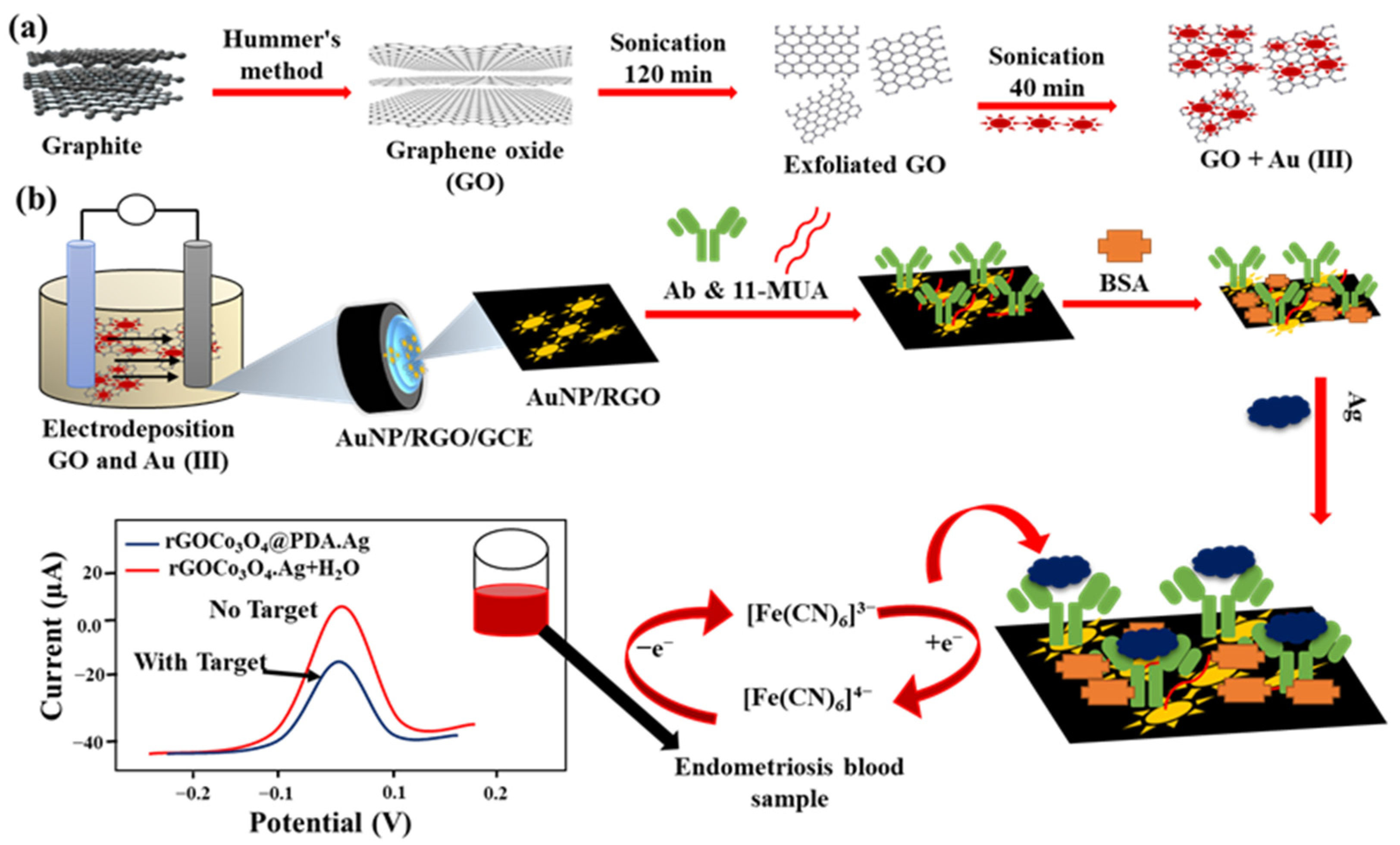
- Sangili, A.; Kalyani, T.; Chen, S.M.; Nanda, A.; Jana, S.K. Label-Free Electrochemical Immunosensor Based on One-Step Electrochemical Deposition of AuNP-RGO Nanocomposites for Detection of Endometriosis Marker CA 125. ACS Appl. Bio Mater. 2020, 3, 7620–7630. [Google Scholar] [CrossRef] [PubMed]
- Zanato, N.; Talamini, L.; Silva, T.R.; Vieira, I.C. Microcystin-LR Label-Free Immunosensor Based on Exfoliated Graphite Nanoplatelets and Silver Nanoparticles. Talanta 2017, 175, 38–45. [Google Scholar] [CrossRef]
- Nanda, S.; Gaur, A.; Kumar Duchaniya, R. Synthesis, Properties and Applications of Graphene Oxide: An Overview. World Sci. News 2020, 143, 17–27. [Google Scholar]
- Sánchez-Tirado, E.; González-Cortés, A.; Yáñez-Sedeño, P.; Pingarrón, J.M. Magnetic Multiwalled Carbon Nanotubes as Nanocarrier Tags for Sensitive Determination of Fetuin in Saliva. Biosens. Bioelectron. 2018, 113, 88–94. [Google Scholar] [CrossRef]
- Samadi Pakchin, P.; Ghanbari, H.; Saber, R.; Omidi, Y. Electrochemical Immunosensor Based on Chitosan-Gold Nanoparticle/Carbon Nanotube as a Platform and Lactate Oxidase as a Label for Detection of CA125 Oncomarker. Biosens. Bioelectron. 2018, 122, 68–74. [Google Scholar] [CrossRef]
- Ma, E.; Wang, P.; Yang, Q.; Yu, H.; Pei, F.; Li, Y.; Liu, Q.; Dong, Y. Electrochemical Immunosensor Based on MoS2 NFs/Au@AgPt YNCs as Signal Amplification Label for Sensitive Detection of CEA. Biosens. Bioelectron. 2019, 142, 111580. [Google Scholar] [CrossRef]
- Li, Y.; Zhang, Y.; Li, F.; Li, M.; Chen, L.; Dong, Y.; Wei, Q. Sandwich-Type Amperometric Immunosensor Using Functionalized Magnetic Graphene Loaded Gold and Silver Core-Shell Nanocomposites for the Detection of Carcinoembryonic Antigen. J. Electroanal. Chem. 2017, 795, 1–9. [Google Scholar] [CrossRef]
- Liao, X.; Wang, X.; Ma, C.; Zhang, L.; Zhao, C.; Chen, S.; Li, K.; Zhang, M.; Mei, L.; Qi, Y.; et al. Enzyme-Free Sandwich-Type Electrochemical Immunosensor for CEA Detection Based on the Cooperation of an Ag/g-C3N4-Modified Electrode and Au@SiO2/Cu2O with Core-Shell Structure. Bioelectrochemistry 2021, 142, 107931. [Google Scholar] [CrossRef]
- Yang, Y.; Yan, Q.; Liu, Q.; Li, Y.; Liu, H.; Wang, P.; Chen, L.; Zhang, D.; Li, Y.; Dong, Y. An Ultrasensitive Sandwich-Type Electrochemical Immunosensor Based on the Signal Amplification Strategy of Echinoidea-Shaped Au@Ag-Cu2O Nanoparticles for Prostate Specific Antigen Detection. Biosens. Bioelectron. 2018, 99, 450–457. [Google Scholar] [CrossRef]
- Lv, H.; Li, Y.; Zhang, X.; Li, X.; Xu, Z.; Chen, L.; Li, D.; Dong, Y. Thionin Functionalized Signal Amplification Label Derived Dual-Mode Electrochemical Immunoassay for Sensitive Detection of Cardiac Troponin I. Biosens. Bioelectron. 2019, 133, 72–78. [Google Scholar] [CrossRef]
- Samadi Pakchin, P.; Fathi, M.; Ghanbari, H.; Saber, R.; Omidi, Y. A Novel Electrochemical Immunosensor for Ultrasensitive Detection of CA125 in Ovarian Cancer. Biosens. Bioelectron. 2020, 153, 112029. [Google Scholar] [CrossRef]
- Dorozhko, E.V.; Gashevskay, A.S.; Korotkova, E.I.; Barek, J.; Vyskocil, V.; Eremin, S.A.; Galunin, E.V.; Saqib, M. A Copper Nanoparticle-Based Electrochemical Immunosensor for Carbaryl Detection. Talanta 2021, 228, 122174. [Google Scholar] [CrossRef]
- Valeria, G.; Lassabe, G.; Gonz, G.; Alicia, M. Development of an Electrochemical Immunosensor for the Determination of Molinate by Using Phages Labeled with CdS Nanocrystals as a Novel Strategy to Signal Amplification Evalo. Sens. Actuators B Chem. 2022, 367, 132126. [Google Scholar] [CrossRef]
- Livas, D.; Trachioti, M.; Banou, S.; Angelopoulou, M.; Kokkinos, C. 3D Printed Microcell Featuring a Disposable Nanocomposite Sb / Sn Immunosensor for Quantum Dot-Based Electrochemical Determination of Adulteration of Ewe / Goat ’ s Cheese with Cow ’ s Milk. Sens. Actuators B Chem. 2021, 334, 129614. [Google Scholar] [CrossRef]
- Cadkova, M.; Kovarova, A.; Dvorakova, V.; Metelka, R.; Bilkova, Z.; Korecka, L. Electrochemical Quantum Dots-Based Magneto-Immunoassay for Detection of HE4 Protein on Metal Fi Lm-Modi Fi Ed Screen-Printed Carbon Electrodes. Talanta 2018, 182, 111–115. [Google Scholar] [CrossRef]
- Qin, X.; Xu, A.; Liu, L.; Sui, Y.; Li, Y.; Tan, Y.; Chen, C.; Xie, Q. Selective Staining of CdS on ZnO Biolabel for Ultrasensitive Sandwich-Type Amperometric Immunoassay of Human Heart-Type Fatty-Acid-Binding Protein and Immunoglobulin G. Biosens. Bioelectron. 2017, 91, 321–327. [Google Scholar] [CrossRef]
- Krishnan, S.; He, X.; Zhao, F.; Zhang, Y.; Liu, S.; Xing, R. Dual Labeled Mesoporous Silica Nanospheres Based Electrochemical Immunosensor for Ultrasensitive Detection of Carcinoembryonic Antigen. Anal. Chim. Acta 2020, 1133, 119–127. [Google Scholar] [CrossRef] [PubMed]
- Wei, S.; Xiao, H.; Gu, M.; Chen, Z.; Cao, L. Ultrasensitive Label-Free Electrochemical Immunosensor Based on Core-Shell Au @ PtNPs Functionalized RGO-TEPA/PB Nanocomposite for HBsAg Detection. J. Electroanal. Chem. 2021, 890, 115216. [Google Scholar] [CrossRef]
- Malla, P.; Liao, H.; Liu, C.; Wu, W. Electrochemical Immunoassay for Serum Parathyroid Hormone Using Screen- Printed Carbon Electrode and Magnetic Beads. J. Electroanal. Chem. 2021, 895, 115463. [Google Scholar] [CrossRef]
- Wu, C.; Zhou, Y.; Wang, H.; Hu, J. P4VP Modified Zwitterionic Polymer for the Preparation of Antifouling Functionalized Surfaces. Nanomaterials 2019, 9, 706. [Google Scholar] [CrossRef] [Green Version]
- Wang, J.; Hui, N. Zwitterionic Poly(Carboxybetaine) Functionalized Conducting Polymer Polyaniline Nanowires for the Electrochemical Detection of Carcinoembryonic Antigen in Undiluted Blood Serum. Bioelectrochemistry 2019, 125, 90–96. [Google Scholar] [CrossRef] [PubMed]
- Serafín, V.; Razzino, C.A.; Gamella, M.; Pedrero, M.; Povedano, E.; Montero-Calle, A.; Barderas, R.; Calero, M.; Lobo, A.O.; Yáñez-Sedeño, P.; et al. Disposable immunoplatforms for the simultaneous determination of biomarkers for neurodegenerative disorders using poly(amidoamine) dendrimer/gold nanoparticle nanocomposite. Anal. Bioanal. Chem. 2021, 413, 799–811. [Google Scholar] [CrossRef] [PubMed]
- Salahandish, R.; Haghayegh, F.; Ayala-charca, G.; Eun, J.; Khalghollah, M.; Zare, A.; Far, B.; Berenger, B.M.; Dong, Y. An Electrochemical Dual-Immuno-Biosensor Accompanied by a Customized Bi-Potentiostat for Clinical Detection of SARS-CoV-2 Nucleocapsid Proteins. Biosens. Bioelectron. 2022, 203, 114018. [Google Scholar] [CrossRef]
- Shen, Z.; Ni, S.; Yang, W.; Sun, W.; Yang, G. Redox Probes Tagged Electrochemical Aptasensing Device for Simultaneous Detection of Multiple Cytokines in Real Time. Sens. Actuators B Chem. 2021, 336, 129747. [Google Scholar] [CrossRef]
- Shu, Y.; Su, T.; Lu, Q.; Shang, Z.; Feng, J.; Jin, D.; Zhu, A.; Xu, Q.; Hu, X. Paper-Based Electrochemical Immunosensor Device via Ni-Co MOF Nanosheet as a Peroxidase Mimic for the Label-Free Detection Of. Sens. Actuators B Chem. 2022, 373, 132736. [Google Scholar] [CrossRef]
- Boonkaew, S.; Jang, I.; Noviana, E.; Siangproh, W.; Chailapakul, O.; Henry, C.S. Electrochemical Paper-Based Analytical Device for Multiplexed, Point-of-Care Detection of Cardiovascular Disease Biomarkers. Sens. Actuators B Chem. 2021, 330, 129336. [Google Scholar] [CrossRef]


| CNT Hybrid Preparation Methods | Electrodes | Sensing Strategies | LOD | LR | Refs. |
|---|---|---|---|---|---|
| Chemical reduction of AuNPs on MWCNT | Anti-KBL/PA/AuNPs-PEI-MWCNTs/GCE | Label-free/DPV | 23 ng/mL | 0.05−100 μg/mL | [69] |
| Chemically synthesized COOH-MWCNTs/AuNPs | Anti-BPA aptamer/COOH-MWCNTs/AuNPs/AuE | Label-free SWV | 114 pg/mL | 22.8–2283 pg/mL | [70] |
| Ultrasonication | α-fetoprotein/N-GQD 1 @SWCNTs/Anti-AFP/BSA/GCE | Label-free/CV and EIS | 0.25 pg/mL | 0.001–200 ng/mL | [73] |
| CNTs/PEI/GE 2 via LBL fashion | CA19-9/PEI-CNTs/EDC-NHS | Label-free/EIS | 0.35 U/mL | 0.05–0.5 U/mL | [78] |
| RGO/CNF and RGO/CNT via sonication and hydrothermal reaction | RGO-CNT-Thi/anti-CA125/AuNPs/GCE | Sandwich reaction/DPV | 0.28 pg/mL | 1–3.2 ng/mL | [74] |
| Electropolymerization | Anti-calreticulin/SWCNTs-PPepx /ITO | Label-free/EIS | 4.6 fg/mL | 0.015–60 pg/mL | [75] |
| Electropolymerization | Anti-CysC/PPy-CNTs/ IDE 3 | Label-free/Capacitance | 28 ng/mL | 30–300 ng/mL | [76] |
Disclaimer/Publisher’s Note: The statements, opinions and data contained in all publications are solely those of the individual author(s) and contributor(s) and not of MDPI and/or the editor(s). MDPI and/or the editor(s) disclaim responsibility for any injury to people or property resulting from any ideas, methods, instructions or products referred to in the content. |
© 2023 by the authors. Licensee MDPI, Basel, Switzerland. This article is an open access article distributed under the terms and conditions of the Creative Commons Attribution (CC BY) license (https://creativecommons.org/licenses/by/4.0/).
Share and Cite
Police Patil, A.V.; Chuang, Y.-S.; Li, C.; Wu, C.-C. Recent Advances in Electrochemical Immunosensors with Nanomaterial Assistance for Signal Amplification. Biosensors 2023, 13, 125. https://doi.org/10.3390/bios13010125
Police Patil AV, Chuang Y-S, Li C, Wu C-C. Recent Advances in Electrochemical Immunosensors with Nanomaterial Assistance for Signal Amplification. Biosensors. 2023; 13(1):125. https://doi.org/10.3390/bios13010125
Chicago/Turabian StylePolice Patil, Avinash V., Yu-Sheng Chuang, Chenzhong Li, and Ching-Chou Wu. 2023. "Recent Advances in Electrochemical Immunosensors with Nanomaterial Assistance for Signal Amplification" Biosensors 13, no. 1: 125. https://doi.org/10.3390/bios13010125