Advances in Microfluidics for Single Red Blood Cell Analysis

 , , and
Abstract
:1. Introduction
2. Integrated Sensors for Microfluidic Platforms for Assessing Single Erythrocytes
2.1. Microscopic RBC Flow Analysis
2.2. Tomographic Analysis of Erythrocyte Flow
2.3. Smartphone-Based Analysis of Single RBCs
2.4. Spectroscopic Analysis of Single RBCs
3. Single RBCs in Microfluidics (State-of-the-Art in Industry and Academia)
3.1. RBC Dynamics of Fluid
3.2. RBC Agglutination/Aggregation in Microfluidic Environment

3.3. RBC Flow Analysis in Bifurcating Channels
3.4. RBC Dielectrophoretic Analysis
3.5. Deformation of Single Erythrocytes in Microchannels

3.6. Miscellaneous Observations
4. Clinical Implications of Microfluidic Based Single RBC Analysis
4.1. Analysis of RBCs Sedimentation Using Microfluidics
4.2. Analysis of Malaria using Erythrocyte Based Microfluidics
4.3. Analysis of Sickle Cells Disease Using RBC Microfluidics
4.4. Sepsis Diagnosis Using RBC Microfluidics
4.5. Cancer Diagnosis with RBC Microfluidic Systems
4.6. Prenatal RBC Diagnosis
4.7. Miscellaneous Areas for RBC Clinical Implications
5. Microfluidics Based Red Blood Cell Sorting
5.1. Cross Flow Filtration of Single RBCs in Microchannels
5.2. Blood Cell Counting and Sorting with Microfluidics
6. Microarrays and Single RBC Trapping Techniques
6.1. The Concept of Microarrays
6.2. State of the Art in single Erythrocyte Trapping Techniques
6.3. Single RBC Trapping Forces
6.3.1. Acoustic Trapping
6.3.2. Dielectrophoretic Trapping
6.3.3. Hydrodynamic Trapping
6.3.4. Magnetic Trapping
6.3.5. Optical Trapping
6.4. Limitations and Future Considerations for Single RBC Trapping Platforms
7. Organ-on-Chips, Multi-Organ Chips and Drug Discovery Involving Single RBC
7.1. Organ-on-Chips: State of the Art
7.2. Antibody Binding and Drug Discovery Applications
8. Future Considerations
Author Contributions
Funding
Institutional Review Board Statement
Informed Consent Statement
Data Availability Statement
Conflicts of Interest
References
- Du, E. Microfluidic approaches for biomechanics of red blood cells. Conf. Proc. Soc. Exp. Mech. Ser. 2017, 6, 89–93. [Google Scholar] [CrossRef]
- Kiran Raj, M.; Bhattacharya, S.; Dasgupta, S.; Chakraborty, S. Collective dynamics of red blood cells on an: In vitro microfluidic platform. Lab Chip 2018, 18, 3939–3948. [Google Scholar] [CrossRef]
- Hassan, U.; Reddy, B.; Damhorst, G.; Sonoiki, O.; Ghonge, T.; Yang, C.; Bashir, R. A microfluidic biochip for complete blood cell counts at the point-of-care. Technology 2015, 03, 201–213. [Google Scholar] [CrossRef] [PubMed] [Green Version]
- Manz, A.; Graber, N.; Widmer, H.M. Miniaturized total chemical analysis systems: A novel concept for chemical sensing. Sens. Actuators B Chem. 1990, 1, 244–248. [Google Scholar] [CrossRef]
- Duncombe, T.A.; Tentori, A.M.; Herr, A.E. Microfluidics: Reframing biological enquiry. Nat. Rev. Mol. Cell Biol. 2015, 16, 554–567. [Google Scholar] [CrossRef] [Green Version]
- Béné, M.C. Microfluidics in flow cytometry and related techniques. Int. J. Lab. Hematol. 2017, 39, 93–97. [Google Scholar] [CrossRef] [Green Version]
- Ito, H.; Kaneko, M. On-chip cell manipulation and applications to deformability measurements. ROBOMECH J. 2020, 7, 3. [Google Scholar] [CrossRef] [Green Version]
- Yaman, S.; Anil-Inevi, M.; Ozcivici, E.; Tekin, H.C. Magnetic force-based microfluidic techniques for cellular and tissue bioengineering. Front. Bioeng. Biotechnol. 2018, 6, 192. [Google Scholar] [CrossRef] [Green Version]
- Avsievich, T.; Zhu, R.; Popov, A.; Bykov, A.; Meglinski, I. The advancement of blood cell research by optical tweezers. Rev. Phys. 2020, 5, 100043. [Google Scholar] [CrossRef]
- Grünberger, A.; Wiechert, W.; Kohlheyer, D. Single-cell microfluidics: Opportunity for bioprocess development. Curr. Opin. Biotechnol. 2014, 29, 15–23. [Google Scholar] [CrossRef]
- Dusny, C.; Grünberger, A. Microfluidic single-cell analysis in biotechnology: From monitoring towards understanding. Curr. Opin. Biotechnol. 2020, 63, 26–33. [Google Scholar] [CrossRef] [PubMed]
- Shinde, P.; Mohan, L.; Kumar, A.; Dey, K.; Maddi, A.; Patananan, A.N.; Tseng, F.G.; Chang, H.Y.; Nagai, M.; Santra, T.S. Current trends of microfluidic single-cell technologies. Int. J. Mol. Sci. 2018, 19, 3143. [Google Scholar] [CrossRef] [PubMed] [Green Version]
- Jammes, F.C.; Maerkl, S.J. How single-cell immunology is benefiting from microfluidic technologies. Microsyst. Nanoeng. 2020, 6, 45. [Google Scholar] [CrossRef] [PubMed]
- Puttaswamy, S.V.; Fishlock, S.J.; Steele, D.; Shi, Q.; Lee, C.; McLaughlin, J. Versatile microfluidic platform embedded with sidewall three-dimensional electrodes for cell manipulation. Biomed. Phys. Eng. Express 2019, 5, 55003. [Google Scholar] [CrossRef]
- Zhong, X.; Mao, W.; Zhang, H. Fluid dynamic simulation of single cell sorting by fiber laser in microfluidics. IOP Conf. Ser. Mater. Sci. Eng. 2020, 892, 012075. [Google Scholar] [CrossRef]
- Zhai, J.; Li, H.; Wong, A.H.H.; Dong, C.; Yi, S.; Jia, Y.; Mak, P.I.; Deng, C.X.; Martins, R.P. A digital microfluidic system with 3D microstructures for single-cell culture. Microsyst. Nanoeng. 2020, 6, 6. [Google Scholar] [CrossRef] [Green Version]
- Nielsen, A.V.; Beauchamp, M.J.; Nordin, G.P.; Woolley, A.T. 3D Printed Microfluidics. Annu. Rev. Anal. Chem. 2020, 13, 45–65. [Google Scholar] [CrossRef]
- Rusling, J.F. Developing Microfluidic Sensing Devices Using 3D Printing. ACS Sens. 2018, 3, 522–526. [Google Scholar] [CrossRef] [Green Version]
- Sochol, R.D.; Sweet, E.; Glick, C.C.; Wu, S.Y.; Yang, C.; Restaino, M.; Lin, L. 3D printed microfluidics and microelectronics. Microelectron. Eng. 2018, 189, 52–68. [Google Scholar] [CrossRef]
- Yazdi, A.A.; Popma, A.; Wong, W.; Nguyen, T.; Pan, Y.; Xu, J. 3D printing: An emerging tool for novel microfluidics and lab-on-a-chip applications. Microfluid. Nanofluidics 2016, 20, 50. [Google Scholar] [CrossRef]
- Amin, R.; Knowlton, S.; Hart, A.; Yenilmez, B.; Ghaderinezhad, F.; Katebifar, S.; Messina, M.; Khademhosseini, A.; Tasoglu, S. 3D-printed microfluidic devices. Biofabrication 2016, 8, 1–16. [Google Scholar] [CrossRef] [PubMed] [Green Version]
- Weisgrab, G.; Ovsianikov, A.; Costa, P.F. Functional 3D Printing for Microfluidic Chips. Adv. Mater. Technol. 2019, 4, 275. [Google Scholar] [CrossRef] [Green Version]
- Nguyen, J.; Wei, Y.; Zheng, Y.; Wang, C.; Sun, Y. On-chip sample preparation for complete blood count from raw blood. Lab Chip 2015, 15, 1533–1544. [Google Scholar] [CrossRef] [PubMed]
- Kim, M.; Alapan, Y.; Adhikari, A.; Little, J.A.; Gurkan, U.A. Hypoxia-enhanced adhesion of red blood cells in microscale flow. Microcirculation 2017, 24, e12374. [Google Scholar] [CrossRef] [Green Version]
- Kaliviotis, E.; Sherwood, J.M.; Balabani, S. Local viscosity distribution in bifurcating microfluidic blood flows. Phys. Fluids 2018, 30. [Google Scholar] [CrossRef]
- Park, H.S.; Ceballos, S.; Eldridge, W.J.; Wax, A. Invited Article: Digital refocusing in quantitative phase imaging for flowing red blood cells. APL Photonics 2018, 3, 030706. [Google Scholar] [CrossRef]
- Tsvirkun, D.; Grichine, A.; Duperray, A.; Misbah, C.; Bureau, L. Microvasculature on a chip: Study of the Endothelial Surface Layer and the flow structure of Red Blood Cells. Sci. Rep. 2017, 7, 45036. [Google Scholar] [CrossRef] [Green Version]
- Park, H.S.; Eldridge, W.J.; Yang, W.H.; Crose, M.; Ceballos, S.; Roback, J.D.; Chi, J.T.A.; Wax, A. Quantitative phase imaging of erythrocytes under microfluidic constriction in a high refractive index medium reveals water content changes. Microsyst. Nanoeng. 2019, 5, 63. [Google Scholar] [CrossRef] [Green Version]
- Memmolo, P.; Miccio, L.; Merola, F.; Mugnano, M.; Ferraro, P. Hydrodynamic Red Blood Cells Deformation by Quantitative Phase Microscopy and Zernike Polynomials. Front. Phys. 2019, 7, 111. [Google Scholar] [CrossRef] [Green Version]
- Ahmad, A.; Dubey, V.; Singh, V.R.; Tinguely, J.C.; Øie, C.I.; Wolfson, D.L.; Mehta, D.S.; So, P.T.C.; Ahluwalia, B.S. Quantitative phase microscopy of red blood cells during planar trapping and propulsion. Lab Chip 2018, 18, 3025–3036. [Google Scholar] [CrossRef]
- Dannhauser, D.; Rossi, D.; Causa, F.; Memmolo, P.; Finizio, A.; Wriedt, T.; Hellmers, J.; Eremin, Y.; Ferraro, P.; Netti, P.A. Optical signature of erythrocytes by light scattering in microfluidic flows. Lab Chip 2015, 15, 3278–3285. [Google Scholar] [CrossRef] [PubMed] [Green Version]
- Ong, J.J.Y.; Oh, J.; Yong Ang, X.; Naidu, R.; Chu, T.T.T.; Hyoung Im, J.; Manzoor, U.; Kha Nguyen, T.; Na, S.-W.; Han, E.-T.; et al. Optical diffraction tomography and image reconstruction to measure host cell alterations caused by divergent Plasmodium species. Spectrochim. Acta Part A Mol. Biomol. Spectrosc. 2023, 286, 122026. [Google Scholar] [CrossRef] [PubMed]
- Ayoub, A.B.; Lim, J.; Saba, A.; Antoine, E.E.; Psaltis, D. Polarization-sensitive optical diffraction tomography. Optica 2021, 8, 402–408. [Google Scholar] [CrossRef]
- Steelman, Z.A.; Coker, Z.N.; Sedelnikova, A.; Keppler, M.A.; Kiester, A.S.; Troyanova-Wood, M.A.; Ibey, B.L.; Bixler, J.N. Comprehensive single-shot biophysical cytometry using simultaneous quantitative phase imaging and Brillouin spectroscopy. Sci. Rep. 2022, 12, 18285. [Google Scholar] [CrossRef] [PubMed]
- Nguyen, T.L.; Pradeep, S.; Judson-Torres, R.L.; Reed, J.; Teitell, M.A.; Zangle, T.A. Quantitative Phase Imaging: Recent Advances and Expanding Potential in Biomedicine. ACS Nano 2022, 16, 11516–11544. [Google Scholar] [CrossRef]
- Li, J.; Mengu, D.; Yardimci, N.T.; Luo, Y.; Li, X.; Veli, M.; Rivenson, Y.; Jarrahi, M.; Ozcan, A. Spectrally encoded single-pixel machine vision using diffractive networks. Sci. Adv. 2021, 7, 7690. [Google Scholar] [CrossRef]
- Balasubramani, V.; Montresor, S.; Tu, H.-Y.; Huang, C.-H.; Picart, P.; Cheng, C.-J. Influence of noise-reduction techniques in sparse-data sample rotation tomographic imaging. Appl. Opt. 2021, 60, B81. [Google Scholar] [CrossRef]
- Li, F.; Chen, X.; Ma, J.; Nie, S.; Ye, R.; Yuan, C.; Zuo, C.; Liu, F.; Yang, J.; Cao, L.; et al. ContransGAN: Convolutional Neural Network Coupling Global Swin-Transformer Network for High-Resolution Quantitative Phase Imaging with Unpaired Data. Cells 2022, 11, 2394. [Google Scholar] [CrossRef]
- Jiang, M.; Shao, M.; Yang, X.; He, L.; Peng, T.; Wang, T.; Ke, Z.; Wang, Z.; Fang, S.; Mao, Y.; et al. Automatic Classification of Red Blood Cell Morphology Based on Quantitative Phase Imaging. Int. J. Opt. 2022, 2022, 1240020. [Google Scholar] [CrossRef]
- Mengu, D.; Ozcan, A. All-Optical Phase Recovery: Diffractive Computing for Quantitative Phase Imaging. Adv. Opt. Mater. 2022, 10, 2200281. [Google Scholar] [CrossRef]
- Visitsattapongse, S.; Thadson, K.; Pechprasarn, S.; Thongpance, N. Analysis of Deep Learning-Based Phase Retrieval Algorithm Performance for Quantitative Phase Imaging Microscopy. Sensors 2022, 22, 3530. [Google Scholar] [CrossRef] [PubMed]
- Hu, C.; He, S.; Lee, Y.J.; He, Y.; Kong, E.M.; Li, H.; Anastasio, M.A.; Popescu, G. Live-dead assay on unlabeled cells using phase imaging with computational specificity. Nat. Commun. 2022, 13, 713. [Google Scholar] [CrossRef] [PubMed]
- Carden, M.A.; Fay, M.; Sakurai, Y.; McFarland, B.; Blanche, S.; DiPrete, C.; Joiner, C.H.; Sulchek, T.; Lam, W.A. Normal saline is associated with increased sickle red cell stiffness and prolonged transit times in a microfluidic model of the capillary system. Microcirculation 2017, 24, 12353. [Google Scholar] [CrossRef] [PubMed]
- Sergunova, V.; Leesment, S.; Kozlov, A.; Inozemtsev, V.; Platitsina, P.; Lyapunova, S.; Onufrievich, A.; Polyakov, V.; Sherstyukova, E. Investigation of Red Blood Cells by Atomic Force Microscopy. Sensors 2022, 22, 2055. [Google Scholar] [CrossRef] [PubMed]
- Besedina, N.A.; Skverchinskaya, E.A.; Shmakov, S.V.; Ivanov, A.S.; Mindukshev, I.V.; Bukatin, A.S. Persistent red blood cells retain their ability to move in microcapillaries under high levels of oxidative stress. Commun. Biol. 2022, 5, 1–9. [Google Scholar] [CrossRef] [PubMed]
- Quint, S.; Christ, A.F.; Guckenberger, A.; Himbert, S.; Kaestner, L.; Gekle, S.; Wagner, C. 3D tomography of cells in micro-channels. Appl. Phys. Lett. 2017, 111, 1–5. [Google Scholar] [CrossRef] [Green Version]
- Chen, C.-L.; Wang, R.K. Optical coherence tomography based angiography. Biomed. Opt. Express 2017, 8, 1056. [Google Scholar] [CrossRef] [Green Version]
- Gnyawali, V.; Strohm, E.M.; Wang, J.Z.; Tsai, S.S.H.; Kolios, M.C. Simultaneous acoustic and photoacoustic microfluidic flow cytometry for label-free analysis. Sci. Rep. 2019, 9, 1585. [Google Scholar] [CrossRef] [Green Version]
- Cacace, T.; Memmolo, P.; Villone, M.M.; De Corato, M.; Mugnano, M.; Paturzo, M.; Ferraro, P.; Maffettone, P.L. Assembling and rotating erythrocyte aggregates by acoustofluidic pressure enabling full phase-contrast tomography. Lab Chip 2019, 19, 3123–3132. [Google Scholar] [CrossRef]
- Merola, F.; Memmolo, P.; Miccio, L.; Savoia, R.; Mugnano, M.; Fontana, A.; D’Ippolito, G.; Sardo, A.; Iolascon, A.; Gambale, A.; et al. Tomographic flow cytometry by digital holography. Light Sci. Appl. 2017, 6, 1–7. [Google Scholar] [CrossRef]
- Jung, J.; Kim, K.; Yoon, J.; Park, Y. Hyperspectral optical diffraction tomography. Opt. Express 2016, 24, 2006. [Google Scholar] [CrossRef] [PubMed] [Green Version]
- Kim, S.C.; Jalal, U.M.; Im, S.B.; Ko, S.; Shim, J.S. A smartphone-based optical platform for colorimetric analysis of microfluidic device. Sens. Actuators B Chem. 2017, 239, 52–59. [Google Scholar] [CrossRef]
- Jalal Uddin, M.; Jin, G.J.; Shim, J.S. Paper-Plastic Hybrid Microfluidic Device for Smartphone-Based Colorimetric Analysis of Urine. Anal. Chem. 2017, 89, 13160–13166. [Google Scholar] [CrossRef]
- Yang, K.; Wu, J.; Peretz-Soroka, H.; Zhu, L.; Li, Z.; Sang, Y.; Hipolito, J.; Zhang, M.; Santos, S.; Hillier, C.; et al. Mkit: A cell migration assay based on microfluidic device and smartphone. Biosens. Bioelectron. 2018, 99, 259–267. [Google Scholar] [CrossRef] [PubMed] [Green Version]
- Zhang, W.; Li, Y.; Chen, B.; Zhang, Y.; Du, Z.; Xiang, F.; Hu, Y.; Meng, X.; Shang, C.; Liang, S.; et al. Fully Integrated Point-of-Care Complete Blood Count Using Multi-Frame Morphology Analysis. SSRN Electron. J. 2022. [Google Scholar] [CrossRef]
- Chattopadhyay, S.; Ram, R.; Sarkar, A.; Chakraborty, S. Smartphone-based automated estimation of plasma creatinine from finger-pricked blood on a paper strip via single-user step sample-to-result integration. Measurement 2022, 199, 111492. [Google Scholar] [CrossRef]
- Ding, S.; Duan, S.; Chen, Y.; Xie, J.; Tian, J.; Li, Y.; Wang, H. Centrifugal microfluidic platform with digital image analysis for parallel red cell antigen typing. Talanta 2023, 252, 123856. [Google Scholar] [CrossRef]
- Li, Q.; He, X.; Wang, Y.; Liu, H.; Xu, D.; Guo, F. Review of spectral imaging technology in biomedical engineering: Achievements and challenges. J. Biomed. Opt. 2013, 18, 100901. [Google Scholar] [CrossRef] [Green Version]
- Paul, R.; Zhou, Y.; Nikfar, M.; Razizadeh, M.; Liu, Y. Quantitative absorption imaging of red blood cells to determine physical and mechanical properties. RSC Adv. 2020, 10, 38923–38936. [Google Scholar] [CrossRef] [PubMed]
- Banoth, E.; Kasula, V.K.; Jagannadh, V.K.; Gorthi, S.S. Optofluidic single-cell absorption flow analyzer for point-of-care diagnosis of malaria. J. Biophotonics 2016, 9, 610–618. [Google Scholar] [CrossRef]
- Ito, H.; Tsai, C.H.D.; Kaneko, M. Integration of fluctuation spectroscopy into a microfluidic platform for novel cellular viscoelastic measurement. In Proceedings of the 2018 IEEE Micro Electro Mechanical Systems (MEMS), Belfast, UK, 21–25 January 2018; Volume 2018, pp. 137–140. [Google Scholar] [CrossRef]
- Pereira, F.M.; Bernacka-Wojcik, I.; Ribeiro, R.S.R.; Lobato, M.T.; Fortunato, E.; Martins, R.; Igreja, R.; Jorge, P.A.S.; Águas, H.; Oliva, A.M.G. Hybrid microfluidic platform for multifactorial analysis based on electrical impedance, refractometry, optical absorption and fluorescence. Micromachines 2016, 7, 181. [Google Scholar] [CrossRef] [PubMed] [Green Version]
- Maji, D.; Suster, M.A.; Mohseni, P. Monitoring Red Blood Cell Aggregation Dynamics in Stasis and under Flow Using a Microfluidic Dielectric Sensor. In Proceedings of the 2018 IEEE Biomedical Circuits and Systems Conference (BioCAS), Cleveland, OH, USA, 17–19 October 2018; pp. 1–4. [Google Scholar] [CrossRef]
- Atkins, C.G.; Buckley, K.; Blades, M.W.; Turner, R.F.B. Raman Spectroscopy of Blood and Blood Components. Appl. Spectrosc. 2017, 71, 767–793. [Google Scholar] [CrossRef] [PubMed] [Green Version]
- Cho, H.Y.; Hossain, M.K.; Lee, J.H.; Han, J.; Lee, H.J.; Kim, K.J.; Kim, J.H.; Lee, K.B.; Choi, J.W. Selective isolation and noninvasive analysis of circulating cancer stem cells through Raman imaging. Biosens. Bioelectron. 2018, 102, 372–382. [Google Scholar] [CrossRef]
- Jahn, I.J.; Radu, A.I.; Weber, K.; Cialla-May, D.; Popp, J. Surface enhanced raman spectroscopy for medical diagnostics. In Nanotechnology Characterization Tools for Biosensing and Medical Diagnosis; Springer: Berlin/Heidelberg, Germany, 2018; pp. 1–66. ISBN 9783662563335. [Google Scholar]
- Neugebauer, U.; Rösch, P.; Popp, J. Raman spectroscopy towards clinical application: Drug monitoring and pathogen identification. Int. J. Antimicrob. Agents 2015, 46, S35–S39. [Google Scholar] [CrossRef]
- Krafft, C.; Popp, J. Micro-Raman spectroscopy in medicine. Phys. Sci. Rev. 2019, 4, 1–15. [Google Scholar] [CrossRef]
- Redding, B.; Schwab, M.; Pan, Y. Le Raman spectroscopy of optically trapped single biological micro-particles. Sensors 2015, 15, 19021–19046. [Google Scholar] [CrossRef] [Green Version]
- Matthiae, M.; Zhu, X.; Marie, R.; Kristensen, A. In-line whole blood fractionation for Raman analysis of blood plasma. Analyst 2019, 144, 602–610. [Google Scholar] [CrossRef] [Green Version]
- Huang, J.-A.; Zhang, Y.-L.; Ding, H.; Sun, H.-B. SERS-Enabled Lab-on-a-Chip Systems. Adv. Opt. Mater. 2015, 3, 618–633. [Google Scholar] [CrossRef]
- Huser, T.; Chan, J. Raman spectroscopy for physiological investigations of tissues and cells. Adv. Drug Deliv. Rev. 2015, 89, 57–70. [Google Scholar] [CrossRef] [Green Version]
- Barkur, S.; Chidangil, S. Surface-enhanced Raman spectroscopy study of red blood cells and platelets. J. Biomol. Struct. Dyn. 2019, 37, 1090–1098. [Google Scholar] [CrossRef] [PubMed]
- Hakkel, O.; Rigo, I.; Veres, M.; Furjes, P. Microfluidically Integrated SERS Active Cell Trap Array for Sensitive Analysis of Red Blood Cells. In Proceedings of the 2019 20th International Conference on Solid-State Sensors, Actuators and Microsystems & Eurosensors XXXIII (Transducers & Eurosensors XXXIII), Berlin, Germany, 23–27 June 2019; pp. 964–967. [Google Scholar] [CrossRef]
- Verma, R.S.; Ahlawat, S.; Uppal, A. Optical guiding-based cell focusing for Raman flow cell cytometer. Analyst 2018, 143, 2648–2655. [Google Scholar] [CrossRef] [PubMed]
- Grigorev, G.V.; Lebedev, A.V.; Wang, X.; Qian, X.; Maksimov, G.V.; Parshina, E.U.; Lin, L. Hemoglobin conformation detection by Raman spectroscopy on single human red blood cells captured in a microfluidic chip. Mendeleev Commun. 2022, 32, 504–506. [Google Scholar] [CrossRef]
- Perez–Guaita, D.; de Veij, M.; Marzec, K.M.; Almohammedi, A.R.D.; McNaughton, D.; Hudson, A.J.; Wood, B.R. Resonance Raman and UV-Visible Microscopy Reveals that Conditioning Red Blood Cells with Repeated Doses of Sodium Dithionite Increases Haemoglobin Oxygen Uptake. ChemistrySelect 2017, 2, 3342–3346. [Google Scholar] [CrossRef] [Green Version]
- Chrimes, A.F.; Khoshmanesh, K.; Stoddart, P.R.; Mitchell, A.; Kalantar-Zadeh, K. Microfluidics and raman microscopy: Current applications and future challenges. Chem. Soc. Rev. 2013, 42, 5880–5906. [Google Scholar] [CrossRef] [PubMed]
- Ashiba, H.; Fujimaki, M.; Awazu, K.; Tanaka, T.; Makishima, M. Microfluidic chips for forward blood typing performed with a multichannel waveguide-mode sensor. Sens. Bio-Sens. Res. 2016, 7, 121–126. [Google Scholar] [CrossRef] [Green Version]
- Zhu, R.; Avsievich, T.; Popov, A.; Meglinski, I. Optical Tweezers in Studies of Red Blood Cells. Cells 2020, 9, 545. [Google Scholar] [CrossRef] [Green Version]
- Jeon, H.J.; Qureshi, M.M.; Lee, S.Y.; Chung, E. Optofluidic laser speckle image decorrelation analysis for the assessment of red blood cell storage. PLoS ONE 2019, 14, 224036. [Google Scholar] [CrossRef]
- Yeom, E.; Lee, S.J. Microfluidic-based speckle analysis for sensitive measurement of erythrocyte aggregation: A comparison of four methods for detection of elevated erythrocyte aggregation in diabetic rat blood. Biomicrofluidics 2015, 9, 1–16. [Google Scholar] [CrossRef] [Green Version]
- Esmaeel, A.M.; ElMelegy, T.T.H.; Abdelgawad, M. Multi-purpose machine vision platform for different microfluidics applications. Biomed. Microdevices 2019, 21, 1–13. [Google Scholar] [CrossRef]
- Yang, Y.; Lü, A.; Li, W.; Qian, Z. Microfluidic-based laser speckle contrast imaging of erythrocyte flow and magnetic nanoparticle retention in blood. AIP Adv. 2019, 9. [Google Scholar] [CrossRef] [Green Version]
- Grundmann, A.; Clavica, F.; Landolt, A.; Barrett, M.; Weber, B.; Obrist, D. Measurement of fluid pressure in microchannels. In Proceedings of the MicroTAS 2015 19th International Conference on Miniaturized Systems for Chemistry and Life Sciences, Gyeongju, Republic of Korea, 25–29 October 2015; pp. 2130–2131. [Google Scholar]
- Gusenbauer, M.; Mazza, G.; Brandl, M.; Schrefl, T.; Tóthová, R.; Jančigová, I.; Cimrák, I. Sensing Platform for Computational and Experimental Analysis of Blood Cell Mechanical Stress and Activation in Microfluidics. Procedia Eng. 2016, 168, 1390–1393. [Google Scholar] [CrossRef]
- Dadvand, A. Simulation of Flowing Red Blood Cells with and without Nanoparticle Dispersion Using Particle-based Numerical Methods. In Computational Approaches in Biomedical Nano-Engineering; John Wiley and Sons: Hoboken, NJ, USA, 2018; pp. 191–225. [Google Scholar] [CrossRef]
- Omori, T.; Imai, Y.; Kikuchi, K.; Ishikawa, T.; Yamaguchi, T. Hemodynamics in the Microcirculation and in Microfluidics. Ann. Biomed. Eng. 2015, 43, 238–257. [Google Scholar] [CrossRef] [PubMed]
- Faivre, M.; Renoux, C.; Bessaa, A.; Da Costa, L.; Joly, P.; Gauthier, A.; Connes, P. Mechanical Signature of Red Blood Cells Flowing Out of a Microfluidic Constriction Is Impacted by Membrane Elasticity, Cell Surface-to-Volume Ratio and Diseases. Front. Physiol. 2020, 11, 576. [Google Scholar] [CrossRef] [PubMed]
- Zhou, Q.; Fidalgo, J.; Calvi, L.; Bernabeu, M.O.; Hoskins, P.R.; Oliveira, M.S.N.; Krüger, T. Spatiotemporal Dynamics of Dilute Red Blood Cell Suspensions in Low-Inertia Microchannel Flow. Biophys. J. 2020, 118, 2561–2573. [Google Scholar] [CrossRef]
- Mauer, J.; Mendez, S.; Lanotte, L.; Nicoud, F.; Abkarian, M.; Gompper, G.; Fedosov, D.A. Flow-Induced Transitions of Red Blood Cell Shapes under Shear. Phys. Rev. Lett. 2018, 121, 118103. [Google Scholar] [CrossRef] [Green Version]
- Amirouche, A.; Esteves, J.; Lavoignat, A.; Picot, S.; Ferrigno, R.; Faivre, M. Dual shape recovery of red blood cells flowing out of a microfluidic constriction. Biomicrofluidics 2020, 14, 24116. [Google Scholar] [CrossRef]
- Lu, H.; Peng, Z. Boundary integral simulations of a red blood cell squeezing through a submicron slit under prescribed inlet and outlet pressures. Phys. Fluids 2019, 31, 5081057. [Google Scholar] [CrossRef]
- Puttaswamy, S.V.; Bhalla, N.; Kelsey, C.; Lubarsky, G.; Lee, C.; McLaughlin, J. Independent and grouped 3D cell rotation in a microfluidic device for bioimaging applications. Biosens. Bioelectron. 2020, 170, 112661. [Google Scholar] [CrossRef]
- Partola, K.R.; Andemariam, B.; Lykotrafitis, G. Microfluidic experimental setup for adhesion and recovery measurements of red blood cells in sickle cell disease. J. Mech. Behav. Biomed. Mater. 2017, 71, 80–84. [Google Scholar] [CrossRef] [PubMed] [Green Version]
- White, J.; Lancelot, M.; Sarnaik, S.; Hines, P. Increased erythrocyte adhesion to VCAM-1 during pulsatile flow: Application of a microfluidic flow adhesion bioassay. Clin. Hemorheol. Microcirc. 2015, 60, 201–213. [Google Scholar] [CrossRef] [PubMed] [Green Version]
- Guruprasad, P.; Mannino, R.G.; Caruso, C.; Zhang, H.; Josephson, C.D.; Roback, J.D.; Lam, W.A. Integrated automated particle tracking microfluidic enables high-throughput cell deformability cytometry for red cell disorders. Am. J. Hematol. 2019, 94, 189–199. [Google Scholar] [CrossRef] [PubMed] [Green Version]
- Zhou, S.; Giannetto, M.; DeCourcey, J.; Kang, H.; Kang, N.; Li, Y.; Zheng, S.; Zhao, H.; Simmons, W.R.; Wei, H.S.; et al. Oxygen tension-mediated erythrocyte membrane interactions regulate cerebral capillary hyperemia. Sci. Adv. 2019, 5, aaw4466. [Google Scholar] [CrossRef] [PubMed] [Green Version]
- Kang, Y.J. Microfluidic-based technique for measuring rbc aggregation and blood viscosity in a continuous and simultaneous fashion. Micromachines 2018, 9, 467. [Google Scholar] [CrossRef] [Green Version]
- Stroobach, M.; Haya, L.; Fenech, M. Effects of red blood cell aggregation on microparticle wall adhesion in circular microchannels. Med. Eng. Phys. 2019, 69, 100–108. [Google Scholar] [CrossRef]
- Lee, K.; Shirshin, E.; Rovnyagina, N.; Yaya, F.; Boujja, Z.; Priezzhev, A.; Wagner, C. Dextran adsorption onto red blood cells revisited: Single cell quantification by laser tweezers combined with microfluidics. Biomed. Opt. Express 2018, 9, 2755. [Google Scholar] [CrossRef]
- Kang, Y.J. Microfluidic-based effective monitoring of bloods by measuring RBC aggregation and blood viscosity under stepwise varying shear rates. Korea Aust. Rheol. J. 2020, 32, 15–27. [Google Scholar] [CrossRef]
- Kang, Y.J. Microfluidic-based measurement of RBC aggregation and the ESR using a driving syringe system. Anal. Methods 2018, 10, 1805–1816. [Google Scholar] [CrossRef]
- Mehri, R.; Mavriplis, C.; Fenech, M. Controlled microfluidic environment for dynamic investigation of red blood cell aggregation. J. Vis. Exp. 2015, 2015, 1–12. [Google Scholar] [CrossRef] [Green Version]
- Mehri, R.; Mavriplis, C.; Fenech, M. Red blood cell aggregates and their effect on non-Newtonian blood viscosity at low hematocrit in a two-fluid low shear rate microfluidic system. PLoS ONE 2018, 13, 199911. [Google Scholar] [CrossRef]
- Windberger, U.; Pöschl, C.; Peters, S.; Huber, J.; van den Hoven, R. Measurement of whole blood of different mammalian species in the oscillating shear field: Influence of erythrocyte aggregation. J. Phys. Conf. Ser. 2017, 790, 012035. [Google Scholar] [CrossRef] [Green Version]
- Pasias, D.; Passos, A.; Constantinides, G.; Balabani, S.; Kaliviotis, E. Surface tension driven flow of blood in a rectangular microfluidic channel: Effect of erythrocyte aggregation. Phys. Fluids 2020, 32, 8939. [Google Scholar] [CrossRef]
- Kang, Y.J.; Kim, B.J. Multiple and periodic measurement of RBC aggregation and ESR in parallel microfluidic channels under on-offblood flow control. Micromachines 2018, 9, 318. [Google Scholar] [CrossRef] [PubMed] [Green Version]
- Kang, Y.J. Periodic and simultaneous quantification of blood viscosity and red blood cell aggregation using a microfluidic platform under in-vitro closed-loop circulation. Biomicrofluidics 2018, 12, 024116. [Google Scholar] [CrossRef] [PubMed]
- Kang, Y.J. Microfluidic-based measurement method of red blood cell aggregation under hematocrit variations. Sensors 2017, 17, 2037. [Google Scholar] [CrossRef] [PubMed] [Green Version]
- Mehri, R.; Niazi, E.; Mavriplis, C.; Fenech, M. An automated method for dynamic red blood cell aggregate detection in microfluidic flow. Physiol. Meas. 2018, 39, 01NT02. [Google Scholar] [CrossRef]
- Kang, Y.J. Microfluidic-based biosensor for sequential measurement of blood pressure and RBC aggregation over continuously varying blood flows. Micromachines 2019, 10, 577. [Google Scholar] [CrossRef]
- Qiu, X.; Huang, J.H.; Westerhof, T.M.; Lombardo, J.A.; Henrikson, K.M.; Pennell, M.; Pourfard, P.P.; Nelson, E.L.; Nath, P.; Haun, J.B. Microfluidic channel optimization to improve hydrodynamic dissociation of cell aggregates and tissue. Sci. Rep. 2018, 8, 2774. [Google Scholar] [CrossRef] [Green Version]
- Chang, Y.J.; Ho, C.Y.; Zhou, X.M.; Yen, H.R. Determination of degree of RBC agglutination for blood typing using a small quantity of blood sample in a microfluidic system. Biosens. Bioelectron. 2018, 102, 234–241. [Google Scholar] [CrossRef]
- Huet, M.; Cubizolles, M.; Buhot, A. Red blood cell agglutination for blood typing within passive microfluidic biochips. High-Throughput 2018, 7, 10. [Google Scholar] [CrossRef] [Green Version]
- Aristov, A.A.; Rozenbaum, Y.A.; Evtushenko, G.S. An Automated Method for Blood Type Determination by Red Blood Cell Agglutination Assay. Biomed. Eng. 2022, 55, 328–332. [Google Scholar] [CrossRef]
- Semenov, A.; Lugovtsov, A.; Ermolinskiy, P.; Lee, K.; Priezzhev, A. Problems of Red Blood Cell Aggregation and Deformation Assessed by Laser Tweezers, Diffuse Light Scattering and Laser Diffractometry. Photonics 2022, 9, 238. [Google Scholar] [CrossRef]
- Trejo-Soto, C.; Lázaro, G.R.; Pagonabarraga, I.; Hernández-Machado, A. Microfluidics Approach to the Mechanical Properties of Red Blood Cell Membrane and Their Effect on Blood Rheology. Membranes 2022, 12, 217. [Google Scholar] [CrossRef] [PubMed]
- Zhu, R.; Avsievich, T.; Su, X.; Bykov, A.; Popov, A.; Meglinski, I. Hemorheological alterations of red blood cells induced by 450-nm and 520-nm laser radiation. J. Photochem. Photobiol. B Biol. 2022, 230, 112438. [Google Scholar] [CrossRef] [PubMed]
- Maurer, F.; John, T.; Makhro, A.; Bogdanova, A.; Minetti, G.; Wagner, C.; Kaestner, L. Continuous Percoll Gradient Centrifugation of Erythrocytes—Explanation of Cellular Bands and Compromised Age Separation. Cells 2022, 11, 1296. [Google Scholar] [CrossRef]
- Vijayaraghavan, M.; Chatterjee, S.; Sumantran, V.N.; Jayavelu, T. Revisiting dextran effect on red blood cell to understand the importance of rouleaux distribution and red blood cell-endothelial cell adhesion. Biomass Convers. Biorefinery 2022, 1–11. [Google Scholar] [CrossRef]
- Wu, K.; Lang, X.; Zhang, Y.; Li, Z.; He, B.; Gao, L.; Chen, J. Ultrasound simulation of blood with different red blood cell aggregations and concentrations. Biomed. Mater. Eng. 2022, 33, 235–257. [Google Scholar] [CrossRef]
- Kuznetsova, P.I.; Raskurazhev, A.A.; Shabalina, A.A.; Melikhyan, A.L.; Subortseva, I.N.; Tanashyan, M.M. Red Blood Cell Morphodynamics in Patients with Polycythemia Vera and Stroke. Int. J. Mol. Sci. 2022, 23, 2247. [Google Scholar] [CrossRef]
- Nader, E.; Nougier, C.; Boisson, C.; Poutrel, S.; Catella, J.; Martin, F.; Charvet, J.; Girard, S.; Havard-Guibert, S.; Martin, M.; et al. Increased blood viscosity and red blood cell aggregation in patients with COVID-19. Am. J. Hematol. 2022, 97, 283–292. [Google Scholar] [CrossRef]
- Bento, D.; Pereira, A.I.; Lima, J.; Miranda, J.M.; Lima, R. Cell-free layer measurements of in vitro blood flow in a microfluidic network: An automatic and manual approach. Comput. Methods Biomech. Biomed. Eng. Imaging Vis. 2018, 6, 629–637. [Google Scholar] [CrossRef] [Green Version]
- Kang, Y.J. Microfluidic-based biosensor for blood viscosity and erythrocyte sedimentation rate using disposable fluid delivery system. Micromachines 2020, 11, 215. [Google Scholar] [CrossRef] [PubMed] [Green Version]
- Hu, R.; Li, F.; Lv, J.; He, Y.; Lu, D.; Yamada, T.; Ono, N. Microfluidic analysis of pressure drop and flow behavior in hypertensive micro vessels. Biomed. Microdevices 2015, 17, 1–9. [Google Scholar] [CrossRef] [PubMed]
- Li, G.; Ye, T.; Li, X. Parallel modeling of cell suspension flow in complex micro-networks with inflow/outflow boundary conditions. J. Comput. Phys. 2020, 401, 109031. [Google Scholar] [CrossRef]
- Zilberman-Rudenko, J.; Sylman, J.L.; Lakshmanan, H.H.S.; McCarty, O.J.T.; Maddala, J. Dynamics of Blood Flow and Thrombus Formation in a Multi-Bypass Microfluidic Ladder Network. Cell. Mol. Bioeng. 2017, 10, 16–29. [Google Scholar] [CrossRef] [PubMed]
- Hymel, S.J.; Lan, H.; Fujioka, H.; Khismatullin, D.B. Cell trapping in Y-junction microchannels: A numerical study of the bifurcation angle effect in inertial microfluidics. Phys. Fluids 2019, 31, 082003. [Google Scholar] [CrossRef]
- Chen, Y.; Li, Y.; Li, D.; Li, J.; Chen, H. Margination mechanism of stiffened red blood cell in microchannel with different cross-section shapes. Microfluid. Nanofluidics 2019, 23, 25. [Google Scholar] [CrossRef]
- Clavica, F.; Homsy, A.; Jeandupeux, L.; Obrist, D. Red blood cell phase separation in symmetric and asymmetric microchannel networks: Effect of capillary dilation and inflow velocity. Sci. Rep. 2016, 6, 36763. [Google Scholar] [CrossRef]
- Waheed, W.; Alazzam, A.; Al-Khateeb, A.N.; Abu-Nada, E. Dissipative particle dynamics for modeling micro-objects in microfluidics: Application to dielectrophoresis. Biomech. Model. Mechanobiol. 2020, 19, 389–400. [Google Scholar] [CrossRef]
- Qiang, Y.; Liu, J.; Mian, M.; Du, E. Experimental Electromechanics of Red Blood Cells Using Dielectrophoresis-Based Microfluidics. In Mechanics of Biological Systems and Materials; Springer: Cham, Switzerland, 2017; Volume 7, pp. 129–134. [Google Scholar]
- Zhu, B.; Cai, Y.; Huang, H.; Liang, X.; Li, X.; Yang, H. Red blood cell stretching manipulation using the microfluidics chip integrated with liquid metal electrode. In Proceedings of the 2019 IEEE/ASME International Conference on Advanced Intelligent Mechatronics (AIM), Hong Kong, China, 8–12 July 2019; pp. 1652–1657. [Google Scholar] [CrossRef]
- Pakhira, W.; Kumar, R.; Ibrahimi, K.M.; Bhattacharjee, R. Design and analysis of a microfluidic lab-on-chip utilizing dielectrophoresis mechanism for medical diagnosis and liquid biopsy. J. Brazilian Soc. Mech. Sci. Eng. 2022, 44, 10. [Google Scholar] [CrossRef]
- Lavi, E.D.; Crivellari, F.; Gagnon, Z. Dielectrophoretic detection of electrical property changes of stored human red blood cells. Electrophoresis 2022, 43, 1297–1308. [Google Scholar] [CrossRef]
- Hu, Q.; Wang, Z.; Shen, L.; Zhao, G. Label-Free and Noninvasive Single-Cell Characterization for the Viscoelastic Properties of Cryopreserved Human Red Blood Cells Using a Dielectrophoresis-On-a-Chip Approach. Anal. Chem. 2022, 94, 10245–10255. [Google Scholar] [CrossRef] [PubMed]
- Salahi, A.; Honrado, C.; Rane, A.; Caselli, F.; Swami, N.S. Modified Red Blood Cells as Multimodal Standards for Benchmarking Single-Cell Cytometry and Separation Based on Electrical Physiology. Anal. Chem. 2022, 94, 2865–2872. [Google Scholar] [CrossRef] [PubMed]
- Jeon, H.J.; Lee, H.; Yoon, D.S.; Kim, B.M. Dielectrophoretic force measurement of red blood cells exposed to oxidative stress using optical tweezers and a microfluidic chip. Biomed. Eng. Lett. 2017, 7, 317–323. [Google Scholar] [CrossRef] [PubMed]
- Qiang, Y.; Liu, J.; Du, E. Dynamic fatigue measurement of human erythrocytes using dielectrophoresis. Acta Biomater. 2017, 57, 352–362. [Google Scholar] [CrossRef]
- Beale, A.D.; Labeed, F.H.; Kitcatt, S.J.; O’Neill, J.S. Detecting Circadian Rhythms in Human Red Blood Cells by Dielectrophoresis. Methods Mol. Biol. 2022, 2482, 255–264. [Google Scholar] [CrossRef]
- Qiang, Y.; Liu, J.; Du, E. Dielectrophoresis testing of nonlinear viscoelastic behaviors of human red blood cells. Micromachines 2018, 9, 21. [Google Scholar] [CrossRef]
- Du, E.; Qiang, Y.; Liu, J. Erythrocyte membrane failure by electromechanical stress. Appl. Sci. 2018, 8, 174. [Google Scholar] [CrossRef] [Green Version]
- Zhu, B.; Cai, Y.; Wu, Z.; Niu, F.; Yang, H. Dielectrophoretic Microfluidic Chip Integrated with Liquid Metal Electrode for Red Blood Cell Stretching Manipulation. IEEE Access 2019, 7, 152224–152232. [Google Scholar] [CrossRef]
- Sang, S.; Feng, Q.; Jian, A.; Li, H.; Ji, J.; Duan, Q.; Zhang, W.; Wang, T. Portable microsystem integrates multifunctional dielectrophoresis manipulations and a surface stress biosensor to detect red blood cells for hemolytic anemia. Sci. Rep. 2016, 6, 33626. [Google Scholar] [CrossRef] [Green Version]
- Huisjes, R.; Bogdanova, A.; van Solinge, W.W.; Schiffelers, R.M.; Kaestner, L.; van Wijk, R. Squeezing for life—Properties of red blood cell deformability. Front. Physiol. 2018, 9, 656. [Google Scholar] [CrossRef] [Green Version]
- Tomaiuolo, G.; Lanotte, L.; D’Apolito, R.; Cassinese, A.; Guido, S. Microconfined flow behavior of red blood cells. Med. Eng. Phys. 2016, 38, 11–16. [Google Scholar] [CrossRef] [PubMed]
- Mehendale, N.; Dhrubaditya, M.; Paul, D. A fast microfluidic device to measure the deformability of red blood cells. bioRxiv 2019. [Google Scholar] [CrossRef]
- Xu, Z.; Pu, H.; Xie, S.; Wang, C.; Sun, Y. Microfluidic measurement of RBC bending stiffness changes in blood storage. In Proceedings of the 2017 19th International Conference on Solid-State Sensors, Actuators and Microsystems (TRANSDUCERS), Kaohsiung, Taiwan, 18–22 June 2017; pp. 734–737. [Google Scholar] [CrossRef]
- Kim, J.; Lee, H.; Shin, S. Advances in the measurement of red blood cell deformability: A brief review. J. Cell. Biotechnol. 2015, 1, 63–79. [Google Scholar] [CrossRef] [Green Version]
- Ye, T.; Phan-Thien, N.; Lim, C.T.; Li, Y. Red blood cell motion and deformation in a curved microvessel. J. Biomech. 2017, 65, 12–22. [Google Scholar] [CrossRef] [PubMed]
- Faustino, V.; Rodrigues, R.O.; Pinho, D.; Costa, E.; Santos-Silva, A.; Miranda, V.; Amaral, J.S.; Lima, R. A Microfluidic Deformability Assessment of Pathological Red Blood Cells Flowing in a Hyperbolic Converging Microchannel. Micromachines 2019, 10, 645. [Google Scholar] [CrossRef]
- Kang, Y.J. A disposable blood-on-a-chip for simultaneous measurement of multiple biophysical properties. Micromachines 2018, 9, 475. [Google Scholar] [CrossRef] [Green Version]
- Matthews, K.; Myrand-Lapierre, M.E.; Ang, R.R.; Duffy, S.P.; Scott, M.D.; Ma, H. Microfluidic deformability analysis of the red cell storage lesion. J. Biomech. 2015, 48, 4065–4072. [Google Scholar] [CrossRef]
- Lim, H.; Back, S.M.; Nam, J.; Choi, H. Determination of red blood cell deformability using centrifugal force in a three-dimensional-printed mini-disk (3D-PMD). PLoS ONE 2018, 13, 0197619. [Google Scholar] [CrossRef] [PubMed]
- Passos, A.; Sherwood, J.M.; Kaliviotis, E.; Agrawal, R.; Pavesio, C.; Balabani, S. The effect of deformability on the microscale flow behavior of red blood cell suspensions. Phys. Fluids 2019, 31, 091903. [Google Scholar] [CrossRef]
- Huang, S.; Hou, H.W.; Kanias, T.; Sertorio, J.T.; Chen, H.; Sinchar, D.; Gladwin, M.T.; Han, J. Towards microfluidic-based depletion of stiff and fragile human red cells that accumulate during blood storage. Lab Chip 2015, 15, 448–458. [Google Scholar] [CrossRef]
- Li, W.; Zhu, B.; Cai, Y.; Wu, Z.; Sun, L.; Yang, H. Analysis of red blood cell deformability using parallel ladder electrodes in a microfluidic manipulation system. Int. J. Adv. Manuf. Technol. 2019, 105, 4919–4928. [Google Scholar] [CrossRef]
- Wu, T.; Guo, Q.; Ma, H.; Feng, J.J. The critical pressure for driving a red blood cell through a contracting microfluidic channel. Theor. Appl. Mech. Lett. 2015, 5, 227–230. [Google Scholar] [CrossRef] [Green Version]
- Reichel, F.; Mauer, J.; Nawaz, A.A.; Gompper, G.; Guck, J.; Fedosov, D.A. High-Throughput Microfluidic Characterization of Erythrocyte Shapes and Mechanical Variability. Biophys. J. 2019, 117, 14–24. [Google Scholar] [CrossRef] [PubMed]
- Bento, D.; Rodrigues, R.O.; Faustino, V.; Pinho, D.; Fernandes, C.S.; Pereira, A.I.; Garcia, V.; Miranda, J.M.; Lima, R. Deformation of red blood cells, air bubbles, and droplets in microfluidic devices: Flow visualizations and measurements. Micromachines 2018, 9, 151. [Google Scholar] [CrossRef] [PubMed] [Green Version]
- Ito, H.; Takeishi, N.; Kirimoto, A.; Chimura, M.; Ohtani, T.; Sakata, Y.; Horade, M.; Takayama, T.; Wada, S.; Kaneko, M. How to Measure Cellular Shear Modulus Inside a Chip: Detailed Correspondence to the Fluid-Structure Coupling Analysis. In Proceedings of the 2019 IEEE 32nd International Conference on Micro Electro Mechanical Systems (MEMS), Seoul, Republic of Korea, 27–31 January 2019; pp. 433–436. [Google Scholar] [CrossRef]
- Kirimoto, A.; Ito, H.; Tsai, C.H.; Kaneko, M. Measurement of both viscous and elastic constants of a red blood cell in a microchannel. In Proceedings of the 2018 IEEE Micro Electro Mechanical Systems (MEMS), Belfast, UK, 21–25 January 2018; pp. 388–391. [Google Scholar] [CrossRef]
- Saadat, A.; Huyke, D.A.; Oyarzun, D.I.; Escobar, P.V.; Øvreeide, I.H.; Shaqfeh, E.S.G.; Santiago, J.G. A system for the high-throughput measurement of the shear modulus distribution of human red blood cells. Lab Chip 2020, 20, 2927–2936. [Google Scholar] [CrossRef]
- Robidoux, J.; Laforce-Lavoie, A.; Charette, S.J.; Shevkoplyas, S.S.; Yoshida, T.; Lewin, A.; Brouard, D. Development of a flow standard to enable highly reproducible measurements of deformability of stored red blood cells in a microfluidic device. Transfusion 2020, 60, 1032–1041. [Google Scholar] [CrossRef]
- Kang, Y.J. RBC deformability measurement based on variations of pressure in multiple micropillar channels during blood delivery using a disposable air-compressed pump. Anal. Methods 2018, 10, 4549–4561. [Google Scholar] [CrossRef]
- Chien, W.; Zhang, Z.; Gompper, G.; Fedosov, D.A. Deformation and dynamics of erythrocytes govern their traversal through microfluidic devices with a deterministic lateral displacement architecture. Biomicrofluidics 2019, 13, 044106. [Google Scholar] [CrossRef] [Green Version]
- Alapan, Y.; Matsuyama, Y.; Little, J.A.; Gurkan, U.A. Dynamic deformability of sickle red blood cells in microphysiological flow. Technology 2016, 4, 71–79. [Google Scholar] [CrossRef] [Green Version]
- Myrand-Lapierre, M.E.; Deng, X.; Ang, R.R.; Matthews, K.; Santoso, A.T.; Ma, H. Multiplexed fluidic plunger mechanism for the measurement of red blood cell deformability. Lab Chip 2015, 15, 159–167. [Google Scholar] [CrossRef] [Green Version]
- Kang, Y.J.; Ha, Y.R.; Lee, S.J. Deformability measurement of red blood cells using a microfluidic channel array and an air cavity in a driving syringe with high throughput and precise detection of subpopulations. Analyst 2016, 141, 319–330. [Google Scholar] [CrossRef] [PubMed]
- Balachandran Nair, A.N.; Pirker, S.; Umundum, T.; Saeedipour, M. A reduced-order model for deformable particles with application in bio-microfluidics. Comput. Part. Mech. 2020, 7, 593–601. [Google Scholar] [CrossRef]
- Moon, J.Y.; Tanner, R.I.; Lee, J.S. A numerical study on the elastic modulus of volume and area dilation for a deformable cell in a microchannel. Biomicrofluidics 2016, 10, 044110. [Google Scholar] [CrossRef] [PubMed]
- Kucukal, E.; Little, J.A.; Gurkan, U.A. Shear dependent red blood cell adhesion in microscale flow. Integr. Biol. 2018, 10, 194–206. [Google Scholar] [CrossRef] [PubMed] [Green Version]
- Xu, Z.; Zheng, Y.; Wang, X.; Shehata, N.; Wang, C.; Sun, Y. Stiffness increase of red blood cells during storage. Microsyst. Nanoeng. 2018, 4, 1–6. [Google Scholar] [CrossRef] [Green Version]
- Wang, Y.; You, G.; Chen, P.; Li, J.; Chen, G.; Wang, B.; Li, P.; Han, D.; Zhou, H.; Zhao, L. The mechanical properties of stored red blood cells measured by a convenient microfluidic approach combining with mathematic model. Biomicrofluidics 2016, 10, 024104. [Google Scholar] [CrossRef] [Green Version]
- Islamzada, E.; Matthews, K.; Guo, Q.; Santoso, A.T.; Duffy, S.P.; Scott, M.D.; Ma, H. Deformability based sorting of stored red blood cells reveals donor-dependent aging curves. bioRxiv 2019. [Google Scholar] [CrossRef]
- Amirouche, A.; Ferrigno, R.; Faivre, M. Impact of Channel Geometry on the Discrimination of Mechanically Impaired Red Blood Cells in Passive Microfluidics. Proceedings 2017, 1, 512. [Google Scholar] [CrossRef] [Green Version]
- Liu, L.; Huang, S.; Xu, X.; Han, J. Study of individual erythrocyte deformability susceptibility to INFeD and ethanol using a microfluidic chip. Sci. Rep. 2016, 6, 22929. [Google Scholar] [CrossRef] [Green Version]
- Gómez Bardón, R.; Passos, A.; Piergiovanni, M.; Balabani, S.; Pennati, G.; Dubini, G. Haematocrit heterogeneity in blood flows past microfluidic models of oxygenating fibre bundles. Med. Eng. Phys. 2019, 73, 30–38. [Google Scholar] [CrossRef] [PubMed]
- Shen, Z.; Coupier, G.; Kaoui, B.; Polack, B.; Harting, J.; Misbah, C.; Podgorski, T. Inversion of hematocrit partition at microfluidic bifurcations. Microvasc. Res. 2016, 105, 40–46. [Google Scholar] [CrossRef] [PubMed] [Green Version]
- Berry, S.B.; Fernandes, S.C.; Rajaratnam, A.; Dechiara, N.S.; Mace, C.R. Measurement of the hematocrit using paper-based microfluidic devices. Lab Chip 2016, 16, 3689–3694. [Google Scholar] [CrossRef] [PubMed]
- Li, P.; Zheng, L.; Zhang, D.; Xie, Y.; Feng, Y.; Xie, G. Investigation of High-Speed Erythrocyte Flow and Erythrocyte-Wall Impact in a Lab-on-a-Chip. Artif. Organs 2016, 40, E203–E218. [Google Scholar] [CrossRef]
- Isiksacan, Z.; Serhatlioglu, M.; Elbuken, C. In vitro analysis of multiple blood flow determinants using red blood cell dynamics under oscillatory flow. Analyst 2020, 145, 5996–6005. [Google Scholar] [CrossRef] [PubMed]
- Xing, F.; Hu, F.; Yang, J.; Pan, L.; Xu, J. Structural and functional studies of erythrocyte membrane-skeleton by single-cell and single-molecule techniques. J. Innov. Opt. Health Sci. 2019, 12, 1–13. [Google Scholar] [CrossRef] [Green Version]
- Huang, L.; Shibajyotl, G.D.; Cansu Agca, Y.A.; Benson, J.D.; Almasri, M. Lab on Chip Microfluidic Sensor for Individual Red Blood Cell Water Permeability Measurement. In Proceedings of the 2018 IEEE 13th Annual International Conference on Nano/Micro Engineered and Molecular Systems (NEMS), Singapore, 22–26 April 2018; pp. 44–47. [Google Scholar] [CrossRef]
- Horade, M.; Tsai, C.H.D.; Ito, H.; Kaneko, M. Red blood cell responses during a long-standing load in a microfluidic constriction. Micromachines 2017, 8, 100. [Google Scholar] [CrossRef] [Green Version]
- Cluitmans, J.C.A.; Gevi, F.; Siciliano, A.; Matte, A.; Leal, J.K.F.; De Franceschi, L.; Zolla, L.; Brock, R.; Adjobo-Hermans, M.J.W.; Bosman, G.J.G.C.M. Red blood cell homeostasis: Pharmacological interventions to explore biochemical, morphological and mechanical properties. Front. Mol. Biosci. 2016, 3, 10. [Google Scholar] [CrossRef] [PubMed] [Green Version]
- Hesh, C.A.; Qiu, Y.; Lam, W.A. Vascularized microfluidics and the blood-endothelium interface. Micromachines 2020, 11, 18. [Google Scholar] [CrossRef] [Green Version]
- Hin, S.; Loskyll, M.; Klein, V.; Keller, M.; Strohmeier, O.; von Stetten, F.; Zengerle, R.; Mitsakakis, K. Membrane-based sample inlet for centrifugal microfluidic cartridges. Microelectron. Eng. 2018, 187, 78–83. [Google Scholar] [CrossRef]
- Reinhart, W.H.; Piety, N.Z.; Goede, J.S.; Shevkoplyas, S.S. Effect of osmolality on erythrocyte rheology and perfusion of an artificial microvascular network. Microvasc. Res. 2015, 98, 102–107. [Google Scholar] [CrossRef] [Green Version]
- Loiseau, E.; Massiera, G.; Mendez, S.; Martinez, P.A.; Abkarian, M. Microfluidic study of enhanced deposition of sickle cells at acute corners. Biophys. J. 2015, 108, 2623–2632. [Google Scholar] [CrossRef] [PubMed] [Green Version]
- Yao, Z.; Kwan, C.C.; Poon, A.W. An optofluidic “tweeze-and-drag” cell stretcher in a microfluidic channel. Lab Chip 2020, 20, 601–613. [Google Scholar] [CrossRef] [PubMed]
- Weng, K.Y.; Chang, Y.J.; Ho, C.Y.; Liou, D.U.; Huang, Y.T.; Chung, W.Y.; Chin, T.Y. Measurement of Impedimetric Ratio of Blood Cells Using Microfluidic Chip with ZnO Nanowires. J. Med. Biol. Eng. 2018, 38, 150–158. [Google Scholar] [CrossRef]
- Richard, C.; Fakhfouri, A.; Colditz, M.; Striggow, F.; Kronstein-Wiedemann, R.; Tonn, T.; Medina-Sánchez, M.; Schmidt, O.G.; Gemming, T.; Winkler, A. Blood platelet enrichment in mass-producible surface acoustic wave (SAW) driven microfluidic chips. Lab Chip 2019, 19, 4043–4051. [Google Scholar] [CrossRef] [PubMed] [Green Version]
- Guo, J.; Ai, Y.; Cheng, Y.; Li, C.M.; Kang, Y.; Wang, Z. Volumetric measurement of human red blood cells by MOSFET-based microfluidic gate. Electrophoresis 2015, 36, 1862–1865. [Google Scholar] [CrossRef]
- Nematbakhsh, Y.; Lim, C.T. Cell biomechanics and its applications in human disease diagnosis. Acta Mech. Sin. Xuebao 2015, 31, 268–273. [Google Scholar] [CrossRef]
- Rajawat, A.; Tripathi, S. Disease diagnostics using hydrodynamic flow focusing in microfluidic devices: Beyond flow cytometry. Biomed. Eng. Lett. 2020, 10, 241–257. [Google Scholar] [CrossRef]
- Hansen, C.E.; Lam, W.A. Clinical Implications of Single-Cell Microfluidic Devices for Hematological Disorders. Anal. Chem. 2017, 89, 11881–11892. [Google Scholar] [CrossRef]
- Pandya, H.J.; Draz, M.S.; Warkiani, M.E.; Shafiee, H. Rapid diagnosis of infectious diseases using microfluidic systems. In Diagnostic Devices with Microfluidics; Taylor and Francis: Abingdon, UK, 2017; pp. 145–161. [Google Scholar] [CrossRef] [Green Version]
- Kar, S.; Maiti, T.K.; Chakraborty, S. Microfluidics-based Low-Cost Medical Diagnostic Devices: Some Recent Developments. Ina. Lett. 2016, 1, 59–64. [Google Scholar] [CrossRef] [Green Version]
- Banerjee, I.; Russom, A. Lab-on-DVD: Optical disk drive-based platforms for point-of-care diagnostics. In Frugal Innovation in Bioengineering for the Detection of Infectious Diseases; Springer: Cham, Switzerland, 2018; pp. 22–38. [Google Scholar] [CrossRef]
- Li, H.; Steckl, A.J. Paper Microfluidics for Point-of-Care Blood-Based Analysis and Diagnostics. Anal. Chem. 2019, 91, 352–371. [Google Scholar] [CrossRef]
- Natoli, M.E.; Schwarz, R.A.; Bond, M.; Majors, C.E.; Rohrman, B.A.; Smith, C.A.; Richards-Kortum, R.R. Advances in Point-of-Care Diagnostics for Infectious Disease. In Frugal Innovation in Bioengineering for the Detection of Infectious Diseases; Springer International Publishing: Cham, Switzerland, 2018; pp. 1–21. ISBN 9783319666471. [Google Scholar]
- Zheng, Y.; Chen, J.; Cui, T.; Shehata, N.; Wang, C.; Sun, Y. Characterization of red blood cell deformability change during blood storage. Lab Chip 2014, 14, 577–583. [Google Scholar] [CrossRef] [PubMed]
- Kartalov, E.P.; Lin, D.H.; Lee, D.T.; Anderson, W.F.; Taylor, C.R.; Scherer, A. Internally calibrated quantification of protein analytes in human serum by fluorescence immunoassays in disposable elastomeric microfluidic devices. Electrophoresis 2008, 29, 5010–5016. [Google Scholar] [CrossRef] [PubMed]
- Maji, D.; Suster, M.A.; Kucukal, E.; Sekhon, U.D.S.; Gupta, A.S.; Gurkan, U.A.; Stavrou, E.X.; Mohseni, P. ClotChip: A Microfluidic Dielectric Sensor for Point-of-Care Assessment of Hemostasis. IEEE Trans. Biomed. Circuits Syst. 2017, 11, 1459–1469. [Google Scholar] [CrossRef] [PubMed]
- Lee, K.; Wagner, C.; Priezzhev, A.V. Assessment of the “cross-bridge”-induced interaction of red blood cells by optical trapping combined with microfluidics. J. Biomed. Opt. 2017, 22, 091516. [Google Scholar] [CrossRef] [PubMed]
- Chang, H.Y.; Li, X.; Karniadakis, G.E. Modeling of Biomechanics and Biorheology of Red Blood Cells in Type 2 Diabetes Mellitus. Biophys. J. 2017, 113, 481–490. [Google Scholar] [CrossRef] [PubMed] [Green Version]
- Schrott, W.; Nebyla, M.; Meisterová, L.; Přibyl, M. Fast ferritin immunoassay on PDMS microchips. Chem. Pap. 2011, 65, 246–250. [Google Scholar] [CrossRef]
- Wu, Z.; Esteban-Fernández De Ávila, B.; Martín, A.; Christianson, C.; Gao, W.; Thamphiwatana, S.K.; Escarpa, A.; He, Q.; Zhang, L.; Wang, J. RBC micromotors carrying multiple cargos towards potential theranostic applications. Nanoscale 2015, 7, 13680–13686. [Google Scholar] [CrossRef]
- Seo, J.; Conegliano, D.; Farrell, M.; Cho, M.; Ding, X.; Seykora, T.; Qing, D.; Mangalmurti, N.S.; Huh, D. A microengineered model of RBC transfusion-induced pulmonary vascular injury. Sci. Rep. 2017, 7, 3413. [Google Scholar] [CrossRef]
- Sebastian, B.; Dittrich, P.S. Microfluidics to Mimic Blood Flow in Health and Disease. Annu. Rev. Fluid Mech. 2018, 50, 483–504. [Google Scholar] [CrossRef]
- Cui, F.; Rhee, M.; Singh, A.; Tripathi, A. Microfluidic Sample Preparation for Medical Diagnostics. Annu. Rev. Biomed. Eng. 2015, 17, 267–286. [Google Scholar] [CrossRef]
- Campbell, J.M.; Balhoff, J.B.; Landwehr, G.M.; Rahman, S.M.; Vaithiyanathan, M.; Melvin, A.T. Microfluidic and paper-based devices for disease detection and diagnostic research. Int. J. Mol. Sci. 2018, 19, 2731. [Google Scholar] [CrossRef] [Green Version]
- Xu, T.; Lizarralde-Iragorri, M.A.; Roman, J.; Ghasemi, R.; Lefèvre, J.P.; Martincic, E.; Brousse, V.; Français, O.; El Nemer, W.; Le Pioufle, B. Characterization of red blood cell microcirculatory parameters using a bioimpedance microfluidic device. Sci. Rep. 2020, 10, 9869. [Google Scholar] [CrossRef]
- Tay, A.; Pavesi, A.; Yazdi, S.R.; Lim, C.T.; Warkiani, M.E. Advances in microfluidics in combating infectious diseases. Biotechnol. Adv. 2016, 34, 404–421. [Google Scholar] [CrossRef]
- Isiksacan, Z.; Erel, O.; Elbuken, C. A portable microfluidic system for rapid measurement of the erythrocyte sedimentation rate. Lab Chip 2016, 16, 4682–4690. [Google Scholar] [CrossRef]
- Chaturvedi, A.; Nagaraj, S.K.; Gorthi, S.S.; Seelamantula, C.S. An Efficient Microscale Technique for Determining the Erythrocyte Sedimentation Rate. SLAS Technol. 2017, 22, 565–572. [Google Scholar] [CrossRef] [Green Version]
- Isiksacan, Z.; Asghari, M.; Elbuken, C. A microfluidic erythrocyte sedimentation rate analyzer using rouleaux formation kinetics. Microfluid. Nanofluidics 2017, 21, 1–11. [Google Scholar] [CrossRef]
- Isiksacan, Z.; Hastar, N.; Erel, O.; Elbuken, C. An optofluidic point-of-care device for quantitative investigation of erythrocyte aggregation during coagulation. Sens. Actuators A Phys. 2018, 281, 24–30. [Google Scholar] [CrossRef]
- Semenov, A.N.; Lugovtsov, A.E.; Shirshin, E.A.; Yakimov, B.P.; Ermolinskiy, P.B.; Bikmulina, P.Y.; Kudryavtsev, D.S.; Timashev, P.S.; Muravyov, A.V.; Wagner, C.; et al. Assessment of fibrinogen macromolecules interaction with red blood cells membrane by means of laser aggregometry, flow cytometry, and optical tweezers combined with microfluidics. Biomolecules 2020, 10, 1448. [Google Scholar] [CrossRef] [PubMed]
- Honrado, C.; Ciuffreda, L.; Spencer, D.; Ranford-Cartwright, L.; Morgan, H. Dielectric characterization of Plasmodium falciparum-infected red blood cells using microfluidic impedance cytometry. J. R. Soc. Interface 2018, 15, 416. [Google Scholar] [CrossRef] [Green Version]
- Liang, T.; Fu, E. High-Performance paper microfluidic malaria test for low-resource settings. Frugal Innov. Bioeng. Detect. Infect. Dis. 2018, 39–55. [Google Scholar] [CrossRef]
- Gitta, B.; Kilian, N. Diagnosis of Malaria Parasites Plasmodium spp. in Endemic Areas: Current Strategies for an Ancient Disease. BioEssays 2020, 42, 1–12. [Google Scholar] [CrossRef] [PubMed] [Green Version]
- Yatsushiro, S.; Yamamoto, T.; Yamamura, S.; Abe, K.; Obana, E.; Nogami, T.; Hayashi, T.; Sesei, T.; Oka, H.; Okello-Onen, J.; et al. Application of a cell microarray chip system for accurate, highly sensitive, and rapid diagnosis for malaria in Uganda. Sci. Rep. 2016, 6, 30136. [Google Scholar] [CrossRef] [Green Version]
- Oleinikov, A.V. Malaria Parasite Plasmodium falciparum Proteins on the Surface of Infected Erythrocytes as Targets for Novel Drug Discovery. Biochemistry 2022, 87, S192–S202. [Google Scholar] [CrossRef] [PubMed]
- Santoso, A.T.; Deng, X.; Lee, J.H.; Matthews, K.; Duffy, S.P.; Islamzada, E.; McFaul, S.M.; Myrand-Lapierre, M.E.; Ma, H. Microfluidic cell-phoresis enabling high-throughput analysis of red blood cell deformability and biophysical screening of antimalarial drugs. Lab Chip 2015, 15, 4451–4460. [Google Scholar] [CrossRef] [PubMed]
- Groomes, P.V.; Kanjee, U.; Duraisingh, M.T. RBC membrane biomechanics and Plasmodium falciparum invasion: Probing beyond ligand–receptor interactions. Trends Parasitol. 2022, 38, 302–315. [Google Scholar] [CrossRef] [PubMed]
- Alapan, Y.; Kim, C.; Adhikari, A.; Gray, K.E.; Gurkan-Cavusoglu, E.; Little, J.A.; Gurkan, U.A. Sickle cell disease biochip: A functional red blood cell adhesion assay for monitoring sickle cell disease. Transl. Res. 2016, 173, 74–91.e8. [Google Scholar] [CrossRef] [Green Version]
- Horton, R.E. Microfluidics for investigating vaso-occlusions in sickle cell disease. Microcirculation 2017, 24, e12373. [Google Scholar] [CrossRef]
- Kucukal, E.; Ilich, A.; Key, N.S.; Little, J.A.; Gurkan, U.A. Red blood cell adhesion to heme-activated endothelial cells reflects clinical phenotype in sickle cell disease. Am. J. Hematol. 2018, 93, 1050–1060. [Google Scholar] [CrossRef] [Green Version]
- Kucukal, E.; Man, Y.; Hill, A.; Liu, S.; Bode, A.; An, R.; Kadambi, J.; Little, J.A.; Gurkan, U.A. Whole blood viscosity and red blood cell adhesion: Potential biomarkers for targeted and curative therapies in sickle cell disease. Am. J. Hematol. 2020, 95, 1246–1256. [Google Scholar] [CrossRef]
- Subramani, K.; Raju, S.P.; Chu, X.; Warren, M.; Pandya, C.D.; Hoda, N.; Fulzele, S.; Raju, R. Effect of plasma-derived extracellular vesicles on erythrocyte deformability in polymicrobial sepsis. Int. Immunopharmacol. 2018, 65, 244–247. [Google Scholar] [CrossRef]
- Ko, E.; Youn, J.M.; Park, H.S.; Song, M.; Koh, K.H.; Lim, C.H. Early red blood cell abnormalities as a clinical variable in sepsis diagnosis. Clin. Hemorheol. Microcirc. 2018, 70, 355–363. [Google Scholar] [CrossRef] [PubMed]
- Ellett, F.; Jorgensen, J.; Marand, A.L.; Liu, Y.M.; Martinez, M.M.; Sein, V.; Butler, K.L.; Lee, J.; Irimia, D. Diagnosis of sepsis from a drop of blood by measurement of spontaneous neutrophil motility in a microfluidic assay. Nat. Biomed. Eng. 2018, 2, 207–214. [Google Scholar] [CrossRef] [PubMed]
- Valdes-Mora, F.; Handler, K.; Law, A.M.K.; Salomon, R.; Oakes, S.R.; Ormandy, C.J.; Gallego-Ortega, D. Single-cell transcriptomics in cancer immunobiology: The future of precision oncology. Front. Immunol. 2018, 9, 2582. [Google Scholar] [CrossRef] [Green Version]
- Magnusson, C.; Augustsson, P.; Lenshof, A.; Ceder, Y.; Laurell, T.; Lilja, H. Clinical-Scale Cell-Surface-Marker Independent Acoustic Microfluidic Enrichment of Tumor Cells from Blood. Anal. Chem. 2017, 89, 11954–11961. [Google Scholar] [CrossRef]
- Khamenehfar, A.; Beischlag, T.V.; Russell, P.J.; Ling, M.T.P.; Nelson, C.; Li, P.C.H. Label-free isolation of a prostate cancer cell among blood cells and the single-cell measurement of drug accumulation using an integrated microfluidic chip. Biomicrofluidics 2015, 9, 064104. [Google Scholar] [CrossRef] [Green Version]
- Mohammadi, R.; Asghari, M.; Colombo, M.; Vaezi, Z.; Richards, D.A.; Stavrakis, S.; Naderi-Manesh, H.; DeMello, A. Hybrid Microfluidic Device for High Throughput Isolation of Cells Using Aptamer Functionalized Diatom Frustules. Chimia 2022, 76, 661. [Google Scholar] [CrossRef]
- Gao, C.; Lin, Z.; Wang, D.; Wu, Z.; Xie, H.; He, Q. Red Blood Cell-Mimicking Micromotor for Active Photodynamic Cancer Therapy. ACS Appl. Mater. Interfaces 2019, 11, 23392–23400. [Google Scholar] [CrossRef]
- Rao, L.; Cai, B.; Bu, L.L.; Liao, Q.Q.; Guo, S.S.; Zhao, X.Z.; Dong, W.F.; Liu, W. Microfluidic Electroporation-Facilitated Synthesis of Erythrocyte Membrane-Coated Magnetic Nanoparticles for Enhanced Imaging-Guided Cancer Therapy. ACS Nano 2017, 11, 3496–3505. [Google Scholar] [CrossRef]
- Byeon, Y.; Ki, C.S.; Han, K.H. Isolation of nucleated red blood cells in maternal blood for Non-invasive prenatal diagnosis. Biomed. Microdevices 2015, 17, 1–7. [Google Scholar] [CrossRef]
- Cheng, W.L.; Hsiao, C.H.; Tseng, H.W.; Lee, T.P. Noninvasive prenatal diagnosis. Taiwan. J. Obstet. Gynecol. 2015, 54, 343–349. [Google Scholar] [CrossRef]
- Huang, C.E.; Ma, G.C.; Jou, H.J.; Lin, W.H.; Lee, D.J.; Lin, Y.S.; Ginsberg, N.A.; Chen, H.F.; Chang, F.M.C.; Chen, M. Noninvasive prenatal diagnosis of fetal aneuploidy by circulating fetal nucleated red blood cells and extravillous trophoblasts using silicon-based nanostructured microfluidics. Mol. Cytogenet. 2017, 10, 1–11. [Google Scholar] [CrossRef] [Green Version]
- Li, H.; Papageorgiou, D.P.; Chang, H.Y.; Lu, L.; Yang, J.; Deng, Y. Synergistic integration of laboratory and numerical approaches in studies of the biomechanics of diseased red blood cells. Biosensors 2018, 8, 76. [Google Scholar] [CrossRef] [Green Version]
- Moon, J.S.; Kim, J.H.; Kim, J.H.; Park, I.R.; Lee, J.H.; Kim, H.J.; Lee, J.; Kim, Y.K.; Yoon, J.S.; Won, K.C.; et al. Impaired RBC deformability is associated with diabetic retinopathy in patients with type 2 diabetes. Diabetes Metab. 2016, 42, 448–452. [Google Scholar] [CrossRef] [PubMed]
- Taparia, N.; Platten, K.C.; Anderson, K.B.; Sniadecki, N.J. A microfluidic approach for hemoglobin detection in whole blood. AIP Adv. 2017, 7, 105102. [Google Scholar] [CrossRef] [Green Version]
- Schoeman, E.M.; Roulis, E.V.; Liew, Y.W.; Martin, J.R.; Powley, T.; Wilson, B.; Millard, G.M.; McGowan, E.C.; Lopez, G.H.; O’Brien, H.; et al. Targeted exome sequencing defines novel and rare variants in complex blood group serology cases for a red blood cell reference laboratory setting. Transfusion 2018, 58, 284–293. [Google Scholar] [CrossRef] [PubMed]
- Huang, H.; Zhu, J.; Fan, L.; Lin, Q.; Fu, D.; Wei, B.; Wei, S. MicroRNA Profiling of Exosomes Derived from Red Blood Cell Units: Implications in Transfusion-Related Immunomodulation. Biomed Res. Int. 2019, 2019. [Google Scholar] [CrossRef] [Green Version]
- Sandor, B.; Marin, M.; Lapoumeroulie, C.; Rabaï, M.; Lefevre, S.D.; Lemonne, N.; El Nemer, W.; Mozar, A.; Français, O.; Le Pioufle, B.; et al. Effects of Poloxamer 188 on red blood cell membrane properties in sickle cell anaemia. Br. J. Haematol. 2016, 173, 145–149. [Google Scholar] [CrossRef] [PubMed]
- Yeom, E.; Kim, H.M.; Park, J.H.; Choi, W.; Doh, J.; Lee, S.J. Microfluidic system for monitoring temporal variations of hemorheological properties and platelet adhesion in LPS-injected rats. Sci. Rep. 2017, 7, 1801. [Google Scholar] [CrossRef] [Green Version]
- Yap, B.K.; Soair, S.N.M.; Talik, N.A.; Lim, W.F.; Lai Mei, I. Potential point-of-care microfluidic devices to diagnose iron deficiency anemia. Sensors 2018, 18, 2625. [Google Scholar] [CrossRef] [Green Version]
- Han, J.Y.; Wiederoder, M.; DeVoe, D.L. Isolation of intact bacteria from blood by selective cell lysis in a microfluidic porous silica monolith. Microsyst. Nanoeng. 2019, 5, 30. [Google Scholar] [CrossRef]
- Paco, D.; Lima, R.; Minas, G.; Catarino, S. Red blood cells deformability as a malaria biomarker. In Proceedings of the 2017 IEEE 5th Portuguese Meeting on Bioengineering (ENBENG), Coimbra, Portugal, 16–18 February 2017; pp. 3–6. [Google Scholar] [CrossRef]
- Wang, R.; Ding, H.; Mir, M.; Tangella, K.; Popescu, G. Effective 3D viscoelasticity of red blood cells measured by diffraction phase microscopy. Biomed. Optics Express 2011, 2, 485–490. [Google Scholar] [CrossRef] [PubMed] [Green Version]
- Yu, Z.T.F.; Aw Yong, K.M.; Fu, J. Microfluidic blood cell sorting: Now and beyond. Small 2014, 10, 1687–1703. [Google Scholar] [CrossRef] [PubMed] [Green Version]
- Chen, Y.; Feng, Y.; Wan, J.; Chen, H. Enhanced separation of aged RBCs by designing channel cross section. Biomicrofluidics 2018, 12, 4598. [Google Scholar] [CrossRef] [PubMed]
- Chung, J.; Shao, H.; Reiner, T.; Issadore, D.; Weissleder, R.; Lee, H. Microfluidic cell sorter (μFCS) for on-chip capture and analysis of single cells. Adv. Healthc. Mater. 2012, 1, 432–436. [Google Scholar] [CrossRef] [PubMed] [Green Version]
- Li, X.; Chen, W.; Liu, G.; Lu, W.; Fu, J. Continuous-flow microfluidic blood cell sorting for unprocessed whole blood using surface-micromachined microfiltration membranes. Lab Chip 2014, 14, 2565–2575. [Google Scholar] [CrossRef] [PubMed] [Green Version]
- Shi, J.; Huang, H.; Stratton, Z.; Huang, Y.; Huang, T.J. Continuous particle separation in a microfluidic channel via standing surface acoustic waves (SSAW). Lab Chip 2009, 9, 3354–3359. [Google Scholar] [CrossRef]
- Cheng, Y.; Ye, X.; Ma, Z.; Xie, S.; Wang, W. High-throughput and clogging-free microfluidic filtration platform for on-chip cell separation from undiluted whole blood. Biomicrofluidics 2016, 10, 1985. [Google Scholar] [CrossRef] [PubMed] [Green Version]
- Nam, J.; Yoon, J.; Kim, J.; Jang, W.S.; Lim, C.S. Continuous erythrocyte removal and leukocyte separation from whole blood based on viscoelastic cell focusing and the margination phenomenon. J. Chromatogr. A 2019, 1595, 230–239. [Google Scholar] [CrossRef]
- Karabacak, N.M.; Spuhler, P.S.; Fachin, F.; Lim, E.J.; Pai, V.; Ozkumur, E.; Martel, J.M.; Kojic, N.; Smith, K.; Chen, P.I.; et al. Microfluidic, marker-free isolation of circulating tumor cells from blood samples. Nat. Protoc. 2014, 9, 694–710. [Google Scholar] [CrossRef]
- Shen, Y.; Yalikun, Y.; Tanaka, Y. Recent advances in microfluidic cell sorting systems. Sens. Actuators B Chem. 2019, 282, 268–281. [Google Scholar] [CrossRef]
- Warkiani, M.E.; Wu, L.; Tay, A.K.P.; Han, J. Large-Volume Microfluidic Cell Sorting for Biomedical Applications. Annu. Rev. Biomed. Eng. 2015, 17, 1–34. [Google Scholar] [CrossRef]
- Catarino, S.O.; Rodrigues, R.O.; Pinho, D.; Miranda, J.M.; Minas, G.; Lima, R. Blood cells separation and sorting techniques of passive microfluidic devices: From fabrication to applications. Micromachines 2019, 10, 593. [Google Scholar] [CrossRef] [PubMed] [Green Version]
- Antfolk, M.; Laurell, T. Continuous flow microfluidic separation and processing of rare cells and bioparticles found in blood—A review. Anal. Chim. Acta 2017, 965, 9–35. [Google Scholar] [CrossRef] [PubMed]
- Shamloo, A.; Selahi, A.A.; Madadelahi, M. Designing and modeling a centrifugal microfluidic device to separate target blood cells. J. Micromech. Microeng. 2016, 26, 35017. [Google Scholar] [CrossRef]
- Tripathi, S.; Varun Kumar, Y.V.B.; Prabhakar, A.; Joshi, S.S.; Agrawal, A. Performance study of microfluidic devices for blood plasma separation—A designer’s perspective. J. Micromech. Microeng. 2015, 25, 84004. [Google Scholar] [CrossRef]
- Henry, E.; Holm, S.H.; Zhang, Z.; Beech, J.P.; Tegenfeldt, J.O.; Fedosov, D.A.; Gompper, G. Sorting cells by their dynamical properties. Sci. Rep. 2016, 6, 34375. [Google Scholar] [CrossRef] [PubMed] [Green Version]
- Amar, L.I.; Hill, M.I.; Faria, M.; Guisado, D.; van Rijn, C.J.M.; Leonard, E.F. Co-current crossflow microfiltration in a microchannel. Biomed. Microdevices 2019, 21, 1–6. [Google Scholar] [CrossRef]
- Amar, L.I.; Guisado, D.; Faria, M.; Jones, J.P.; van Rijn, C.J.M.; Hill, M.I.; Leonard, E.F. Erythrocyte fouling on micro-engineered membranes. Biomed. Microdevices 2018, 20, 1–11. [Google Scholar] [CrossRef]
- Wang, Z.; Cheng, L.; Wei, X.; Cai, B.; Sun, Y.; Zhang, Y.; Liao, L.; Zhao, X.Z. High-throughput isolation of fetal nucleated red blood cells by multifunctional microsphere-assisted inertial microfluidics. Biomed. Microdevices 2020, 22, 1–9. [Google Scholar] [CrossRef]
- Lee, D.H.; Li, X.; Jiang, A.; Lee, A.P. An integrated microfluidic platform for size-selective single-cell trapping of monocytes from blood. Biomicrofluidics 2018, 12, 054104. [Google Scholar] [CrossRef] [Green Version]
- Faustino, V.; Catarino, S.O.; Pinho, D.; Lima, R.A.; Minas, G. A passive microfluidic device based on crossflow filtration for cell separation measurements: A spectrophotometric characterization. Biosensors 2018, 8, 125. [Google Scholar] [CrossRef] [PubMed] [Green Version]
- Smith, S.; Madzivhandila, P.; Sewart, R.; Govender, U.; Becker, H.; Roux, P.; Land, K. Microfluidic Cartridges for Automated, Point-of-Care Blood Cell Counting. SLAS Technol. 2017, 22, 176–185. [Google Scholar] [CrossRef] [PubMed] [Green Version]
- Zhao, Y.; Li, Q.; Hu, X.; Lo, Y. Microfluidic cytometers with integrated on-chip optical systems for red blood cell and platelet counting. Biomicrofluidics 2016, 10, 064119. [Google Scholar] [CrossRef] [PubMed] [Green Version]
- Rodrigues, R.O.; Pinho, D.; Faustino, V.; Lima, R. A simple microfluidic device for the deformability assessment of blood cells in a continuous flow. Biomed. Microdevices 2015, 17, 1–9. [Google Scholar] [CrossRef] [PubMed] [Green Version]
- Wyatt Shields IV, C.; Reyes, C.D.; López, G.P. Microfluidic cell sorting: A review of the advances in the separation of cells from debulking to rare cell isolation. Lab Chip 2015, 15, 1230–1249. [Google Scholar] [CrossRef] [Green Version]
- Sahin, O.; Elitas, M.; Yapici, M.K. Simulation of Dielectrophoresis based Separation of Red Blood Cells (RBC) from Bacteria Cells. In Proceedings of the 2020 21st International Conference on Thermal, Mechanical and Multi-Physics Simulation and Experiments in Microelectronics and Microsystems (EuroSimE), Cracow, Poland, 5–8 July 2020; pp. 8–11. [Google Scholar] [CrossRef]
- Qiang, Y.; Liu, J.; Dieujuste, D.; Ramsamooj, K.; Du, S.E. Continuous cell sorting by dielectrophoresis in a straight microfluidic channel. ASME Int. Mech. Eng. Congr. Expo. Proc. 2018, 3, 1–5. [Google Scholar] [CrossRef]
- Göllner, M.; Toma, A.C.; Strelnikova, N.; Deshpande, S.; Pfohl, T. A self-filling microfluidic device for noninvasive and time-resolved single red blood cell experiments. Biomicrofluidics 2016, 10, 054121. [Google Scholar] [CrossRef] [Green Version]
- Ahmed, F.; Mehrabadi, M.; Liu, Z.; Barabino, G.A.; Aidun, C.K. Internal Viscosity-Dependent Margination of Red Blood Cells in Microfluidic Channels. J. Biomech. Eng. 2018, 140, 061013. [Google Scholar] [CrossRef]
- Jeon, H.; Jundi, B.; Choi, K.; Ryu, H.; Levy, B.D.; Lim, G.; Han, J. Fully-Automated and field-deployable blood leukocyte separation platform using multi-dimensional double spiral (MDDS) inertial microfluidics. Lab Chip 2020, 20, 3612–3624. [Google Scholar] [CrossRef]
- Sun, Y.; Sethu, P. Low-stress Microfluidic Density-gradient Centrifugation for Blood Cell Sorting. Biomed. Microdevices 2018, 20, 1–10. [Google Scholar] [CrossRef]
- Yu, Z.T.F.; Joseph, J.G.; Liu, S.X.; Cheung, M.K.; Haffey, P.J.; Kurabayashi, K.; Fu, J. Centrifugal microfluidics for sorting immune cells from whole blood. Sens. Actuators B Chem. 2017, 245, 1050–1061. [Google Scholar] [CrossRef] [PubMed] [Green Version]
- Islam, M.; Brink, H.; Blanche, S.; DiPrete, C.; Bongiorno, T.; Stone, N.; Liu, A.; Philip, A.; Wang, G.; Lam, W.; et al. Microfluidic Sorting of Cells by Viability Based on Differences in Cell Stiffness. Sci. Rep. 2017, 7, 1–12. [Google Scholar] [CrossRef] [PubMed]
- Zhang, Z.; Chien, W.; Henry, E.; Fedosov, D.A.; Gompper, G. Sharp-edged geometric obstacles in microfluidics promote deformability-based sorting of cells. arXiv 2019, arXiv:024201. [Google Scholar] [CrossRef] [Green Version]
- Kabacaoǧlu, G.; Biros, G. Sorting same-size red blood cells in deep deterministic lateral displacement devices. J. Fluid Mech. 2018, 859, 433–475. [Google Scholar] [CrossRef] [Green Version]
- Holm, S.H.; Beech, J.P.; Barrett, M.P.; Tegenfeldt, J.O. Separation of parasites from human blood using deterministic lateral displacement. Lab Chip 2011, 11, 1326–1332. [Google Scholar] [CrossRef] [PubMed]
- Holm, S.H.; Zhang, Z.; Beech, J.P.; Gompper, G.; Fedosov, D.A.; Tegenfeldt, J.O. Microfluidic Particle Sorting in Concentrated Erythrocyte Suspensions. Phys. Rev. Appl. 2019, 12, 1. [Google Scholar] [CrossRef] [Green Version]
- Roman, S.; Merlo, A.; Duru, P.; Risso, F.; Lorthois, S. Going beyond 20 μm-sized channels for studying red blood cell phase separation in microfluidic bifurcations. Biomicrofluidics 2016, 10, 955. [Google Scholar] [CrossRef] [Green Version]
- Lopes, R.; Rodrigues, R.O.; Pinho, D.; Garcia, V.; Schutte, H.; Lima, R.; Gassmann, S. Low cost microfluidic device for partial cell separation: Micromilling approach. In Proceedings of the 2015 IEEE International Conference on Industrial Technology (ICIT), Seville, Spain , 17–19 March 2015; Volume 2015, pp. 3347–3350. [Google Scholar] [CrossRef]
- Shirinkami, H.; Wang, G.; Park, J.; Ahn, J.; Choi, Y.; Chun, H. Red blood cell and white blood cell separation using a lateral-dimension scalable microchip based on hydraulic jump and sedimentation. Sens. Actuators B Chem. 2020, 307, 127412. [Google Scholar] [CrossRef]
- Kuan, D.-H.; Wu, C.-C.; Su, W.-Y.; Huang, N.-T. A Microfluidic Device for Simultaneous Extraction of Plasma, Red Blood Cells, and On-Chip White Blood Cell Trapping. Sci. Rep. 2018, 8, 15345. [Google Scholar] [CrossRef] [Green Version]
- Robinson, M.; Marks, H.; Hinsdale, T.; Maitland, K.; Coté, G. Rapid isolation of blood plasma using a cascaded inertial microfluidic device. Biomicrofluidics 2017, 11, 024109. [Google Scholar] [CrossRef] [PubMed] [Green Version]
- Sesen, M.; Whyte, G. Image-Based Single Cell Sorting Automation in Droplet Microfluidics. Sci. Rep. 2020, 10, 8736. [Google Scholar] [CrossRef] [PubMed]
- Ossowski Pawełand Raiter-Smiljanic, A.; Szkulmowska, A.; Bukowska, D.; Wiese, M.; Derzsi, L.; Eljaszewicz, A.; Garstecki, P.; Wojtkowski, M. Differentiation of morphotic elements in human blood using optical coherence tomography and a microfluidic setup. Opt. Express 2015, 23, 27724. [Google Scholar] [CrossRef]
- Simon, P.; Frankowski, M.; Bock, N.; Neukammer, J. Label-free whole blood cell differentiation based on multiple frequency AC impedance and light scattering analysis in a micro flow cytometer. Lab Chip 2016, 16, 2326–2338. [Google Scholar] [CrossRef] [PubMed]
- Fang, T.; Shang, W.; Liu, C.; Xu, J.; Zhao, D.; Liu, Y.; Ye, A. Nondestructive identification and accurate isolation of single cells through a chip with raman optical tweezers. Anal. Chem. 2019, 91, 9932–9939. [Google Scholar] [CrossRef]
- Wu, W.T.; Martin, A.B.; Gandini, A.; Aubry, N.; Massoudi, M.; Antaki, J.F. Design of microfluidic channels for magnetic separation of malaria-infected red blood cells. Microfluid. Nanofluidics 2016, 20, 1–11. [Google Scholar] [CrossRef] [PubMed] [Green Version]
- Carlo, D.D.; Wu, L.Y.; Lee, L.P. Dynamic single cell culture array. Lab Chip 2006, 6, 1445–1449. [Google Scholar] [CrossRef]
- Deng, B.; Wang, H.; Tan, Z.; Quan, Y. Microfluidic cell trapping for single-cell analysis. Micromachines 2019, 10, 409. [Google Scholar] [CrossRef]
- Seo, J.; Ionescu-Zanetti, C.; Diamond, J.; Lal, R.; Lee, L.P. Integrated multiple patch-clamp array chip via lateral cell trapping junctions. Appl. Phys. Lett. 2004, 84, 1973–1975. [Google Scholar] [CrossRef]
- Wood, D.K.; Weingeist, D.M.; Bhatia, S.N.; Engelward, B.P. Single cell trapping and DNA damage analysis using microwell arrays. Proc. Natl. Acad. Sci. USA 2010, 107, 10008–10013. [Google Scholar] [CrossRef] [Green Version]
- Khalili, A.A.; Ahmad, M.R. Numerical analysis of hydrodynamic flow in microfluidic biochip for single-cell trapping application. Int. J. Mol. Sci. 2015, 16, 26770–26785. [Google Scholar] [CrossRef] [PubMed] [Green Version]
- Narayanamurthy, V.; Nagarajan, S.; Firus Khan, A.Y.; Samsuri, F.; Sridhar, T.M. Microfluidic hydrodynamic trapping for single cell analysis: Mechanisms, methods and applications. Anal. Methods 2017, 9, 3751–3772. [Google Scholar] [CrossRef]
- Mi, L.; Huang, L.; Li, J.; Xu, G.; Wu, Q.; Wang, W. A fluidic circuit based, high-efficiency and large-scale single cell trap. Lab Chip 2016, 16, 4507–4511. [Google Scholar] [CrossRef] [PubMed]
- Tu, C.; Huang, B.; Zhou, J.; Liang, Y.; Tian, J.; Ji, L.; Liang, X.; Ye, X. A microfluidic chip for cell patterning utilizing paired microwells and protein patterns. Micromachines 2017, 8, 1. [Google Scholar] [CrossRef] [Green Version]
- Mohammadi, M.; Madadi, H.; Casals-Terré, J.; Sellarès, J. Hydrodynamic and direct-current insulator-based dielectrophoresis (H-DC-iDEP) microfluidic blood plasma separation. Anal. Bioanal. Chem. 2015, 407, 4733–4744. [Google Scholar] [CrossRef] [PubMed]
- Takeuchi, M.; Nagasaka, K.; Yoshida, M.; Kawata, Y.; Miyagawa, Y.; Tago, S.; Hiraike, H.; Wada-Hiraike, O.; Oda, K.; Osuga, Y.; et al. On-chip immunofluorescence analysis of single cervical cells using an electroactive microwell array with barrier for cervical screening. Biomicrofluidics 2019, 13, 044107. [Google Scholar] [CrossRef]
- Mencattini, A.; Di Giuseppe, D.; D’Orazio, M.; Rizzuto, V.; Manu Pereira, M.M.; Colomba Comes, M.; Lopez-Martinez, M.J.; Samitier, J.; Martinelli, E. A microfluidic device for shape measurement in red blood cells (RBCs). In Proceedings of the 2020 IEEE International Symposium on Medical Measurements and Applications (MeMeA), Bari, Italy, 1 June 2020; IEEE: Piscataway, NJ, USA, 2020; pp. 1–5. [Google Scholar]
- Duez, J.; Carucci, M.; Garcia-Barbazan, I.; Corral, M.; Perez, O.; Presa, J.L.; Henry, B.; Roussel, C.; Ndour, P.A.; Rosa, N.B.; et al. High-throughput microsphiltration to assess red blood cell deformability and screen for malaria transmission-blocking drugs. Nat. Protoc. 2018, 13, 1362–1376. [Google Scholar] [CrossRef]
- Duez, J.; Holleran, J.P.; Ndour, P.A.; Loganathan, S.; Amireault, P.; Français, O.; El Nemer, W.; Le Pioufle, B.; Amado, I.F.; Garcia, S.; et al. Splenic retention of Plasmodium falciparum gametocytes to block the transmission of malaria. Antimicrob. Agents Chemother. 2015, 59, 4206–4214. [Google Scholar] [CrossRef]
- Zhou, A.E.; Jain, A.; Nakajima, R.; Shrestha, B.; Stucke, E.M.; Joshi, S.; Strauss, K.A.; Hedde, P.N.; Berry, A.A.; Felgner, P.L.; et al. Protein Microarrays as a Tool to Analyze Antibody Responses to Variant Surface Antigens Expressed on the Surface of Plasmodium falciparum–Infected Erythrocytes. Methods Mol. Biol. 2022, 2470, 343–358. [Google Scholar] [CrossRef]
- de Jong, R.M.; Alkema, M.; Oulton, T.; Dumont, E.; Teelen, K.; Nakajima, R.; de Assis, R.R.; Press, K.W.D.; Ngotho, P.; Tetteh, K.K.A.; et al. The acquisition of humoral immune responses targeting Plasmodium falciparum sexual stages in controlled human malaria infections. Front. Immunol. 2022, 13. [Google Scholar] [CrossRef]
- Kim, Y.; Hyun, J.Y.; Shin, I. Glycan microarrays from construction to applications. Chem. Soc. Rev. 2022, 51, 8276–8299. [Google Scholar] [CrossRef] [PubMed]
- Onukwugha, N.E.; Kang, Y.T.; Nagrath, S. Emerging micro-nanotechnologies for extracellular vesicles in immuno-oncology: From target specific isolations to immunomodulation. Lab Chip 2022, 22, 3314–3339. [Google Scholar] [CrossRef]
- Wang, X.; Wang, Z.; Yu, C.; Ge, Z.; Yang, W. Advances in precise single-cell capture for analysis and biological applications. Anal. Methods 2022, 14, 3047–3063. [Google Scholar] [CrossRef] [PubMed]
- Man, Y.; Kucukal, E.; An, R.; Bode, A.; Little, J.A.; Gurkan, U.A. Standardized Microfluidic Assessment of Red Blood Cell–Mediated Microcapillary Occlusion: Association with Clinical Phenotype and Hydroxyurea Responsiveness in Sickle Cell Disease. Microcirculation 2020, 28, e12662. [Google Scholar] [CrossRef] [PubMed]
- Scheim, D. From Cold to Killer: How SARS-CoV-2 Evolved without Hemagglutinin Esterase to Agglutinate and Then Clot Blood Cells; OSF Preprints: Charlottesville, VA, USA, 2022. [Google Scholar] [CrossRef]
- Ananthaseshan, S.; Bojakowski, K.; Sacharczuk, M.; Poznanski, P.; Skiba, D.S.; Prahl Wittberg, L.; McKenzie, J.; Szkulmowska, A.; Berg, N.; Andziak, P.; et al. Red blood cell distribution width is associated with increased interactions of blood cells with vascular wall. Sci. Rep. 2022, 12, 13676. [Google Scholar] [CrossRef] [PubMed]
- Adderley, J.; O’Donoghue, F.; Doerig, C.; Davis, S. MAPPINGS, a tool for network analysis of large phospho-signalling datasets: Application to host erythrocyte response to Plasmodium infection. Curr. Res. Microb. Sci. 2022, 3, 100149. [Google Scholar] [CrossRef]
- Yang, Y.; Zhao, M.; Liu, X.; Ge, P.; Zheng, F.; Chen, T.; Sun, X. Two-way detection of image features and immunolabeling of lymphoma cells with one-step microarray analysis. Biomicrofluidics 2018, 12, 064106. [Google Scholar] [CrossRef]
- Pipatpanukul, C.; Takeya, S.; Baba, A.; Amarit, R.; Somboonkaew, A.; Sutapun, B.; Kitpoka, P.; Kunakorn, M.; Srikhirin, T. Rh blood phenotyping (D, E, e, C, c) microarrays using multichannel surface plasmon resonance imaging. Biosens. Bioelectron. 2018, 102, 267–275. [Google Scholar] [CrossRef]
- Pipatpanukul, C.; Amarit, R.; Somboonkaew, A.; Sutapun, B.; Vongsakulyanon, A.; Kitpoka, P.; Srikhirin, T.; Kunakorn, M. Microfluidic PMMA-based microarray sensor chip with imaging analysis for ABO and RhD blood group typing. Vox Sang. 2016, 110, 60–69. [Google Scholar] [CrossRef]
- Pinho, D.; Rodrigues, R.O.; Faustino, V.; Yaginuma, T.; Exposto, J.; Lima, R. Red blood cells radial dispersion in blood flowing through microchannels: The role of temperature. J. Biomech. 2016, 49, 2293–2298. [Google Scholar] [CrossRef]
- Iliescu, C.; Xu, G.; Tong, W.H.; Yu, F.; Bălan, C.M.; Tresset, G.; Yu, H. Cell patterning using a dielectrophoretic–hydrodynamic trap. Microfluid. Nanofluidics 2015, 19, 363–373. [Google Scholar] [CrossRef] [Green Version]
- Deng, Y.; Guo, Y.; Xu, B. Recent development of microfluidic technology for cell trapping in single cell analysis: A review. Processes 2020, 8, 1253. [Google Scholar] [CrossRef]
- Yatsushiro, S.; Yamamura, S.; Yamaguchi, Y.; Shinohara, Y.; Tamiya, E.; Horii, T.; Baba, Y.; Kataoka, M. Rapid and highly sensitive detection of malaria-infected erythrocytes using a cell microarray chip. PLoS ONE 2010, 5, 13179. [Google Scholar] [CrossRef] [PubMed] [Green Version]
- Ebrahimi, S.; Moradi, A.-R.; Anand, A.; Javidi, B. Digital holographic microscopy with coupled optical fiber trap for cell measurement and manipulation. Opt. Lett. 2014, 39, 2916. [Google Scholar] [CrossRef] [PubMed]
- Kim, M.G.; Park, J.; Lim, H.G.; Yoon, S.; Lee, C.; Chang, J.H.; Kirk Shung, K. Label-free analysis of the characteristics of a single cell trapped by acoustic tweezers. Sci. Rep. 2017, 7, 14092. [Google Scholar] [CrossRef] [Green Version]
- Bellini, N.; Vishnubhatla, K.; Ramponi, R.; Osellame, R.; Bragheri, F.; Ferrara, L.; Minzioni, P.; Cristiani, I. Trapping and stretching of single cells in an optofluidic chip fabricated by a femtosecond laser. Opt. InfoBase Conf. Pap. 2010, 18, 4679–4688. [Google Scholar] [CrossRef]
- Campbell, C.J.; O’Looney, N.; Kwan, M.C.; Robb, J.S.; Ross, A.J.; Beattie, J.S.; Petrik, J.; Ghazal, P. Cell interaction microarray for blood phenotyping. Anal. Chem. 2006, 78, 1930–1938. [Google Scholar] [CrossRef]
- O’Looney, N.; Burgess, S.T.G.; Kwan, M.C.; Ross, A.J.; Robb, J.; Forster, T.; Beattie, J.S.; Ghazal, P.; Petrik, J.; Campbell, C.J. Evaluation of a protein microarray method for immuno-typing erythrocytes in whole blood. J. Immunoass. Immunochem. 2008, 29, 197–209. [Google Scholar] [CrossRef]
- Burgess, S.T.G.; Kenyon, F.; O’Looney, N.; Ross, A.J.; Kwan, M.C.; Beattie, J.S.; Petrik, J.; Ghazal, P.; Campbell, C.J. A multiplexed protein microarray for the simultaneous serodiagnosis of human immunodeficiency virus/hepatitis C virus infection and typing of whole blood. Anal. Biochem. 2008, 382, 9–15. [Google Scholar] [CrossRef] [Green Version]
- Ye, L.; Zhang, J.; Wang, C.; Yang, Q.; Han, S.; Zhu, Z. Performance of a microarray-based genotyping system for red cell and platelet antigens in China. Blood Transfus. 2015, 13, 690–693. [Google Scholar] [CrossRef]
- Arita, T.; Fukata, M.; Maruyama, T.; Odashiro, K.; Fujino, T.; Wakana, C.; Sato, A.; Takahashi, K.; Iida, Y.; Mawatari, S.; et al. Comparison of Human Erythrocyte Filterability with Trapping Rate Obtained by Nickel Mesh Filtration Technique: Two Independent Parameters of Erythrocyte Deformability. Int. Blood Res. Rev. 2018, 8, 1–8. [Google Scholar] [CrossRef]
- Alrifaiy, A.; Ramser, K. How to integrate a micropipette into a closed microfluidic system: Absorption spectra of an optically trapped erythrocyte. Biomed. Opt. Express 2011, 2, 2299. [Google Scholar] [CrossRef] [PubMed] [Green Version]
- Lim, H.G.; Shung, K.K. Quantification of Inter-Erythrocyte Forces with Ultra-High Frequency (410 MHz) Single Beam Acoustic Tweezer. Ann. Biomed. Eng. 2017, 45, 2174–2183. [Google Scholar] [CrossRef]
- Zennadi, R.; Moeller, B.J.; Whalen, E.J.; Batchvarova, M.; Xu, K.; Shan, S.; Delahunty, M.; Dewhirst, M.W.; Telen, M.J. Epinephrine-induced activation of LW-mediated sickle cell adhesion and vaso-occlusion in vivo. Blood 2007, 110, 2708–2717. [Google Scholar] [CrossRef] [PubMed]
- Hashimoto, M.; Numata, M.; Yatsushiro, S.; Ido, Y.; Tanaka, M.; Kajimoto, K.; Kataoka, M. Pseudo-Infected Red Blood Cell Beads as Positive Control for Cell Microarray Chip-Based Detection of Plasmodium-Infected RBCs. J. Parasitol. 2018, 104, 283–288. [Google Scholar] [CrossRef]
- Farzam Rad, V.; Tavakkoli, R.; Moradi, A.R.; Anand, A.; Javidi, B. Calcium effect on membrane of an optically trapped erythrocyte studied by digital holographic microscopy. Appl. Phys. Lett. 2017, 111, 083701. [Google Scholar] [CrossRef]
- Link, A.; Franke, T. Acoustic erythrocytometer for mechanically probing cell viscoelasticity. Lab Chip 2020, 20, 1991–1998. [Google Scholar] [CrossRef]
- Liu, Y.; Xin, F. Nonlinear large deformation of a spherical red blood cell induced by ultrasonic standing wave. Biomech. Model. Mechanobiol. 2022, 21, 589–604. [Google Scholar] [CrossRef]
- Liu, H.-C.; Li, Y.; Chen, R.; Jung, H.; Shung, K.K. Single-Beam Acoustic Trapping of Red Blood Cells and Polystyrene Microspheres in Flowing Red Blood Cell Saline and Plasma Suspensions. Ultrasound Med. Biol. 2017, 43, 852–859. [Google Scholar] [CrossRef]
- Du, E.; Dao, M.; Suresh, S. Quantitative biomechanics of healthy and diseased human red blood cells using dielectrophoresis in a microfluidic system. Extrem. Mech. Lett. 2014, 1, 35–41. [Google Scholar] [CrossRef] [Green Version]
- Jones, P.V.; Staton, S.J.R.; Hayes, M.A. Blood cell capture in a sawtooth dielectrophoretic microchannel. Anal. Bioanal. Chem. 2011, 401, 2103–2111. [Google Scholar] [CrossRef] [PubMed]
- Rousselet, J.; Markx, G.H.; Pethig, R. Separation of erythrocytes and latex beads by dielectrophoretic levitation and hyperlayer field-flow fractionation. Colloids Surf. A Physicochem. Eng. Asp. 1998, 140, 209–216. [Google Scholar] [CrossRef]
- Lipp, C.; Koebel, L.; Loyon, R.; Bolopion, A.; Spehner, L.; Gauthier, M.; Borg, C.; Bertsch, A.; Renaud, P. Microfluidic device combining hydrodynamic and dielectrophoretic trapping for the controlled contact between single micro-sized objects and application to adhesion assays. bioRxiv 2022. [Google Scholar] [CrossRef]
- Tan, W.-H.; Takeuchi, S. A trap-and-release integrated microfluidic system for dynamic microarray applications. Proc. Natl. Acad. Sci. USA 2007, 104, 1146–1151. [Google Scholar] [CrossRef] [PubMed] [Green Version]
- Grigorev, G.V.; Nikitin, N.O.; Hvatov, A.; Kalyuzhnaya, A.V.; Lebedev, A.V.; Wang, X.; Qian, X.; Maksimov, G.V.; Lin, L. Single Red Blood Cell Hydrodynamic Traps via the Generative Design. Micromachines 2022, 13, 367. [Google Scholar] [CrossRef] [PubMed]
- Nikitin, N.O.; Hvatov, A.; Polonskaia, I.S.; Kalyuzhnaya, A.V.; Grigorev, G.V.; Wang, X.; Qian, X. Generative Design of Microfluidic Channel Geometry Using Evolutionary Approach; In Proceedings of the Genetic and Evolutionary Computation Conference Companion, Lille, France, 10–14 July 2021. [CrossRef]
- Zborowski, M.; Ostera, G.R.; Moore, L.R.; Milliron, S.; Chalmers, J.J.; Schechter, A.N. Red blood cell magnetophoresis. Biophys. J. 2003, 84, 2638–2645. [Google Scholar] [CrossRef] [PubMed]
- Watarai, H.; Namba, M. Capillary magnetophoresis of human blood cells and their magnetophoretic trapping in a flow system. J. Chromatogr. A 2002, 961, 3–8. [Google Scholar] [CrossRef]
- Lee, K.; Danilina, A.V.; Kinnunen, M.; Priezzhev, A.V.; Meglinski, I. Probing the Red Blood Cells Aggregating Force With Optical Tweezers. IEEE J. Sel. Top. Quantum Electron. 2016, 22, 365–370. [Google Scholar] [CrossRef]
- Lee, K.; Kinnunen, M.; Khokhlova, M.D.; Lyubin, E.V.; Priezzhev, A.V.; Meglinski, I.; Fedyanin, A.A. Optical tweezers study of red blood cell aggregation and disaggregation in plasma and protein solutions. J. Biomed. Opt. 2016, 21, 35001. [Google Scholar] [CrossRef]
- Khokhlova, M.D. Normal and system lupus erythematosus red blood cell interactions studied by double trap optical tweezers: Direct measurements of aggregation forces. J. Biomed. Opt. 2012, 17, 25001. [Google Scholar] [CrossRef] [Green Version]
- Khokhlova, M.D.; Lyubin, E.V.; Zhdanov, A.G.; Rykova, S.Y.; Krasnova, T.N.; Sokolova, I.A.; Fedyanin, A.A. Peculiarities of RBC aggregation studied by double trap optical tweezers. Biophotonics Photonic Solut. Better Health Care II 2010, 7715, 77150M. [Google Scholar] [CrossRef]
- Zhong, M.-C.; Wei, X.-B.; Zhou, J.-H.; Wang, Z.-Q.; Li, Y.-M. Trapping red blood cells in living animals using optical tweezers. Nat. Commun. 2013, 4, 1768. [Google Scholar] [CrossRef] [PubMed] [Green Version]
- Zhong, M.-C.; Gong, L.; Zhou, J.-H.; Wang, Z.-Q.; Li, Y.-M. Optical trapping of red blood cells in living animals with a water immersion objective. Opt. Lett. 2013, 38, 5134. [Google Scholar] [CrossRef] [PubMed]
- Ahluwalia, B.S.; McCourt, P.; Huser, T.; Hellesø, O.G. Optical trapping and propulsion of red blood cells on waveguide surfaces. Opt. Express 2010, 18, 21053. [Google Scholar] [CrossRef] [Green Version]
- Lee, W.G.; Park, J.; Bang, H.; Chung, S.; Cho, K.; Chung, C.; Han, D.; Chang, J.K. Single Red Blood Cell Deformability Test Using Optical Trapping in Plastic Microfluidic Chip. In Proceedings of the 2005 3rd IEEE/EMBS Special Topic Conference on Microtechnology in Medicine and Biology, Oahu, HI, USA, 12–15 May 2005; pp. 389–390. [Google Scholar]
- Mao, F.L.; Xing, Q.R.; Wang, K.; Lang, L.Y.; Wang, Z.; Chai, L.; Wang, Q.Y. Optical trapping of red blood cells and two-photon excitation-based photodynamic study using a femtosecond laser. Opt. Commun. 2005, 256, 358–363. [Google Scholar] [CrossRef]
- Kumar, R.; Srivastava, V.; Mehta, D.S.; Shakher, C. Role of arbitrary intensity profile laser beam in trapping of RBC for phase-imaging. J. Opt. Soc. Korea 2016, 20, 78–87. [Google Scholar] [CrossRef]
- Chen, X.; Li, H.; Wu, T.; Gong, Z.; Guo, J.; Li, Y.; Li, B.; Ferraro, P.; Ferraro, P.; Zhang, Y.; et al. Optical-force-controlled red-blood-cell microlenses for subwavelength trapping and imaging. Biomed. Opt. Express 2022, 13, 2995–3004. [Google Scholar] [CrossRef]
- Bambardekar, K.; Dharmadhikari, A.K.; Dharmadhikari, J.A.; Mathur, D.; Sharma, S. Measuring erythrocyte deformability with fluorescence, fluid forces, and optical trapping. J. Biomed. Opt. 2008, 13, 64021. [Google Scholar] [CrossRef] [Green Version]
- Gu, M.; Kuriakose, S.; Gan, X. A single beam near-field laser trap for optical stretching, folding and rotation of erythrocytes. Opt. Express 2007, 15, 1369. [Google Scholar] [CrossRef]
- Hussein, A.; Saadon, H.; Khalaf, A.; Abdulah, M. Optical laser trapping for studying the deformability of sickle red blood cells in response to hydroxyurea. Iraqi J. Hematol. 2018, 7, 79. [Google Scholar] [CrossRef]
- Ghosh, A.; Sinha, S.; Dharmadhikari, J.A.; Roy, S.; Dharmadhikari, A.K.; Samuel, J.; Sharma, S.; Mathur, D. Euler buckling-induced folding and rotation of red blood cells in an optical trap. Phys. Biol. 2006, 3, 67–73. [Google Scholar] [CrossRef] [Green Version]
- Paiva, J.S.; Ribeiro, R.S.R.; Jorge, P.A.S.; Rosa, C.C.; Cunha, J.P.S. Computational modeling of red blood cells trapping using Optical Fiber Tweezers. In Proceedings of the 2017 IEEE 5th Portuguese Meeting on Bioengineering (ENBENG), Coimbra, Portugal, 16–18 February 2017. [Google Scholar] [CrossRef]
- Ermolinskiy, P.B.; Lugovtsov, A.E.; Semenov, A.N.; Priezzhev, A.V. Red blood cell in the field of a beam of optical tweezers. Quantum Electron. 2022, 52, 22–27. [Google Scholar] [CrossRef]
- Hinge, S.; Banpurkar, A.G.; Kulkarni, G.R. Optical trapping of cord blood and adult blood: RBC. Imaging Manip. Anal. Biomol. Cells Tissues XX 2022, 11964, 62–66. [Google Scholar] [CrossRef]
- Cooper, J.; Devito, D.; Solomon, R.; Brown, C.; Crogman, H.; Farone, A.; Farone, M.; Erenso, D.; Aguinaga, M.D.P. Laser Trapping for Single Red Blood Cell (RBC) Ionization and Measurement of Charge. Biomed. Opt. 2014, 2, BT3A.34. [Google Scholar] [CrossRef]
- Pasquerilla, M.; Kelley, M.; Mushi, R.; Aguinaga, M.D.P.; Erenso, D. Laser trapping ionization of single human red blood cell. Biomed. Phys. Eng. Express 2018, 4, 045020. [Google Scholar] [CrossRef]
- Kelley, M.; Cooper, J.; Devito, D.; Mushi, R.; Aguinaga, M.d.P.; Erenso, D.B. Laser trap ionization for identification of human erythrocytes with variable hemoglobin quantitation. J. Biomed. Opt. 2018, 23, 1. [Google Scholar] [CrossRef] [Green Version]
- Jijo, L.; Ganesh, M.; Mithun, N.; Shamee, S.; Santhosh, C. Optical Trap Combined with Raman Spectroscopy to Probe Red Blood Cell Behaviour in Certain Intravenous Fluids. J. Biomed. Photonics Eng. 2019, 5, 1–8. [Google Scholar] [CrossRef]
- Ramser, K.; Logg, K.; Enger, J.; Goksor, M.; Kall, M.; Hanstorp, D. Resonance Raman study of the oxygenation cycle of optically trapped single red blood cells in a microfluidic system. Opt. Trapp. Opt. Micromanip. 2004, 5514, 560. [Google Scholar] [CrossRef]
- Lin, M.; Xu, B.; Yao, H.; Shen, A.; Hu, J. An in vivo quantitative Raman-pH sensor of arterial blood based on laser trapping of erythrocytes. Analyst 2016, 141, 3027–3032. [Google Scholar] [CrossRef]
- Barkur, S.; Mathur, D.; Chidangil, S. A laser Raman tweezers study of eryptosis. J. Raman Spectrosc. 2018, 49, 1155–1164. [Google Scholar] [CrossRef]
- Guillermo, L.; Brugarolas, R. Organ-on-a-Chip Microfluidic Devices Mimicking Human Splenic Functions; Universitat de Barcelona: Barcelona, Spain, 2016. [Google Scholar]
- Mi, S.; Du, Z.; Xu, Y.; Sun, W. The crossing and integration between microfluidic technology and 3D printing for organ-on-chips. J. Mater. Chem. B 2018, 6, 6191–6206. [Google Scholar] [CrossRef] [PubMed]
- Knowlton, S.; Yenilmez, B.; Tasoglu, S. Towards Single-Step Biofabrication of Organs on a Chip via 3D Printing. Trends Biotechnol. 2016, 34, 685–688. [Google Scholar] [CrossRef] [PubMed]
- Dadras-Toussi, O.; Khorrami, M.; Louis Sam Titus, A.S.C.; Majd, S.; Mohan, C.; Abidian, M.R. Multiphoton Lithography of Organic Semiconductor Devices for 3D Printing of Flexible Electronic Circuits, Biosensors, and Bioelectronics. Adv. Mater. 2022, 34, 2200512. [Google Scholar] [CrossRef] [PubMed]
- Li, H.; Cheng, F.; Wang, Z.; Li, W.; Robledo-Lara, J.A.; Zhang, Y.S. 3D-printed, configurable, paper-based, and autonomous multi-organ-on-paper platforms. Mol. Syst. Des. Eng. 2022, 7, 1538–1548. [Google Scholar] [CrossRef]
- Prinz, V.Y.; Fritzler, K.B. 3D Printed Biohybrid Microsystems. Adv. Mater. Technol. 2022, 2101633. [Google Scholar] [CrossRef]
- Caplin, J.D.; Granados, N.G.; James, M.R.; Montazami, R.; Hashemi, N. Microfluidic Organ-on-a-Chip Technology for Advancement of Drug Development and Toxicology. Adv. Healthc. Mater. 2015, 4, 1426–1450. [Google Scholar] [CrossRef] [Green Version]
- Zheng, F.; Fu, F.; Cheng, Y.; Wang, C.; Zhao, Y.; Gu, Z. Organ-on-a-Chip Systems: Microengineering to Biomimic Living Systems. Small 2016, 12, 2253–2282. [Google Scholar] [CrossRef]
- Ingber, D.E. Human organs-on-chips for disease modelling, drug development and personalized medicine. Nat. Rev. Genet. 2022, 23, 467–491. [Google Scholar] [CrossRef]
- Imparato, G.; Urciuolo, F.; Netti, P.A. Organ on Chip Technology to Model Cancer Growth and Metastasis. Bioengineering 2022, 9, 28. [Google Scholar] [CrossRef]
- Zuchowska, A.; Skorupska, S. Multi-organ-on-chip approach in cancer research. Organs-on-a-Chip 2022, 4, 100014. [Google Scholar] [CrossRef]
- Dornhof, J.; Kieninger, J.; Muralidharan, H.; Maurer, J.; Urban, G.A.; Weltin, A. Microfluidic organ-on-chip system for multi-analyte monitoring of metabolites in 3D cell cultures. Lab Chip 2022, 22, 225–239. [Google Scholar] [CrossRef] [PubMed]
- Messelmani, T.; Morisseau, L.; Sakai, Y.; Legallais, C.; Le Goff, A.; Leclerc, E.; Jellali, R. Liver organ-on-chip models for toxicity studies and risk assessment. Lab Chip 2022, 22, 2423–2450. [Google Scholar] [CrossRef] [PubMed]
- Dsouza, V.L.; Kuthethur, R.; Kabekkodu, S.P.; Chakrabarty, S. Organ-on-Chip platforms to study tumor evolution and chemosensitivity. Biochim. Biophys. Acta—Rev. Cancer 2022, 1877, 188717. [Google Scholar] [CrossRef] [PubMed]
- Ahadian, S.; Civitarese, R.; Bannerman, D.; Mohammadi, M.H.; Lu, R.; Wang, E.; Davenport-Huyer, L.; Lai, B.; Zhang, B.; Zhao, Y.; et al. Organ-On-A-Chip Platforms: A Convergence of Advanced Materials, Cells, and Microscale Technologies. Adv. Healthc. Mater. 2018, 7, 1–53. [Google Scholar] [CrossRef]
- Morsink, M.A.J.; Willemen, N.G.A.; Leijten, J.; Bansal, R.; Shin, S.R. Immune organs and immune cells on a chip: An overview of biomedical applications. Micromachines 2020, 11, 849. [Google Scholar] [CrossRef]
- Rigat-Brugarolas, L.G.; Bernabeu, M.; Elizalde, A.; de Niz, M.; Martin-Jaular, L.; Fernandez-Becerra, C.; Homs-Corbera, A.; del Portillo, H.A.; Samitier, J. Human Splenon-on-a-chip: Design and Validation of a Microfluidic Model Resembling the Interstitial Slits and the Close/Fast and Open/Slow Microcirculations. In Proceedings of the IFMBE Proceedings 41 XIII Mediterranean Conference on Medical and Biological Engineering and Computing 2013, Seville, Spain, 25–28 September 2013; Springer: Cham, Switzerland, 2014; pp. 884–887, ISBN 9783319008462. [Google Scholar]
- Rigat-Brugarolas, L.G.; Elizalde, A.; Del Portillo, H.A.; Homs-Corbera, A.; Samitier, J. Selective cell culturing step using laminar co-flow to enhance cell culture in splenon-on-a-chip biomimetic platform. In Proceedings of the 18th International Conference on MiniaturizedSystems for Chemistry and Life Sciences, San Antonio, TX, USA, 26–30 October 2014; pp. 769–771. [Google Scholar]
- Gambhire, P.; Atwell, S.; Iss, C.; Bedu, F.; Ozerov, I.; Badens, C.; Helfer, E.; Viallat, A.; Charrier, A. High Aspect Ratio Sub-Micrometer Channels Using Wet Etching: Application to the Dynamics of Red Blood Cell Transiting through Biomimetic Splenic Slits. Small 2017, 13, 1–11. [Google Scholar] [CrossRef] [Green Version]
- Picot, J.; Ndour, P.A.; Lefevre, S.D.; El Nemer, W.; Tawfik, H.; Galimand, J.; Da Costa, L.; Ribeil, J.A.; de Montalembert, M.; Brousse, V.; et al. A biomimetic microfluidic chip to study the circulation and mechanical retention of red blood cells in the spleen. Am. J. Hematol. 2015, 90, 339–345. [Google Scholar] [CrossRef]
- Pivkin, I.V.; Peng, Z.; Karniadakis, G.E.; Buffet, P.A.; Dao, M.; Suresh, S. Biomechanics of red blood cells in human spleen and consequences for physiology and disease. Proc. Natl. Acad. Sci. USA 2016, 113, 7804–7809. [Google Scholar] [CrossRef] [Green Version]
- Li, H.; Lu, L.; Li, X.; Buffet, P.A.; Dao, M.; Karniadakis, G.E.; Suresh, S. Mechanics of diseased red blood cells in human spleen and consequences for hereditary blood disorders. Proc. Natl. Acad. Sci. USA 2018, 115, 9574–9579. [Google Scholar] [CrossRef] [Green Version]
- Freund, J.B. The flow of red blood cells through a narrow spleen-like slit. Phys. Fluids 2013, 25, 110807. [Google Scholar] [CrossRef] [Green Version]
- Maschmeyer, I.; Hasenberg, T.; Jaenicke, A.; Lindner, M.; Lorenz, A.K.; Zech, J.; Garbe, L.A.; Sonntag, F.; Hayden, P.; Ayehunie, S.; et al. Chip-based human liver-intestine and liver-skin co-cultures—A first step toward systemic repeated dose substance testing in vitro. Eur. J. Pharm. Biopharm. 2015, 95, 77–87. [Google Scholar] [CrossRef] [PubMed] [Green Version]
- Herricks, T.; Seydel, K.B.; Molyneux, M.; Taylor, T.; Rathod, P.K. Estimating physical splenic filtration of Plasmodium falciparum-infected red blood cells in malaria patients. Cell. Microbiol. 2012, 14, 1880–1891. [Google Scholar] [CrossRef] [PubMed] [Green Version]
- Stauber, H.; Waisman, D.; Korin, N.; Sznitman, J. Red blood cell dynamics in biomimetic microfluidic networks of pulmonary alveolar capillaries. Biomicrofluidics 2017, 11, 014803. [Google Scholar] [CrossRef] [PubMed] [Green Version]
- Maschmeyer, I.; Lorenz, A.K.; Schimek, K.; Hasenberg, T.; Ramme, A.P.; Hübner, J.; Lindner, M.; Drewell, C.; Bauer, S.; Thomas, A.; et al. A four-organ-chip for interconnected long-term co-culture of human intestine, liver, skin and kidney equivalents. Lab Chip 2015, 15, 2688–2699. [Google Scholar] [CrossRef] [Green Version]
- Torisawa, Y.S.; Mammoto, T.; Jiang, E.; Jiang, A.; Mammoto, A.; Watters, A.L.; Bahinski, A.; Ingber, D.E. Modeling Hematopoiesis and Responses to Radiation Countermeasures in a Bone Marrow-on-a-Chip. Tissue Eng.—Part C Methods 2016, 22, 509–515. [Google Scholar] [CrossRef]
- Low, L.A.; Tagle, D.A. Organs-on-chips: Progress, challenges, and future directions. Exp. Biol. Med. 2017, 242, 1573–1578. [Google Scholar] [CrossRef] [Green Version]
- Bhise, N.S.; Manoharan, V.; Massa, S.; Tamayol, A.; Ghaderi, M.; Miscuglio, M.; Lang, Q.; Zhang, Y.S.; Shin, S.R.; Calzone, G.; et al. A liver-on-a-chip platform with bioprinted hepatic spheroids. Biofabrication 2016, 8, 014101. [Google Scholar] [CrossRef] [Green Version]
- Hamid, Q.; Wang, C.; Snyder, J.; Williams, S.; Liu, Y.; Sun, W. Maskless fabrication of cell-laden microfluidic chips with localized surface functionalization for the co-culture of cancer cells. Biofabrication 2015, 7, 015012. [Google Scholar] [CrossRef]
- Cui, P.; Wang, S. Application of microfluidic chip technology in pharmaceutical analysis: A review. J. Pharm. Anal. 2019, 9, 238–247. [Google Scholar] [CrossRef]
- Lin, J.-H.; Tsai, T.-T.; Zeng, Q.; Chang, C.-Y.; Guo, J.-Y.; Lin, C.-J.; Chen, C.-F. A Multifunctional Microfluidic Device for Blood Typing and Primary Screening of Blood Diseases. ACS Sens. 2020, 5, 3082–3090. [Google Scholar] [CrossRef]
- Villa, C.H.; Anselmo, A.C.; Mitragotri, S.; Muzykantov, V. Red blood cells: Supercarriers for drugs, biologicals, and nanoparticles and inspiration for advanced delivery systems. Adv. Drug Deliv. Rev. 2016, 106, 88–103. [Google Scholar] [CrossRef] [PubMed] [Green Version]
- Liu, R.; Chu, C.H.; Wang, N.; Ozkaya-Ahmadov, T.; Civelekoglu, O.; Lee, D.; Arifuzzman, A.K.M.; Sarioglu, A.F. Combinatorial Immunophenotyping of Cell Populations with an Electronic Antibody Microarray. Small 2019, 15, 1–12. [Google Scholar] [CrossRef]
- Klein, M.N.; Larkin, E.J.; Marshall, J.N.; Fan, X.; Parry, P.; Tirouvanziam, R.; Fontaine, M.J. Autoantibodies to red blood cell surface Glycophorin A impact the activation poise of circulating leukocytes. Transfusion 2022, 62, 217–226. [Google Scholar] [CrossRef] [PubMed]
- D’Apolito, R.; Taraballi, F.; Minardi, S.; Liu, X.; Caserta, S.; Cevenini, A.; Tasciotti, E.; Tomaiuolo, G.; Guido, S. Microfluidic interactions between red blood cells and drug carriers by image analysis techniques. Med. Eng. Phys. 2016, 38, 17–23. [Google Scholar] [CrossRef] [PubMed] [Green Version]
- Shen, H.; Su, R.; Peng, J.; Zhu, L.; Deng, K.; Niu, Q.; Song, Y.; Yang, L.; Wu, L.; Zhu, Z.; et al. Antibody-engineered red blood cell interface for high-performance capture and release of circulating tumor cells. Bioact. Mater. 2022, 11, 32–40. [Google Scholar] [CrossRef]
- Xing, F.; Xun, S.; Zhu, Y.; Hu, F.; Drevenšek-Olenik, I.; Zhang, X.; Pan, L.; Xu, J. Microfluidic assemblies designed for assessment of drug effects on deformability of human erythrocytes. Biochem. Biophys. Res. Commun. 2019, 512, 303–309. [Google Scholar] [CrossRef]
- Mock, D.M.; Stowell, S.R.; Franco, R.S.; Kyosseva, S.V.; Nalbant, D.; Schmidt, R.L.; Cress, G.A.; Strauss, R.G.; Cancelas, J.A.; von Goetz, M.; et al. Antibodies against biotin-labeled red blood cells can shorten posttransfusion survival. Transfusion 2022, 62, 770–782. [Google Scholar] [CrossRef]
- Piergiovanni, M.; Casagrande, G.; Taverna, F.; Corridori, I.; Frigerio, M.; Bianchi, E.; Arienti, F.; Mazzocchi, A.; Dubini, G.; Costantino, M.L. Shear-Induced Encapsulation into Red Blood Cells: A New Microfluidic Approach to Drug Delivery. Ann. Biomed. Eng. 2020, 48, 236–246. [Google Scholar] [CrossRef]
- Xia, Q.; Zhang, Y.; Li, Z.; Hou, X.; Feng, N. Red blood cell membrane-camouflaged nanoparticles: A novel drug delivery system for antitumor application. Acta Pharm. Sin. B 2019, 9, 675–689. [Google Scholar] [CrossRef]
- Grau, M.; Ibershoff, L.; Zacher, J.; Bros, J.; Tomschi, F.; Diebold, K.F.; Predel, H.G.; Bloch, W. Even patients with mild COVID-19 symptoms after SARS-CoV-2 infection show prolonged altered red blood cell morphology and rheological parameters. J. Cell. Mol. Med. 2022, 26, 3022–3030. [Google Scholar] [CrossRef] [PubMed]
- Swan, D.; Quinn, J.; Glavey, S.; Murphy, P. Erythrocytes in COVID-19: Effects on Morphology, Function, and Potential Role in Disease Pathogenesis. Med. Res. Arch. 2022, 10. [Google Scholar] [CrossRef]
- Piagnerelli, M.; Vanderelst, J.; Rousseau, A.; Monteyne, D.; Perez-Morga, D.; Biston, P.; Zouaoui Boudjeltia, K. Red Blood Cell Shape and Deformability in Patients With COVID-19 Acute Respiratory Distress Syndrome. Front. Physiol. 2022, 13. [Google Scholar] [CrossRef] [PubMed]
- Russo, A.; Tellone, E.; Barreca, D.; Ficarra, S.; Laganà, G. Implication of COVID-19 on Erythrocytes Functionality: Red Blood Cell Biochemical Implications and Morpho-Functional Aspects. Int. J. Mol. Sci. 2022, 23, 2171. [Google Scholar] [CrossRef] [PubMed]
- Arkew, M.; Gemechu, K.; Haile, K.; Asmerom, H. Red Blood Cell Distribution Width as Novel Biomarker in Cardiovascular Diseases: A Literature Review. J. Blood Med. 2022, 13, 413. [Google Scholar] [CrossRef]
- Lazar, I.M. Microfluidic devices in diagnostics: What does the future hold? Bioanalysis 2015, 7, 2677–2680. [Google Scholar] [CrossRef]
- Gale, B.K.; Jafek, A.R.; Lambert, C.J.; Goenner, B.L.; Moghimifam, H.; Nze, U.C.; Kamarapu, S.K. A review of current methods in microfluidic device fabrication and future commercialization prospects. Inventions 2018, 3, 60. [Google Scholar] [CrossRef] [Green Version]
- Bhattacharjee, N.; Urrios, A.; Kang, S.; Folch, A. The upcoming 3D-printing revolution in microfluidics. Lab Chip 2016, 16, 1720–1742. [Google Scholar] [CrossRef] [Green Version]
- Li, Z.; Shum, H.C. Nanotechnology and Microfluidics for Biosensing and Biophysical Property Assessment. In Nanotechnology and Microfluidics; Wiley: Hoboken, NJ, USA, 2020; pp. 83–107. [Google Scholar]
- Sackmann, E.K.; Fulton, A.L.; Beebe, D.J. The present and future role of microfluidics in biomedical research. Nature 2014, 507, 181–189. [Google Scholar] [CrossRef]
- Chin, C.D.; Linder, V.; Sia, S.K. Lab-on-a-chip devices for global health: Past studies and future opportunities. Lab Chip 2007, 7, 41–57. [Google Scholar] [CrossRef]
- Gomez, F.A. The future of microfluidic point-of-care diagnostic devices. Bioanalysis 2013, 5, 1–3. [Google Scholar] [CrossRef] [PubMed]
- Wu, H.; Zhu, J.; Huang, Y.; Wu, D.; Sun, J. Microfluidic-based single-cell study: Current status and future perspective. Molecules 2018, 23, 2347. [Google Scholar] [CrossRef] [PubMed] [Green Version]
- Rothbauer, M.; Wartmann, D.; Charwat, V.; Ertl, P. Recent advances and future applications of microfluidic live-cell microarrays. Biotechnol. Adv. 2015, 33, 948–961. [Google Scholar] [CrossRef] [PubMed]
- Asaro, R.J.; Cabrales, P. Red Blood Cells: Tethering, Vesiculation, and Disease in Micro-Vascular Flow. Diagnostics 2021, 11, 971. [Google Scholar] [CrossRef]
- Trends in biomedical analysis of red blood cells—Raman spectroscopy against other spectroscopic, microscopic and classical techniques. TrAC Trends Anal. Chem. 2022, 146, 116481. [CrossRef]
- Deshpande, N.M.; Gite, S.; Aluvalu, R. A review of microscopic analysis of blood cells for disease detection with AI perspective. PeerJ Comput. Sci. 2021, 7, 1–27. [Google Scholar] [CrossRef]
- Bachratý, H.; Bachratá, K.; Chovanec, M.; Jančigová, I.; Smiešková, M.; Kovalčíková, K. Applications of machine learning for simulations of red blood cells in microfluidic devices. BMC Bioinform. 2020, 21, 1–15. [Google Scholar] [CrossRef] [Green Version]
- Praljak, N.; Iram, S.; Goreke, U.; Singh, G.; Hill, A.; Gurkan, U.A.; Hinczewski, M. Integrating deep learning with microfluidics for biophysical classification of sickle red blood cells. bioRxiv 2020. [Google Scholar] [CrossRef]
- Glassman, P.M.; Hood, E.D.; Ferguson, L.T.; Zhao, Z.; Siegel, D.L.; Mitragotri, S.; Brenner, J.S.; Muzykantov, V.R. Red blood cells: The metamorphosis of a neglected carrier into the natural mothership for artificial nanocarriers. Adv. Drug Deliv. Rev. 2021, 178, 113992. [Google Scholar] [CrossRef]
- Lo, R. Microfluidics technology: Future prospects for molecular diagnostics. Adv. Health Care Technol. 2017, 3, 3–17. [Google Scholar] [CrossRef] [Green Version]
- Kuo, F.-J.; Ho, M.-S.; Dai, J.; Fan, M.-H. Atomic force microscopy for dynamic observation of human erythrocytes in a microfluidic system. RSC Adv. 2015, 5, 101319–101326. [Google Scholar] [CrossRef]
- Kumar, A.; Schmidt, B.R.; Sanchez, Z.A.C.; Yazar, F.; Davis, R.W.; Ramasubramanian, A.K.; Saha, A.K. Automated Motion Tracking and Data Extraction for Red Blood Cell Biomechanics. Curr. Protoc. Cytom. 2020, 93, 1–12. [Google Scholar] [CrossRef] [PubMed]
- Consolo, F.; Dimasi, A.; Rasponi, M.; Valerio, L.; Pappalardo, F.; Bluestein, D.; Slepian, M.J.; Fiore, G.B.; Redaelli, A. Microfluidic approaches for the assessment of blood cell trauma: A focus on thrombotic risk in mechanical circulatory support devices. Int. J. Artif. Organs 2016, 39, 184–193. [Google Scholar] [CrossRef] [PubMed]
- Branchford, B.R.; Ng, C.J.; Neeves, K.B.; Di Paola, J. Microfluidic technology as an emerging clinical tool to evaluate thrombosis and hemostasis. Thromb. Res. 2015, 136, 13–19. [Google Scholar] [CrossRef] [PubMed] [Green Version]
- Briones, J.; Espulgar, W.; Koyama, S.; Takamatsu, H.; Tamiya, E.; Saito, M. The future of microfluidics in immune checkpoint blockade. Cancer Gene Ther. 2020, 1–16. [Google Scholar] [CrossRef] [PubMed]
- Wei, X.; Chen, K.; Guo, S.; Liu, W.; Zhao, X.Z. Emerging Microfluidic Technologies for the Detection of Circulating Tumor Cells and Fetal Nucleated Red Blood Cells. ACS Appl. Bio Mater. 2021, 4, 1140–1155. [Google Scholar] [CrossRef] [PubMed]


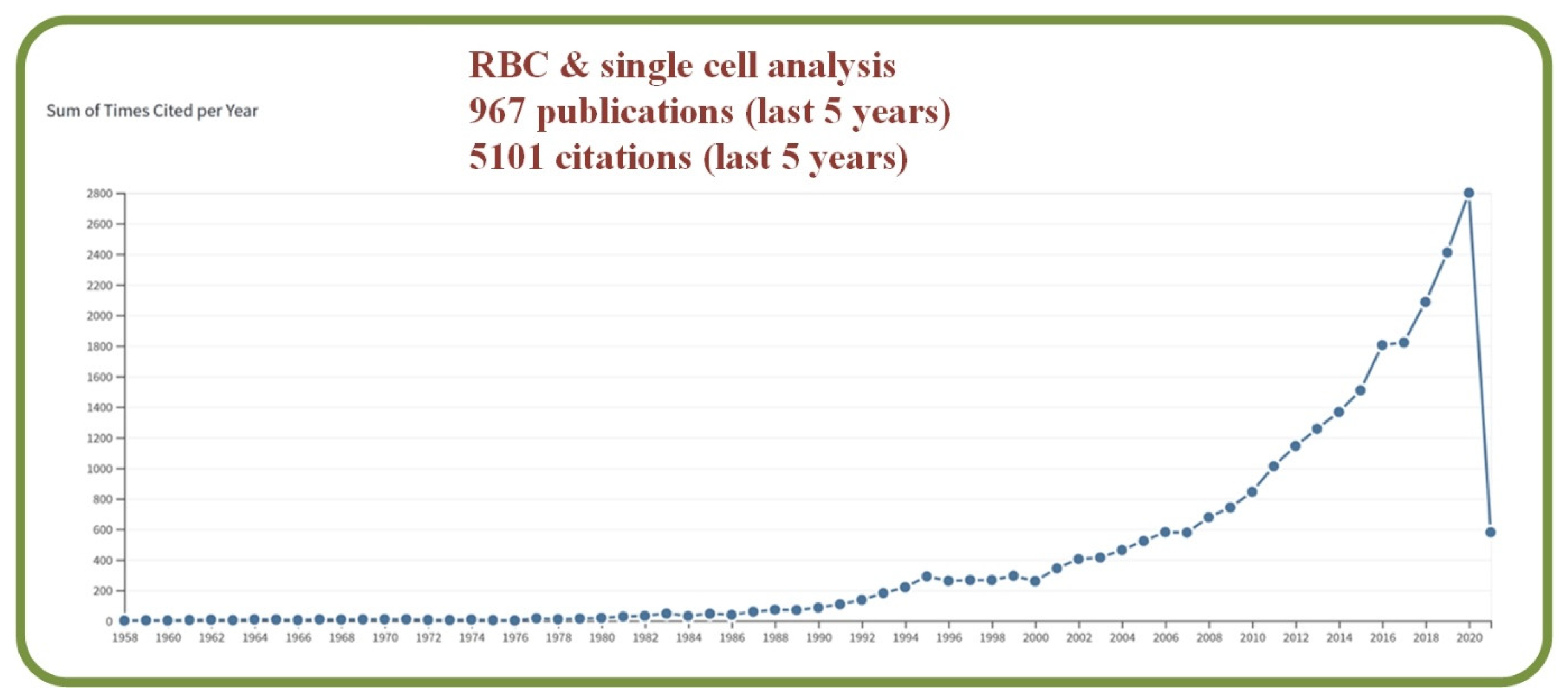
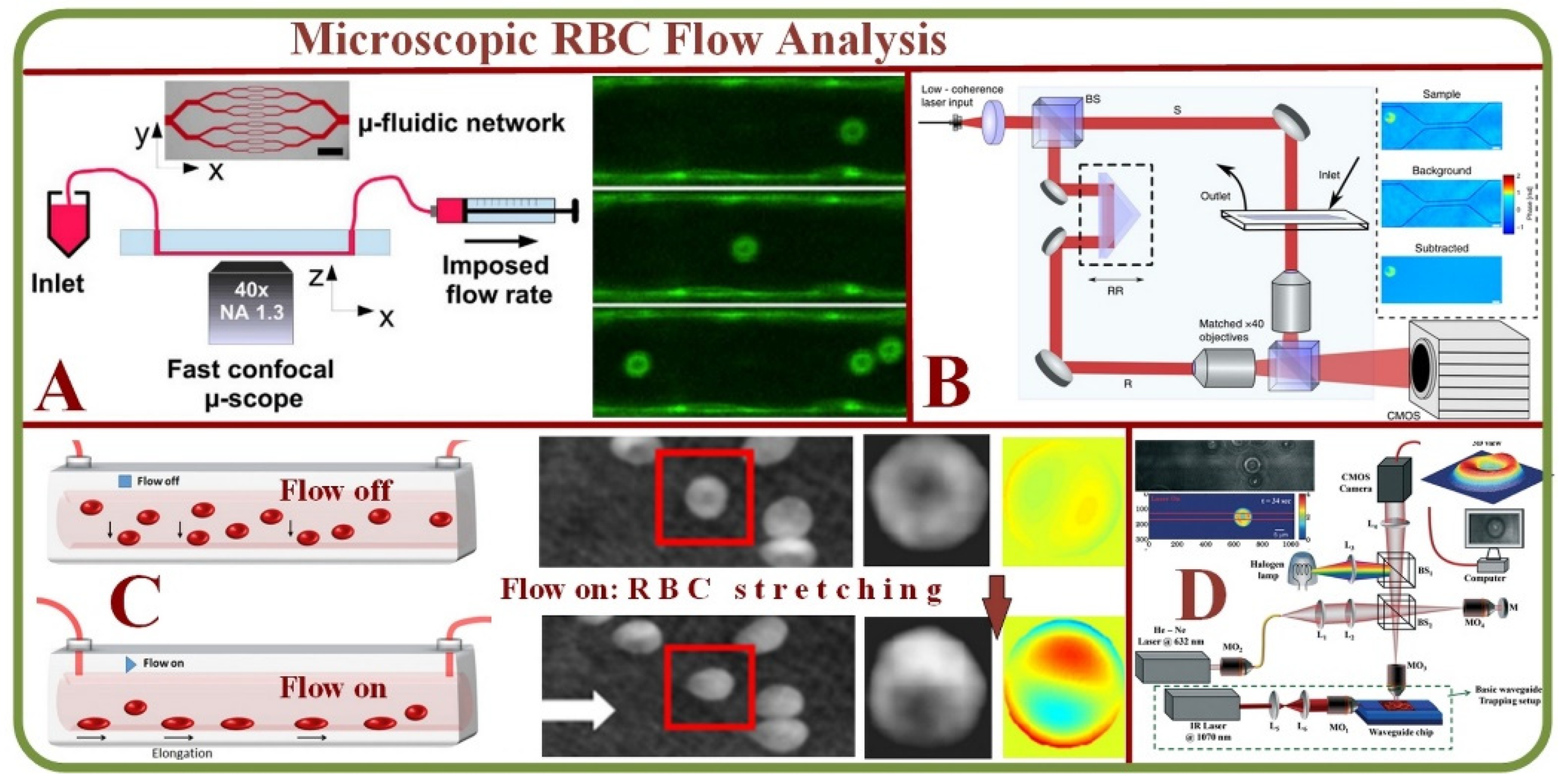
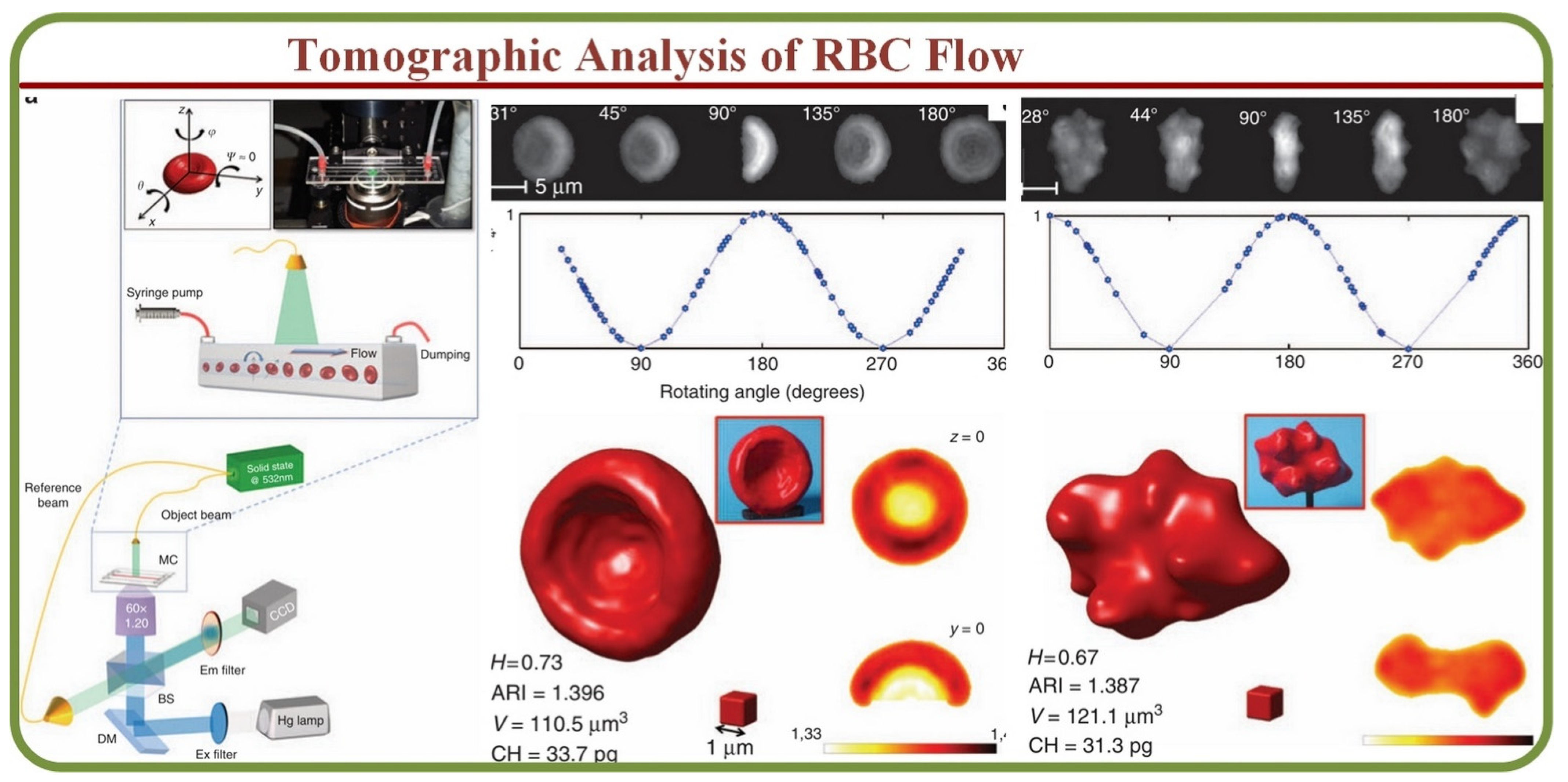
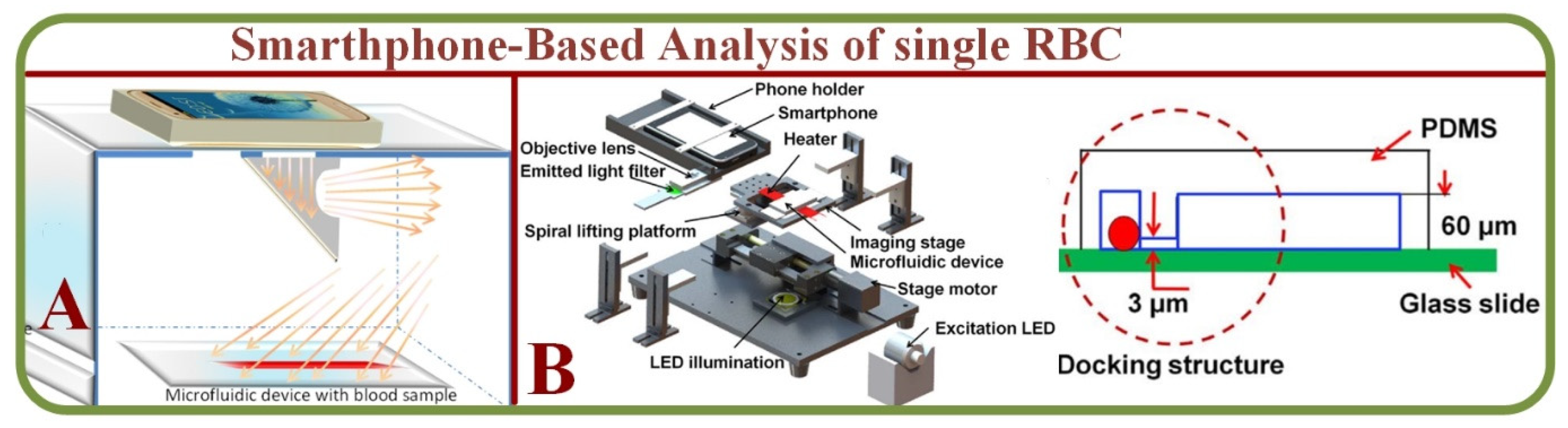
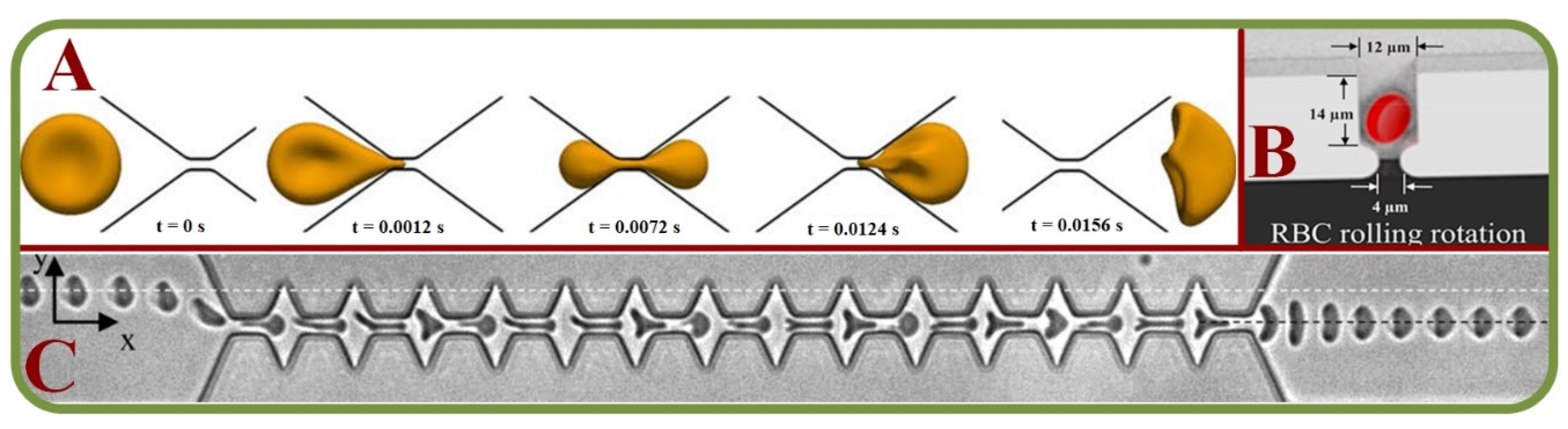
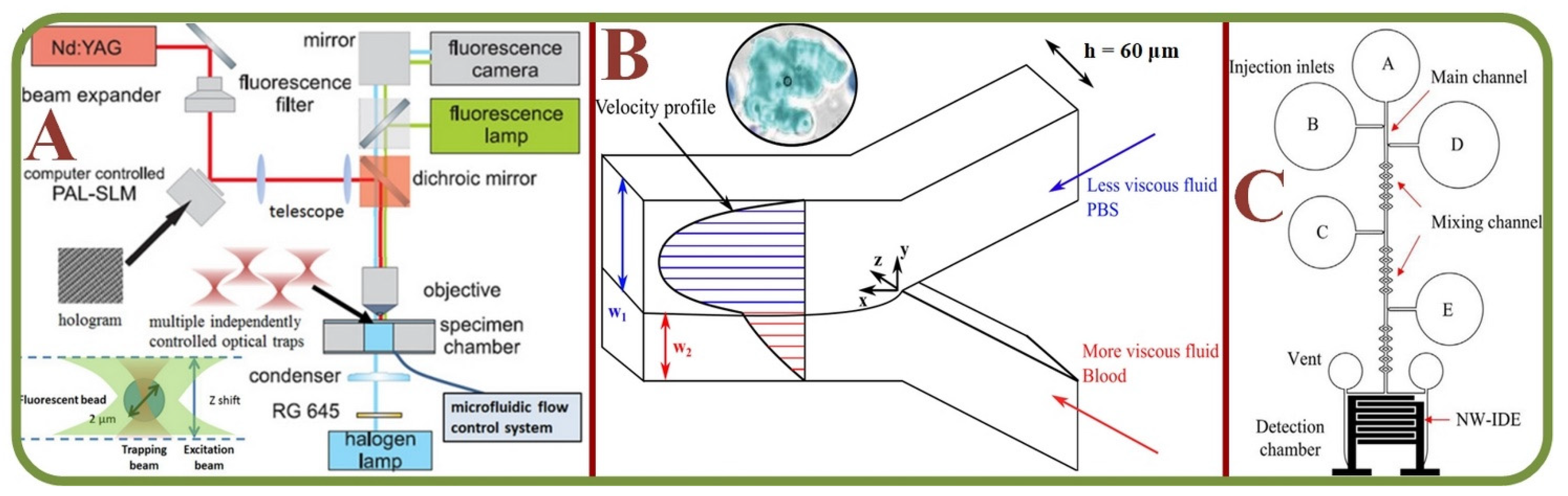
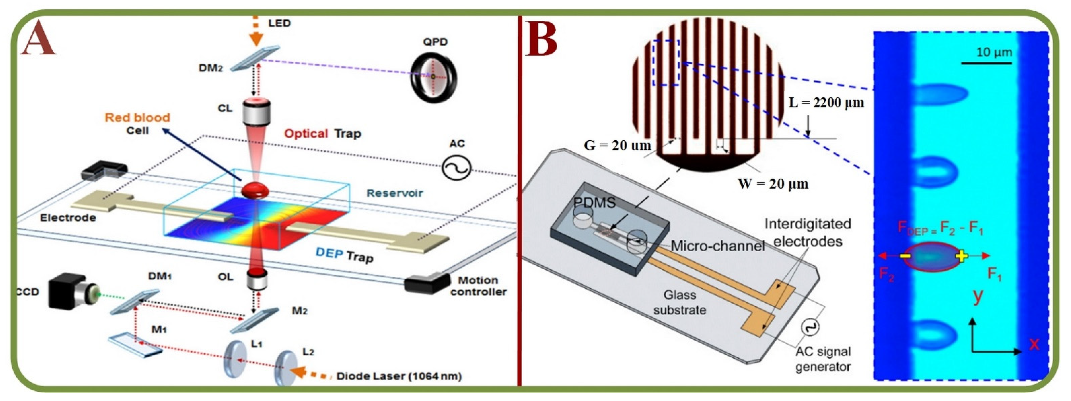
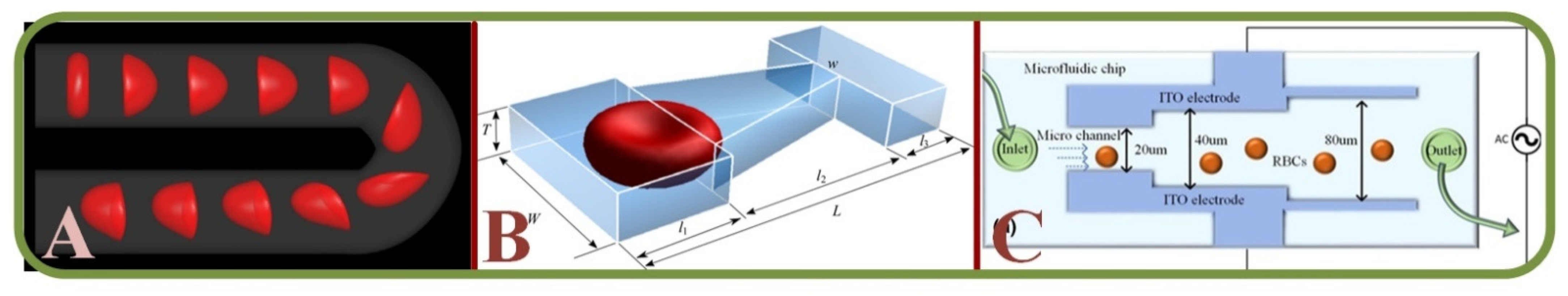
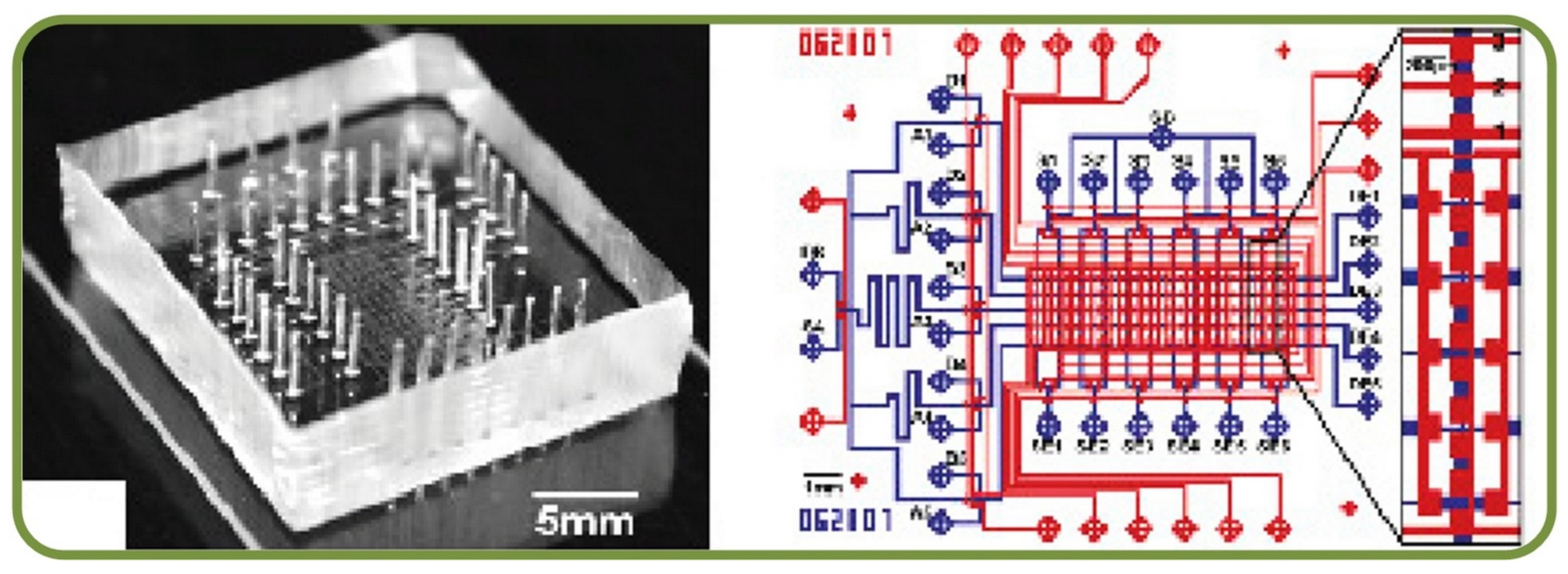
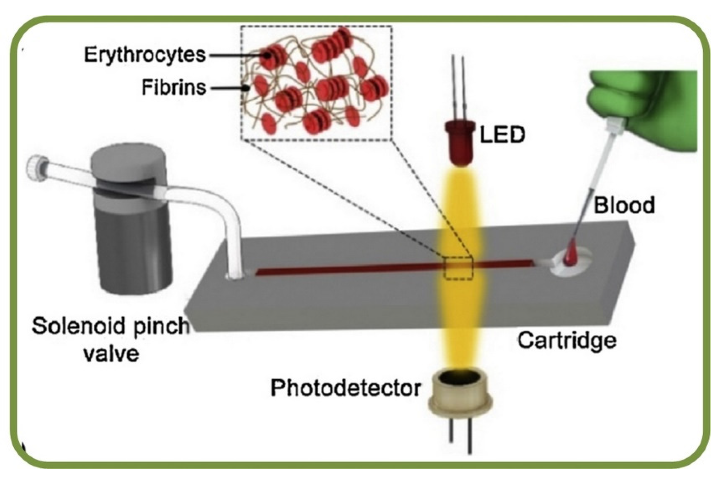
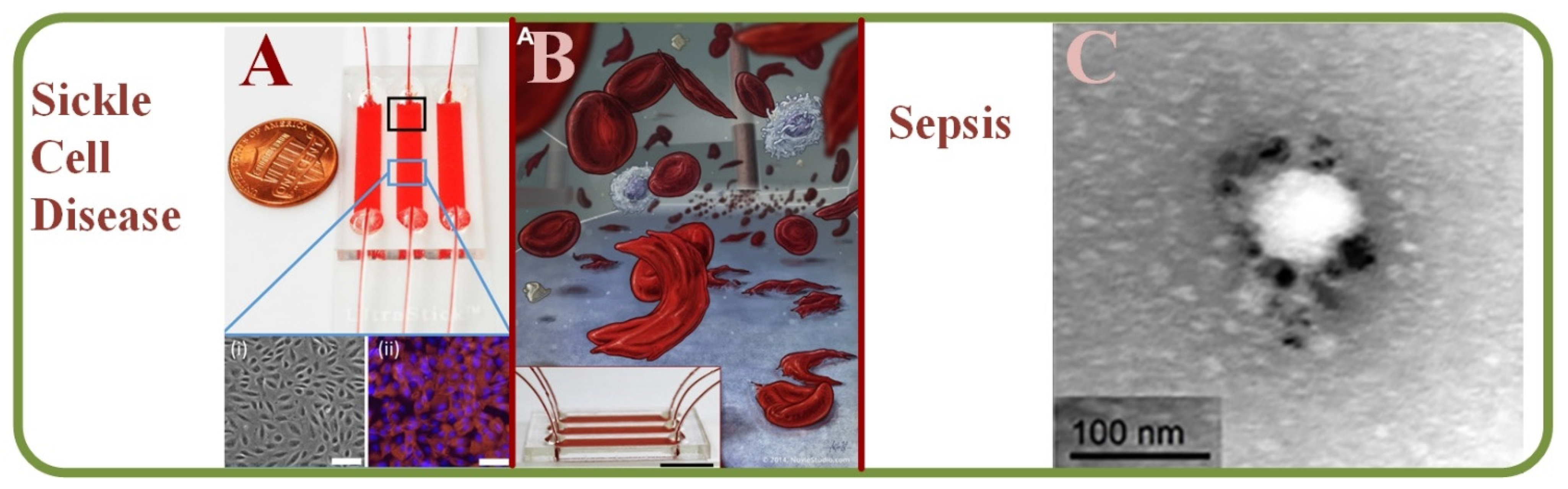
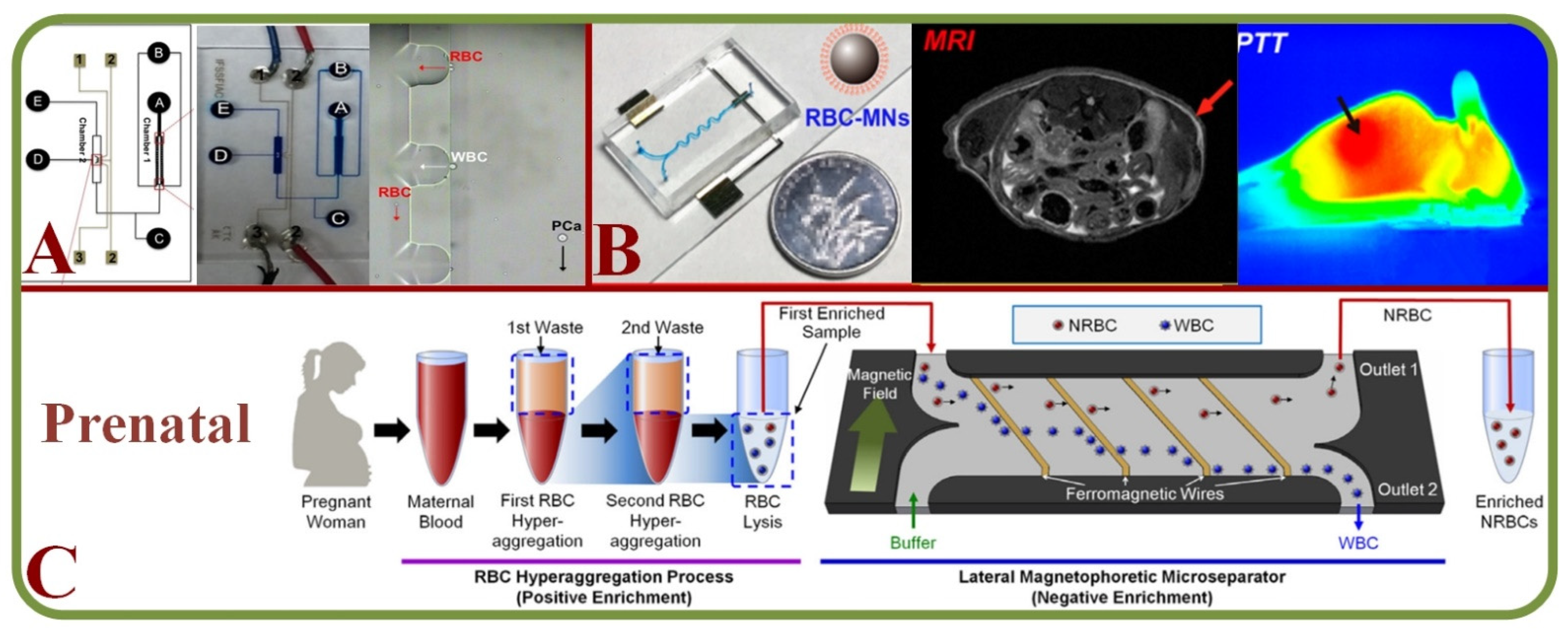
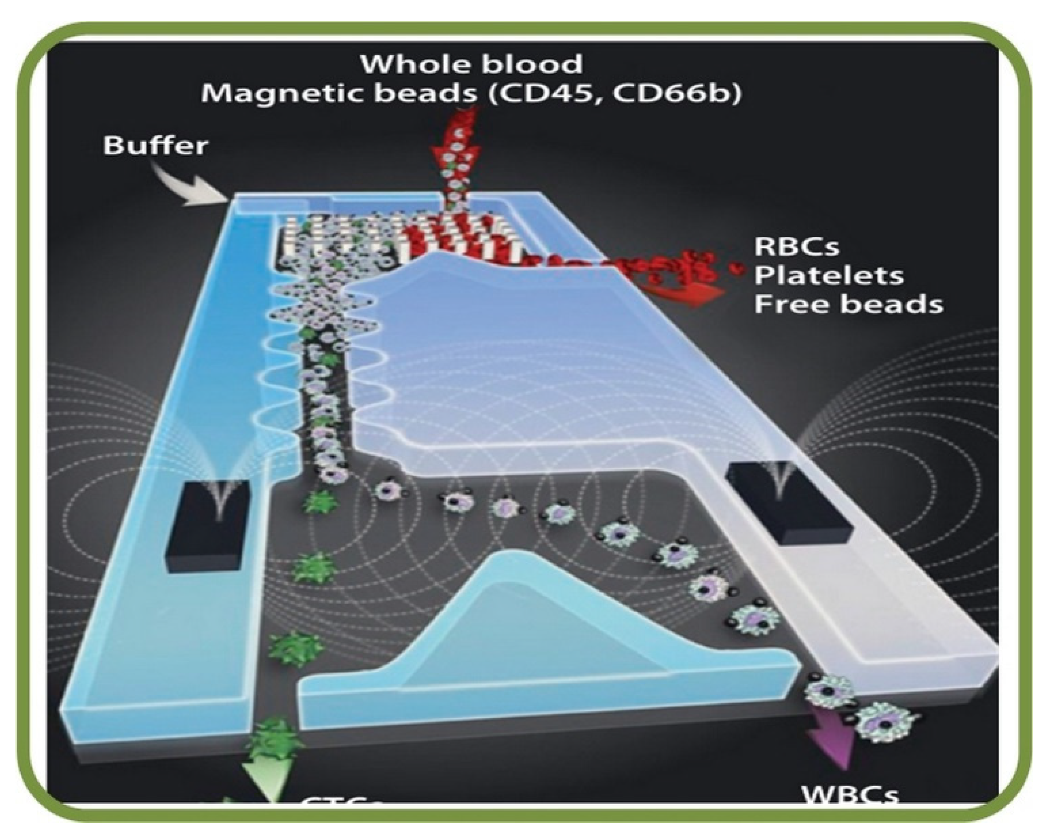
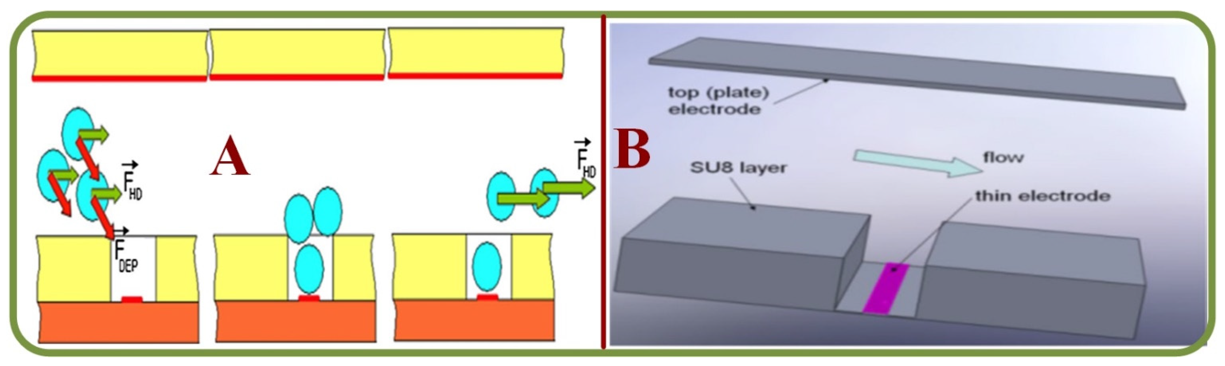
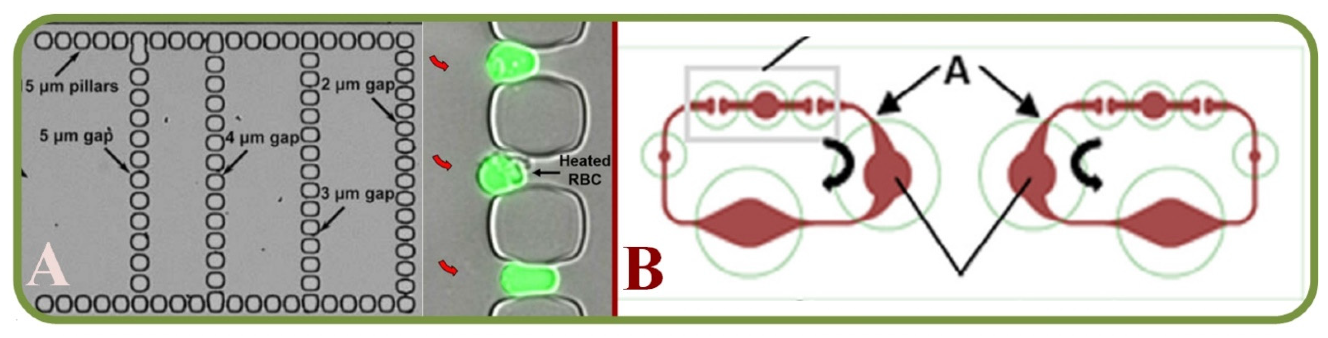
| Methodology | Existing Knowledge | Current Limitations | Future Prospects |
|---|---|---|---|
| Microscopic RBC/ Erythrocyte analysis | Viscosity aggregation of blood through T-junctions [25], RBC Quantitative Phase Imaging (QPI) [26,28] Quantitative Phase Microscopy (QPM) [29,30] Microvasculature on a chip [27], light scattering [31], saline induced stiffness [43] | Relationships between RBC aggregation and blood viscosity; refocusing issues in phase imaging systems and spatiotemporal phase sensitivity. | Viscosity in complex networks, phase imaging cytometry, cell aggregation, waveguide-based microfluidic platforms, multicellular diagnosis. |
| Spectral Imaging | State-of-the-art [58], RBC physics and mechanics [59,61], POC malaria diagnosis [60], aggregation dynamics [63], hybrid analytical platforms [62] | Low processing speeds, limitations in wavelength and challenges due to the variation of device elements. | Multispectral disease diagnosis, spectral cellular deformability analysis, spectral POC systems. |
| Raman Spectra Applications | State-of-the-art [64], isolation of circulating cells [65], clinical diagnosis [66,67,68], bio-particle trapping [69], blood plasma [70], SERS RBC analysis [73,74], optical-guided cell focusing [75], Hb oxygenation uptake [77] | Raman scattering related issues, signal overlap, long exposure time, channel dimension and optical scattering trade-off, integrating SERS systems and dynamic trapping of cells, bulky components. | Precision diagnostics for cancer metastasis, advanced laser-embedded microfluidic chips, multi-channel Raman analytics and sorting, multiplexing of analytes, real time analytics, reusability. |
| Tomographic Analysis | 3D microchannel tomography [46], OCT angiography [47], label-free cytometry [48,50], phase-contrast tomography (PCT) [49], hyperspectral tomography [51] | Flow cytometry relies on 1D assessment, limited wavelength, false-positive signals, acquisition of flow signal intensity, limited field of view and acquisition time. | Shape-based cellular identification, fast acquisition speed, optimal wavelengths, motion correction, detection of circulating tumour cells, 3D image reconstruction. |
| Miscellaneous Studies | Waveguide based blood typing [79], optical tweezers [80], speckle analysis [81,82,84], machine vision analytics [83] | Equipment handling, uncertainties in the relationship between parameters, scatter concentrations affect speckle contrast | Stability and accuracy in agglutination detection, stem cell-based therapy, moving trajectory-based deformation, speckle decorrelation time. |
| Domain | State of the Art | Limitations | Future Prospects |
|---|---|---|---|
| General [197,198,199,200,201,202,203,204,213,214,215,216,217] | RBC Sedimentation, real-time tracking, finite element based analysis, POC systems, coagulation–aggregation, fibrinogen-RBC interactions, optical detections | Device reliability, component incompatibility, assess limits in immunological profiles, occlusions, quality control, the high surface area-volume ratio to modify adsorption-desorption characterics. | Detection of tumor cells, advanced data acquisitions, macromolecules for drug delivery, hydrodynamic focusing, smartphone integrated devices, reduced testing time, simultaneous assaying, microfluidic vascular models, precise fabrication of sub-components. |
| Malaria infected RBC/Erythrocytes [224,225,226,228,255] | Deformation analysis, paper-based POC devices, microarray and microchannel analytics, cell-phoresis analytics, impedance cytometry | Automated devices, high-performance reagents, integrated systems, detection limitations, false-positive cases, drug reactions | Centralized geographical tracking, drug discovery platform, anti-malarial therapy, detection of RBC deformability with label-free identification and sample pre-enrichment |
| Sickle Cell Disease [230,231,232,233] | RBC adherence and cell phenotyping, hemoglobin content and hemolysis, vaso-occlusion due to SCD, micro-particle image velocimetry | Custom design for RBC adhesion studies, a traditional dish based cell culturing, internal bubble growth, vaso-occlusive crisis | Hemoglobin, reticulocyte, and lactate dehydrogenase count, RBC adhesion biophysics and hemolysis, vaso occlusion on sickling kinetics, poloxamer for decreasing blood viscosity, hematological parameters, drug interaction study |
| Sepsis [234,235] | RBC deformation mechanics, neutrophil motility analysis, laser diffraction studies | Limited knowledge on the impacts of neutrophils in the cytokine promotion | The correlation between neutrophils and septic responses, neutrophil activation, identification of inflammatory sequences, and erythrocyte rigidity |
| Cancer [237,238,239,241,242] | Droplet-Based ScRNAseq, acoustic cell separation, micro-filters and dielectrophoresis, magnetic cell navigation for photodynamic therapy, image-guided therapies | Lymphocyte contamination, cell concentration, processing rate dependence during sorting, loss of residual samples | Post-separation cell culturing, ex-vivo drug screening, anticancer drug accumulation, multidrug resistance, targeted drug delivery, synthesis and storage of theranostic nanoparticles, microfluidic electroporation, cell membrane coated nanoparticles |
| Prenatal Diagnostics [243,244,245] | Fetal RBC collection, loss of diseased RBC, magnetic enrichment, and nucleated RBC isolation | Scarcity of fetal cells in maternal circulation, high priced detection equipment | Rare cell separation, advanced molecular biology analytics, capture of limited fetal cells, microarray-based detections, circulating tumor cells for genetic tests |
| Miscellaneous [207,209,211,212,246,247,248,249,250,251,252,253,256] | Numerical and computational cell biomechanics, anemia detection, blood vessel interactions, drug interaction models, optical tweezers-based devices, silica-monolith platforms, spectroscopic techniques. | Interaction of blood cells under varying flows, metabolism-related alterations, immuno-suppression due to allogenic RBC transfusion, limited sampling volume, lack of knowledge on cell flow behavior, manufacturing cost | RBC biomechanics in metabolic disorders and diabetic neuropathy, construction of kinetic-particle models, POC based hemoglobin tests, RBC transfusion induced vascular injuries, assessment of protein-concentration effects, nanofluidic filtration and spectroscopic coupling, RBC micro motor based theranostics, blood coagulation and platelet defects |
| Method | Advantages | Disadvantages |
|---|---|---|
| Physical filtration | High separation and sorting efficiency | Clogging and fouling of cells |
| Hydrodynamic and hemodynamic processes | Enhanced separation and sorting with narrowed sheath flows | Stress on cells, altering molecular mechanisms, inhomogeneity |
| Surface Affinity and Topography | Specificity and accuracy | Altering cell physiology |
| Magnetophoresis | Differentiating cells without additives, efficiency up to 90% | Magnetic flux gradients on cells |
| Electrical Methods and Acoustophoresis | Sensitive, rapid, convenient, and robust. | Electrolysis, temperature elevations, phenotypic changes |
| Technique | State of the Art | Future Prospects |
|---|---|---|
| Immuno-phenotyping applications [335,336,337,338] | Detection of complex mixtures and antigens, pre-transfusion testing, serodiagnosis, and genotyping | Futuristic assaying methodology, device miniaturization, and combined testing |
| Mesh filtration and Erythrocyte deformability analysis [339] | RBC rheology through a nickel mesh filter | Further studies of trapping rates and rheology |
| Trapping Spectroscopy [340] | Micropipette based multifunctional microfluidic trapping | Cell sorting, pharmaceutics, Point of Care testing |
| UHF Single Beam acoustic tweezers [341] | Effective measurement of inter RBC forces | Other types of cells |
| Sickle Cell adhesion [342] | Sickle cell adhesion analysis, vaso occlusion studies | Development of anti-adhesive/occlusion agents, stress-dependent vaso-occlusion studies |
| Malaria Detection [331,343] | Highly sensitive detection of infection rates, remote analysis | Large-scale and mobile diagnostics |
| Digital Holographic Microscopy [332,344] | Optical trapping and recording via digital holograms | Detailed cellular dynamics, morphology-based identification |
| Trapping | State of the Art | Current Limitations | Future Prospects |
|---|---|---|---|
| Acoustic | Label-free approach for the analysis of physical properties [333], elastic and viscous parameters [345], trapping under suspensions [347] | Isolation and characterization of single cells, high-intensity ultrasound for cell trapping, increase in local temperature, acoustic rupture | Relation between particle diameter and ultrasound wavelength for trapping, automated analysis of erythrocyte population, in vivo application of single beam tweezers |
| Dielectrophoretic | single cell deformation mechanics [143,348], insulator based gradient dielectrophoretics [349], erythrocyte separation [350] | Particle sub-populations | Biophysical property determination, separation of bio analytes from complex fluids, treated surface coating, DC-micro devices, ellipsoid model-based RBC analysis |
| Hydro-dynamic | DC insulator-based [311], Steady stream flows, stagnation traps. hydrodynamic traps [308,329,352] | Cell retention vs. trapping efficiency trade-offs, complex fabrication processes, choking due to high cell density, loss of smaller cells due to non-uniformity | Enhanced particle (cell) separation, automated pumping systems, genetic/ biochemical/ physiological cell studies, reduced mechanical shear on cells, biocompatible materials |
| Magnetic | Improved magnetic field gradients and counter-current flow trapping [356], magnetophoretic RBC migration analysis [355] | Low volume analysis, pre-treatment methods, pre-labeling | Oxygenated erythrocyte mobility analysis, paramagnetic behavior studies, superconductive fractionation, collection throughput, low cost, rapid and automation |
| Optical | Cell aggregation [357,358,359], objective immersion-based [362], deformability analysis [361], waveguide surface-based [363] | Optical assessment of RBC mechanics, spontaneous aggregation modeling, stiffness calibration | Improved RBC shape recovery, cell positioning, aggregation rate, cell sorting, drug delivery |
Disclaimer/Publisher’s Note: The statements, opinions and data contained in all publications are solely those of the individual author(s) and contributor(s) and not of MDPI and/or the editor(s). MDPI and/or the editor(s) disclaim responsibility for any injury to people or property resulting from any ideas, methods, instructions or products referred to in the content. |
© 2023 by the authors. Licensee MDPI, Basel, Switzerland. This article is an open access article distributed under the terms and conditions of the Creative Commons Attribution (CC BY) license (https://creativecommons.org/licenses/by/4.0/).
Share and Cite
Grigorev, G.V.; Lebedev, A.V.; Wang, X.; Qian, X.; Maksimov, G.V.; Lin, L. Advances in Microfluidics for Single Red Blood Cell Analysis. Biosensors 2023, 13, 117. https://doi.org/10.3390/bios13010117
Grigorev GV, Lebedev AV, Wang X, Qian X, Maksimov GV, Lin L. Advances in Microfluidics for Single Red Blood Cell Analysis. Biosensors. 2023; 13(1):117. https://doi.org/10.3390/bios13010117
Chicago/Turabian StyleGrigorev, Georgii V., Alexander V. Lebedev, Xiaohao Wang, Xiang Qian, George V. Maksimov, and Liwei Lin. 2023. "Advances in Microfluidics for Single Red Blood Cell Analysis" Biosensors 13, no. 1: 117. https://doi.org/10.3390/bios13010117