Use of Cysteamine and Glutaraldehyde Chemicals for Robust Functionalization of Substrates with Protein Biomarkers—An Overview on the Construction of Biosensors with Different Transductions


Abstract
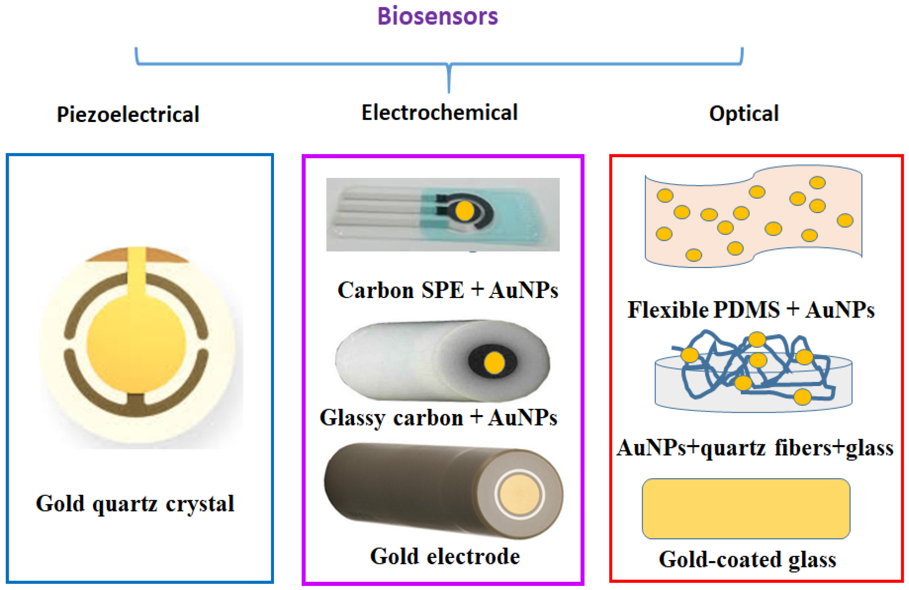
:1. Introduction
2. Piezoelectric Immunosensors
3. Electrochemical Immunosensors

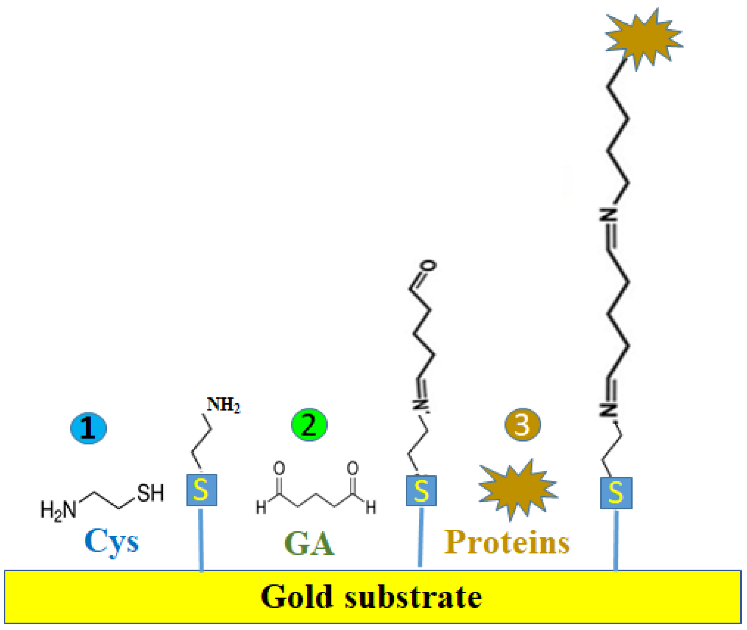
| Electrode | Size (Ø) | Cleaning | Cys Activation | GA Activation | Dilution Buffer | Ab | Incu-bation Time/°C (Ab) | Blocking the Non-Specific Sizes | Storage | Ag2 | Detection Method | Ref. |
|---|---|---|---|---|---|---|---|---|---|---|---|---|
| Au disc (GDE) | 1.6 mm | 30% H2O2; conc H2SO4, 1:3 (v/v) + polished alumina powder (0.3 and 0.5 μm) + water + ethanol + CV in 0.1 M H2SO4+ CV in KOH + water + ethanol (1) | 10 mM Cys in an ethanolic solution for 16 h at 25 °C (2) | 2.5% GA for 60 min (3) | 0.05 M PBS, pH 7.4 | Anti-GLY Ab 10 × 106 pg/mL 40 min (4) | 1 h at 37 °C | 2% BSA (0.05 M PBS, pH 7.4, for 30 min at 25 °C (5) | 4 °C in PBS (pH 7.4) (after 30 days) 9% loss for 10 ng GLY Ag (7) | GLY protein 103–106 pg/mL for 30 min at 25 °C (6) | SWV | [49] |
| Au | 3 mm | SR + Al2O3 < 50 nm + Drops UPW + UPW + absolute ethanol (99.9%) for 5 min + UPW for 5 min in the ultrasonic + dried with pure Ar (1) | 100 mM Cys in absolute ethanol for 1 h (2) | 1% GA for 10 min + 1.5% PAMAM in methanol for 1 h (3) | UPW + AS | 2.5 ng/anti-TSH for 1 h (5 µL) (4) | 1 h (4) | x | x | 0.1–0.6 mIUL−1 TSH in artificial serum (AS) (5) | EIS | [41] |
| Au | 2 mm | Polished with 0.3 and 0.05 mm alumina slurry + acetone/water (1:1) for 30 min + 0.1 M H2SO4 (1) | 10 mM solution Cys in 1 mM ethanol 200 µL, 3 h in dark at RT (2) | GA for 30 min RT (3) | 0.1 M PBS pH 7 | Anti-Cyfra 21.1 Ab (50 µL) (4) | 12 h at 4 °C | BSA for 1 h (5) | 4 °C | Cyfra 21.1 Ag (2.5, 5, 10, 25, 50) × 103 pg/mL human saliva (6) | SWV | [48] |
| Au | x | 0.1 M H2SO4 + CV + polish with alumina slurry, sized 1.0, 0.3, 0.05 µm (1) | 100 mM aqueous Cys for 1 h (20 μL) + wash DI water (2) | 2.5 % GA in WEB (20 μL) + 100 × 106 pg/mL Ab in WEB (20 μL) (3) | 0.05 M PBS pH 7.4 (WEB) | x | x | x | x | DPV: (2.5, 10, 25, 50, 100, and 200) × 103 pg/mL WEB DHEA−S (10 μL) for 30 min (4) | DPV | [46] |
| Au | x | 0.1 M H2SO4 + 15 CVs (1) | 10 mM Cys fo1r 1 h + drying (2) | GA for 1 h (3) | PBS, pH 7.4 | x | x | x | 4 °C for 24 h | 60 CVs for polymerization 0.5 mM TB*c in PBS (pH 7.4) + PSA (1–60) × 103 pg/mL (4) + 60 CVs for polymerization 1 M KNO3 in PBS (pH 7.4) (5) +100 CVs with 0.1 M NaOH (6) | DPV | [45] |
| Au | 1.6 mm | 0.05 and 0.3 µm alumina + rinsed with ddwater + 0.1 M H2SO4 + H2O2/H2SO4, 1/3 v/v) for 3 min + ultra-pure water 10× + dry in pure argon + hehexane-dithiol solution (0.1 M in pure ethanol) for 24 h + ethanol + argon (1) | 10 mM Cys in absolute ethanol for 3 h in dark (2) | 2.5% GA in water for 30 min (200 µL) (3) | PBS, pH 7 | 10 µg/mL (200 µL) (4) | Over-night at 4 °C (4) | 1% milk 1 h at RT (5) | x | Depleted plasma (pg/mL to ×106 pg/mL) for 15 min (20 µL) (6) | EIS | [39] |
| AuNps inks/carbon ink/polyimide sheet + 150 °C for 10 min (1) | ≈2 mm2 electrode with < 60 nm AuNps | Polyimide: ultrasonication with acetone | 20 mM Cys for 30 min(5 µL) + ddwater + N2 dry (2) | 4% GA for 30 min (5 μL) + ddwater + N2 dry (3) | DI water vs. PBS, pH 7.4 | x | x | x | x | 7 × 10⁹ pg/mL GOx (5 μL) overnight (4) …… wash PBS + N2 dry (5) | ChA | [37] |
| Au | 2.01 mm2 | Polished with 0.05 μm alumina + ultrasonicaltion in ethanol for 5 min (1) | 0.5 M Cys in pure ethanol overnight in dark (2) | 5% GA (5 µL) + 5 × 10⁹ pg/mL anti-HER-3 (5 μL) for 1 h in wet atm (3) | Sterile 0.01 M PBS (pH 7) | x | x | 1% BSA (10 μL) for 1 h in wet atm (4) | Anti-HER-3 and HER-3 solutions at −20 °C | 0.2 to 1.0 pg/mL HER-3 solution (5 μL) for 1 h in wet atm (5) | EIS | [40] |
| Carbon + AuNPs by electro-deposition (1) | x | x | Cys 2 h at RT (2) | 2.5% (v/v) GA in 200 mM PBS (pH 7.4) for 1 h (3) | x | Anti-STAT3, anti-PGM3, anti-DOCK8 10 × 106 pg/mL PBS, pH 8.5 (4) | 1 h (4) | 0.1 M ethanol-amine for 30 min (5) | 4 °C wet atm | 1 pg/mL to 105 pg/mL STAT3 (for 30 min), PGM3, and DOCK3 for 45 min (6) | SVW | [47] |
| GCE | 3 mm | 0.3µm and 0.05 µm Al2O3 slurry + Ultrason (59 kHz, 200 W) with UPW + absolute ethanol + BP (3 μL) + PG (4.2 μL) + IR dried + AuNP solution in dark for 24 h (1) | 60 mM Cys in pure ethanol + overnight in the dark (2) | 0.1% GA for 15 min (3) | 0.1 M PBS, pH 7.4 | 20 × 103 pg/mL anti-leptin solution (10 μL) (4) | In dark for 120 min (4) | 1% BSA (10 μL) (5) | 4 °C for 1 week | 0.15, 1, 10, 100, 312, 625, 1250 and 2500 pg/mL leptin for 2 h (6) | SWV | [50] |
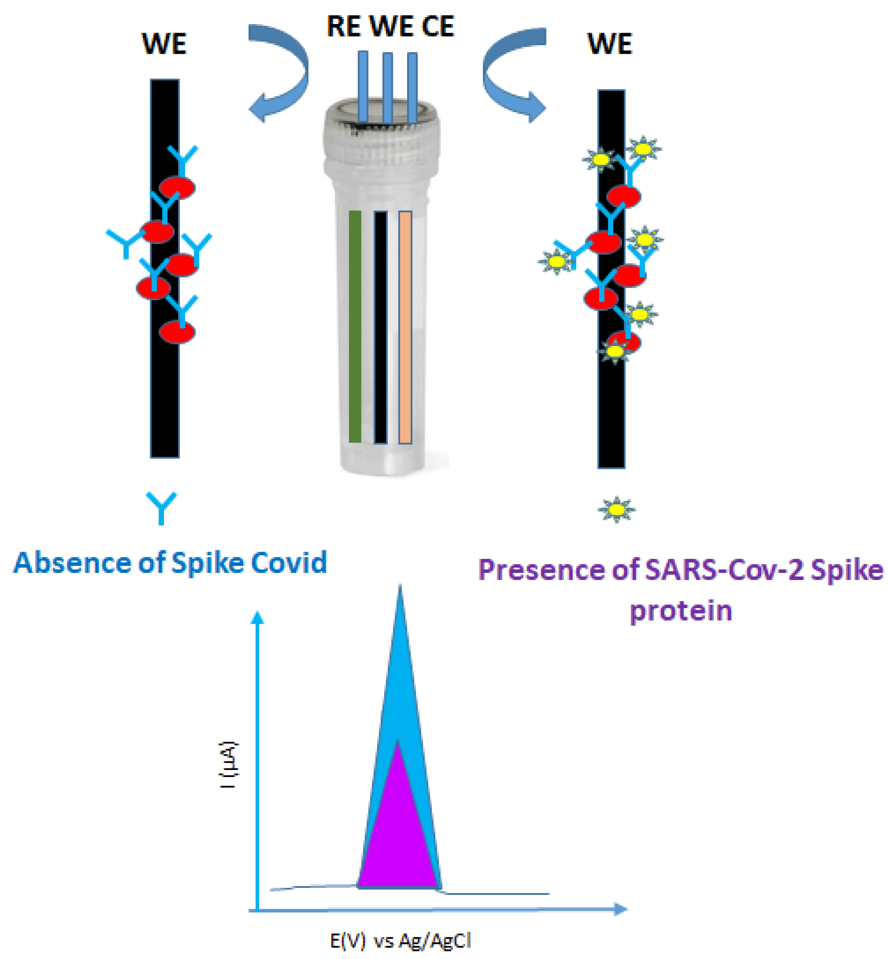
| Graphite pencils | 1 cm lengh (Ø 0.7 mm) (1) | Polish sand-paper (2000-grit) (2) | AuNP-Cys (pH 7.4) for 75 min (4) ………… 50 mM EDC + 25 mM NHS + 10 × 106 pg/mL ACE2 (5) | 2.5% (v/v) GA for 1 h at 37 °C (3) | 0.1 M PBS, pH 7.4 | x | 30 min at 37 °C | 1% BSA (w/v) for 30 min (6) | 4 °C dry (stable 24 h) or in PBS (pH 7.4) (stable for 120 h) | SARS-CoV-2 spike protein (SP) (7) | SWV | [53] |
| GCE | Au clusters on GCE after 20 CV cycles of Au solution (2) | 0.05 µm alumina suspension on felt + water rinsing + ultasonic ethanol/water (1:1) for 5 min (1) | 20 mM Cys for 1 h (25 μL) (3) | 7.5% GA in dimethyl formamide for 1 h (25 μL) (4) | PBS tablet: 0.01 M PBS + 0.0027 M KCl + 0.137 M NaCl (pH 7.5) at 25 °C | (0.1–1000) × 10−6 pg/mL 0.01 M (pH 7.5) PBS solution anti-spike antibody (7) | 30 min at RT | 2% BSA for 20 min (6) | 4 °C | 5 × 10⁶ pg/mL SARS-CoV-2 (2019-nCoV) spike S1-his recombinant protein for 45 min (10 μL) (5) | SWV | [54] |
| GSPE | 2 mm | Acetone 15 min (1) | 20 × 109 pg/mL Cys in water (2 μL) for 2 h at RT (2) | 5% GA in PBS for 1 h at RT (3) | *AB PBS + filtered through 0.22 μm PES mb | 0.81 × 10⁹ pg Ab/mL−1 PBS (4) 0.46 × 10⁹ pg/mLAb-HRP (2 μL) (7) | Over-night at 4 °C (4) …… 1 h (7) | 1% BSA in AB for 1 h at RT (5) | 4 °C (dry electrode with Ab) (8) | Melissococcus bacteria in PBS (105 to 109 CFU mL−1) for 1 h (6) | Ampe-rometry + H2O2/1 mM TMB | [36] |
| GCE | 4 mm | 0.3 μm and 0.05 μm alumina slurries + sonication in distilled water and ethanol for 2 min + dry in the air (1) ……… HAuCl4 solution (1% wt) (2) | 0.1 M Cys for 12 h at 4 °C (3) | 2.5% GA for 2 h (4) | 0.01 M PBS pH 7.4 (5) | MC-LR-BSA conjugate 50 × 106 pg/mL (5 μL) (6) | 6 h at 4 °C (6) | 0.01 M PBS pH 7.4 + 2 wt % BSA for 1 h at RT (5 µL) (7) | Dry at 4 °C (8) | 10 to 105 pg/mL MC-LR (2.5 μL) + 100 × 106 pg/mL HRP-mAb (2.5 μL) for 40 min at RT (9) 1.0 mM 4-CN and 0.15 mM H2O2, for 15 min at RT (10) | EIS | [42] |
| GSPE | 2 mm | Acetone for 20 min (1) | 20 × 109 pg/mL−1 Cys in water 2 h (2) | 3% in PBS for 1 h at RT (3) | Filtered PBS, pH 7.4 | 100 × 106 pg/mL in PBS (4) | Over-night at 4 °C (4) | BSA in PBS + 0.01% Tween 20 or milk 30 min (5) | Dry at 4 °C (6) | 103–108 CFU/mL Salmonella in tube 1 mL or 10 µL in PBS or milk 15 min RT (7) | EIS | [43] |
| PCB | Formation 75–100 nm AuNPs (1) | EC and AC ** (2) | 10 mM Cys in absolute ethanol (20 µL) (3) | 2.5% (v/v) GA in DI water (10 µL) for 2.5 h (4) | 1 × filtered PBS * | SARS-CoV-2 spike protein polyclonal Ab (10 × 106 pg/mL, 10 µL) (5) | 12 h at 4 °C | 1% BSA (7 µL) 3 h at 4 °C (6) | 4 °C (7) | Spike protein 0.1 × 10³ pg/mL to 500 × 103 pg/mL. 7 µL for 5 min (8) | DPV | [52] |
| NC-mb + 0.2% CHIT (in 2% acetic acid) for 24 h at RT + 10% methanol + 30 min drying (4) + urease NPs (0.5 mL) + GA/NC mb overnight at 4 °C “WM” (6) | Preparation: urease NPs (ethane/urease = 2:1) 20–100 nm NPs pH 5.5 vs. 13 nm urease pH 7 (1) | x | 0.12 g Cys under stirring for 5–6 h (3) | 2.5% GA stirring 500 rpm at 4 °C for 24 h (2) …… 2.5% GA in 0.1 M PB, pH 7.3 at RT for 2 h (5) | 0.1 M sodium acetate buffer, pH 5.5 | x | x | x | WM in 0.1 M sodium acetate buffer, pH 5.5, at 4 °C | Urea 2 to 80 µM in 0.1 M sodium acetate buffer, pH 5.5, at 40 °C (7) | Poten-tiometry AISE | [51] |
4. Optical Immunosensors
5. Conclusions and Perspectives
Funding
Institutional Review Board Statement
Informed Consent Statement
Acknowledgments
Conflicts of Interest
References
- Ye, M.; Wang, J.; Pan, S.; Lihong, Z.; Wang, Z.-W.; Zhu, X. Nucleic acids and proteins carried by exosomes of different origins as potential biomarkers for gynecologic cancers. Mol. Ther. Oncolytics 2022, 24, 101–113. [Google Scholar] [CrossRef]
- Karki, H.P.; Jang, Y.; Jung, J.; Oh, J. Advances in the development paradigm of biosample-based biosensors for early ultrasensitive detection of alzheimer’s disease. J. Nanobiotechnol. 2021, 19, 72. [Google Scholar] [CrossRef] [PubMed]
- Landegren, U.; Hammond, M. Cancer diagnostics based on plasma protein biomarkers: Hard times but great expectations. Mol. Oncol. 2021, 15, 1715–1726. [Google Scholar] [CrossRef]
- Karmacharya, M.; Kumar, S.; Lee, C.; Cho, Y.-K. Lab-on-a-disc for ultrafast plasmonic assay of cysteamine. Biosens. Bioelectron. 2021, 194, 113584. [Google Scholar] [CrossRef] [PubMed]
- Wai, J.L.; New, S.Y. Cysteamine-coated gold nanoparticles for bimodal colorimetric detection with inverse sensitivity. A proof-of-concept with lysozyme. RSC. Adv. 2019, 10, 1088–1094. [Google Scholar] [CrossRef] [Green Version]
- Gukowsky, J.C.; Tan, C.; Han, Z.; He, L. Cysteamine-Modified Gold Nanoparticles as a Colorimetric Sensor for the Rapid Detection of Gentamicin. J. Food Sci. 2018, 83, 1631–1638. [Google Scholar] [CrossRef]
- Vasconcelos, E.A.; Peres, N.G.; Pereira, C.O.; Silva, V.L.; Silva, E.F.; Dutra, R.F. Potential of a simplified measurement scheme and device structure for a low cost label-free point-of-care capacitive biosensor. Biosens. Bioelectron. 2009, 25, 870–876. [Google Scholar] [CrossRef] [PubMed]
- Wang, Q.; Zhang, B.; Lin, X.; Weng, W. Hybridization biosensor based on the covalent immobilization of probe DNA on chitosan–mutiwalled carbon nanotubes nanocomposite by using glutaraldehyde as an arm linker. Sens. Actuators B 2011, 156, 599–605. [Google Scholar] [CrossRef]
- Richards, F.M.; Knowles, J.R. Glutaraldehyde as a protein cross-linkage reagent. J. Mol. Biol. 1968, 37, 231–233. [Google Scholar] [CrossRef]
- Barbosa, O.; Ortiz, C.; Berenguer-Murcia, A.; Torres, R.; Rodrigues, R.C.; Fernandez-Lafuente, R. Glutaraldehyde in bio-catalysts design: A useful crosslinker and a versatile tool in enzyme immobilization. RSC Adv. 2014, 4, 1583–1600. [Google Scholar] [CrossRef] [Green Version]
- Yorganci, E.; Akyilmaz, E. Alkaline phosphatase based amperometric biosensor immobilized by cysteamine-glutaraldehyde modified self-assembled monolayer. Artif. Cells Blood Substit. Biotechnol. 2011, 39, 317–323. [Google Scholar] [CrossRef] [PubMed]
- Sahajpal, K.; Shekhar, S.; Kumar, A.; Sharma, B.; Meena, M.K.; Bhagi, A.K.; Sharma, S. Dynamic protein and polypeptide hydrogels based on Schiff base co-assembly for biomedicine. J. Mater. Chem. B 2022, 10, 3173–3198. [Google Scholar] [CrossRef] [PubMed]
- Mera, K.; Nagai, M.; Brock, J.W.C.; Fujiwara, Y.; Murata, T.; Maruyama, T.; Baynes, J.W.; Otagiri, M.; Nagai, R. Glutaraldehyde is an effective cross-linker for production of antibodies against advanced glycation end-products. J. Immunol. Methods 2008, 334, 82–90. [Google Scholar] [CrossRef] [Green Version]
- Naresh, V.; Lee, N. A review on biosensors and recent development of nanostructured materials-enabled biosensors. Sensors 2021, 21, 1109. [Google Scholar] [CrossRef] [PubMed]
- Cui, M.; Liu, Z.; Tang, Y. Application of visual biosensors based on gold nanoparticles for detection of target molecules. Proc. SPIE 2022, 12164, 136–142. [Google Scholar] [CrossRef]
- Arshad, R.; Fatima, I.; Sargazi, S.; Rahdar, A.; Karamzadeh-Jahromi, M.; Pandey, S.; Díez-Pascual, A.M.; Bilal, M. Novel perspectives towards rna-based nano-theranostic approaches for cancer management. Nanomaterials 2021, 11, 3330. [Google Scholar] [CrossRef]
- Pohanka, M. Quartz Crystal Microbalance (QCM) Sensing Materials in Biosensors Development. J. Electrochem. Sci. 2021, 16, 211220. [Google Scholar] [CrossRef]
- Pohanka, M. Overview of Piezoelectric Biosensors. Immunosens. DNA Sens. Appl. Mater. 2018, 11, 448. [Google Scholar]
- Songkhla, S.N.; Nakamoto, T. Overview of quartz crystal microbalance behavior analysis and measurement. Chemosensors 2021, 9, 350. [Google Scholar] [CrossRef]
- Wang, X.; Yu, H.; Lu, D.; Zhang, J.; Deng, W. Label free detection of the breast cancer biomarker CA15.3 using ZnO nanorods coated quartz crystal microbalance. Sens. Actuators B Chem. 2014, 195, 630–634. [Google Scholar] [CrossRef]
- Ayhan, F. QCM-Based Biosensor for the Detection of Homocysteine. Eur. J. Sci. Technol. 2020, 20, 835–843. [Google Scholar]
- Mattos, A.B.; Freitas, T.A.; Silva, V.L.; Dutra, R.F. A dual quartz crystal microbalance for human cardiac troponin T in real time detection. Sens. Actuators B Chem. B 2012, 161, 439–446. [Google Scholar] [CrossRef]
- Ramos-Jesus, J.; Carvalho, K.A.; Fonseca, R.A.S.; Oliveira, G.G.S.; Barrouin Melo, S.M.; Alcântara-Neves, N.M.; Dutra, R.F. A piezoelectric immunosensor for Leishmania chagasi antibodies in canine serum. Anal. Bioanal. Chem. 2011, 401, 917–925. [Google Scholar] [CrossRef] [PubMed]
- Pohanka, M.; Treml, F.; Hubálek, M.; Banďouchová, H.; Beklová, M.; Pikula, J. Piezoelectric biosensor for a simple serological diagnosis of tularemia in infected European brown hares (Lepus europaeus). Sensors 2007, 7, 2825–2834. [Google Scholar] [CrossRef] [PubMed] [Green Version]
- Zou, S.; Wei, H.; Cui, X.; Cheung, W.; Lid, X.; Liu, G. Intercalating methylene blue in molecular beacon for sensitive detection of salivary TNF-α towards early diagnosis of oral cancer. Sens. Diagn. 2022, 1, 731–738. [Google Scholar] [CrossRef]
- Lin, C.-Y.; Nhat Nguyen, U.T.; Hsieh, H.-Y.; Tahara, H.; Chang, Y.-S.; Wang, B.-Y.; Gu, B.-C.; Wu, C.-C.; Tsai, I.-J.; Fan, Y.-J. Peptide-based electrochemical sensor with nanogold enhancement for detecting rheumatoid arthritis. Talanta 2022, 236, 122886. [Google Scholar] [CrossRef] [PubMed]
- Aydin, E.B. Highly sensitive impedimetric immunosensor for determination of interleukin 6 as a cancer biomarker by using conjugated polymer containing epoxy side groups modified disposable ITO electrode. Talanta 2020, 215, 120909. [Google Scholar] [CrossRef] [PubMed]
- Gwiazda, M.; Bhardwaj, S.K.; Kijeńska-Gawrońska, E.; Wojciech, S.; Sivasankaran, U.; Kaushik, A. Impedimetric and plasmonic sensing of collagen i using a half-antibody-supported, au-modified, self-assembled monolayer system. Biosensors 2021, 11, 227. [Google Scholar] [CrossRef]
- Shao, M.; Shi, Z.; Pu, S.; Sun, J.; Bai, Y. Development of nanostructured enzymic amperometric biosensor based on gold nanoparticles for detection of pyruvate in natural samples. Int. J. Electrochem. Sci. 2022, 17, 220423. [Google Scholar] [CrossRef]
- Arévalo, B.; Blázquez, M.; Serafín, V.; Montero-Calle, A.; Calero, M.; Valverde, A.; Barderas, R.; Campuzano, S.; Yáñez-Sedeño, P.; Pingarrón, J.M. Unraveling autoimmune and neurodegenerative diseases by amperometric serological detection of antibodies against aquaporin-4. Bioelectrochemistry 2022, 144, 108041. [Google Scholar] [CrossRef]
- Bekhit, M.; Gorski, W. Electroanalysis of Infection with Methyl Pyruvate. ACS Sens. 2020, 5, 535–540. [Google Scholar] [CrossRef] [PubMed]
- Martínez-García, G.; Sánchez-Tirado, E.; González-Cortés, A.; Yáñez-Sedeño, P.; Pingarrón, J.M. Amperometric immunoassay for the obesity biomarker amylin using a screen printed carbon electrode functionalized with an electropolymerized carboxylated polypyrrole. Microchim. Acta 2018, 185, 323. [Google Scholar] [CrossRef]
- Yang, J.C.; Cho, C.H.; Choi, D.Y.; Park, J.P.; Park, J. Microcontact surface imprinting of affinity peptide for electrochemical impedimetric detection of neutrophil gelatinase-associated lipocalin. Sens. Actuators B Chem. 2022, 364, 131916. [Google Scholar] [CrossRef]
- Zhai, X.-J.; Wang, Q.-L.; Cui, H.-F.; Song, X.; Lv, Q.-Y.; Guo, Y.A. DNAzyme-catalyzed label-free aptasensor based on multifunctional dendrimer-like DNA assembly for sensitive detection of carcinoembryonic antigen. Biosens. Bioelectron. 2021, 194, 113618. [Google Scholar] [CrossRef] [PubMed]
- Hassanain, W.A.; Sivanesan, A.; Izake, E.L.; Ayoko, G.A. An electrochemical biosensor for the rapid detection of erythropoietin in blood. Talanta 2018, 189, 636–640. [Google Scholar] [CrossRef] [PubMed]
- Mikušová, Z.; Farka, Z.; Pastucha, M.; Poláchová, V.; Obořilová, R.; Skládal, P. Amperometric Immunosensor for Rapid Detection of Honeybee Pathogen Melissococcus plutonius. Electroanalysis 2019, 31, 1969–1976. [Google Scholar] [CrossRef]
- Benson, J.; Fung, C.M.; Lloyd, J.S.; Deganello, D.; Smith, N.A.; Teng, K.S. Direct patterning of gold nanoparticles using flexographic printing for biosensing applications. Nanoscale Res. Lett. 2015, 10, 127. [Google Scholar] [CrossRef] [PubMed] [Green Version]
- Mehmandoust, M.; Gumus, Z.P.; Soylak, M.; Erk, N. Electrochemical immunosensor for rapid and highly sensitive detection of SARS-CoV-2 antigen in the nasal sample. Talanta 2022, 240, 133211. [Google Scholar] [CrossRef]
- Garyfallou, G.-Z.; Ketebu, O.; Şahin, S.; Mukaetova-Ladinska, E.B.; Catt, M.; Yu, E.H. Electrochemical detection of plasma immunoglobulin as a biomarker for Alzheimer’s disease. Sensors 2017, 17, 2464. [Google Scholar] [CrossRef] [PubMed] [Green Version]
- Canbaz, M.C.; Şimşek, C.S.; Sezgintürk, M.K. Electrochemical biosensor based on self-assembled monolayers modified with gold nanoparticles for detection of HER-3. Anal. Chem. Acta 2014, 814, 31–38. [Google Scholar] [CrossRef]
- Ozcan, H.M.; Aydin, U.D. A simple immunosensor for thyroid stimulating hormone. Artif. Cells Nanomed. Biotechnol. 2021, 49, 61–70. [Google Scholar] [CrossRef] [PubMed]
- Hou, L.; Ding, Y.; Zhang, L.; Li, M.; Chen, Z.; Wu, X. An ultrasensitive competitive immunosensor for impedimetric detection of microcystin-LR via antibody-conjugated enzymatic biocatalytic precipitation. Sens. Actuators B Chem. 2016, 233, 63–70. [Google Scholar] [CrossRef]
- Farka, Z.; Juřík, T.; Pastucha, M.; Kovr, D.; Lacina, K.; Skládal, P. Rapid Immunosensing of Salmonella Typhimurium Using Electrochemical Impedance Spectroscopy: The Effect of sample treatment. Electroanalysis 2016, 28, 1803–1809. [Google Scholar] [CrossRef]
- Moradkhani, M.; Farshchi, F.; Hasanzadeh, M.; Mokhtarzadeh, A. A novel bioassay for the monitoring of carcinoembryonic antigen in human biofluid using polymeric interface and immunosensing method. Mol. Recognit. 2020, 33, e2852. [Google Scholar] [CrossRef] [PubMed]
- Abbasy, L.; Mohammadzadeh, A.; Hasanzadeh, M.; Razmi, N. Development of a reliable bioanalytical method based on prostate specific antigen trapping on the cavity of molecular imprinted polymer towards sensing of PSA using binding affinity of PSA-MIP receptor: A novel biosensor. J. Pharm. Biomed. Anal. 2020, 188, 113447. [Google Scholar] [CrossRef] [PubMed]
- Balaban, S.; Durmus, C.; Aydindogan, E.; Gumus, Z.P.; Timur, S. An Electrochemical Biosensor Platform for Testing of Dehydroepiandrosterone 3-Sulfate (DHEA−S) as a Model for Doping Materials. Electroanalysis 2020, 32, 128–134. [Google Scholar] [CrossRef]
- Eissa, S.; Abdulkarim, H.; Dasouki, M.; Al Mousac, H.; Arnout, R.; Al Saudc, B.; Rahman, A.A.; Zourob, M. Multiplexed detection of DOCK8, PGM3 and STAT3 proteins for the diagnosis of Hyper-Immunoglobulin E syndrome using gold nanoparticles-based immunosensor array platform. Biosens. Bioelectron. 2018, 117, 613–619. [Google Scholar] [CrossRef] [PubMed]
- Jafari, M.; Hasanzadeh, M. Non-invasive bioassay of Cytokeratin Fragment 21.1 (Cyfra 21.1) protein in human saliva samples using immunoreaction method: An efficient platform for early-stage diagnosis of oral cancer based on biomedicine. Biomed. Pharmacother. 2020, 131, 110671. [Google Scholar] [CrossRef]
- Kalyani, T.; Nanda, A.; Jana, S.K. Detection of a novel glycodelin biomarker using electrochemical immunosensor for endometriosis. Anal. Chim. Acta 2021, 1146, 146–154. [Google Scholar] [CrossRef]
- Cai, J.; Gou, X.; Sun, B.; Li, W.; Li, D.; Liu, J.; Hu, F.; Li, Y. Porous graphene-black phosphorus nanocomposite modified electrode for detection of leptin. Biosens. Bioelectron. 2019, 137, 88–95. [Google Scholar] [CrossRef] [PubMed]
- Jakhar, S.; Pundir, C.S. Preparation, characterization and application of urease nanoparticles for construction of an improved potentiometric urea biosensor. Biosens. Bioelectron. 2018, 100, 242–250. [Google Scholar] [CrossRef]
- Nandeshwar, R.; Kumar, M.S.; Kondabagli, K.; Tallur, S. Electrochemical Immunosensor Platform Using Low-Cost ENIG PCB Finish Electrodes: Application for SARS-CoV-2 Spike Protein Sensing. IEEE Access 2021, 9, 1543686154377. [Google Scholar] [CrossRef]
- de Lima, L.F.; Ferreira, A.L.; Torres, M.D.T.; de Araujo, W.R.; de la Fuente-Nunez, C. Minute-scale detection of SARS-CoV-2 using a low-cost biosensor composed of pencil graphite electrodes. Proc. Natl. Acad. Sci. USA 2021, 118, e2106724118. [Google Scholar] [CrossRef]
- Liv, L. Electrochemical immunosensor platform based on gold-clusters, cysteamine and glutaraldehyde modified electrode for diagnosing COVID-19. Microchem. J. 2021, 168, 106445. [Google Scholar] [CrossRef]
- Tai, J.; Fan, S.; Ding, S.; Ren, L. Gold Nanoparticles Based Optical Biosensors for Cancer Biomarker Proteins: A Review of the Current Practices. Front. Bioeng. Biotechnol. 2022, 10, 877193. [Google Scholar] [CrossRef]
- Im, H.; Shao, H.; Park, Y.I.; Peterson, V.M.; Castro, C.M.; Weissleder, R.; Lee, H. Label-free detection and molecular profiling of exosomes with a nanoplasmonic sensor. Nat. Biotechnol. 2014, 3, 490–495. [Google Scholar] [CrossRef] [Green Version]
- Letchumanan, I.; Gopinath, S.C.B.; Md Arshad, M.K.; Saheed, M.S.M.; Perumal, V.; Voon, C.H.; Hashim, U. Gold-Nanohybrid Biosensors for Analyzing Blood Circulating Clinical Biomacromolecules: Current Trend toward Future Remote Digital Monitoring. Crit. Rev. Anal. Chem. 2022, 52, 577–592. [Google Scholar] [CrossRef] [PubMed]
- Halas, N.J.; Lal, S.; Chang, W.S.; Link, S.; Nordlander, P. Plasmons in strongly coupled metallic nanostructures. Chem. Rev. 2011, 111, 3913–3961. [Google Scholar] [CrossRef]
- Tessaro, L.; Aquino, A.; Carvalho, A.P.A.D.; Conte-Junior, C.A. A systematic review on gold nanoparticles based-optical biosensors for Influenza virus detection. Sens. Actuators Rep. 2021, 3, 100060. [Google Scholar] [CrossRef]
- Fattahi, Z.; Khosroushahi, A.Y.; Hasanzadeh, M. Recent progress on developing of plasmon biosensing of tumor biomarkers: Efficient method towards early stage recognition of cancer. Biomed. Pharmacother. 2020, 132, 110850. [Google Scholar] [CrossRef]
- Koster, H.J.; Rojalin, T.; Powell, A.; Pham, D.; Mizenko, R.R.; Andrew, C.; Birkeland, A.C.; Carney, R.P. Surface enhanced Raman scattering of extracellular vesicles for cancer diagnostics despite isolation dependent lipoprotein contamination. Nanoscale 2021, 13, 14760–14776. [Google Scholar] [CrossRef]
- Choi, M.; Kang, T.; Choi, S.H.O.; Byun, K.M. Dual modal plasmonic substrates based on a convective self-assembly technique for enhancement in SERS and LSPR detection. Opt. Express 2021, 29, 6179–6187. [Google Scholar] [CrossRef] [PubMed]
- Focsan, M.; Craciun, A.M.; Potara, M.; Leordean, C.; Vulpoi, A.; Maniu, D. Flexible and tunable ED gold nanocups platform as plasmonic biosensor for specific dual LSPR-SERS immuno-detection. Sci. Rep. 2017, 7, 14240. [Google Scholar] [CrossRef] [PubMed] [Green Version]
- Chou, S.-F.; Hsu, W.-L.; Hwang, J.-M.; Chen, C.-Y. Development of an immunosensor for human ferritin, a nonspecific tumor marker, based on surface plasmon resonance. Biosens. Bioelectron. 2004, 19, 999–1005. [Google Scholar] [CrossRef]
- Wu, B.; Zhang, G.; Shuang, S.; Choi, M.M.F. Biosensors for determination of glucose with glucose oxidase immobilized on an eggshell membrane. Talanta 2004, 64, 546–553. [Google Scholar] [CrossRef]
- Migneault, L.; Dartiguenave, C.; Bertrand, M.J.; Waldron, K.C. Glutaraldehyde: Behaviour in aqueous solution, reaction with proteins, and application to enzyme crosslinking. BioTechniques 2004, 37, 790–802. [Google Scholar] [CrossRef]
- Farris, S.; Song, J.; Huang, Q. Alternative reaction mechanism for the cross-linking of gelatin with glutaraldehyde. J. Agric. Food Chem. 2010, 58, 998–1003. [Google Scholar] [CrossRef]
- Rodrigues, R.C.; Berenguer-Murciz, Ā.; Carballares, D.; Morellon-Sterling, R.; Fernandez-Lafuente, R. Stabilization of enzymes via immobilization: Multipoint covalent attachment and other stabilization strategies. Biotechnol. Adv. 2021, 52, 107821. [Google Scholar] [CrossRef]
- Shiue, A.; Chen, J.-H.; Hsiao, C.-Y.; Chang, S.-M.; Hwa, K.-Y.; Leggett, G. Preparation of substrates for microarray protein chips with different ending functional groups. J. Immunol. Methods 2022, 502, 113218. [Google Scholar] [CrossRef]
- Welch, N.G.; Scoble, J.A.; Muir, B.W.; Pigram, P.J. Orientation and characterization of immobilized antibodies for improved immunoassays (review). Biointerphases 2017, 12, 02D301. [Google Scholar] [CrossRef] [Green Version]
- Gao, S.; Rojas-Vega, F.; Rocha-Martin, J.; Guisan, J.M. Oriented immobilization of antibodies through different surface regions containing amino groups: Selective immobilization through the bottom of the Fc region. Int. J. Biol. Macromol. 2021, 177, 19–28. [Google Scholar] [CrossRef] [PubMed]
- Fisher, S.A.; Baker, A.E.G.; Shoichet, M.S. Designing peptide and protein modified hydrogels selecting the optimal conjugation strategy. J. Am. Chem. Soc. 2017, 139, 7416–7427. [Google Scholar] [CrossRef] [PubMed]
- Suni, I.I. Substrate materials for biomolecular immobilization within electrochemical biosensors. Biosensors 2021, 11, 239. [Google Scholar] [CrossRef] [PubMed]
- Aquino, A.; Paschoalin, V.M.F.; Tessaro, L.L.G.; Raymundo-Pereira, P.A.; Conte-Junior, C.A. Updating the use of nano-biosensors as promising devices for the diagnosis of coronavirus family members: A systematic review. J. Pharm. Biomed. Anal. 2022, 211, 114608. [Google Scholar] [CrossRef] [PubMed]
- Tan, D.; Li, F.; Zhou, B. Antifouling self-assembled monolayers for designing of electrochemical biosensors. Int. J. Electrochem. Sci. 2020, 15, 9446–9458. [Google Scholar] [CrossRef]
- Vaisocherová-Lísalová, H.; Víšová, I.; Ermini, M.L.; Springer, T.; Song, X.C.; Mrazek, J.; Lamacova, J.; Lynn, N.C.; Šedivák, P.; Homola, J. Low-fouling surface plasmon resonance biosensor for multi-step detection of foodborne bacterial pathogens in complex food samples. Biosens. Bioelectron. 2016, 80, 84–90. [Google Scholar] [CrossRef]
- Yang, G.; Wei, L.; Thong, B.K.S.; Fu, Y.; Cheong, I.H.; Kozlakidis, Z.; Li, X.; Wang, H.; Li, X. A systematic review of oral biopsies, sample types, and detection techniques applied in relation to oral cancer detection. BioTech 2022, 11, 5. [Google Scholar] [CrossRef]
- Wang, L.; Skotland, T.; Berge, V.; Sandvig, K.; Llorente, A. Exosomal proteins as prostate cancer biomarkers in urine: From mass spectrometry discovery to immunoassay-based validation. Eur. J. Pharm. Sci. 2017, 98, 80–85. [Google Scholar] [CrossRef]
- Wuethrich, A.; Quirino, J.P. A decade of microchip electrophoresis for clinical diagnostics—A review of 2008–2017. Anal. Chim. Acta 2019, 1045, 42–66. [Google Scholar] [CrossRef] [Green Version]
- Tseng, J.-Y.; Yang, C.-Y.; Liang, S.-C.; Liu, R.-S.; Jiang, J.-K.; Lin, C.-H. Dynamic changes in numbers and properties of circulating tumor cells and their potential applications. Cancers 2014, 6, 2369–2386. [Google Scholar] [CrossRef] [Green Version]
- Ibau, C.; Md Arshad, M.K.; Subash, C.B.G. Current advances and future visions on bioelectronic immunosensing for prostate-specific antigen. Biosens. Bioelectron. 2017, 98, 267–284. [Google Scholar] [CrossRef]
- Rehman, A.; Zeng, X. Monitoring the cellular binding events with quartz crystal microbalance (QCM) biosensors. Methods Mol. Biol. 2017, 1572, 313–326. [Google Scholar] [PubMed]
- Dastidar, M.G.; Murugappan, K.; Damry, A.M.; Nisbet, D.R.; Nolan, C.J.; Tricoli, A. When less gold is more: Selective attomolar biosensing at the nanoscale. Adv. Funct. Mater. 2022, 32, 2105433. [Google Scholar] [CrossRef]
- Zhang, M.; Cui, X.; Li, N. Smartphone-based mobile biosensors for the point-of-care testing of human metabolites. Mater. Today Bio 2022, 14, 100254. [Google Scholar] [CrossRef]
- Orrego, A.H.; Romero-Ferández, M.; Millán-Linares, M.C.; Just, M.M.; Guisán, J.M.; Rocha-Martin, J. Stabilization of enzymes by multipoint covalent attachment on aldehyde-supports 2-picoline borane as an alternative reducing agent. Catalysts 2018, 8, 333. [Google Scholar] [CrossRef] [Green Version]
- Oliverion, M.; Perotto, S.; Messina, G.C.; Lovato, L.; De Angelis, F. Chemical functionalization of plasmonic surface biosensors: A tutorial review on issues, strategies, and costs. ACS Appl. Mater. Interfaces 2017, 9, 29394–29411. [Google Scholar] [CrossRef] [Green Version]
- Rosy; Goyal, R.N.; Shim, Y.-B. Glutaraldehyde sandwiched amino functionalized polymer based aptasensor for the determination and quantification of chloramphenicol. RSC Adv. 2015, 5, 69356–69364. [Google Scholar] [CrossRef]
- Atallah, C.; Charcosset, C.; Greige-Gerges, H. Challenges for cysteamine stabilization, quantification, and biological effects improvement. J. Phram. Anal. 2020, 10, 499–516. [Google Scholar] [CrossRef]
- Mao, K.; Min, X.; Zhang, H.; Zhang, K.; Cao, H.; Guo, Y.; Yang, Z. Paper-based microfluidics for rapid diagnostics and drug delivery. J. Control. Release 2020, 322, 187–199. [Google Scholar] [CrossRef] [PubMed]
- Lee, W.-C.; Ng, H.-Y.; Hou, C.-Y.; Lee, C.-T.; Fu, L.-M. Recent advances in lab-on-paper diagnostic devices using blood samples. Lab Chip 2021, 21, 1422–1453. [Google Scholar] [CrossRef] [PubMed]
- Hou, Y.; Lv, C.-C.; Guo, Y.-L.; Ma, X.-H.; Liu, W.; Jin, Y.; Li, B.-X.; Yang, M.; Yao, S.Y. Recent advances and applications in paper-based devices for point-of-care testing. J. Anal. Test. 2022, in press. [Google Scholar] [CrossRef]
- Zhou, L.; Poggesi, S.; Bariani, G.C.; Mittapalli, R.; Adam, P.-M.; Manzano, M.; Ionescu, R.E. Robust SERS platforms based on annealed gold nanostructures formed on ultrafine glass substrates for various (bio)applications. Biosensors 2019, 9, 53. [Google Scholar] [CrossRef] [PubMed] [Green Version]
- Jia, K.; Bijeon, J.L.; Adam, P.M.; Ionescu, R.E. Sensitive localized surface plasmon resonance multiplexing protocols. Anal. Chem. 2012, 84, 8020–8027. [Google Scholar] [CrossRef] [PubMed]
- Alba-Patino, A.; Vaquer, A.; Baron, E.; Rusell, S.M.; Borges, M.; de la Rica, R. Micro- and nanosensors for detecting blood pathogens and biomarkers at different points of sepsis care. Microchim. Acta 2022, 189, 74. [Google Scholar] [CrossRef] [PubMed]
- Pavel, I.-A.; Lakard, S.; Lakard, B. Flexible sensors based on conductive polymers. Chemosensors 2022, 10, 97. [Google Scholar] [CrossRef]
- Wen, N.; Zhang, L.; Jiang, D.; Wu, Z.; Li, B.; Sun, C.; Guo, Z. Emerging flexible sensors based on nanomaterials: Recent status and applications. J. Mater. Chem. A 2020, 8, 25499–25527. [Google Scholar] [CrossRef]
- Reddy, B.; Salm, E.; Bashir, R. Electrical chips for biological point-of-care detection. Annu. Rev. Biomed. Eng. 2016, 18, 329–355. [Google Scholar] [CrossRef] [PubMed] [Green Version]
- Li, D.; Yuan, Z.; Huang, X.; Li, H.; Guo, X.; Zhang, H.; Sang, S. Surface functionalization, bioanalysis, and applications: Progress of new magnetoelastic biosensors. Adv. Eng. Mater. 2022, 24, 2101216. [Google Scholar] [CrossRef]
- Qian, S.; Cui, Y.; Cai, Z.; Li, L. Applications of smartphone-based colorimetric biosensors. Biosens. Bioelectron. 2022, 11, 1000173. [Google Scholar] [CrossRef]
- Mukherjee, S.; Suleman, S.; Pilloton, R.; Narang, J.; Rani, K. State of the art in smart portable, wearable, ingestible and implant-table devices for health status monitoring and disease management. Sensors 2022, 22, 4228. [Google Scholar] [CrossRef]


| Electrode | Size (Ø) | Cleaning | Cys Activation | GA Activation | Buffer | Ab | Incubation Time/°C (Ab) | Blocking Sites | Storage | Ag | Detection Method | Ref. |
|---|---|---|---|---|---|---|---|---|---|---|---|---|
| Ag-QCM 10 MHz | 5 mm | 0.5 M NaOH + acetone + methanol for 30 min + DI water + drying at 37 °C for 30 min (1) | 18 mM Cys in 0.1 M PBS pH 7 for 2 h in dark (2) | 0.66 M in sodium tetraborate/HCl buffer pH 8.2 for 2 h in dark (3) | 0.1 M PBS, pH 7.4 (for dil.) | 1/10,000 (v/v) anti-Hcy Ab (3 mL) for 30 min on stirred (4) | RT | x | Stock solutions at 4 °C for one week before use | 10 μM–50 μM Hcy (3 mL) for 30 min on stirred at RT (5) | QCM | [21] |
| Au-QCM 10 MHz (flow) | 8 mm | 1:3 mixture of 30%, (v/v), H2O2/conc H2SO4 for 2 min + UPW + ethanol for 5 min (1) | 25 mM Cys in ethanol for 2 h (static regime) + PBS flow 4 min (2) | 2.5% (v/v) GA in 50 mM PBS (pH 7.4) for 45 min (static regime) (3) | 0.01 M PBS *, pH 7.4 | 1.2 × 10⁶ pg/mL mAb-cTnT in PBS, (15 μL) in wet condition (4) | 1 h, 25 °C (4) | 0.1 M glycine (pH 7.4) for 1 h, in static regime (5) | x | cTnT in PBS or serum 800 s (static regime) + PBS wash at flow 100 μL/min for 4 min at 25 °C (6) | QCM | [22] |
| AuQCM 9 MHz | 0.8 cm | 0.5 M NaOH for 3 min + 3 × washing with ethanol and DI water (1) | 50 mM Cys in PBS (pH 7.4) for 2 h, at RT (2) | 2.5% (v/v) GA for 45 min (3) | PBS pH 7.4 | Canine serum positive to L. chagasi in dilution with 1:3200, 1:1600, 1:800, 1:400 (200 µL) (6) | 15 min (6) | 50 mM glycine (5) | 4 to 8 °C | 3 × 10⁶ pg/mL rLci2BNH6 antigen for 1 h (4) | QCM | [23] |
| Au-QCM 10 MHz flow | 5 mm | Acetone for 30 min + drying (1) | 10 × 109 pg/mL Cys for 2 h (20 μL) (2) | 3% GA in water for 2 h (3) | Wash: PBS/0.5% Triton x 100/PBS (7) ………… 0.1 M glycine buffer of pH 2.2 with 0.5% Triton x 100 (8) | Sera sample for 10 min (20 μL) (6) | RT | 10 × 109 pg/mL BSA (5) | x | 1 × 10⁹ pg/mL Ag (lipid fraction from liver cells) + overnight at 4 °C (20 μL) (4) | QCM | [24] |
| Electrode | Size Ø | Cleaning | Cys Activation | GA Activation | Dilution/Washing Buffer | Ab | Incubation Time/°C (Ab) | Blocking Sites | Storage | Ag/Analyte | Detection Method | Ref. |
|---|---|---|---|---|---|---|---|---|---|---|---|---|
| AuNPs on quartz fibers | 40–60 nm | x | 20 mM Cys in 95% ethanol for 1 h (10 μL) (1) | GA for 15 min (for SEM) | UPW (2), (4) | x | x | x | x | 100 × EV in UVW for 2 h at RT (40 μL) (3) | SERS | [61] |
| NSF10 glass | 5 nm Ti + 45 nm Au (2) | Sonication in acetone/ethyl (10 min) + rinsed DIW + ethyl alcohol (5 min) + N2 drying (1) | 1 mM Cys for 24 h + 30 nm AuNPs at 50 °C to obtain 5OD (3) | x ……. GA 30 min (4LSPR) | x | 500 nM IgG + ethyl alcohol and distilled water for 10 min (5LSPR) | x | x | x | 4-ABT 10−8 to 10−4 M for 30 min + ethyl alcohol and distilled water for 5 min (4SERS) | SERS & LSPR | [62] |
| Au filmed PDMS | 1 cm2 | Glass slide: UV ozone for 20 min + PS + PDMS + 1 h at 60 °C Pelled off PDMS + DMF Coating: 50 nm Au (1) | 0.2 M Cys aqueous solution in dark at RT for 15 h (2) | 4% GA at RTfor 4 h (3) | PBS pH 7.4 (4) | Anti-human IgG 1.5 × 10⁶ pg/mL (50 μL) (7) | 4 h (7) | 5 × 10⁹ pg/mL of BSA in PBS for 1 h (6) | x | 1 × 10⁹ pg/mL human IgG in PBS (pH 7.4) at 20 °C for 15 h (5) | LSPR & SERS | [63] |
| Glass slide + 5 nm Cr + 50 nm Au | x | 1.2 M NaOH for 10 min + 1.2 M HCl for 5 min + one drop of HCl for 30 s (1) | 10 mM Cys in 50 mM PBS, pH 7.0, for 1 h + DI + PBS + dry (2) | 10% GA (v/v eau) for 30 min + DI wash (3) | PBS * + DI water + dry (6), (8) | 1 × 10⁹ pg/mL anti-ferritin MAbs (4) | 1 h (5) | 0.1 M glycine in 50 mM PBS pH 7.0 for 30 min (7) | Signal stability for 15 days | Human ferritin 0.2 × 10³–200 × 103 pg/mL for 30 min (3 µL) (9) …… 0.1 M HCl buffer, pH 2.1 (10) | SPR | [64] |
Publisher’s Note: MDPI stays neutral with regard to jurisdictional claims in published maps and institutional affiliations. |
© 2022 by the author. Licensee MDPI, Basel, Switzerland. This article is an open access article distributed under the terms and conditions of the Creative Commons Attribution (CC BY) license (https://creativecommons.org/licenses/by/4.0/).
Share and Cite
Ionescu, R.E. Use of Cysteamine and Glutaraldehyde Chemicals for Robust Functionalization of Substrates with Protein Biomarkers—An Overview on the Construction of Biosensors with Different Transductions. Biosensors 2022, 12, 581. https://doi.org/10.3390/bios12080581
Ionescu RE. Use of Cysteamine and Glutaraldehyde Chemicals for Robust Functionalization of Substrates with Protein Biomarkers—An Overview on the Construction of Biosensors with Different Transductions. Biosensors. 2022; 12(8):581. https://doi.org/10.3390/bios12080581
Chicago/Turabian StyleIonescu, Rodica Elena. 2022. "Use of Cysteamine and Glutaraldehyde Chemicals for Robust Functionalization of Substrates with Protein Biomarkers—An Overview on the Construction of Biosensors with Different Transductions" Biosensors 12, no. 8: 581. https://doi.org/10.3390/bios12080581