Recent Developments and Future Perspective on Electrochemical Glucose Sensors Based on 2D Materials


Abstract
:1. Introduction
- (1)
- A biological element that distinguish the analyte from other substances;
- (2)
- A transducer, which converts the biorecognition event into a quantifiable signal;
- (3)
- A processing system that converts a quantifiable signal into a readable signal [5].
Commercialy Available Electrochemical Glucose Sensors
2. Electrochemical Glucose Sensors
2.1. Glucose Sensing Mechanism
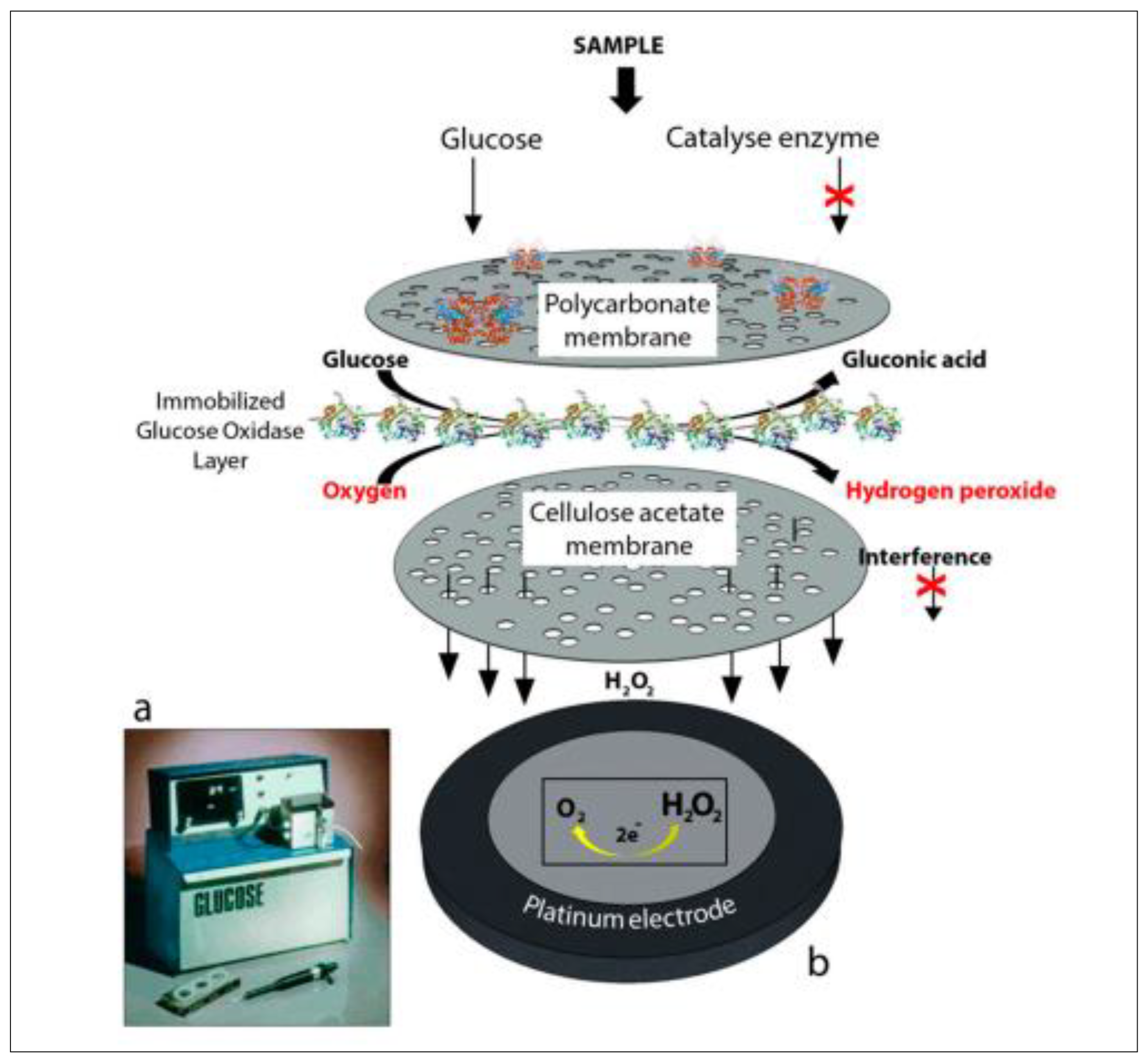
2.1.1. The Sensing Mechanism of the Enzymatic Glucose Sensor
2.1.2. The Sensing Mechanism of Non-Enzymatic Glucose Sensors
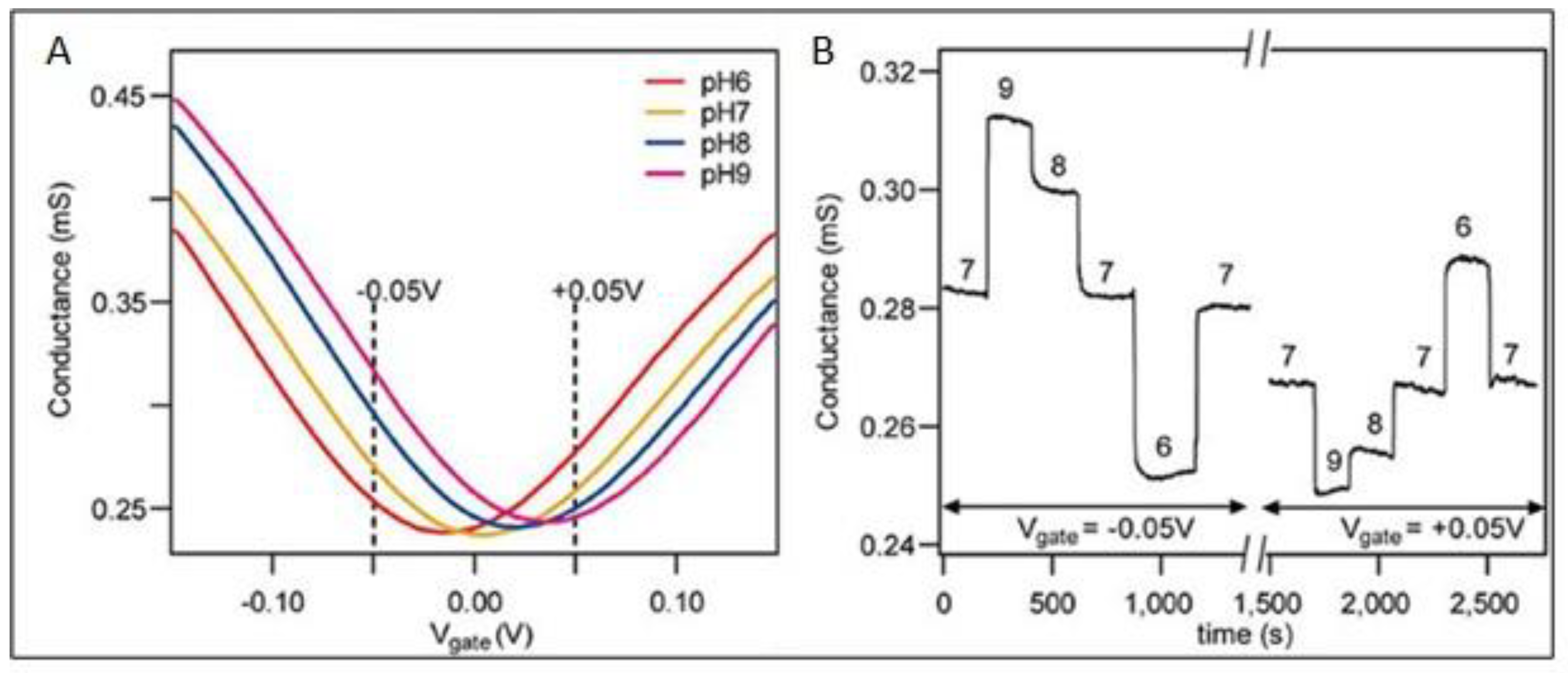
2.2. Factors Influencing the Electrochemical Glucose Sensing Performance and Monitoring
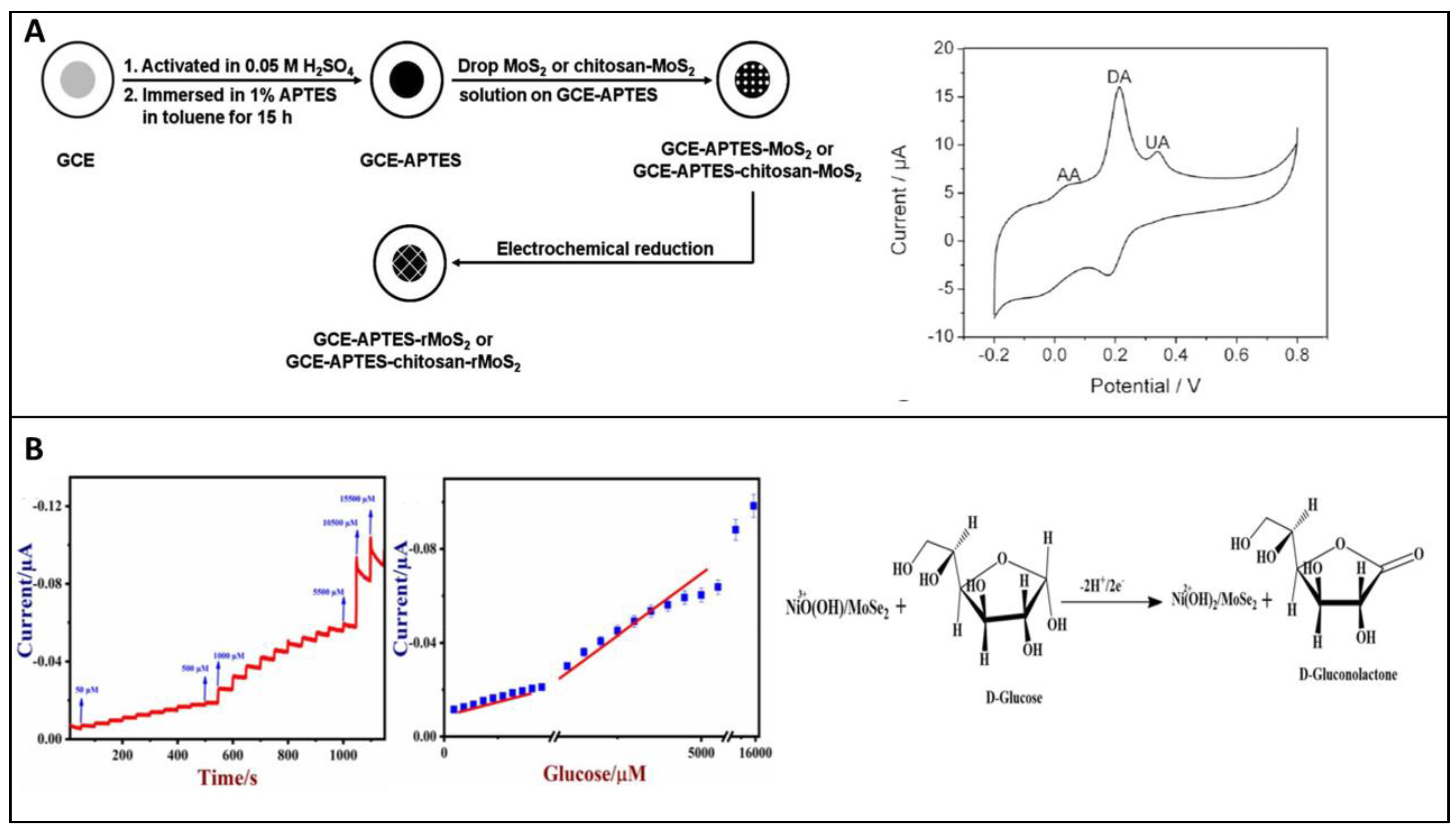
2.3. Fabrication Procedure
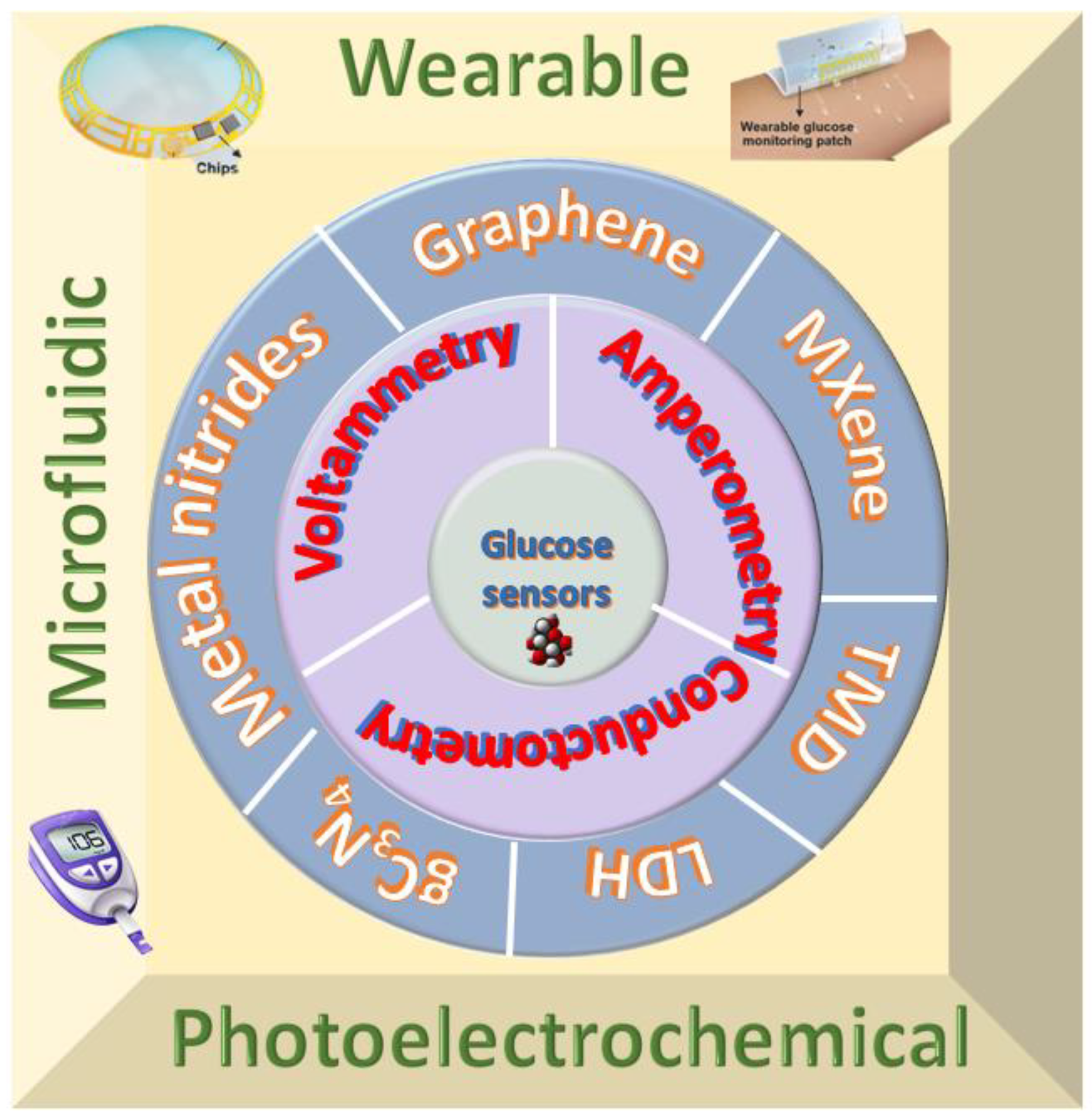
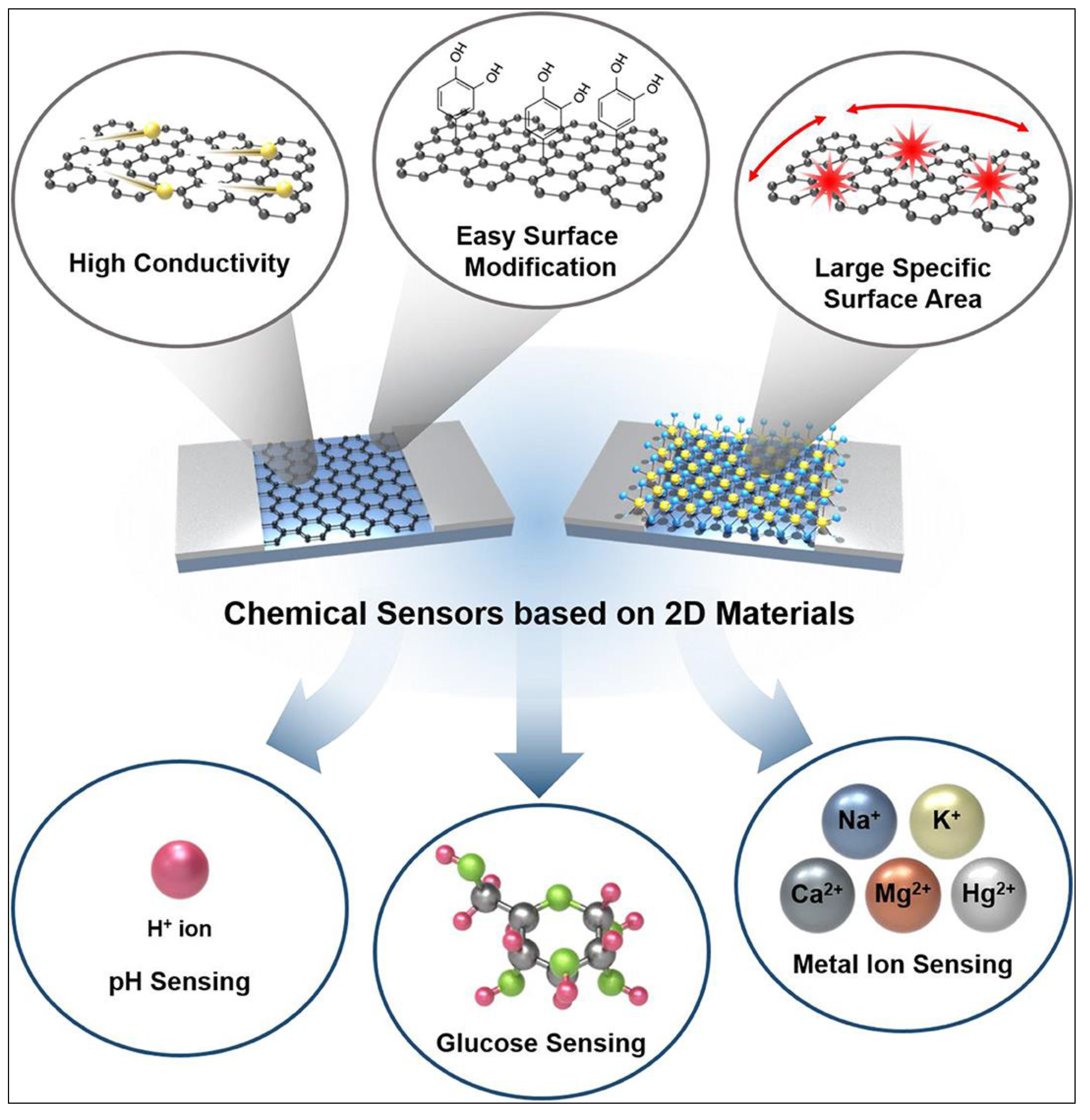
3. 2D Materials for Electrochemical Glucose Sensing
3.1. Graphene
3.1.1. Enzymatic Glucose Sensors Based on Graphene
3.1.2. Non-Enzymatic Glucose Sensors Based on Graphene
3.2. MXene
3.2.1. Enzymatic Glucose Sensors Based on MXene
3.2.2. Non-Enzymatic Glucose Sensors Based on MXene
3.3. TMD Based Electrochemical Glucose Sensors
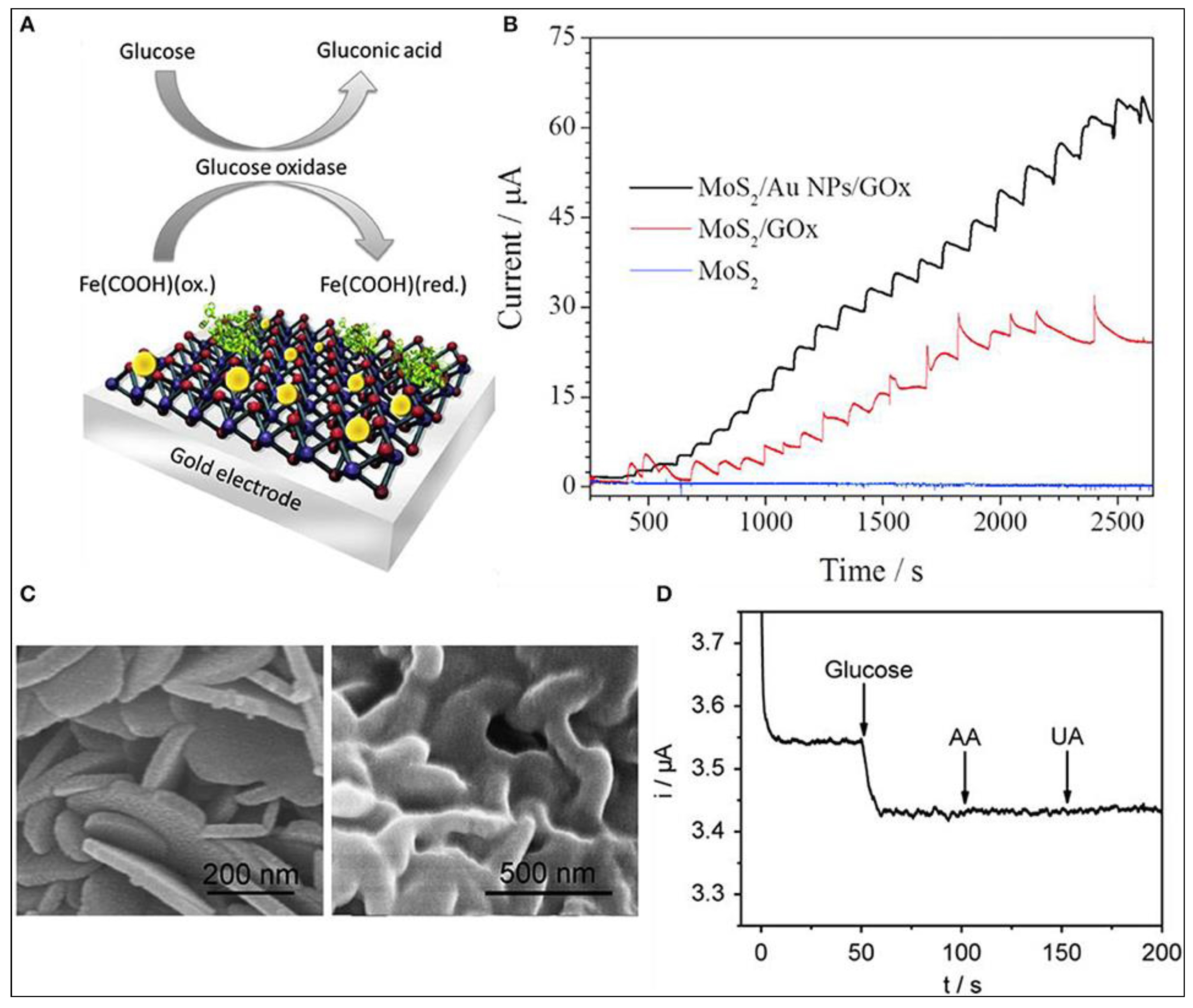
3.3.1. Enzymatic Glucose Sensors Based on TMD
3.3.2. Non-Enzymatic Glucose Sensors Based on TMD
3.4. Layered Double Hydroxides (LDHs)
3.4.1. Enzymatic Glucose Sensors Based on LDH
3.4.2. Non-Enzymatic Glucose Sensors Based on LDH
3.5. Other 2D Materials
4. Wearable and Flexible Electrochemical Glucose Sensors
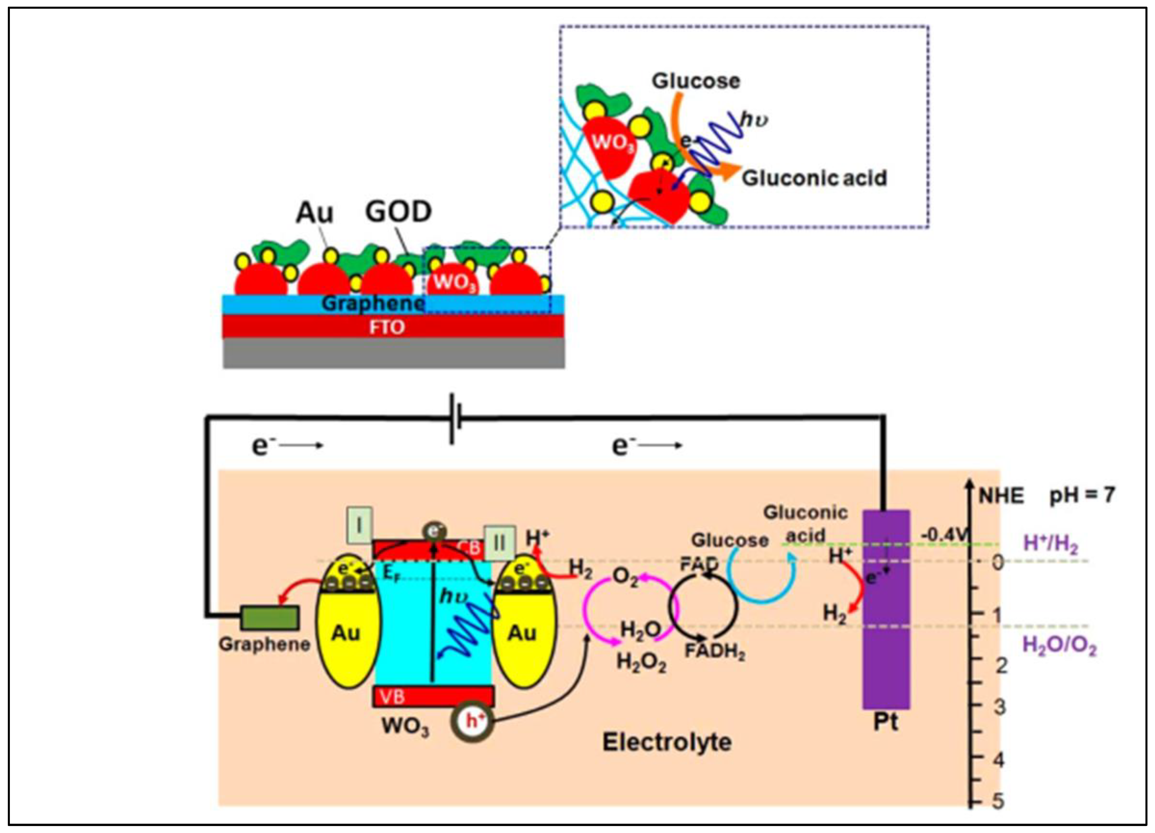
5. Photoelectrochemical Sensors
6. Theoretical Perspective for Glucose Sensing by 2D Materials
6.1. Theoretical Modelling
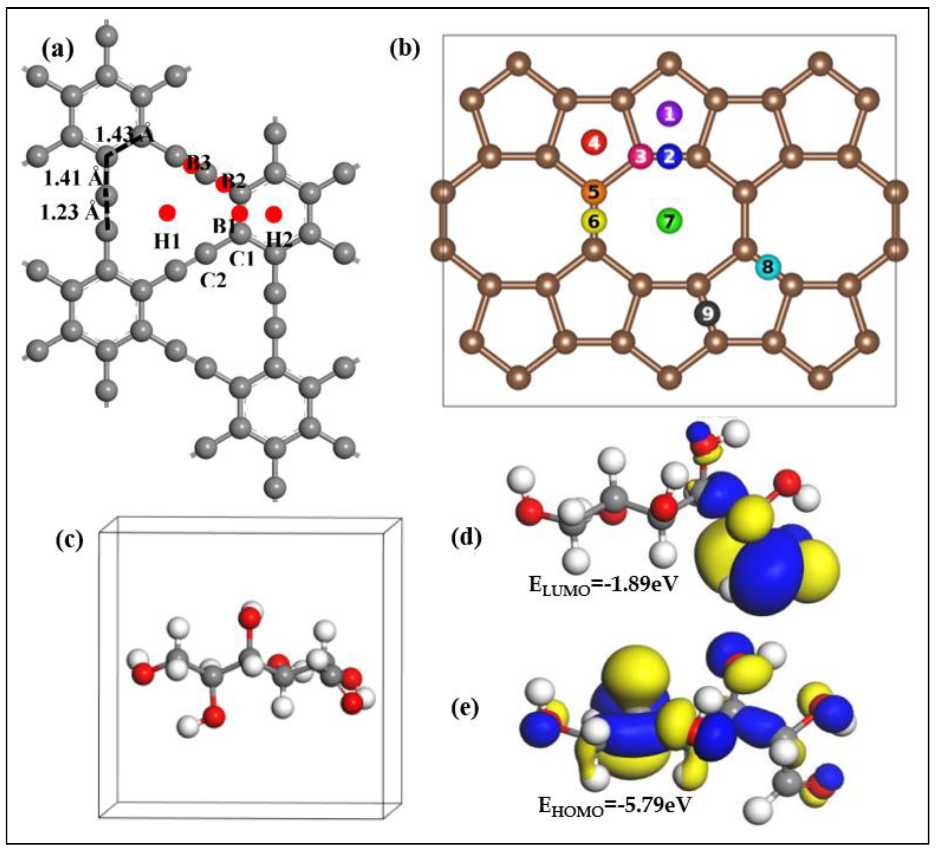
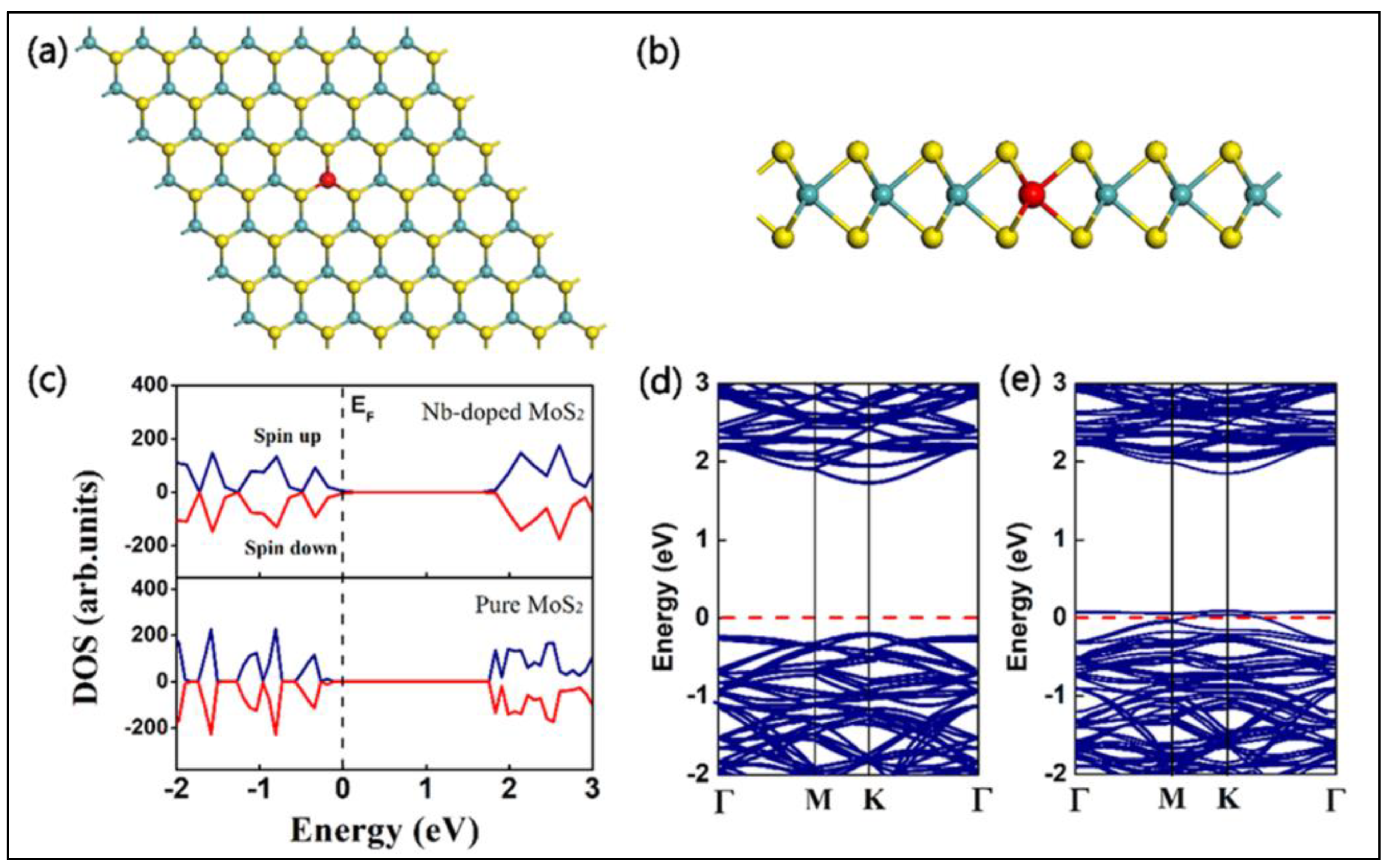
6.2. DFT Simulation of Glucose Sensing by 2D Materials
- i.
- Adsorption sites and configuration
- ii.
- Adsorption energy
- iii.
- Variations in the electronic properties
- iv.
- Charge transfer
- v.
- Orbital interactions
6.3. Theoretical Analysis of the Practical Feasibility of the Glucose Sensor
- (i)
- Thermal stability
- (ii)
- Sensitivity of glucose adsorption analysis based on the band structure
- (iii)
- Recovery time
7. Future Perspectives
- (1)
- Electron transfer between the current collector and the catalytic active sites is governed by the electrical conductivity of the catalyst. The use of conductive materials can promote interfacial contact by lowering interfacial charge resistance and overpotential. Because of their great charge mobility and strong electrical conductivity, 2D materials can be employed to accomplish this. High stability, little residual current, high capacitance, vast flexibility, and substantial robustness characterize 2D-material-based electrochemical glucose sensors.
- (2)
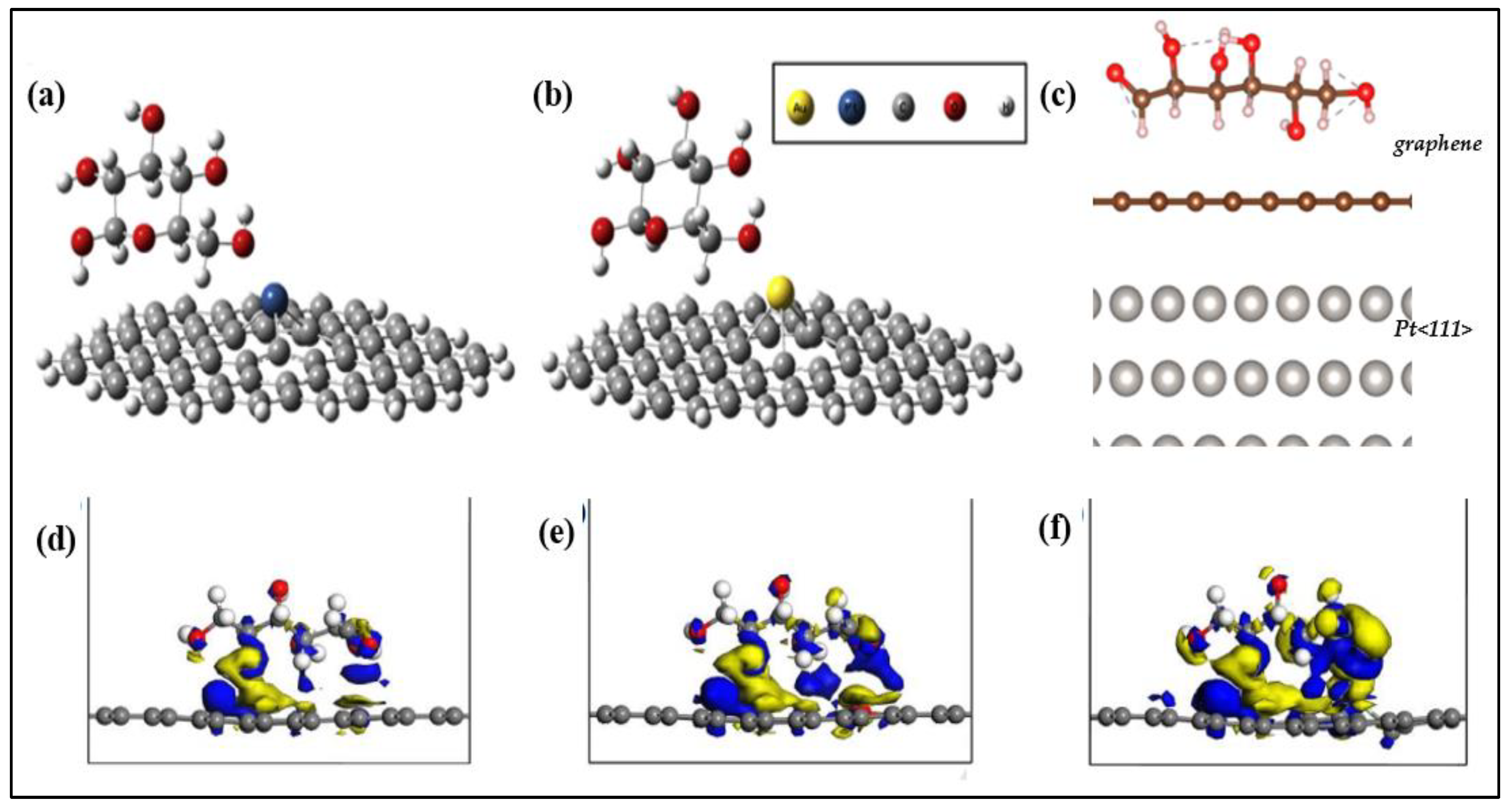
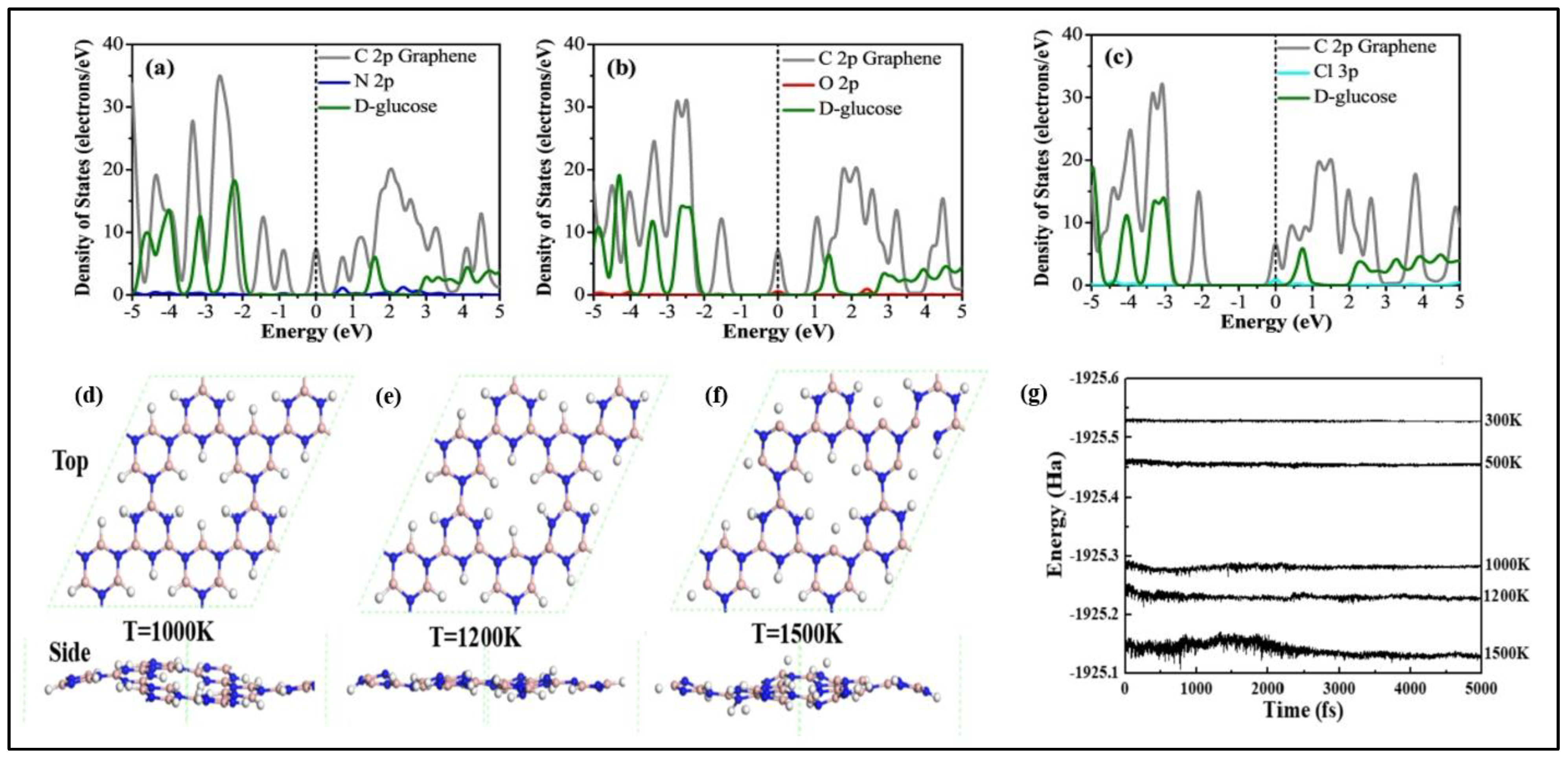
- Another important quality of a glucose sensor is judged by the stability, which is reliant on a number of parameters. Graphene and BN have the highest chemical and structural stability of any 2D material, whereas other 2D layered materials can react with water and oxygen. Black phosphorus (BP) has a high reaction rate when compared to other stacked 2D materials. As a result, publications based on BP for glucose detection are relatively rare. Gu et al. recently described an enzymatic biofuel cell based on BP and explored the BP mimicking enzyme properties [207]. Although certain physical and chemical modifications can be made to improve the stability of 2D materials, they nonetheless present numerous barriers and challenges. One major challenge is that existing solutions for enhancing the stability of 2D-material-based devices are still in the early stages of commercialization and manufacture [208].
- (3)
- Since glucose monitoring is connected with direct contact with real samples such as blood, urine, serum, and so on, the use of a biocompatible catalyst that inhibits biological reactions and cell damage is critical. Based on synthesis technique, structure, layer number and elemental composition, the compatibility of 2D materials may vary. For example, Tao et al. studied the biocompatibility of TMDs and found out that WSe2 shows high toxicity compared to WS2 and MoS2, which can be attributed to the toxic effects of Se. Similar studies were used to examine the role of the chalcogen atom in biocompatibility and found that VS2 is less toxic compared to VSe2 and VTe2. The biocompatibility of graphene and analogues has already been well-studied and find their place in glucose sensors. Aside from graphene and TMDs, the biocompatibility of additional 2D family members such as h-BN, MXenes, and black phosphorus is understudied. MXenes are quickly finding a place in healthcare applications; certain studies proved their biocompatibility. However, the biocompatibility of these electrochemical glucose sensors based on 2D materials should be further explored because in vivo devices must meet more stringent biocompatibility requirements than in vitro diagnostics.
- (4)
- The accurate detection of glucose in complicated physiological media (e.g., blood, interstitial fluid, or saliva) is critical for commercially viable biosensors, including those based on 2D materials. Because of the presence of non-target species in these media, there is an additional difficulty known as biofouling. When exposed to a biofluid, undesired molecules and other species (e.g., proteins, bacteria, platelets, etc.) might build on the sensor’s surface, interfering with and deteriorating its capabilities. Surprisingly, 2D materials can have antifouling capabilities by repelling hydrophilic interferents and so maintaining stability. In general, the electrooxidation of poisoning intermediates results in a large reduction in active surface area, which reduces sensitivity. Because of the huge surface area and edge-plane defect sites in 2D materials, this constraint can be efficiently eliminated. This needs a thorough assessment of the performance of these 2D-material-based biosensors in real-world matrices (beyond PBS or other buffers).
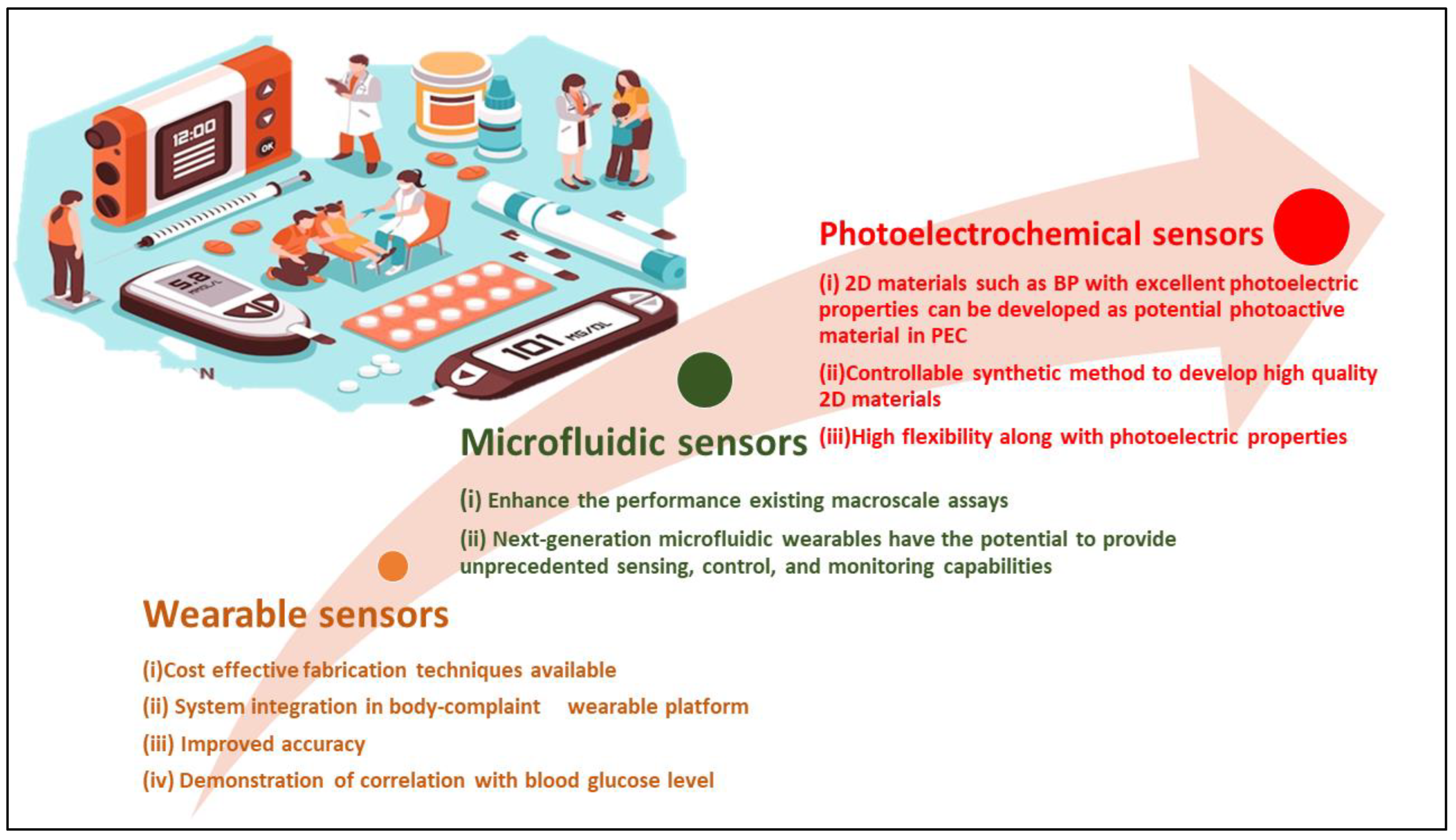
- (5)
- The use of 2D materials in wearable devices, particularly sweat monitoring platforms, has recently gained popularity. Such 2D-based platforms have demonstrated promising performance in sensing and self-powered energy harvesting devices. The use of cost-effective fabrication techniques (e.g., screen printing), improved reproducibility and accuracy of the measured glucose signal, system integration in body-compliant flexible wearable platforms, validating sensors with blood glucose levels, and the improved (stimulated) collection of non-invasive biofluids have all resulted in significant advances. Despite these exciting recent improvements, properly deploying non-invasive glucose monitoring systems requires additional tuning followed by large-scale validation studies to critically analyse the reliability and accuracy of these devices. These lengthy investigations will establish the potential of such non-invasive approaches to dependably monitor glucose and the association of these non-invasive generated data with gold-standard blood glucose readings. Other issues that need to be addressed in epidermal non-invasive sensing include irregular biofluid extraction, surface contamination, and the effect of some physiological factors (e.g., temperature, pH) on measurement accuracy. Despite these obstacles, the next decade will see a surge in activity in the development of non-invasive glucose monitoring systems based on 2D materials.
- (6)
- The incorporation of 2D materials into various semiconductor-based photoactive materials might increase their PEC sensing performance. Despite significant advances, there is still a long way to go in the research and implementation of 2D photoactive materials. Two-dimensional materials with a suitable band gap and strong optical absorption such as BP and 2D perovskites have not yet been reported. The lack of a suitable synthetic method also challenges the use of 2D materials in PEC sensing. However, the flexibility of ultrathin nanosheets, along with their strong photoelectric characteristics, makes 2D materials ideal for developing wearable PEC sensors for non-invasive health detection.
8. Conclusions
Author Contributions
Funding
Institutional Review Board Statement
Informed Consent Statement
Data Availability Statement
Acknowledgments
Conflicts of Interest
References
- Reddy, V.S.; Agarwal, B.; Ye, Z.; Zhang, C.; Roy, K.; Chinnappan, A.; Narayan, R.J.; Ramakrishna, S.; Ghosh, R. Recent Advancement in Biofluid-Based Glucose Sensors Using Invasive, Minimally Invasive, and Non-Invasive Technologies: A Review. Nanomaterials 2022, 12, 1082. [Google Scholar] [CrossRef] [PubMed]
- Sharma, P. Over 95,000 Children Suffer from Type-1 Diabetes: ICMR. Available online: https://www.livemint.com/science/health/over-95-000-children-suffer-from-type-1-diabetes-icmr-11654542643638.html (accessed on 16 June 2022).
- Witkowska Nery, E.; Kundys, M.; Jeleń, P.S.; Jönsson-Niedziółka, M. Electrochemical Glucose Sensing: Is There Still Room for Improvement? Anal. Chem. 2016, 88, 11271–11282. [Google Scholar] [CrossRef] [PubMed]
- Nava, C.; Modiano Hedenmalm, A.; Borys, F.; Hooft, L.; Bruschettini, M.; Jenniskens, K. Accuracy of Continuous Glucose Monitoring in Preterm Infants: A Systematic Review and Meta-Analysis. BMJ Open 2020, 10, e045335. [Google Scholar] [CrossRef] [PubMed]
- Reghunath, R.; devi, K.; Singh, K.K. Recent Advances in Graphene Based Electrochemical Glucose Sensor. Nano Struct. Nano Objects 2021, 26, 100750. [Google Scholar] [CrossRef]
- Danielsson, B.; Hedberg, U.; Rank, M.; Xie, B. Recent Investigations on Calorimetric Biosensors. Sens. Actuators B Chem. 1992, 6, 138–142. [Google Scholar] [CrossRef]
- Chen, Y.-T.; Kolhatkar, A.G.; Zenasni, O.; Xu, S.; Lee, T.R. Biosensing Using Magnetic Particle Detection Techniques. Sensors 2017, 17, 2300. [Google Scholar] [CrossRef]
- Damborský, P.; Švitel, J.; Katrlík, J. Optical Biosensors. Essays Biochem. 2016, 60, 91–100. [Google Scholar] [CrossRef] [Green Version]
- Pohanka, M. Overview of Piezoelectric Biosensors, Immunosensors and DNA Sensors and Their Applications. Materials 2018, 11, 448. [Google Scholar] [CrossRef] [Green Version]
- Zaidi, S.A.; Shin, J.H. Recent Developments in Nanostructure Based Electrochemical Glucose Sensors. Talanta 2016, 149, 30–42. [Google Scholar] [CrossRef]
- Teymourian, H.; Barfidokht, A.; Wang, J. Electrochemical Glucose Sensors in Diabetes Management: An Updated Review (2010–2020). Chem. Soc. Rev. 2020, 49, 7671–7709. [Google Scholar] [CrossRef]
- Castle, J.R.; Kenneth Ward, W. Amperometric Glucose Sensors: Sources of Error and Potential Benefit of Redundancy. J. Diabetes Sci. Technol. 2010, 4, 221–225. [Google Scholar] [CrossRef] [PubMed] [Green Version]
- Soldatkin, A.P.; El’skaya, A.V.; Shul’ga, A.A.; Jdanova, A.S.; Dzyadevich, S.V.; Jaffrezic-Renault, N.; Martelet, C.; Clechet, P. Glucose Sensitive Conductometric Biosensor with Additional Nafion Membrane: Reduction of Influence of Buffer Capacity on the Sensor Response and Extension of Its Dynamic Range. Anal. Chim. Acta 1994, 288, 197–203. [Google Scholar] [CrossRef]
- Walker, N.L.; Roshkolaeva, A.B.; Chapoval, A.I.; Dick, J.E. Recent Advances in Potentiometric Biosensing. Curr. Opin. Electrochem. 2021, 28, 100735. [Google Scholar] [CrossRef]
- Khan, R.; Radoi, A.; Rashid, S.; Hayat, A.; Vasilescu, A.; Andreescu, S. Two-Dimensional Nanostructures for Electrochemical Biosensor. Sensors 2021, 21, 3369. [Google Scholar] [CrossRef] [PubMed]
- Heller, A.; Feldman, B. Electrochemical Glucose Sensors and Their Applications in Diabetes Management. Chem. Rev. 2008, 108, 2482–2505. [Google Scholar] [CrossRef] [Green Version]
- Wang, J. Glucose Biosensors: 40 Years of Advances and Challenges. Electroanalysis 2001, 13, 983–988. [Google Scholar] [CrossRef]
- Zhang, C.; Zhang, Z.; Yang, Q.; Chen, W. Graphene-Based Electrochemical Glucose Sensors: Fabrication and Sensing Properties. Electroanalysis 2018, 30, 2504–2524. [Google Scholar] [CrossRef]
- Wu, I.-C.; Weng, Y.-H.; Lu, M.-Y.; Jen, C.-P.; Fedorov, V.E.; Chen, W.C.; Wu, M.T.; Kuo, C.-T.; Wang, H.-C. Nano-Structure ZnO/Cu2O Photoelectrochemical and Self-Powered Biosensor for Esophageal Cancer Cell Detection. Opt. Express OE 2017, 25, 7689–7706. [Google Scholar] [CrossRef]
- Shu, J.; Tang, D. Recent Advances in Photoelectrochemical Sensing: From Engineered Photoactive Materials to Sensing Devices and Detection Modes. Anal. Chem. 2020, 92, 363–377. [Google Scholar] [CrossRef]
- Graphene Market & 2D Materials Assessment 2021–2031. 2020. Available online: https://www.idtechex.com/zh/research-report/graphene-market-and-2d-materials-assessment-2021-2031/789 (accessed on 11 May 2022).
- Team Develops Novel Chemical Glucose Sensing Method Based on Boronic Acids and Graphene Foam. Available online: https://phys.org/news/2022-02-team-chemical-glucose-method-based.html (accessed on 16 June 2022).
- Hassan, M.H.; Vyas, C.; Grieve, B.; Bartolo, P. Recent advances in enzymatic and non-enzymatic electrochemical glucose sensing. Sensors 2021, 21, 4672. [Google Scholar] [CrossRef]
- Yoo, E.-H.; Lee, S.-Y. Glucose Biosensors: An Overview of Use in Clinical Practice. Sensors 2010, 10, 4558–4576. [Google Scholar] [CrossRef] [PubMed] [Green Version]
- Clark, L.C.; Lyons, C. Electrode Systems for Continuous Monitoring in Cardiovascular Surgery. Ann. N. Y. Acad. Sci. 1962, 102, 29–45. [Google Scholar] [CrossRef] [PubMed]
- D’Orazio, P. Biosensors in clinical chemistry. Clin. Chim. Acta 2003, 334, 41–69. [Google Scholar] [CrossRef]
- Newman, J.D.; Turner, A.P. Home blood glucose biosensors: A commercial perspective. Biosens. Bioelectron. 2005, 20, 2435–2453. [Google Scholar] [CrossRef] [Green Version]
- Juska, V.B.; Pemble, M.E. A Critical Review of Electrochemical Glucose Sensing: Evolution of Biosensor Platforms Based on Advanced Nanosystems. Sensors 2020, 20, 6013. [Google Scholar] [CrossRef]
- Matthews, D.R.; Holman, R.R.; Bown, E.; Steemson, J.; Watson, A.; Hughes, S.; Scott, D. Pen-sized digital 30-second blood glucose meter. Lancet 1987, 1, 778–779. [Google Scholar] [CrossRef]
- Ipekci, H.H.; Kazak, O.; Tor, A.; Uzunoglu, A. Tuning Active Sites of N-Doped Porous Carbon Catalysts Derived from Vinasse for High-Performance Electrochemical Sensing. Part. Sci. Technol. 2022, 1–12. [Google Scholar] [CrossRef]
- Quintero-Jaime, A.F.; Conzuelo, F.; Schuhmann, W.; Cazorla-Amorós, D.; Morallón, E. Multi-wall Carbon Nanotubes Electrochemically Modified with Phosphorus and Nitrogen Functionalities as a Basis for Bioelectrodes with Improved Performance. Electrochim. Acta 2021, 387, 138530. [Google Scholar] [CrossRef]
- Quintero-Jaime, A.F.; Conzuelo, F.; Cazorla-Amorós, D.; Morallón, E. Pyrroloquinoline Quinone-Dependent Glucose Dehydrogenase Bioelectrodes Based on One-Step Electrochemical Entrapment over Single-Wall Carbon Nanotubes. Talanta 2021, 232, 122386. [Google Scholar] [CrossRef]
- Jiang, D.; Chu, Z.; Peng, J.; Luo, J.; Mao, Y.; Yang, P.; Jin, W. One-Step Synthesis of Three-Dimensional Co(OH)2/RGO Nano-Flowers as Enzyme-Mimic Sensors for Glucose Detection. Electrochim. Acta 2018, 270, 147–155. [Google Scholar] [CrossRef]
- Xue, B.; Li, K.; Feng, L.; Lu, J.; Zhang, L. Graphene Wrapped Porous Co3O4/NiCo2O4 Double-Shelled Nanocages with Enhanced Electrocatalytic Performance for Glucose Sensor. Electrochim. Acta 2017, 239, 36–44. [Google Scholar] [CrossRef]
- Thatikayala, D.; Ponnamma, D.; Sadasivuni, K.K.; Cabibihan, J.J.; Al-Ali, A.K.; Malik, R.A.; Min, B. Progress of Advanced Nanomaterials in the Non-Enzymatic Electrochemical Sensing of Glucose and H2O2. Biosensors 2020, 10, 151. [Google Scholar] [CrossRef] [PubMed]
- Zhang, L.; Wang, J.; Liu, F.; Xiong, Y.; Liu, Z.; Jiang, D.; Pu, X. Rapid detection of blaNDM-1 in multidrug-resistant organisms using a novel electrochemical biosensor. RSC Adv. 2017, 7, 12576–12585. [Google Scholar] [CrossRef] [Green Version]
- Li, K.; Liu, W.; Ni, Y.; Li, D.; Lin, D.; Su, Z.; Wei, G. Technical Synthesis and Biomedical Applications of Graphene Quantum Dots. J. Mater. Chem. B 2017, 5, 4811–4826. [Google Scholar] [CrossRef]
- Mohammadniaei, M.; Yoon, J.; Lee, T.; Bharate, B.G.; Jo, J.; Lee, D.; Choi, J.W. Electrochemical Biosensor Composed of Silver Ion-Mediated DsDNA on Au-Encapsulated Bi2Se3 Nanoparticles for the Detection of H2O2 Released from Breast Cancer Cells. Small 2018, 14, 1703970. [Google Scholar] [CrossRef]
- Fu, Y.; An, Q.; Ni, R.; Zhang, Y.; Li, Y.; Ke, H. Preparation of Polyaniline-Encapsulated Carbon/Copper Composite Nanofibers for Detection of Polyphenol Pollutant. Colloids Surf. A Physicochem. Eng. Asp. 2018, 559, 289–296. [Google Scholar] [CrossRef]
- Wei, M.; Qiao, Y.; Zhao, H.; Liang, J.; Li, T.; Luo, Y.; Sun, X. Electrochemical non-enzymatic glucose sensors: Recent progress and perspectives. Chem. Commun. 2020, 56, 14553–14569. [Google Scholar] [CrossRef]
- Lee, C.W.; Suh, J.M.; Jang, H.W. Chemical Sensors Based on Two-Dimensional (2D) Materials for Selective Detection of Ions and Molecules in Liquid. Front. Chem. 2019, 7, 708. [Google Scholar] [CrossRef]
- Georgakilas, V.; Otyepka, M.; Bourlinos, A.B.; Chandra, V.; Kim, N.; Kemp, K.C.; Kim, K.S. Functionalization of Graphene: Covalent and Non-Covalent Approaches, Derivatives and Applications. Chem. Rev. 2012, 112, 6156–6214. [Google Scholar] [CrossRef]
- Moses, P.G.; Mortensen, J.J.; Lundqvist, B.I.; Nørskov, J.K. Density Functional Study of the Adsorption and van Der Waals Binding of Aromatic and Conjugated Compounds on the Basal Plane of MoS2. J. Chem. Phys. 2009, 130, 104709. [Google Scholar] [CrossRef] [Green Version]
- Berger, C.; Song, Z.; Li, X.; Wu, X.; Brown, N.; Naud, C.; Heer, W.A. Electronic Confinement and Coherence in Patterned Epitaxial Graphene. Science 2006, 312, 1191–1196. [Google Scholar] [CrossRef] [PubMed] [Green Version]
- Wang, Y.; Shao, Y.; Matson, D.W.; Li, J.; Lin, Y. Nitrogen-doped graphene and its application in electrochemical biosensing. ACS Nano 2010, 4, 1790–1798. [Google Scholar] [CrossRef] [PubMed]
- Luo, J.; Jiang, S.; Zhang, H.; Jiang, J.; Liu, X. A novel non-enzymatic glucose sensor based on Cu nanoparticle modified graphene sheets electrode. Analyt. Chim. Acta 2012, 709, 47–53. [Google Scholar] [CrossRef] [PubMed]
- Kwak, Y.H.; Choi, D.S.; Kim, Y.N.; Kim, H.; Yoon, D.H.; Ahn, S.-S.; Yang, J.-W.; Yang, W.S.; Seo, S. Flexible Glucose Sensor Using CVD-Grown Graphene-Based Field Effect Transistor. Biosens. Bioelectron. 2012, 37, 82–87. [Google Scholar] [CrossRef]
- Parlak, O.; ncel, A.; Uzun, L.; Turner, A.P.F.; Tiwari, A. Biosens. Bioelectron 2017, 89, 545–550. [Google Scholar] [CrossRef]
- Su, S.; Zhang, C.; Yuwen, L.; Liu, X.; Wang, L.; Fan, C.; Wang, L. Uniform Au@ Pt Core–Shell Nanodendrites Supported on Molybdenum Disulfide Nanosheets for the Methanol Oxidation Reaction. Nanoscale 2016, 8, 602–608. [Google Scholar] [CrossRef]
- Huang, J.; He, Y.; Jin, J.; Li, Y.; Dong, Z.; Li, R. A Novel Glucose Sensor Based on MoS2 Nanosheet Functionalized with Ni Nanoparticles. Electrochim. Acta 2014, 136, 41–46. [Google Scholar] [CrossRef]
- Lin, X.; Ni, Y.; Kokot, S. Electrochemical and Bio-Sensing Platform Based on a Novel 3D Cu Nano-Flowers/Layered MoS2 Composite. Biosens. Bioelectron. 2016, 79, 685–692. [Google Scholar] [CrossRef]
- Zhong, X.; Yang, H.; Guo, S.; Li, S.; Gou, G.; Niu, Z.; Ma, J. In Situ Growth of Ni–Fe Alloy on Graphene-like MoS2 for Catalysis of Hydrazine Oxidation. J. Mater. Chem. 2012, 22, 13925–13927. [Google Scholar] [CrossRef]
- Sun, Z.; Zhao, Q.; Zhang, G.; Li, Y.; Zhang, G.; Zhang, F.; Fan, X. Exfoliated MoS2 Supported Au–Pd Bimetallic Nanoparticles with Core–Shell Structures and Superior Peroxidase-like Activities. RSC Adv. 2015, 5, 10352–10357. [Google Scholar] [CrossRef]
- Li, J.; Yang, Z.; Tang, Y.; Zhang, Y.; Hu, X. Carbon nanotubes-nanoflake-like SnS2 nanocomposite for direct electrochemistry of glucose oxidase and glucose sensing. Biosens. Bioelectron. 2013, 41, 698–703. [Google Scholar] [CrossRef] [PubMed]
- Guo, C.X.; Li, C.M. Direct Electron Transfer of Glucose Oxidase and Biosensing of Glucose on Hollow Sphere-Nanostructured Conducting Polymer/Metal Oxide Composite. Phys. Chem. Chem. Phys. 2010, 12, 12153–12159. [Google Scholar] [CrossRef] [PubMed]
- Zhu, J.; Jiang, J.; Liu, J.; Ding, R.; Li, Y.; Ding, H.; Huang, X. CNT-Network Modified Ni Nanostructured Arrays for High Performance Non-Enzymatic Glucose Sensors. RSC Adv. 2011, 1, 1020–1025. [Google Scholar] [CrossRef]
- Ginsberg, B.H. Factors Affecting Blood Glucose Monitoring: Sources of Errors in Measurement. J. Diabetes Sci. Technol. 2009, 3, 903–913. [Google Scholar] [CrossRef] [Green Version]
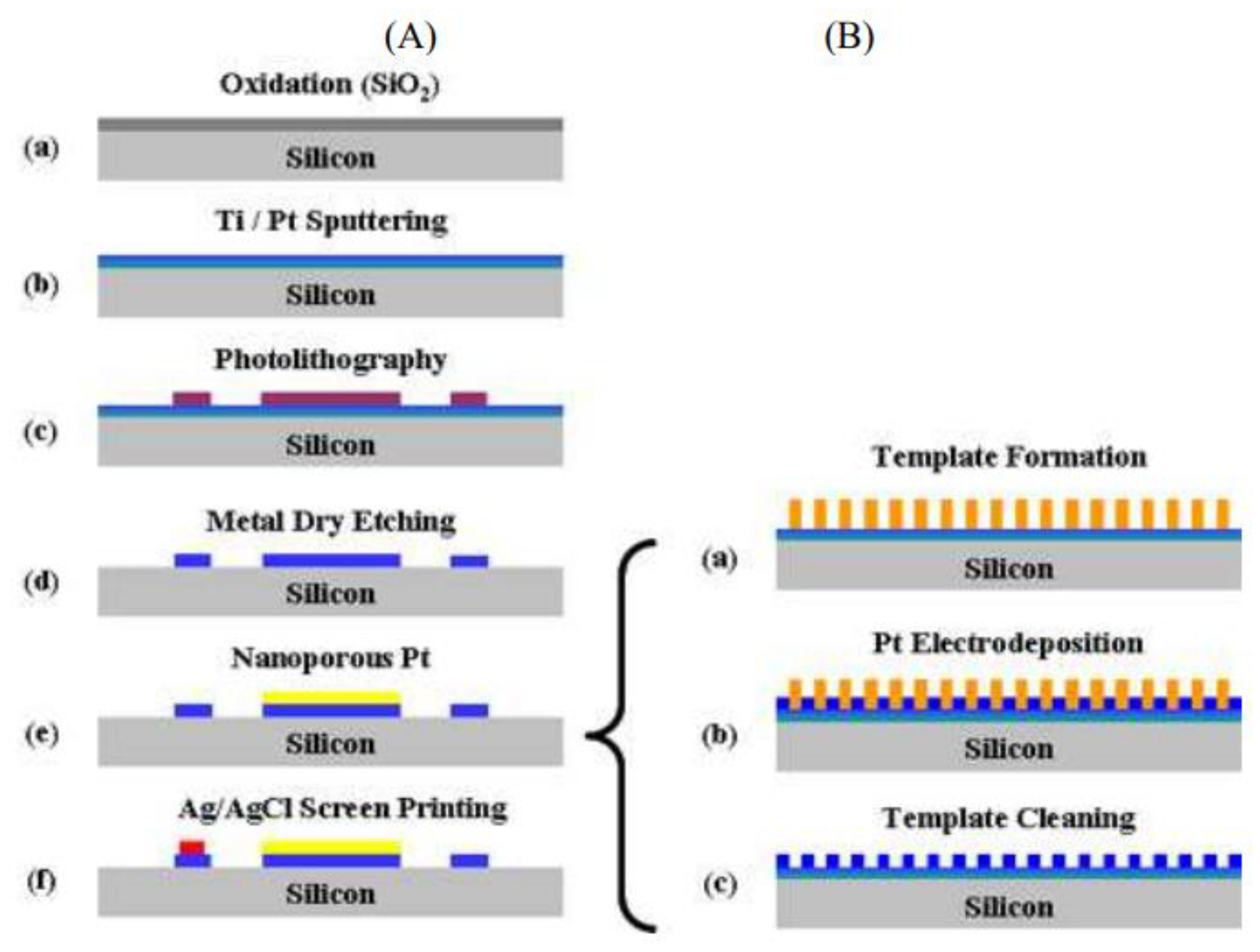
- Lee, Y.J.; Park, D.J.; Park, J.Y.; Kim, Y. Fabrication and Optimization of a Nanoporous Platinum Electrode and a Non-Enzymatic Glucose Micro-Sensor on Silicon. Sensors 2008, 8, 6154–6164. [Google Scholar] [CrossRef]
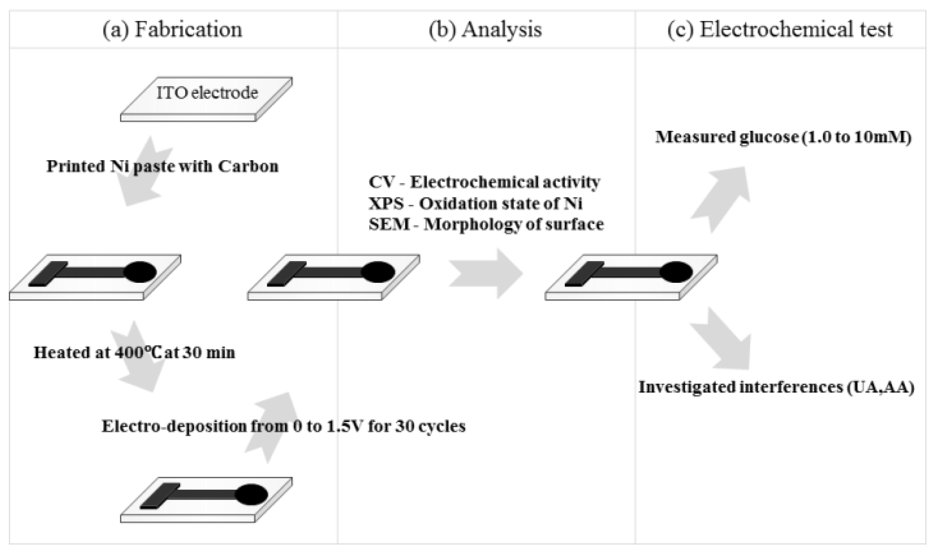
- Jeon, W.Y.; Choi, Y.B.; Kim, H.H. Disposable Non-Enzymatic Glucose Sensors Using Screen-Printed Nickel/Carbon Composites on Indium Tin Oxide Electrodes. Sensors 2015, 15, 31083–31091. [Google Scholar] [CrossRef]
- Yu, X.; Zhang, J.; Tang, X.; Wei, Y.; Kou, S.; Niu, J.; Yao, B. Preparation and Performance of Non-Enzymatic Glucose Sensor Electrode Based on Nanometer Cuprous Oxide. Nanomater. Nanotechnol. 2018, 8, 1847980418793526. [Google Scholar] [CrossRef] [Green Version]
- Gong, X.; Gu, Y.; Zhang, F.; Liu, Z.; Li, Y.; Chen, G.; Wang, B. High-Performance Non-Enzymatic Glucose Sensors Based on CoNiCu Alloy Nanotubes Arrays Prepared by Electrodeposition. Front. Mater. 2019, 6, 1–9. [Google Scholar] [CrossRef] [Green Version]
- Shabbir, S.A.; Tariq, S.; Ashiq, M.; Khan, W.A. Non-Enzymatic Glucose Sensor with Electrodeposited Silver/Carbon Nanotubes Composite Electrode. Biosci. Rep. 2019, 39, BSR20181983. [Google Scholar] [CrossRef] [Green Version]
- Amin, B.G.; Masud, J.; Nath, M. A Non-Enzymatic Glucose Sensor Based on a CoNi2Se4/RGO Nanocomposite with Ultrahigh Sensitivity at Low Working Potential. J. Mater. Chem. B 2019, 7, 2338–2348. [Google Scholar] [CrossRef]
- Hasanzadeh, M.; Hasanzadeh, Z.; Alizadeh, S.; Sayadi, M.; Nasiri Nezhad, M.; Sabzi, R.E.; Ahmadi, S. Copper-Nickel Oxide Nanofilm Modified Electrode for Non-Enzymatic Determination of Glucose. J. Electrochem. Sci. Eng. 2020, 10, 245–255. [Google Scholar] [CrossRef]
- More, K.D.; Dadge, J.W.; Khairnar, R.S.; Bogle, K.A. Development of Nano-TiO2/Al Electrode for Non-Enzymatic Glucose Bio-Sensing Application. AIP Conf. Proc. 2020, 2220, 020003. [Google Scholar]
- Espro, C.; Marini, S.; Giusi, D.; Ampelli, C.; Neri, G. Non-Enzymatic Screen Printed Sensor Based on Cu2O Nanocubes for Glucose Determination in Bio-Fermentation Processes. J. Electroanal. Chem. 2020, 873, 114354. [Google Scholar] [CrossRef]
- Baek, S.H.; Roh, J.; Park, C.Y.; Kim, M.W.; Shi, R.; Kailasa, S.K.; Park, T.J. Cu-Nanoflower Decorated Gold Nanoparticles-Graphene Oxide Nanofiber as Electrochemical Biosensor for Glucose Detection. Mater. Sci. Eng. C 2020, 107, 110273. [Google Scholar] [CrossRef] [PubMed]
- Mao, Q.; Jing, W.; Zhou, F.; Liu, S.; Gao, W.; Wei, Z.; Jiang, Z. Depositing Reduced Graphene Oxide on ZnO Nanorods to Improve the Performance of Enzymatic Glucose Sensors. Mater. Sci. Semicond. Process. 2021, 121, 105391. [Google Scholar] [CrossRef]
- Hossain, M.F.; Slaughter, G. PtNPs Decorated Chemically Derived Graphene and Carbon Nanotubes for Sensitive and Selective Glucose Biosensing. J. Electroanal. Chem. 2020, 861, 113990. [Google Scholar] [CrossRef]
- Sakr, M.A.; Elgammal, K.; Delin, A.; Serry, M. Performance-Enhanced Non-Enzymatic Glucose Sensor Based on Graphene-Heterostructure. Sensors 2020, 20, 145. [Google Scholar] [CrossRef] [Green Version]
- Scandurra, A.; Ruffino, F.; Sanzaro, S.; Grimaldi, M.G. Laser and Thermal Dewetting of Gold Layer onto Graphene Paper for Non-Enzymatic Electrochemical Detection of Glucose and Fructose. Sens. Actuators B Chem. 2019, 301, 127113. [Google Scholar] [CrossRef]
- Jothi, L.; Jayakumar, N.; Jaganathan, S.K.; Nageswaran, G. Ultrasensitive and Selective Non-Enzymatic Electrochemical Glucose Sensor Based on Hybrid Material of Graphene Nanosheets/Graphene Nanoribbons/Nickel Nanoparticle. Mater. Res. Bull. 2018, 98, 300–307. [Google Scholar] [CrossRef]
- Ren, Z.; Mao, H.; Luo, H.; Liu, Y. Glucose Sensor Based on Porous Ni by Using a Graphene Bottom Layer Combined with a Ni Middle Layer. Carbon 2019, 149, 609–617. [Google Scholar] [CrossRef]
- Deng, Z.-P.; Sun, Y.; Wang, Y.-C.; Gao, J.-D. A NiFe Alloy Reduced on Graphene Oxide for Electrochemical Nonenzymatic Glucose Sensing. Sensors 2018, 18, 3972. [Google Scholar] [CrossRef] [PubMed] [Green Version]
- Ayranci, R.; Demirkan, B.; Sen, B.; Şavk, A.; Ak, M.; Şen, F. Use of the Monodisperse Pt/Ni@rGO Nanocomposite Synthesized by Ultrasonic Hydroxide Assisted Reduction Method in Electrochemical Nonenzymatic Glucose Detection. Mater. Sci. Eng. C 2019, 99, 951–956. [Google Scholar] [CrossRef] [PubMed]
- Yan, X.; Gu, Y.; Li, C.; Zheng, B.; Li, Y.; Zhang, T.; Zhang, Z.; Yang, M. A Non-Enzymatic Glucose Sensor Based on the CuS Nanoflakes–Reduced Graphene Oxide Nanocomposite. Anal. Methods 2018, 10, 381–388. [Google Scholar] [CrossRef]
- Terán-Alcocer, Á.; Bravo-Plascencia, F.; Cevallos-Morillo, C.; Palma-Cando, A. Electrochemical Sensors Based on Conducting Polymers for the Aqueous Detection of Biologically Relevant Molecules. Nanomaterials 2021, 11, 252. [Google Scholar] [CrossRef]
- Lei, W.; Si, W.; Xu, Y.; Gu, Z.; Hao, Q. Conducting Polymer Composites with Graphene for Use in Chemical Sensors and Biosensors. Microchim. Acta 2014, 181, 707–722. [Google Scholar] [CrossRef]
- Muthusankar, E.; Ragupathy, D. Graphene/Poly(Aniline-Co-Diphenylamine) Nanohybrid for Ultrasensitive Electrochemical Glucose Sensor. Nano Struct. Nano Objects 2019, 20, 100390. [Google Scholar] [CrossRef]
- Deshmukh, M.A.; Kang, B.-C.; Ha, T.-J. Non-Enzymatic Electrochemical Glucose Sensors Based on Polyaniline/Reduced-Graphene-Oxide Nanocomposites Functionalized with Silver Nanoparticles. J. Mater. Chem. C 2020, 8, 5112–5123. [Google Scholar] [CrossRef]
- Eryiğit, M.; Çepni, E.; Kurt Urhan, B.; Öztürk Doğan, H.; Öznülüer Özer, T. Nonenzymatic Glucose Sensor Based on Poly(3,4-Ethylene Dioxythiophene)/Electroreduced Graphene Oxide Modified Gold Electrode. Synth. Met. 2020, 268, 116488. [Google Scholar] [CrossRef]
- Sivasankarapillai, V.S.; Sharma, T.S.K.; Hwa, K.-Y.; Wabaidur, S.M.; Angaiah, S.; Dhanusuraman, R. MXene Based Sensing Materials: Current Status and Future Perspectives. ES Energy Environ. 2022, 15, 4–14. [Google Scholar] [CrossRef]
- Kalambate, P.K.; Gadhari, N.S.; Li, X.; Rao, Z.; Navale, S.T.; Shen, Y.; Patil, V.R.; Huang, Y. Recent Advances in MXene–Based Electrochemical Sensors and Biosensors. TrAC Trends Anal. Chem. 2019, 120, 115643. [Google Scholar] [CrossRef]
- Wu, X.; Ma, P.; Sun, Y.; Du, F.; Song, D.; Xu, G. Application of MXene in Electrochemical Sensors: A Review. Electroanalysis 2021, 33, 1827–1851. [Google Scholar] [CrossRef]
- Mathew, M.; Rout, C.S. Electrochemical Biosensors Based on Ti3C2Tx MXene: Future Perspectives for on-Site Analysis. Curr. Opin. Electrochem. 2021, 30, 100782. [Google Scholar] [CrossRef]
- Zhu, J.; Ha, E.; Zhao, G.; Zhou, Y.; Huang, D.; Yue, G.; Hu, L.; Sun, N.; Wang, Y.; Lee, L.Y.S.; et al. Recent Advance in MXenes: A Promising 2D Material for Catalysis, Sensor and Chemical Adsorption. Coord. Chem. Rev. 2017, 352, 306–327. [Google Scholar] [CrossRef]
- Rakhi, R.B.; Nayak, P.; Xia, C.; Alshareef, H.N. Novel Amperometric Glucose Biosensor Based on MXene Nanocomposite. Sci. Rep. 2016, 6, 36422. [Google Scholar] [CrossRef] [PubMed] [Green Version]
- Gu, H.; Xing, Y.; Xiong, P.; Tang, H.; Li, C.; Chen, S.; Zeng, R.; Han, K.; Shi, G. Three-Dimensional Porous Ti3C2Tx MXene–Graphene Hybrid Films for Glucose Biosensing. ACS Appl. Nano Mater. 2019, 2, 6537–6545. [Google Scholar] [CrossRef]
- Wu, M.; Zhang, Q.; Fang, Y.; Deng, C.; Zhou, F.; Zhang, Y.; Wang, X.; Tang, Y.; Wang, Y. Polylysine-Modified MXene Nanosheets with Highly Loaded Glucose Oxidase as Cascade Nanoreactor for Glucose Decomposition and Electrochemical Sensing. J. Colloid Interface Sci. 2021, 586, 20–29. [Google Scholar] [CrossRef]
- Li, M.; Fang, L.; Zhou, H.; Wu, F.; Lu, Y.; Luo, H.; Zhang, Y.; Hu, B. Three-Dimensional Porous MXene/NiCo-LDH Composite for High Performance Non-Enzymatic Glucose Sensor. Appl. Surf. Sci. 2019, 495, 143554. [Google Scholar] [CrossRef]
- Gopal, T.S.; Jeong, S.K.; Alrebdi, T.A.; Pandiaraj, S.; Alodhayb, A.; Muthuramamoorthy, M. Andrews Nirmala Grace MXene-Based Composite Electrodes for Efficient Electrochemical Sensing of Glucose by Non-Enzymatic Method. Mater. Today Chem. 2022, 24, 100891. [Google Scholar] [CrossRef]
- Mphuthi, N.; Sikhwivhilu, L.; Ray, S.S. Functionalization of 2D MoS2 Nanosheets with Various Metal and Metal Oxide Nanostructures: Their Properties and Application in Electrochemical Sensors. Biosensors 2022, 12, 386. [Google Scholar] [CrossRef]
- SpringerLink. Direct Electrochemistry of Glucose Oxidase and a Biosensor for Glucose Based on a Glass Carbon Electrode Modified with MoS2 Nanosheets Decorated with Gold Nanoparticles. Available online: https://link.springer.com/article/10.1007/s00604-014-1178-9 (accessed on 23 April 2022).
- Jeong, J.-M.; Yang, M.; Kim, D.S.; Lee, T.J.; Choi, B.G.; Kim, D.H. High Performance Electrochemical Glucose Sensor Based on Three-Dimensional MoS2/Graphene Aerogel. J. Colloid Interface Sci. 2017, 506, 379–385. [Google Scholar] [CrossRef]
- Huang, J.; Dong, Z.; Li, Y.; Li, J.; Tang, W.; Yang, H.; Wang, J.; Bao, Y.; Jin, J.; Li, R. MoS2 nanosheet functionalized with Cu nanoparticles and its application for glucose detection. Mater. Res. Bull. 2013, 48, 4544–4547. [Google Scholar] [CrossRef]
- He, W.; Huang, Y.; Wu, J. Enzyme-Free Glucose Biosensors Based on MoS2 Nanocomposites. Nanoscale Res. Lett. 2020, 15, 60. [Google Scholar] [CrossRef] [PubMed]
- Geng, D.; Bo, X.; Guo, L. Ni-Doped Molybdenum Disulfide Nanoparticles Anchored on Reduced Graphene Oxide as Novel Electroactive Material for a Non-Enzymatic Glucose Sensor. Sens. Actuators B Chem. 2017, 244, 131–141. [Google Scholar] [CrossRef]
- Ji, S.; Yang, Z.; Zhang, C.; Miao, Y.-E.; Tjiu, W.W.; Pan, J.; Liu, T. Nonenzymatic Sensor for Glucose Based on a Glassy Carbon Electrode Modified with Ni(OH)2 Nanoparticles Grown on a Film of Molybdenum Sulfide. Microchim. Acta 2013, 180, 1127–1134. [Google Scholar] [CrossRef]
- Fang, L.; Wang, F.; Chen, Z.; Qiu, Y.; Zhai, T.; Hu, M.; Zhang, C.; Huang, K. Flower-like MoS2 Decorated with Cu2O Nanoparticles for Non-Enzymatic Amperometric Sensing of Glucose. Talanta 2017, 167, 593–599. [Google Scholar] [CrossRef]
- Wu, S.; Zeng, Z.; He, Q.; Wang, Z.; Wang, S.J.; Du, Y.; Yin, Z.; Sun, X.; Chen, W.; Zhang, H. Electrochemically Reduced Single-Layer MoS2 Nanosheets: Characterization, Properties, and Sensing Applications. Small 2012, 8, 2264–2270. [Google Scholar] [CrossRef]
- Scientific Reports. Fabrication of 2D-MoSe2 Incorporated NiO Nanorods Modified Electrode for Selective Detection of Glucose in Serum Samples. Available online: https://www.nature.com/articles/s41598-021-92620-2 (accessed on 24 April 2022).
- Mani, S.; Ramaraj, S.; Chen, S.-M.; Dinesh, B.; Chen, T.-W. Two-Dimensional Metal Chalcogenides Analogous NiSe2 Nanosheets and Its Efficient Electrocatalytic Performance towards Glucose Sensing. J. Colloid Interface Sci. 2017, 507, 378–385. [Google Scholar] [CrossRef]
- Wan, L.; Liu, J.; Huang, X.-J. Novel Magnetic Nickel Telluride Nanowires Decorated with Thorns: Synthesis and Their Intrinsic Peroxidase-like Activity for Detection of Glucose. Chem. Commun. 2014, 50, 13589–13591. [Google Scholar] [CrossRef]
- Farid, A.; Pan, L.; Usman, M.; Khan, I.A.; Khan, A.S.; ul Ahmad, A.; Javid, M. In-Situ Growth of Porous CoTe2 Nanosheets Array on 3D Nickel Foam for Highly Sensitive Binder-Free Non-Enzymatic Glucose Sensor. J. Alloys Compd. 2021, 861, 158642. [Google Scholar] [CrossRef]
- Colombari, M.; Ballarin, B.; Carpani, I.; Guadagnini, L.; Mignani, A.; Scavetta, E.; Tonelli, D. Glucose Biosensors Based on Electrodes Modified with Ferrocene Derivatives Intercalated into Mg/Al Layered Double Hydroxides. Electroanalysis 2007, 19, 2321–2327. [Google Scholar] [CrossRef]
- Shan, D.; Cosnier, S.; Mousty, C. HRP Wiring by Redox Active Layered Double Hydroxides: Application to the Mediated H2O2 Detection. Anal. Lett. 2003, 36, 909–922. [Google Scholar] [CrossRef]
- Shan, D.; Yao, W.; Xue, H. Electrochemical Study of Ferrocenemethanol-Modified Layered Double Hydroxides Composite Matrix: Application to Glucose Amperometric Biosensor. Biosens. Bioelectron. 2007, 23, 432–437. [Google Scholar] [CrossRef]
- Mousty, C.; Prevot, V. Hybrid and Biohybrid Layered Double Hydroxides for Electrochemical Analysis. Anal. Bioanal. Chem. 2013, 405, 3513–3524. [Google Scholar] [CrossRef] [PubMed]
- Shi, Q.; Han, E.; Shan, D.; Yao, W.; Xue, H. Development of a High Analytical Performance Amperometric Glucose Biosensor Based on Glucose Oxidase Immobilized in a Composite Matrix: Layered Double Hydroxides/Chitosan. Bioprocess Biosyst. Eng. 2008, 31, 519–526. [Google Scholar] [CrossRef] [PubMed]
- Tonelli, D.; Scavetta, E.; Giorgetti, M. Layered-Double-Hydroxide-Modified Electrodes: Electroanalytical Applications. Anal. Bioanal. Chem. 2013, 405, 603–615. [Google Scholar] [CrossRef]
- Zhang, Y.; Chen, X.; Wang, J.; Yang, W. The Direct Electrochemistry of Glucose Oxidase Based on Layered Double-Hydroxide Nanosheets. Electrochem. Solid State Lett. 2008, 11, F19. [Google Scholar] [CrossRef]
- Scavetta, E.; Ballarin, B.; Gazzano, M.; Tonelli, D. Electrochemical Behaviour of Thin Films of Co/Al Layered Double Hydroxide Prepared by Electrodeposition. Electrochim. Acta 2009, 54, 1027–1033. [Google Scholar] [CrossRef]
- Lu, Y.; Jiang, B.; Fang, L.; Fan, S.; Wu, F.; Hu, B.; Meng, F. Highly Sensitive Nonenzymatic Glucose Sensor Based on 3D Ultrathin NiFe Layered Double Hydroxide Nanosheets. Electroanalysis 2017, 29, 1755–1761. [Google Scholar] [CrossRef]
- Zhao, Z.; Sun, Y.; Song, J.; Li, Y.; Xie, Y.; Cui, H.; Gong, W.; Hu, J.; Chen, Y. Highly Sensitive Nonenzymetic Glucose Sensing Based on Multicomponent Hierarchical NiCo-LDH/CCCH/CuF Nanostructures. Sens. Actuators B Chem. 2021, 326, 128811. [Google Scholar] [CrossRef]
- Hai, B.; Zou, Y.-Q.; Guo, G.-B.; Wang, Z.-D.; Liu, Y.-Y.; Wang, X.-X.; Yan, H.; Ma, L.-T.; Bai, Y.-C. A Novel Strategy to Prepare LDH Networks Loaded Carbon Structure by C-MEMS Techniques for Glucose Detection. Chin. Chem. Lett. 2017, 28, 149–152. [Google Scholar] [CrossRef]
- Fu, S.; Fan, G.; Yang, L.; Li, F. Non-Enzymatic Glucose Sensor Based on Au Nanoparticles Decorated Ternary Ni-Al Layered Double Hydroxide/Single-Walled Carbon Nanotubes/Graphene Nanocomposite. Electrochim. Acta 2015, 152, 146–154. [Google Scholar] [CrossRef]
- Hai, B.; Wei, H. Facile Preparation of Three-Dimensional NiO Nanosheets Array on Carbon Sphere and Its High-Performance Nonenzymatic Glucose Detection. IOP Conf. Ser. Earth Environ. Sci. 2019, 300, 052037. [Google Scholar] [CrossRef]
- Asadian, E.; Shahrokhian, S.; Iraji Zad, A. Highly Sensitive Nonenzymetic Glucose Sensing Platform Based on MOF-Derived NiCo LDH Nanosheets/Graphene Nanoribbons Composite. J. Electroanal. Chem. 2018, 808, 114–123. [Google Scholar] [CrossRef]
- Shahrokhian, S.; Sanati, E.K.; Hosseini, H. Advanced On-Site Glucose Sensing Platform Based on a New Architecture of Free-Standing Hollow Cu(OH)2 Nanotubes Decorated with CoNi-LDH Nanosheets on Graphite Screen-Printed Electrode. Nanoscale 2019, 11, 12655–12671. [Google Scholar] [CrossRef] [PubMed]
- Balasubramanian, P.; He, S.-B.; Deng, H.-H.; Peng, H.-P.; Chen, W. Defects Engineered 2D Ultrathin Cobalt Hydroxide Nanosheets as Highly Efficient Electrocatalyst for Non-Enzymatic Electrochemical Sensing of Glucose and l-Cysteine. Sens. Actuators B Chem. 2020, 320, 128374. [Google Scholar] [CrossRef]
- Sahoo, R.K.; Das, A.; Samantaray, K.; Singh, S.K.; Mane, R.S.; Shin, H.-C.; Yun, J.M.; Kim, K.H. Electrochemical Glucose Sensing Characteristics of Two-Dimensional Faceted and Non-Faceted CuO Nanoribbons. CrystEngComm 2019, 21, 1607–1616. [Google Scholar] [CrossRef]
- Chia, H.L.; Mayorga-Martinez, C.C.; Gusmão, R.; Novotny, F.; Webster, R.D.; Pumera, M. A Highly Sensitive Enzyme-Less Glucose Sensor Based on Pnictogens and Silver Shell–Gold Core Nanorod Composites. Chem. Commun. 2020, 56, 7909–7912. [Google Scholar] [CrossRef]
- Aziz, A.; Asif, M.; Ashraf, G.; Azeem, M.; Majeed, I.; Ajmal, M.; Wang, J.; Liu, H. Advancements in Electrochemical Sensing of Hydrogen Peroxide, Glucose and Dopamine by Using 2D Nanoarchitectures of Layered Double Hydroxides or Metal Dichalcogenides. A Review. Microchim. Acta 2019, 186, 671. [Google Scholar] [CrossRef]
- Ping, J.; Fan, Z.; Sindoro, M.; Ying, Y.; Zhang, H. Recent Advances in Sensing Applications of Two-Dimensional Transition Metal Dichalcogenide Nanosheets and Their Composites. Adv. Funct. Mater. 2017, 27, 1605817. [Google Scholar] [CrossRef]
- Zhao, A.; Zhang, Z.; Zhang, P.; Xiao, S.; Wang, L.; Dong, Y.; Yuan, H.; Li, P.; Sun, Y.; Jiang, X.; et al. 3D Nanoporous Gold Scaffold Supported on Graphene Paper: Freestanding and Flexible Electrode with High Loading of Ultrafine PtCo Alloy Nanoparticles for Electrochemical Glucose Sensing. Anal. Chim. Acta 2016, 938, 63–71. [Google Scholar] [CrossRef]
- He, W.; Sun, Y.; Xi, J.; Abdurhman, A.A.M.; Ren, J.; Duan, H. Printing Graphene-Carbon Nanotube-Ionic Liquid Gel on Graphene Paper: Towards Flexible Electrodes with Efficient Loading of PtAu Alloy Nanoparticles for Electrochemical Sensing of Blood Glucose. Anal. Chim. Acta 2016, 903, 61–68. [Google Scholar] [CrossRef] [PubMed]
- Abellán-Llobregat, A.; Jeerapan, I.; Bandodkar, A.; Vidal, L.; Canals, A.; Wang, J.; Morallón, E. A Stretchable and Screen-Printed Electrochemical Sensor for Glucose Determination in Human Perspiration. Biosens. Bioelectron. 2017, 91, 885–891. [Google Scholar] [CrossRef] [PubMed] [Green Version]
- Xuan, X.; Yoon, H.S.; Park, J.Y. A Wearable Electrochemical Glucose Sensor Based on Simple and Low-Cost Fabrication Supported Micro-Patterned Reduced Graphene Oxide Nanocomposite Electrode on Flexible Substrate. Biosens. Bioelectron. 2018, 109, 75–82. [Google Scholar] [CrossRef] [PubMed]
- Cao, L.; Han, G.-C.; Xiao, H.; Chen, Z.; Fang, C. A Novel 3D Paper-Based Microfluidic Electrochemical Glucose Biosensor Based on RGO-TEPA/PB Sensitive Film. Anal. Chim. Acta 2020, 1096, 34–43. [Google Scholar] [CrossRef]
- Zhang, Y.; Li, N.; Xiang, Y.; Wang, D.; Zhang, P.; Wang, Y.; Lu, S.; Xu, R.; Zhao, J. A Flexible Non-Enzymatic Glucose Sensor Based on Copper Nanoparticles Anchored on Laser-Induced Graphene. Carbon 2020, 156, 506–513. [Google Scholar] [CrossRef]
- Lipani, L.; Dupont, B.G.R.; Doungmene, F.; Marken, F.; Tyrrell, R.M.; Guy, R.H.; Ilie, A. Non-Invasive, Transdermal, Path-Selective and Specific Glucose Monitoring via a Graphene-Based Platform. Nat. Nanotechnol. 2018, 13, 504–511. [Google Scholar] [CrossRef]
- Kim, J.; Kim, M.; Lee, M.-S.; Kim, K.; Ji, S.; Kim, Y.-T.; Park, J.; Na, K.; Bae, K.-H.; Kyun Kim, H.; et al. Wearable Smart Sensor Systems Integrated on Soft Contact Lenses for Wireless Ocular Diagnostics. Nat. Commun. 2017, 8, 14997. [Google Scholar] [CrossRef] [Green Version]
- Lee, H.; Choi, T.K.; Lee, Y.B.; Cho, H.R.; Ghaffari, R.; Wang, L.; Choi, H.J.; Chung, T.D.; Lu, N.; Hyeon, T.; et al. A Graphene-Based Electrochemical Device with Thermoresponsive Microneedles for Diabetes Monitoring and Therapy. Nat. Nanotechnol. 2016, 11, 566–572. [Google Scholar] [CrossRef]
- Lin, S.; Feng, W.; Miao, X.; Zhang, X.; Chen, S.; Chen, Y.; Wang, W.; Zhang, Y. A Flexible and Highly Sensitive Nonenzymatic Glucose Sensor Based on DVD-Laser Scribed Graphene Substrate. Biosens. Bioelectron. 2018, 110, 89–96. [Google Scholar] [CrossRef]
- Yoon, H.; Nah, J.; Kim, H.; Ko, S.; Sharifuzzaman, M.; Barman, S.C.; Xuan, X.; Kim, J.; Park, J.Y. A Chemically Modified Laser-Induced Porous Graphene Based Flexible and Ultrasensitive Electrochemical Biosensor for Sweat Glucose Detection. Sens. Actuators B Chem. 2020, 311, 127866. [Google Scholar] [CrossRef]
- Lei, Y.; Zhao, W.; Zhang, Y.; Jiang, Q.; He, J.-H.; Baeumner, A.J.; Wolfbeis, O.S.; Wang, Z.L.; Salama, K.N.; Alshareef, H.N. A MXene-Based Wearable Biosensor System for High-Performance In Vitro Perspiration Analysis. Small 2019, 15, 1901190. [Google Scholar] [CrossRef] [PubMed] [Green Version]
- Li, M.; Wang, L.; Liu, R.; Li, J.; Zhang, Q.; Shi, G.; Li, Y.; Hou, C.; Wang, H. A Highly Integrated Sensing Paper for Wearable Electrochemical Sweat Analysis. Biosens. Bioelectron. 2021, 174, 112828. [Google Scholar] [CrossRef] [PubMed]
- Myndrul, V.; Coy, E.; Babayevska, N.; Zahorodna, V.; Balitskyi, V.; Baginskiy, I.; Gogotsi, O.; Bechelany, M.; Giardi, M.T.; Iatsunskyi, I. MXene Nanoflakes Decorating ZnO Tetrapods for Enhanced Performance of Skin-Attachable Stretchable Enzymatic Electrochemical Glucose Sensor. Biosens. Bioelectron. 2022, 207, 114141. [Google Scholar] [CrossRef] [PubMed]
- Yoon, J.; Lee, S.N.; Shin, M.K.; Kim, H.-W.; Choi, H.K.; Lee, T.; Choi, J.-W. Flexible Electrochemical Glucose Biosensor Based on GOx/Gold/MoS2/Gold Nanofilm on the Polymer Electrode. Biosens. Bioelectron. 2019, 140, 111343. [Google Scholar] [CrossRef]
- Vishnu, N.; Sahatiya, P.; Kong, C.Y.; Badhulika, S. Large Area, One Step Synthesis of NiSe2 Films on Cellulose Paper for Glucose Monitoring in Bio-Mimicking Samples for Clinical Diagnostics. Nanotechnology 2019, 30, 355502. [Google Scholar] [CrossRef]
- Liu, S.; Hui, K.S.; Hui, K.N. Flower-like Copper Cobaltite Nanosheets on Graphite Paper as High-Performance Supercapacitor Electrodes and Enzymeless Glucose Sensors. ACS Appl. Mater. Interfaces 2016, 8, 3258–3267. [Google Scholar] [CrossRef]
- Wang, X.; Zheng, Y.; Yuan, J.; Shen, J.; Hu, J.; Wang, A.-J.; Wu, L.; Niu, L. Three-Dimensional NiCo Layered Double Hydroxide Nanosheets Array on Carbon Cloth, Facile Preparation and Its Application in Highly Sensitive Enzymeless Glucose Detection. Electrochim. Acta 2017, 224, 628–635. [Google Scholar] [CrossRef]
- Hai, B.; Zou, Y. Carbon Cloth Supported NiAl-Layered Double Hydroxides for Flexible Application and Highly Sensitive Electrochemical Sensors. Sens. Actuators B Chem. 2015, 208, 143–150. [Google Scholar] [CrossRef]
- Katz, E.; Bückmann, A.F.; Willner, I. Self-Powered Enzyme-Based Biosensors. J. Am. Chem. Soc. 2001, 123, 10752–10753. [Google Scholar] [CrossRef]
- Cho, E.; Mohammadifar, M.; Choi, S. A Single-Use, Self-Powered, Paper-Based Sensor Patch for Detection of Exercise-Induced Hypoglycemia. Micromachines 2017, 8, 265. [Google Scholar] [CrossRef] [Green Version]
- Qiu, Z.; Shu, J.; Tang, D. Near-Infrared-to-Ultraviolet Light-Mediated Photoelectrochemical Aptasensing Platform for Cancer Biomarker Based on Core–Shell NaYF4:Yb,Tm@TiO2 Upconversion Microrods. Anal. Chem. 2018, 90, 1021–1028. [Google Scholar] [CrossRef]
- Wang, S.; Li, S.; Wang, W.; Zhao, M.; Liu, J.; Feng, H.; Chen, Y.; Gu, Q.; Du, Y.; Hao, W. A Non-Enzymatic Photoelectrochemical Glucose Sensor Based on BiVO4 Electrode under Visible Light. Sens. Actuators B Chem. 2019, 291, 34–41. [Google Scholar] [CrossRef]
- Luo, Z.; Qi, Q.; Zhang, L.; Zeng, R.; Su, L.; Tang, D. Branched Polyethylenimine-Modified Upconversion Nanohybrid-Mediated Photoelectrochemical Immunoassay with Synergistic Effect of Dual-Purpose Copper Ions. Anal. Chem. 2019, 91, 4149–4156. [Google Scholar] [CrossRef] [PubMed]
- Wang, H.; Zhang, L.; Chen, Z.; Hu, J.; Li, S.; Wang, Z.; Liu, J.; Wang, X. Semiconductor Heterojunction Photocatalysts: Design, Construction, and Photocatalytic Performances. Chem. Soc. Rev. 2014, 43, 5234–5244. [Google Scholar] [CrossRef] [PubMed]
- Xu, H.; Chang, Y.; Shen, X.; Liu, Z.; Zhu, B.; Macharia, D.K.; Wang, Z.; Chen, Z.; Zhang, L. Construction of Ag/AgCl-CN Heterojunctions with Enhanced Photocatalytic Activities for Degrading Contaminants in Wastewater. J. Colloid Interface Sci. 2019, 543, 25–33. [Google Scholar] [CrossRef]
- Devadoss, A.; Sudhagar, P.; Das, S.; Lee, S.Y.; Terashima, C.; Nakata, K.; Fujishima, A.; Choi, W.; Kang, Y.S.; Paik, U. Synergistic Metal–Metal Oxide Nanoparticles Supported Electrocatalytic Graphene for Improved Photoelectrochemical Glucose Oxidation. ACS Appl. Mater. Interfaces 2014, 6, 4864–4871. [Google Scholar] [CrossRef]
- Zhang, X.; Xu, F.; Zhao, B.; Ji, X.; Yao, Y.; Wu, D.; Gao, Z.; Jiang, K. Synthesis of CdS Quantum Dots Decorated Graphene Nanosheets and Non-Enzymatic Photoelectrochemical Detection of Glucose. Electrochim. Acta 2014, 133, 615–622. [Google Scholar] [CrossRef]
- Ma, Z.; Li, S. A Novel Photoelectrochemical Glucose Sensor Based on Graphene-CdS Nanocomposites Decorated with CoOx Nanosheets. Int. J. Electrochem. Sci. 2019, 14, 11445–11455. [Google Scholar] [CrossRef]
- Jafari, F.; Salimi, A.; Navaee, A. Electrochemical and Photoelectrochemical Sensing of Dihydronicotinamide Adenine Dinucleotide and Glucose Based on Noncovalently Functionalized Reduced Graphene Oxide-Cadmium Sulfide Quantum Dots/Poly-Nile Blue Nanocomposite. Electroanalysis 2014, 26, 1782–1793. [Google Scholar] [CrossRef]
- Li, H.; Guo, C.; Liu, C.; Ge, L.; Li, F. Laser-Induced Graphene Hybrid Photoelectrode for Enhanced Photoelectrochemical Detection of Glucose. Analyst 2020, 145, 4041–4049. [Google Scholar] [CrossRef]
- Çakıroğlu, B.; Özacar, M. Photoelectrochemical and Non-Enzymatic Glucose Sensor Based on Modified Fehling’s Test by Using Ti/TiO2 NTs-RGO-Cu2O Electrode. J. Electrochem. Soc. 2019, 166, B728. [Google Scholar] [CrossRef]
- Zhao, C.; Jing, T.; Tian, J.; Guo, J.; Wu, M.; Shi, D.; Zhao, Z.; Guo, Z. Visible Light-Driven Photoelectrochemical Enzyme Biosensor Based on Reduced Graphene Oxide/Titania for Detection of Glucose. J. Nanostruct. Chem. 2022, 12, 193–205. [Google Scholar] [CrossRef]
- Gopalan, A.I.; Muthuchamy, N.; Lee, K.P. A Novel Bismuth Oxychloride-Graphene Hybrid Nanosheets Based Non-Enzymatic Photoelectrochemical Glucose Sensing Platform for High Performances. Biosens. Bioelectron. 2017, 89, 352–360. [Google Scholar] [CrossRef]
- Wu, Z.; Zhao, J.; Yin, Z.; Wang, X.; Li, Z.; Wang, X. Highly Sensitive Photoelectrochemical Detection of Glucose Based on BiOBr/TiO2 Nanotube Array p-n Heterojunction Nanocomposites. Sens. Actuators B Chem. 2020, 312, 127978. [Google Scholar] [CrossRef]
- Zhang, X.Y.; Liu, S.G.; Zhang, W.J.; Wang, X.H.; Han, L.; Ling, Y.; Li, N.B.; Luo, H.Q. Photoelectrochemical Platform for Glucose Sensing Based on G-C3N4/ZnIn2S4 Composites Coupled with Bi-Enzyme Cascade Catalytic in-Situ Precipitation. Sens. Actuators B Chem. 2019, 297, 126818. [Google Scholar] [CrossRef]
- Li, W.; Jiang, D.; Yan, P.; Dong, J.; Qian, J.; Chen, J.; Xu, L. Graphitic Carbon Nitride/α-Fe2O3 Heterostructures for Sensitive Photoelectrochemical Non-Enzymatic Glucose Sensor. Inorg. Chem. Commun. 2019, 106, 211–216. [Google Scholar] [CrossRef]
- Chen, D.; Jiang, D.; Du, X.; Zhou, L.; Huang, L.; Qian, J.; Liu, Q.; Hao, N.; Li, Y.; Wang, K. Engineering Efficient Charge Transfer Based on Ultrathin Graphite-like Carbon Nitride/WO3 Semiconductor Nanoheterostructures for Fabrication of High-Performances Non-Enzymatic Photoelectrochemical Glucose Sensor. Electrochim. Acta 2016, 215, 305–312. [Google Scholar] [CrossRef]
- Çakıroğlu, B.; Demirci, Y.C.; Gökgöz, E.; Özacar, M. A Photoelectrochemical Glucose and Lactose Biosensor Consisting of Gold Nanoparticles, MnO2 and g-C3N4 Decorated TiO2. Sens. Actuators B Chem. 2019, 282, 282–289. [Google Scholar] [CrossRef]
- Li, M.; Wang, H.; Wang, X.; Lu, Q.; Li, H.; Zhang, Y.; Yao, S. Ti3C2/Cu2O Heterostructure Based Signal-off Photoelectrochemical Sensor for High Sensitivity Detection of Glucose. Biosens. Bioelectron. 2019, 142, 111535. [Google Scholar] [CrossRef]
- Chen, G.; Wang, H.; Wei, X.; Wu, Y.; Gu, W.; Hu, L.; Xu, D.; Zhu, C. Efficient Z-Scheme Heterostructure Based on TiO2/Ti3C2Tx/Cu2O to Boost Photoelectrochemical Response for Ultrasensitive Biosensing. Sens. Actuators B Chem. 2020, 312, 127951. [Google Scholar] [CrossRef]
- Wu, S.; Huang, H.; Shang, M.; Du, C.; Wu, Y.; Song, W. High Visible Light Sensitive MoS2 Ultrathin Nanosheets for Photoelectrochemical Biosensing. Biosens. Bioelectron. 2017, 92, 646–653. [Google Scholar] [CrossRef] [PubMed]
- Fang, Y.; Cao, Y.; Chen, Q. Synthesis of an Ag2WO4/Ti3C2 Schottky composite by electrostatic traction and its photocatalytic activity. Ceram. Int. 2019, 45, 22298–22307, ISSN 0272-8842. [Google Scholar] [CrossRef]
- Lathe, A.; Ansari, A.; Badhe, R.; Palve, A.M.; Garje, S.S. Single-Step Production of a TiO2@MoS2 Heterostructure and Its Applications as a Supercapacitor Electrode and Photocatalyst for Reduction of Cr(VI) to Cr(III). ACS Omega 2021, 6, 13008–13014. [Google Scholar] [CrossRef] [PubMed]
- Samoson, K.; Thavarungkul, P.; Kanatharana, P.; Limbut, W. A Nonenzymatic Glucose Sensor Based on the Excellent Dispersion of a Graphene Oxide-Poly(Acrylic Acid)-Palladium Nanoparticle-Modified Screen-Printed Carbon Electrode. J. Electrochem. Soc. 2019, 166, B1079. [Google Scholar] [CrossRef]
- Ansari, S.; Cho, M. Simple and Large Scale Construction of MoS2-g-C3N4 Heterostructures Using Mechanochemistry for High Performance Electrochemical Supercapacitor and Visible Light Photocatalytic Applications. Sci. Rep. 2017, 7, 43055. [Google Scholar] [CrossRef]
- Fu, X.; Chen, Z.; Shen, S.; Xu, L.; Luo, Z. Highly Sensitive Nonenzymatic Glucose Sensor Based on Reduced Graphene Oxide/Ultrasmall Pt Nanowire Nanocomposites. Int. J. Electrochem. Sci. 2018, 13, 4817–4826. [Google Scholar] [CrossRef]
- Farhat, H.; Célier, J.; Forano, C.; Mousty, C. Evaluation of Hierarchical Glucose Oxidase/Co3Mn-CO3 LDH Modified Electrodes for Glucose Detection. Electrochim. Acta 2021, 376, 138050. [Google Scholar] [CrossRef]
- Çakıroğlu, B.; Özacar, M. A Photoelectrochemical Biosensor Fabricated using Hierarchically Structured Gold Nanoparticle and MoS2 on Tannic Acid Templated Mesoporous TiO2. Electroanalysis 2019, 32, 166–177. [Google Scholar] [CrossRef]
- Deepalakshmi, T.; Tran, D.T.; Kim, N.H.; Chong, K.T.; Lee, J.H. Nitrogen-Doped Graphene-Encapsulated Nickel Cobalt Nitride as a Highly Sensitive and Selective Electrode for Glucose and Hydrogen Peroxide Sensing Applications. ACS Appl. Mater. Interfaces 2018, 10, 35847–35858. [Google Scholar] [CrossRef]
- Xie, F.; Liu, T.; Xie, L.; Sun, X.; Luo, Y. Metallic Nickel Nitride Nanosheet: An Efficient Catalyst Electrode for Sensitive and Selective Non-Enzymatic Glucose Sensing. Sens. Actuators B Chem. 2018, 255, 2794–2799. [Google Scholar] [CrossRef]
- Chen, J.; Yin, H.; Zhou, J.; Gong, J.; Wang, L.; Zheng, Y.; Nie, Q. Non-Enzymatic Glucose Sensor Based on Nickel Nitride Decorated Nitrogen Doped Carbon Spheres (Ni3N/NCS) via Facile One Pot Nitridation Process. J. Alloys Compd. 2019, 797, 922–930. [Google Scholar] [CrossRef]
- Zheng, L.; Teng, F.; Ye, X.; Zheng, H.; Fang, X. Photo/Electrochemical Applications of Metal Sulfide/TiO2 Heterostructures. Adv. Energy Mater. 2019, 10, 1902355. [Google Scholar] [CrossRef]
- Makov, G.; Payne, M.C. Periodic Boundary Conditions in Ab Initio Calculations. Phys. Rev. B 1995, 51, 4014–4022. [Google Scholar] [CrossRef] [PubMed] [Green Version]
- Parr, R.G. Density Functional Theory. Annu. Rev. Phys. Chem. 1983, 34, 631–656. [Google Scholar] [CrossRef]
- Mooney, C.Z. Monte Carlo Simulation; Sage Publications, Inc.: Thousand Oaks, CA, USA, 1997; Volume 8, p. 103. ISBN 978-0-8039-5943-9. [Google Scholar]
- Slater, J.C. A Simplification of the Hartree-Fock Method. Phys. Rev. 1951, 81, 385–390. [Google Scholar] [CrossRef]
- Zhang, X.; Fang, R.; Chen, D.; Zhang, G. Using Pd-Doped γ-Graphyne to Detect Dissolved Gases in Transformer Oil: A Density Functional Theory Investigation. Nanomaterials 2019, 9, 1490. [Google Scholar] [CrossRef] [Green Version]
- Gehringer, D.; Dengg, T.; Popov, M.; Holec, D. Interactions between a H2 Molecule and Carbon Nanostructures: A DFT Study. C 2020, 6, 16. [Google Scholar] [CrossRef] [Green Version]
- Opoku, F.; Osikoya, A.O.; Dikio, E.D.; Govender, P.P. One-Step Synthesized 2D Heteroatom Doped Graphene for High Throughput Electrochemical Biosensing: A Combined Experimental and Computational Studies. Diam. Relat. Mater. 2019, 100, 107592. [Google Scholar] [CrossRef]
- Huang, Z.; Liu, H.; Hu, R.; Qiao, H.; Wang, H.; Liu, Y.; Qi, X.; Zhang, H. Structures, Properties and Application of 2D Monoelemental Materials (Xenes) as Graphene Analogues under Defect Engineering. Nano Today 2020, 35, 100906. [Google Scholar] [CrossRef]
- Mostufa, S.; Paul, A.K.; Chakrabarti, K. Detection of Hemoglobin in Blood and Urine Glucose Level Samples Using a Graphene-Coated SPR Based Biosensor. OSA Contin. 2021, 4, 2164–2176. [Google Scholar] [CrossRef]
- Chia, H.L.; Mayorga-Martinez, C.C.; Sofer, Z.; Lazar, P.; Webster, R.D.; Pumera, M. Vanadium Dopants: A Boon or a Bane for Molybdenum Dichalcogenides-Based Electrocatalysis Applications. Adv. Funct. Mater. 2021, 31, 2009083. [Google Scholar] [CrossRef]
- Zhang, Y.-H.; Chen, Y.-B.; Zhou, K.-G.; Liu, C.-H.; Zeng, J.; Zhang, H.-L.; Peng, Y. Improving Gas Sensing Properties of Graphene by Introducing Dopants and Defects: A First-Principles Study. Nanotechnology 2009, 20, 185504. [Google Scholar] [CrossRef] [PubMed] [Green Version]
- Li, M.; Yao, J.; Wu, X.; Zhang, S.; Xing, B.; Niu, X.; Yan, X.; Yu, Y.; Liu, Y.; Wang, Y. P-Type Doping in Large-Area Monolayer MoS2 by Chemical Vapor Deposition. ACS Appl. Mater. Interfaces 2020, 12, 6276–6282. [Google Scholar] [CrossRef] [PubMed]
- Grimme, S. Semiempirical GGA-type density functional constructed with a long-range dispersion correction. J. Comput. Chem. 2006, 27, 1787–1799. [Google Scholar] [CrossRef]
- Stampfl, C.; Van de Walle, C.G. Density-Functional Calculations for III-V Nitrides Using the Local-Density Approximation and the Generalized Gradient Approximation. Phys. Rev. B 1999, 59, 5521–5535. [Google Scholar] [CrossRef] [Green Version]
- Moellmann, J.; Grimme, S. DFT-D3 Study of Some Molecular Crystals. J. Phys. Chem. C 2014, 118, 7615–7621. [Google Scholar] [CrossRef]
- Ebadi, M.; Reisi-Vanani, A. Methanol and Carbon Monoxide Sensing and Capturing by Pristine and Ca-Decorated Graphdiyne: A DFT-D2 Study. Phys. E Low Dimens. Syst. Nanostruct. 2021, 125, 114425. [Google Scholar] [CrossRef]
- Caglar, A.; Düzenli, D.; Onal, I.; Tezsevin, I.; Sahin, O.; Kivrak, H. A Comparative Experimental and Density Functional Study of Glucose Adsorption and Electrooxidation on the Au-Graphene and Pt-Graphene Electrodes. Int. J. Hydrogen Energy 2020, 45, 490–500. [Google Scholar] [CrossRef]
- Panigrahi, P.; Sajjad, M.; Singh, D.; Hussain, T.; Andreas Larsson, J.; Ahuja, R.; Singh, N. Two-Dimensional Nitrogenated Holey Graphene (C2N) Monolayer Based Glucose Sensor for Diabetes Mellitus. Appl. Surf. Sci. 2022, 573, 151579. [Google Scholar] [CrossRef]
- Govindasamy, P.; Gunasekaran, S.; Srinivasan, S. Molecular Geometry, Conformational, Vibrational Spectroscopic, Molecular Orbital and Mulliken Charge Analysis of 2-Acetoxybenzoic Acid. Spectrochim. Acta Part A Mol. Biomol. Spectrosc. 2014, 130, 329–336. [Google Scholar] [CrossRef]
- Ertural, C.; Steinberg, S.; Dronskowski, R. Development of a Robust Tool to Extract Mulliken and Löwdin Charges from Plane Waves and Its Application to Solid-State Materials. RSC Adv. 2019, 9, 29821–29830. [Google Scholar] [CrossRef] [PubMed] [Green Version]
- Lakshmy, S.; Sanyal, G.; Vaidyanathan, A.; Joseph, S.; Kalarikkal, N.; Chakraborty, B. Catechol Detection in Pure and Transition Metal Decorated 2D MoS2: Acumens from Density Functional Theory Approaches. Appl. Surf. Sci. 2021, 562, 150216. [Google Scholar] [CrossRef]
- Vaidyanathan, A.; Lakshmy, S.; Sanyal, G.; Joseph, S.; Kalarikkal, N.; Chakraborty, B. Nitrobenzene Sensing in Pristine and Metal Doped 2D Dichalcogenide MoS2: Insights from Density Functional Theory Investigations. Appl. Surf. Sci. 2021, 550, 149395. [Google Scholar] [CrossRef]
- Sanyal, G.; Lakshmy, S.; Vaidyanathan, A.; Kalarikkal, N.; Chakraborty, B. Detection of Nitrobenzene in Pristine and Metal Decorated 2D Dichalcogenide VSe2: Perspectives from Density Functional Theory. Surf. Interfaces 2022, 29, 101816. [Google Scholar] [CrossRef]
- Yong, Y.; Gao, R.; Wang, X.; Yuan, X.; Hu, S.; Zhao, Z.; Li, X.; Kuang, Y. Highly sensitive and selective room-temperature gas sensors based on B6N6H6 monolayer for sensing SO2 and NH3: A first-principles study. Results Phys. 2022, 33, 105208. [Google Scholar] [CrossRef]
- Zhao, Z.; Yong, Y.; Hu, S.; Li, C.; Kuang, Y. Adsorption of Gas Molecules on a C3N Monolayer and the Implications for NO2 Sensors. AIP Adv. 2019, 9, 125308. [Google Scholar] [CrossRef] [Green Version]
- Ivády, V.; Armiento, R.; Szász, K.; Janzén, E.; Gali, A.; Abrikosov, I.A. Theoretical Unification of Hybrid-DFT and DFT + U Methods for the Treatment of Localized Orbitals. Phys. Rev. B 2014, 90, 035146. [Google Scholar] [CrossRef] [Green Version]
- Shishkin, M.; Marsman, M.; Kresse, G. Accurate Quasiparticle Spectra from Self-Consistent GW Calculations with Vertex Corrections. Phys. Rev. Lett. 2007, 99, 246403. [Google Scholar] [CrossRef] [Green Version]
- Deák, P.; Aradi, B.; Frauenheim, T.; Janzén, E.; Gali, A. Accurate Defect Levels Obtained from the HSE06 Range-Separated Hybrid Functional. Phys. Rev. B 2010, 81, 153203. [Google Scholar] [CrossRef] [Green Version]
- Yong, Y.; Cui, H.; Zhou, Q.; Su, X.; Kuang, Y.; Li, X. C2N Monolayer as NH3 and NO Sensors: A DFT Study. Appl. Surf. Sci. 2019, 487, 488–495. [Google Scholar] [CrossRef]
- ACS Sustainable Chemistry & Engineering. Glucose Dehydrogenase-like Nanozyme Based on Black Phosphorus Nanosheets for High-Performance Biofuel Cells. Available online: https://pubs.acs.org/doi/abs/10.1021/acssuschemeng.0c05743 (accessed on 9 May 2022).
- Wang, X.; Sun, Y.; Liu, K. Chemical and Structural Stability of 2D Layered Materials. 2D Mater. 2019, 6, 042001. [Google Scholar] [CrossRef]


| Pros | Cons |
|---|---|
| Low power requirements, linear output, and good resolution Excellent repeatability and accuracy Less expensive Fast response time with high sensitivity and low detection limit Multianalyte detection [11] | Narrow or limited temperature range Short or limited shelf life |
| Amperometry | Conductometry | Voltammetry | Potentiometry |
|---|---|---|---|
| Amperometry means the measurement of the current flow in a closed loop of cells using an excitation signal produced by the generator Advantages are
| Response of the conductometric enzyme biosensors is mainly due to protons generated by a biocatalytic reaction inside the layer of immobilized enzyme Advantages are
Not very specific (less selective) [13] | Here measurements are related to the recording of either the current–time or the current–voltage relationship by applying known potential varying between the WE and REF electrodes Advantages are Simplicity, sensitivity, speed, and low costs | In this technique, negligible bias current flows as the potential between a working electrode and a reference electrode is measured across some interface Advantages are
Not very specific (less selective) [14] |
| 2D Materials | Properties Required for Sensing |
|---|---|
| Graphene |
|
| MXene |
|
| TMDs |
|
| LDH |
|
| Electrode | Analayte | Linear Range | Stability with Response Current | Detection Limit | Ref | |
|---|---|---|---|---|---|---|
| Ni–MoS2 | Glucose | 0–2.4 mM | 96.6% after 4 weeks | 0.31 μ M | [50] | |
| Cu-nanoflower@AuNPs-GO NFs | Glucose | 0.001–0.1 mM | 91% after 20 days | 0.018 μM | [67] | |
| GOx/rGO/ZnO/Au/PET | Glucose | 0.1 to 12 mM | 90% after 10 scan cycle | 301.9 μM | [68] | |
| Au/CG/C@MWCNTs/PtNPs/GOx/nafion | Glucose | 0.5 to 13.5 mM | 91.5% after 4 weeks | 1.3 μM | [69] | |
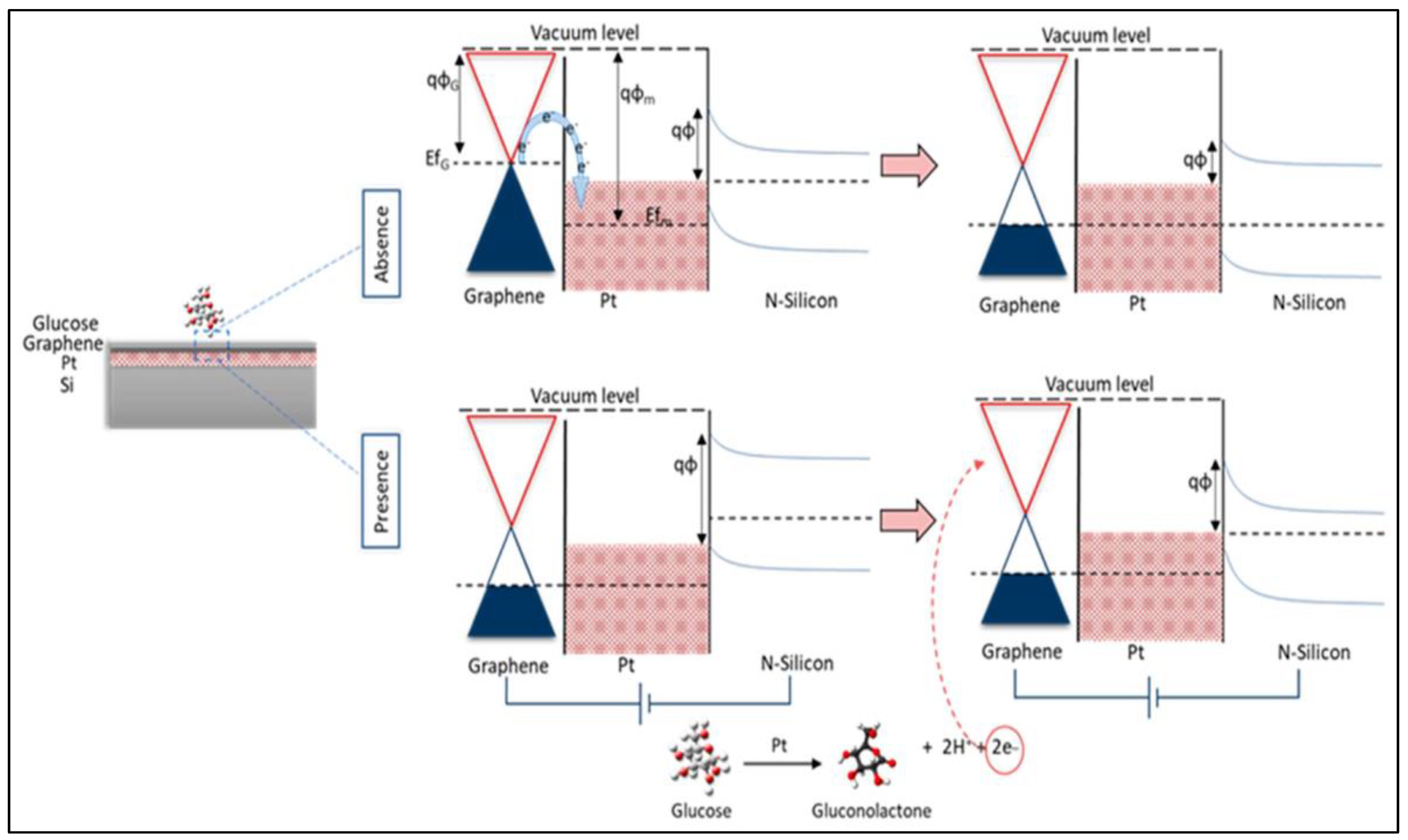
| Graphene (G)/platinum oxide (PtO)/n-silicon (Si) heterostructure | Glucose | 2–20 mM | - | Sensitivity around 30 μA/mM.cm2 | [70] | |
| Graphene paper/AuNPs laser dewetted | Glucose and fructose detection | 20 μM–8 mM | - | 2.5 μM | [71] | |
| GS/GNR/Ni | Glucose | 5 nM–5 mM | 91.3% after 30 days | 2.5 nM | [72] | |
| Cu/Ni-EG/pNi | Human blood serum | 0.0005–1.0 mM | 92% after 8 days | 6161 mA/mM−1 cm−2 | [73] | |
| NiFe/GO | Glucose | 0.05 to 5 mM | Recovery value close to 100% | 9 μM | [74] | |
| Pt/Ni@rGO | Glucose | 0.02–5.0 mM | 98% after 10 weeks | 6.3 μM | [75] | |
| rGO/CuSNFs/GCE | Glucose | 1 to 2000 µM | 97% after 30 days | 0.19 µM | [76] | |
| Gra-PANI-co-PDPA-ME | Glucose | 1 to 10 µM | 94% after 20 days | 0.1 µM | [79] | |
| Ag-PANI/rGO | Organic fluids (orange juice, apple juice, mango juice, Coke, and milk) | 50 μM to 0.1 μM | 91.3 after 30 days | 0.79 μM | [80] | |
| Au–PEDOT–ERGO | Glucose | 0.1–100 mM | 90% after 15 days | 0.12 μM | [81] | |
| GOx/Au/MXene/Nafion/GCE | Glucose | 0.1 to 18 mM | - | 5.9 μM | [87] | |
| Ti3C2Tx MXene–graphene (MG) hybrid | PBS | - | - | 0.10 mM in PBS and 0.13 mM in O2- saturated PBS | [88] | |
| Ti3C2/poly-L-lysine (PLL)/glucose oxidase (GOx) | Glucose | 4.0–20 µM and 0.02–1.1 mM | 85% after 100 cycles | 2.6 μM | [89] | |
| MXene/Nickel–Cobalt layered double hydroxide (NiCo-LDH) | Glucose | 0.002–4.096 mM | 92.5% after 15 days | 0.53 μM | [91] | |
| MXene–Cu2O | Glucose | 0.01–30 mM | - | 2.83 μM | [90] | |
| GOx/3D MGA/GC | Glucose | 2 to 20mM | - | 0.29 mM | [94] | |
| Cu–MoS2 hybrid | Glucose | Up to 4 mM | - | sensitivity 1055 µA mM−1 cm−2 | [95] | |
| Ni–MoS2/rGO | Glucose | 0.005–8.2 mM | 90% after 5 days | 2.7 μM | [97] | |
| Ni(OH)2/MoSx | Glucose | 10–1300 μM | 90% after 2 weeks | 5.8 μM | [98] | |
| Cu2O–MoS2/GCE | Glucose | 0.01 to 4.0 mM | 90% after 10 weeks | 1.0 μM | [99] | |
| MoSe2/NiO nanorod | Blood and serum | 50 µM to 15.5 mM | 91%after 50 cycles. | 0.6 µM | [101] | |
| Ni3Te2 | Glucose | 0.1–0.5 µM | - | 25 nM | [103] | |
| 3D CoTe2 | Glucose | - | 96% after one month | 0.59 μM | [104] | |
| Zn–Cr–ABTS LDH | Glucose | 1.3 10−8 –6.3 10−7 M | - | 10 nm | [106] | |
| LDHs/MeOHFc/GOD | Glucose | 6.7 × 10⁻⁶ to 3.86 × 10⁻⁴ M | - | 2.25 mM | [107] | |
| LDHs/CHT/GOD | Glucose | 1 × 10⁻⁶ to 3 × 10⁻³ M | 70% after 60 days | 0.1 µM | [109] | |
| NiFe–LDH/NF hybrid | Blood and serum | Up to 0.8 mM | - | 0.59 μM | [113] | |
| NiAl–LDH/CMC | Glucose | 0.2–18.6 mmol L−1 | - | 0.12 mmol L−1 | [115] | |
| Au/LDH-CNTs-G | Glucose | 10 μM to 6.1 mM | 95% after 30 days | 1.0 μM | [116] | |
| NiCo NSs/GNR-GCE | Glucose | 5 μM–0.8 mM 1–10 mM | 93% after 3 weeks | 0.6 μM | [118] | |
| NiCo-LDH/CCCH/CuF | Glucose | 0.001–1.5 mM | - | 0.68 μM | [114] | |
| Cu(OH)2@CoNi-LDH NT-NSs/GSPE | Glucose | 0.002–3.2 3.2–7.7 | - | 0.6 µM | [119] | |
| VCo-Co(OH)2 | Glucose | 0.4 μM–8.23 mM | - | 295 nM | [120] | |
| 2D CuO nanoribbon | Glucose | Up to 2 mM | Similar current response after 30 days | 58 µM | [121] | |
| Wearable electrochemical sensors | ||||||
| Ag/Pt @rGO | Sweat | 48 μA/mMcm2 | [128] | |||
| PtCo/NPG/GP | Blood | 35 μM–30 mM | 88% after 30 days | 5 μM | [125] | |
| GOx/PtNP/acetic acid-treated LIG | Sweat | 2.1 mM | - | 0.3 µM | [135] | |
| CNTs/Ti3C2Tx/PB/CFMs | Sweat | 10 × 10−6–22 × 10−3 M | - | Glucose | Lactate | [136] |
| 0.33 × 10⁻⁶ M | 0.67 × 10⁻⁶ M | |||||
| rGO-TEPA/PB | Human sweat and blood | 0.1–25 mM | 75.1% after 2 weeks | 25 μM | [130] | |
| Ti3C2Tx/MB | Sweat | 0.08–1.25 mM | - | Glucose | Lactate | [137] |
| 17.05 μM | 3.73 μM | |||||
| ZnO TPs/MXene | Sweat | 0.05–0.7 mM | - | 17.05 μM | [138] | |
| GOx/gold/MoS2/gold | Glucose | 500–100 nm | - | 10 nM | [139] | |
| NiCo LDH/CC | Glucose | 1 μM to 1.5 mM | 95% after one month | 0.12 μM | [142] | |
| NiSe2 | Glucose | 0.1–1 mM | - | 24.8 μM | [140] | |
| NiAl-LDH | Glucose | 1 to 329 µM | 94.7% after one month | 0.22 µM | [143] | |
| Photoelectrochemical sensor | ||||||
| Graphene–CdS hybrid | Glucose | 0.1∼4mmol dm−3 | - | 7 μmol dm-3 | [152] | |
| CoOx/graphene-CdS/GCE | Urine | 5–370 μM | - | 0.5 μM | [153] | |
| rGO-CdS QDs/PBA/P-NB/GDH | Glucose | Upto 200 μM | 95% response to NADH oxidation | <1mM | [154] | |
| LI-NiEC-CdS-G@ITO | Glucose | - | - | 0.4 μM | [155] | |
| Cu2O-rGO TiO2 NTs/Ti electrode | Glucose | 0.0007–20 mM | Photocurrent response retained upto 94% after 7 weeks | 0.21 μM | [156] | |
| rGO/TiO2 nanotube | Glucose | - | - | 5 μM | [157] | |
| BiOCl-G NHS | Human serum | 500 µM–10mM | 97% after 4 weeks | 0.22 mM | [158] | |
| BiOBr-TNTA | Human serum | 5 × 10⁻²–3 × 10⁷ nM | 95% after 4 weeks | 10 nM | [159] | |
| g-C3N4/ZnIn2S4 | Glucose | 1–10,000 μM | 0.28 μM | [160] | ||
| g-C3N4/WO3 | Glucose | 0.01~7.12 mM | 89.5% after 30 days | 0.1 μM | [162] | |
| g-C3N4/Fe2O3 | Glucose | g 0.1 to 11.5 mg L−1 | - | 0.03 mg/L | [161] | |
| ITO/TiO2-Au NPs- g-C3N4 -MnO2 | Glucose and lactose | 0.004–1.75 mM | 92.3% after 5 weeks | 0.12 µM | [163] | |
| Ti3C2 MXene/Cu2O | Human serum samples | - | - | 0.17 nM | [164] | |
| TiO2/Ti3C2/Cu2O | Human serum samples | - | - | 33.75 nM | [165] | |
| MoS2 ultrathin nanosheets | Glucose | - | 98% | 0.61 nmol/L | [166] | |
| Other glucose sensors | ||||||
| rGO-PtNW | Glucose | 0.032–1.89 mmol/L | - | 4.6 μmol/L | [171] | |
| GOx/Co3Mn-CO3/CF | Glucose | - | - | 0.02 mM | [172] | |
| NiCo2N/N-doped graphene | Glucose | 2.008 µM to 7.15 mM | 98.52% | 0.05 µM | [174] | |
| Ni3N nanosheets | Glucose | 0.2 μM to 1.5 mM | 91.3% after one month | 0.06 µM | [175] | |
| Ni3N/NCS | Glucose | 1 μM–3000 μM and 3000 μM–7000 μM | - | 0.1 μM and 0.35 μM | [176] | |
| System | Adsorption Energy (Ev) | Bond Distance (Å) | Charge Transfer ∆q (E) | Ref. |
|---|---|---|---|---|
| Graphene + Au + glucose | −0.71 | Au—O = 2.28 | −0.21 | [194] |
| Graphene + Pt + glucose | −0.38 | Pt—O = 2.39 | −0.17 | [194] |
| C2N + glucose | −0.93 (gas phase) −1.32 (aq. phase) | −0.01 | [195] | |
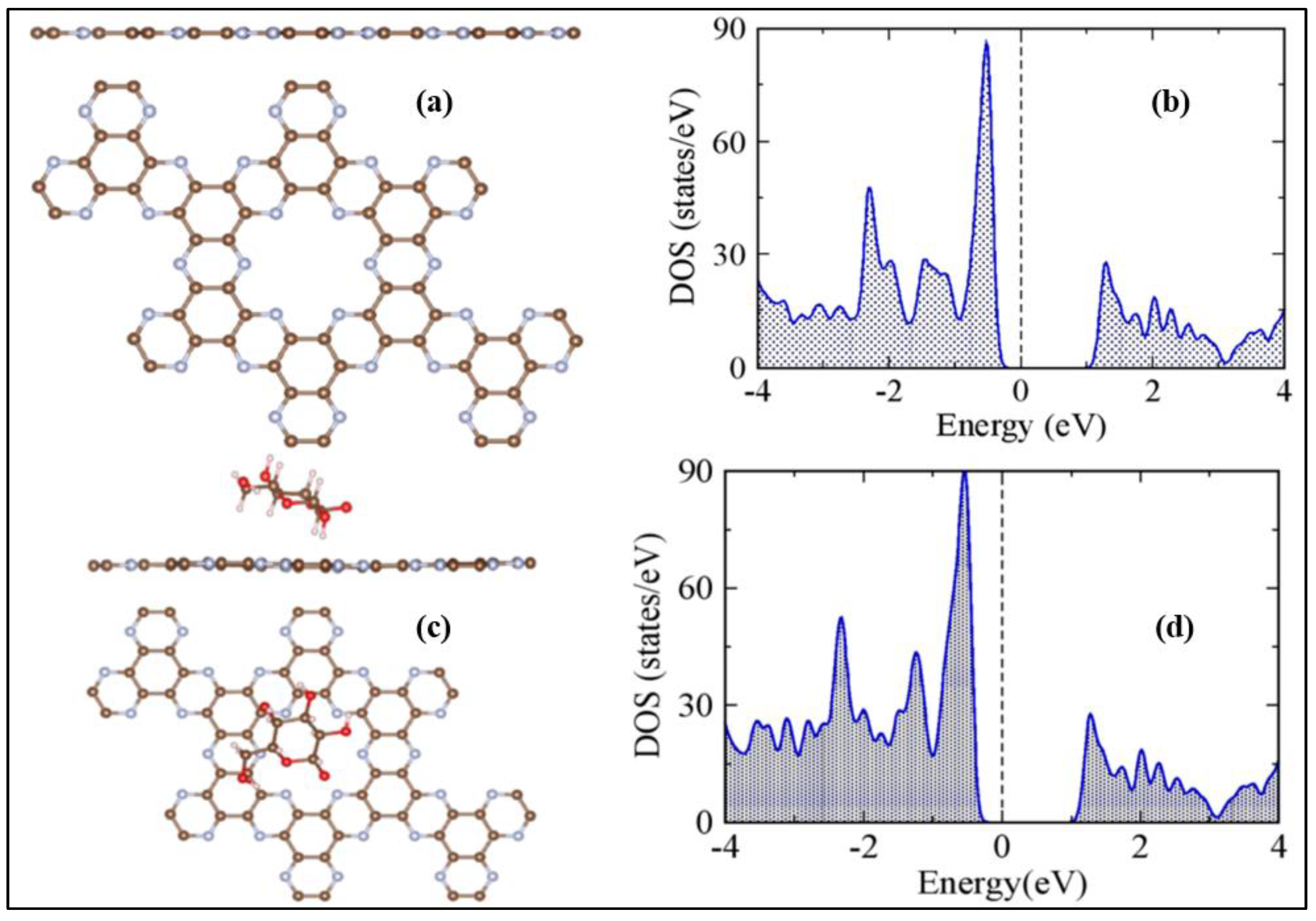
| HGr + Cl + glucose | −1.86 | Cl—O = 2.850 H—C = 1.746 | −0.20 | [184] |
| HGr + N + glucose | −1.69 | N—O = 3.372 H—C = 2.642 | −0.08 | [184] |
| HGr + O + glucose | −1.73 | O—O = 2.926 H—C = 2.371 | −0.12 | [184] |
Publisher’s Note: MDPI stays neutral with regard to jurisdictional claims in published maps and institutional affiliations. |
© 2022 by the authors. Licensee MDPI, Basel, Switzerland. This article is an open access article distributed under the terms and conditions of the Creative Commons Attribution (CC BY) license (https://creativecommons.org/licenses/by/4.0/).
Share and Cite
Radhakrishnan, S.; Lakshmy, S.; Santhosh, S.; Kalarikkal, N.; Chakraborty, B.; Rout, C.S. Recent Developments and Future Perspective on Electrochemical Glucose Sensors Based on 2D Materials. Biosensors 2022, 12, 467. https://doi.org/10.3390/bios12070467
Radhakrishnan S, Lakshmy S, Santhosh S, Kalarikkal N, Chakraborty B, Rout CS. Recent Developments and Future Perspective on Electrochemical Glucose Sensors Based on 2D Materials. Biosensors. 2022; 12(7):467. https://doi.org/10.3390/bios12070467
Chicago/Turabian StyleRadhakrishnan, Sithara, Seetha Lakshmy, Shilpa Santhosh, Nandakumar Kalarikkal, Brahmananda Chakraborty, and Chandra Sekhar Rout. 2022. "Recent Developments and Future Perspective on Electrochemical Glucose Sensors Based on 2D Materials" Biosensors 12, no. 7: 467. https://doi.org/10.3390/bios12070467