Novel Optical Fiber-Based Structures for Plasmonics Sensors

 , , ,
, , , 
Abstract
:1. Introduction
2. Processing Technology
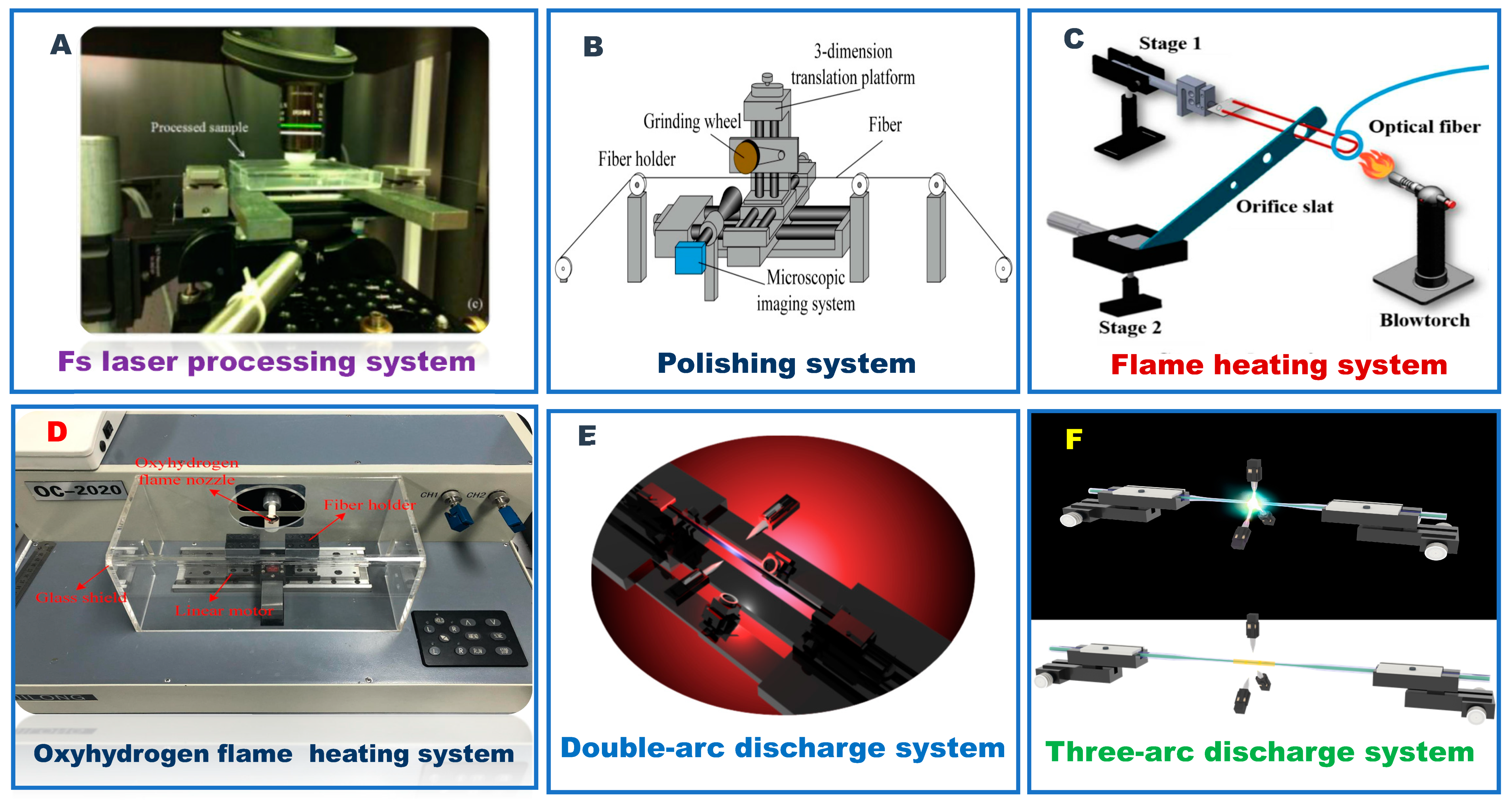
2.1. Optical Fiber Processing Technology
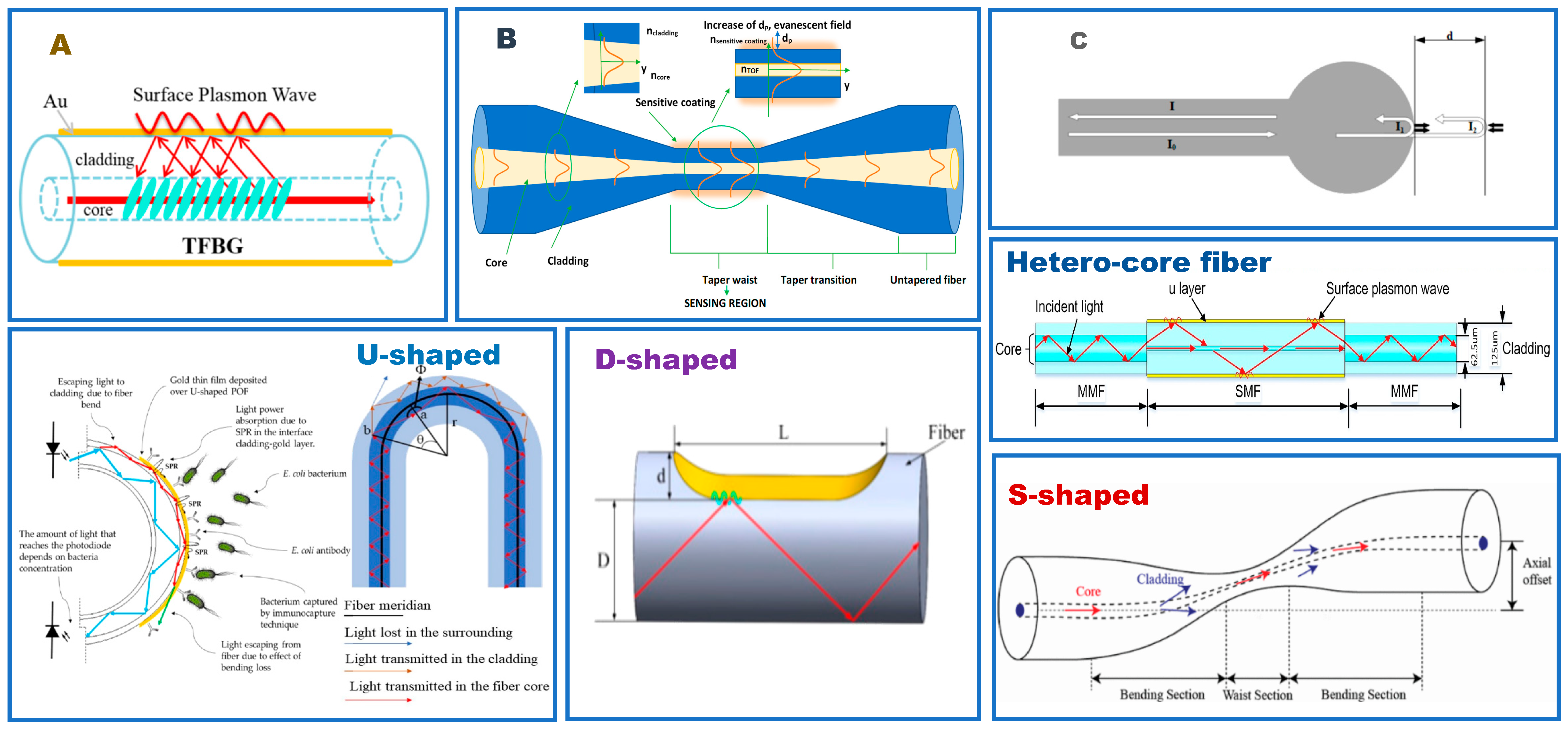
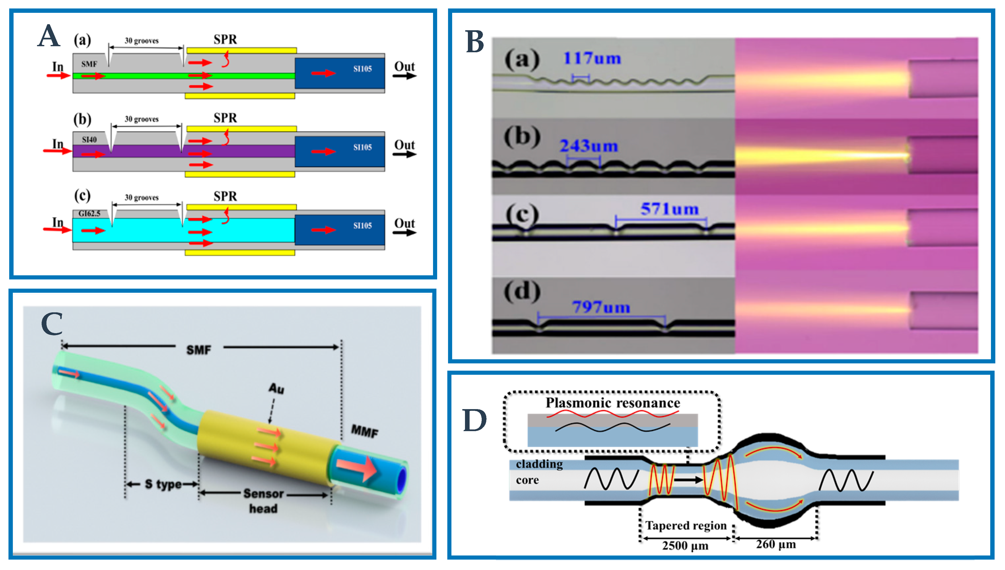
2.2. Fiber Grating
2.3. Taper Optical Fiber
2.4. Fiber Ball
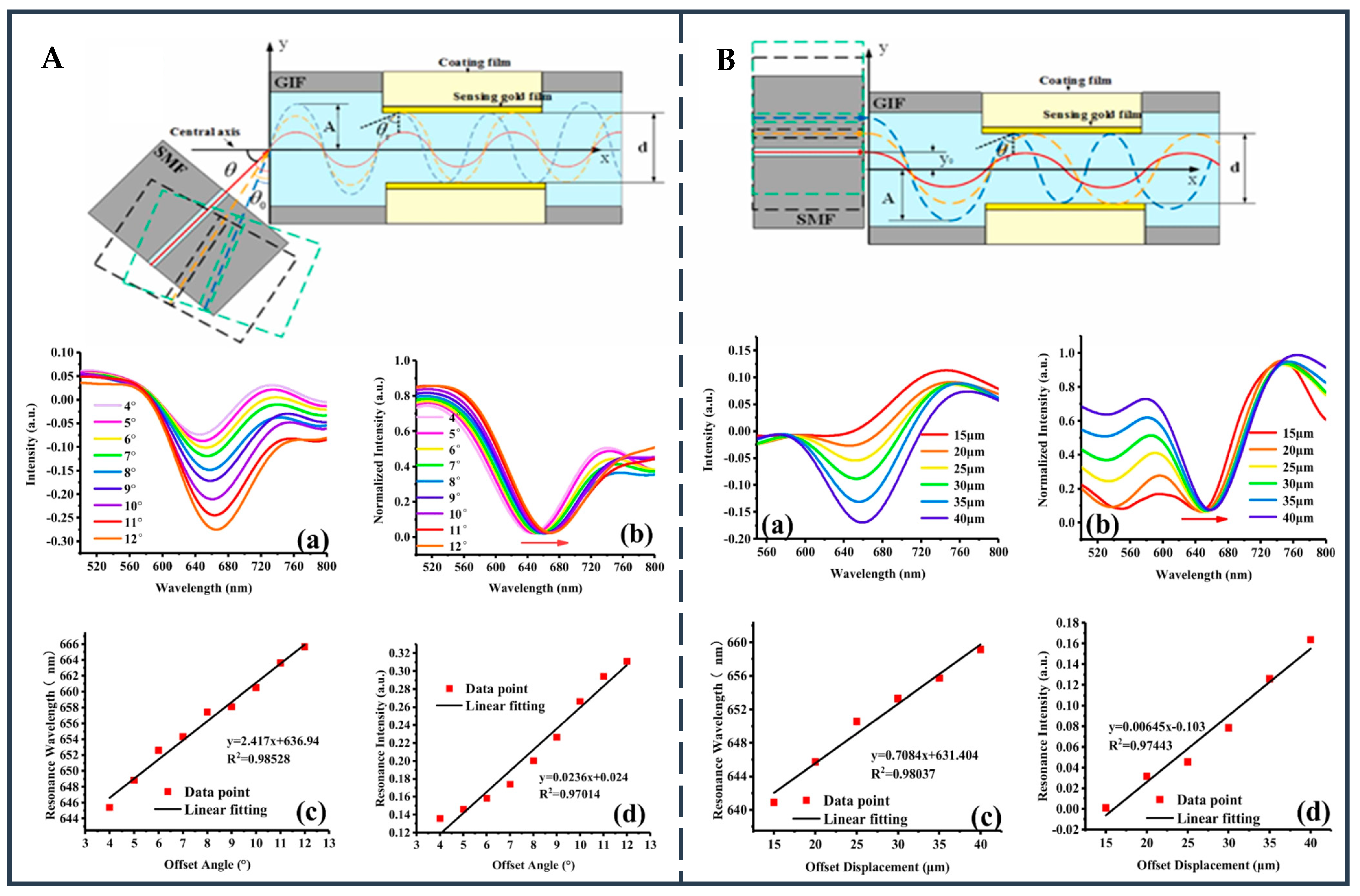
2.5. Hetero-Core Fiber Structure
2.6. D-Shape Fiber
2.7. S-Shape Fiber
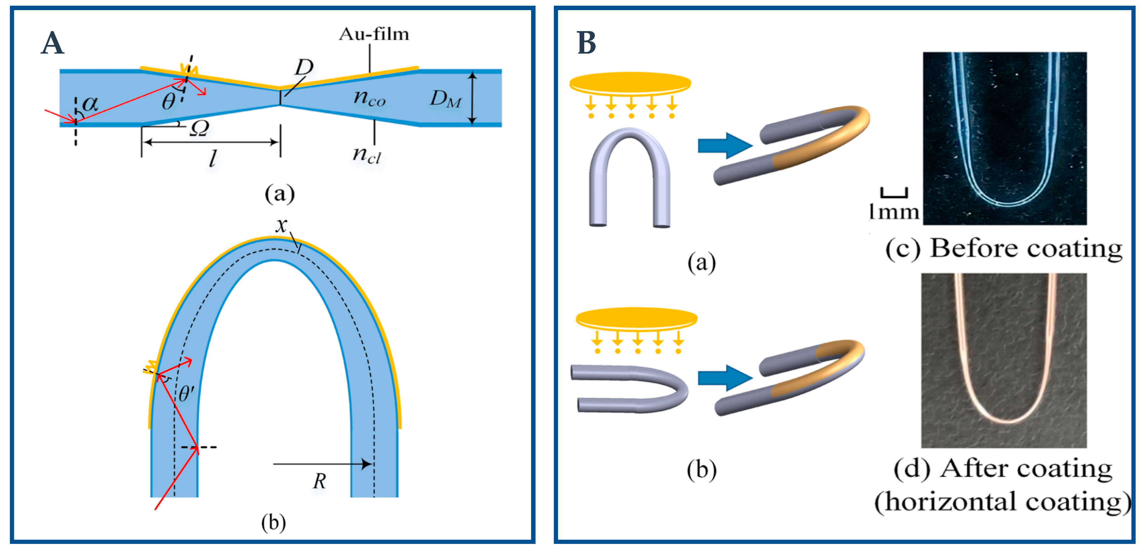
2.8. U-Shaped Fiber
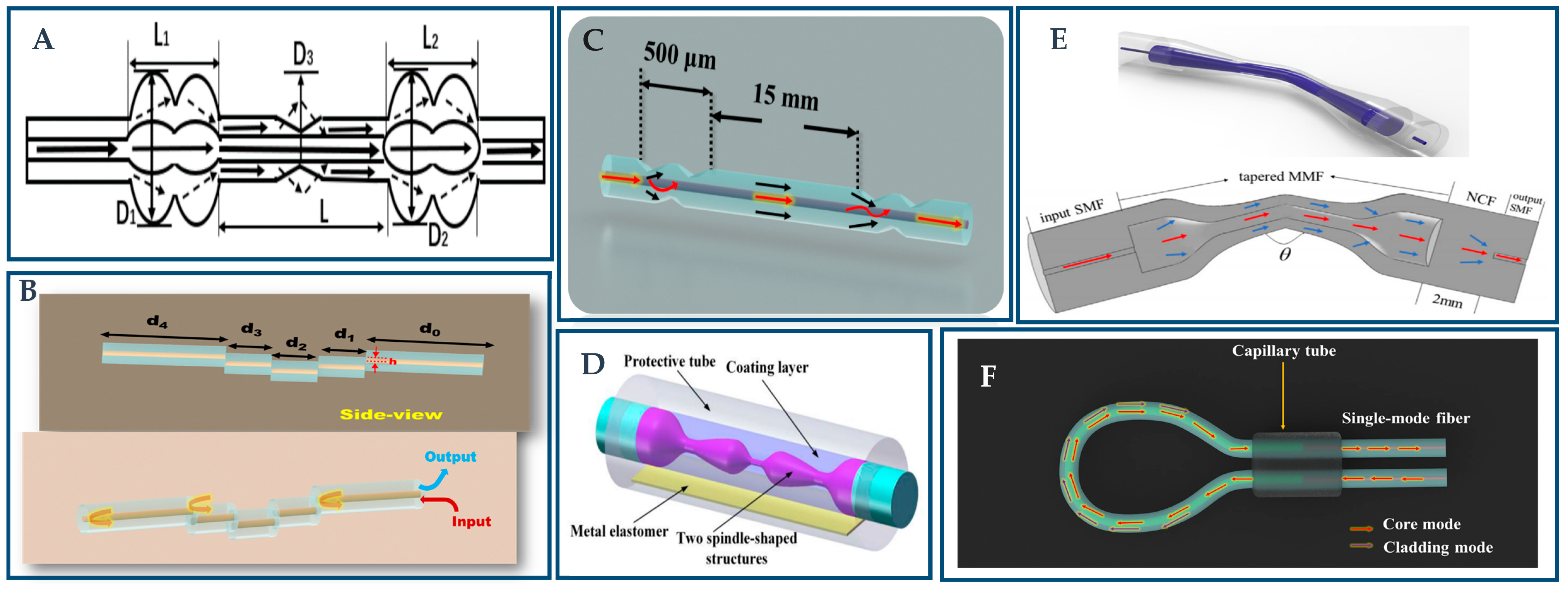
3. Novel Plasmonic Fiber Structure
3.1. Polymer Optical Fiber-Based Novel Structure
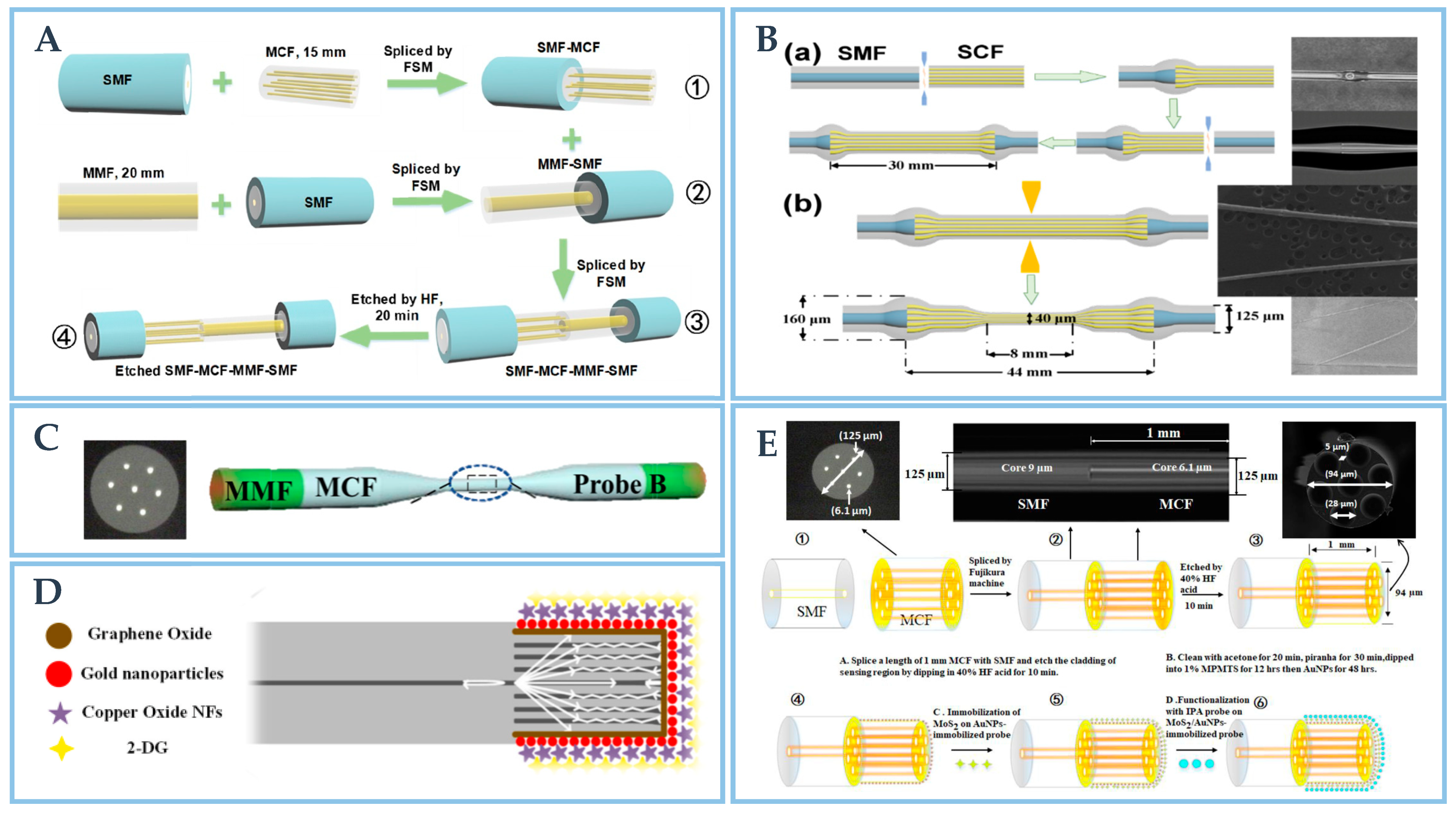
3.2. Multi-Core Fiber-Based Novel Structure

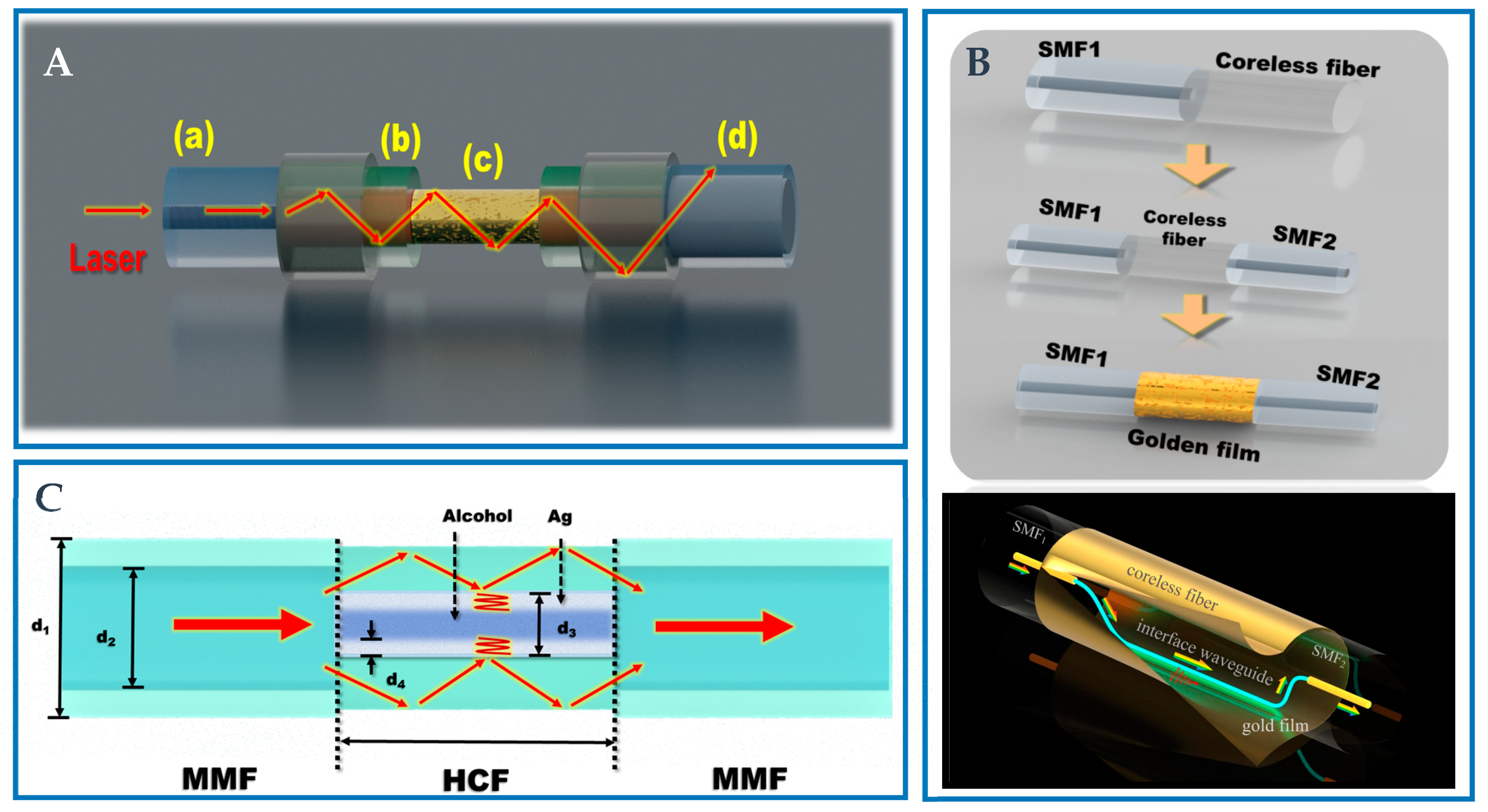
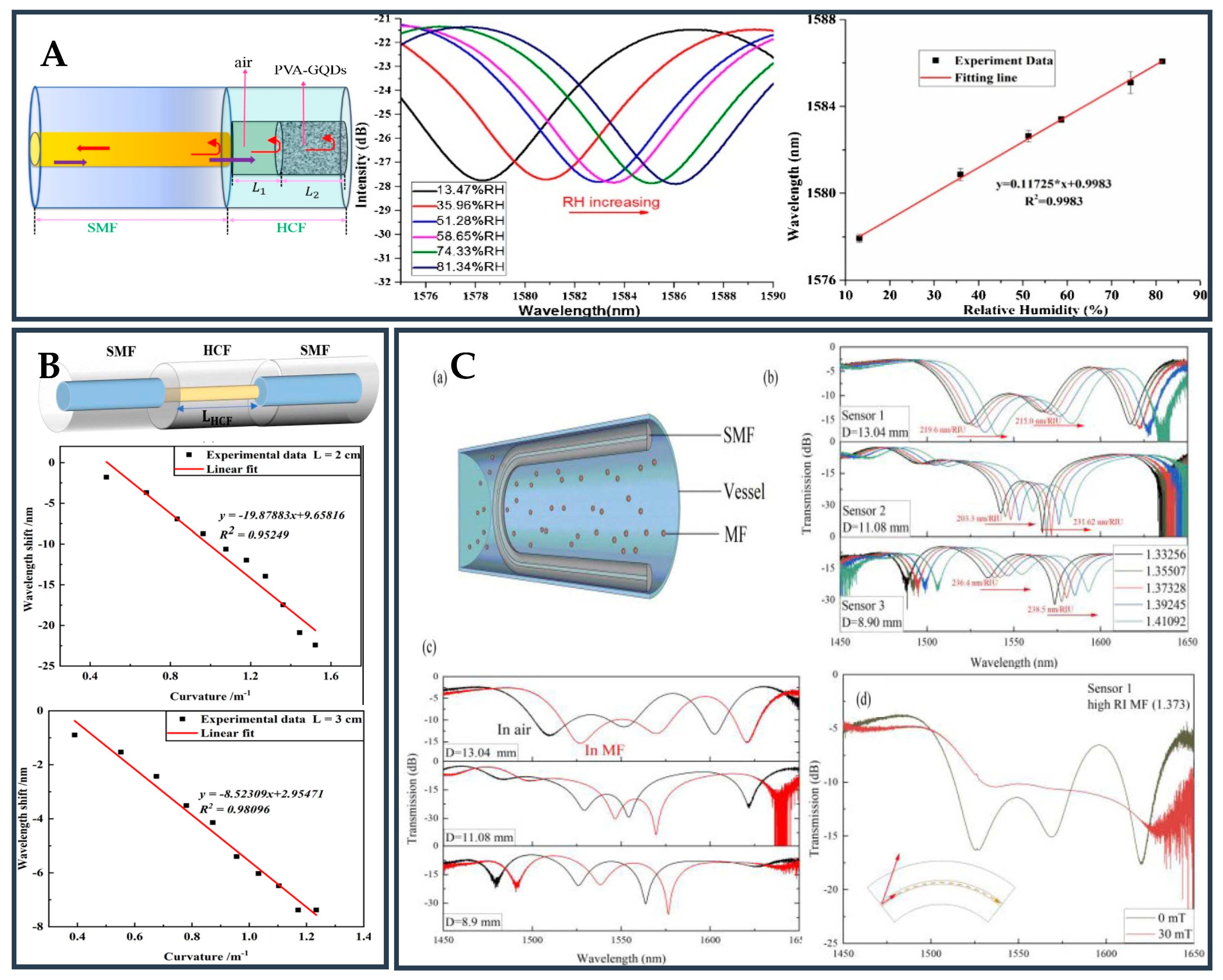
3.3. Hollow Core Fiber-Based Novel Structure
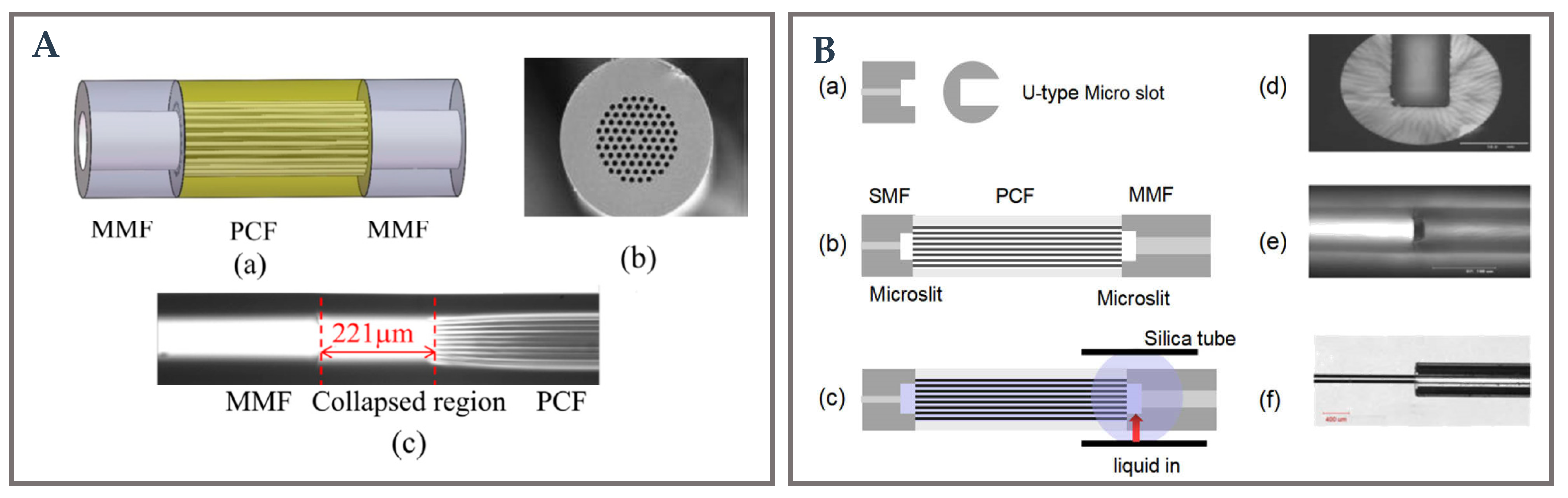
3.4. Photonic Crystal Fiber-Based Novel Structure
3.5. Special Fusion Splicing Structure
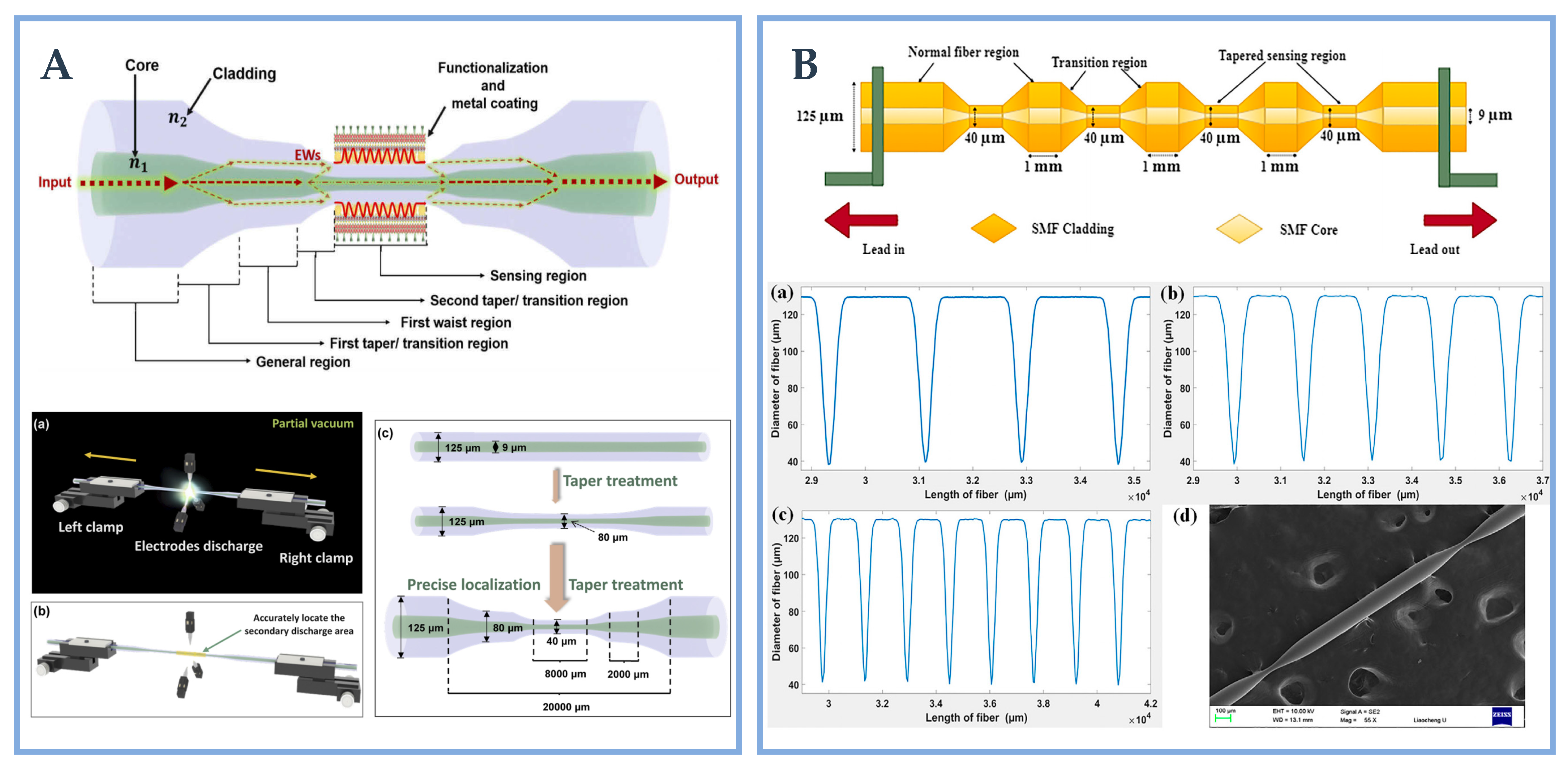
3.6. Special Tapering Strcuture
3.7. Other Special Novel Structure
3.8. Other Noval Fiber Structurals

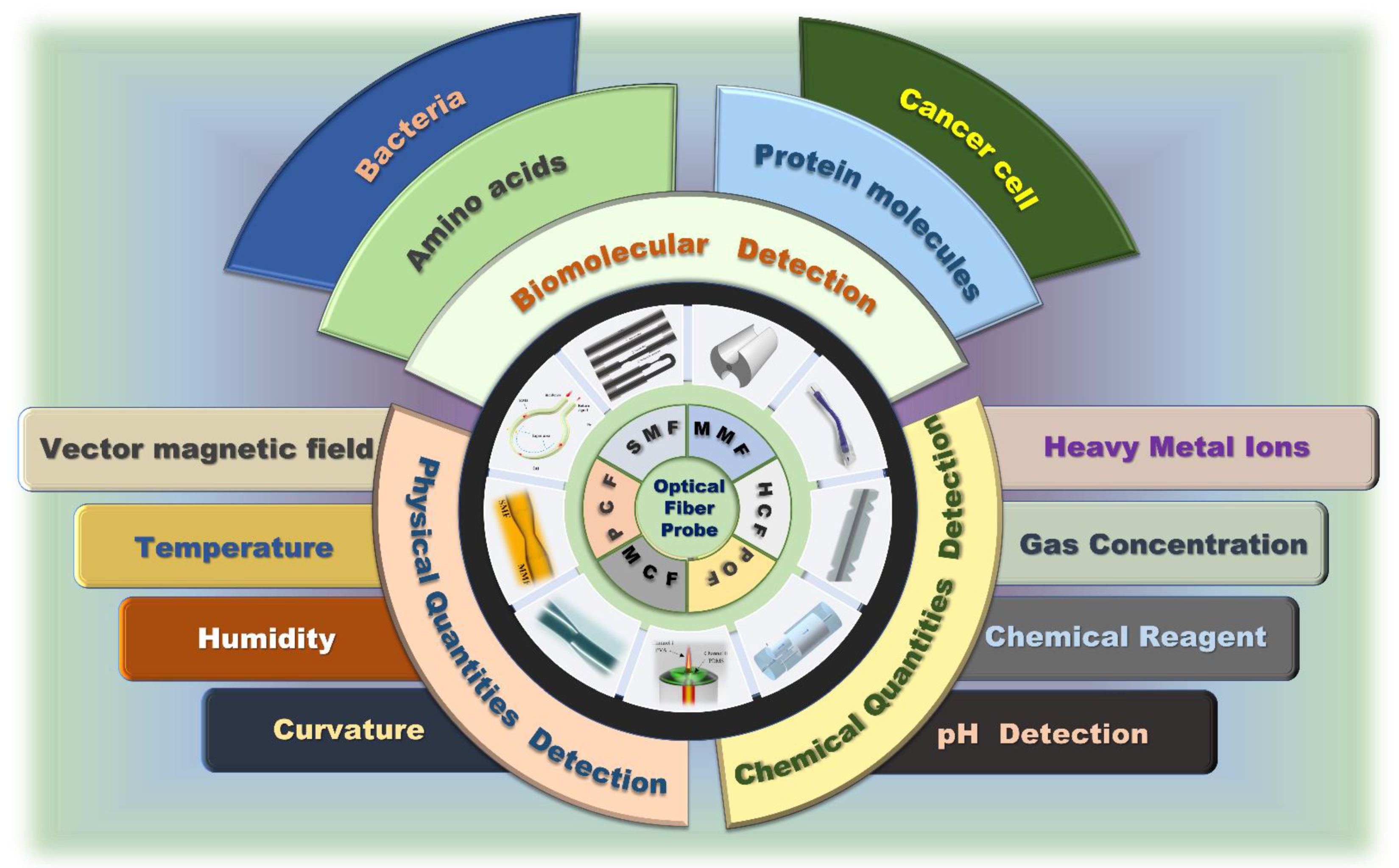
4. Special Optical Fiber Structure-Based Biosensing Applications
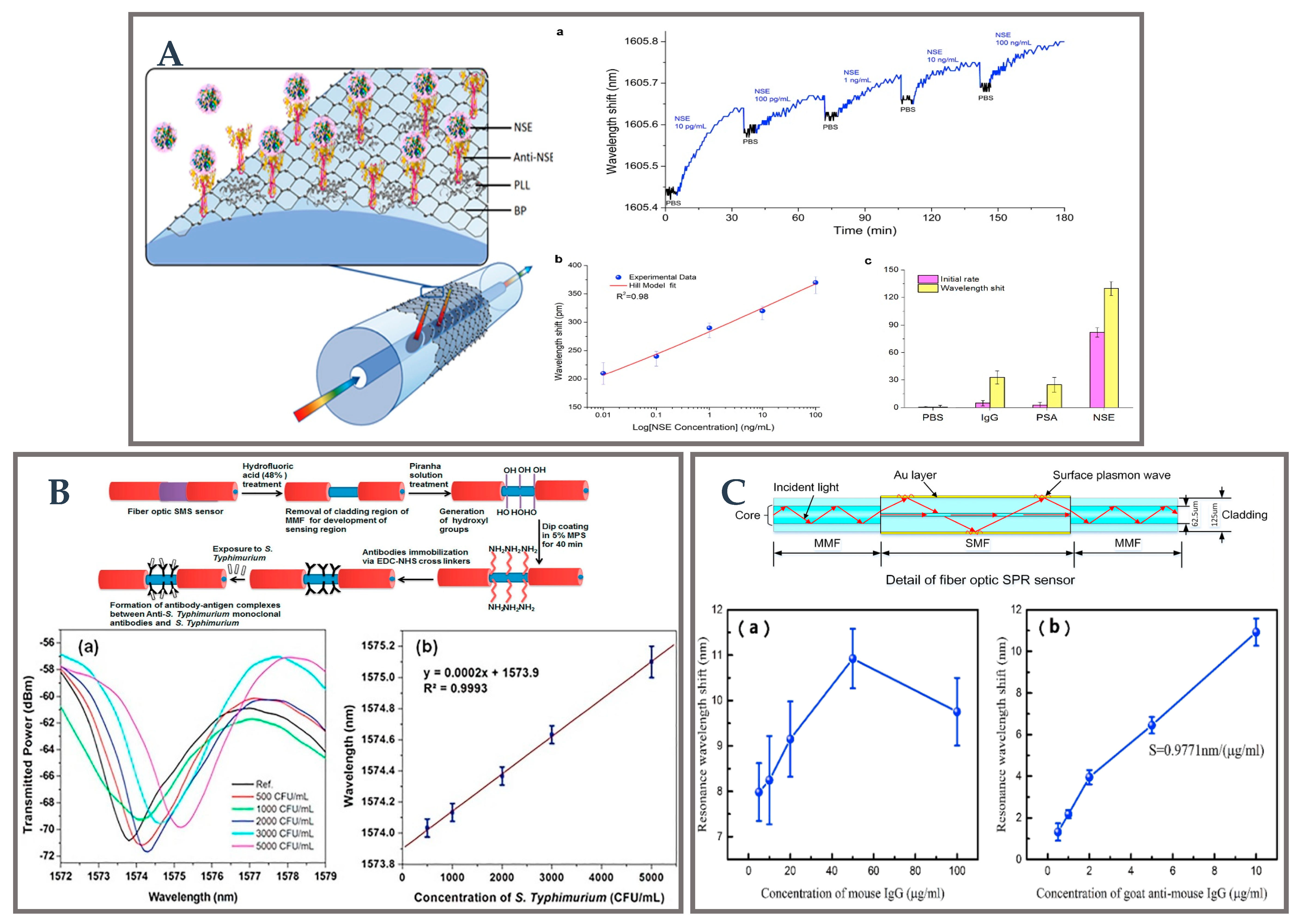
4.1. Biomolecular Detection

| Characteristics | Nanomaterials | Measured Parameter | Sensing Range /LOD | Sensitivity | Ref. |
|---|---|---|---|---|---|
| SMF–MCF–MMF–SMF | GO/AuNPs /MoS2-NPs/CA | Creatinine | 0–2000 μM /128.4 μM | 0.0025 nm/μM | [50] |
| Micro-ball | Au | Protein | 0.4–100 pM | 1273.74 nm/RIU | [84] |
| Convex fiber-tapered seven core fiber-Convex fiber (CTC) | AuNPs /Nb2CTx Mxene /CA | Creatinine | 86.12 μM | 3.1 pm/μM | [103] |
| Tapered | AuNRs/AuTNPs/ AuNPs | microRNA | 103 aM–261 aM | 0.92 nm/nM 0.97 nm/nM | [148] |
| U-shaped | Au/miRNA-133a | mRNA | 0.0133 ng/mL | 27.352 dB/log ng/mL | [149] |
| Tapered POF | molecularly imprinted polymer | L-nicotine | n.r. a | 1.3 × 10⁴ nm/M −1.7 × 10³ nm/M | [150] |
| U-bent fiber | Au layer/ITO nanorods /graphene | DNA | 0.1–100 nM/0.10 nM | 690.7 nm/RIU | [151] |
| D-shape plastic optical fiber | Graphene/Au | DNA | n.r. a | 1227 nm/RIU | [152] |
| D-shaped POF | HMM/ graphene | DNA | 10 pM–100 Nm /10 pM | 0.26 nm/ nM | [153] |
| SMF-NCF_SMF | Au | cDNA | 80 nm | n.r. a | [154] |
| Tapered SMS | AgNPs/GO | L-Cysteine | 10 nM–1 mM/63.25 μM | 7.0 nm/ mM | [155] |
| TFBG | Au/ graphene | Dopamine | 10−13–10−8 M /10−13 M | 0.29 dB/ logM | [156] |
| Tapered | AgNPs | Dopamine | 0.58 μM | n.r. a | [157] |
| U-shaped MMF-MSM | Au | IgG | 0.104 μg/mL | 0.192 nm/ wt./mL | [158] |
| Dual channel | Au layer/GO /anti-IgG/ AuNPs-IgG | IgG | 1–35 μg/mL /0.015 μg/mL | 1.36 nm/(μg/mL) | [159] |
| MMF-NCF-MMF structure | Polyelectrolyte self-assembled multilayers | IgG | 1.75 μg/mL | 57.06 nm /(mg/mL) | [160] |
| Tapered | AuNPs | Uric acid | 280.07 μM | 0.0131 nm/μM | [161] |
| SMF and TFBG | Au/AuNPs | Thrombin | 1 nm | n.r. a | [162] |
| SMF and TFBG | Au | Formaldehyde | 0–17 ppm | 2.10 pm/ppm | [163] |
4.2. Chemical Quantities Detection
| Characteristics | Nanomaterials | Measured Parameter | Sensing Range/LOD | Sensitivity | Ref. |
|---|---|---|---|---|---|
| Unclad fiber | ITO/BCP | Ammonia gas | 1~10 ppm | 1.891 nm/ppm | [13] |
| U-shaped | Au | Pb2+ | 2.1 ppb | n.r. a | [164] |
| D-shaped | Au | Pb2+ | n.r. a | 0.116 nm/ppm | [167] |
| MMF-SMF-MMF | Hydrogel/Au | pH | 8–10 pH | 13 nm/pH | [168] |
| SMF-FBG | Cu/WS2/PAAG | pH | 1–9 | −4.42 nm/pH | [169] |
| TFBG | Ag | H2O2 | 0.2 μM | n.r. a | [170] |
| Unclad fiber | Cu/ZnO | H2S gas | n.r. a | n.r. a | [171] |
| SMF-MMF-SMF | Onic liquid gel coatings | CO2 gas | n.r. a | n.r. a | [172] |
| MMF-Tapered HCF-MMF | Au | RI | 1.335–1.40 | 7592.25 nm/RIU | [173] |
| Tapered and U-shaped (MMF-TUMMF-MMF) | Au | RI | n.r. a | X: 1415/RIU y: 1293/RIU | [174] |
| D-shaped | Graphene/Ag | RI | n.r. a | 9000 nm/RIU 14,500 nm/RIU | [175] |
| D-shaped | Au | RI | n.r. a | 30 nm/RIU | [176] |
| U-shaped | Au/PML | RI | 1.33–1.44 | 66,000 nm/RIU | [177] |
| D-shaped | Au/self-assembled monolayer | Cu2+ | 4 × 10⁻⁶–2 × 10⁻⁴ M | n.r. a | [178] |
| MMF-NCF-MMF | Au | Cu2+ | n.r. a | 0.1184 nm/μM | [179] |
4.3. Physical Quantities Detection
| Characteristics | Nanomaterials | Measured Parameter | Sensing Range/LOD | Sensitivity | Ref. |
|---|---|---|---|---|---|
| Tapered MMF bent into loop | Au | Load | 0–20 kPa | 1.47 nm/kPa | [34] |
| SMF-GI MMF-SI MMF-SMF-SI MMF | Au | RI/Temperature | 0–25 nm 20–60 °C | DSR: 4.24 nm/μm −0.19 nm/°C TSR: 0.46 nm/μm −2.485 nm/°C | [37] |
| MMF-PCF-MMF | Au/PDMS | Temperature | 35–100 °C | −1.551 nm/°C | [40] |
| Double-side polished U-shape POF | Au/PDMS | RI/Temperature | n.r. a | 1258 nm/RIU –0.596 nm/°C | [185] |
| MMF-SMF-MMF | Au | RI/Temperature | n.r. a | 2323.4 nm/RIU 2.850 nm/°C | [186] |
| SMF-NCF-SMF | Au | RI/Temperature | 2061.6 nm | 2061.6 nm/RIU −0.0379 nm/°C | [187] |
| Two opposite D–shaped | Au/Ethanol/polyvinyl alcohol | Temperature/Humidity | 10–70 °C, 20–80% RH | 2.9 nm/°C 11.6 nm/%RH | [188] |
| Single mode-side polished multimode-singlemode (SSPMS) | gelatin material | Humidity | 40–90 %RH | 0.14 dB/%RH | [189] |
| Spiral twisted LPFG | Au/WS2 | Humidity | n.r. a | 37.3 pm/% RH | [190] |
| MMF-SMF | Au | Temperature/Salinity | n.r. a | −4.418nm/°C 0.558 nm/‰ | [191] |
| MMF-SMF | Au | Temperature/Salinity | n.r. a | −2.02 nm/°C 0.31 nm/‰ | [192] |
| MMF-PCF-SMF | Au | Temperature/Pressure | n.r. a | −1.802 nm/°C 2.838 nm/MPa | [193] |
| C-type micro-structured fiber | Au | Temperature/Salinity | 5–35 °C 30–40‰ | −7.609 nm/°C 1.402 nm/‰ | [194] |
5. Future Prospects
6. Conclusions
Author Contributions
Funding
Institutional Review Board Statement
Informed Consent Statement
Data Availability Statement
Acknowledgments
Conflicts of Interest
References
- Luo, X.; Qiu, T.; Lu, W.; Ni, Z. Plasmons in graphene: Recent progress and applications. Mater. Sci. Eng. R Rep. 2013, 74, 351–376. [Google Scholar] [CrossRef] [Green Version]
- Ritchie, R.H. Plasma Losses by Fast Electrons in Thin Films. Phys. Rev. 1957, 106, 874–881. [Google Scholar] [CrossRef]
- Choi, I.; Choi, Y. Plasmonic Nanosensors: Review and Prospect. IEEE J. Sel. Top. Quantum Electron. 2012, 18, 1110–1121. [Google Scholar] [CrossRef]
- Zeng, Y.; Hu, R.; Wang, L.; Gu, D.; He, J.; Wu, S.-Y.; Ho, H.-P.; Li, X.; Qu, J.; Gao, B.; et al. Recent advances in surface plasmon resonance imaging: Detection speed, sensitivity, and portability. Nanophotonics 2017, 6, 1017–1030. [Google Scholar] [CrossRef]
- Leal-Junior, A.; Frizera, A.; Pontes, M.J.; Fasano, A.; Woyessa, G.; Bang, O.; Marques, C.A.F. Dynamic mechanical characterization with respect to temperature, humidity, frequency and strain in mPOFs made of different materials. Opt. Mater. Express 2018, 8, 804–815. [Google Scholar] [CrossRef] [Green Version]
- Li, M.; Cushing, S.; Wu, N. Plasmon-Enhanced Optical Sensors: A Review. Analyst 2014, 140, 386–406. [Google Scholar] [CrossRef] [Green Version]
- Nylander, C.; Liedberg, B.; Lind, T. Gas detection by means of surface plasmon resonance. Sens. Actuators 1982, 3, 79–88. [Google Scholar] [CrossRef]
- Liu, Y.; Peng, W. Fiber-Optic Surface Plasmon Resonance Sensors and Biochemical Applications: A Review. J. Light. Technol. 2021, 39, 3781–3791. [Google Scholar] [CrossRef]
- Jorgenson, R.C.; Yee, S.S. A fiber-optic chemical sensor based on surface plasmon resonance. Sens. Actuators B Chem. 1993, 12, 213–220. [Google Scholar] [CrossRef]
- Kim, Y.-C.; Masson, J.-F.; Booksh, K.S. Single-crystal sapphire-fiber optic sensors based on surface plasmon resonance spectroscopy for in situ monitoring. Talanta 2005, 67, 908–917. [Google Scholar] [CrossRef]
- Yuzhi, C.; Xuejin, L. Single mode-no core-single mode fiber based surface plasmon resonance sensor. Infrared Laser Eng. 2020, 49, 20201055-1. [Google Scholar] [CrossRef]
- Yang, Q.; Zhang, X.; Kumar, S.; Singh, R.; Zhang, B.; Bai, C.-L.; Pu, X. Development of Glucose Sensor Using Gold Nanoparticles and Glucose-Oxidase Functionalized Tapered Fiber Structure. Plasmonics 2020, 15, 841–848. [Google Scholar] [CrossRef]
- Mishra, S.K.; Bhardwaj, S.; Gupta, B.D. Surface Plasmon Resonance-Based Fiber Optic Sensor for the Detection of Low Concentrations of Ammonia Gas. IEEE Sens. J. 2015, 15, 1235–1239. [Google Scholar] [CrossRef]
- Li, X.; Gong, P.; Zhao, Q.; Zhou, X.; Zhang, Y.; Zhao, Y. Plug-in optical fiber SPR biosensor for lung cancer gene detection with temperature and pH compensation. Sens. Actuators B Chem. 2022, 359, 131596. [Google Scholar] [CrossRef]
- Liu, L.; Liu, Z.; Zhang, Y.; Liu, S. Side-Polished D-Type Fiber SPR Sensor for RI Sensing with Temperature Compensation. IEEE Sens. J. 2021, 21, 16621–16628. [Google Scholar] [CrossRef]
- Liedberg, B.; Nylander, C.; Lunström, I. Surface plasmon resonance for gas detection and biosensing. Sens. Actuators 1983, 4, 299–304. [Google Scholar] [CrossRef]
- Zakaria, R.; Zainuddin, N.A.a.M.; Leong, T.C.; Rosli, R.; Rusdi, M.F.; Harun, S.W.; Sadegh Amiri, I. Investigation of Surface Plasmon Resonance (SPR) in MoS2- and WS2-Protected Titanium Side-Polished Optical Fiber as a Humidity Sensor. Micromachines 2019, 10, 465. [Google Scholar] [CrossRef] [Green Version]
- Zhao, Y.; Tong, R.-j.; Xia, F.; Peng, Y. Current status of optical fiber biosensor based on surface plasmon resonance. Biosens. Bioelectron. 2019, 142, 111505. [Google Scholar] [CrossRef] [PubMed]
- Teng, C.; Li, M.; Cheng, Y.; Peng, H.; Deng, S.; Deng, H.; Yuan, L.; Chen, M. Investigation of U-shape tapered plastic optical fibers based surface plasmon resonance sensor for RI sensing. Optik 2022, 251, 168461. [Google Scholar] [CrossRef]
- Haider, F.; Aoni, R.A.; Ahmed, R.; Mahdiraji, G.; Azman, F.; Adikan, F. Mode-multiplex plasmonic sensor for multi-analyte detection. Opt. Lett. 2020, 45, 3945–3948. [Google Scholar] [CrossRef]
- Pesavento, M.; Zeni, L.; De Maria, L.; Alberti, G.; Cennamo, N. SPR-Optical Fiber-Molecularly Imprinted Polymer Sensor for the Detection of Furfural in Wine. Biosensors 2021, 11, 72. [Google Scholar] [CrossRef]
- Leitão, C.; Leal-Junior, A.; Almeida, A.R.; Pereira, S.O.; Costa, F.M.; Pinto, J.L.; Marques, C. Cortisol AuPd plasmonic unclad POF biosensor. Biotechnol. Rep. 2021, 29, e00587. [Google Scholar] [CrossRef]
- Aoni, R.A.; Mahdiraji, G.A.; Sua, Y.M.; Ahmed, R.; Shee, Y.G.; Adikan, F. Highly sensitive multi-core flat fiber surface plasmon resonance refractive index sensor. Opt. Express 2016, 24, 2485–2495. [Google Scholar] [CrossRef]
- Sakib, M.; Hossain, M.B.; Altabatabaie, K.; Mehedi, I.; Hasan, M.T.; Hossain, M.; Sadegh Amiri, I. High performance dual core D-shape PCF-SPR sensor modeling employing gold coat. Results Phys. 2019, 15, 102788. [Google Scholar] [CrossRef]
- Jain, S.; Choudhary, K.; Kumar, S. Photonic crystal fiber-based SPR sensor for broad range of refractive index sensing applications. Opt. Fiber Technol. 2022, 73, 103030. [Google Scholar] [CrossRef]
- Zhao, Y.; Zhao, H.; Lv, R.-Q.; Zhao, J. Review of optical fiber Mach–Zehnder interferometers with micro-cavity fabricated by femtosecond laser and sensing applications. Opt. Lasers Eng. 2019, 117, 7–20. [Google Scholar] [CrossRef]
- Liu, Z.; Liu, W.; Yang, X.; Zhang, Y.; Zhang, Y.; Song, H.; Zhang, J.; Yang, J.; Yuan, L. Hollow hanging-core fiber SPR sensor for microfluidic chip with large size channel. Sens. Actuators A Phys. 2020, 309, 111993. [Google Scholar] [CrossRef]
- Lin, H.Y.; Huang, C.H.; Cheng, G.L.; Chen, N.K.; Chui, H.C. Tapered optical fiber sensor based on localized surface plasmon resonance. Opt Express 2012, 20, 21693–21701. [Google Scholar] [CrossRef]
- Liu, C.; Wang, J.; Wang, F.; Su, W.; Yang, L.; Lv, J.; Fu, G.; Li, X.; Liu, Q.; Sun, T.; et al. Surface plasmon resonance (SPR) infrared sensor based on D-shape photonic crystal fibers with ITO coatings. Opt. Commun. 2020, 464, 125496. [Google Scholar] [CrossRef]
- Gupta, B.; Kant, R. Recent advances in surface plasmon resonance based fiber optic chemical and biosensors utilizing bulk and nanostructures. Opt. Laser Technol. 2018, 101, 144–161. [Google Scholar] [CrossRef]
- Chen, M.-q.; He, T.-y.; Zhao, Y.; Yang, G. Ultra-short phase-shifted fiber Bragg grating in a microprobe for refractive index sensor with temperature compensation. Opt. Laser Technol. 2023, 157, 108672. [Google Scholar] [CrossRef]
- Liu, Z.; Liu, L.; Zhu, Z.; Zhang, Y.; Wei, Y.; Zhang, Y.; Yang, J.; Yuan, L. Dual-channel surface plasmon resonance refractive index sensor based on modified hetero-core structure fiber. Opt. Commun. 2017, 403, 290–295. [Google Scholar] [CrossRef]
- Gomez-Cardona, N.; Reyes-Vera, E.; Torres, P. High Sensitivity Refractive Index Sensor Based on the Excitation of Long-Range Surface Plasmon Polaritons in H-Shaped Optical Fiber. Sensors 2020, 20, 2111. [Google Scholar] [CrossRef] [PubMed] [Green Version]
- Wu, Q.L.; Zhao, Y.; Zhang, Y.N.; Yang, Y. High sensitive applied load measurement using optical fiber tapered-loop probe with SPR effect. Opt. Laser Technol. 2019, 114, 95–102. [Google Scholar] [CrossRef]
- Ren, Z.H.; Wang, Q.; Zhao, W.M.; Wang, L.; Jiang, C.Q.; Cong, X.W.; Yan, X.; Zhu, A.S.; Qiu, F.M.; Chen, B.H.; et al. A High-FOM surface plasmon resonance sensor based on MMF-TUMMF-MMF structure of optical fiber. Opt. Fiber Technol. 2022, 72, 102970. [Google Scholar] [CrossRef]
- Kong, L.-X.; Chi, M.-J.; Ren, C.; Ni, L.-F.; Li, Z.; Zhang, Y.-S. Micro-Lab on Tip: High-Performance Dual-Channel Surface Plasmon Resonance Sensor Integrated on Fiber-Optic End Facet. Sens. Actuators B Chem. 2022, 351, 130978. [Google Scholar] [CrossRef]
- Wei, Y.; Wu, P.; Zhu, Z.D.; Liu, L.; Liu, C.L.; Hu, J.X.; Wang, S.F.; Zhang, Y.H. Surface-Plasmon-Resonance-Based Optical-Fiber Micro-Displacement Sensor with Temperature Compensation. Sensors 2018, 18, 3210. [Google Scholar] [CrossRef] [Green Version]
- Wang, F.; Pang, K.; Ma, T.; Wang, X.; Liu, Y. Folded-tapered multimode-no-core fiber sensor for simultaneous measurement of refractive index and temperature. Opt. Laser Technol. 2020, 130, 106333. [Google Scholar] [CrossRef]
- Lin, Z.-T.; Zhao, Y.; Lv, R.-Q.; Zheng, H.-K.; Zhao, Q. High-sensitivity salinity sensor based on etched C-type micro-structured fiber sensing structure. Sens. Actuators A Phys. 2022, 339, 113518. [Google Scholar] [CrossRef]
- Ma, Y.; Yi, Y.; Li, X.; Su, C.; Zhang, M.; Geng, T.; Sun, W.; Yuan, L. Refractometer based on fiber Mach-Zehnder interferometer composed of two micro bending cores. Opt. Express 2021, 29, 31443–31454. [Google Scholar] [CrossRef]
- Pospori, A.; Marques, C.A.F.; Bang, O.; Webb, D.J.; André, P. Polymer optical fiber Bragg grating inscription with a single UV laser pulse. Opt. Express 2017, 25, 9028–9038. [Google Scholar] [CrossRef] [PubMed] [Green Version]
- Sola, D.; Alamri, S.; Lasagni, A.F.; Artal, P. Fabrication and characterization of diffraction gratings in ophthalmic polymers by using UV direct laser interference patterning. Appl. Surf. Sci. 2019, 476, 128–135. [Google Scholar] [CrossRef]
- Ma, Y.; Sun, X.; Si, X.; Peng, L.; Wang, H.; Zhu, Y.-F.; Wu, L.; Yi, L.; Li, L.; Zhao, X.; et al. Thermal stability of fiber Bragg gratings fabricated by 193 nm excimer laser. Opt. Commun. 2022, 516, 128286. [Google Scholar] [CrossRef]
- Hu, Q.; Zhao, X.; Tian, X.; Li, H.; Wang, M.; Wang, Z.; Xu, X. Raman suppression in 5 kW fiber amplifier using long period fiber grating fabricated by CO2 laser. Opt. Laser Technol. 2022, 145, 107484. [Google Scholar] [CrossRef]
- Wang, Z.; Zhu, G.; Wang, Y.; Li, M.; Singh, R.; Zhang, B.; Kumar, S. Fabrication techniques and stability analysis of SMF-/MMF-based differently tapered optical fiber structures. Appl. Opt. 2021, 60, 2077–2082. [Google Scholar] [CrossRef] [PubMed]
- Zhang, W.; Wang, Z.; Singh, R.; Wang, Y.; Xie, Y.; Su, X.; Gao, F.; Li, G.; Swarnakar, S.; Min, R.; et al. Performance analysis of an SMF-/MMF-based single/double/quadruple tapered optical fiber structure. Appl. Opt. 2022, 61, 2140–2146. [Google Scholar] [CrossRef]
- Liu, X.; Li, M.; Singh, R.; Wang, Y.; Xie, Y.; Su, X.; Gao, F.; Li, G.; Kumar, D.; Zhang, B.; et al. Feasibility analysis of an SMS-/MSM-/SMSMS-based optical fiber sensor structure. Appl. Opt. 2022, 61, 2327–2332. [Google Scholar] [CrossRef]
- Liu, L.; Deng, S.; Zheng, J.; Yuan, L.; Deng, H.; Teng, C. An Enhanced Plastic Optical Fiber-Based Surface Plasmon Resonance Sensor with a Double-Sided Polished Structure. Sensors 2021, 21, 1516. [Google Scholar] [CrossRef]
- Wen, H.-Y.; Hsu, H.-C.; Tsai, Y.-T.; Feng, W.-K.; Lin, C.-L.; Chiang, C.-C. U-Shaped Optical Fiber Probes Coated with Electrically Doped GQDs for Humidity Measurements. Polymers 2021, 13, 2696. [Google Scholar] [CrossRef]
- Li, M.; Singh, R.; Marques, C.; Zhang, B.; Kumar, S. 2D material assisted SMF-MCF-MMF-SMF based LSPR sensor for creatinine detection. Opt. Express 2021, 29, 38150–38167. [Google Scholar] [CrossRef]
- Wang, Z.; Singh, R.; Marques, C.; Jha, R.; Zhang, B.; Kumar, S. Taper-in-taper fiber structure-based LSPR sensor for alanine aminotransferase detection. Opt. Express 2021, 29, 43793–43810. [Google Scholar] [CrossRef]
- Min, R.; Marques, C.; Nielsen, K.; Bang, O.; Ortega, B. Fast Inscription of Long Period Gratings in Microstructured Polymer Optical Fibers. IEEE Sens. J. 2018, 18, 1919–1923. [Google Scholar] [CrossRef]
- Leitão, C.; Pereira, S.O.; Alberto, N.; Lobry, M.; Loyez, M.; Costa, F.M.; Pinto, J.L.; Caucheteur, C.; Marques, C. Cortisol In-Fiber Ultrasensitive Plasmonic Immunosensing. IEEE Sens. J. 2021, 21, 3028–3034. [Google Scholar] [CrossRef]
- Long, S.; Cao, J.; Wang, Y.; Gao, S.; Xu, N.; Gao, J.; Wan, W. Grating coupled SPR sensors using off the shelf compact discs and sensitivity dependence on grating period. Sens. Actuators Rep. 2020, 2, 100016. [Google Scholar] [CrossRef]
- Long, S.; Cao, J.; Geng, S.; Xu, N.; Qian, W.; Gao, S. Optimization of plasmonic sensors based on sinusoidal and rectangular gratings. Opt. Commun. 2020, 476, 126310. [Google Scholar] [CrossRef]
- Iadicicco, A.; Cusano, A.; Campopiano, S.; Cutolo, A.; Giordano, M. Thinned fiber Bragg gratings as refractive index sensors. IEEE Sens. J. 2006, 5, 1288–1295. [Google Scholar] [CrossRef]
- Liu, X.; Zhang, X.; Cong, J.; Xu, J.; Chen, K. Demonstration of etched cladding fiber Bragg grating-based sensors with hydrogel coating. Sens. Actuators B Chem. 2003, 96, 468–472. [Google Scholar] [CrossRef]
- Guo, T. Fiber Grating Assisted Surface Plasmon Resonance for Biochemical and Electrochemical Sensing. J. Light. Technol. 2016, 35, 1. [Google Scholar] [CrossRef]
- Zhang, Y.; Wang, F.; Qian, S.; Liu, Z.; Wang, Q.; Gu, Y.; Wu, Z.; Jing, Z.; Sun, C.; Peng, W. A Novel Fiber Optic Surface Plasmon Resonance Biosensors with Special Boronic Acid Derivative to Detect Glycoprotein. Sensors 2017, 17, 2259. [Google Scholar] [CrossRef] [Green Version]
- Fu, X.; Zhou, J.; Fu, Z.; Xu, M.; Huang, S.; Jin, W.; Fu, G.; Bi, W. A Multiparameter Sensor Based on Dumbbell-Shaped Double-Cladding Fiber Structure Cascaded Long Period Fiber Grating. IEEE Sens. J. 2022, 22, 1. [Google Scholar] [CrossRef]
- Ma, H.-Z.; Zhang, Y.; Zhang, W.; Gao, H.; Ma, L.; Lai, M.; Kong, L.; Yan, T. Polymer-coated polishing seven-core Mach-Zehnder interferometer for temperature sensitivity enhancement. Opt. Laser Technol. 2022, 149, 107774. [Google Scholar] [CrossRef]
- Li, J.-X.; Zhang, W.-H.; Tong, Z.-R.; Liu, J.-W. Fiber optic sensor modified by graphene oxide-glucose oxidase for glucose detection. Opt. Commun. 2021, 492, 126983. [Google Scholar] [CrossRef]
- Agrawal, N.; Saha, C.; Kumar, C.; Singh, R.; Zhang, B.; Kumar, S. Development of Uric Acid Sensor Using Copper Oxide and Silver Nanoparticles Immobilized SMSMS Fiber Structure-Based Probe. IEEE Trans. Instrum. Meas. 2020, 69, 9097–9104. [Google Scholar] [CrossRef]
- Niu, H.; Zhang, S.; Chen, W.; Liu, Y.; Li, X.; Yan, Y.; Wang, S.; Geng, T.; Sun, W.; Yuan, L. Optical Fiber Sensors Based on Core-Offset Structure: A Review. IEEE Sens. J. 2021, 21, 22388–22401. [Google Scholar] [CrossRef]
- Soares, M.S.; Silva, L.C.B.; Vidal, M.; Loyez, M.; Facão, M.; Caucheteur, C.; Segatto, M.E.V.; Costa, F.M.; Leitão, C.; Pereira, S.O.; et al. Label-free plasmonic immunosensor for cortisol detection in a D-shaped optical fiber. Biomed. Opt. Express 2022, 13, 3259–3274. [Google Scholar] [CrossRef] [PubMed]
- Rui, Y.; Yu, Y.S.; Xue, Y.; Chao, C.; Sun, H.B. Single S-tapered fiber Mach-Zehnder interferometers. Opt. Lett. 2011, 36, 4482–4484. [Google Scholar]
- Sai, V.; Kundu, T.; Mukherji, S. Novel U-bent fiber optic probe for localized surface plasmon resonance based biosensor. Biosens. Bioelectron. 2009, 24, 2804. [Google Scholar] [CrossRef]
- Gupta, B.D. Surface Plasmon Resonance Based Fiber Optic Sensors; Springer: New York, NY, USA, 2012. [Google Scholar]
- Gupta, B.D.; Dodeja, H.; Tomar, A.K. Fibre-optic evanescent field absorption sensor based on a U-shaped probe. Opt. Quantum Electron. 1996, 28, 1629–1639. [Google Scholar] [CrossRef]
- Stasiewicz, K.A.; Jakubowska, I.; Moś, J.E.; Marć, P.; Paczesny, J.; Zbonikowski, R.; Jaroszewicz, L.R. Optical Properties of a Tapered Optical Fiber Coated with Alkanes Doped with Fe3O4 Nanoparticles. Sensors 2022, 22, 7801. [Google Scholar] [CrossRef]
- Kumar, S.; Singh, R.; Zhu, G.; Yang, Q.; Zhang, X.; Cheng, S.; Zhang, B.; Kaushik, B.K.; Liu, F.Z. Development of Uric Acid Biosensor Using Gold Nanoparticles and Graphene Oxide Functionalized Micro-Ball Fiber Sensor Probe. IEEE Trans. Nanobiosci. 2020, 19, 173–182. [Google Scholar] [CrossRef]
- Mai, Z.; Zhang, J.; Chen, Y.; Wang, J.; Hong, X.; Su, Q.; Li, X. A disposable fiber optic SPR probe for immunoassay. Biosens. Bioelectron. 2019, 144, 111621. [Google Scholar] [CrossRef] [PubMed]
- Arcas, A.D.S.; Dutra, F.D.S.; Allil, R.C.S.B.; Werneck, M.M. Surface Plasmon Resonance and Bending Loss-Based U-Shaped Plastic Optical Fiber Biosensors. Sensors 2018, 18, 648. [Google Scholar] [CrossRef] [PubMed] [Green Version]
- Liu, H.; Sun, Y.; Guo, J.; Liu, W.; Liu, L.; Meng, Y.; Yu, X. Temperature-Insensitive Label-Free Sensors for Human IgG Based on S-Tapered Optical Fiber Sensors. IEEE Access 2021, 9, 116286–116293. [Google Scholar] [CrossRef]
- Lin, W.; Huang, W.; Liu, Y.; Chen, X.; Qu, H.; Hu, X. Cladding Mode Fitting-Assisted Automatic Refractive Index Demodulation Optical Fiber Sensor Probe Based on Tilted Fiber Bragg Grating and SPR. Sensors 2022, 22, 32. [Google Scholar] [CrossRef] [PubMed]
- Baiad, M.D.; Gagne, M.; Madore, W.J.; De Montigny, E.; Godbout, N.; Boudoux, C.; Kashyap, R. Surface plasmon resonance sensor interrogation with a double-clad fiber coupler and cladding modes excited by a tilted fiber Bragg grating. Opt. Lett. 2013, 38, 4911–4914. [Google Scholar] [CrossRef] [PubMed]
- Ortega-Gomez, A.; Loyez, M.; Lobry, M.; Chah, K.; Zubia, J.; Villatoro, J.; Caucheteur, C. Plasmonic sensors based on tilted Bragg gratings in multicore optical fibers. Opt. Express 2021, 29, 18469–18480. [Google Scholar] [CrossRef] [PubMed]
- Liu, F.; Zhang, X.; Li, K.; Guo, T.; Ianoul, A.; Albert, J. Discrimination of Bulk and Surface Refractive Index Change in Plasmonic Sensors with Narrow Bandwidth Resonance Combs. ACS Sens. 2021, 6, 3013–3023. [Google Scholar] [CrossRef]
- Vikas; Verma, R.K. Design considerations of a surface plasmon resonance (SPR) based tapered fiber optic bio-sensing probe with graphene-MoS2 over layers. Optik 2019, 180, 330–343. [Google Scholar] [CrossRef]
- Herrera, N.D.; Esteban, O.; Navarrete, M.C.; Gonzalez-Cano, A.; Benito-Pena, E.; Orellana, G. Improved performance of SPR sensors by a chemical etching of tapered optical fibers. Opt. Lasers Eng. 2011, 49, 1065–1068. [Google Scholar] [CrossRef]
- Yang, Q.S.; Zhu, G.; Singh, L.; Wang, Y.; Singh, R.; Zhang, B.Y.; Zhang, X.; Kumar, S. Highly sensitive and selective sensor probe using glucose oxidase/gold nanoparticles/graphene oxide functionalized tapered optical fiber structure for detection of glucose. Optik 2020, 208, 164536. [Google Scholar] [CrossRef]
- Korec, J.; Stasiewicz, K.A.; Garbat, K.; Jaroszewicz, L.R. Enhancement of the SPR Effect in an Optical Fiber Device Utilizing a Thin Ag Layer and a 3092A Liquid Crystal Mixture. Molecules 2021, 26, 7553. [Google Scholar] [CrossRef] [PubMed]
- Chen, X.H.; Li, X.J.; Yi, D.; Hong, X.M.; Chen, Y.Z. Plasmonic tapered-fiber interference sensor for simultaneously detecting refractive index and temperature. Opt. Lett. 2021, 46, 6071–6074. [Google Scholar] [CrossRef] [PubMed]
- Ayupova, T.; Shaimerdenova, M.; Sypabekova, M.; Vangelista, L.; Tosi, D. Picomolar detection of thrombin with fiber-optic ball resonator sensor using optical backscatter reflectometry. Optik 2021, 241, 166969. [Google Scholar] [CrossRef]
- Rao, Y.J.; Deng, M.; Duan, D.W.; Zhu, T. In-line fiber Fabry-Perot refractive-index tip sensor based on endlessly photonic crystal fiber. Sens. Actuators A Phys. 2008, 148, 33–38. [Google Scholar] [CrossRef]
- Yi, D.; Chen, Y.Z.; Geng, Y.F.; Teng, F.; Li, Y.; Liu, F.; Li, X.J.; Hong, X.M. Interrogation technique analyses of a hybrid fiber optic sensor based on SPR and MMI. Opt. Express 2020, 28, 20764–20772. [Google Scholar] [CrossRef] [PubMed]
- Singh, L.; Singh, R.; Kumar, S.; Zhang, B.; Kaushik, B.K. Development of Collagen-IV Sensor Using Optical Fiber-Based Mach-Zehnder Interferometer Structure. IEEE J. Quantum Electron. 2020, 56, 1–8. [Google Scholar] [CrossRef]
- Zhu, J.J.; Zhang, A.P.; Xia, T.H.; He, S.L.; Xue, W. Fiber-Optic High-Temperature Sensor Based on Thin-Core Fiber Modal Interferometer. IEEE Sens. J. 2010, 10, 1415–1418. [Google Scholar] [CrossRef]
- Zhu, F.; Hao, X.; Zhang, Y.; Jia, P.; Su, J.; Wang, L.; Liu, L.; Li, X.; An, G. D-shaped optic fiber temperature and refractive index sensor assisted by tilted fiber bragg grating and PDMS film. Sens. Actuators A Phys. 2022, 346, 113870. [Google Scholar] [CrossRef]
- Yu, H.X.; Chong, Y.; Zhang, P.H.; Ma, J.M.; Li, D.C. A D-shaped fiber SPR sensor with a composite nanostructure of MoS2-graphene for glucose detection. Talanta 2020, 219, 121324. [Google Scholar] [CrossRef]
- Liu, W.; Hu, C.J.; Zhou, L.; Yi, Z.; Liu, C.; Lv, J.W.; Yang, L.; Chu, P.K. A square-lattice D-shaped photonic crystal fiber sensor based on SPR to detect analytes with large refractive indexes. Phys. E Low Dimens. Syst. Nanostruct. 2022, 138, 115106. [Google Scholar] [CrossRef]
- Zeng, W.Y.; Wang, Q.L.; Xu, L. Plasmonic refractive index sensor based on D-shaped photonic crystal fiber for wider range of refractive index detection. Optik 2020, 223, 165463. [Google Scholar] [CrossRef]
- Lidiya, A.E.; Raja, R.V.J.; Pham, V.D.; Ngo, Q.M.; Vigneswaran, D. Detecting hemoglobin content blood glucose using surface plasmon resonance in D-shaped photonic crystal fiber. Opt. Fiber Technol. 2019, 50, 132–138. [Google Scholar] [CrossRef]
- Wang, Q.; Du, N.N.; Zhao, W.M.; Wang, L.; Cong, X.W.; Zhu, A.S.; Qiu, F.M.; Zhang, K.K. Highly Sensitive U-Shaped Optical Fiber Refractometer Based on Bi2O2Se-Assisted Surface Plasmon Resonance. IEEE Trans. Instrum. Meas. 2022, 71, 1–8. [Google Scholar] [CrossRef]
- Meng, X.; Kun, L.; Tao, W.; Peng-Xiang, C.; Jun-Feng, J.; Tie-Gen, L. Single Mode Fiber SPR Refractive Index Sensor Based on U-shaped Structure. Acta Photonica Sin. 2017, 46, 1006004. [Google Scholar] [CrossRef]
- Zhong, N.A.B.; Zhao, M.F.; Li, Y.S. U-shaped, double-tapered, fiber-optic sensor for effective biofilm growth monitoring. Biomed. Opt. Express 2016, 7, 352–368. [Google Scholar] [CrossRef] [Green Version]
- Wang, R.; Liu, C.L.; Wei, Y.; Jiang, T.C.; Liu, C.B.; Shi, C.; Zhao, X.L.; Li, L.L. Research and application of multi-channel SPR sensor cascaded with fiber U-shaped structure. Optik 2022, 266, 169603. [Google Scholar] [CrossRef]
- Liu, L.; Zheng, J.; Deng, S.; Yuan, L.; Teng, C. Parallel Polished Plastic Optical Fiber-Based SPR Sensor for Simultaneous Measurement of RI and Temperature. IEEE Trans. Instrum. Meas. 2021, 70, 1–8. [Google Scholar] [CrossRef]
- Teng, C.; Wang, Y.; Min, R.; Deng, S.; Deng, H.; Li, Y.; Yuan, L. Plastic Optical Fiber Based SPR Sensor for Simultaneous Measurement of Refractive Index and Liquid Level. IEEE Sens. J. 2022, 22, 6677–6684. [Google Scholar] [CrossRef]
- Zhou, R.; Qiao, X.; Wang, R.; Chen, F.; Ma, W. An Optical Fiber Sensor Based on Lateral-Offset Spliced Seven-Core Fiber for Bending and Stretching Strain Measurement. IEEE Sens. J. 2020, 20, 5915–5920. [Google Scholar] [CrossRef]
- Kumar, S.; Guo, Z.; Singh, R.; Wang, Q.; Zhang, B.; Cheng, S.; Liu, F.-Z.; Marques, C.; Kaushik, B.K.; Jha, R. MoS2 Functionalized Multicore Fiber Probes for Selective Detection of Shigella Bacteria Based on Localized Plasmon. J. Light. Technol. 2021, 39, 4069–4081. [Google Scholar] [CrossRef]
- Chunxia, Y.; Hui, D.; Wei, D.; Chaowei, X. Weakly-coupled multicore optical fiber taper-based high-temperature sensor. Sens. Actuators A Phys. 2018, 280, 139–144. [Google Scholar] [CrossRef]
- Li, M.Y.; Singh, R.; Soares, M.S.; Marques, C.; Zhang, B.Y.; Kumar, S. Convex fiber-tapered seven core fiber-convex fiber (CTC) structure-based biosensor for creatinine detection in aquaculture. Opt. Express 2022, 30, 13898–13914. [Google Scholar] [CrossRef] [PubMed]
- Zhu, G.; Wang, Y.; Wang, Z.; Singh, R.; Marques, C.; Wu, Q.; Kaushik, B.K.; Jha, R.; Zhang, B.; Kumar, S. Localized Plasmon-Based Multicore Fiber Biosensor for Acetylcholine Detection. IEEE Trans. Instrum. Meas. 2022, 71, 1–9. [Google Scholar] [CrossRef]
- Singh, R.; Kumar, S.; Liu, F.-Z.; Shuang, C.; Zhang, B.; Jha, R.; Kaushik, B.K. Etched multicore fiber sensor using copper oxide and gold nanoparticles decorated graphene oxide structure for cancer cells detection. Biosens. Bioelectron. 2020, 168, 112557. [Google Scholar] [CrossRef]
- Zhu, Z.; Liu, L.; Liu, Z.; Zhang, Y.; Zhang, Y. Surface-plasmon-resonance-based optical-fiber temperature sensor with high sensitivity and high figure of merit. Opt. Lett. 2017, 42, 2948–2951. [Google Scholar] [CrossRef]
- Liu, Z.; Yang, X.; Zhang, Y.; Zhang, Y.; Zhu, Z.; Yang, X.; Zhang, J.; Yang, J.; Yuan, L. Hollow fiber SPR sensor available for microfluidic chip. Sens. Actuators B Chem. 2018, 265, 211–216. [Google Scholar] [CrossRef]
- Zhao, Y.; Tong, R.-J.; Chen, M.-Q.; Xia, F. Relative humidity sensor based on hollow core fiber filled with GQDs-PVA. Sens. Actuators B Chem. 2019, 284, 96–102. [Google Scholar] [CrossRef]
- Liu, C.; Zhang, X.; Gao, Y.; Wei, Y.; Wu, P.; Su, Y.; Wu, P. Fiber SPR refractive index sensor with the variable core refractive index. Appl. Opt. 2020, 59, 1323–1328. [Google Scholar] [CrossRef]
- Zhang, Y.; Liao, C.; Lin, C.; Shao, Y.; Wang, Y.; Wang, Y. Surface plasmon resonance refractive index sensor based on fiber-interface waveguide inscribed by femtosecond laser. Opt. Lett. 2019, 44, 2434–2437. [Google Scholar] [CrossRef] [Green Version]
- Zhou, X.; Li, S.; Li, X.; Yan, X.; Zhang, X.; Wang, F.; Cheng, T. High-Sensitivity SPR Temperature Sensor Based on Hollow-Core Fiber. IEEE Trans. Instrum. Meas. 2020, 69, 8494–8499. [Google Scholar] [CrossRef]
- Zhang, Q.; Liu, H.; Li, B.; Zhang, F.; Yan, X.; Zhang, X.; Wang, F.; Cheng, T. A Dual-Channel Surface Plasmon Resonance Sensor for the Liquid Refractive Index and Temperature Measurement Based on Hollow-Core Fiber. IEEE Sens. J. 2022, 22, 7785–7791. [Google Scholar] [CrossRef]
- Hassani, A.; Skorobogatiy, M. Practical design of Microstructured Optical Fibers for Surface Plasmon Resonance sensing. In Proceedings of the Optical Fiber Communication Conference 2007, Anaheim, CA, USA, 25–29 March 2007. [Google Scholar] [CrossRef]
- Liu, W.; Liu, Z.; Zhang, Y.; Li, S.; Zhang, Y.; Yang, X.; Zhang, J.; Yuan, L. Specialty optical fibers and 2D materials for sensitivity enhancement of fiber optic SPR sensors: A review. Opt. Laser Technol. 2022, 152, 108167. [Google Scholar] [CrossRef]
- Wang, Q.; Wang, B. Sensitivity enhanced SPR immunosensor based on graphene oxide and SPA co-modified photonic crystal fiber. Opt. Laser Technol. 2018, 107, 210–215. [Google Scholar] [CrossRef]
- Yi, D.; Su, M.; Tan, X.; Geng, Y.; Li, X.; Wang, L.; Hong, X. Four-wave mixing-based photonic crystal fiber microfluid sensor with embedded U-shape microslits. Opt. Express 2021, 29, 15434–15442. [Google Scholar] [CrossRef] [PubMed]
- Kaur, V.; Singh, S. Design of D-Shaped PCF-SPR sensor with dual coating of ITO and ZnO conducting metal oxide. Optik 2020, 220, 165135. [Google Scholar] [CrossRef]
- Yao, S.; Yu, Y.; Qin, S.; Wang, D.; Yan, P.; Zhang, Z. Research on optimization of magnetic field sensing characteristics of PCF sensor based on SPR. Opt. Express 2022, 30, 16405–16418. [Google Scholar] [CrossRef]
- Meng, F.; Wang, H.; Fang, D. Research on D-Shape Open-Loop PCF Temperature Refractive Index Sensor Based on SPR Effect. IEEE Photonics J. 2022, 14, 1–5. [Google Scholar] [CrossRef]
- Li, M.; Singh, R.; Zhang, B.; Kumar, S.; Wu, R.; Matoba, O.; Wang, Y.; Kidger, T.E. Gold/zinc oxide nanoparticles functionalized tapered SMF-MMF-SMF-based sensor probe for uric acid detection. In Proceedings of the Optical Design and Testing XI, Nantong, China, 10–19 October 2021. [Google Scholar]
- Fernandes, C.S.; Giraldi, M.T.M.R.; de Sousa, M.J.; Costa, J.C.W.A.; Gouveia, C.; Jorge, P.; Franco, M.A.R. Curvature and Vibration Sensing Based on Core Diameter Mismatch Structures. IEEE Trans. Instrum. Meas. 2016, 65, 2120–2128. [Google Scholar] [CrossRef] [Green Version]
- Meng, X.; Li, S.; Li, J.; Guo, Y.; Han, Y.; Wang, Y.; Gong, L.; Yuan, J.; Qiu, S.; Shen, J. Refractive index sensing of twin-core fiber based on Mach Zehnder interference and SPR effect. J. Phys. D Appl. Phys. 2022, 55, 185103. [Google Scholar] [CrossRef]
- Dong, B.; Ge, Y.; Wang, Y.; Yu, C. High extinction-ratio dual thin-taper fiber interferometer fabricated by arc-discharge and its performance as sensors. Opt. Commun. 2015, 355, 225–229. [Google Scholar] [CrossRef]
- Liu, T.; Ding, H.; Zhan, C.; Huang, J.; Wang, S. Simply and cost-effectively fabricated AuNP-based fusion spliced transmissive optical fiber LSPR probes. Opt. Express 2021, 29, 7398–7409. [Google Scholar] [CrossRef] [PubMed]
- Zhu, G.; Agrawal, N.; Singh, R.; Kumar, S.; Zhang, B.; Saha, C.; Kumar, C. A novel periodically tapered structure-based gold nanoparticles and graphene oxide—Immobilized optical fiber sensor to detect ascorbic acid. Opt. Laser Technol. 2020, 127, 106156. [Google Scholar] [CrossRef]
- Li, L.; Wei, Y.; Tan, W.; Zhang, Y.; Liu, C.; Ran, Z.; Tang, Y.; Su, Y.; Liu, Z.; Zhang, Y. Fiber cladding SPR sensor based on V-groove structure. Opt. Commun. 2023, 526, 128944. [Google Scholar] [CrossRef]
- Wei, Y.; Shi, C.; Zhao, X.; Liu, C.; Li, L.; Wang, R.; Liu, C.; Zhu, D.; Zhang, Y.; Liu, Z. S-type fiber surface plasmon resonance strain sensor. Appl. Opt. 2022, 61, 7912–7916. [Google Scholar] [CrossRef] [PubMed]
- Jin, B.; Wang, D.N. Multimode fiber surface plasmon resonance sensor based on a down–up taper. Opt. Lett. 2022, 47, 5329–5332. [Google Scholar] [CrossRef] [PubMed]
- Lin, W.; Zhao, F.; Shao, L.-Y.; Vai, M.I.; Shum, P.P.; Sun, S. Temperature Sensor Based on Er-Doped Cascaded-Peanut Taper Structure In-Line Interferometer in Fiber Ring Laser. IEEE Sens. J. 2021, 21, 21594–21599. [Google Scholar] [CrossRef]
- Zhao, F.; Xiao, D.; Lin, W.; Chen, Y.; Wang, G.; Hu, J.; Liu, S.; Yu, F.; Xu, W.; Yang, X.; et al. Sensitivity Enhanced Refractive Index Sensor with In-Line Fiber Mach-Zehnder Interferometer Based on Double-Peanut and Er-Doped Fiber Taper Structure. J. Light. Technol. 2022, 40, 245–251. [Google Scholar] [CrossRef]
- Fan, H.; Fan, H.; Lu, C.; Yin, Q.; Bao, X. U-shape core-offset fiber sensor with submicrostrain resolution over a 35 millistrain range. Appl. Opt. 2022, 61, 1150–1155. [Google Scholar] [CrossRef]
- Tan, J.; Chen, X.; Yang, C.; Zhou, H.; Zhou, S. Double-Sphere Tapered Fiber RI, Temperature, and Strain Sensor Based on Micro-Spherical Mode Controller. IEEE Sens. J. 2021, 21, 1568–1579. [Google Scholar] [CrossRef]
- Gang, T.T.; Tong, R.X.; Bian, C. A Novel Strain Sensor Using a Fiber Taper Cascaded with an Air Bubble Based on Fabry–Perot Interferometer. IEEE Sens. J. 2021, 21, 4618–4622. [Google Scholar] [CrossRef]
- Zhu, X.; Geng, J.; Sun, D.; Ji, Y.; Shi, Y.; Cao, J.; Zhang, G. A Novel Optical Fiber Sensor Based on Microfiber Mach-Zehnder Interferometer with Two Spindle-Shaped Structures. IEEE Photonics J. 2021, 13, 1–9. [Google Scholar] [CrossRef]
- Tian, K.; Farrell, G.; Wang, X.; Lewis, E.; Wang, P. Highly sensitive displacement sensor based on composite interference established within a balloon-shaped bent multimode fiber structure. Appl. Opt. 2018, 57, 9662–9668. [Google Scholar] [CrossRef] [PubMed]
- Umapathi, R.; Park, B.; Sonwal, S.; Rani, G.M.; Cho, Y.; Huh, Y.S. Advances in optical-sensing strategies for the on-site detection of pesticides in agricultural foods. Trends Food Sci. Technol. 2022, 119, 69–89. [Google Scholar] [CrossRef]
- Zhou, L.; Liu, C.; Sun, Z.; Mao, H.; Zhang, L.; Yu, X.; Zhao, J.; Chen, X. Black phosphorus based fiber optic biosensor for ultrasensitive cancer diagnosis. Biosens. Bioelectron. 2019, 137, 140–147. [Google Scholar] [CrossRef]
- Esposito, F.; Sansone, L.; Srivastava, A.; Baldini, F.; Campopiano, S.; Chiavaioli, F.; Giordano, M.; Giannetti, A.; Iadicicco, A. Fiber optic biosensor for inflammatory markers based on long period grating. In Proceedings of the 2020 IEEE SENSORS, Amsterdam, The Netherlands, 25–28 October 2020; pp. 1–4. [Google Scholar]
- Esposito, F.; Sansone, L.; Srivastava, A.; Cusano, A.M.; Campopiano, S.; Giordano, M.; Iadicicco, A. Fiber optic biosensor based on long period grating for the detection of vitamin D. In Proceedings of the 2021 IEEE Sensors, Sydney, Australia, 31 October–3 November 2021; pp. 1–4. [Google Scholar]
- Kaushik, S.; Pandey, A.; Tiwari, U.K.; Sinha, R.K. A label-free fiber optic biosensor for Salmonella Typhimurium detection. Opt. Fiber Technol. 2018, 46, 95–103. [Google Scholar] [CrossRef]
- Wang, Y.; Singh, R.; Li, M.; Min, R.; Wu, Q.; Kaushik, B.K.; Jha, R.; Zhang, B.; Kumar, S. Cardiac Troponin I Detection using Gold/Cerium-Oxide Nanoparticles assisted Hetro-Core Fiber Structure. IEEE Trans. Nanobiosci. 2022, 35853044. [Google Scholar] [CrossRef] [PubMed]
- Wang, Y.; Singh, R.; Chaudhary, S.; Zhang, B.; Kumar, S. 2-D Nanomaterials Assisted LSPR MPM Optical Fiber Sensor Probe for Cardiac Troponin I Detection. IEEE Trans. Instrum. Meas. 2022, 71, 1–9. [Google Scholar] [CrossRef]
- Xiao, P.; Xu, Z.; Hu, D.; Liang, L.; Sun, L.; Li, J.; Ran, Y.; Guan, B.-O. Efficiently Writing Bragg Grating in High-Birefringence Elliptical Microfiber for Label-Free Immunosensing with Temperature Compensation. Adv. Fiber Mater. 2021, 3, 321–330. [Google Scholar] [CrossRef]
- Sun, D. In-line taper fiber-optic interferometer and FBG for label-free detection of cancer biomarker. In Proceedings of the SPIE/COS Photonics Asia, Beijing, China, 11–13 October 2018; p. 139913687. [Google Scholar]
- Li, G.; Xu, Q.; Singh, R.; Zhang, W.; Marques, C.; Xie, Y.; Zhang, B.; Kumar, S. Graphene Oxide/Multiwalled Carbon Nanotubes Assisted Serial Quadruple Tapered Structure-Based LSPR Sensor for Glucose Detection. IEEE Sens. J. 2022, 22, 1. [Google Scholar] [CrossRef]
- Zhu, G.; Singh, L.; Wang, Y.; Singh, R.; Zhang, B.; Liu, F.; Kaushik, B.K.; Kumar, S. Tapered Optical Fiber-Based LSPR Biosensor for Ascorbic Acid Detection. Photonic Sens. 2020, 11, 418–434. [Google Scholar] [CrossRef]
- Park, B.; Koo, B.; Kim, J.; Lee, K.; Bang, H.; Kim, S.H.; Jhang, K.Y.; Shin, Y.; Lee, S. Rapid Molecular Diagnostic Sensor Based on Ball-Lensed Optical Fibers. Biosensors 2021, 11, 125. [Google Scholar] [CrossRef] [PubMed]
- Liyanage, T.; Lai, M.M.; Slaughter, G. Label-free tapered optical fiber plasmonic biosensor. Anal. Chim. Acta 2021, 1169, 338629. [Google Scholar] [CrossRef] [PubMed]
- Wen, H.Y.; Huang, C.W.; Li, Y.L.; Chen, J.L.; Yeh, Y.T.; Chiang, C.C. A Lamping U-Shaped Fiber Biosensor Detector for MicroRNA. Sensors 2020, 20, 1509. [Google Scholar] [CrossRef] [PubMed] [Green Version]
- Cennamo, N.; D’Agostino, G.; Pesavento, M.; Zeni, L. High selectivity and sensitivity sensor based on MIP and SPR in tapered plastic optical fibers for the detection of L-nicotine. Sens. Actuators B Chem. 2014, 191, 529–536. [Google Scholar] [CrossRef]
- Yang, W.; Yu, J.; Xi, X.; Sun, Y.; Shen, Y.; Yue, W.; Zhang, C.; Jiang, S. Preparation of Graphene/ITO Nanorod Metamaterial/U-Bent-Annealing Fiber Sensor and DNA Biomolecule Detection. Nanomaterials 2019, 9, 1154. [Google Scholar] [CrossRef] [Green Version]
- Gong, W.; Jiang, S.Z.; Li, Z.; Li, C.H.; Xu, J.H.; Pan, J.; Huo, Y.Y.; Man, B.Y.; Liu, A.H.; Zhang, C. Experimental and theoretical investigation for surface plasmon resonance biosensor based on graphene/Au film/D-POF. Opt. Express 2019, 27, 3483–3495. [Google Scholar] [CrossRef]
- Li, C.; Gao, J.; Shafi, M.; Liu, R.; Zha, Z.; Feng, D.; Liu, M.; Du, X.; Yue, W.; Jiang, S. Optical fiber SPR biosensor complying with a 3D composite hyperbolic metamaterial and a graphene film. Photonics Res. 2021, 9, 379–388. [Google Scholar] [CrossRef]
- Gong, P.Q.; Wang, Y.M.; Zhou, X.; Wang, S.K.; Zhang, Y.A.; Zhao, Y.; Nguyen, L.V.; Ebendorff-Heidepriem, H.; Peng, L.; Warren-Smith, S.C.; et al. In Situ Temperature-Compensated DNA Hybridization Detection Using a Dual-Channel Optical Fiber Sensor. Anal. Chem. 2021, 93, 10561–10567. [Google Scholar] [CrossRef]
- Agrawal, N.; Saha, C.; Kumar, C.; Singh, R.; Zhang, B.; Jha, R.; Kumar, S. Detection of L-Cysteine Using Silver Nanoparticles and Graphene Oxide Immobilized Tapered SMS Optical Fiber Structure. IEEE Sens. J. 2020, 20, 11372–11379. [Google Scholar] [CrossRef]
- Hu, W.; Huang, Y.; Chen, C.; Liu, Y.; Guo, T.; Guan, B.-O. Highly sensitive detection of dopamine using a graphene functionalized plasmonic fiber-optic sensor with aptamer conformational amplification. Sens. Actuators B Chem. 2018, 264, 440–447. [Google Scholar] [CrossRef]
- Agrawal, N.; Zhang, B.Y.; Saha, C.; Kumar, C.; Kaushik, B.K.; Kumar, S. Development of Dopamine Sensor Using Silver Nanoparticles and PEG-Functionalized Tapered Optical Fiber Structure. IEEE Trans. Biomed. Eng. 2020, 67, 1542–1547. [Google Scholar] [CrossRef] [PubMed]
- Wang, X.; Deng, H.; Yuan, L. Ultra-high sensitivity SPR temperature sensor based on a helical-core fiber. Opt. Express 2021, 29, 22417–22426. [Google Scholar] [CrossRef] [PubMed]
- Wang, Q.; Wang, X.-Z.; Song, H.; Zhao, W.-M.; Jing, J.-Y. A dual channel self-compensation optical fiber biosensor based on coupling of surface plasmon polariton. Opt. Laser Technol. 2020, 124, 106002. [Google Scholar] [CrossRef]
- Hosoki, A.; Nishiyama, M.; Kumekawa, N.; Watanabe, K.; Yatabe, R.; Tahara, Y.; Onodera, T.; Sugiyama, A.; Sakurai, N. Hetero-core structured fiber optic chemical sensor based on surface plasmon resonance using Au/lipid films. Opt. Commun. 2022, 524, 128751. [Google Scholar] [CrossRef]
- Singh, L.; Zhu, G.; Singh, R.; Zhang, B.Y.; Wang, W.J.; Kaushik, B.K.; Kumar, S. Gold Nanoparticles and Uricase Functionalized Tapered Fiber Sensor for Uric Acid Detection. IEEE Sens. J. 2020, 20, 219–226. [Google Scholar] [CrossRef]
- Lao, J.; Han, L.; Wu, Z.; Zhang, X.; Huang, Y.; Tang, Y.; Guo, T. Gold Nanoparticle-Functionalized Surface Plasmon Resonance Optical Fiber Biosensor: In Situ Detection of Thrombin with 1 n·M Detection Limit. J. Light. Technol. 2019, 37, 2748–2755. [Google Scholar] [CrossRef]
- González-Vila, Á.; Debliquy, M.; Lahem, D.; Zhang, C.; Mégret, P.; Caucheteur, C. Molecularly imprinted electropolymerization on a metal-coated optical fiber for gas sensing applications. Sens. Actuators B Chem. 2017, 244, 1145–1151. [Google Scholar] [CrossRef]
- Boruah, B.S.; Biswas, R. Localized surface plasmon resonance based U-shaped optical fiber probe for the detection of Pb2+ in aqueous medium. Sens. Actuators B Chem. 2018, 276, 89–94. [Google Scholar] [CrossRef]
- Singh, S.; Gupta, B.D. Fabrication and characterization of a highly sensitive surface plasmon resonance based fiber optic pH sensor utilizing high index layer and smart hydrogel. Sens. Actuators B Chem. 2012, 173, 268–273. [Google Scholar] [CrossRef]
- Li, L.; Zhang, Y.N. Fiber-Optic SPR pH Sensor Based on MMF–NCF–MMF Structure and Self-Assembled Nanofilm. IEEE Trans. Instrum. Meas. 2021, 70, 1–9. [Google Scholar] [CrossRef]
- Alwahib, A.A.; Alhasan, S.F.; Yaacob, M.H.; Lim, H.N.; Mahdi, M.A. Surface plasmon resonance sensor based on D-shaped optical fiber using fiberbench rotating wave plate for sensing pb ions. Optik 2020, 202, 163724. [Google Scholar] [CrossRef]
- Zhao, Y.; Lei, M.; Liu, S.X.; Zhao, Q. Smart hydrogel-based optical fiber SPR sensor for pH measurements. Sens. Actuators B Chem. 2018, 261, 226–232. [Google Scholar] [CrossRef]
- Li, L.J.; Wang, X.Q.; Li, J.W.; Jia, Q.Y.; Yang, H.J.; Bo, Y.Q.; Liu, Z.Q.; Zhang, P.F.; Kong, L.X. Sensitivity-enhanced fiber-optic surface plasmon resonance sensor utilizing Cu/WS2/PAAG composite film for pH measurement. Optik 2022, 260, 169075. [Google Scholar] [CrossRef]
- Zhang, X.; Wu, Z.; Liu, F.; Fu, Q.; Chen, X.; Xu, J.; Zhang, Z.; Huang, Y.; Tang, Y.; Guo, T.; et al. Hydrogen peroxide and glucose concentration measurement using optical fiber grating sensors with corrodible plasmonic nanocoatings. Biomed. Opt. Express 2018, 9, 1735–1744. [Google Scholar] [CrossRef] [Green Version]
- Tabassum, R.; Mishra, S.K.; Gupta, B.D. Surface plasmon resonance-based fiber optic hydrogen sulphide gas sensor utilizing Cu-ZnO thin films. Phys. Chem. Chem. Phys. 2013, 15, 11868–11874. [Google Scholar] [CrossRef]
- Suzuki, M.; Nishiyama, M.; Watanabe, K.; Ida, J. Evaluation of hetero-core fiber SPR sensor with different ionic liquid gel coatings for CO2 detection. In Proceedings of the Photonic Instrumentation Engineering IX, San Francisco, CA, USA, 22 January–28 February 2022. [Google Scholar]
- Teng, C.; Li, M.; Min, R.; Deng, S.; Chen, M.; Xue, M.; Yuan, L.; Deng, H. A High-Sensitivity SPR Sensor Based on MMF-Tapered HCF-MMF Fiber Structure for Refractive Index Sensing. IEEE Sens. J. 2022, 22, 18517–18523. [Google Scholar] [CrossRef]
- Islam, M.S.; Sultana, J.; Rifat, A.A.; Ahmed, R.; Dinovitser, A.; Ng, B.W.H.; Ebendorff-Heidepriem, H.; Abbott, D. Dual-polarized highly sensitive plasmonic sensor in the visible to near-IR spectrum. Opt. Express 2018, 26, 30347–30361. [Google Scholar] [CrossRef]
- Bai, G.; Li, S.; Fan, X.; Meng, X.; Wang, Y.; Gao, Z. Highly sensitive refractive index sensor based on a D-shaped few-mode fiber with silver and graphene film. Optik 2022, 267, 169653. [Google Scholar] [CrossRef]
- Liao, C.R.; Wang, Q.; Xu, L.; Liu, S.; He, J.; Zhao, J.; Li, Z.Y.; Wang, Y.P. D-shaped fiber grating refractive index sensor induced by an ultrashort pulse laser. Appl. Opt. 2016, 55, 1525–1529. [Google Scholar] [CrossRef] [Green Version]
- Ahmed, T.; Haider, F.; Aoni, R.A.; Ahmed, R. Highly Sensitive U-Shaped Micro-channel Photonic Crystal Fiber-Based Plasmonic Biosensor. Plasmonics 2021, 16, 2215–2223. [Google Scholar] [CrossRef]
- Pesavento, M.; Profumo, A.; Merli, D.; Cucca, L.; Zeni, L.; Cennamo, N. An Optical Fiber Chemical Sensor for the Detection of Copper(II) in Drinking Water. Sensors 2019, 19, 5246. [Google Scholar] [CrossRef] [PubMed] [Green Version]
- Ding, Z.W.; Ravikumar, R.; Zhao, C.L.; Chen, L.H.; Chan, C.C. Chitosan/Poly (Acrylic Acid) Based Fiber-Optic Surface Plasmon Resonance Sensor for Cu2+ Ions Detection. J. Light. Technol. 2019, 37, 2246–2252. [Google Scholar] [CrossRef]
- Yang, W.; Wang, W.; Chen, H.; Zhang, X.; Guo, Z.; Zhang, J. Fiber-optic multimode interferometric curvature sensor based on small-inner-diameter hollow core fiber. Opt. Fiber Technol. 2021, 67, 102749. [Google Scholar] [CrossRef]
- Li, Y.; Pu, S.; Hao, Z.; Yan, S.; Zhang, Y.; Lahoubi, M. Vector magnetic field sensor based on U-bent single-mode fiber and magnetic fluid. Opt. Express 2021, 29, 5236–5246. [Google Scholar] [CrossRef] [PubMed]
- Chiang, H.P.; Wang, Y.C.; Leung, P.T.; Tse, W.S. A theoretical model for the temperature-dependent sensitivity of the optical sensor based on surface plasmon resonance. Opt. Commun. 2001, 188, 283–289. [Google Scholar] [CrossRef]
- Mohanraj, J.; Velmurugan, V.; Sathiyan, S.; Sivabalan, S. All fiber-optic ultra-sensitive temperature sensor using few-layer MoS2 coated D-shaped fiber. Opt. Commun. 2018, 406, 139–144. [Google Scholar] [CrossRef]
- Liu, C.; Wu, P.; Wei, Y.; Li, B.; Su, Y.; Zhao, X.; Wang, R.; Li, L. Dual-detection-parameter SPR sensor based on graded index multimode fiber. Sens. Actuators A Phys. 2022, 335, 113360. [Google Scholar] [CrossRef]
- Teng, C.; Shao, P.; Li, S.; Li, S.; Liu, H.; Deng, H.; Chen, M.; Yuan, L.; Deng, S. Double-side polished U-shape plastic optical fiber based SPR sensor for the simultaneous measurement of refractive index and temperature. Opt. Commun. 2022, 525, 128844. [Google Scholar] [CrossRef]
- Velazquez-Gonzalez, J.S.; Monzon-Hernandez, D.; Moreno-Hernandez, D.; Martinez-Pinon, F.; Hernandez-Romano, I. Simultaneous measurement of refractive index and temperature using a SPR-based fiber optic sensor. Sens. Actuators B Chem. 2017, 242, 912–920. [Google Scholar] [CrossRef]
- Zhang, Y.; Liu, M.; Zhang, Y.; Liu, Z.; Yang, X.; Zhang, J.; Yang, J.; Yuan, L. Simultaneous measurement of temperature and refractive index based on a hybrid surface plasmon resonance multimode interference fiber sensor. Appl. Opt. 2020, 59, 1225–1229. [Google Scholar] [CrossRef]
- Wang, J.K.; Ying, Y.; Hu, N.; Cheng, S.Y. Double D-shaped optical fiber temperature and humidity sensor based on ethanol and polyvinyl alcohol. Optik 2021, 242, 166972. [Google Scholar] [CrossRef]
- Wang, X.F.; Farrell, G.; Lewis, E.; Tian, K.; Yuan, L.B.; Wang, P.F. A Humidity Sensor Based on a Singlemode-Side Polished Multimode-Singlemode Optical Fibre Structure Coated with Gelatin. J. Light. Technol. 2017, 35, 4087–4094. [Google Scholar] [CrossRef]
- Wang, J. Surface plasmon resonance humidity sensor based on twisted long period fiber grating coated with tungsten disulfide film. Optik 2021, 236, 166616. [Google Scholar] [CrossRef]
- Wu, Q.-L.; Zhao, Y.; Si-Yu, E.; Zhang, Y.-N. Reflex optical fiber probe for simultaneous determination of seawater salinity and temperature by surface plasmon resonance. Instrum. Sci. Technol. 2019, 47, 374–388. [Google Scholar] [CrossRef]
- Li, H.; Qian, X.; Zheng, W.; Lu, Y.; Siyu, E.; Zhang, Y.-N. Theoretical and experimental characterization of a salinity and temperature sensor employing optical fiber surface plasmon resonance (SPR). Instrum. Sci. Technol. 2020, 48, 601–615. [Google Scholar] [CrossRef]
- Zhao, Y.; Wu, Q.-L.; Zhang, Y.-N. Simultaneous measurement of salinity, temperature and pressure in seawater using optical fiber SPR sensor. Measurement 2019, 148, 106792. [Google Scholar] [CrossRef]
- Zhao, Y.; Wu, Q.-L.; Zhang, Y.-N. Theoretical analysis of high-sensitive seawater temperature and salinity measurement based on C-type micro-structured fiber. Sens. Actuators B Chem. 2018, 258, 822–828. [Google Scholar] [CrossRef]
- Chu, S.; Nakkeeran, K.; Abobaker, A.M.; Aphale, S.S.; Sivabalan, S.; Babu, P.R.; Senthilnathan, K. A Surface Plasmon Resonance Bio-Sensor Based on Dual Core D-Shaped Photonic Crystal Fibre Embedded with Silver Nanowires for Multisensing. IEEE Sens. J. 2021, 21, 76–84. [Google Scholar] [CrossRef] [Green Version]
- Han, F.; Lang, T.; Mao, B.; Zhao, C.; Kang, J.; Shen, C.; Wang, D. Surface plasmon resonance sensor based on coreless fiber for high sensitivity. Opt. Fiber Technol. 2019, 50, 172–176. [Google Scholar] [CrossRef]
- Wang, Q.; Jing, J.Y.; Wang, X.Z.; Niu, L.Y.; Zhao, W.M. A D-Shaped Fiber Long-Range Surface Plasmon Resonance Sensor with High Q-Factor and Temperature Self-Compensation. IEEE Trans. Instrum. Meas. 2020, 69, 2218–2224. [Google Scholar] [CrossRef]
- Peng, Y.; Zhao, Y.; Hu, X.-G.; Yang, Y. Optical fiber quantum biosensor based on surface plasmon polaritons for the label-free measurement of protein. Sens. Actuators B Chem. 2020, 316, 128097. [Google Scholar] [CrossRef]
- Tabassum, R.; Gupta, B.D. Simultaneous estimation of vitamin K1 and heparin with low limit of detection using cascaded channels fiber optic surface plasmon resonance. Biosens. Bioelectron. 2016, 86, 48–55. [Google Scholar] [CrossRef] [PubMed]


| Characteristics | Fabrication Method | Materials | Sensitivity | Ref. | |
|---|---|---|---|---|---|
| FBG | TFBG | Ultraviolet laser phase mask technique | Au | −6721 dB/RIU | [75] |
| TFBG | Scanning phase mask technique and laser | Au | 510.5 nm/RIU | [76] | |
| TFBG | n.r. a | Au | 100 nm/RIU | [77] | |
| TFBG | Laser irradiation through a phase mask | Au | n.r. a | [78] | |
| Tapered optical fiber | Tapered MMF | Flame brush method | Platinum | 7.04 μm/RIU | [79] |
| Double-layer uniform-waist tapered fibers | Heat fiber to melting point then stretch in opposite directions | Al/TiO2 | 2400 nm/RIU | [80] | |
| Tapered SMF | Heat and stretch with a motorized linear positioning stage | AuNPs/GO | 1.06 nm/mM | [81] | |
| Tapered SMF | Fiber-optic taper element technology | Ag | n.r. a | [82] | |
| Tapered SMF | n.r. a | Au | 2021.07 nm/RIU | [83] | |
| Micro-ball | Micro-ball | CO2 laser splicer | Au | 1273.74 nm/RIU | [84] |
| Hetero-core fiber | SMF-EPCF-SMF | n.r. a | - | 4.16 nm/°C | [85] |
| SMF-HCF-SMF | n.r. a | Au | 1037 nm/RIU | [86] | |
| SMF-HCF-SMF | n.r. a | Au | 2061.6 nm/RIU | [87] | |
| SMF-thin core fiber-SMF | n.r. a | - | 18.3 pm/°C | [88] | |
| D-shaped optical fiber | D-shaped | Femtosecond laser system | Polydimethylsiloxane | 521.92 nm/RIU | [89] |
| D-shaped | Side polishing technique | MoS2-graphene composite | 6708.87 nm/RIU | [90] | |
| D-shaped | Side polishing technique | ITO | 60,000 nm/RIU | [91] | |
| D-shaped | Wheel polishing technology | Two nanoscale gold belts | 1700–12600 nm/RIU | [92] | |
| D-shaped | Wheel polishing and side polishing technology | TiO2 layer with Au nanoparticles embedded | 0.83 nm/(g/L) | [93] | |
| U-shaped optical fiber | U-shaped | Burn into U-shape | Au and Bi2O2Se | 6827.41 nm/RIU | [94] |
| U-shaped | Fiber fixed to rubber column | TiO2/Au | 5959 nm/RIU (nd = 1.333) 11,092 nm/RIU (nd = 1.383) | [95] | |
| U-shaped | Etch using hydrofluoric acid | Polyimide–silica hybrid film | n.r. a | [96] | |
| Double U-shaped | n.r. a | Au | 0.172 nm/μg/mL | [97] |
Publisher’s Note: MDPI stays neutral with regard to jurisdictional claims in published maps and institutional affiliations. |
© 2022 by the authors. Licensee MDPI, Basel, Switzerland. This article is an open access article distributed under the terms and conditions of the Creative Commons Attribution (CC BY) license (https://creativecommons.org/licenses/by/4.0/).
Share and Cite
Wang, Z.; Zhang, W.; Liu, X.; Li, M.; Lang, X.; Singh, R.; Marques, C.; Zhang, B.; Kumar, S. Novel Optical Fiber-Based Structures for Plasmonics Sensors. Biosensors 2022, 12, 1016. https://doi.org/10.3390/bios12111016
Wang Z, Zhang W, Liu X, Li M, Lang X, Singh R, Marques C, Zhang B, Kumar S. Novel Optical Fiber-Based Structures for Plasmonics Sensors. Biosensors. 2022; 12(11):1016. https://doi.org/10.3390/bios12111016
Chicago/Turabian StyleWang, Zhi, Wen Zhang, Xuecheng Liu, Muyang Li, Xianzheng Lang, Ragini Singh, Carlos Marques, Bingyuan Zhang, and Santosh Kumar. 2022. "Novel Optical Fiber-Based Structures for Plasmonics Sensors" Biosensors 12, no. 11: 1016. https://doi.org/10.3390/bios12111016