Next Issue
Volume 11, February
Previous Issue
Volume 10, December


Biosensors, Volume 11, Issue 1 (January 2021) – 26 articles

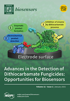
Cover Story (view full-size image): Dithiocarbamate fungicides (DTFs) are widely used to control various fungal diseases in crops and ornamental plants. Maximum residue limits in the order of ppb–ppm are imposed by the current legislation. The specific analytical determination of DTFs is complicated by their low solubility in water and organic solvents. This review summarizes the current analytical procedures used for the analysis of DTF, including chromatography, spectroscopy, and sensor-based methods, and discusses the challenges related to selectivity, sensitivity, and sample preparation. Biosensors based on enzymatic inhibition demonstrated potential as analytical tools for DTFs. Meanwhile, recent studies highlight Raman spectroscopy and various sensors as very promising, provided their selectivity issues are solved. View this paper.
  • Issues are regarded as officially published after their release is announced to the table of contents alert mailing list.
  • You may sign up for e-mail alerts to receive table of contents of newly released issues.
  • PDF is the official format for papers published in both, html and pdf forms. To view the papers in pdf format, click on the "PDF Full-text" link, and use the free Adobe Reader to open them.
Order results
Result details
Section
Select all
Export citation of selected articles as:
16 pages, 554 KiB  
Review
The Ketogenic Diet: Breath Acetone Sensing Technology
by Omar Alkedeh and Ronny Priefer
Biosensors 2021, 11(1), 26; https://doi.org/10.3390/bios11010026 - 19 Jan 2021
Cited by 20 | Viewed by 5308
Abstract
The ketogenic diet, while originally thought to treat epilepsy in children, is now used for weight loss due to increasing evidence indicating that fat is burned more rapidly when there is a low carbohydrate intake. This low carbohydrate intake can lead to elevated [...] Read more.
The ketogenic diet, while originally thought to treat epilepsy in children, is now used for weight loss due to increasing evidence indicating that fat is burned more rapidly when there is a low carbohydrate intake. This low carbohydrate intake can lead to elevated ketone levels in the blood and breath. Breath and blood ketones can be measured to gauge the level of ketosis and allow for adjustment of the diet to meet the user’s needs. Blood ketone levels have been historically used, but now breath acetone sensors are becoming more common due to less invasiveness and convenience. New technologies are being researched in the area of acetone sensors to capitalize on the rising popularity of the diet. Current breath acetone sensors come in the form of handheld breathalyzer devices. Technologies in development mostly consist of semiconductor metal oxides in different physio-chemical formations. These current devices and future technologies are investigated here with regard to utility and efficacy. Technologies currently in development do not have extensive testing of the selectivity of the sensors including the many compounds present in human breath. While some sensors have undergone human testing, the sample sizes are very small, and the testing was not extensive. Data regarding current devices is lacking and more research needs to be done to effectively evaluate current devices if they are to have a place as medical devices. Future technologies are very promising but are still in early development stages. Full article
Show Figures

Figure 1

17 pages, 4710 KiB  
Article
Quantitative E. coli Enzyme Detection in Reporter Hydrogel-Coated Paper Using a Smartphone Camera
by Kawaljit Kaur, Winny Chelangat, Sergey I. Druzhinin, Nancy Wangechi Karuri, Mareike Müller and Holger Schönherr
Biosensors 2021, 11(1), 25; https://doi.org/10.3390/bios11010025 - 19 Jan 2021
Cited by 17 | Viewed by 3908
Abstract
There is a growing demand for rapid and sensitive detection approaches for pathogenic bacteria that can be applied by non-specialists in non-laboratory field settings. Here, the detection of the typical E. coli enzyme β-glucuronidase using a chitosan-based sensing hydrogel-coated paper sensor and the [...] Read more.
There is a growing demand for rapid and sensitive detection approaches for pathogenic bacteria that can be applied by non-specialists in non-laboratory field settings. Here, the detection of the typical E. coli enzyme β-glucuronidase using a chitosan-based sensing hydrogel-coated paper sensor and the detailed analysis of the reaction kinetics, as detected by a smartphone camera, is reported. The chromogenic reporter unit affords an intense blue color in a two-step reaction, which was analyzed using a modified Michaelis–Menten approach. This generalizable approach can be used to determine the limit of detection and comprises an invaluable tool to characterize the performance of lab-in-a-phone type approaches. For the particular system analyzed, the ratio of reaction rate and equilibrium constants of the enzyme–substrate complex are 0.3 and 0.9 pM−1h−1 for β-glucuronidase in phosphate buffered saline and lysogeny broth, respectively. The minimal degree of substrate conversion for detection of the indigo pigment formed during the reaction is 0.15, while the minimal time required for detection in this particular system is ~2 h at an enzyme concentration of 100 nM. Therefore, this approach is applicable for quantitative lab-in-a-phone based point of care detection systems that are based on enzymatic substrate conversion via bacterial enzymes. Full article
(This article belongs to the Section Biosensor Materials)
Show Figures

Graphical abstract

15 pages, 3017 KiB  
Article
Influence of the Electrolyte Salt Concentration on DNA Detection with Graphene Transistors
by Agnes Purwidyantri, Telma Domingues, Jérôme Borme, Joana Rafaela Guerreiro, Andrey Ipatov, Catarina M. Abreu, Marco Martins, Pedro Alpuim and Marta Prado
Biosensors 2021, 11(1), 24; https://doi.org/10.3390/bios11010024 - 17 Jan 2021
Cited by 17 | Viewed by 6085
Abstract
Liquid-gated Graphene Field-Effect Transistors (GFET) are ultrasensitive bio-detection platforms carrying out the graphene’s exceptional intrinsic functionalities. Buffer and dilution factor are prevalent strategies towards the optimum performance of the GFETs. However, beyond the Debye length (λD), the role of the graphene-electrolytes’ ionic species [...] Read more.
Liquid-gated Graphene Field-Effect Transistors (GFET) are ultrasensitive bio-detection platforms carrying out the graphene’s exceptional intrinsic functionalities. Buffer and dilution factor are prevalent strategies towards the optimum performance of the GFETs. However, beyond the Debye length (λD), the role of the graphene-electrolytes’ ionic species interactions on the DNA behavior at the nanoscale interface is complicated. We studied the characteristics of the GFETs under different ionic strength, pH, and electrolyte type, e.g., phosphate buffer (PB), and phosphate buffer saline (PBS), in an automatic portable built-in system. The electrostatic gating and charge transfer phenomena were inferred from the field-effect measurements of the Dirac point position in single-layer graphene (SLG) transistors transfer curves. Results denote that λD is not the main factor governing the effective nanoscale screening environment. We observed that the longer λD was not the determining characteristic for sensitivity increment and limit of detection (LoD) as demonstrated by different types and ionic strengths of measuring buffers. In the DNA hybridization study, our findings show the role of the additional salts present in PBS, as compared to PB, in increasing graphene electron mobility, electrostatic shielding, intermolecular forces and DNA adsorption kinetics leading to an improved sensitivity. Full article
(This article belongs to the Special Issue Field-Effect Transistors for Biosensing Applications)
Show Figures

Figure 1

11 pages, 2281 KiB  
Letter
Effective Isolation for Lung Carcinoma Cells Based on Immunomagnetic Separation in a Microfluidic Channel
by Hien Vu-Dinh, Hui Feng and Chun-Ping Jen
Biosensors 2021, 11(1), 23; https://doi.org/10.3390/bios11010023 - 16 Jan 2021
Cited by 6 | Viewed by 2624
Abstract
In this paper, we developed an isolation system for A549 human lung carcinoma cells as an effective factor for the early diagnosis of lung cancer. A microfluidic immunomagnetic method was used, in which the combination of immunomagnetic separation and a microfluidic system allowed [...] Read more.
In this paper, we developed an isolation system for A549 human lung carcinoma cells as an effective factor for the early diagnosis of lung cancer. A microfluidic immunomagnetic method was used, in which the combination of immunomagnetic separation and a microfluidic system allowed for increased isolation efficiency with uncomplicated manipulation. In the microfluidic immunomagnetic strategy, A549 cells were combined with aptamer-conjugated carboxylated magnetic beads and then collected in a specified region by applying a magnetic field. The results were recorded using a fluorescence microscope, and the captured targets were then quantified. The isolation efficiency of A549 cells is up to 77.8%. This paper developed a simple working procedure, which is less time consuming, high-throughput, and trustworthy for the isolation of A549 cells. This procedure can be a useful reference method for the development of an effective diagnosis and treatment method for lung cancer in the future. Full article
(This article belongs to the Special Issue Aptamers to Replace Antibodies for in vitro Diagnostics)
Show Figures

Figure 1

7 pages, 525 KiB  
Brief Report
Performance of the Intermittently Scanned Continuous Glucose Monitoring (isCGM) System during a High Oral Glucose Challenge in Adults with Type 1 Diabetes—A Prospective Secondary Outcome Analysis
by Othmar Moser, Norbert Tripolt, Peter Pferschy, Anna Obermayer, Harald Kojzar, Alexander Mueller, Hakan Yildirim, Caren Sourij, Max Eckstein and Harald Sourij
Biosensors 2021, 11(1), 22; https://doi.org/10.3390/bios11010022 - 15 Jan 2021
Cited by 6 | Viewed by 2495
Abstract
To assess intermittently scanned continuous glucose monitoring (isCGM) performance for different rates of change in plasma glucose (RCPG) during glycemic challenges in type 1 diabetes (T1D). Nineteen people with T1D (7 females; age 35 ± 11 years; HbA1c 7.3 ± 0.6% (56 [...] Read more.
To assess intermittently scanned continuous glucose monitoring (isCGM) performance for different rates of change in plasma glucose (RCPG) during glycemic challenges in type 1 diabetes (T1D). Nineteen people with T1D (7 females; age 35 ± 11 years; HbA1c 7.3 ± 0.6% (56 ± 7 mmol/mol)) performing two glycemic challenges (OGTT) were included. During OGTTs, plasma glucose was compared against sensor glucose for timepoints 0 min (pre-OGTT), +15 min, +30 min, +60 min, +120 min, +180 min, and +240 min by means of median absolute (relative) difference (MARD and MAD) and Clarke Error Grid (CEG), then was stratified for RCPG and glycemic ranges. Overall, MARD was 8.3% (4.0–14.8) during hypoglycemia level 1 18.8% (15.8–22.0), euglycemia 9.5% (4.3–15.1), hyperglycemia level 1 9.4% (4.0–17.2), and hyperglycemia level 2 7.1% (3.3–11.9). The MARD was associated with the RCPG (p < 0.0001), detailing significant differences in comparison of low, moderate, high, and very high RCPG (p = 0.014). Overall, CEG resulted in 88% (212 values) of comparison points in zone A, 12% (29 values) in zone B, and 0.4% (1 value) in zone D. The isCGM system was accurate during OGTTs. Its performance was dependent on the RCPG and showed an overestimation of the actual reference glucose during hypoglycemia. Full article
(This article belongs to the Section Biosensors and Healthcare)
Show Figures

Figure 1

14 pages, 5392 KiB  
Article
Surface Plasmon Resonance Based on Molecularly Imprinted Polymeric Film for l-Phenylalanine Detection
by Duygu Çimen, Nilay Bereli and Adil Denizli
Biosensors 2021, 11(1), 21; https://doi.org/10.3390/bios11010021 - 15 Jan 2021
Cited by 20 | Viewed by 3033
Abstract
In this study, we designed a simple, rapid, sensitive and selective surface plasmon resonance (SPR) sensor for detection of L-phenylalaine by utilizing molecular imprinting technology. l-phenylalanine imprinted and non-imprinted poly(2-hydroxyethyl methacrylate-methacryloyl-l-phenylalanine) polymeric films were synthesized onto SPR chip surfaces using [...] Read more.
In this study, we designed a simple, rapid, sensitive and selective surface plasmon resonance (SPR) sensor for detection of L-phenylalaine by utilizing molecular imprinting technology. l-phenylalanine imprinted and non-imprinted poly(2-hydroxyethyl methacrylate-methacryloyl-l-phenylalanine) polymeric films were synthesized onto SPR chip surfaces using ultraviolet polymerization. l-phenyalanine imprinted and non-imprinted SPR sensors were characterized by using contact angle, atomic force microscopy and ellipsometry. After characterization studies, kinetic studies were carried out in the concentration range of 5.0–400.0 μM. The limit of detection and quantification were obtained as 0.0085 and 0.0285 μM, respectively. The response time for the test including equilibration, adsorption and desorption was approximately 9 min. The selectivity studies of the l-phenylalanine imprinted SPR sensor was performed in the presence of d-phenylalanine and l-tryptophan. Validation studies were carried out via enzyme-linked immunosorbent analysis technique in order to demonstrate the applicability and superiority of the l-phenylalanine imprinted SPR sensor. Full article
Show Figures

Figure 1

15 pages, 1648 KiB  
Article
Kynurenic Acid Electrochemical Immunosensor: Blood-Based Diagnosis of Alzheimer’s Disease
by Jose Marrugo-Ramírez, Montserrat Rodríguez-Núñez, M.-Pilar Marco, Mónica Mir and Josep Samitier
Biosensors 2021, 11(1), 20; https://doi.org/10.3390/bios11010020 - 12 Jan 2021
Cited by 12 | Viewed by 3949
Abstract
Alzheimer’s disease (AD) is a neurodegenerative disorder, characterized by a functional deterioration of the brain. Currently, there are selected biomarkers for its diagnosis in cerebrospinal fluid. However, its extraction has several disadvantages for the patient. Therefore, there is an urgent need for a [...] Read more.
Alzheimer’s disease (AD) is a neurodegenerative disorder, characterized by a functional deterioration of the brain. Currently, there are selected biomarkers for its diagnosis in cerebrospinal fluid. However, its extraction has several disadvantages for the patient. Therefore, there is an urgent need for a detection method using sensitive and selective blood-based biomarkers. Kynurenic acid (KYNA) is a potential biomarker candidate for this purpose. The alteration of the KYNA levels in blood has been related with inflammatory processes in the brain, produced as a protective function when neurons are damaged. This paper describes a novel electrochemical immunosensor for KYNA detection, based on successive functionalization multi-electrode array. The resultant sensor was characterized by cyclic voltammetry (CV), chronoamperometry (CA), and electrochemical impedance spectroscopy (EIS). The proposed biosensor detects KYNA within a linear calibration range from 10 pM to 100 nM using CA and EIS, obtaining a limit of detection (LOD) of 16.9 pM and 37.6 pM in buffer, respectively, being the lowest reported LOD for this biomarker. Moreover, to assess our device closer to the real application, the developed immunosensor was also tested under human serum matrix, obtaining an LOD of 391.71 pM for CA and 278.8 pM for EIS with diluted serum. Full article
(This article belongs to the Special Issue Biosensors for Neurodegenerative Diseases and Related Disorders)
Show Figures

Graphical abstract

10 pages, 1805 KiB  
Article
Detection of Hypoxanthine from Inosine and Unusual Hydrolysis of Immunosuppressive Drug Azathioprine through the Formation of a Diruthenium(III) System
by Marta Orts-Arroyo, Isabel Castro and José Martínez-Lillo
Biosensors 2021, 11(1), 19; https://doi.org/10.3390/bios11010019 - 11 Jan 2021
Cited by 8 | Viewed by 2734
Abstract
Hypoxanthine (hpx) is an important molecule for both biochemistry research and biomedical applications. It is involved in several biological processes associated to energy and purine metabolism and has been proposed as a biomarker for a variety of disease states. Consequently, the discovery and [...] Read more.
Hypoxanthine (hpx) is an important molecule for both biochemistry research and biomedical applications. It is involved in several biological processes associated to energy and purine metabolism and has been proposed as a biomarker for a variety of disease states. Consequently, the discovery and development of systems suitable for the detection of hypoxanthine is pretty appealing in this research field. Thus, we have obtained a stable diruthenium (III) compound in its dehydrated and hydrated forms with formula [{Ru(µ-Cl)(µ-hpx)}2Cl4] (1a) and [{Ru(µ-Cl)(µ-hpx)}2Cl4]·2H2O (1b), respectively. This purine-based diruthenium(III) system was prepared from two very different starting materials, namely, inosine and azathioprine, the latter being an immunosuppressive drug. Remarkably, it was observed that an unusual azathioprine hydrolysis occurs in the presence of ruthenium, thus generating hypoxanthine instead of the expected 6-mercaptopurine antimetabolite, so that the hpx molecule is linked to two ruthenium(III) ions. 1a and 1b were characterized through IR, SEM, powder and single-crystal X-ray Diffraction and Cyclic Voltammetry (CV). The electrochemical studies allowed us to detect the hpx molecule when coordinated to ruthenium in the reported compound. The grade of sensitivity, repeatability and stability reached by this diruthenium system make it potentially useful and could provide a first step to develop new sensor devices suitable to detect hypoxanthine. Full article
(This article belongs to the Section Biosensor and Bioelectronic Devices)
Show Figures

Graphical abstract

18 pages, 2426 KiB  
Article
Aptamer–Target–Gold Nanoparticle Conjugates for the Quantification of Fumonisin B1
by Vicente Antonio Mirón-Mérida, Yadira González-Espinosa, Mar Collado-González, Yun Yun Gong, Yuan Guo and Francisco M. Goycoolea
Biosensors 2021, 11(1), 18; https://doi.org/10.3390/bios11010018 - 08 Jan 2021
Cited by 17 | Viewed by 3651
Abstract
Fumonisin B1 (FB1), a mycotoxin classified as group 2B hazard, is of high importance due to its abundance and occurrence in varied crops. Conventional methods for detection are sensitive and selective; however, they also convey disadvantages such as long assay times, expensive equipment [...] Read more.
Fumonisin B1 (FB1), a mycotoxin classified as group 2B hazard, is of high importance due to its abundance and occurrence in varied crops. Conventional methods for detection are sensitive and selective; however, they also convey disadvantages such as long assay times, expensive equipment and instrumentation, complex procedures, sample pretreatment and unfeasibility for on-site analysis. Therefore, there is a need for quick, simple and affordable quantification methods. On that note, aptamers (ssDNA) are a good alternative for designing specific and sensitive biosensing techniques. In this work, the assessment of the performance of two aptamers (40 and 96 nt) on the colorimetric quantification of FB1 was determined by conducting an aptamer–target incubation step, followed by the addition of gold nanoparticles (AuNPs) and NaCl. Although MgCl2 and Tris-HCl were, respectively, essential for aptamer 96 and 40 nt, the latter was not specific for FB1. Alternatively, the formation of Aptamer (96 nt)–FB1–AuNP conjugates in MgCl2 exhibited stabilization to NaCl-induced aggregation at increasing FB1 concentrations. The application of asymmetric flow field-flow fractionation (AF4) allowed their size separation and characterization by a multidetection system (UV-VIS, MALS and DLS online), with a reduction in the limit of detection from 0.002 µg/mL to 56 fg/mL. Full article
Show Figures

Figure 1

12 pages, 2158 KiB  
Communication
Detection of CRISPR-Cas9-Mediated Mutations Using a Carbon Nanotube-Modified Electrochemical Genosensor
by Ezgi Kivrak, Tekle Pauzaite, Nikki A. Copeland, John G. Hardy, Pinar Kara, Melike Firlak, Atike I. Yardimci, Selahattin Yilmaz, Fahreddin Palaz and Mehmet Ozsoz
Biosensors 2021, 11(1), 17; https://doi.org/10.3390/bios11010017 - 08 Jan 2021
Cited by 8 | Viewed by 5804
Abstract
The CRISPR-Cas9 system has facilitated the genetic modification of various model organisms and cell lines. The outcomes of any CRISPR-Cas9 assay should be investigated to ensure/improve the precision of genome engineering. In this study, carbon nanotube-modified disposable pencil graphite electrodes (CNT/PGEs) were used [...] Read more.
The CRISPR-Cas9 system has facilitated the genetic modification of various model organisms and cell lines. The outcomes of any CRISPR-Cas9 assay should be investigated to ensure/improve the precision of genome engineering. In this study, carbon nanotube-modified disposable pencil graphite electrodes (CNT/PGEs) were used to develop a label-free electrochemical nanogenosensor for the detection of point mutations generated in the genome by using the CRISPR-Cas9 system. Carbodiimide chemistry was used to immobilize the 5′-aminohexyl-linked inosine-substituted probe on the surface of the sensor. After hybridization between the target sequence and probe at the sensor surface, guanine oxidation signals were monitored using differential pulse voltammetry (DPV). Optimization of the sensitivity of the nanogenoassay resulted in a lower detection limit of 213.7 nM. The nanogenosensor was highly specific for the detection of the precisely edited DNA sequence. This method allows for a rapid and easy investigation of the products of CRISPR-based gene editing and can be further developed to an array system for multiplex detection of different-gene editing outcomes. Full article
(This article belongs to the Special Issue Application of CRISPR Cas Systems for Biosensing)
Show Figures

Figure 1

12 pages, 1934 KiB  
Article
Development of a Sensitive Self-Powered Glucose Biosensor Based on an Enzymatic Biofuel Cell
by Kantapat Chansaenpak, Anyanee Kamkaew, Sireerat Lisnund, Pannaporn Prachai, Patipat Ratwirunkit, Thitichaya Jingpho, Vincent Blay and Piyanut Pinyou
Biosensors 2021, 11(1), 16; https://doi.org/10.3390/bios11010016 - 07 Jan 2021
Cited by 35 | Viewed by 8331
Abstract
Biofuel cells allow for constructing sensors that leverage the specificity of enzymes without the need for an external power source. In this work, we design a self-powered glucose sensor based on a biofuel cell. The redox enzymes glucose dehydrogenase (NAD-GDH), glucose oxidase (GOx), [...] Read more.
Biofuel cells allow for constructing sensors that leverage the specificity of enzymes without the need for an external power source. In this work, we design a self-powered glucose sensor based on a biofuel cell. The redox enzymes glucose dehydrogenase (NAD-GDH), glucose oxidase (GOx), and horseradish peroxidase (HRP) were immobilized as biocatalysts on the electrodes, which were previously engineered using carbon nanostructures, including multi-wall carbon nanotubes (MWCNTs) and reduced graphene oxide (rGO). Additional polymers were also introduced to improve biocatalyst immobilization. The reported design offers three main advantages: (i) by using glucose as the substrate for the both anode and cathode, a more compact and robust design is enabled, (ii) the system operates under air-saturating conditions, with no need for gas purge, and (iii) the combination of carbon nanostructures and a multi-enzyme cascade maximizes the sensitivity of the biosensor. Our design allows the reliable detection of glucose in the range of 0.1–7.0 mM, which is perfectly suited for common biofluids and industrial food samples. Full article
(This article belongs to the Special Issue Electrochemical Biosensors)
Show Figures

Figure 1

15 pages, 4180 KiB  
Article
An Electrochemical Strategy for the Simultaneous Detection of Doxorubicin and Simvastatin for Their Potential Use in the Treatment of Cancer
by Iulia Rus, Mihaela Tertiș, Cristina Barbălată, Alina Porfire, Ioan Tomuță, Robert Săndulescu and Cecilia Cristea
Biosensors 2021, 11(1), 15; https://doi.org/10.3390/bios11010015 - 03 Jan 2021
Cited by 10 | Viewed by 2701
Abstract
The aim of this study was to develop a disposable, simple, fast, and sensitive sensor for the simultaneous electrochemical detection of doxorubicin (DOX) and simvastatin (SMV), which could be used in preclinical studies for the development of new pharmaceutical formulations for drug delivery. [...] Read more.
The aim of this study was to develop a disposable, simple, fast, and sensitive sensor for the simultaneous electrochemical detection of doxorubicin (DOX) and simvastatin (SMV), which could be used in preclinical studies for the development of new pharmaceutical formulations for drug delivery. Firstly, the electrochemical behavior of each molecule was analyzed regarding the influence of electrode material, electrolyte solution, and scan rate. After this, the proper electrode material, electrolyte solution, and scan rate for both active substances were chosen, and a linear sweep voltammetry procedure was optimized for simultaneous detection. Two chronoamperometry procedures were tested, one for the detection of DOX in the presence of SMV, and the other one for the detection of DOX and SMV together. Finally, calibration curves for DOX and SMV in the presence of each other were obtained using both electrochemical methods and the results were compared. The use of amperometry allowed for a better limit of detection (DOX: 0.1 μg/mL; SMV: 0.7 μg/mL) than the one obtained in voltammetry (1.5 μg/mL for both drugs). The limits of quantification using amperometry were 0.5 μg/mL for DOX (dynamic range: 0.5–65 μg/mL) and 2 μg/mL for SMV (dynamic range: 2–65 μg/mL), while using voltammetry 1 μg/mL was obtained for DOX (dynamic range: 1–100 μg/mL) and 5 μg/mL for SMV (dynamic range: 5–100 μg/mL). This detection strategy represents a promising tool for the analysis of new pharmaceutical formulations for targeted drug delivery containing both drugs, whose association was proven to bring benefits in the treatment of cancer. Full article
(This article belongs to the Special Issue Electrochemical Biosensors)
Show Figures

Graphical abstract

32 pages, 9919 KiB  
Review
Survey of Saliva Components and Virus Sensors for Prevention of COVID-19 and Infectious Diseases
by Priya Kishor Dave, Roberto Rojas-Cessa, Ziqian Dong and Vatcharapan Umpaichitra
Biosensors 2021, 11(1), 14; https://doi.org/10.3390/bios11010014 - 31 Dec 2020
Cited by 19 | Viewed by 9424
Abstract
The United States Centers for Disease Control and Prevention considers saliva contact the lead transmission mean of severe acute respiratory syndrome coronavirus 2 (SARS-CoV-2), which causes the coronavirus disease 2019 (COVID-19). Saliva droplets or aerosols expelled by sneezing, coughing, breathing, and talking may [...] Read more.
The United States Centers for Disease Control and Prevention considers saliva contact the lead transmission mean of severe acute respiratory syndrome coronavirus 2 (SARS-CoV-2), which causes the coronavirus disease 2019 (COVID-19). Saliva droplets or aerosols expelled by sneezing, coughing, breathing, and talking may carry this virus. People in close distance may be exposed directly to these droplets or indirectly when touching the droplets that fall on surrounding surfaces and ending up contracting COVID-19 after touching the mucosa tissue of their faces. It is of great interest to quickly and effectively detect the presence of SARS-CoV-2 in an environment, but the existing methods only work in laboratory settings, to the best of our knowledge. However, it may be possible to detect the presence of saliva in the environment and proceed with prevention measures. However, detecting saliva itself has not been documented in the literature. On the other hand, many sensors that detect different organic components in saliva to monitor a person’s health and diagnose different diseases, ranging from diabetes to dental health, have been proposed and they may be used to detect the presence of saliva. This paper surveys sensors that detect organic and inorganic components of human saliva. Humidity sensors are also considered in the detection of saliva because a large portion of saliva is water. Moreover, sensors that detect infectious viruses are also included as they may also be embedded into saliva sensors for a confirmation of the presence of the virus. A classification of sensors by their working principles and the substances they detect is presented, including the sensors’ specifications, sample size, and sensitivity. Indications of which sensors are portable and suitable for field application are presented. This paper also discusses future research and challenges that must be resolved to realize practical saliva sensors. Such sensors may help minimize the spread of not only COVID-19 but also other infectious diseases. Full article
(This article belongs to the Section Biosensor and Bioelectronic Devices)
Show Figures

Figure 1

12 pages, 2387 KiB  
Article
Display of Microbial Glucose Dehydrogenase and Cholesterol Oxidase on the Yeast Cell Surface for the Detection of Blood Biochemical Parameters
by Shiyao Zhao, Dong Guo, Quanchao Zhu, Weiwang Dou and Wenjun Guan
Biosensors 2021, 11(1), 13; https://doi.org/10.3390/bios11010013 - 30 Dec 2020
Cited by 12 | Viewed by 3338
Abstract
High levels of blood glucose are always associated with numerous complications including cholesterol abnormalities. Therefore, it is important to simultaneously monitor blood glucose and cholesterol levels in patients with diabetes during the management of chronic diseases. In this study, a glucose dehydrogenase from [...] Read more.
High levels of blood glucose are always associated with numerous complications including cholesterol abnormalities. Therefore, it is important to simultaneously monitor blood glucose and cholesterol levels in patients with diabetes during the management of chronic diseases. In this study, a glucose dehydrogenase from Aspergillus oryzae TI and a cholesterol oxidase from Chromobacterium sp. DS-1 were displayed on the surface of Saccharomyces cerevisiae, respectively, using the yeast surface display system at a high copy number. In addition, two whole-cell biosensors were constructed through the immobilization of the above yeast cells on electrodes, for electrochemical detection of glucose and cholesterol. The assay time was 8.5 s for the glucose biosensors and 30 s for the cholesterol biosensors. Under optimal conditions, the cholesterol biosensor exhibited a linear range from 2 to 6 mmol·L−1. The glucose biosensor responded efficiently to the presence of glucose at a concentration range of 20–600 mg·dL−1 (1.4–33.3 mmol·L−1) and showed excellent anti-xylose interference properties. Both biosensors exhibited good performance at room temperature and remained stable over a three-week storage period. Full article
(This article belongs to the Special Issue Biosensors for Diagnosis and Monitoring)
Show Figures

Figure 1

26 pages, 3379 KiB  
Review
Advances in the Detection of Dithiocarbamate Fungicides: Opportunities for Biosensors
by Pablo Fanjul-Bolado, Ronen Fogel, Janice Limson, Cristina Purcarea and Alina Vasilescu
Biosensors 2021, 11(1), 12; https://doi.org/10.3390/bios11010012 - 30 Dec 2020
Cited by 29 | Viewed by 4572
Abstract
Dithiocarbamate fungicides (DTFs) are widely used to control various fungal diseases in crops and ornamental plants. Maximum residual limits in the order of ppb-ppm are currently imposed by legislation to prevent toxicity problems associated with excessive use of DTFs. The specific analytical determination [...] Read more.
Dithiocarbamate fungicides (DTFs) are widely used to control various fungal diseases in crops and ornamental plants. Maximum residual limits in the order of ppb-ppm are currently imposed by legislation to prevent toxicity problems associated with excessive use of DTFs. The specific analytical determination of DTFs is complicated by their low solubility in water and organic solvents. This review summarizes the current analytical procedures used for the analysis of DTF, including chromatography, spectroscopy, and sensor-based methods and discusses the challenges related to selectivity, sensitivity, and sample preparation. Biosensors based on enzymatic inhibition demonstrated potential as analytical tools for DTFs and warrant further research, considering novel enzymes from extremophilic sources. Meanwhile, Raman spectroscopy and various sensors appear very promising, provided the selectivity issues are solved. Full article
(This article belongs to the Special Issue Biosensors: 10th Anniversary Feature Papers)
Show Figures

Figure 1

2 pages, 152 KiB  
Editorial
Biosensors for the Multiplex Detection of Inflammatory Disease Biomarkers
by Ali Mobasheri
Biosensors 2021, 11(1), 11; https://doi.org/10.3390/bios11010011 - 28 Dec 2020
Cited by 4 | Viewed by 2324
Abstract
A biosensor is an analytical device used for the real-time detection and measurement of a chemical or biochemical substance [...] Full article
Show Figures

Graphical abstract

14 pages, 2004 KiB  
Article
Potentiometric Biosensing of Ascorbic Acid, Uric Acid, and Cysteine in Microliter Volumes Using Miniaturized Nanoporous Gold Electrodes
by Christopher J. Freeman, Borkat Ullah, Md. Shafiul Islam and Maryanne M. Collinson
Biosensors 2021, 11(1), 10; https://doi.org/10.3390/bios11010010 - 28 Dec 2020
Cited by 17 | Viewed by 4215
Abstract
Potentiometric redox sensing is a relatively inexpensive and passive approach to evaluate the overall redox state of complex biological and environmental solutions. The ability to make such measurements in ultra-small volumes using high surface area, nanoporous electrodes is of particular importance as such [...] Read more.
Potentiometric redox sensing is a relatively inexpensive and passive approach to evaluate the overall redox state of complex biological and environmental solutions. The ability to make such measurements in ultra-small volumes using high surface area, nanoporous electrodes is of particular importance as such electrodes can improve the rates of electron transfer and reduce the effects of biofouling on the electrochemical signal. This work focuses on the fabrication of miniaturized nanoporous gold (NPG) electrodes with a high surface area and a small footprint for the potentiometric redox sensing of three biologically relevant redox molecules (ascorbic acid, uric acid, and cysteine) in microliter volumes. The NPG electrodes were inexpensively made by attaching a nanoporous gold leaf prepared by dealloying 12K gold in nitric acid to a modified glass capillary (1.5 mm id) and establishing an electrode connection with copper tape. The surface area of the electrodes was ~1.5 cm2, providing a roughness factor of ~16 relative to the geometric area of 0.09 cm2. Scanning electron microscopy confirmed the nanoporous framework. A linear dependence between the open-circuit potential (OCP) and the logarithm of concentration (e.g., Nernstian-like behavior) was obtained for all three redox molecules in 100 μL buffered solutions. As a first step towards understanding a real system, the response associated with changing the concentration of one redox species in the presence of the other two was examined. These results show that at NPG, the redox potential of a solution containing biologically relevant concentrations of ascorbic acid, uric acid, and cysteine is strongly influenced by ascorbic acid. Such information is important for the measurement of redox potentials in complex biological solutions. Full article
(This article belongs to the Section Biosensor and Bioelectronic Devices)
Show Figures

Figure 1

31 pages, 3357 KiB  
Review
Solid State Sensors for Hydrogen Peroxide Detection
by Vinay Patel, Peter Kruse and Ponnambalam Ravi Selvaganapathy
Biosensors 2021, 11(1), 9; https://doi.org/10.3390/bios11010009 - 25 Dec 2020
Cited by 38 | Viewed by 6603
Abstract
Hydrogen peroxide (H2O2) is a key molecule in numerous physiological, industrial, and environmental processes. H2O2 is monitored using various methods like colorimetry, luminescence, fluorescence, and electrochemical methods. Here, we aim to provide a comprehensive review of [...] Read more.
Hydrogen peroxide (H2O2) is a key molecule in numerous physiological, industrial, and environmental processes. H2O2 is monitored using various methods like colorimetry, luminescence, fluorescence, and electrochemical methods. Here, we aim to provide a comprehensive review of solid state sensors to monitor H2O2. The review covers three categories of sensors: chemiresistive, conductometric, and field effect transistors. A brief description of the sensing mechanisms of these sensors has been provided. All three sensor types are evaluated based on the sensing parameters like sensitivity, limit of detection, measuring range and response time. We highlight those sensors which have advanced the field by using innovative materials or sensor fabrication techniques. Finally, we discuss the limitations of current solid state sensors and the future directions for research and development in this exciting area. Full article
(This article belongs to the Section Nano- and Micro-Technologies in Biosensors)
Show Figures

Figure 1

13 pages, 2629 KiB  
Article
Development of Fluorescence In Situ Hybridization as a Rapid, Accurate Method for Detecting Coliforms in Water Samples
by Jong-Tar Kuo, Li-Li Chang, Chia-Yuan Yen, Teh-Hua Tsai, Yu-Chi Chang, Yu-Tang Huang and Ying-Chien Chung
Biosensors 2021, 11(1), 8; https://doi.org/10.3390/bios11010008 - 24 Dec 2020
Cited by 13 | Viewed by 3919
Abstract
Coliform bacteria are indicators of water quality; however, most detection methods for coliform bacteria are time-consuming and nonspecific. Here, we developed a fluorescence in situ hybridization (FISH) approach to detect four types of coliform bacteria, including Escherichia coli, Klebsiella pneumoniae, Enterobacter aerogenes [...] Read more.
Coliform bacteria are indicators of water quality; however, most detection methods for coliform bacteria are time-consuming and nonspecific. Here, we developed a fluorescence in situ hybridization (FISH) approach to detect four types of coliform bacteria, including Escherichia coli, Klebsiella pneumoniae, Enterobacter aerogenes, and Citrobacter freundii, simultaneously in water samples using specific probes for 16S rRNA. This FISH method was applied to detect coliform bacteria in simulated water and domestic wastewater samples and compared with traditional detection methods (e.g., plate counting, multiple-tube fermentation (MTF) technique, and membrane filter (MF) technique). Optimal FISH conditions for detecting the four types of coliforms were found to be fixation in 3% paraformaldehyde at 4 °C for 2 h and hybridization at 50 °C for 1.5 h. By comparing FISH with plate counting, MTF, MF, and a commercial detection kit, we found that FISH had the shortest detection time and highest accuracy for the identification of coliform bacteria in simulated water and domestic wastewater samples. Moreover, the developed method could simultaneously detect individual species and concentrations of coliform bacteria. Overall, our findings indicated that FISH could be used as a rapid, accurate biosensor system for simultaneously detecting four types of coliform bacteria to ensure water safety. Full article
Show Figures

Figure 1

9 pages, 1903 KiB  
Article
Sandwich ELISA-Based Electrochemical Biosensor for Leptin in Control and Diet-Induced Obesity Mouse Model
by Ryong Sung and Yun Seok Heo
Biosensors 2021, 11(1), 7; https://doi.org/10.3390/bios11010007 - 24 Dec 2020
Cited by 11 | Viewed by 5127
Abstract
Leptin is a peptide hormone produced primarily in adipose tissues. Leptin is considered a biomarker associated with obesity and obesity-mediated diseases. Biosensor detection of leptin in the blood may play a critical role as an indicator of dynamic pathological changes. In this paper, [...] Read more.
Leptin is a peptide hormone produced primarily in adipose tissues. Leptin is considered a biomarker associated with obesity and obesity-mediated diseases. Biosensor detection of leptin in the blood may play a critical role as an indicator of dynamic pathological changes. In this paper, we introduce an electrochemical biosensor that adopts o-Phenylenediamine (oPD) on screen-printed gold electrodes (SPGEs) for detecting the leptin from a mouse model of diet-induced obesity (DIO). A linear calibration curve for the leptin concentration was obtained in the ranges from 0.1 to 20 ng/mL with a lower detection limit of 0.033 ng/mL. The leptin concentration was quantified with HRP (horseradish peroxidase)-catalyzed oxidation of oPD by two voltammetry methods: cyclic voltammetry (CV) and square-wave voltammetry (SWV). The proposed sandwich enzyme-linked immunosorbent assay (ELISA)-based electrochemical biosensor for the leptin in mouse blood serum showed high stability, sensitivity, selectivity, and effectivity compared to the commercial Leptin ELISA measurement. Thus, we believe that this leptin biosensor can be a sensitive analytical tool to detect low-levels of biomarkers in clinics and point-of-care testing (POCT). Full article
(This article belongs to the Section Biosensors and Healthcare)
Show Figures

Figure 1

13 pages, 3234 KiB  
Article
Fluorescent Probe for Ag+ Detection Using SYBR GREEN I and C-C Mismatch
by Xiaohong Zhou, Abdul Ghaffar Memon, Weiming Sun, Fang Fang and Jinsong Guo
Biosensors 2021, 11(1), 6; https://doi.org/10.3390/bios11010006 - 24 Dec 2020
Cited by 9 | Viewed by 2623
Abstract
Among heavy metals silver ions (Ag+) severely impact water, the environment and have serious side effects on human health. This article proposes a facile and ultrasensitive fluorescent probe for the detection of Ag+ ions using SYBR Green I (SGI) and [...] Read more.
Among heavy metals silver ions (Ag+) severely impact water, the environment and have serious side effects on human health. This article proposes a facile and ultrasensitive fluorescent probe for the detection of Ag+ ions using SYBR Green I (SGI) and cytosine-rich (C-rich) silver-specific oligonucleotide (SSO). Maximum fluorescent intensities with the highest sensitivity were obtained using a 0.61 dye/SSO base ratio (DBR). The established sensing principle using the optimized parameters for bath temperature, SSO concentration, DBR, ionic strength, pH, reaction time, incubation duration and temperature effect achieved a sensitive limit of detection of 59.9 nM for silver ions (calculated through 3σ, n = 11) with a linear working range of 100–1000 nM and 0.997 R2. The total time for one assay is below 10 min; The relative standard derivation for ten repeated measurements is 8.6%. No blatant interferences were observed in the selectivity test when fluorescent probe is evaluated by investigating the effects of 11 common interference factors in the aqueous matrix. In extreme cases, three false-negative factors were observed, including calcium hardness, magnesium hardness, and hypochlorite. The recovery ratios were within the range of 79~110% for three types of diluted water. Full article
(This article belongs to the Special Issue Last Advances in Optical Biosensors)
Show Figures

Figure 1

13 pages, 2263 KiB  
Article
Label-Free Amperometric Immunosensor Based on Versatile Carbon Nanofibers Network Coupled with Au Nanoparticles for Aflatoxin B1 Detection
by Yunhong Huang, Fei Zhu, Jinhua Guan, Wei Wei and Long Zou
Biosensors 2021, 11(1), 5; https://doi.org/10.3390/bios11010005 - 24 Dec 2020
Cited by 22 | Viewed by 3022
Abstract
Facile detection methods for mycotoxins with high sensitivity are of great significance to prevent potential harm to humans. Herein, a label-free amperometric immunosensor based on a 3-D interconnected carbon nanofibers (CNFs) network coupled with well-dispersed Au nanoparticles (AuNPs) is proposed for the quantitative [...] Read more.
Facile detection methods for mycotoxins with high sensitivity are of great significance to prevent potential harm to humans. Herein, a label-free amperometric immunosensor based on a 3-D interconnected carbon nanofibers (CNFs) network coupled with well-dispersed Au nanoparticles (AuNPs) is proposed for the quantitative determination of aflatoxin B1 (AFB1) in wheat samples. In comparison to common carbon nanotubes (CNTs), the CNFs network derived from bacterial cellulose biomass possesses a unique hierarchically porous structure for fast electrolyte diffusion and a larger electrochemical active area, which increases the peak current of differential pulse voltammetry curves for an immunosensor. Combined with AuNPs that are incorporated into CNFs by using linear polyethyleneimine (PEI) as a soft template, the developed Au@PEI@CNFs-based immunosensor showed a good linear response to AFB1 concentrations in a wide range from 0.05 to 25 ng mL−1. The limit of detection was 0.027 ng mL−1 (S/N = 3), more than three-fold lower than that of an Au@PEI@CNTs-based sensor. The reproducibility, storage stability and selectivity of the immunosensor were proved to be satisfactory. The developed immunosensor with appropriate sensitivity and reliable accuracy can be used for the analysis of wheat samples. Full article
(This article belongs to the Special Issue Nanocarbon-Based Biosensors)
Show Figures

Figure 1

13 pages, 3782 KiB  
Article
Demonstration of a Label-Free and Low-Cost Optical Cavity-Based Biosensor Using Streptavidin and C-Reactive Protein
by Donggee Rho and Seunghyun Kim
Biosensors 2021, 11(1), 4; https://doi.org/10.3390/bios11010004 - 24 Dec 2020
Cited by 5 | Viewed by 2974
Abstract
An optical cavity-based biosensor (OCB) has been developed for point-of-care (POC) applications. This label-free biosensor employs low-cost components and simple fabrication processes to lower the overall cost while achieving high sensitivity using a differential detection method. To experimentally demonstrate its limit of detection [...] Read more.
An optical cavity-based biosensor (OCB) has been developed for point-of-care (POC) applications. This label-free biosensor employs low-cost components and simple fabrication processes to lower the overall cost while achieving high sensitivity using a differential detection method. To experimentally demonstrate its limit of detection (LOD), we conducted biosensing experiments with streptavidin and C-reactive protein (CRP). The optical cavity structure was optimized further for better sensitivity and easier fluid control. We utilized the polymer swelling property to fine-tune the optical cavity width, which significantly improved the success rate to produce measurable samples. Four different concentrations of streptavidin were tested in triplicate, and the LOD of the OCB was determined to be 1.35 nM. The OCB also successfully detected three different concentrations of human CRP using biotinylated CRP antibody. The LOD for CRP detection was 377 pM. All measurements were done using a small sample volume of 15 µL within 30 min. By reducing the sensing area, improving the functionalization and passivation processes, and increasing the sample volume, the LOD of the OCB are estimated to be reduced further to the femto-molar range. Overall, the demonstrated capability of the OCB in the present work shows great potential to be used as a promising POC biosensor. Full article
(This article belongs to the Special Issue Photonic Biosensors: Detection, Analysis and Medical Diagnostics)
Show Figures

Figure 1

18 pages, 2796 KiB  
Article
Molecularly Imprinted Polyscopoletin for the Electrochemical Detection of the Chronic Disease Marker Lysozyme
by Tiziano Di Giulio, Elisabetta Mazzotta and Cosimino Malitesta
Biosensors 2021, 11(1), 3; https://doi.org/10.3390/bios11010003 - 23 Dec 2020
Cited by 33 | Viewed by 4243
Abstract
Herein we report the electropolymerization of a scopoletin based molecularly imprinted polymer (MIP) for the detection of lysozyme (Lyz), an enzymatic marker of several diseases in mammalian species. Two different approaches have been used for the imprinting of lysozyme based, respectively, on the [...] Read more.
Herein we report the electropolymerization of a scopoletin based molecularly imprinted polymer (MIP) for the detection of lysozyme (Lyz), an enzymatic marker of several diseases in mammalian species. Two different approaches have been used for the imprinting of lysozyme based, respectively, on the use of a monomer-template mixture and on the covalent immobilization of the enzyme prior to polymer synthesis. In the latter case, a multi-step protocol has been exploited with preliminary functionalization of gold electrode with amino groups, via 4-aminothiophenol, followed by reaction with glutaraldehyde, to provide a suitable linker for lysozyme. Each step of surface electrode modification has been followed by cyclic voltammetry and electrochemical impedance spectroscopy, which has been also employed to test the electrochemical responses of the developed MIP. The sensors show good selectivity to Lyz and detect the enzyme at concentrations up to 292 mg/L (20 μM), but with different performances, depending on the used imprinting approach. An imprinting factor equal to 7.1 and 2.5 and a limit of detection of 0.9 mg/L (62 nM) and 2.1 mg/L (141 nM) have been estimated for MIPs prepared with and without enzyme immobilization, respectively. Competitive rebinding experiment results show that this sensing material is selective for Lyz determination. Tests were performed using synthetic saliva to evaluate the potential application of the sensors in real matrices for clinical purposes. Full article
(This article belongs to the Special Issue Molecularly Imprinted Polymers (MIPs) Biosensors)
Show Figures

Figure 1

18 pages, 3736 KiB  
Article
A Label-Free Optical Detection of Pathogens in Isopropanol as a First Step towards Real-Time Infection Prevention
by Julie Claudinon, Siegfried Steltenkamp, Manuel Fink, Taras Sych, Benoît Verreman, Winfried Römer and Morgan Madec
Biosensors 2021, 11(1), 2; https://doi.org/10.3390/bios11010002 - 23 Dec 2020
Cited by 2 | Viewed by 2425
Abstract
The detection of pathogens is a major public health issue. Every year, thousands of people die because of nosocomial infections. It is therefore important to be able to detect possible outbreaks as early as possible, especially in the hospital environment. Various pathogen detection [...] Read more.
The detection of pathogens is a major public health issue. Every year, thousands of people die because of nosocomial infections. It is therefore important to be able to detect possible outbreaks as early as possible, especially in the hospital environment. Various pathogen detection techniques have already been demonstrated. However, most of them require expensive and specific equipment, and/or complex protocols, which, most of the time, involve biochemical reaction and labelling steps. In this paper, a new method that combines microscopic imaging and machine learning is described. The main benefits of this approach are to be low-cost, label-free and easy to integrate in any suitable medical device, such as hand hygiene dispensers. The suitability of this pathogen detection method is validated using four bacteria, both in PBS (Phosphate Buffered Saline) and in isopropanol. In particular, we demonstrated an efficient pathogenic detection that is sensible to changes in the composition of a mixture of pathogens, even in alcohol-based solutions. Full article
(This article belongs to the Section Intelligent Biosensors and Bio-Signal Processing)
Show Figures

Figure 1

10 pages, 3086 KiB  
Article
An Electrochemical Aptasensor for Pb2+ Detection Based on Metal–Organic-Framework-Derived Hybrid Carbon
by Jina Ding, Dongwei Zhang, Yang Liu, Xuejia Zhan, Yitong Lu, Pei Zhou and Dan Zhang
Biosensors 2021, 11(1), 1; https://doi.org/10.3390/bios11010001 - 22 Dec 2020
Cited by 17 | Viewed by 3576
Abstract
A new double-shelled carbon nanocages material was synthesized and developed an aptasensor for determining Pb2+ in aqueous solution. Herein, nanoporous carbon materials derived from core–shell zeolitic imidazolate frameworks (ZIFs) demonstrated excellent electrochemical activity, stability, and high specificity surface area, consequently resulting in [...] Read more.
A new double-shelled carbon nanocages material was synthesized and developed an aptasensor for determining Pb2+ in aqueous solution. Herein, nanoporous carbon materials derived from core–shell zeolitic imidazolate frameworks (ZIFs) demonstrated excellent electrochemical activity, stability, and high specificity surface area, consequently resulting in the strong binding with aptamers. The aptamer strands would be induced to form G-quadruplex structure when Pb2+ was introduced. Under optimal conditions, the aptasensor exhibited a good linear relationship of Pb2+ concentration ranging from 0.1 to 10 μg L−1 with the detection limits of 0.096 μg L−1. The feasibility was proved by detecting Pb2+ in spiked water samples and polluted soil digestion solution. The proposed aptasensor showed excellent selectivity and reproducibility, indicating promising applications in environmental monitoring. Full article
(This article belongs to the Special Issue New Developments for Efficient Rapid Bioassays)
Show Figures

Figure 1

Previous Issue
Next Issue
Back to TopTop