Recent Advances in Portable Biosensors for Biomarker Detection in Body Fluids


Abstract
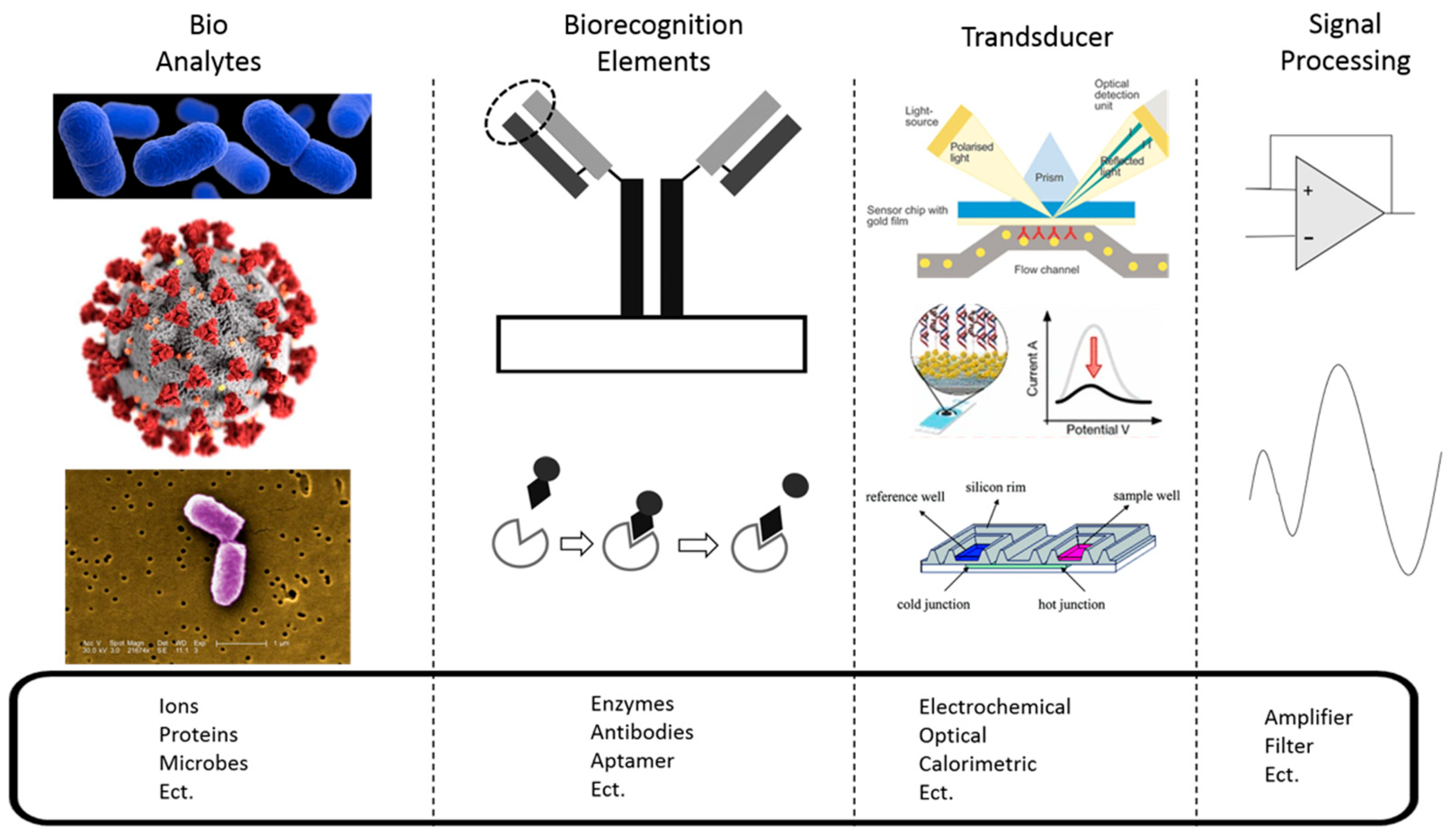
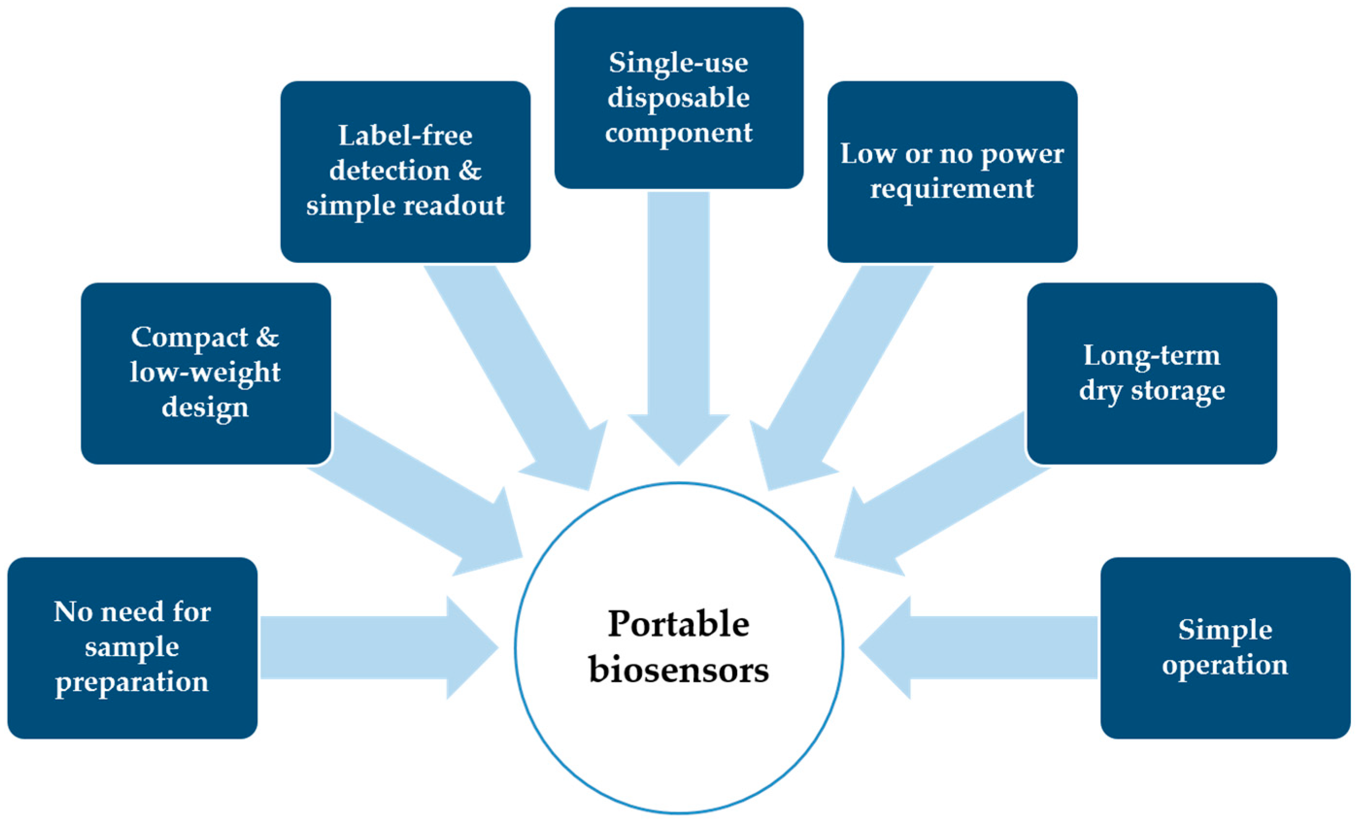
:1. Introduction
2. Analytes
2.1. Saliva
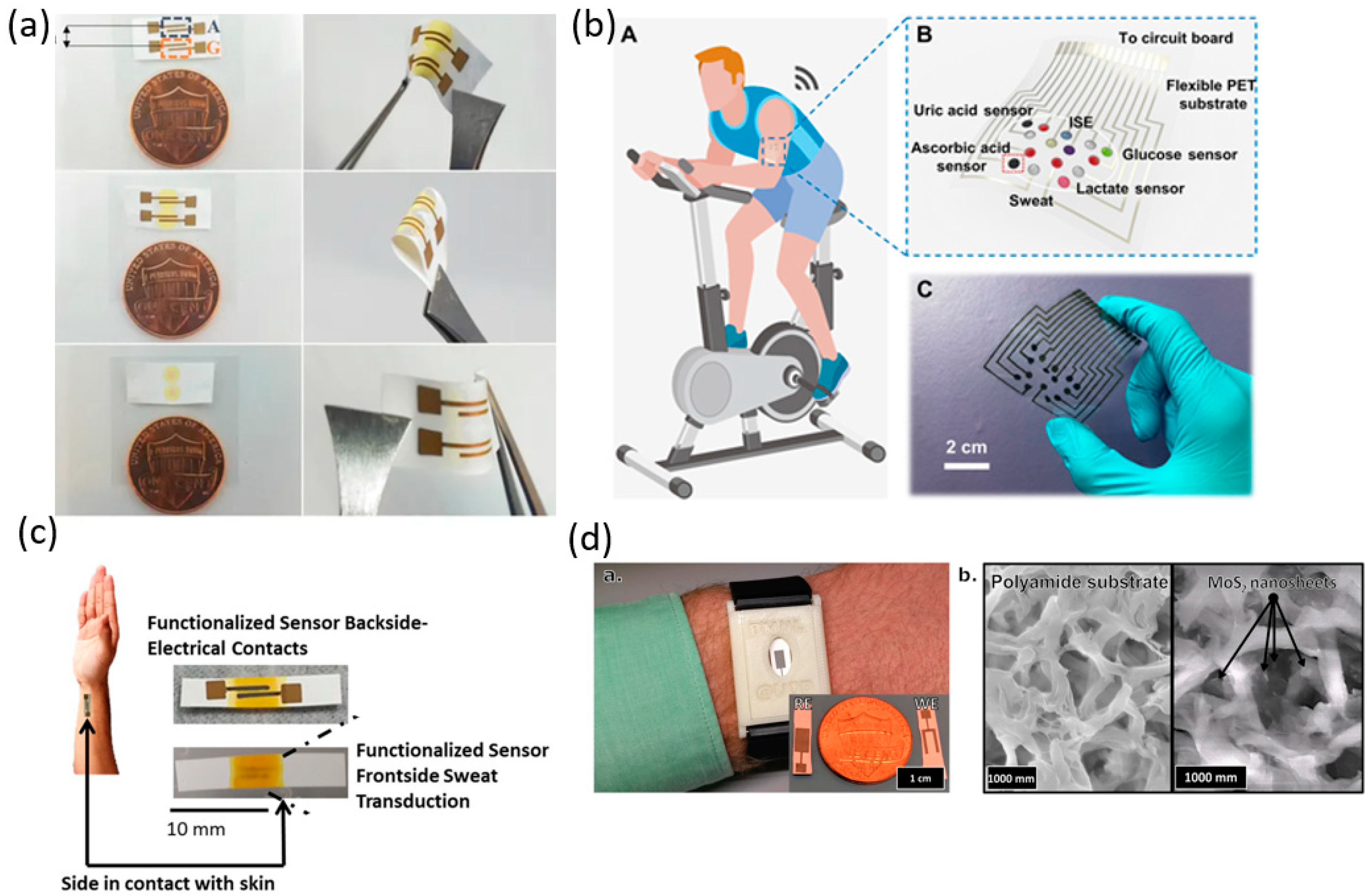
2.2. Sweat
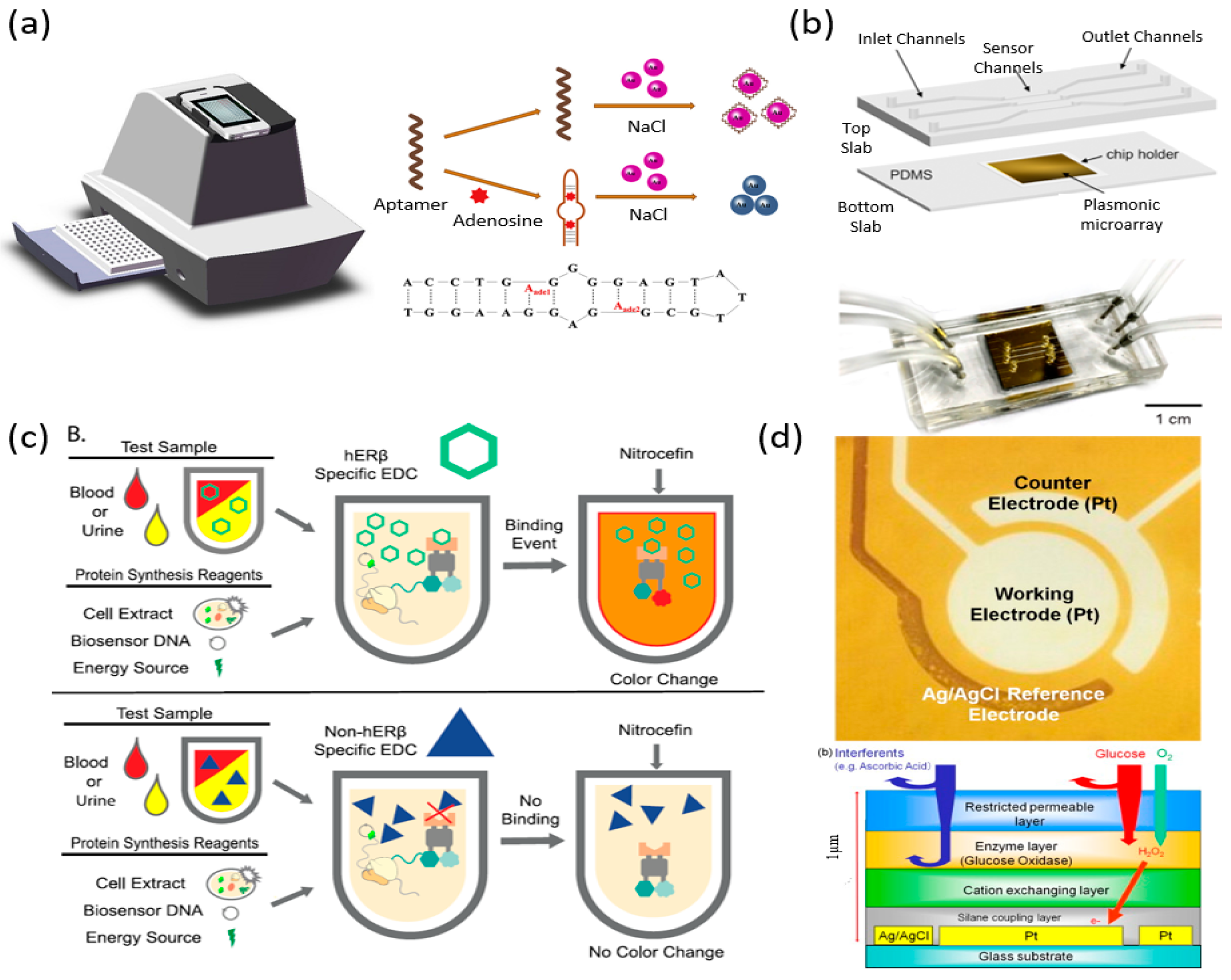
2.3. Urine
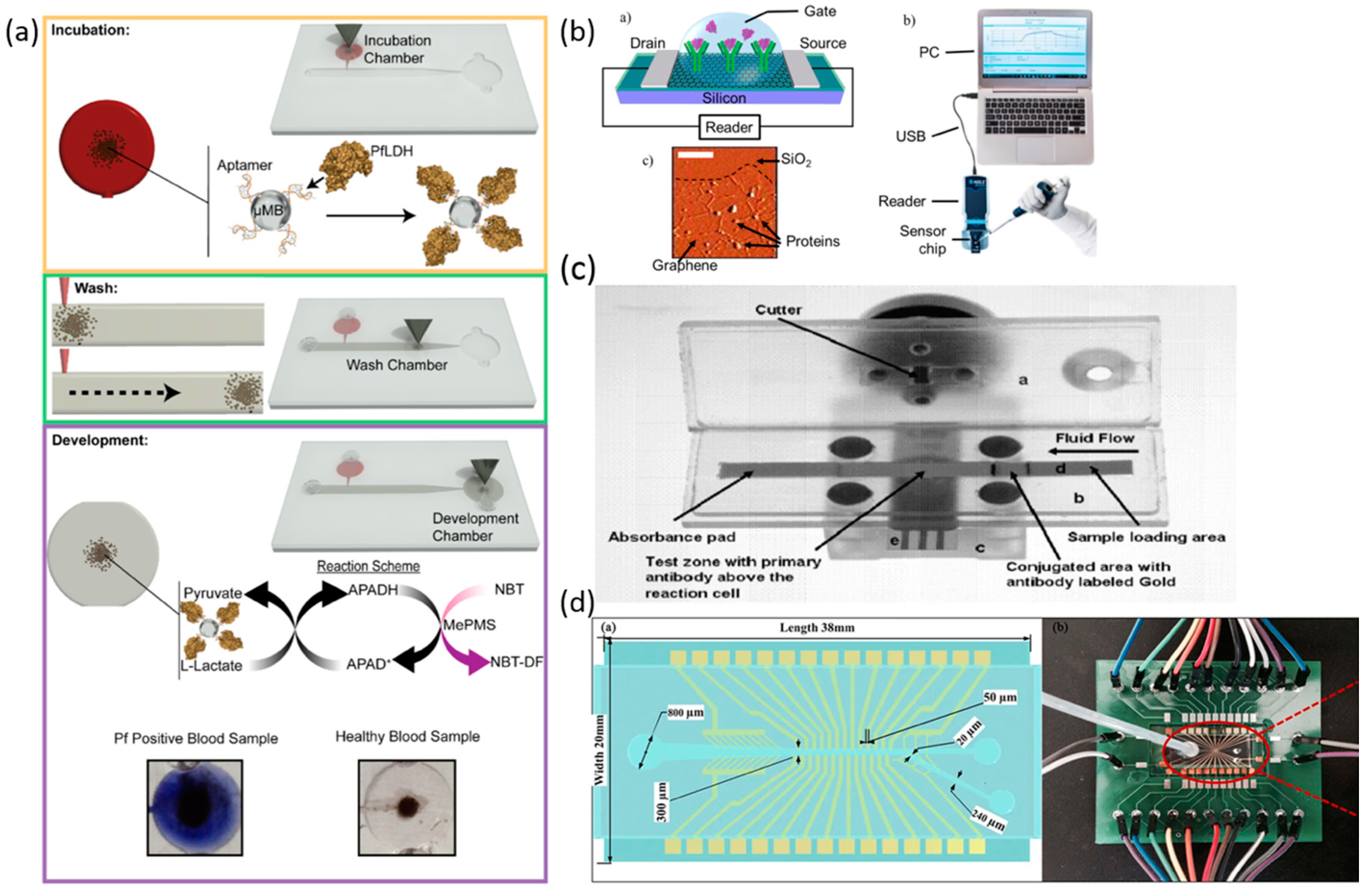
2.4. Blood
2.5. Tears/Breath
3. Discussion
4. Conclusions and Outlooks
Author Contributions
Funding
Conflicts of Interest
References
- Thévenot, D.R.; Toth, K.; Durst, R.A.; Wilson, G.S. Electrochemical Biosensors: Recommended Definitions and Classification1International Union of Pure and Applied Chemistry: Physical Chemistry Division, Commission I.7 (Biophysical Chemistry); Analytical Chemistry Division, Commission V.5 (Electroanalytical Chemistry).1. Biosens. Bioelectron. 2001, 16, 121–131. [Google Scholar] [PubMed]
- Reilly, R.B.; Lee, T.C. Biosensors. Technol. Health Care 2011, 19, 285–293. [Google Scholar] [CrossRef] [PubMed]
- OADC; DNEM. Image Library|CDC Online Newsroom|CDC. 2020. Available online: https://www.cdc.gov/media/subtopic/images.htm (accessed on 13 August 2020).
- Morales, M.A.; Mark Halpern, J. Guide to Selecting a Biorecognition Element for Biosensors. Bioconjug. Chem. 2018, 29, 3231–3239. [Google Scholar] [CrossRef] [PubMed]
- Khansili, N.; Rattu, G.; Krishna, P.M. Label-Free Optical Biosensors for Food and Biological Sensor Applications. Sens. Actuators B Chem. 2018, 265, 35–49. [Google Scholar] [CrossRef]
- Nordin, N.; Yusof, N.A.; Abdullah, J.; Radu, S.; Hushiarian, R. A Simple, Portable, Electrochemical Biosensor to Screen Shellfish for Vibrio Parahaemolyticus. AMB Express 2017, 7, 41. [Google Scholar] [CrossRef] [Green Version]
- Vermeir, S.; Nicolaï, B.M.; Verboven, P.; Van Gerwen, P.; Baeten, B.; Hoflack, L.; Vulsteke, V.; Lammertyn, J. Microplate Differential Calorimetric Biosensor for Ascorbic Acid Analysis in Food and Pharmaceuticals. Anal. Chem. 2007, 79, 6119–6127. [Google Scholar] [CrossRef]
- Heineman, W.R.; Jensen, W.B. Leland C. Clark Jr. (1918–2005). Biosens. Bioelectron. 2006, 21, 1403–1404. [Google Scholar] [CrossRef]
- Nguyen, H.H.; Lee, S.H.; Lee, U.J.; Fermin, C.D.; Kim, M. Immobilized Enzymes in Biosensor Applications. Materials 2019, 12, 121. [Google Scholar] [CrossRef] [Green Version]
- Sadana, A.; Sadana, N. Handbook of Biosensors and Biosensor Kinetics; Elsevier Science & Technology, Elsevier Science: Oxford, UK, 2010. [Google Scholar]
- Tabeta, I.; Ueshiba, H.; Ichijo, T.; Hiroi, N.; Yakushiji, F.; Simojo, M.; Tsuboi, K.; Miyachi, Y. The Corticotropin-Releasing Hormone Stimulation Test in White Coat Hypertension. J. Clin. Endocrinol. Metab. 2002, 87, 3672–3675. [Google Scholar] [CrossRef]
- Niu, Z.; Zhang, W.; Yu, C.; Zhang, J.; Wen, Y. Recent Advances in Biological Sample Preparation Methods Coupled with Chromatography, Spectrometry and Electrochemistry Analysis Techniques. TrAC Trends Anal. Chem. 2018, 102, 123–146. [Google Scholar] [CrossRef]
- Soltani Zarrin, P.; Ibne Jamal, F.; Roeckendorf, N.; Wenger, C. Development of a Portable Dielectric Biosensor for Rapid Detection of Viscosity Variations and Its In Vitro Evaluations Using Saliva Samples of COPD Patients and Healthy Control. Healthcare 2019, 7, 11. [Google Scholar] [CrossRef] [Green Version]
- Petropoulos, K.; Piermarini, S.; Bernardini, S.; Palleschi, G.; Moscone, D. Development of a Disposable Biosensor for Lactate Monitoring in Saliva. Sens. Actuators B Chem. 2016, 237, 8–15. [Google Scholar] [CrossRef]
- Roda, A.; Guardigli, M.; Calabria, D.; Calabretta, M.M.; Cevenini, L.; Michelini, E. A 3D-Printed Device for a Smartphone-Based Chemiluminescence Biosensor for Lactate in Oral Fluid and Sweat. Analyst 2014, 139, 6494–6501. [Google Scholar] [CrossRef] [PubMed]
- Yao, Y.; Li, H.; Wang, D.; Liu, C.; Zhang, C. An Electrochemiluminescence Cloth-Based Biosensor with Smartphone-Based Imaging for Detection of Lactate in Saliva. Analyst 2017, 142, 3715–3724. [Google Scholar] [CrossRef]
- Ahmed, A.; Rushworth, J.V.; Wright, J.D.; Millner, P.A. Novel Impedimetric Immunosensor for Detection of Pathogenic Bacteria Streptococcus Pyogenes in Human Saliva. Anal. Chem. 2013, 85, 12118–12125. [Google Scholar] [CrossRef]
- Wang, R.; Lin, J.; Lassiter, K.; Srinivasan, B.; Lin, L.; Lu, H.; Tung, S.; Hargis, B.; Bottje, W.; Berghman, L.; et al. Evaluation Study of a Portable Impedance Biosensor for Detection of Avian Influenza Virus. J. Virol. Methods 2011, 178, 52–58. [Google Scholar] [CrossRef]
- Hao, Z.; Pan, Y.; Shao, W.; Lin, Q.; Zhao, X. Graphene-Based Fully Integrated Portable Nanosensing System for on-Line Detection of Cytokine Biomarkers in Saliva. Biosens. Bioelectron. 2019, 134, 16–23. [Google Scholar] [CrossRef]
- Ferguson, B.S.; Buchsbaum, S.F.; Wu, T.-T.; Hsieh, K.; Xiao, Y.; Sun, R.; Soh, H.T. Genetic Analysis of H1N1 Influenza Virus from Throat Swab Samples in a Microfluidic System for Point-of-Care Diagnostics. J. Am. Chem. Soc. 2011, 133, 9129–9135. [Google Scholar] [CrossRef] [Green Version]
- Piermarini, S.; Volpe, G.; Federico, R.; Moscone, D.; Palleschi, G. Detection of Biogenic Amines in Human Saliva Using a Screen-Printed Biosensor. Anal. Lett. 2010, 43, 1310–1316. [Google Scholar] [CrossRef]
- Stevens, R.C.; Soelberg, S.D.; Near, S.; Furlong, C.E. Detection of Cortisol in Saliva with a Flow-Filtered, Portable Surface Plasmon Resonance Biosensor System. Anal. Chem. 2008, 80, 6747–6751. [Google Scholar] [CrossRef] [Green Version]
- Ma, X.; Chen, Z.; Zhou, J.; Weng, W.; Zheng, O.; Lin, Z.; Guo, L.; Qiu, B.; Chen, G. Aptamer-Based Portable Biosensor for Platelet-Derived Growth Factor-BB (PDGF-BB) with Personal Glucose Meter Readout. Biosens. Bioelectron. 2014, 55, 412–416. [Google Scholar] [PubMed]
- Lu, Y.-P.; Huang, J.-W.; Lee, I.-N.; Weng, R.-C.; Lin, M.-Y.; Yang, J.-T.; Lin, C.-T. A Portable System to Monitor Saliva Conductivity for Dehydration Diagnosis and Kidney Healthcare. Sci. Rep. 2019, 9, 1–9. [Google Scholar] [CrossRef] [Green Version]
- García-Carmona, L.; Martín, A.; Sempionatto, J.R.; Moreto, J.R.; González, M.C.; Wang, J.; Escarpa, A. Pacifier Biosensor: Toward Noninvasive Saliva Biomarker Monitoring. Anal. Chem. 2019, 91, 13883–13891. [Google Scholar]
- CDC. Avian Influenza A Virus Infections in Humans. Available online: https://www.cdc.gov/flu/avianflu/avian-in-humans.htm (accessed on 10 January 2020).
- Lee, J.; Choi, B.Y.; Jung, E. Metapopulation Model Using Commuting Flow for National Spread of the 2009 H1N1 Influenza Virus in the Republic of Korea. J. Theor. Biol. 2018, 454, 320–329. [Google Scholar] [CrossRef]
- Scully, C. Halitosis. BMJ Clin. Evid. 2014, 2014, 1305. [Google Scholar] [CrossRef]
- Bhide, A.; Muthukumar, S.; Saini, A.; Prasad, S. Simultaneous Lancet-Free Monitoring of Alcohol and Glucose from Low-Volumes of Perspired Human Sweat. Sci. Rep. 2018, 8, 1–11. [Google Scholar] [CrossRef] [Green Version]
- He, W.; Wang, C.; Wang, H.; Jian, M.; Lu, W.; Liang, X.; Zhang, X.; Yang, F.; Zhang, Y. Integrated Textile Sensor Patch for Real-Time and Multiplex Sweat Analysis. Sci. Adv. 2019, 5, eaax0649. [Google Scholar]
- Munje, R.D.; Muthukumar, S.; Jagannath, B.; Prasad, S. A New Paradigm in Sweat Based Wearable Diagnostics Biosensors Using Room Temperature Ionic Liquids (RTILs). Sci. Rep. 2017, 7, 1–12. [Google Scholar] [CrossRef] [Green Version]
- Kinnamon, D.; Ghanta, R.; Lin, K.-C.; Muthukumar, S.; Prasad, S. Portable Biosensor for Monitoring Cortisol in Low-Volume Perspired Human Sweat. Sci. Rep. 2017, 7, 13312. [Google Scholar] [CrossRef] [Green Version]
- Gamella, M.; Campuzano, S.; Manso, J.; Rivera GG de López-Colino, F.; Reviejo, A.J.; Pingarrón, J.M. A Novel Non-Invasive Electrochemical Biosensing Device for in Situ Determination of the Alcohol Content in Blood by Monitoring Ethanol in Sweat. Anal. Chim. Acta 2014, 806, 1–7. [Google Scholar]
- Gao, W.; Emaminejad, S.; Nyein, H.Y.Y.; Challa, S.; Chen, K.; Peck, A.; Fahad, H.M.; Ota, H.; Shiraki, H.; Kiriya, D.; et al. Fully Integrated Wearable Sensor Arrays for Multiplexed in Situ Perspiration Analysis. Nature 2016, 529, 509–514. [Google Scholar] [CrossRef] [PubMed] [Green Version]
- Zhou, S.; Gan, Y.; Kong, L.; Sun, J.; Liang, T.; Wang, X.; Wan, H.; Wang, P. A Novel Portable Biosensor Based on Aptamer Functionalized Gold Nanoparticles for Adenosine Detection. Anal. Chim. Acta 2020, 1120, 43–49. [Google Scholar] [CrossRef] [PubMed]
- Soler, M.; Belushkin, A.; Cavallini, A.; Kebbi-Beghdadi, C.; Greub, G.; Altug, H. Multiplexed Nanoplasmonic Biosensor for One-Step Simultaneous Detection of Chlamydia Trachomatis and Neisseria Gonorrhoeae in Urine. Biosens. Bioelectron. 2017, 94, 560–567. [Google Scholar] [CrossRef]
- Huang, C.Y.; Hsieh, C.H.; Chen, Y.L.; Lee, M.H.; Lin, C.F.; Tsai, H.H.; Juang, Y.Z.; Liu, B.D.; Lin, H.Y. Portable Potentiostatic Sensor Integrated with Neopterin-Imprinted Poly(Ethylene-Co-Vinyl Alcohol)-Based Electrode. IET Nanobiotechnol. 2011, 5, 126–131. [Google Scholar] [CrossRef]
- Salehi, A.S.M.; Yang, S.O.; Earl, C.C.; Shakalli Tang, M.J.; Porter Hunt, J.; Smith, M.T.; Wood, D.W.; Bundy, B.C. Biosensing Estrogenic Endocrine Disruptors in Human Blood and Urine: A RAPID Cell-Free Protein Synthesis Approach. Toxicol. Appl. Pharmacol. 2018, 345, 19–25. [Google Scholar] [CrossRef]
- Miyashita, M.; Ito, N.; Ikeda, S.; Murayama, T.; Oguma, K.; Kimura, J. Development of Urine Glucose Meter Based on Micro-Planer Amperometric Biosensor and Its Clinical Application for Self-Monitoring of Urine Glucose. Biosens. Bioelectron. 2009, 24, 1336–1340. [Google Scholar] [CrossRef]
- Nikolelis, D.P.; Raftopoulou, G.; Chatzigeorgiou, P.; Nikoleli, G.-P.; Viras, K. Optical Portable Biosensors Based on Stabilized Lipid Membrane for the Rapid Detection of Doping Materials in Human Urine. Sens. Actuators B Chem. 2008, 130, 577–582. [Google Scholar] [CrossRef]
- Dirkzwager, R.M.; Liang, S.; Tanner, J. Development of Aptamer-Based Point-of-Care Diagnostic Devices for Malaria Using Three-Dimensional Printing Rapid Prototyping. ACS Sens. 2016, 1, 420–426. [Google Scholar] [CrossRef]
- Fraser, L.A.; Kinghorn, A.B.; Dirkzwager, R.M.; Liang, S.; Cheung, Y.-W.; Lim, B.; Shiu, S.C.-C.; Tang, M.S.L.; Andrew, D.; Manitta, J.; et al. A Portable Microfluidic Aptamer-Tethered Enzyme Capture (APTEC) Biosensor for Malaria Diagnosis. Biosens. Bioelectron. 2018, 100, 591–596. [Google Scholar] [CrossRef]
- Afsahi, S.; Lerner, M.B.; Goldstein, J.M.; Lee, J.; Tang, X.; Bagarozzi, D.A.; Pan, D.; Locascio, L.; Walker, A.; Barron, F.; et al. Novel Graphene-Based Biosensor for Early Detection of Zika Virus Infection. Biosens. Bioelectron. 2018, 100, 85–88. [Google Scholar] [CrossRef]
- Zaytseva, N.V.; Montagna, R.A.; Lee, E.M.; Baeumner, A.J. Multi-Analyte Single-Membrane Biosensor for the Serotype-Specific Detection of Dengue Virus. Anal. Bioanal. Chem. 2004, 380, 46–53. [Google Scholar] [CrossRef]
- Wei, H.; Guo, Z.; Zhu, Z.; Tan, Y.; Du, Z.; Yang, R. Sensitive Detection of Antibody against Antigen F1 of Yersinia Pestis by an Antigen Sandwich Method Using a Portable Fiber Optic Biosensor. Sens. Actuators B Chem. 2007, 127, 525–530. [Google Scholar] [CrossRef]
- Zou, Z.; Du, D.; Wang, J.; Smith, J.N.; Timchalk, C.; Li, Y.; Lin, Y. Quantum Dot-Based Immunochromatographic Fluorescent Biosensor for Biomonitoring Trichloropyridinol, a Biomarker of Exposure to Chlorpyrifos. Anal. Chem. 2010, 82, 5125–5133. [Google Scholar] [CrossRef]
- Wang, L.; Lu, D.; Wang, J.; Du, D.; Zou, Z.; Wang, H.; Smith, J.N.; Timchalk, C.; Liu, F.; Lin, Y. A Novel Immunochromatographic Electrochemical Biosensor for Highly Sensitive and Selective Detection of Trichloropyridinol, a Biomarker of Exposure to Chlorpyrifos. Biosens. Bioelectron. 2011, 26, 2835–2840. [Google Scholar] [CrossRef]
- Ming, J.; Fan, W.; Jiang, T.-F.; Wang, Y.-H.; Lv, Z.-H. Portable and Sensitive Detection of Copper(II) Ion Based on Personal Glucose Meters and a Ligation DNAzyme Releasing Strategy. Sens. Actuators B Chem. 2017, 240, 1091–1098. [Google Scholar] [CrossRef]
- Sang, S.; Feng, Q.; Jian, A.; Li, H.; Ji, J.; Duan, Q.; Zhang, W.; Wang, T. Portable Microsystem Integrates Multifunctional Dielectrophoresis Manipulations and a Surface Stress Biosensor to Detect Red Blood Cells for Hemolytic Anemia. Sci. Rep. 2016, 6, 33626. [Google Scholar] [CrossRef] [Green Version]
- Zhou, H.; Ran, G.; Masson, J.-F.; Wang, C.; Zhao, Y.; Song, Q. Novel Tungsten Phosphide Embedded Nitrogen-Doped Carbon Nanotubes: A Portable and Renewable Monitoring Platform for Anticancer Drug in Whole Blood. Biosens. Bioelectron. 2018, 105, 226–235. [Google Scholar] [CrossRef] [PubMed]
- Davis, C.E.; Bogan, M.J.; Sankaran, S.; Molina, M.A.; Loyola, B.R.; Zhao, W.; Benner, W.H.; Schivo, M.; Farquar, G.R.; Kenyon, N.J.; et al. Analysis of Volatile and Non-Volatile Biomarkers in Human Breath Using Differential Mobility Spectrometry (DMS). IEEE Sens. J. 2010, 10, 114–122. [Google Scholar] [CrossRef]
- Sempionatto, J.R.; Brazaca, L.C.; García-Carmona, L.; Bolat, G.; Campbell, A.S.; Martin, A.; Tang, G.; Shah, R.; Mishra, R.K.; Kim, J.; et al. Eyeglasses-Based Tear Biosensing System: Non-Invasive Detection of Alcohol, Vitamins and Glucose. Biosens. Bioelectron. 2019, 137, 161–170. [Google Scholar] [CrossRef]
- Yao, H.; Shum, A.J.; Cowan, M.; Lähdesmäki, I.; Parviz, B.A. A Contact Lens with Embedded Sensor for Monitoring Tear Glucose Level. Biosens. Bioelectron. 2011, 26, 3290–3296. [Google Scholar] [CrossRef] [Green Version]
- Chu, M.; Shirai, T.; Takahashi, D.; Arakawa, T.; Kudo, H.; Sano, K.; Sawada, S.; Yano, K.; Iwasaki, Y.; Akiyoshi, K.; et al. Biomedical Soft Contact-Lens Sensor for in Situ Ocular Biomonitoring of Tear Contents. Biomed. Microdevices 2011, 13, 603–611. [Google Scholar] [CrossRef]
- Sreekumar, J.; France, N.; Taylor, S.; Matthews, T.; Turner, P.; Bliss, P.; Brook, A.H.; Watson, A. Diagnosis of Helicobacter Pylori by Carbon-13 Urea Breath Test Using a Portable Mass Spectrometer. SAGE Open Med. 2015, 3, 2050312115569565. [Google Scholar] [CrossRef] [Green Version]
- Righettoni, M.; Tricoli, A.; Gass, S.; Schmid, A.; Amann, A.; Pratsinis, S.E. Breath Acetone Monitoring by Portable Si:WO3 Gas Sensors. Anal. Chim. Acta 2012, 738, 69–75. [Google Scholar] [CrossRef] [Green Version]
- Bagchi, S.; SenGupta, S.; Mondal, S. Development and Characterization of Carbonic Anhydrase-Based CO2 Biosensor for Primary Diagnosis of Respiratory Health. IEEE Sens. J. 2017, 17, 1384–1390. [Google Scholar] [CrossRef]
- Strand, N.; Bhushan, A.; Schivo, M.; Kenyon, N.J.; Davis, C.E. Chemically Polymerized Polypyrrole for On-Chip Concentration of Volatile Breath Metabolites. Sens. Actuators B Chem. 2010, 143, 516–523. [Google Scholar] [CrossRef] [Green Version]
- Yu, J.; Wang, S.; Ge, L.; Ge, S. A Novel Chemiluminescence Paper Microfluidic Biosensor Based on Enzymatic Reaction for Uric Acid Determination. Biosens. Bioelectron. 2011, 26, 3284–3289. [Google Scholar] [CrossRef]
- Chuang, C.-H.; Chiang, Y.-Y. Bio-O-Pump: A Novel Portable Microfluidic Device Driven by Osmotic Pressure. Sens. Actuators B Chem. 2019, 284, 736–743. [Google Scholar] [CrossRef]


| Biomarker | Target Disease/Area | Sensor Type | Detection Limit * Sensitivity ** Specificity | Dynamic Range | Analysis Time |
|---|---|---|---|---|---|
| Lactate [14] | Respiratory insufficiency, shocks, heart failure and metabolic disorders | Modified screen printed electrode | 0.01 mM | 0.025–0.25 mM | <60 s |
| Lactate [15] | Diabetes, sports medicine, critical care | 3D printed chemiluminescence biosensor | 0.1 mmol/L | NA | <5 min |
| Lactate [16] | Clinical diagnosis, sport physiology and food analysis | Cloth-based electrochemiluminescence (ECL) | 0.035 mM | 0.05–2.5 mM | NA |
| Streptococcus [17] | Streptococcus Pyogenes | Impedimetric Immunosensor | NA | 100 to 105 cells/10 µL cumulative incubation 100 to 104 cells/10 µL single-shot | NA |
| Avian Influenza Virus [18] | Avian influenza | Impedance biosensor | 1 × 102.2 ELD50/mL Tracheal = 100% * Cloacal = 55% * | NA | <1 h |
| Cytokine biomarkers [19] | Disease detection such as cancer | Graphene-based fully integrated portable nanosensing biosensor | 12 pM | NA | Real-time |
| H1N1 [20] | Influenza detection | Magnetic Integrated Microfluidic Electrochemical Detector | 10 TCID50 | NA | 3.5 h |
| Biogenic Amines [21] | Halitosis | Diamine Oxidase Electrochemical screen printed electrode Biosensor | 1 × 10⁻⁵ M | 2 × 10⁻⁵–3× 10⁻⁴ M | NA |
| Cortisol [22] | Stress | Surface plasmon resonance biosensor | 1.0 ng/mL | 1.5 ng/mL–10 ng/mL | <10 min |
| PDGF [23] | Cell growth and division | Aptamer-based biosensor PGM | 2.9 fM | 1.0 × 10⁻¹⁴ M to 3.16 × 10⁻¹² M | 20 min |
| Saliva Conductivity [24] | Dehydration and Kidney function | Au Electrode biosensor | 93.3% * 80% ** | NA | Real-time |
| Metabolites (Glucose) [25] | Metabolite pacifier biosensor for infants | Glucose-oxidase based enzyme detection electrode biosensor | 0.04 mM | 0.1 to 1.4 mM | Real-time |
| Biomarker | Target Disease/Area | Sensor Type | Detection Limit * Sensitivity | Dynamic Range | Analysis Time |
|---|---|---|---|---|---|
| Alcohol [33] | Noninvasive measurement | Bienzyme amperometric composite biosensors | 0.0005 g/L | 0.0005–0.6 g/L | Real-time |
| Glucose [29] | Diabetes | Zinc Oxide Thin film nanoporous electrode biosensor | 0.1 mg/dL | 0.01–200 mg/dL | NA |
| Multiplexed (Metabolites/electrolytes/temperature) [34] | Physiological monitoring | Flexible sweat sensor array with wireless FPCB | 2.35 nA/μM Glucose * 220 nA/mM Lactate * | NA | |
| glucose, lactate, ascorbic acid, uric acid, Na+ and K+ [30] | Multipurpose healthcare monitoring | Silk fabric–derived intrinsically nitrogen (N)–doped carbon textile (SilkNCT) flexible biosensor | Glucose: 5 μM Lactate: 0.5 mM UA: 0.1 μM AA: 1 μM Na+: 1 mM K+: 0.5 mM | Glucose: 25 to 300 μM Lactate: 5 to 35 mM UA: 2.5 to 115 μM AA: 20 to 300 μM Na+: 5 to 100 mM K+: 1.25 to 40 mM | Real-time |
| Interleukin [31] | Immune response | BMIM[BF4] RTIL stability enhancing capture probe immunoassay functionalized ZnO thin films deposited on nanoporous polyamide membrane biosensor | 0.2 pg/mL for 0–24 h and 2 pg/mL for 24–48 h post-antibody sensor functionalization | 0.2–200 pg/mL continuous detection | NA |
| Lactate [15] | Diabetes, sports medicine, critical care | 3D printed chemiluminescence biosensor | 0.1 mmol/L | NA | <5 min |
| Cortisol [32] | Stress | Non-faradaic label-free cortisol biosensor | 1 ng/mL | 1–500 ng/mL | Continuous for 3+ hours |
| Biomarker | Target Disease/Area | Sensor Type | Detection Limit * Sensitivity | Dynamic Range | Analysis Time |
|---|---|---|---|---|---|
| Adenosine [35] | Lung Cancer | Colorimetric aptasensor | 0.17 μM | 5.0 μM–60.0 μM | <20 min |
| Chlamydia trachomatis [36] | Chlamydia | Nanoplasmonic biosensor | 300 CFU/mL | NA | Real time |
| Neisseria gonorrhoeae [36] | Gonorrhoeae | Nanoplasmonic biosensor | 150 CFU/mL | NA | Real time |
| Neopterin [37] | Aging | Molecularly Imprinted Polymer integrated Potentiostat | 0.025 pg/mL 0.041 pg/mL compared to reference of 35–55 ng/mL * | NA | NA |
| Estrogenic Endocrine Disruptor [38] | Obesity, birth defects, cancer, reproductive impairment | In-vitro Detection biosensor platform | urine 4 nM Blood 8 nM | 4–100 nM, urine | 2.5 h |
| Glucose [39] | Diabetes | Micro-Planer amperometric biosensor | NA | 0–2000 mg/dL | 6 s |
| Dopamine [40] | Doping | Stabilized lipid Membrane optical Biosensor | 10−9 M | 0 to 100 nM | <1 min |
| Ephedrin [40] | Doping | Stabilized lipid Membrane optical Biosensor | 10−9 M | 0 to 100 nM | <1 min |
| Biomarker | Sample Type | Target Disease/Area | Sensor Type | Detection Limit * Sensitivity ** Specificity | Dynamic Range | Analysis Time |
|---|---|---|---|---|---|---|
| Malaria [41] | Whole blood | Malaria− | Aptamer Tethered Enzyme Capture assay | 4.9 ng/mL | NA | <1 h |
| Malaria [42] | Blood | Malaria | Aptamer-Tethered Enzyme Capture (APTEC) biosensor | 250 parasites/µL | NA | <20 min |
| Zika [43] | Simulated Serum | Zika | graphene-based biosensor | 0.45 nM | NA | Real time |
| Dengue [44] | Blood | Dengue fever | multi-analyte biosensor based on nucleic acid hybridization and liposome signal amplification | 50 RNA molecules for serotype 2, 500 RNA molecules for serotypes 3 and 4, and 50,000 molecules for serotype 1 | NA | <25 min |
| Yersinia Pestis Antibody [45] | Rabbit serum | Etiological agent of plague | Antigen sandwich method using a portable fiber optic biosensor | 10 ng/mL 100% * 94.7% ** | NA | 40 min |
| Trichloropyridino [46] | Rat Blood | Exposure to organophosphorus insecticides | Quantum Dot integrated Fluorescent biosensor | 1.0 ng/mL | 1–50 ng/ml | 15 min |
| Trichloropyridino [47] | Rat plasma | Exposure to organophosphorus insecticides | Immunochromatographic electrochemical biosensor | 0.1 ng/ml | 0.1–100 ng/ml | 15 min |
| Copper [48] | Serum | Copper Toxicity | Cu2+-dependent DNA ligation DNAzyme PGMs | 1 nM possible | 10–600 mg/dL | NA |
| Estrogenic Endocrine Disruptor [38] | Blood | Obesity, birth defects, cancer, reproductive impairment | In-vitro Detection biosensor platform | 8 nM | 8–300 nM in blood 4–100 nM urine | 2.5 h |
| Red blood [49] | Blood | Anemia | Surface Stress Biosensor | NA | NA | NA |
| Anti-Cancer Drugs [50] | Blood | Toxicity | Novel Tungsten Phosphide Embedded Nitrogen-Doped Carbon Nanotubes biosensor | 45 nM | 0.01–45 µM | NA |
| Biomarker | Sample Type | Target Disease/Area | Sensor Type | Detection Limit * Sensitivity | Dynamic Range | Analysis Time |
|---|---|---|---|---|---|---|
| Alcohol/Glucose/Vitamins (B2,B6,C) [52] | Tear | Various disease/Health Monitoring | Alcohol-oxidase (AOx) biosensing fluidic system | NA | NA | Real-time |
| Glucose [53] | Tear | Diabetes | Amperometric glucose biosensor | 0.01 mM 240 uA/(mM·cm2) * | Linearity 0.1–0.6 mM | 20 s |
| Glucose [54] | Tear | Diabetes | SCL-biosensor | NA | 0.03–5.0 mmol/L | Real-time |
| helicobacter pylori [55] | breath | Chronic gastritis/(gastric/duodenal ulcers)/gastric cancer | Quadrupole mass Spectrometer biosensor | NA | NA | NA |
| Acetone [56] | Breath | Various disease/Health Monitoring | portable Si:WO3 gas sensors | 20 ppb | NA | 10–15 s |
| Volatile and non-volatile biomarkers [51] | Breath | Disease or chemical exposure | Differential Mobility Spectrometry | Toluene: 200 ppb Angiotensin: 1 pM | NA | Near real-time |
| CO2 [57] | Breath | Respiratory health | Carbonic Anhydrase-Based enzyme biosensor | 0.132 mV/ppm * | 160–2677 ppm CO2 linear response | 12 s |
| Biomarker | Sample Type | Target Disease/Area |
|---|---|---|
| Lactate | Saliva and sweat | Respiratory insufficiency, shocks, heart failure, metabolic disorders, diabetes, sports medicine, critical care, and food analysis |
| Glucose | Sweat, urine, and tears | Diabetes and general healthcare monitoring |
| Alcohol | Sweat tears, and breath | BAC for drivers and diabetes treatment for hypoglycemia prevention |
© 2020 by the authors. Licensee MDPI, Basel, Switzerland. This article is an open access article distributed under the terms and conditions of the Creative Commons Attribution (CC BY) license (http://creativecommons.org/licenses/by/4.0/).
Share and Cite
Senf, B.; Yeo, W.-H.; Kim, J.-H. Recent Advances in Portable Biosensors for Biomarker Detection in Body Fluids. Biosensors 2020, 10, 127. https://doi.org/10.3390/bios10090127
Senf B, Yeo W-H, Kim J-H. Recent Advances in Portable Biosensors for Biomarker Detection in Body Fluids. Biosensors. 2020; 10(9):127. https://doi.org/10.3390/bios10090127
Chicago/Turabian StyleSenf, Brian, Woon-Hong Yeo, and Jong-Hoon Kim. 2020. "Recent Advances in Portable Biosensors for Biomarker Detection in Body Fluids" Biosensors 10, no. 9: 127. https://doi.org/10.3390/bios10090127