Next Issue
Volume 7, February
Previous Issue
Volume 6, December


Nanomaterials, Volume 7, Issue 1 (January 2017) – 22 articles

Cover Story (view full-size image): 
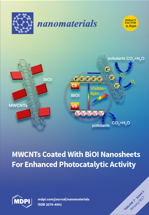
We developed a novel heterostructure of multiwalled carbon nanotubes (MWCNTs) coated with BiOI nanosheets via a facile one-pot solvothermal method as an efficient visible-light-driven (VLD) photocatalyst. The MWCNTs/BiOI composite can efficiently degrade diverse organic pollutants (RhB/MO/4-CP) with good stability due to the strong coupling interface between MWCNTs and BiOI. Therefore, MWCNTs/BiOI has great potential as an efficient and stable VLD photocatalyst for wastewater treatment. View the paper
  • Issues are regarded as officially published after their release is announced to the table of contents alert mailing list.
  • You may sign up for e-mail alerts to receive table of contents of newly released issues.
  • PDF is the official format for papers published in both, html and pdf forms. To view the papers in pdf format, click on the "PDF Full-text" link, and use the free Adobe Reader to open them.
Order results
Result details
Section
Select all
Export citation of selected articles as:
6336 KiB  
Article
A Novel Heterostructure of BiOI Nanosheets Anchored onto MWCNTs with Excellent Visible-Light Photocatalytic Activity
by Shijie Li, Shiwei Hu, Kaibing Xu, Wei Jiang, Jianshe Liu and Zhaohui Wang
Nanomaterials 2017, 7(1), 22; https://doi.org/10.3390/nano7010022 - 23 Jan 2017
Cited by 48 | Viewed by 6536
Abstract
Developing efficient visible-light-driven (VLD) photocatalysts for environmental decontamination has drawn significant attention in recent years. Herein, we have reported a novel heterostructure of multiwalled carbon nanotubes (MWCNTs) coated with BiOI nanosheets as an efficient VLD photocatalyst, which was prepared via a simple solvothermal [...] Read more.
Developing efficient visible-light-driven (VLD) photocatalysts for environmental decontamination has drawn significant attention in recent years. Herein, we have reported a novel heterostructure of multiwalled carbon nanotubes (MWCNTs) coated with BiOI nanosheets as an efficient VLD photocatalyst, which was prepared via a simple solvothermal method. The morphology and structure were characterized by powder X-ray diffraction (XRD), scanning electron microscopy (SEM), transmission electron microscopy (TEM), UV-Vis diffuse reflectance spectroscopy (DRS), and specific surface area measurements. The results showed that BiOI nanosheets were well deposited on MWCNTs. The MWCNTs/BiOI composites exhibited remarkably enhanced photocatalytic activity for the degradation of rhodamine B (RhB), methyl orange (MO), and para-chlorophenol (4-CP) under visible-light, compared with pure BiOI. When the MWCNTs content is 3 wt %, the MWCNTs/BiOI composite (3%M-Bi) achieves the highest activity, which is even higher than that of a mechanical mixture (3 wt % MWCNTs + 97 wt % BiOI). The superior photocatalytic activity is predominantly due to the strong coupling interface between MWCNTs and BiOI, which significantly promotes the efficient electron-hole separation. The photo-induced holes (h+) and superoxide radicals (O2) mainly contribute to the photocatalytic degradation of RhB over 3%M-Bi. Therefore, the MWCNTs/BiOI composite is expected to be an efficient VLD photocatalyst for environmental purification. Full article
(This article belongs to the Special Issue Nanoscale in Photocatalysis)
Show Figures

Graphical abstract

2569 KiB  
Review
Behavior and Potential Impacts of Metal-Based Engineered Nanoparticles in Aquatic Environments
by Cheng Peng, Wen Zhang, Haiping Gao, Yang Li, Xin Tong, Kungang Li, Xiaoshan Zhu, Yixiang Wang and Yongsheng Chen
Nanomaterials 2017, 7(1), 21; https://doi.org/10.3390/nano7010021 - 22 Jan 2017
Cited by 116 | Viewed by 9687
Abstract
The specific properties of metal-based nanoparticles (NPs) have not only led to rapidly increasing applications in various industrial and commercial products, but also caused environmental concerns due to the inevitable release of NPs and their unpredictable biological/ecological impacts. This review discusses the environmental [...] Read more.
The specific properties of metal-based nanoparticles (NPs) have not only led to rapidly increasing applications in various industrial and commercial products, but also caused environmental concerns due to the inevitable release of NPs and their unpredictable biological/ecological impacts. This review discusses the environmental behavior of metal-based NPs with an in-depth analysis of the mechanisms and kinetics. The focus is on knowledge gaps in the interaction of NPs with aquatic organisms, which can influence the fate, transport and toxicity of NPs in the aquatic environment. Aggregation transforms NPs into micrometer-sized clusters in the aqueous environment, whereas dissolution also alters the size distribution and surface reactivity of metal-based NPs. A unique toxicity mechanism of metal-based NPs is related to the generation of reactive oxygen species (ROS) and the subsequent ROS-induced oxidative stress. Furthermore, aggregation, dissolution and ROS generation could influence each other and also be influenced by many factors, including the sizes, shapes and surface charge of NPs, as well as the pH, ionic strength, natural organic matter and experimental conditions. Bioaccumulation of NPs in single organism species, such as aquatic plants, zooplankton, fish and benthos, is summarized and compared. Moreover, the trophic transfer and/or biomagnification of metal-based NPs in an aquatic ecosystem are discussed. In addition, genetic effects could result from direct or indirect interactions between DNA and NPs. Finally, several challenges facing us are put forward in the review. Full article
(This article belongs to the Special Issue Environmental Applications and Implications of Nanotechnology)
Show Figures

Figure 1

3778 KiB  
Article
Enhanced Visible Light Photocatalytic Activity of ZnO Nanowires Doped with Mn2+ and Co2+ Ions
by Wei Li, Guojing Wang, Chienhua Chen, Jiecui Liao and Zhengcao Li
Nanomaterials 2017, 7(1), 20; https://doi.org/10.3390/nano7010020 - 19 Jan 2017
Cited by 144 | Viewed by 8307
Abstract
In this research, ZnO nanowires doped with Mn2+ and Co2+ ions were synthesized through a facile and inexpensive hydrothermal approach, in which Mn2+ and Co2+ ions successfully substituted Zn2+ in the ZnO crystal lattice without changing the morphology [...] Read more.
In this research, ZnO nanowires doped with Mn2+ and Co2+ ions were synthesized through a facile and inexpensive hydrothermal approach, in which Mn2+ and Co2+ ions successfully substituted Zn2+ in the ZnO crystal lattice without changing the morphology and crystalline structure of ZnO. The atomic percentages of Mn and Co were 6.29% and 1.68%, respectively, in the doped ZnO nanowires. The photocatalytic results showed that Mn-doped and Co-doped ZnO nanowires both exhibited higher photocatalytic activities than undoped ZnO nanowires. Among the doped ZnO nanowires, Co-doped ZnO, which owns a twice active visible-light photocatalytic performance compared to pure ZnO, is considered a more efficient photocatalyst material. The enhancement of its photocatalytic performance originates from the doped metal ions, which enhance the light absorption ability and inhibit the recombination of photo-generated electron-hole pairs as well. The effect of the doped ion types on the morphology, crystal lattice and other properties of ZnO was also investigated. Full article
(This article belongs to the Special Issue Nanoscale in Photocatalysis)
Show Figures

Graphical abstract

3496 KiB  
Article
Enhanced Photocatalytic Performance under Visible and Near-Infrared Irradiation of Cu1.8Se/Cu3Se2 Composite via a Phase Junction
by Li-Na Qiao, Huan-Chun Wang, Yang Shen, Yuan-Hua Lin and Ce-Wen Nan
Nanomaterials 2017, 7(1), 19; https://doi.org/10.3390/nano7010019 - 18 Jan 2017
Cited by 28 | Viewed by 5922
Abstract
A novel Cu1.8Se/Cu3Se2 composite photocatalyst was prepared by the simple precipitation method. This composite possesses a wide photoabsorption until the range of near-infrared light, and exhibits significantly enhanced photocatalytic activity for methyl orange degradation under visible and near-infrared [...] Read more.
A novel Cu1.8Se/Cu3Se2 composite photocatalyst was prepared by the simple precipitation method. This composite possesses a wide photoabsorption until the range of near-infrared light, and exhibits significantly enhanced photocatalytic activity for methyl orange degradation under visible and near-infrared light irradiation compared with bare Cu1.8Se and Cu3Se2. The mechanism of this outstanding photocatalytic behavior can be explained by the calculated energy band positions. The efficient charge separation via a phase junction of Cu1.8Se/Cu3Se2 composite would make a great contribution to its much-enhanced photocatalytic efficiency. Full article
(This article belongs to the Special Issue Nanoscale in Photocatalysis)
Show Figures

Graphical abstract

4533 KiB  
Article
Amorphous Silica Particles Relevant in Food Industry Influence Cellular Growth and Associated Signaling Pathways in Human Gastric Carcinoma Cells
by Anja Wittig, Helge Gehrke, Giorgia Del Favero, Eva-Maria Fritz, Marco Al-Rawi, Silvia Diabaté, Carsten Weiss, Haider Sami, Manfred Ogris and Doris Marko
Nanomaterials 2017, 7(1), 18; https://doi.org/10.3390/nano7010018 - 13 Jan 2017
Cited by 14 | Viewed by 8029
Abstract
Nanostructured silica particles are commonly used in biomedical and biotechnical fields, as well as, in cosmetics and food industry. Thus, their environmental and health impacts are of great interest and effects after oral uptake are only rarely investigated. In the present study, the [...] Read more.
Nanostructured silica particles are commonly used in biomedical and biotechnical fields, as well as, in cosmetics and food industry. Thus, their environmental and health impacts are of great interest and effects after oral uptake are only rarely investigated. In the present study, the toxicological effects of commercially available nano-scaled silica with a nominal primary diameter of 12 nm were investigated on the human gastric carcinoma cell line GXF251L. Besides the analysis of cytotoxic and proliferative effects and the comparison with effects of particles with a nominal primary diameter of 200 nm, emphasis was also given to their influence on the cellular epidermal growth factor receptor (EGFR) and mitogen-activated protein kinases (MAPK) signaling pathways—both of them deeply involved in the regulation of cellular processes like cell cycle progression, differentiation or proliferation. The investigated silica nanoparticles (NPs) were found to stimulate cell proliferation as measured by microscopy and the sulforhodamine B assay. In accordance, the nuclear level of the proliferation marker Ki-67 was enhanced in a concentration-dependent manner. At high particle concentrations also necrosis was induced. Finally, silica NPs affected the EGFR and MAPK pathways at various levels dependent on concentration and time. However, classical activation of the EGFR, to be reflected by enhanced levels of phosphorylation, could be excluded as major trigger of the proliferative stimulus. After 45 min of incubation the level of phosphorylated EGFR did not increase, whereas enhanced levels of total EGFR protein were observed. These results indicate interference with the complex homeostasis of the EGFR protein, whereby up to 24 h no impact on the transcription level was detected. In addition, downstream on the level of the MAP kinases ERK1/2 short term incubation appeared to affect total protein levels without clear increase in phosphorylation. Depending on the concentration range, enhanced levels of ERK1/2 phosphorylation were only observed after 24 h of incubation. Taken together, the present study demonstrates the potential of the tested silica particles to enhance the growth of gastric carcinoma cells. Although interference with the EGFR/MAPK cascade is observed, additional mechanisms are likely to be involved in the onset of the proliferative stimulus. Full article
(This article belongs to the Special Issue Cytotoxicity of Nanoparticles)
Show Figures

Graphical abstract

3966 KiB  
Article
Nano-Photonic Structures for Light Trapping in Ultra-Thin Crystalline Silicon Solar Cells
by Prathap Pathi, Akshit Peer and Rana Biswas
Nanomaterials 2017, 7(1), 17; https://doi.org/10.3390/nano7010017 - 13 Jan 2017
Cited by 45 | Viewed by 9778
Abstract
Thick wafer-silicon is the dominant solar cell technology. It is of great interest to develop ultra-thin solar cells that can reduce materials usage, but still achieve acceptable performance and high solar absorption. Accordingly, we developed a highly absorbing ultra-thin crystalline Si based solar [...] Read more.
Thick wafer-silicon is the dominant solar cell technology. It is of great interest to develop ultra-thin solar cells that can reduce materials usage, but still achieve acceptable performance and high solar absorption. Accordingly, we developed a highly absorbing ultra-thin crystalline Si based solar cell architecture using periodically patterned front and rear dielectric nanocone arrays which provide enhanced light trapping. The rear nanocones are embedded in a silver back reflector. In contrast to previous approaches, we utilize dielectric photonic crystals with a completely flat silicon absorber layer, providing expected high electronic quality and low carrier recombination. This architecture creates a dense mesh of wave-guided modes at near-infrared wavelengths in the absorber layer, generating enhanced absorption. For thin silicon (<2 μm) and 750 nm pitch arrays, scattering matrix simulations predict enhancements exceeding 90%. Absorption approaches the Lambertian limit at small thicknesses (<10 μm) and is slightly lower (by ~5%) at wafer-scale thicknesses. Parasitic losses are ~25% for ultra-thin (2 μm) silicon and just 1%–2% for thicker (>100 μm) cells. There is potential for 20 μm thick cells to provide 30 mA/cm2 photo-current and >20% efficiency. This architecture has great promise for ultra-thin silicon solar panels with reduced material utilization and enhanced light-trapping. Full article
(This article belongs to the Special Issue Nanostructured Solar Cells)
Show Figures

Graphical abstract

2703 KiB  
Article
Influence of Solution Properties and Process Parameters on the Formation and Morphology of YSZ and NiO Ceramic Nanofibers by Electrospinning
by Gerard Cadafalch Gazquez, Vera Smulders, Sjoerd A. Veldhuis, Paul Wieringa, Lorenzo Moroni, Bernard A. Boukamp and Johan E. Ten Elshof
Nanomaterials 2017, 7(1), 16; https://doi.org/10.3390/nano7010016 - 13 Jan 2017
Cited by 44 | Viewed by 5676
Abstract
The fabrication process of ceramic yttria-stabilized zirconia (YSZ) and nickel oxide nanofibers by electrospinning is reported. The preparation of hollow YSZ nanofibers and aligned nanofiber arrays is also demonstrated. The influence of the process parameters of the electrospinning process, the physicochemical properties of [...] Read more.
The fabrication process of ceramic yttria-stabilized zirconia (YSZ) and nickel oxide nanofibers by electrospinning is reported. The preparation of hollow YSZ nanofibers and aligned nanofiber arrays is also demonstrated. The influence of the process parameters of the electrospinning process, the physicochemical properties of the spinning solutions, and the thermal treatment procedure on spinnability and final microstructure of the ceramic fibers was determined. The fiber diameter can be varied from hundreds of nanometers to more than a micrometer by controlling the solution properties of the electrospinning process, while the grain size and surface roughness of the resulting fibers are mainly controlled via the final thermal annealing process. Although most observed phenomena are in qualitative agreement with previous studies on the electrospinning of polymeric nanofibers, one of the main differences is the high ionic strength of ceramic precursor solutions, which may hamper the spinnability. A strategy to control the effective ionic strength of precursor solutions is also presented. Full article
Show Figures

Graphical abstract

6157 KiB  
Article
Modification of the Surface Topography and Composition of Ultrafine and Coarse Grained Titanium by Chemical Etching
by Denis V. Nazarov, Elena G. Zemtsova, Alexandr Yu. Solokhin, Ruslan Z. Valiev and Vladimir M. Smirnov
Nanomaterials 2017, 7(1), 15; https://doi.org/10.3390/nano7010015 - 13 Jan 2017
Cited by 45 | Viewed by 8113
Abstract
In this study, we present the detailed investigation of the influence of the etching medium (acidic or basic Piranha solutions) and the etching time on the morphology and surface relief of ultrafine grained (UFG) and coarse grained (CG) titanium. The surface relief and [...] Read more.
In this study, we present the detailed investigation of the influence of the etching medium (acidic or basic Piranha solutions) and the etching time on the morphology and surface relief of ultrafine grained (UFG) and coarse grained (CG) titanium. The surface relief and morphology have been studied by means of scanning electron microscopy (SEM), atomic force microscopy (AFM), and the spectral ellipsometry. The composition of the samples has been determined by X-ray fluorescence analysis (XRF) and X-ray Photoelectron Spectroscopy (XPS). Significant difference in the etching behavior of UFG and CG titanium has been found. UFG titanium exhibits higher etching activity independently of the etching medium. Formed structures possess higher homogeneity. The variation of the etching medium and time leads to micro-, nano-, or hierarchical micro/nanostructures on the surface. Significant difference has been found between surface composition for UFG titanium etched in basic and acidic Piranha solution. Based on the experimental data, the possible reasons and mechanisms are considered for the formation of nano- and microstructures. The prospects of etched UFG titanium as the material for implants are discussed. Full article
Show Figures

Figure 1

3011 KiB  
Article
Effects of Iodine Doping on Optoelectronic and Chemical Properties of Polyterpenol Thin Films
by Kateryna Bazaka and Mohan V. Jacob
Nanomaterials 2017, 7(1), 11; https://doi.org/10.3390/nano7010011 - 13 Jan 2017
Cited by 37 | Viewed by 5556
Abstract
Owing to their amorphous, highly cross-liked nature, most plasma polymers display dielectric properties. This study investigates iodine doping as the means to tune optoelectronic properties of plasma polymer derived from a low-cost, renewable resource, i.e., Melaleuca alternifolia oil. In situ exposure of polyterpenol [...] Read more.
Owing to their amorphous, highly cross-liked nature, most plasma polymers display dielectric properties. This study investigates iodine doping as the means to tune optoelectronic properties of plasma polymer derived from a low-cost, renewable resource, i.e., Melaleuca alternifolia oil. In situ exposure of polyterpenol to vapors of electron-accepting dopant reduced the optical band gap to 1.5 eV and increased the conductivity from 5.05 × 10⁻⁸ S/cm to 1.20 × 10⁻⁶ S/cm. The increased conductivity may, in part, be attributed to the formation of charge-transfer complexes between the polymer chain and halogen, which act as a cation and anion, respectively. Higher levels of doping notably increased the refractive index, from 1.54 to 1.70 (at 500 nm), and significantly reduced the transparency of films. Full article
Show Figures

Figure 1

397 KiB  
Editorial
Acknowledgement to Reviewers of Nanomaterials in 2016
by Nanomaterials Editorial Office
Nanomaterials 2017, 7(1), 14; https://doi.org/10.3390/nano7010014 - 12 Jan 2017
Cited by 1 | Viewed by 4132
Abstract
The editors of Nanomaterials would like to express their sincere gratitude to the following reviewers for assessing manuscripts in 2016.[...] Full article
1864 KiB  
Article
Parametrically Optimized Carbon Nanotube-Coated Cold Cathode Spindt Arrays
by Xuesong Yuan, Matthew T. Cole, Yu Zhang, Jianqiang Wu, William I. Milne and Yang Yan
Nanomaterials 2017, 7(1), 13; https://doi.org/10.3390/nano7010013 - 12 Jan 2017
Cited by 20 | Viewed by 6085
Abstract
Here, we investigate, through parametrically optimized macroscale simulations, the field electron emission from arrays of carbon nanotube (CNT)-coated Spindts towards the development of an emerging class of novel vacuum electron devices. The present study builds on empirical data gleaned from our recent experimental [...] Read more.
Here, we investigate, through parametrically optimized macroscale simulations, the field electron emission from arrays of carbon nanotube (CNT)-coated Spindts towards the development of an emerging class of novel vacuum electron devices. The present study builds on empirical data gleaned from our recent experimental findings on the room temperature electron emission from large area CNT electron sources. We determine the field emission current of the present microstructures directly using particle in cell (PIC) software and present a new CNT cold cathode array variant which has been geometrically optimized to provide maximal emission current density, with current densities of up to 11.5 A/cm2 at low operational electric fields of 5.0 V/μm. Full article
(This article belongs to the Special Issue Computational Modeling and Simulations of Carbon Nanomaterials)
Show Figures

Figure 1

4195 KiB  
Article
Preparation of g-C3N4/Graphene Composite for Detecting NO2 at Room Temperature
by Shaolin Zhang, Nguyen Thuy Hang, Zhijun Zhang, Hongyan Yue and Woochul Yang
Nanomaterials 2017, 7(1), 12; https://doi.org/10.3390/nano7010012 - 12 Jan 2017
Cited by 61 | Viewed by 8976
Abstract
Graphitic carbon nitride (g-C3N4) nanosheets were exfoliated from bulk g-C3N4 and utilized to improve the sensing performance of a pure graphene sensor for the first time. The role of hydrochloric acid treatment on the exfoliation result [...] Read more.
Graphitic carbon nitride (g-C3N4) nanosheets were exfoliated from bulk g-C3N4 and utilized to improve the sensing performance of a pure graphene sensor for the first time. The role of hydrochloric acid treatment on the exfoliation result was carefully examined. The exfoliated products were characterized by X-ray diffraction (XRD) patterns, scanning electron microscopy (SEM), atomic force microscopy (AFM), and UV-Vis spectroscopy. The exfoliated g-C3N4 nanosheets exhibited a uniform thickness of about 3–5 nm and a lateral size of about 1–2 µm. A g-C3N4/graphene nanocomposite was prepared via a self-assembly process and was demonstrated to be a promising sensing material for detecting nitrogen dioxide gas at room temperature. The nanocomposite sensor exhibited better recovery as well as two-times the response compared to pure graphene sensor. The detailed sensing mechanism was then proposed. Full article
(This article belongs to the Special Issue Nanocomposite Coatings)
Show Figures

Figure 1

805 KiB  
Article
Synthesis of Vertically-Aligned Zinc Oxide Nanowires and Their Application as a Photocatalyst
by Qiong Zhou, John Z. Wen, Pei Zhao and William A. Anderson
Nanomaterials 2017, 7(1), 9; https://doi.org/10.3390/nano7010009 - 11 Jan 2017
Cited by 61 | Viewed by 8324
Abstract
Vertically aligned zinc oxide (ZnO) nanowires were hydrothermally synthesized on a glass substrate with the assistance of a pre-coated ZnO seeding layer. The crystalline structure, morphology and transmission spectrum of the as-synthesized sample were characterized by X-ray diffraction (XRD), field-emission scanning electron microscopy [...] Read more.
Vertically aligned zinc oxide (ZnO) nanowires were hydrothermally synthesized on a glass substrate with the assistance of a pre-coated ZnO seeding layer. The crystalline structure, morphology and transmission spectrum of the as-synthesized sample were characterized by X-ray diffraction (XRD), field-emission scanning electron microscopy (FE-SEM), and ultraviolet-visible (UV-Vis) spectrophotometry, respectively, indicating a wurzite ZnO material of approximately 100 nm wire diameter and absorbance at 425 nm and lower wavelengths. The photocatalytic activity of the sample was tested via the degradation of methyl orange in aqueous solution under UV-A irradiation. The synthesized nanowires showed a high photocatalytic activity, which increased up to 90% degradation in 2 h as pH was increased to 12. It was shown that the photocatalytic activity of the nanowires was proportional to the length to diameter ratio of the nanowires, which was in turn controlled by the growth time and grain size of the seed layer. Estimates suggest that diffusion into the regions between nanowires may be significantly hindered. Finally, the reusability of the prepared ZnO nanowire samples was also investigated, with results showing that the nanowires still showed 97% of its original photoactivity after ten cycles of use. Full article
(This article belongs to the Special Issue Nanomaterials for Water Treatment)
Show Figures

Figure 1

1038 KiB  
Article
Gold Nanobeacons for Tracking Gene Silencing in Zebrafish
by Milton Cordeiro, Lara Carvalho, Joana Silva, Leonor Saúde, Alexandra R. Fernandes and Pedro V. Baptista
Nanomaterials 2017, 7(1), 10; https://doi.org/10.3390/nano7010010 - 11 Jan 2017
Cited by 18 | Viewed by 5321
Abstract
The use of gold nanoparticles for effective gene silencing has demonstrated its potential as a tool for gene expression experiments and for the treatment of several diseases. Here, we used a gold nanobeacon designed to specifically silence the enhanced green fluorescence protein (EGFP) [...] Read more.
The use of gold nanoparticles for effective gene silencing has demonstrated its potential as a tool for gene expression experiments and for the treatment of several diseases. Here, we used a gold nanobeacon designed to specifically silence the enhanced green fluorescence protein (EGFP) mRNA in embryos of a fli-EGFP transgenic zebrafish line, while simultaneously allowing the tracking and localization of the silencing events via the beacon’s emission. Fluorescence imaging measurements demonstrated a decrease of the EGFP emission with a concomitant increase in the fluorescence of the Au-nanobeacon. Furthermore, microinjection of the Au-nanobeacon led to a negligible difference in mortality and malformations in comparison to the free oligonucleotide, indicating that this system is a biocompatible platform for the administration of gene silencing moieties. Together, these data illustrate the potential of Au-nanobeacons as tools for in vivo zebrafish gene modulation with low toxicity which may be used towards any gene of interest. Full article
Show Figures

Figure 1

2539 KiB  
Article
SERS-Based Flavonoid Detection Using Ethylenediamine-β-Cyclodextrin as a Capturing Ligand
by Jae Min Choi, Eunil Hahm, Kyeonghui Park, Daham Jeong, Won-Yeop Rho, Jaehi Kim, Dae Hong Jeong, Yoon-Sik Lee, Sung Ho Jhang, Hyun Jong Chung, Eunae Cho, Jae-Hyuk Yu, Bong-Hyun Jun and Seunho Jung
Nanomaterials 2017, 7(1), 8; https://doi.org/10.3390/nano7010008 - 06 Jan 2017
Cited by 15 | Viewed by 7217
Abstract
Ethylenediamine-modified β-cyclodextrin (Et-β-CD) was immobilized on aggregated silver nanoparticle (NP)-embedded silica NPs (SiO2@Ag@Et-β-CD NPs) for the effective detection of flavonoids. Silica NPs were used as the template for embedding silver NPs to create hot spots and enhance surface-enhanced Raman scattering (SERS) [...] Read more.
Ethylenediamine-modified β-cyclodextrin (Et-β-CD) was immobilized on aggregated silver nanoparticle (NP)-embedded silica NPs (SiO2@Ag@Et-β-CD NPs) for the effective detection of flavonoids. Silica NPs were used as the template for embedding silver NPs to create hot spots and enhance surface-enhanced Raman scattering (SERS) signals. Et-β-CD was immobilized on Ag NPs to capture flavonoids via host-guest inclusion complex formation, as indicated by enhanced ultraviolet absorption spectra. The resulting SiO2@Ag@Et-β-CD NPs were used as the SERS substrate for detecting flavonoids, such as hesperetin, naringenin, quercetin, and luteolin. In particular, luteolin was detected more strongly in the linear range 10−7 to 10−3 M than various organic molecules, namely ethylene glycol, β-estradiol, isopropyl alcohol, naphthalene, and toluene. In addition, the SERS signal for luteolin captured by the SiO2@Ag@Et-β-CD NPs remained even after repeated washing. These results indicated that the SiO2@Ag@Et-β-CD NPs can be used as a rapid, sensitive, and selective sensor for flavonoids. Full article
Show Figures

Graphical abstract

1503 KiB  
Article
Bulk Surfaces Coated with Triangular Silver Nanoplates: Antibacterial Action Based on Silver Release and Photo-Thermal Effect
by Agnese D’Agostino, Angelo Taglietti, Roberto Desando, Marcella Bini, Maddalena Patrini, Giacomo Dacarro, Lucia Cucca, Piersandro Pallavicini and Pietro Grisoli
Nanomaterials 2017, 7(1), 7; https://doi.org/10.3390/nano7010007 - 06 Jan 2017
Cited by 88 | Viewed by 7900
Abstract
A layer of silver nanoplates, specifically synthesized with the desired localized surface plasmon resonance (LSPR) features, was grafted on amino-functionalized bulk glass surfaces to impart a double antibacterial action: (i) the well-known, long-term antibacterial effect based on the release of Ag+; [...] Read more.
A layer of silver nanoplates, specifically synthesized with the desired localized surface plasmon resonance (LSPR) features, was grafted on amino-functionalized bulk glass surfaces to impart a double antibacterial action: (i) the well-known, long-term antibacterial effect based on the release of Ag+; (ii) an “on demand” action which can be switched on by the use of photo-thermal properties of silver nano-objects. Irradiation of these samples with a laser having a wavelength falling into the so called “therapeutic window” of the near infrared region allows the reinforcement, in the timescale of minutes, of the classical antibacterial effect of silver nanoparticles. We demonstrate how using the two actions allows for almost complete elimination of the population of two bacterial strains of representative Gram-positive and Gram-negative bacteria. Full article
(This article belongs to the Special Issue Antimicrobial Nanomaterials and Nanotechnology)
Show Figures

Figure 1

2489 KiB  
Article
Spectroscopic Characterization of Copper-Chitosan Nanoantimicrobials Prepared by Laser Ablation Synthesis in Aqueous Solutions
by Maria Chiara Sportelli, Annalisa Volpe, Rosaria Anna Picca, Adriana Trapani, Claudio Palazzo, Antonio Ancona, Pietro Mario Lugarà, Giuseppe Trapani and Nicola Cioffi
Nanomaterials 2017, 7(1), 6; https://doi.org/10.3390/nano7010006 - 30 Dec 2016
Cited by 24 | Viewed by 6080
Abstract
Copper-chitosan (Cu-CS) nanoantimicrobials are a novel class of bioactive agents, providing enhanced and synergistic efficiency in the prevention of biocontamination in several application fields, from food packaging to biomedical. Femtosecond laser pulses were here exploited to disrupt a Cu solid target immersed into [...] Read more.
Copper-chitosan (Cu-CS) nanoantimicrobials are a novel class of bioactive agents, providing enhanced and synergistic efficiency in the prevention of biocontamination in several application fields, from food packaging to biomedical. Femtosecond laser pulses were here exploited to disrupt a Cu solid target immersed into aqueous acidic solutions containing different CS concentrations. After preparation, Cu-CS colloids were obtained by tuning both Cu/CS molar ratios and laser operating conditions. As prepared Cu-CS colloids were characterized by Fourier transform infrared spectroscopy (FTIR), to study copper complexation with the biopolymer. X-ray photoelectron spectroscopy (XPS) was used to elucidate the nanomaterials’ surface chemical composition and chemical speciation of the most representative elements. Transmission electron microscopy was used to characterize nanocolloids morphology. For all samples, ξ-potential measurements showed highly positive potentials, which could be correlated with the XPS information. The spectroscopic and morphological characterization herein presented outlines the characteristics of a technologically-relevant nanomaterial and provides evidence about the optimal synthesis parameters to produce almost monodisperse and properly-capped Cu nanophases, which combine in the same core-shell structure two renowned antibacterial agents. Full article
(This article belongs to the Special Issue New Developments in Nanomaterial Analysis)
Show Figures

Figure 1

5980 KiB  
Article
Synthesis of Polyhydroxybutyrate Particles with Micro-to-Nanosized Structures and Application as Protective Coating for Packaging Papers
by Vibhore Kumar Rastogi and Pieter Samyn
Nanomaterials 2017, 7(1), 5; https://doi.org/10.3390/nano7010005 - 30 Dec 2016
Cited by 18 | Viewed by 7003
Abstract
This study reports on the development of bio-based hydrophobic coatings for packaging papers through deposition of polyhydroxybutyrate (PHB) particles in combination with nanofibrillated cellulose (NFC) and plant wax. In the first approach, PHB particles in the micrometer range (PHB-MP) were prepared through a [...] Read more.
This study reports on the development of bio-based hydrophobic coatings for packaging papers through deposition of polyhydroxybutyrate (PHB) particles in combination with nanofibrillated cellulose (NFC) and plant wax. In the first approach, PHB particles in the micrometer range (PHB-MP) were prepared through a phase-separation technique providing internally-nanosized structures. The particles were transferred as a coating by dip-coating filter papers in the particle suspension, followed by sizing with a carnauba wax solution. This approach allowed partial to almost full surface coverage of PHB-MP over the paper surface, resulting in static water contact angles of 105°–122° and 129°–144° after additional wax coating. In the second approach, PHB particles with submicron sizes (PHB-SP) were synthesized by an oil-in-water emulsion (o/w) solvent evaporation method and mixed in aqueous suspensions with 0–7 wt % NFC. After dip-coating filter papers in PHB-SP/NFC suspensions and sizing with a carnauba wax solution, static water contact angles of 112°–152° were obtained. The intrinsic properties of the particles were analyzed by scanning electron microscopy, thermal analysis and infrared spectroscopy, indicating higher crystallinity for PHB-SP than PHB-MP. The chemical interactions between the more amorphous PHB-MP particles and paper fibers were identified as an esterification reaction, while the morphology of the NFC fibrillar network was playing a key role as the binding agent in the retention of more crystalline PHB-SP at the paper surface, hence contributing to higher hydrophobicity. Full article
(This article belongs to the Special Issue Nanomaterials in Food Safety)
Show Figures

Graphical abstract

3593 KiB  
Article
On the Use of the Electrospinning Coating Technique to Produce Antimicrobial Polyhydroxyalkanoate Materials Containing In Situ-Stabilized Silver Nanoparticles
by Jinneth Lorena Castro-Mayorga, Maria Jose Fabra, Luis Cabedo and Jose Maria Lagaron
Nanomaterials 2017, 7(1), 4; https://doi.org/10.3390/nano7010004 - 29 Dec 2016
Cited by 57 | Viewed by 5806
Abstract
Electro-hydrodynamic processing, comprising electrospraying and electrospinning techniques, has emerged as a versatile technology to produce nanostructured fiber-based and particle-based materials. In this work, an antimicrobial active multilayer system comprising a commercial polyhydroxyalkanoate substrate (PHA) and an electrospun PHA coating containing in situ-stabilized silver [...] Read more.
Electro-hydrodynamic processing, comprising electrospraying and electrospinning techniques, has emerged as a versatile technology to produce nanostructured fiber-based and particle-based materials. In this work, an antimicrobial active multilayer system comprising a commercial polyhydroxyalkanoate substrate (PHA) and an electrospun PHA coating containing in situ-stabilized silver nanoparticles (AgNPs) was successfully developed and characterized in terms of morphology, thermal, mechanical, and barrier properties. The obtained materials reduced the bacterial population of Salmonella enterica below the detection limits at very low silver loading of 0.002 ± 0.0005 wt %. As a result, this study provides an innovative route to generate fully renewable and biodegradable materials that could prevent microbial outbreaks in food packages and food contact surfaces. Full article
(This article belongs to the Special Issue Multifunctional Polymer-Based Nanocomposites)
Show Figures

Figure 1

3073 KiB  
Article
Pnma-BN: Another Boron Nitride Polymorph with Interesting Physical Properties
by Zhenyang Ma, Zheng Han, Xuhong Liu, Xinhai Yu, Dayun Wang and Yi Tian
Nanomaterials 2017, 7(1), 3; https://doi.org/10.3390/nano7010003 - 28 Dec 2016
Cited by 42 | Viewed by 5284
Abstract
Structural, mechanical, electronic properties, and stability of boron nitride (BN) in Pnma structure were studied using first-principles calculations by Cambridge Serial Total Energy Package (CASTEP) plane-wave code, and the calculations were performed with the local density approximation and generalized gradient approximation in the [...] Read more.
Structural, mechanical, electronic properties, and stability of boron nitride (BN) in Pnma structure were studied using first-principles calculations by Cambridge Serial Total Energy Package (CASTEP) plane-wave code, and the calculations were performed with the local density approximation and generalized gradient approximation in the form of Perdew–Burke–Ernzerhof. This BN, called Pnma-BN, contains four boron atoms and four nitrogen atoms buckled through sp3-hybridized bonds in an orthorhombic symmetry unit cell with Space group of Pnma. Pnma-BN is energetically stable, mechanically stable, and dynamically stable at ambient pressure and high pressure. The calculated Pugh ratio and Poisson’s ratio revealed that Pnma-BN is brittle, and Pnma-BN is found to turn brittle to ductile (~94 GPa) in this pressure range. It shows a higher mechanical anisotropy in Poisson’s ratio, shear modulus, Young’s modulus, and the universal elastic anisotropy index AU. Band structure calculations indicate that Pnma-BN is an insulator with indirect band gap of 7.18 eV. The most extraordinary thing is that the band gap increases first and then decreases with the increase of pressure from 0 to 60 GPa, and from 60 to 100 GPa, the band gap increases first and then decreases again. Full article
(This article belongs to the Special Issue Computational Modeling and Simulations of Carbon Nanomaterials)
Show Figures

Figure 1

2583 KiB  
Article
Visible Light-Responsive Platinum-Containing Titania Nanoparticle-Mediated Photocatalysis Induces Nucleotide Insertion, Deletion and Substitution Mutations
by Der-Shan Sun, Yao-Hsuan Tseng, Wen-Shiang Wu, Ming-Show Wong and Hsin-Hou Chang
Nanomaterials 2017, 7(1), 2; https://doi.org/10.3390/nano7010002 - 28 Dec 2016
Cited by 8 | Viewed by 4532
Abstract
Conventional photocatalysts are primarily stimulated using ultraviolet (UV) light to elicit reactive oxygen species and have wide applications in environmental and energy fields, including self-cleaning surfaces and sterilization. Because UV illumination is hazardous to humans, visible light-responsive photocatalysts (VLRPs) were discovered and are [...] Read more.
Conventional photocatalysts are primarily stimulated using ultraviolet (UV) light to elicit reactive oxygen species and have wide applications in environmental and energy fields, including self-cleaning surfaces and sterilization. Because UV illumination is hazardous to humans, visible light-responsive photocatalysts (VLRPs) were discovered and are now applied to increase photocatalysis. However, fundamental questions regarding the ability of VLRPs to trigger DNA mutations and the mutation types it elicits remain elusive. Here, through plasmid transformation and β-galactosidase α-complementation analyses, we observed that visible light-responsive platinum-containing titania (TiO2) nanoparticle (NP)-mediated photocatalysis considerably reduces the number of Escherichia coli transformants. This suggests that such photocatalytic reactions cause DNA damage. DNA sequencing results demonstrated that the DNA damage comprises three mutation types, namely nucleotide insertion, deletion and substitution; this is the first study to report the types of mutations occurring after photocatalysis by TiO2-VLRPs. Our results may facilitate the development and appropriate use of new-generation TiO2 NPs for biomedical applications. Full article
(This article belongs to the Special Issue Antimicrobial Nanomaterials and Nanotechnology)
Show Figures

Graphical abstract

3623 KiB  
Article
Improving Powder Magnetic Core Properties via Application of Thin, Insulating Silica-Nanosheet Layers on Iron Powder Particles
by Toshitaka Ishizaki, Hideyuki Nakano, Shin Tajima and Naoko Takahashi
Nanomaterials 2017, 7(1), 1; https://doi.org/10.3390/nano7010001 - 23 Dec 2016
Cited by 14 | Viewed by 5514
Abstract
A thin, insulating layer with high electrical resistivity is vital to achieving high performance of powder magnetic cores. Using layer-by-layer deposition of silica nanosheets or colloidal silica over insulating layers composed of strontium phosphate and boron oxide, we succeeded in fabricating insulating layers [...] Read more.
A thin, insulating layer with high electrical resistivity is vital to achieving high performance of powder magnetic cores. Using layer-by-layer deposition of silica nanosheets or colloidal silica over insulating layers composed of strontium phosphate and boron oxide, we succeeded in fabricating insulating layers with high electrical resistivity on iron powder particles, which were subsequently used to prepare toroidal cores. The compact density of these cores decreased after coating with colloidal silica due to the substantial increase in the volume, causing the magnetic flux density to deteriorate. Coating with silica nanosheets, on the other hand, resulted in a higher electrical resistivity and a good balance between high magnetic flux density and low iron loss due to the thinner silica layers. Transmission electron microscopy images showed that the thickness of the colloidal silica coating was about 700 nm, while that of the silica nanosheet coating was 30 nm. There was one drawback to using silica nanosheets, namely a deterioration in the core mechanical strength. Nevertheless, the silica nanosheet coating resulted in nanoscale-thick silica layers that are favorable for enhancing the electrical resistivity. Full article
Show Figures

Graphical abstract

Previous Issue
Next Issue
Back to TopTop