Article

CrPS4 Nanoflakes as Stable Direct-Band-Gap 2D Materials for Ultrafast Pulse Laser Applications

1
Key Laboratory of Optoelectronic Devices and Systems of Ministry of Education and Guangdong Province, College of Physics and Optoelectronic Engineering, Shenzhen University, Shenzhen 518060, China
2
Xinjiang Key for Laboratory of Solid State Physics and Devices, Xinjiang University, 777 Huarui Street, Urumqi 830017, China
*
Author to whom correspondence should be addressed.
These authors contributed equally to this work.
Nanomaterials 2023, 13(6), 1128; https://doi.org/10.3390/nano13061128
Submission received: 27 February 2023 / Revised: 17 March 2023 / Accepted: 20 March 2023 / Published: 22 March 2023
(This article belongs to the Special Issue 2D Structured Materials: Synthesis, Properties and Applications)

Abstract

:
Two-dimensional (2D) materials have attracted considerable attention due to their potential for generating ultrafast pulsed lasers. Unfortunately, the poor stability of most layered 2D materials under air exposure leads to increased fabrication costs; this has limited their development for practical applications. In this paper, we describe the successful preparation of a novel, air-stable, and broadband saturable absorber (SA), the metal thiophosphate CrPS4, using a simple and cost-effective liquid exfoliation method. The van der Waals crystal structure of CrPS4 consists of chains of CrS6 units interconnected by phosphorus. In this study, we calculated the electronic band structures of CrPS4, revealing a direct band gap. The nonlinear saturable absorption properties, which were investigated using the P-scan technique at 1550 nm, revealed that CrPS4-SA had a modulation depth of 12.2% and a saturation intensity of 463 MW/cm2. Integration of the CrPS4-SA into Yb-doped fiber and Er-doped fiber laser cavities led to mode-locking for the first time, resulting in the shortest pulse durations of 298 ps and 500 fs at 1 and 1.5 µm, respectively. These results indicate that CrPS4 has great potential for broadband ultrafast photonic applications and could be developed into an excellent candidate for SA devices, providing new directions in the search for stable SA materials and for their design.

Graphical Abstract

1. Introduction

Ultrafast laser pulses can be generated by converting a continuous laser wave into a short pulse train via a mode-locking method that has attracted considerable interest from various fields of science and technology, including material processing, the semiconductor industry, and advanced instrumentation [1,2,3,4,5,6]. A saturable absorber (SA) is a vital component of mode-locking technology. An SA generates ultrafast pulsed lasers through its nonlinear optical properties, which can periodically modulate the circulated light field in the laser cavity and thus satisfy the ever-growing demand for multiple technological applications [7,8,9,10]. As a result of the sustained efforts of scientists to discover SA materials with valuable properties, such as the semiconductor saturable-absorber mirror (SESAM) [11,12], many two-dimensional (2D) layered materials have now been considered as alternative systems [13,14,15,16]. 2D materials are characterized by their chemical diversity and structural complexity, as well as their unique optical and magnetic properties [17,18,19]. Moreover, semiconductor materials with direct band gaps have become important in a range of technologies such as solar cells and lasers. Their strong light absorption and ability to facilitate efficient light emission make them highly desirable for these applications [20,21,22,23,24]. As a result, several direct-band-gap semiconductors have been developed, including black phosphorus (BP) [25,26,27] and transition metal dichalcogenides (TMDs), for designing mode-locked fiber lasers [28] such as WSe2 and MoS2 [29,30]. BP has gained a great deal of attention on account of its outstanding performance and inherent layer-dependent direct-band-gap energy. However, utilizing BP in practical applications remains a challenge due to its vulnerability to instability when exposed to ambient conditions [31,32,33]. In recent years, to improve the stability of BP, researchers have used various physical processing routes, such as surface passivation, O2 plasma etching, ALD, transfer techniques, and self-assembly, to form different capping layers such as graphene, AlOx, SiO2, TiOx, and polymers [34,35]. Scientists have also employed a variety of strategies to chemically protect BP materials, such as covalent functionalization (the formation of P–Ti, P=N, and P–O bonds) and elemental doping, as well as surface treatment [36,37,38]. However, the optimal means of protecting and passivating BP remains to be determined, and there is still an urgent need to discover novel and air-stable 2D materials for use as SAs.
Metal thiophosphates, such as CrPS4, are 2D materials with excellent optical, electronic, and magnetic properties [39,40,41,42,43,44]. In the case of monolayer CrPS4, the density-functional theory (DFT) describes it as a bipolar ferromagnetic semiconductor with a Curie temperature of 58 K. In other words, CrPS4 is a candidate magnetic van der Waals (vdW) material [45]. Moreover, the peculiar in-plane anisotropy makes few-layer CrPS4 flakes a birefringent material having monoclinic symmetry with space group C2 (No. 5), with two optical axes [46]. In addition, a lack of inversion symmetry in the crystal structure means that CrPS4 may be used as a second-harmonic generation optical material. In short, CrPS4 has attracted considerable interest in a number of fields due to its superior physical properties. However, to the best of our knowledge, the saturable absorption properties of CrPS4 have yet to be fully studied. The potential of ultrafast photonic devices based on CrPS4 flakes also remains unexplored. This prompted us to investigate whether CrPS4 flakes could be a promising SA material. Furthermore, compared to other SAs with binary elements (WSe2 and MoS2), the incorporation of a third element in 2D ternary materials might introduce new degrees of freedom, potentially leading to more intriguing device applications.
In the study reported below, theoretical calculations show that CrPS4 is a direct-band-gap semiconductor; that is, it can emit almost all energy in the form of light, indicating that it has potentially good saturable absorption properties, like monolayer MoS2 and BP. It is also worth noting that, due to their multibonded crystal structure and chemical composition, CrPS4 flakes exhibit remarkable stability even when exposed to air; in other words, the material could potentially be air-stable [47]. We then demonstrate the nonlinear optical absorption of CrPS4 flakes prepared by the ultrasound-assisted liquid-phase exfoliation (LPE) method via the P-scan technique, and determine the modulation depth and the saturation intensity to be 12.2% and 463 MW/cm2, respectively. Finally, by dripping few-layer CrPS4 onto D-shaped fiber, a few-layer CrPS4-SA is fabricated and applied for passively mode-locked YDFLs and EDFLs, with ultrashort pulse widths of 298 ps and 500 fs, respectively. By such means, we confirm that CrPS4 has great potential in broadband ultrafast photonic applications. Our work provides a new paradigm for exploring the applications of metal thiophosphates in mode-locked fiber lasers.

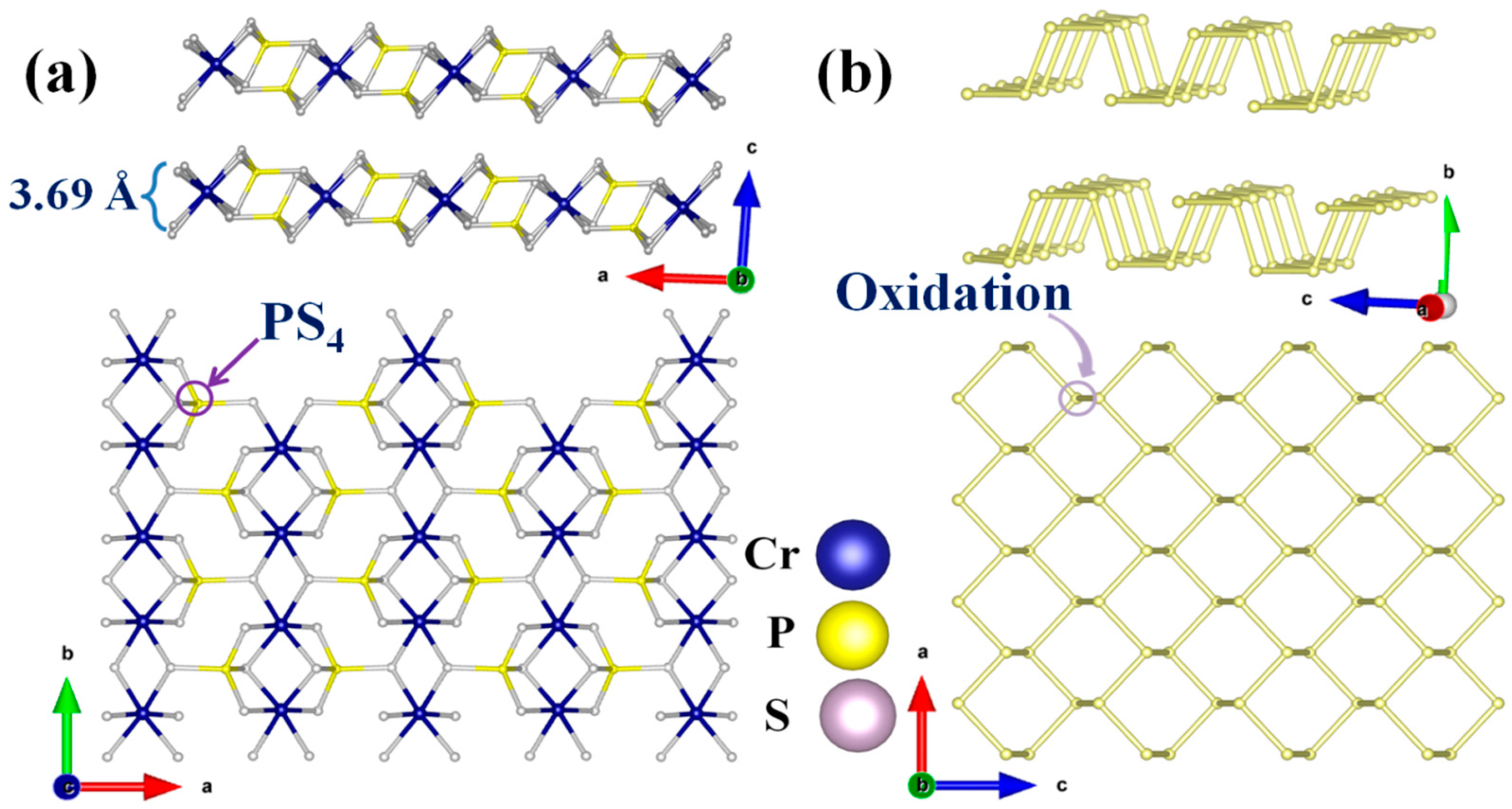
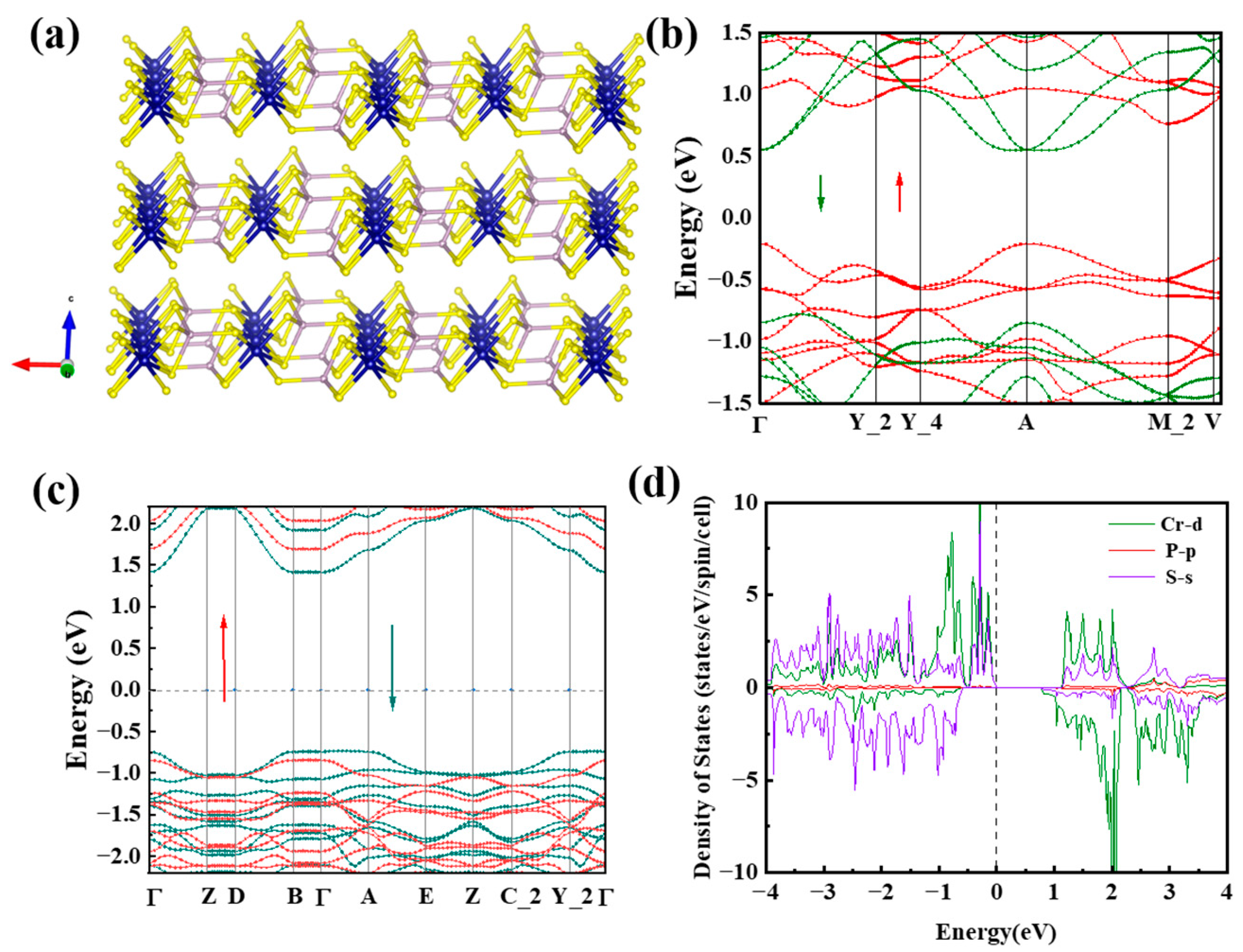
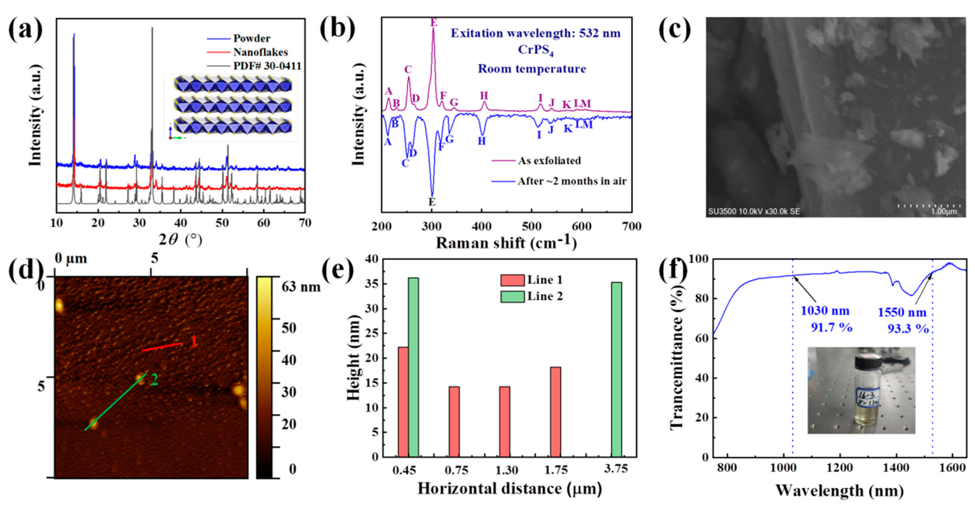
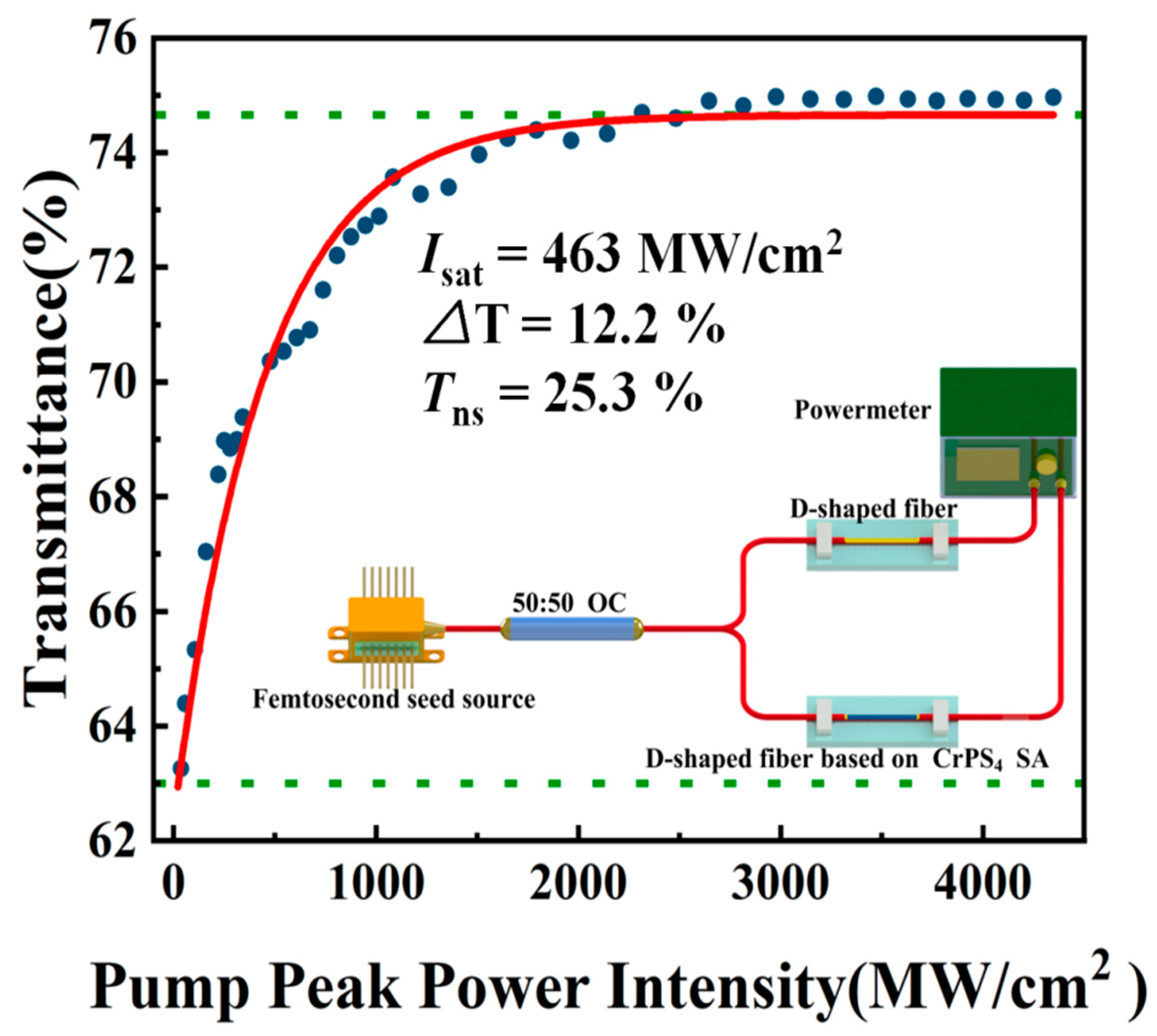
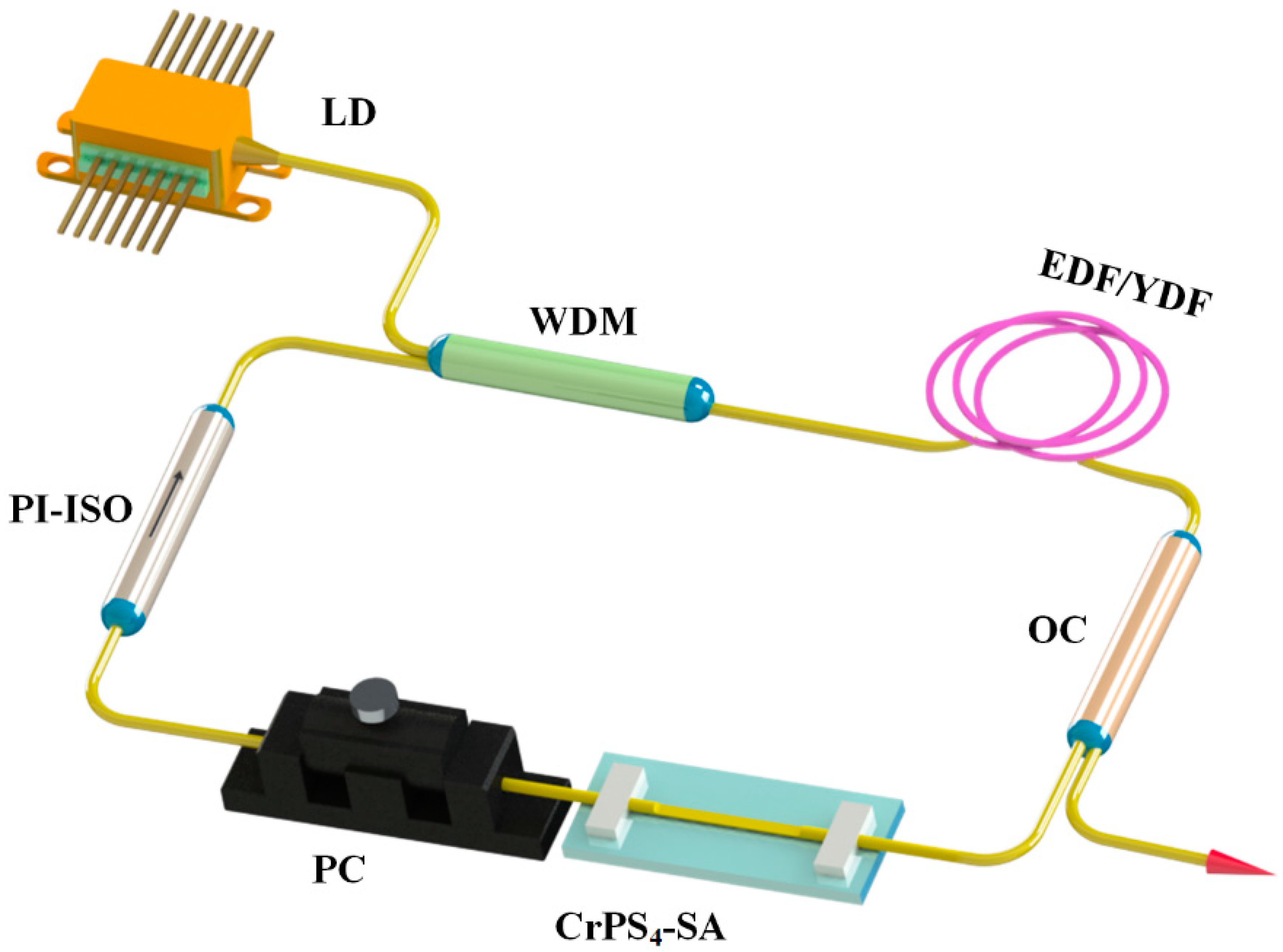
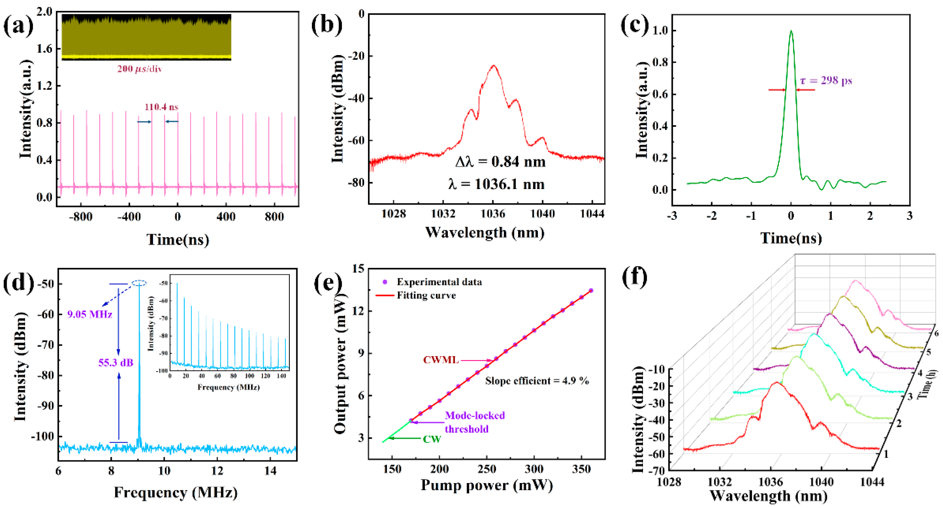
2. Results and Discussion

Bulk CrPS4 crystallized in the non-centrosymmetric monoclinic space group C2 (No. 5) at room temperature. The structure of layered CrPS4 is shown in Figure 1a. The crystal structure was composed of distorted CrS6 octahedra and PS4 tetrahedra connected into a 2D double layer through chemical covalent bonds in a horizontal direction and vdWs forces along the c axis. The sheet thickness was 3.69 Å. Structurally, according to previous reports, the weak vdWs gap between layers was about 2.46 Å; this means that the few-layer form could be readily obtained from bulk samples by the LPE method. More importantly, the multibonded crystal structure of CrPS4, in contrast to the lone-pair electrons found in BP, gave it the potential for air stability. It is commonly known that layered BP possesses a honeycomb structure in which a phosphorus atom is covalently bonded to three neighboring atoms through their p-orbitals, exposing a pair of lone-pair electrons (Figure 1b). The lone pairs of phosphorus atoms can readily react with oxygen to form PxOy; this ultimately leads to the formation of phosphoric acid and H2O, and thereby presents a significant obstacle to the use of BP in applications involving flexible electronics and photoelectronics.
To gain deeper insights into the band-structure properties of CrPS4, the CASTEP mode in Material Studio software was utilized to perform DFT calculations of the band structure and density of states (DOS) of both bulk and monolayer forms of CrPS4 [48]. The calculated electronic band structures of bulk and monolayer CrPS4 are illustrated in Figure 2b,c; these indicate that CrPS4 is a direct-band-gap semiconductor, in agreement with previous research [49]. From Figure 2b, it can be seen that the bulk CrPS4 nanoflakes exhibited a band-gap energy of 0.97 eV. As the number of layers decreased, the energy of the band gap increased. When the CrPS4 nanoflakes were monolayered, the band-gap energy increased to about 2.15 eV. As illustrated in Figure 2d, the DOS plots reveal significant spin splitting in the d-orbitals of Cr atoms, indicating the presence of a sizable spin polarization in the bulk system.
Using the LPE method, few-layer CrPS4 nanoflakes were exfoliated simply and effectively. Subsequently, we demonstrated the powder X-ray diffraction (XRD) patterns of CrPS4 and its samples after exfoliation treatment by XRD spectroscopy, as illustrated in Figure 3a. It can be seen that the positions of the diffraction peaks on the two patterns were in almost perfect agreement with a standard PDF card of CrPS4 (PDF #30-0411), indicating the integrity of the structure after exfoliated CrPS4 nanoflakes were obtained. In addition, the diffraction pattern from the CrPS4 sample was analyzed using X’pert Highscore Plus 3.0 [50]. The characterization of the diffraction pattern occurred in the range of 2ϴ = 10–70°, as shown in Figure S1. The diffraction pattern from the sample matched well with the diffraction pattern from the previous report. The reference code of CrPS4 is 00-033-0404 [51]. The vibrational modes of the CrPS4 were verified by the Raman spectrum and detected in the range 200–700 cm−1 (excitation wavelength: 532 nm, inVia, Renishaw, Wotton-under-Edge, UK) at room temperature. As shown in Figure 3b, about 13 vibration peaks were found in the Raman spectrum; this was in agreement with previously reported results [46], and further confirmed the rationality of the structure of the CrPS4. The surface morphology of the samples as exfoliated was then analyzed via scanning electron microscope (SEM, JSM-5910LV, JEOL, Tokyo, Japan). As depicted in Figure 3c, the CrPS4 nanoflakes exhibited an obvious layered structure, indicating that the nanoflakes were successfully fabricated based on the LPE method. The morphology of the CrPS4 nanoflakes was also tested using an atomic force microscope (AFM, MFP-3D Infinity, Asylum Research, Oxford, UK), which can observe the lateral size of nanoflakes. Figure 3d,e show CrPS4 nanoflakes with an average thickness of ~25 nm. The linear optical transmission spectrum of exfoliated CrPS4 was also detected using a UV-vis-NIR spectrophotometer (LAMBDA, Perkin Elmer Inc., Waltham, MA, USA). We found that, at wavelengths of 1030 and 1530 nm, the transmittances were approximately 91.7% and 93.3%, respectively (Figure 3f). BP has attracted tremendous interest because of its natural layer-dependent direct-band-gap energy, but layered BP is unstable and degrades rapidly in ambient conditions within hours. Calculated results indicated that CrPS4 is also a direct-band-gap semiconductor, and this made us keen to discover if it was stable in ambient conditions for long time periods. Firstly, taken together, the powder XRD and Raman spectroscopy studies clearly demonstrated that the CrPS4 structure remained stable after months in ambient conditions. As shown in Figure S1a, the intensity and peak position of CrPS4, both as exfoliated and after ~2 months in air, remained essentially unchanged. The intensity and peak position of the Raman modes of CrPS4 also remained essentially unchanged for ~2 months, indicating the air-stability of CrPS4 (shown in Figure 3b). The CrPS4 nanoflakes were then analyzed via energy-dispersive X-ray spectroscopy (EDS, Oxford Instruments, Oxford, UK). As depicted in Figure S1c,d, an average Cr/P/S molar ratio of 1.0:1.0:4.0 was recorded for CrPS4 nanoflakes as exfoliated and again after ~2 months in air, further confirming the air-stability of the samples. To more intuitively demonstrate this stability after exposure to air for about 2 months, the surface morphology of the sample was analyzed via SEM. As can be seen in Figure S1c, the CrPS4 nanoflakes continued to exhibit an obvious layered structure.
The obtained few-layer CrPS4 nanoflakes were then dripped onto the D-shaped fiber to form an SA device. To investigate the nonlinear optical properties of the CrPS4-SA, a dual-channel balanced detection measurement system based on an erbium-doped fiber laser (1550 nm, 100 fs, 8.05 MHz) was employed, as shown in Figure 4 inset. Equation (1) only considers the case of single-photon absorption, and the nonlinear saturated absorption curve of CrPS4-SA was obtained after fitting [52,53].
T ( I ) = 1 Δ T × exp ( I I s a t ) T n s
In Equation (1), T(I) is the transmission rate, T is the modulation depth (MD), I is the input intensity, I s a t is the saturated intensity, and T n s is the nonsaturable loss (NL). The fitting results are shown in Figure 4. Values of MD, I s a t , and NL were found to be ~12.2%, ~463 MW/cm2, and ~25.3%, respectively.
Next, to validate the excellent potential of the layered CrPS4 for ultrafast laser applications, we constructed 1.0 μm and 1.5 μm all-fiber laser cavities using Er-doped or Yb-doped fibers. The structural diagram of the optical fiber laser structure is shown in Figure 5. The laser cavity consisted of a section of Er-doped or Yb-doped fiber, a laser diode (980 nm Pump Laser, Hanyu, Shanghai, China), a wavelength division multiplexer (WDM, Mingchuang, Shenzhen, China), an optical coupler (OC, Mingchuang, Shenzhen, China), a polarization-independent optical isolator (ISO, Mingchuang, Shenzhen, China), a polarization controller (PC, General Photonics, Losa Angeles, US), and a D-shaped fiber based on CrPS4-SA. ISO and PC were used to ensure the unidirectional propagation of light and to adjust its polarization state, respectively. The evanescent field length of the D-shaped fiber optic bare leak was 10 mm, and the distance between the surface and the core was 1 μm. The Er-doped fiber laser cavity length was 21.96 m, including 4 m EDF (Nufern EDFC-980-HP, Hanyu, Shanghai, China) and 17.96 m single-mode fiber (SMF, Hanyu, Shanghai, China), with dispersion parameters at 1530 nm of −12.2 ps/(km·nm) and 18 ps/(km·nm), respectively. The net dispersion was estimated as −0.39 ps2. Similarly, the cavity length of Yb-doped laser consisted of 1 m YDF (Nufern SM-YSF-HI-6/125, Hanyu, Shanghai, China) and 15.56 m single-mode fiber (HI1060), and the net dispersion of the cavity was estimated at 0.35 ps2. The pulse performance of the laser output was determined by a power meter, a spectrum analyzer (AQ6370C, Yokogawa, Tokyo, Japan), a photodetector (DET08CFC/M 5 GHz, Thorlabs, Newton, MA, USA), a hybrid oscilloscope (DP04104B 1 GHz/s, Tektronix, OR, USA), and autocorrelator monitoring (PulseCheck, APE, Berlin, Germany). The temperature and humidity of the ultra-clean laboratory were 16.8 °C and 58%, respectively.
Importantly, we performed pre-experiments to demonstrate that, in the absence of SA, no mode-locking occurred in the laser cavity, regardless of PC modulation or pump power. By such means, we confirmed the authenticity of the experiment. After SA was applied to the laser cavity, we adjusted the PC while continuously increasing the pump power, and observed the output waveform of the oscilloscope. We achieved continuous-wave mode-locking (CWML) when the pump power was higher than 170 wm. When the pump power was increased to 300 mW, the output power was 10.64 mW, and pulse energy and peak power were 1.174 nJ and 3.94 W, respectively. The mode-locking sequence diagram is presented in Figure 6a, which shows a 110.4 ns time interval between adjacent pulses, which corresponds well to the pulse repetition rate of 9.05 MHz. The inset in Figure 6a shows the amplitude intensity plot of the mode-locked pulse sequence; this indicates that the mode-locked sequence existed stably for a long time. The spectrum with a central wavelength of 1036.1 nm had a 3 dB spectral width of 0.84 nm, as shown in Figure 6b. The autocorrelation trace corresponding to the measured pulse at this time is shown in Figure 6c. It can be seen that the pulse width is about 298 ps, and the time-bandwidth product (TBP) is 69.9, indicating that the pulse has a serious chirp. In Figure 6d, a signal-to-noise ratio (SNR) measurement of approximately 55.3 dB can be observed with a higher signal peak at a laser cavity repetition rate of 9.05 MHz. The relationship between the mode-locked output power and pump power is shown in Figure 6e. When the pump power was 130 mW, the laser cavity output a continuous wave (CW). When the pump power was increased to 170 mW, the output was CWML, and the measured slope efficiency was about 4.9%. Subsequently, we measured the output spectrum of the laser cavity over a longer timescale, at time intervals of 1 h, for a total of 6 h. As shown in Figure 6f, the long-term spectrum was quite stable, indicating that a ytterbium-doped laser has the potential of highly stable operation.
In order to verify that CrPS4 could work in a wide wavelength range, we placed the additionally prepared CrPS4-SA into an erbium-doped fiber laser cavity for debugging. Stable CWML output was achieved by adjusting the PC when the pump power was above 150 mW. The output power, pulse energy, and peak power were 6.1 mW, 0.893 nJ, and 1786 W, respectively, when the pump power was 270 mW. The output pulse characteristics are shown in Figure 7. The mode-locked pulse sequence is shown in Figure 7a. The pulse period was 146.4 ns, which corresponded to a pulse repetition rate of 6.83 MHz. The inset shows that the mode-locking was quite stable. Figure 7b shows the laser spectrum centered at 1531.6 nm with a 3 dB bandwidth of 5.6 nm. A measured pulse width of 500 fs resulted in a TBP of 0.35, as shown in Figure 7c. It can be clearly observed that the peak value of the high signal at the mode-locked repetition frequency was 6.83 MHz, and the signal-to-noise ratio was about 64 dB, as shown in Figure 7d. Figure 7e shows that the average output power varied linearly with increasing pump power, with a slope efficiency of 2.2%. Subsequently, we also measured the spectrum for 6 h, at time intervals of 1 h, and found that the erbium-doped laser mode-locking was very stable, as shown in Figure 7f.
To demonstrate the stability of CrPS4, we placed the previously prepared saturable absorber back into an ytterbium-doped fiber laser, 40 days after the first experiment, and found that the laser could still output mode-locked pulses after adjusting the pump and PC. The mode-locking output results are shown in Figure S3. The threshold of mode-locking was 180 mW, a slight increase (of 10 mW) compared to the first experiment. Figure S3a shows the pulse sequence with a pulse interval of 107.2 ns. The inset shows that the laser continued to work with high stability. Figure S3b shows that, in comparison with the first experiment, the central wavelength of the mode-locked spectrum remained the same, at 1036.1 nm, while the 3 dB bandwidth changed only slightly, from 0.84 nm to 0.8 nm. The measured mode-locking pulse width was 400 ps, as shown in Figure S3c, which was wider than the first measurement. The repetition frequency of the mode-locked strong signal peak was 9.32 MHz, and the SNR was about 52.6 dB, as shown in Figure S3d. The relationship between average output power and pump power is shown in Figure S3e, and the slope efficiency was 3.6%, which was slightly lower than the first time. We then measured the spectrum for 6 h, and the results are shown in Figure S3f. The spectrum remained unchanged, indicating that the laser worked stably, and that the CrPS4 saturable absorber we made exhibited high stability.
Once again, 40 days after the first experiment, we put the previously used saturable absorber back into the erbium-doped fiber laser, using the adjustment method described above. To achieve CWML, pump power needed to be increased to more than 150 mw. The mode-locking output characteristics are shown in Figure S4. Figure S4a shows that the pulse interval was 136.9 ns, and the inset shows that the laser cavity remained in a stable state. Figure S4b shows that the central wavelength of the spectrum was 1531.4 nm, and the 3dB bandwidth was 6.2 nm, which was stable compared with the first experiment. Figure S4c shows that the mode-locked pulse width was 594 fs, which was slightly wider than the first experiment, with a TBP of 0.47. The signal-to-noise ratio of a strong signal peak at a repetition frequency of 7.33 MHz was approximately 69 dB. Figure S4e shows that the slope efficiency between the average output power and the pump power was 2.4%. Finally, we measured the long-term spectral changes, which indicated the excellent stability of the laser cavity, as shown in Figure S4f.
We then compared the data of the two experiments, as set out in Table 1. Although the two experiments were separated by 40 days, we found few differences in the results, indicating that the CrPS4-SA we prepared had excellent stability. However, in order to more intuitively observe any variation in the experimental results, we drew a coefficient of variation diagram to represent the degree of dispersion of the pulse parameters of the two experiments, as shown in Figure 8. It can be seen that the coefficients of variation of the pulse width, output power, pulse energy, and coefficients of variation of the Yb-doped fiber laser were close to 30%, while the coefficient of variation of the peak power was as high as 59%. For Er-doped fiber lasers, the coefficients of variation of its pulse parameters were all less than 20%; among these, the coefficients of variation of pulse energy, peak power and slope efficiency were all less than 10%. In short, the output variation of the erbium-doped fiber laser cavity was lower, and this finding is related to the performance of the laser cavity itself. The ytterbium-doped fiber laser cavity worked in the total positive dispersion region, while the erbium-doped laser cavity worked in the anomalous dispersion region, and the optical solitons formed through the balance of dispersion and nonlinear effects were more stable.
The pulse performance of the laser has a crucial influence on the application. In Table S1, we summarize the performance of mode-locked ytterbium-doped lasers for several representative 2D materials. It can be seen that the output of these ytterbium-doped lasers was in the order of picoseconds. In contrast, the output of our CrPS4 ytterbium-doped fiber laser was 298 ps. These results show that CrPS4 has higher generation efficiency for ultra-short pulse output, and has certain advantages in terms of pulse width. The maximum output power of our laser was 10.63 mW, which is 28.7 times that of the graphene previously reported by Zhao et al. [54], and 1.38 times that of the Mo2C previously reported by Liu et al. [55]. The single-pulse energy of most YDF lasers using 2D materials as SA is usually limited to below 1 nJ; however, our laser achieved an output of 1.174 nJ, with a peak power of 3.94 W, which is 5.5 times that of the WS2 reported by Mao et al. [56], and 2.3 times that of the NiPS3 reported by Liu et al. [56]. In a similar way, we compared erbium-doped fiber lasers based on other 2D slave materials, as shown in Table S2. As can be readily observed, because of the remarkable nonlinear optical properties of these 2D materials, their modulation depths vary in percentage terms from low single digits to many tens. In contrast, the modulation depth of our prepared CrPS4 was 12.2%, which is higher than the 10.9% for BP reported by Chao et al. [57] and the 5.1% for Mo2C reported by Liu et al. [55]. At the same time, our pulse output was 500 fs, which is similar to that achieved by other lasers. In addition, our output reached a level of 6.1 mW, which is twice that of the graphene reported by Bao et al. [7], and 2.3 times that of the BP reported by Chao et al. [57]. The single pulse energy and peak power were 893 pJ and 1786 W, respectively. Not only did we measure the stability of the spectrum over 6 h, we also re-experimented with the previously fabricated saturable absorber 40 days later and found that it still achieved mode locking. These experimental results confirm that CrPS4 is a competitive direct-band-gap material with excellent nonlinear optical modulation properties and great potential for broadband ultrafast photonics applications.

3. Conclusions

In conclusion, we fabricated high-quality CrPS4-SA by the LPE method. Theoretical calculations of the electronic band structures of CrPS4 revealed a direct band gap. We studied the applications of few-layer CrPS4-SA in ultrafast photonics for the first time. The saturated intensity and modulation depth of CrPS4-SA were 463 MW/cm2 and 12.2%, respectively, at 1.5 µm. Moreover, based on the excellent saturable absorption of the D-shaped CrPS4 SA, the pulse characteristics of fiber lasers operating in conventional soliton states were measured. We successfully obtained picosecond mode-locked pulses of 298 ps and ultrashort femtosecond pulses of 500 fs in the 1 µm and 1.5 µm regions, respectively. The signal-to-noise ratio (SNR) of the mode-locked operation was as high as 55.3 dB at 9.05 MHz (YDFL), and 64 dB at 6.83 MHz (EDFL). More importantly, the few-layer CrPS4 exhibited excellent stability during exposure to air for a period of time. Our experimental results show that CrPS4 is an air-stable and broadband SA, with promising potential for ultrafast laser applications.

4. Experimental Section

Fabrication of CrPS4. An LPE method was used to exfoliate few-layer CrPS4 nanoflakes, in which the vdWs forced between the layers of CrPS4 were broken by an ultrasonic wave. Firstly, commercially available high-purity CrPS4 (Shenzhen six carbon) powder (about 23 mg) was ground in a mortar and dispersed into N-methyl-2-pyrrolidone (NMP, 30 mL), which was exfoliated in an ultrasonic cell disruptor for 20 h at power of 400 W. In order to make the solute to form nanoscale flakes, the solvent was ultrasonicated in an ultrasound cleaner for 24 h. Then, the mixture was centrifugally treated at a speed of 5000 rpm for 20 min to separate precipitation, and the few-layer CrPS4 containing supernatant was obtained. All experimental procedures were conducted at room temperature (16.8 °C) and a relative humidity of 58%.
Characterization. The powder X-ray diffraction (XRD) patterns of CrPS4 and its samples after exfoliated treatment were further demonstrated by XRD spectroscopy; the vibrational modes of the CrPS4 were verified by Raman spectrum, and detected in the range 200–700 cm−1 (excitation wavelength: 532 nm, inVia, Renishaw, Wotton-under-Edge, UK) at room temperature. The surface morphology of the samples as exfoliated was analyzed via a scanning electron microscope. The morphology of CrPS4 nanoflakes was also tested using an atomic force microscope and the linear optical transmission spectrum of exfoliated CrPS4 was detected by an UV-vis-NIR spectrophotometer.
DFT calculation details. Here, the Vienna Ab initio Simulation Package (VASP, University of Vienna) was utilized to optimize the crystal structures and calculate electronic structures [58,59,60]. The exchange and correlative potentials of electron−electron interactions were accounted for using the generalized gradient approximation (GGA) within the Perdew−Burke−Eruzerhof (PBE) scheme [61,62]. More specifically, an energy cutoff of 500 eV and a Monkhorst–Pack Brillouin zone sampling grid [63] with a resolution 0.02 × 2π Å−1 were applied.

Supplementary Materials

The following supporting information can be downloaded at: https://www.mdpi.com/article/10.3390/nano13061128/s1: Figure S1: The diffraction pattern from CrPS4 sample was analyzed using X’pert Highscore Plus 3.0; Figure S2: XRD spectra, SEM image, EDX spectra with an atomic ratio of Cr, P, and S elements for CrPS4 flakes as exfoliated and after ~2 months in air; Figure S3: the results of typical mode-locked pulse characteristics (second experiment) at 1 μm after ~40 days; Figure S4: the results of typical mode-locked pulse characteristics(second experiment) at 1.5 μm after ~40 days; Table S1: comparison of SA with other 2D materials at 1 μm laser output parameters; Table S2: comparison of SA with other 2D materials at 1.5 μm laser output parameters. References [64,65,66,67,68,69,70,71,72,73,74,75] are cited in the supplementary materials.

Author Contributions

Conceptualization, Q.W.; methodology, W.Z.; software, Y.Z. and Q.W.; validation, Y.Z., W.Z., and X.L.; formal analysis, W.Z. and Q.J.; investigation, W.Z. and Y.Z.; resources, Y.Z.; data curation, W.Z.; writing—original draft preparation, W.Z. and Y.Z.; writing—review and editing, W.Z. and Y.Z.; visualization, W.Z. and Y.Z.; supervision, Q.W. and Q.J.; project administration, Q.W.; funding acquisition, Q.W. All authors have read and agreed to the published version of the manuscript.

Funding

This work was financially supported by the National Natural Science Foundation of China (6217030813), the Guangdong Basic and Applied Basic Research Foundation (2021A1515010964), and the Science and Technology Innovation Commission of Shenzhen Municipality (SGDX20190919094803949 and JCYJ20200109105810074).

Data Availability Statement

The data presented in this study are available on request from the corresponding author.

Conflicts of Interest

The authors declare no competing financial interest.

References

  1. Zhang, Y.; Zhu, J.Q.; Li, P.X.; Wang, X.X.; Yu, H.; Xiao, K.; Li, C.Y.; Zhang, G.Y. All-fiber Yb-doped fiber laser passively mode-locking by monolayer MoS2 saturable absorber. Opt. Commun. 2018, 413, 236–241. [Google Scholar] [CrossRef]
  2. Sugioka, K.; Cheng, Y. Ultrafast lasers—Reliable tools for advanced materials processing. Light Sci. Appl. 2014, 3, e149. [Google Scholar] [CrossRef] [Green Version]
  3. Keller, U. Recent developments in compact ultrafast lasers. Nature 2003, 424, 831–838. [Google Scholar] [CrossRef]
  4. Gu, T.; Petrone, N.; McMillan, J.F.; van der Zande, A.; Yu, M.; Lo, G.Q.; Kwong, D.L.; Hone, J.; Wong, C.W. Regenerative oscillation and four-wave mixing in graphene optoelectronics. Nat. Photonics 2012, 6, 554–559. [Google Scholar] [CrossRef] [Green Version]
  5. Fermann, M.E.; Hartl, I. Ultrafast fibre lasers. Nat. Photonics 2013, 7, 868–874. [Google Scholar] [CrossRef]
  6. Du, Y.; Zhao, K.; Zhu, Z.L.; Wang, J.; Deng, W.J.; Liang, X.D. Research and application of ultrafast laser precision manufacturing technology. Laser Infrared 2020, 50, 1419–1425. [Google Scholar] [CrossRef]
  7. Bao, Q.L.; Zhang, H.; Wang, Y.; Ni, Z.H.; Yan, Y.L.; Shen, Z.X.; Loh, K.P.; Tang, D.Y. Atomic-layer graphene as a saturable absorber for ultrafast pulsed lasers. Adv. Funct. Mater. 2009, 19, 3077–3083. [Google Scholar] [CrossRef]
  8. Dong, Y.C.; Chertopalov, S.; Maleski, K.; Anasori, B.; Hu, L.Y.; Bhattacharya, S.; Rao, A.M.; Gogotsi, Y.; Mochalin, V.N.; Podila, R. Saturable absorption in 2D Ti3C2 MXene thin films for passive photonic diodes. Adv. Mater. 2018, 30, 1705714. [Google Scholar] [CrossRef]
  9. Liu, S.X.; Huang, H.F.; Lu, J.S.; Xu, N.; Qu, J.L.; Wen, Q. Liquid-phase exfoliation of Ta2NiS5 and Its application in near-infrared mode-locked fiber lasers with evanescent field interactions and passively Q-switched bulk laser. Nanomaterials 2022, 12, 695. [Google Scholar] [CrossRef]
  10. Zhang, A.J.; Wang, Z.H.; Ou-Yang, H.; Lyu, W.H.; Sun, J.X.; Cheng, Y.; Fu, B. Recent progress of two-dimensional materials for ultrafast photonics. Nanomaterials 2021, 11, 1778. [Google Scholar] [CrossRef]
  11. Chen, L.L.; Zhang, M.; Zhou, C.; Cai, Y.; Ren, L.; Zhang, Z.G. Ultra-Low Repetition Rate SESAM-mode-locked Linear-cavity Erbium-doped Fiber Laser. In Proceedings of the 2009 Conference on Lasers & Electro Optics & The Pacific Rim Conference on Lasers and Electro-Optics, Shanghai, China, 20 August–3 September 2009; pp. 587–588. [Google Scholar]
  12. Cabasse, A.; Ortac, B.; Martel, G.; Hideur, A.; Limpere, J. Highly normal dispersion Er-doped fiber laser mode-locked with a SESAM. In Proceedings of the 2008 Conference on Lasers and Electro-Optics and 2008 Conference on Quantum Electronics and Laser Science, San Jose, CA, USA, 4–9 May 2008; pp. 1–2. [Google Scholar]
  13. Li, P.F.; Chen, Y.; Yang, T.S.; Wang, Z.Y.; Lin, H.; Xu, Y.H.; Li, L.; Mu, H.R.; Shivananju, B.N.; Zhang, Y.P.; et al. Two-dimensional CH3NH3PbI3 perovskite nanosheets for ultrafast pulsed fiber lasers. ACS Appl. Mater. Interfaces 2017, 9, 12759–12765. [Google Scholar] [CrossRef]
  14. Guo, B.; Xiao, Q.L.; Wang, S.H.; Zhang, H. 2D layered materials: Synthesis, nonlinear optical properties, and device applications. Laser Photonics Rev. 2019, 13, 1800327. [Google Scholar] [CrossRef]
  15. Guo, X.; Wang, S.; Yan, P.G.; Wang, J.Z.; Yu, L.P.; Liu, W.J.; Zheng, Z.J.; Guo, C.Y.; Ruan, S.C. High modulation depth enabled by Mo2Ti2C3Tx MXene for Q-switched pulse generation in a mid-infrared fiber laser. Nanomaterials 2022, 12, 1343. [Google Scholar] [CrossRef]
  16. Zhou, L.L.; Fu, H.G.; Lv, T.; Wang, C.B.; Gao, H.; Li, D.Q.; Deng, L.M.; Xiong, W. Nonlinear optical characterization of 2D materials. Nanomaterials 2020, 10, 2263. [Google Scholar] [CrossRef]
  17. Jia, L.; Lei, T.M. Research progress on physical properties and chemical stability of two-dimensional black phosphorus. Mater. Rev. 2018, 32, 1100–1106. [Google Scholar] [CrossRef]
  18. He, J.S.; Tao, L.L.; Zhang, H.; Zhou, B.; Li, J.B. Emerging 2D materials beyond graphene for ultrashort pulse generation in fiber lasers. Nanoscale 2019, 11, 2577–2593. [Google Scholar] [CrossRef]
  19. Bundulis, A.; Alnis, J.; Shuklov, I.A.; Kim, V.V.; Lizunova, A.A.; Mardini, A.A.; Grube, J.; Razumov, V.F.; Ganeev, R.A. Nonlinear absorption and refraction of picosecond and femtosecond pulses in HgTe quantum dot films. Nanomaterials 2021, 11, 3351. [Google Scholar] [CrossRef]
  20. Shah, A.; Torres, P.; Tscharner, R.; Wyrsch, N.; Keppner, H. Photovoltaic technology: The case for thin-film solar cells. Science 1999, 285, 692–698. [Google Scholar] [CrossRef] [Green Version]
  21. Guo, Q.J.; Ford, G.M.; Yang, W.-C.; Walker, B.C.; Stach, E.A.; Hillhouse, H.W.; Agrawal, R. Fabrication of 7.2% efficient CZTSSe solar cells using CZTS nanocrystals. J. Am. Chem. Soc. 2010, 132, 17384–17386. [Google Scholar] [CrossRef]
  22. Watanabe, K.; Taniguchi, T.; Kanda, H. Direct-bandgap properties and evidence for ultraviolet lasing of hexagonal boron nitride single crystal. Nat. Mater. 2004, 3, 404–409. [Google Scholar] [CrossRef]
  23. Hochbaum, A.I.; Yang, P. Semiconductor nanowires for energy conversion. Chem. Rev. 2010, 110, 527–546. [Google Scholar] [CrossRef]
  24. Bhaskar, S.; Visweswar Kambhampati, N.S.; Ganesh, K.M.; Srinivasan, V.; Ramamurthy, S.S. Metal-free, graphene oxide-based tunable soliton and plasmon engineering for biosensing applications. ACS Appl. Mater. Interfaces 2021, 13, 17046–17061. [Google Scholar] [CrossRef] [PubMed]
  25. Sun, M.K.; Wang, Y.; Hu, H.; Zhang, H.; Li, W.J.; Lv, B.; Zhu, Z.; Guan, C.Y.; Shi, J.H. Optical properties and dynamic extrinsic chirality of structured monolayer black phosphorus. Front. Mater. 2022, 9, 826795. [Google Scholar] [CrossRef]
  26. Zhang, M.; Wu, Q.; Zhang, F.; Chen, L.L.; Jin, X.X.; Hu, Y.W.; Zheng, Z.; Zhang, H. 2D black phosphorus saturable absorbers for ultrafast photonics. Adv. Opt. Mater. 2019, 7, 1800224. [Google Scholar] [CrossRef] [Green Version]
  27. Li, L.; Wang, Y.G.; Wang, X. Ultrafast pulse generation with black phosphorus solution saturable absorber. Laser Phys. 2017, 27, 085104. [Google Scholar] [CrossRef]
  28. Liu, X.; Gao, Q.; Zheng, Y.; Mao, D.; Zhao, J.L. Recent progress of pulsed fiber lasers based on transition-metal dichalcogenides and black phosphorus saturable absorbers. Nanophotonics 2020, 9, 2215–2231. [Google Scholar] [CrossRef] [Green Version]
  29. Wang, J.T.; Chen, H.; Jiang, Z.K.; Yin, J.D.; Wang, J.Z.; Zhang, M.; He, T.C.; Li, J.Z.; Yan, P.G.; Ruan, S.C. Mode-locked thulium-doped fiber laser with chemical vapor deposited molybdenum ditelluride. Opt. Lett. 2018, 43, 1998–2001. [Google Scholar] [CrossRef]
  30. Wang, J.L.; Wang, X.L.; Lei, J.J.; Ma, M.Y.; Wang, C.; Ge, Y.Q.; Wei, Z.Y. Recent advances in mode-locked fiber lasers based on two-dimensional materials. Nanophotonics 2020, 9, 2315–2340. [Google Scholar] [CrossRef]
  31. Zhao, Y.T.; Wang, H.Y.; Huang, H.; Xiao, Q.L.; Xu, Y.H.; Guo, Z.; Xie, H.H.; Shao, J.D.; Sun, Z.B.; Han, W.J.; et al. Surface coordination of black phosphorus for robust air and water stability. Angew. Chem. Int. Ed. 2016, 55, 5003–5007. [Google Scholar] [CrossRef]
  32. Wood, J.D.; Wells, S.A.; Jariwala, D.; Chen, K.-S.; Cho, E.; Sangwan, V.K.; Liu, X.L.; Lauhon, L.J.; Marks, T.J.; Hersam, M.C. Effective passivation of exfoliated black phosphorus transistors against ambient degradation. Nano Lett. 2014, 14, 6964–6970. [Google Scholar] [CrossRef] [Green Version]
  33. Ziletti, A.; Carvalho, A.; Campbell, D.K.; Coker, D.F.; Castro Neto, A.H. Oxygen defects in phosphorene. Phys. Rev. Lett. 2015, 114, 046801. [Google Scholar] [CrossRef] [Green Version]
  34. Zhao, Y.K.; Sun, Z.J.; Zhang, B.W.; Yan, Q.F. Unveiling the degradation chemistry of fibrous red phosphorus under ambient conditions. ACS Appl. Mater. Interfaces 2022, 14, 9925–9932. [Google Scholar] [CrossRef] [PubMed]
  35. Tan, S.J.R.; Abdelwahab, I.; Chu, L.Q.; Poh, S.M.; Liu, Y.P.; Lu, J.; Chen, W.; Loh, K.P. Quasi-monolayer black phosphorus with high mobility and air stability. Adv. Mater. 2018, 30, 1704619. [Google Scholar] [CrossRef] [PubMed]
  36. Luo, W.; Zemlyanov, D.Y.; Milligan, C.A.; Du, Y.C.; Yang, L.M.; Wu, Y.Q.; Ye, P.D. Surface chemistry of black phosphorus under a controlled oxidative environment. Nanotechnology 2016, 27, 434002. [Google Scholar] [CrossRef] [PubMed] [Green Version]
  37. Song, H.Z.; Wu, H.; Ren, T.Q.; Yan, S.C.; Chen, T.H.; Shi, Y. Developments in stability and passivation strategies for black phosphorus. Nano Res. 2021, 14, 4386–4397. [Google Scholar] [CrossRef]
  38. Pei, J.J.; Gai, X.; Yang, J.; Wang, X.B.; Yu, Z.F.; Choi, D.-Y.; Luther-Davies, B.; Lu, Y.R. Producing air-stable monolayers of phosphorene and their defect engineering. Nat. Commun. 2016, 7, 10450. [Google Scholar] [CrossRef] [Green Version]
  39. Neal, S.N.; O’Neal, K.R.; Haglund, A.V.; Mandrus, D.G.; Bechtel, H.A.; Carr, G.L.; Haule, K.; Vanderbilt, D.; Kim, H.-S.; Musfeldt, J.L. Exploring few and single layer CrPS4 with near-field infrared spectroscopy. 2D Mater. 2021, 8, 035020. [Google Scholar] [CrossRef]
  40. Synnatschke, K.; Shao, S.Q.; van Dinter, J.; Hofstetter, Y.J.; Kelly, D.J.; Grieger, S.; Haigh, S.J.; Vaynzof, Y.; Bensch, W.; Backes, C. Liquid exfoliation of Ni2P2S6: Structural characterization, size-dependent properties, and degradation. Chem. Mater. 2019, 31, 9127–9139. [Google Scholar] [CrossRef]
  41. Sibley, S.P.; Francisa, A.H.; Lifshitzb, E.; Clkmen, R. Magnetic resonance studies of intercalated, twodimensional transition metal chalcogenophosphate. Colloids Surf. A Physicochem. Eng. Asp. 1994, 82, 205–215. [Google Scholar] [CrossRef] [Green Version]
  42. Budniak, A.K.; Killilea, N.A.; Zelewski, S.J.; Sytnyk, M.; Kauffmann, Y.; Amouyal, Y.; Kudrawiec, R.; Heiss, W.; Lifshitz, E. Exfoliated CrPS4 with promising photoconductivity. Small 2020, 16, 1905924. [Google Scholar] [CrossRef]
  43. Calder, S.; Haglund, A.V.; Liu, Y.; Pajerowski, D.M.; Cao, H.B.; Williams, T.J.; Garlea, V.O.; Mandrus, D. Magnetic structure and exchange interactions in the layered semiconductor CrPS4. Phys. Rev. B 2020, 102, 024408. [Google Scholar] [CrossRef]
  44. Kim, S.; Lee, J.; Jin, G.; Jo, M.-H.; Lee, C.G.; Ryu, S.M. Crossover between photochemical and photothermal oxidations of atomically thin magnetic semiconductor CrPS4. Nano Lett. 2019, 19, 4043–4051. [Google Scholar] [CrossRef]
  45. Peng, Y.X.; Ding, S.L.; Cheng, M.; Hu, Q.F.; Yang, J.; Wang, F.G.; Xue, M.Z.; Liu, Z.; Lin, Z.C.; Avdeev, M.; et al. Magnetic structure and metamagnetic transitions in the van der Waals antiferromagnet CrPS4. Adv. Mater. 2020, 32, 2001200. [Google Scholar] [CrossRef]
  46. Lee, J.; Ko, T.Y.; Kim, J.H.; Bark, H.; Kang, B.; Jung, S.-G.; Park, T.; Lee, Z.; Ryu, S.; Lee, C. Structural and optical properties of single- and few-layer magnetic semiconductor CrPS4. ACS Nano 2017, 11, 10935–10944. [Google Scholar] [CrossRef]
  47. Son, J.; Son, S.; Park, P.; Kim, M.; Tao, Z.; Oh, J.; Lee, T.; Lee, S.; Kim, J.; Zhang, K.X.; et al. Air-stable and layer-dependent ferromagnetism in atomically thin van der Waals CrPS4. ACS Nano 2021, 15, 16904–16912. [Google Scholar] [CrossRef] [PubMed]
  48. Momma, K.; Izumi, F. VESTA 3 for three-dimensional visualization of crystal, volumetric and morphology data. J. Appl. Crystallogr. 2011, 44, 1272–1276. [Google Scholar] [CrossRef]
  49. Deng, J.; Guo, J.; Hosono, H.; Ying, T.; Chen, X. Two-dimensional bipolar ferromagnetic semiconductors from layered antiferromagnets. Phys. Rev. Mater. 2021, 5, 034005. [Google Scholar] [CrossRef]
  50. Degen, T.; Sadki, M.; Bron, E.; König, U.; Nénert, G. HighScore Suite; Cambridge University Press: Cambridge, UK, 2014; Volume 29, pp. S13–S18. [Google Scholar]
  51. Diehl, R.; Carpentier, C.-D. The crystal structure of chromium thiophosphate, CrPS4. Acta Crystallogr. Sect. B Struct. Sci. Cryst. Eng. Mater. 1977, B33, 1399–1404. [Google Scholar] [CrossRef] [Green Version]
  52. Andrianov, A.; Kim, A.; Muraviov, S.; Sysoliatin, A. Wavelength-tunable few-cycle optical pulses directly from an all-fiber Er-doped laser setup. Opt. Lett. 2009, 34, 3193–3195. [Google Scholar] [CrossRef]
  53. Chi, C.; Lee, J.; Koo, J.; Han Lee, J. All-normal-dispersion dissipative-soliton fiber laser at 1.06 µm using a bulk-structured Bi2Te3 topological insulator-deposited side-polished fiber. Laser Phys. 2014, 24, 105106. [Google Scholar] [CrossRef]
  54. Zhao, L.M.; Tang, D.Y.; Zhang, H.; Wu, X.; Bao, Q.L.; Loh, K.P. Dissipative soliton operation of an ytterbium-doped fiber laser mode locked with atomic multilayer graphene. Opt. Lett. 2010, 35, 3622–3624. [Google Scholar] [CrossRef]
  55. Liu, S.X.; Lu, J.S.; Huang, H.F.; Xu, N.; Qu, J.L.; Wen, Q. Ultrafast photonics applications based on evanescent field interactions with 2D molybdenum carbide (Mo2C). J. Mater. Chem. C 2021, 9, 6187–6192. [Google Scholar] [CrossRef]
  56. Mao, D.; Zhang, S.L.; Wang, Y.D.; Gan, X.T.; Zhang, W.D.; Mei, T.; Wang, Y.G.; Wang, Y.S.; Zeng, H.B.; Zhao, J.L. WS2 saturable absorber for dissipative soliton mode locking at 1.06 and 1.55 microm. Opt. Express 2015, 23, 27509–27519. [Google Scholar] [CrossRef] [PubMed]
  57. Chao, L.Z.; Meng, L.; Nan, G.Z.; Fang, J.X.; Ping, L.A.; Jun, Z.H.; Feng, Y.X.; Cheng, X.W.; Han, Z. Microfiber-based few-layer black phosphorus saturable absorber for ultra-fast fiber laser. Opt. Express 2015, 23, 20030–20039. [Google Scholar] [CrossRef] [Green Version]
  58. Kresse, G.; Hafner, J. Norm-conserving and ultrasoft pseudopotentials for first-row and transition elements. J. Phys. Condens. Matter 1994, 6, 8245–8257. [Google Scholar] [CrossRef]
  59. Kresse, G.; Furthmüller, J. Efficiency of ab-initio total energy calculations for metals and semiconductors using a plane-wave basis set. Comput. Mater. Sci. 1996, 6, 15–50. [Google Scholar] [CrossRef]
  60. Kresse, G.; Furthmüller, J. Efficient iterative schemes for ab initio total-energy calculations using a plane-wave basis set. Phys. Rev. B Cover. Condens. Matter Mater. Phys. 1996, 54, 11169–11186. [Google Scholar] [CrossRef]
  61. Bader, R.F.W. A quantum theory of molecular structure and its applications. Chem. Rev. 1991, 91, 893–928. [Google Scholar] [CrossRef]
  62. Perdew, J.P.; Burke, K.; Ernzerhof, M. Generalized gradient approximation made simple. Phys. Rev. Lett. 1996, 77, 3865–3868. [Google Scholar] [CrossRef] [PubMed] [Green Version]
  63. Monkhorst, H.J.; Pack, J.D. Special points for Brillouin-zone integrations. Phys. Rev. B 1976, 13, 5188–5192. [Google Scholar] [CrossRef]
  64. Huang, S.S.; Wang, Y.G.; Guang, Y.P.; Zhang, G.L.; Zhao, J.Q.; Li, H.Q.; Lin, R.Y.; Cao, G.Z.; Duan, J.A. Observation of multipulse bunches in a graphene oxide passively mode-locked ytterbium-doped fiber laser with all-normal dispersion. Appl. Phys. B 2014, 116, 939–946. [Google Scholar] [CrossRef]
  65. Al-Masoodi, A.H.H.; Yasin, M.; Ahmed, M.H.M.; Latiff, A.A.; Arof, H.; Harun, S.W. Mode-locked ytterbium-doped fiber laser using mechanically exfoliated black phosphorus as saturable absorber. Optik 2017, 147, 52–58. [Google Scholar] [CrossRef]
  66. Song, H.; Wang, Q.; Zhang, Y.; Li, L. Mode-locked ytterbium-doped all-fiber lasers based on few-layer black phosphorus saturable absorbers. Opt. Commun. 2017, 394, 157–160. [Google Scholar] [CrossRef]
  67. Jiang, X.T.; Liu, S.X.; Liang, W.Y.; Luo, S.j.; He, Z.L.; Ge, Y.Q.; Wang, H.D.; Cao, R.; Zhang, F.; Wen, Q.; et al. Broadband nonlinear photonics in few-layer MXene Ti3C2Tx (T = F, O, or OH). Laser Photonics Rev. 2018, 12. [Google Scholar] [CrossRef]
  68. Samikannu, S.; Sivaraj, S. Dissipative soliton generation in an all-normal dispersion ytterbium-doped fiber laser using few-layer molybdenum diselenide as a saturable absorber. Optical Eng. 2016, 55. [Google Scholar] [CrossRef]
  69. Du, J.; Wang, Q.K.; Jiang, G.B.; Xu, C.W.; Zhao, C.J.; Xiang, Y.J.; Chen, Y.; Wen, S.C.; Zhang, H. Ytterbium-doped fiber laser passively mode locked by few-layer Molybdenum Disulfide (MoS2) saturable absorber functioned with evanescent field interaction. Sci. Rep. 2014, 4, 6346. [Google Scholar] [CrossRef] [PubMed] [Green Version]
  70. Zhang, H.; Lu, S.B.; Zheng, J.; Du, J.; Wen, S.C.; Tang, D.Y.; Loh, K.P. Molybdenum disulfide (MoS2) as a broadband saturable absorber for ultra-fast photonics. Optics Express 2014, 22, 7249–7260. [Google Scholar] [CrossRef] [PubMed]
  71. Park, K.; Lee, J.; Lee, Y.T.; Choi, W.-K.; Lee, J.H.; Song, Y.-W. Black phosphorus saturable absorber for ultrafast mode-locked pulse laser via evanescent field interaction. Ann. Phys. 2015, 527, 770–776. [Google Scholar] [CrossRef]
  72. Jhon, Y.I.; Koo, J.; Anasori, B.; Seo, M.; Lee, J.H.; Gogotsi, Y.; Jhon, Y.M. Metallic MXene saturable absorber for femtosecond mode-locked lasers. Adv. Mater. 2017, 29. [Google Scholar] [CrossRef]
  73. Aiub, E.J.; Steinberg, D.; Thoroh de Souza, E.A.; Saito, L.A.M. 200-fs mode-locked Erbium-doped fiber laser by using mechanically exfoliated MoS2 saturable absorber onto D-shaped optical fiber. Optics Express 2017, 25, 10546–10552. [Google Scholar] [CrossRef]
  74. Khazaeinezhad, R.; Kassani, S.H.; Jeong, H.; Nazari, T.; Yeom, D.-I.; Oh, K. Mode-locked all-fiber lasers at both anomalous and normal dispersion regimes based on spin-coated MoS2 nano-Sheets on a side-polished fiber. IEEE Photonics J. 2015, 7, 1–9. [Google Scholar] [CrossRef]
  75. Long, H.; Liu, S.X.; Wen, Q.; Yuan, H.Y.; Tang, C.Y.; Qu, J.L.; Ma, S.N.; Qarony, W.; Zeng, L.H.; Tsang, Y.H. In2Se3 nanosheets with broadband saturable absorption used for near-infrared femtosecond laser mode locking. Nanotechnology 2019, 30, 465704. [Google Scholar] [CrossRef] [PubMed]
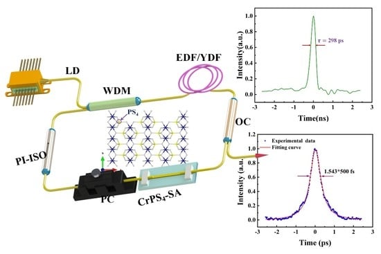
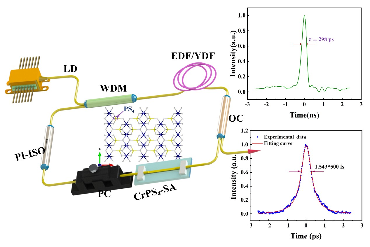
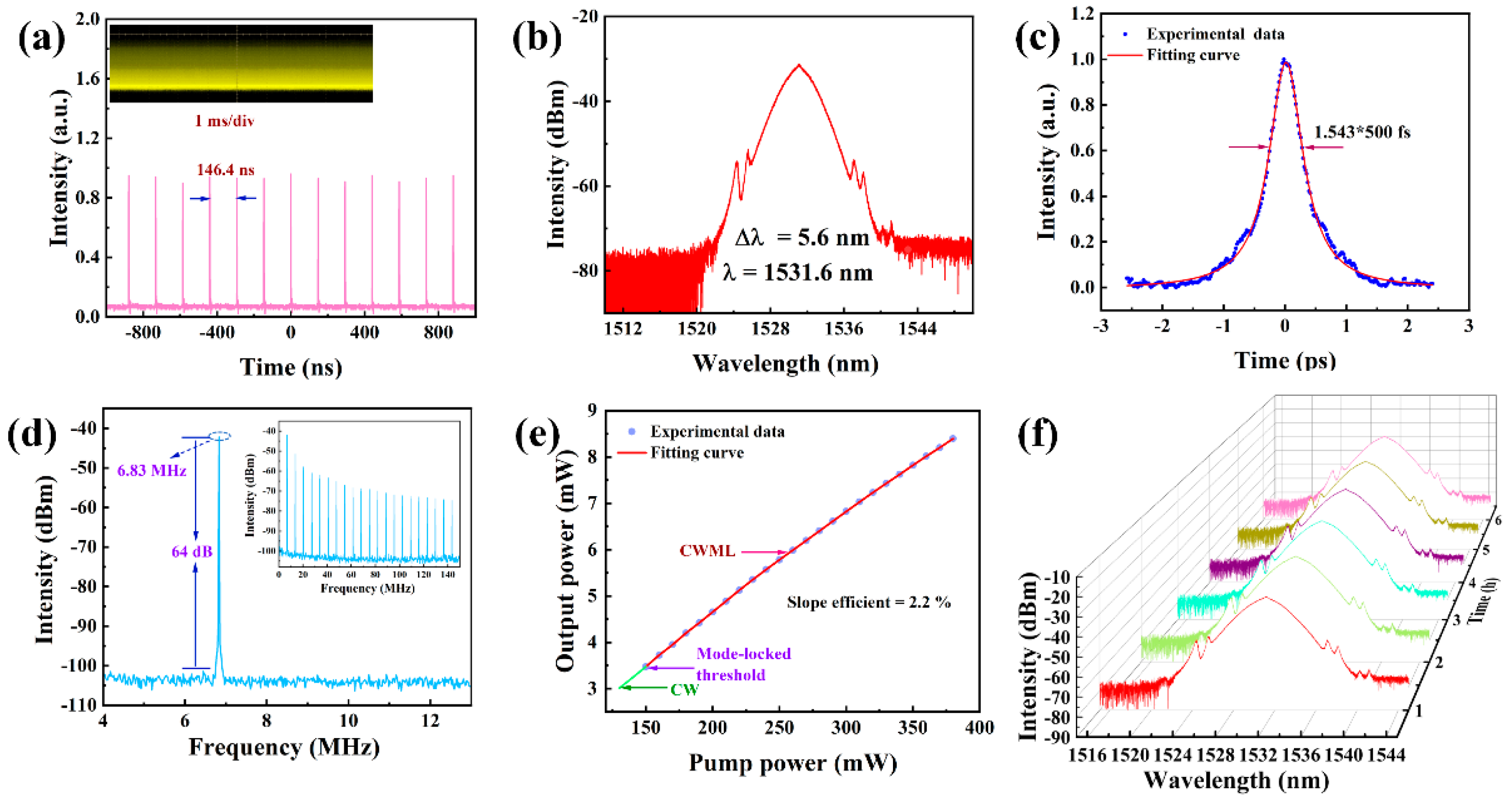
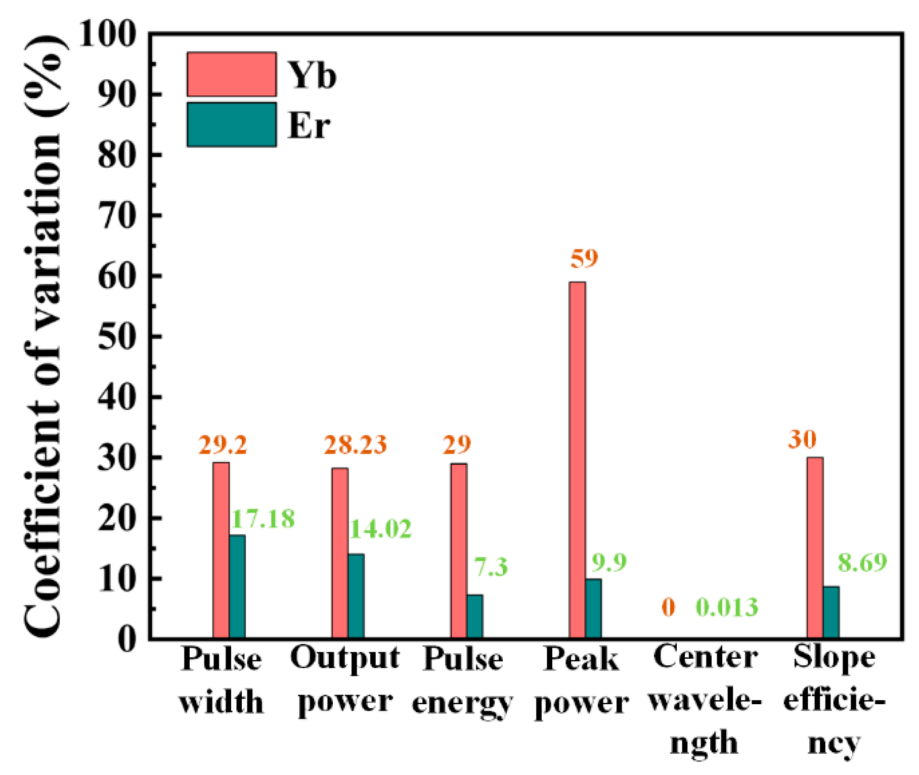
Figure 1. Schematic diagrams of crystal structures of layered (a) CrPS4 and (b) BP. (The arrows represent the three coordinate axes of the crystallographic coordinate system.)
Nanomaterials 13 01128 g001
Figure 2. (a) Structure of CrPS4 (The arrows represent the three coordinate axes of the crystallographic coordinate system); (b,c) electronic band structures of the bulk and monolayer CrPS4 materials calculated by the HSE06 function (The red arrow denotes spin-up bands, while the green arrow represents spin-down bands in the energy band structure.); (d) DOS of the bulk CrPS4.
Nanomaterials 13 01128 g002
Figure 3. Characterizations of as-synthesized CrPS4 nanoflakes after liquid-phase exfoliation: (a) the XRD patterns of CrPS4; (b) the Raman spectrum of few-layer CrPS4 nanoflakes on a Si substrate; (c) SEM image of CrPS4; (d,e) AFM image of few-layer CrPS4 nanoflakes on a Si substrate and corresponding height profile ( The red line and the green line represent the positions of the measured material thickness in the sample); (f) UV/Vis/NIR absorption spectrum of few-layer CrPS4 nanosheets in NMP.
Nanomaterials 13 01128 g003
Figure 4. P-scan measurement system diagram and the corresponding nonlinear saturable absorption curve of CrPS4.
Nanomaterials 13 01128 g004
Figure 5. The cavity schematic for the fiber laser.
Nanomaterials 13 01128 g005
Figure 6. Classical mode-locked output characteristics: (a) pulse train; (b) optical spectrum; (c) pulse width; (d) RF spectrum (inset: broadband RF spectrum); (e) relative change of output power and pump power; (f) long-period spectroscopic measurement (1 h intervals, 6 h in total).
Nanomaterials 13 01128 g006
Figure 7. Classical mode-locked output characteristics: (a) pulse train; (b) optical spectrum; (c) pulse width; (d) RF spectrum (inset: broadband RF spectrum); (e) relative change of output power and pump power; (f) long-period spectroscopic measurement (1 h intervals, 6 h in total).
Nanomaterials 13 01128 g007
Figure 8. Diagram of the degree of dispersion of the output pulse parameters of the two experiments.
Nanomaterials 13 01128 g008
Table 1. Comparison of data before and after the two experiments.
Gain FiberExperimental Conditions (mW)/DaysPulse WidthOutput Power (mW)Pulse Energy (nJ)Peak Power
(W)
Center WaveLength
(nm)
Slope Efficiency
(%)
Yb300/1298 ps10.631.1743.941036.14.9
300/40400 ps80.8762.1441036.13.6
Er270/1500 fs6.10.89317861531.62.2
270/40594 fs7.020.9611617.81531.42.4
Note: The experimental conditions include the pump power in milliwatts and the time interval between the two experiments.
Disclaimer/Publisher’s Note: The statements, opinions and data contained in all publications are solely those of the individual author(s) and contributor(s) and not of MDPI and/or the editor(s). MDPI and/or the editor(s) disclaim responsibility for any injury to people or property resulting from any ideas, methods, instructions or products referred to in the content.

