Article

Preparation and Characterization of Nano-Fe3O4 and Its Application for C18-Functionalized Magnetic Nanomaterials Used as Chromatographic Packing Materials

1
College of Environment, Zhejiang University of Technology, Hangzhou 310014, China
2
College of Biological and Environmental Engineering, Zhejiang Shuren University, Hangzhou 310015, China
*
Authors to whom correspondence should be addressed.
Nanomaterials 2023, 13(6), 1111; https://doi.org/10.3390/nano13061111
Submission received: 24 February 2023 / Revised: 11 March 2023 / Accepted: 16 March 2023 / Published: 20 March 2023
(This article belongs to the Special Issue Advanced Nanomaterials for Adsorption Purposes)

Abstract

:
A new type of magnetic nanomaterial with Fe3O4 as the core and organic polymer as the shell was synthesized by seed emulsion polymerization. This material not only overcomes the problem of insufficient mechanical strength of the organic polymer, it also solves the problem that Fe3O4 is prone to oxidation and agglomeration. In order to make the particle size of Fe3O4 meet the requirement of the seed, the solvothermal method was used to prepare Fe3O4. The effects of the reaction time, amount of solvent, pH value, and polyethylene glycol (PEG) on the particle size of Fe3O4 were investigated. In addition, in order to accelerate the reaction rate, the feasibility of preparing Fe3O4 by microwave was studied. The results showed that under the optimum conditions, the particle size of Fe3O4 could reach 400 nm and had good magnetic properties. After three stages of oleic acid coating, seed emulsion polymerization, and C18 modification, the obtained C18-functionalized magnetic nanomaterials were used for the preparation of the chromatographic column. Under optimal conditions, stepwise elution significantly shortened the elution time of sulfamethyldiazine, sulfamethazine, sulfamethoxypyridazine, and sulfamethoxazole while still achieving a baseline separation.

1. Introduction

At present, chromatographic packing materials are mainly silicon-based materials because the silicon-based materials have a strong chemical stability, and the surface is rich in silicon hydroxyl groups [1,2,3]. Therefore, it can be modified according to the needs of separation and analysis. In addition, silicon-based materials also have high mechanical strength and stability. All these ensure that the chromatographic column prepared by silicon-based materials has the advantages of wide range of application, good reproducibility, high stability, high column efficiency, low resistance, and so on [4,5,6,7]. However, the preparation process of silicon-based materials is complex, and the manufacturing cost is high. In order to find a substitute with a simple preparation process and low manufacturing cost, organic polymer has become an optional choice. Because it is simple to prepare an organic polymer, the polymerization can be initiated by evenly mixing and degassing the polymerization solution composed of a functional monomer, a cross-linking agent, an initiator, and a pore-forming agent under the condition of light or heating. In the preparation process, the pore size and the particle size can be controlled by adjusting the ratio of the pore-inducing agent and the functional monomer. However, the mechanical strength of organic polymers is low, which is the main factor hindering their application in chromatographic packing materials.
In order to solve this problem, it is necessary to combine organic polymers with other materials. The Fe3O4 nano-material is one of the materials that can be compounded because it contains hydroxyl groups and can be chemically modified. Additionally, Fe3O4 has high magnetization, superparamagnetism, excellent physical properties, stability [8,9,10], good biocompatibility, and unique electromagnetic properties [11,12,13]. Owing to its above properties, Fe3O4 is used in the fields of separation [14,15,16], protein immobilization [17], catalysis [18,19,20], medicine [21,22], environment [23,24], and so on. However, Fe3O4 nanoparticles have a huge defect, which is that it is easy to agglomerate and oxidize because of its high energy state [25,26]. Therefore, in order to prevent the agglomeration and oxidation of Fe3O4, it is necessary to form the structure of Fe3O4 encapsulated by an organic polymer when compounded with an organic polymer. This structure not only ensures the sufficient mechanical strength of the organic polymer, it also ensures the oxidation resistance and dispersion of Fe3O4.
In order to better realize the structure of the organic polymer-coated Fe3O4, suspension polymerization and seed emulsion polymerization were investigated [27]. The preparation process of suspension polymerization is simple, but the particle size uniformity of the prepared nanoparticles is poor. The range of particle size distribution and the position of Fe3O4 are both difficult to control. Although the preparation process of seed emulsion polymerization is relatively complex, the particle size is more uniform, and the control over the particle size is simpler. The most important thing is that Fe3O4 can be used as the seed of organic polymerization, which can accurately control the position of Fe3O4 in the microspheres. However, only when the particle size of ferric oxide is large enough can it be used as seed for seed emulsion polymerization.
Generally speaking, the preparation methods of Fe3O4 nanoparticles include coprecipitation [28,29], pyrolysis [30], microemulsion [31], the solvothermal method [32,33,34,35], the modified polyol method [36], the electrochemically assisted synthesis method [37], the sonochemical method [38], and so on. Of all these methods, coprecipitation is the first choice for most researchers because the reaction can be carried out with water as a solvent under mild conditions. However, the shape of the finished product is difficult to control, and the particle size is generally smaller than 20 nm. Pyrolysis is considered to be the best way to obtain nanomaterials with controllable sizes and morphologies. However, the precursors of the Fe3O4 are organometallic compounds, such as Fe(CO)5, metal copper ferrate, and metal acetoacetate, which are toxic. In addition, the pyrolysis of organometallic compounds often requires a very high temperature, which is a great challenge for many populations. The microemulsion method can be used to synthesize nanoparticles with specific morphologies and uniform sizes. Unfortunately, this method consumes a lot of organic solvents and requires organic surfactants to control the size and shape of the product. The Fe3O4 prepared by the polyol method has hydrophilic polyol groups on its surface and can be dispersed in water or other polar solvents with high magnetic properties, but the cost of this method is high, and the residue can easily cause environmental pollution. The sonochemical method has the advantages of simple operation, easy control, high efficiency, narrow particle size distribution, and no need for an additional initiator. However, the nanoparticles prepared by the sonochemical method are prone to agglomeration and other problems. Compared with the above methods, the solvothermal method has the advantages of high yield and controllable particle size. Additionally, almost all materials can be dissolved at higher pressure and higher temperature, which greatly increases the solubility of solids, resulting in the synthesis of high-quality, nanostructured materials. To sum up, the solvothermal method has become the best choice to obtain Fe3O4 particles with larger particle sizes.
In this study, Fe3O4 prepared by the solvothermal method was used for seed emulsion polymerization to prepare a new type of composite material, with magnetic material for the core and organic polymer for the shell (Figure 1). During the preparation of Fe3O4, the reaction time, pH, the amount of solvent, and polyethylene glycol were optimized. Then, the optimized Fe3O4 was coated with oleic acid using seeded emulsion polymerization and C18 functionalization. The magnetic nanocomposites were used as the chromatographic packing materials to prepare the reversed-phase chromatographic column. Finally, the obtained column was applied in the analysis of four sulfonamides.

2. Experimental Section

2.1. Reagents and Materials

Ferric chloride hexahydrate (FeCl3·6H2O), sodium acetate (NaAc), oleic acid (OA), ethylene glycol (EG), polyethylene glycol (PEG), polyvinyl pyrrolidone (PVP), styrene (ST), divinylbenzene (DVB), 2,2′-Azobis(2-methylpropionitrile) (AIBN), sodium dodecyl sulfate (SDS), dibutyl phthalate (DBP), polyvinyl alcohol (PVA), benzoyl peroxide (BPO), N, N-dimethyloctadecylamine, and glycidyl methacrylate (GMA) of analytical grade were purchased from Sinopharm Chemical Reagent Co., Ltd. (Shanghai, China) (Table S1). Methanol of HPLC grade and the standard solution of sulfamethyldiazine, sulfamethazine, sulfamethoxypyridazine, and sulfamethoxazole (1000 mg/L) were purchased from Aladdin.

2.2. Equipment

The magnetic and morphological characteristics of Fe3O4 were carried out by using an S4800 field-emission scanning electron microscope (SEM, Hitachi, Tokyo, Japan), an Ultima IV polycrystal (powder) X-ray diffractometer (XRD, Tokyo, Japan), and a Lake Shore 7404 vibrating sample magnetometer (VSM, Westerville, USA). The specific surface area and pore size of the adsorbent were measured and calculated by Brunauer–Emmett–Teller (BET) and Barrett–Joyner–Halenda (BJH) (Micromeritics, Norcross, USA).

2.3. Preparation of Magnetic Fe3O4 Particles

The preparation of Fe3O4 nanoparticles by the solvothermal method was as follows: 2.0 g of FeCl3·6H2O was added to a certain amount of EG solution, and a certain amount of PEG was added to the solution, which was completely dissolved by ultrasound. Then, 6.0 g of NaAc was added to the mixture and stirred evenly by ultrasound. It was transferred and sealed in a 100 mL Teflon-lined stainless-steel autoclave and reacted at 200 °C. After 8 h of reaction, it was cooled to room temperature and washed several times in sequence with deionized water and anhydrous ethanol. Finally, the product was collected using a magnet and baked in an oven at 70 °C for 12 h to obtain Fe3O4 nanoparticles. In order to explore the mechanism of Fe3O4 synthesis and the factors affecting the particle size, the effects of NaOH concentration (A), reaction time (B), usage of PEG (C), solvent volume (D), and microwave preparation time (E) were set (Table 1).

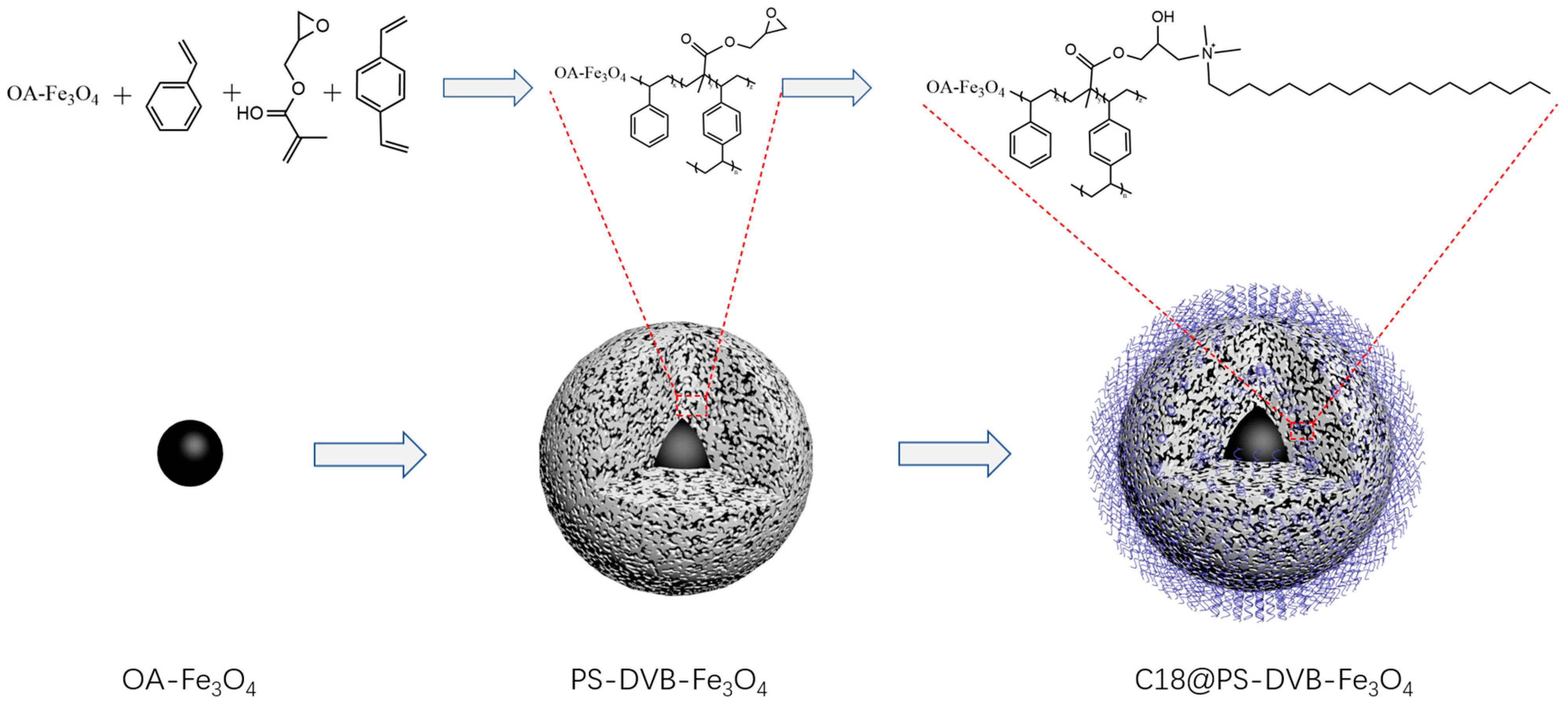
2.4. Preparation of C18-Functionalized Magnetic Nanomaterials

2.4.1. Preparation of Magnetic PS-DVB-GMA Materials

In this work, magnetic PS-DVB-GMA materials were prepared by seeded emulsion polymerization as follows:
(1)
A total of 1.0 g of Fe3O4 was mixed with 200 mL of anhydrous ethanol. Then, 5.0 mL of OA was slowly dripped at 80 °C. After that, the reaction lasted for 1.0 h.
(2)
An amount of 1.0 g OA-Fe3O4 was mixed with 100 mL of 95% ethanol aqueous solution. Then, certain amounts of PVP, ST, and AIBN were added to it, mixed evenly, and transferred to the three-neck flask of 250 mL. The reaction was carried out in a nitrogen atmosphere at 70 °C for 24 h.
(3)
A total of 4 mL of DBP were dispersed in 50 mL of SDS (0.2%, m/v) solution and emulsified in the ice bath for 1 h. Then, it was mixed with the prepared seeds and transferred to a 500 mL three-neck flask and stirred at 30 °C for 24 h.
(4)
A certain amount of SDS and BPO were added to the 250 mL PVA. Toluene, DVB, and GMA were added to the solution at a ratio of 3:1:1. Then, it was emulsified in the ice bath for 2 h. After that, it was poured into the three-neck flask in step 2 and stirred continuously at 30 °C for 24 h.
(5)
After passing nitrogen for 30 min, the temperature rose to 70 °C for the 24 h reaction. After the reaction, the particles were washed with hot water and anhydrous ethanol several times and then dried at 70 °C.

2.4.2. C18-Functionalized Magnetic Nanomaterials

The 4.0 g magnetic PS-DVB-GMA materials were uniformly mixed with 100 mL of methanol and transferred to the 250 mL three-neck flask. Under the conditions of 80 °C and 500 rpm, 10 mL of N, N-dimethyloctadecylamine was slowly added. Then, there was a reflux reaction for 8 h. At the end of the reaction, the nanoparticles were washed with methanol and anhydrous ethanol several times and then dried. Finally, C18-functionalized magnetic nanomaterials were obtained.
The as-prepared C18-functionalized magnetic nanomaterials were ultrasonically suspended in 50.0 mL of cyclohexanol, and then the slurry-packed sorbent was transferred to stainless-steel column tubes (4.6 × 250 mm) at 40 MPa. The packed column was separated and plugged at the column inlets. After being eluted with 200 mL of methyl alcohol, the C18 column was successfully prepared and expected to be applied in the separation of sulfamethyldiazine, sulfamethazine, sulfamethoxypyridazine, and sulfamethoxazole.

2.5. Standard Preparation

The standard solutions of single-standard sulfamethyldiazine, sulfamethazine, sulfamethoxypyridazine, and sulfamethoxazole, with 1 mL concentrations of 1000 mg/L, were taken in a 10 mL volumetric flask, had the volumes fixed with methanol, and were prepared into a standard solution with a concentration of 100 μg/mL. Then, five groups of mixed standard solutions with different concentrations of 0.1 μg/mL, 0.5 μg/mL, 1.0 μg/mL, 5.0 μg/mL, and 10 μg/mL were fixed with methanol.

2.6. Chromatographic Conditions

The chromatographic separation was performed on the prepared C18 column (4.6 × 250 mm) by using 0.1% formic acid (v/v) in water as the eluent (A) and methanol as the eluent (B) in the mobile phase. Detector wavelengths were 270 nm, and the temperature of the column thermostat was 33 °C. The injection volume was 10 μL. The separation was accomplished at a constant flow of 1.0 mL/min. Elution gradients: 0.0–6.0 min, 17–20%B; 6.0–11.0 min, 20–25%B; 11.0–35.0 min, 25–75%B; 35.0–40.0 min, 75–17%B; 40.0–45.0 min, 17%B.

3. Results and Discussion

3.1. Analysis of Synthesis Mechanism

In the reaction system, the role of EG was to provide a mild environment for the reaction. While at a high temperature, trivalent iron was reduced to divalent iron as a reductant; sodium acetate was a strong alkali and a weak acid salt, and acetate reacted with crystalline water in ferric chloride to hydrolyze and produce OH, forming a weak alkaline environment. Then, Fe3+ and Fe2+ reacted with OH under high temperature and high pressure to form Fe3O4 nanoparticles. The reaction equations involved in the experimental system were as follows:
CH3COO + H2O → CH3COOH + OH
Fe2+ + 2Fe3+ + 8OH → Fe3O4 + 4H2O

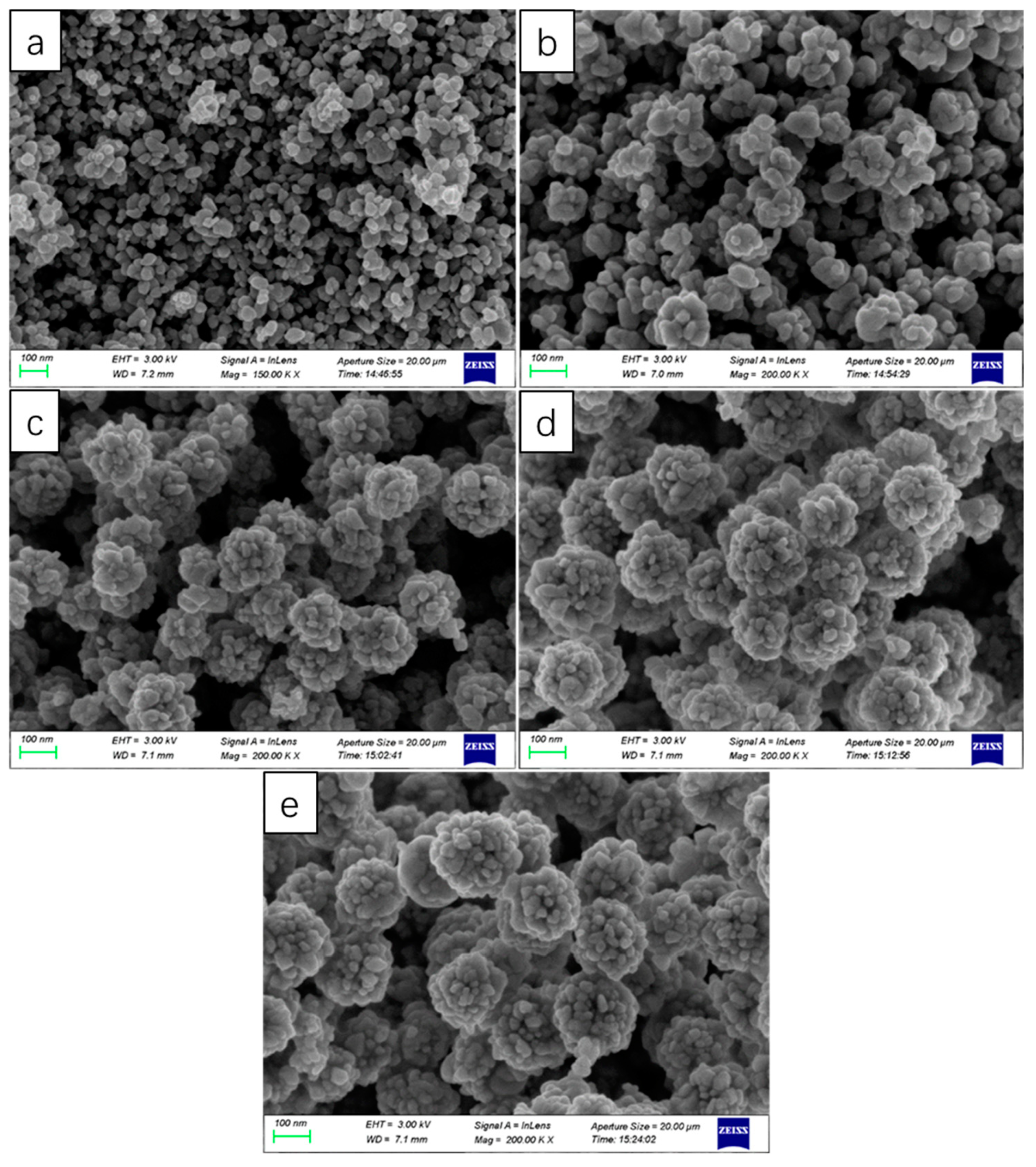
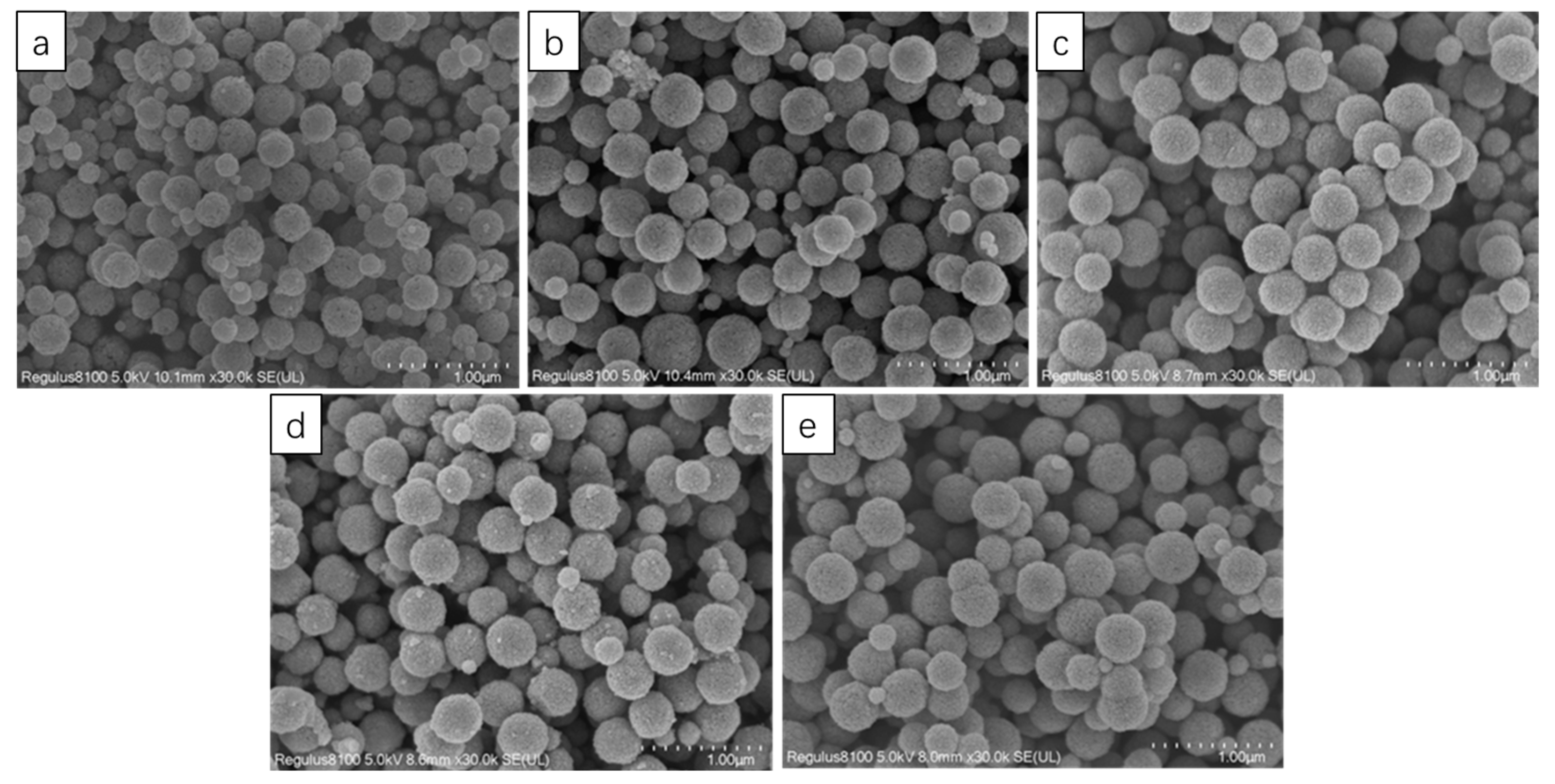
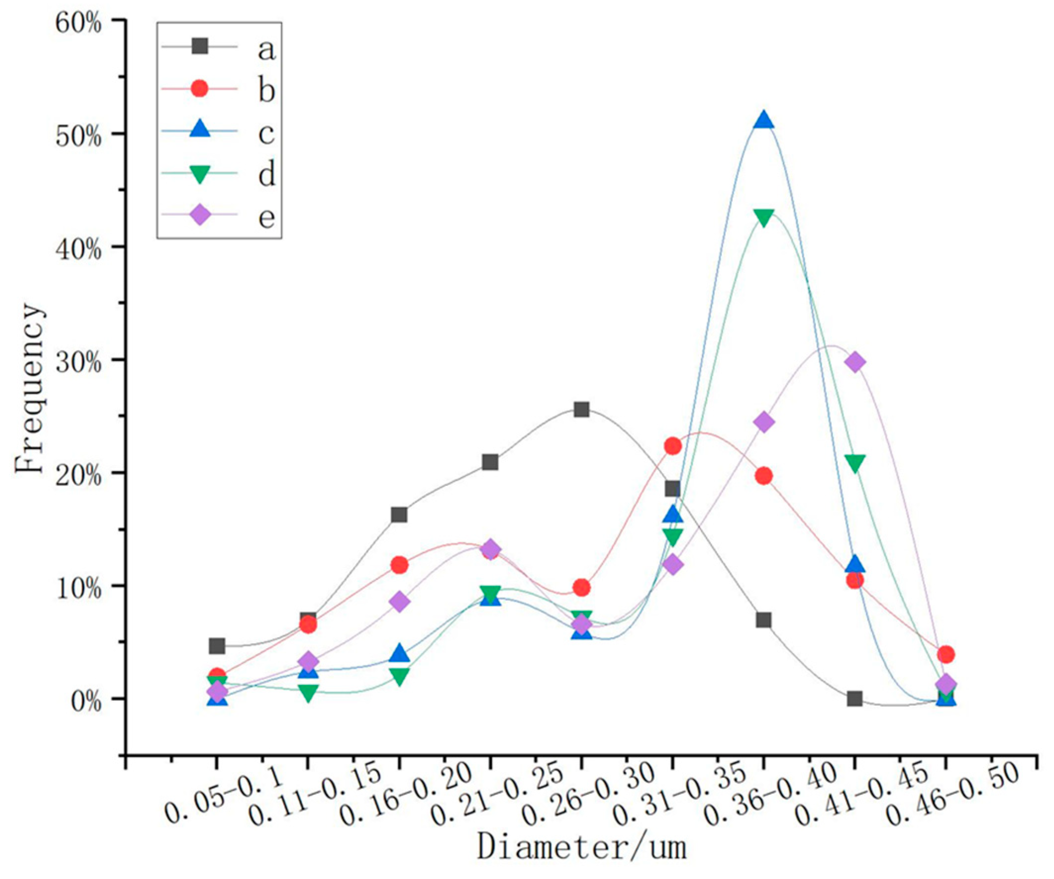
3.2. Effect of the Usage Amount of NaOH on the Product

The effect of the usage amount of NaOH on the particle size of the product was investigated deeply, and the results are shown in Figure 2. When the usage amount of NaOH was 0.4 g, the particle size of the magnetic microspheres was the smallest (only about 20 nm), while the particle size of the magnetic microspheres was about 200 nm without using NaOH. The magnetic microsphere was formed by the agglomeration of many small particles (about 20 nm). As shown in Figure 3, the crystalline phases and chemical structures of the prepared particles were determined by XRD. In the XRD diagram, the main peaks of synthesized magnetic microspheres were located at 30.17°, 35.56°, 43.19°, 53.6°, 57.11°, and 62.69°, in line with previously reported works about Fe3O4 [20]. Therefore, it can be concluded that the prepared particles were magnetic Fe3O4.
The formation process of the particles from the solution went through the process of nucleation, growth, agglomeration, and so on. The morphology and size of the particles mainly depended on the competition between the nucleation and the growth rate when the particles were precipitated from the solution. When the pH value was high, the metal ions nucleated separately under the alkaline environment. Then, the concentration of metal ions in the solution decreased rapidly, and the particles grew slowly, that is to say, the nucleation rate was greater than the growth rate, which was beneficial to the formation of small particles. When the pH value decreased, there were fewer OH in the solution; some of the metal ions combined with these OH to form a nucleus, and the other part was attached to the surface of the crystal nucleus, causing the particles to grow up. At this time, the growth rate was dominant and tended to the formation of large particles. Summing up all of the above analysis results, it was concluded that the phase transformation pH value was an important factor affecting the purity and size of the synthesized nano-Fe3O4. In order to obtain nanometer particles of three-body tetroxide with a larger particle size, there was no need to add alkaline substances in the reaction solvent, and the pH of the reaction solvent should be controlled in the range of 8–10.

3.3. Effect of Reaction Time on Products

Time was one of the important factors in the reaction process. There were four time points: 6 h, 8 h, 10 h, and 12 h. As shown in Figure 4a, it can be seen that when the reaction time was 6 h, the surfaces of the particles were very rough, and there was adhesion between the balls; it was obvious that a large ball was formed by the polymerization of many small balls. This was due to insufficient reaction time. In Figure 4b, the surfaces of the microspheres were much smoother, and the morphology was stable, indicating that the crystal had been fully grown at this time. In Figure 4c, we can see that there were dents or even cracks on the surface of some microspheres, indicating that the crystal had been overgrown at this time, destroying the crystal structure of the microspheres. Figure 4d also confirms the above statement; the crystal had been overgrown in 12 h, and the crystal had seriously collapsed, resulting in crystal splitting and an unstable morphology. From this, it can be concluded that the best reaction time was 8 h.

3.4. Effect of the Amount of PEG on the Product

PEG was discussed in this lab. Because PEG can be used as a surfactant, the addition of PEG could improve the dispersion of nanoparticles. As can be seen from Figure 5, the particle size was obviously much more uniform. In addition, statistics were carried out using the software Nano Measurer, and 150 balls were randomly selected from each picture for statistics. The results are shown in Figure 6. The particle size distribution, without adding PEG, was relatively dispersed, while the particle size distribution, with PEG, was concentrated, and the proportion of particle size in the range of 300–450 nm was 76%. It can be concluded that PEG had a great effect on the particle size of Fe3O4, and the addition of PEG improved the dispersion of nanoparticles in the reaction system, which made the polymerized Fe3O4 nanoparticles more uniform.

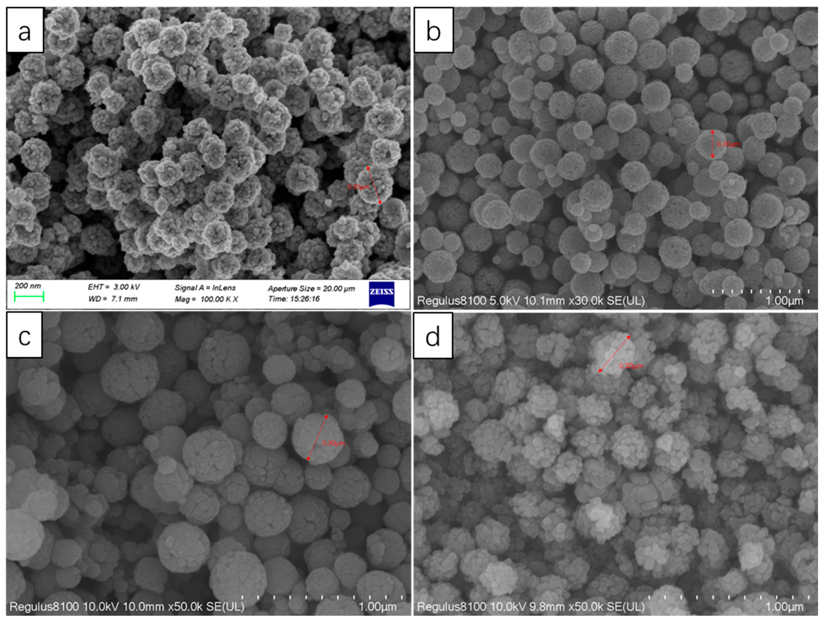
3.5. The Mechanism of the Effect of Reaction Solvent Dosage on the Product

The polymerization of nanoparticles was affected by pressure. The pressure of the reaction environment was affected by the amount of solvent. There were five solvent volumes: 40 mL, 50 mL, 60 mL, 70 mL, and 80 mL. A total of 150 microspheres were randomly selected for statistics, as shown in Figure 7, and Figure 8 was obtained. As shown in Figure 7 and Figure 8, in the range of 40 mL–80 mL, the particle size increased with the increase in the amount of solution, which was because the increase in the amount of solution led to the increase in pressure in the container, which made the particles polymerize more easily. When the solvent dosage was 60 mL, the distribution of particle size was the most concentrated, and the proportion of microspheres with particle sizes of 300–450 nm reached 79%. When the solution dose was 40 mL, 74% of the microspheres were smaller than 300 nm, and when the solution dose was 50 mL, the microspheres smaller than 300 nm were reduced to 43%. When the solution dose was 70 mL, the microspheres with particle sizes larger than 400 nm increased by 10%, compared with 60 mL; when the solution dose was 80 mL, the microspheres with particle sizes larger than 400 nm increased by 18%, compared with 60 mL, and the proportion of microspheres with particle sizes less than 300 nm increased to 32%. As a result, it can be seen that the amount of solvent had a great effect on the particle size; with the increase in pressure, there was an increase in the amount of solvent, and the particles were more likely to polymerize under higher pressure. However, too much solvent would widen the range of particle size distribution because a large number of Fe ions were polymerized into large spheres, which reduced the concentration of Fe ions in the reaction system, and could not continue to polymerize to form large spheres.

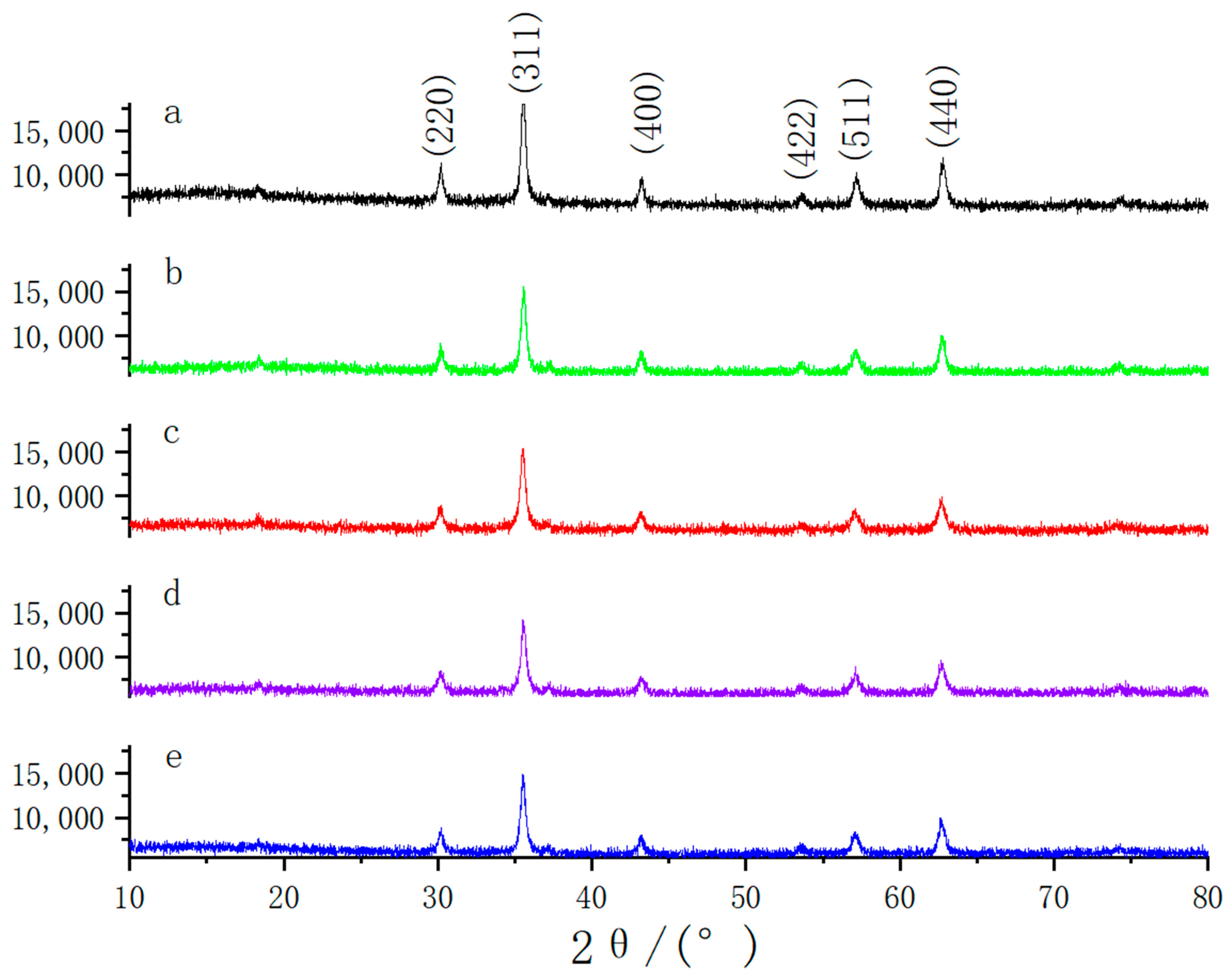
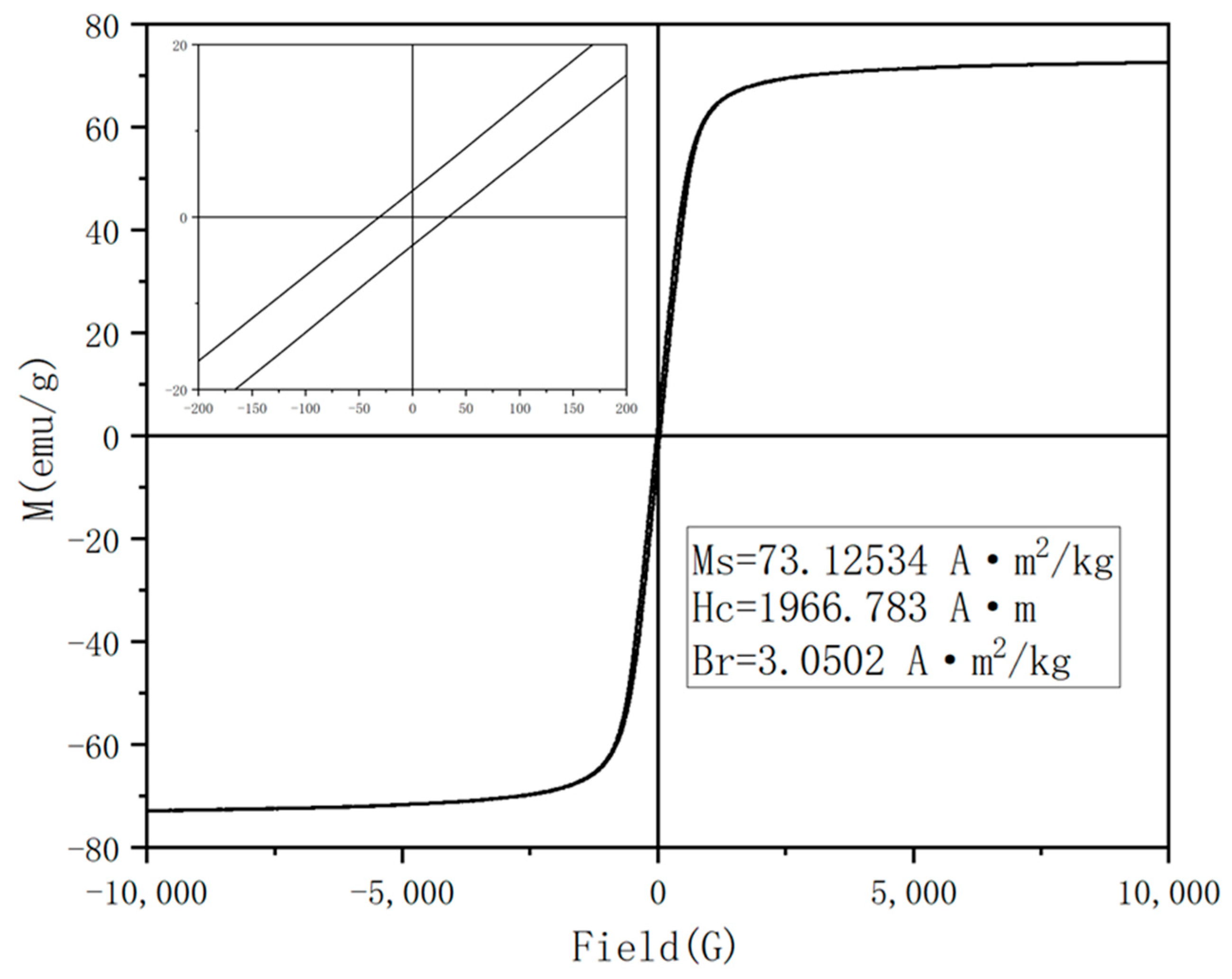
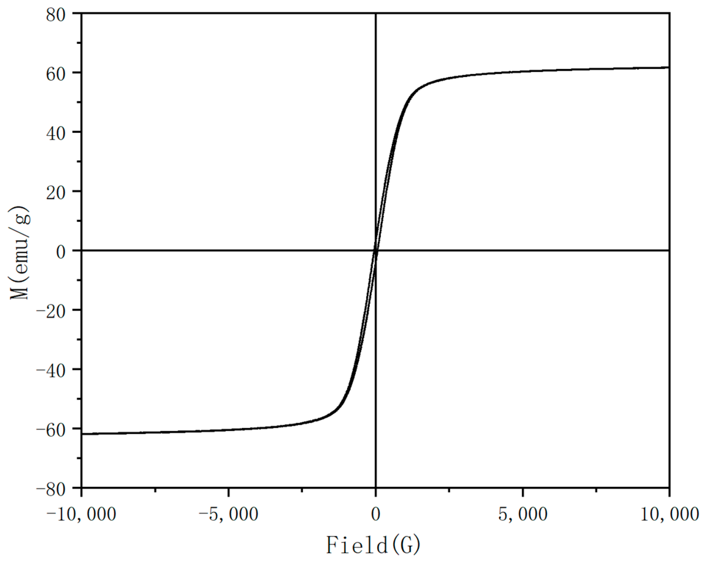
3.6. Analysis of Magnetic Properties of Fe3O4 Nanoparticles

Based on the reaction time, the pH, and the polyethylene glycol, the optimum synthesis conditions were determined as the 8 h reaction and a pH of 8~10, and a certain amount of PEG was added. Under these conditions, Fe3O4 with a uniform particle size was prepared, and its magnetic properties were also tested. The hysteresis loop in the greenhouse was shown in Figure 9. It can be seen that the curve was closed and showed a typical “S” shape. The saturation magnetization of the obtained Fe3O4 nanospheres was 73.125 emu/g, which had high magnetic properties. In addition, the coercive force (Hc) and remanence (Br) of Fe3O4 nanoparticles, shown in the upper left magnification, were 1966.78 A/m and 3.0502 A·m2/kg, respectively.
Compared with the previously reported work on the preparation of Fe3O4, the Fe3O4 prepared in this study had much more advantages in particle size control and magnetic properties. For example, the average particle size of the Fe3O4 particles prepared by using the chemical precipitation method was 10 nm, and the magnetization saturation intensity was 60 emu/g [39]. Similar results can be obtained by using the microwave hydrothermal method [40]; the Fe3O4 particle size was about 20 nm, and the magnetization saturation intensity was also 60 emu/g. As a result, it can be concluded that the Fe3O4 particles obtained by using the solvothermal method possessed much higher magnetization saturation intensities and larger particle sizes.

3.7. Preparation of Fe3O4 by Microwave

In order to shorten the reaction time, the synthesis of Fe3O4 by microwave was studied. Microwave could make the reaction reach the required temperature faster and heat more evenly. The time of the microwave was controlled under the conditions of 60 mL, adding 1.0 g of PEG, and keeping the temperature at 200 °C. As shown in the Figure 10, when the reaction time was 2 h, the shapes of the particles were unstable, and there was a flaky structure in addition to the sphere, which shows that the reaction was not complete. When the reaction time was 3 h, there was still a small amount of sheet structures, and the particle size distribution was not uniform. When the reaction time was 4 h, the flake structure disappeared, indicating that the reaction was complete, but the particle size distribution was still uneven. In addition, the particles with a reaction time of 4 h were characterized by VSM, and the range of particle size distribution was calculated. As shown in Figure 11, the Fe3O4 particles had strong magnetic properties, and its magnetization saturation intensity was 62.49 emu/g. A total of 300 Fe3O4 particles were randomly selected for statistics, as shown in Figure 12a,b was obtained. It can be concluded that the number of microspheres with particle sizes in the range of 60–100nm was the largest, reaching 35%, and the proportion of microspheres with particle sizes larger than 200 nm was 34%. Compared with previous literature, the particle size of Fe3O4 prepared by Savvidou [41] was only 20 nm. The particle size of Fe3O4 prepared by Aivazoglou [41] was 9 nm. It can be seen that the microwave method was mainly used in the preparation of small particle size Fe3O4. This study provided a certain reference significance for the preparation of large-size Fe3O4 by microwave.

4. Application

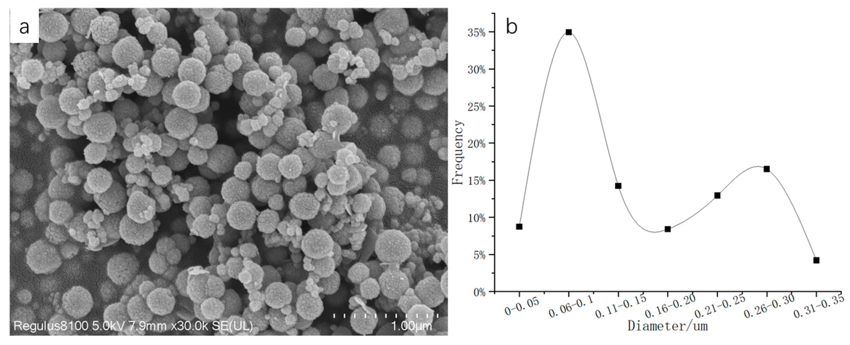
4.1. Performance Characterization of Materials

According to Figure 13, it can be seen that the new magnetic organic composites had a stable shape, the range of the particle size distribution was narrow, and the average particle size was 4 μm. Through the test, this new type of magnetic composite material had a specific surface area of 300.8128 m2/g and a magnetic induction intensity of 27.68 emu/g, which could be used as a filling material for a chromatographic column.

4.2. Column Efficiency Analysis

The new type of magnetic composite was prepared into a chromatographic column, and its stereoselectivity and hydrophobicity were tested. The result is shown in Figure 14. The graph shown in Figure 14a is the chromatogram of naphthalene, which was used for the hydrophobicity test. The asymmetry was 0.99, and the number of plates was 11,272. The chromatographic column had a good ability to select and recognize the hydrophobic groups of the separated substances. As shown in Figure 14b, the chromatograms of uracil, ortho-terphenyl, and chrysene were used for stereoselectivity testing, and the stereoselectivity factor was 1.822, which showed that the chromatographic column had a good ability to select and recognize the molecular shape of the separated substance.

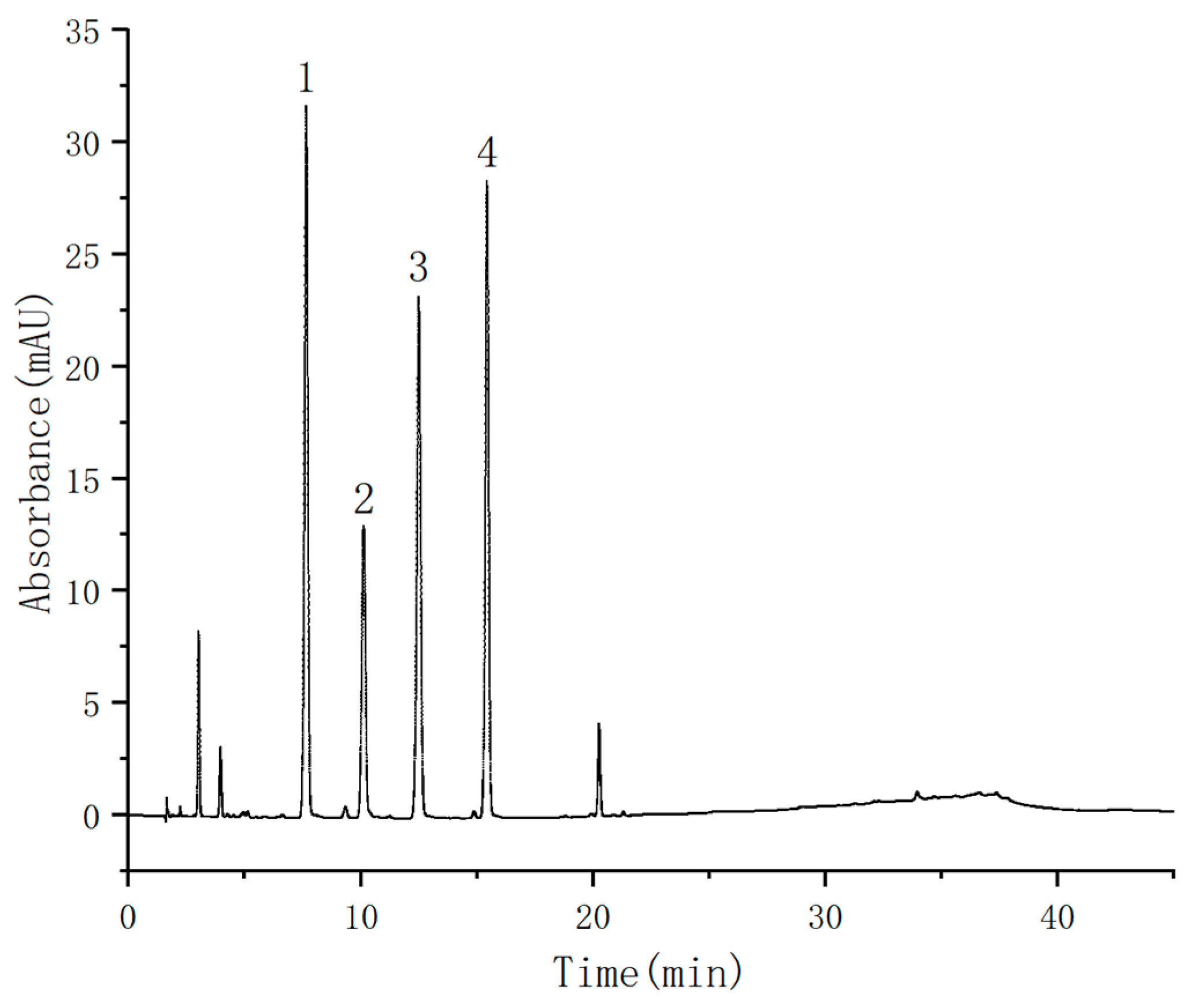
4.3. Sample Analysis

In order to test the effect of the prepared chromatographic column in the analysis of actual compounds, it was applied to the detection of sulfonamides (Figure 15). It can be seen from Table 2 that the linear range of four kinds of sulfonamides was 0.1–10 μg/mL. The linear correlation coefficients were all above 0.9992. Six groups of parallel experiments were conducted to calculate the relative standard deviation (RSD). The intraday and inter-day RSDs of the four sulfonamides were less than 10.0%, respectively. In addition, the comparison of the prepared chromatographic column with the Diamonsil C18 (5 μm, 4.6 × 250 mm) commercial chromatographic column (Figure S1) was studied, and the results showed that the retention times of four sulfonamides were basically consistent. It could be seen that the magnetic material had a great advantage in the preparation of the chromatographic column.

5. Conclusions

To sum up, the preparation of Fe3O4 using the solvothermal method could be controlled by the pH, the reaction time, the amount of solvent, and polyethylene glycol, and the particle size could be adjusted in the range of 20–400 nm. Among them, the best conditions for preparing Fe3O4 with a 400 nm particle size was 8 h of reaction time, 60 mL of EG, pH = 8–10, and 1.0 g of PEG. In addition, this study carried out a preliminary study on the preparation of Fe3O4 using the microwave solvothermal method, which provided a powerful theoretical basis for the preparation of large-size Fe3O4 using the microwave method.
The magnetic nanocomposite prepared by the optimized Fe3O4 was used as the chromatographic packing material, and the prepared column had good hydrophobic and stereoselective properties. It also had a good separation effect in the analysis of four sulfonamides.

Supplementary Materials

The following supporting information can be downloaded at: https://www.mdpi.com/article/10.3390/nano13061111/s1, Figure S1. Chromatograms of the four sulfonamides: (1) sulfamethyldiazine; (2) sulfamethazine; (3) sulfamethoxypyridazine; and (4) sulfamethoxazole. (a) Diamonsil C18 (5 μm, 4.6 × 250 mm); (b) Self-made chromatographic column (5 μm, 4.6 × 250 mm). Table S1. Chemical structure of reagents used for the preparation of C18@PS-DVB-Fe3O4 particles.

Author Contributions

Methodology, W.-X.L.; validation, W.-X.L. and W.-N.Z.; resources, S.S.; writing—original draft preparation, W.-X.L. and W.-N.Z.; writing—review and editing, Y.-G.Z.; funding acquisition, Y.L. All authors have read and agreed to the published version of the manuscript.

Funding

Zhejiang Provincial Key Research and Development Project (No. 2021C03176), top young talents of the Ten Thousand Talents Program of the Zhejiang Province (ZJWR0308003).

Data Availability Statement

Not Applicable.

Acknowledgments

We would like to thank the Zhejiang Provincial Key Research and Development Project (No. 2021C03176), top young talents of the Ten Thousand Talents Program of the Zhejiang Province (ZJWR0308003), State Key Laboratory of Environmental Chemistry and Ecotoxicology, Research Center for Eco-Environmental Sciences, and the Chinese Academy of Sciences (No. KF2021-24). The funders had no roles in study design, in the data collection and analysis, in the decision to publish, or in the preparation of the manuscript.

Conflicts of Interest

The authors declare no conflict of interest.

References

  1. Buszewski, B.; Noga, S. Hydrophilic interaction liquid chromatography (HILIC)—A powerful separation technique. Anal. Bioanal. Chem. 2012, 402, 231–247. [Google Scholar] [CrossRef] [Green Version]
  2. Kohler, J.; Kirkland, J.J. Improved silica-based column packings for high-performance liquid chromatography. J. Chromatogr. A 1987, 385, 125–150. [Google Scholar] [CrossRef]
  3. Liang, Y.; Wang, C.; Liang, Z.; Zhang, L.; Zhang, Y. C18-functionalized amine-bridged hybrid monoliths for mass spectrometry-friendly peptide separation and highly sensitive proteomic analysis. Anal. Chem. 2022, 94, 6084–6088. [Google Scholar] [CrossRef]
  4. Zuvela, P.; Skoczylas, M.; Jay Liu, J.; Ba Czek, T.; Kaliszan, R.; Wong, M.W.; Buszewski, B.; Heberger, K. Column characterization and selection systems in reversed-phase high-performance liquid chromatography. Chem. Rev. 2019, 119, 3674–3729. [Google Scholar] [CrossRef]
  5. Liu, B.; Li, H.; Quan, K.; Chen, J.; Qiu, H. Periodic mesoporous organosilica for chromatographic stationary phases: From synthesis strategies to applications. TrAC Trends Anal. Chem. 2023, 158, 116895. [Google Scholar] [CrossRef]
  6. Wu, Y.; Sun, X.; Zhu, J.; Shen, J.; Ke, Y. Monodisperse core–shell silica particles as a high-performance liquid chromatography packing material: Facile in situ silica sol-gel synthesis. J. Chromatogr. A 2020, 1625, 461282. [Google Scholar] [CrossRef]
  7. Grecco, C.F.; Miranda, L.F.C.; Costa Queiroz, M.E. Aminopropyl hybrid silica monolithic capillary containing mesoporous SBA-15 particles for in-tube SPME-HILIC-MS/MS to determine levodopa, carbidopa, benserazide, dopamine, and 3-O-methyldopa in plasma samples. Microchem. J. 2020, 157, 105106. [Google Scholar] [CrossRef]
  8. Laconte, L.; Nitin, N.; Gang, B. Magnetic nanoparticle probes. Mater. Today 2005, 8, 32–38. [Google Scholar] [CrossRef]
  9. Khiabani, S.S.; Farshbaf, M.; Akbarzadeh, A.; Davaran, S. Magnetic nanoparticles: Preparation methods, applications in cancer diagnosis and cancer therapy. Artif. Cell. Nanomed. B 2017, 45, 6–17. [Google Scholar] [CrossRef]
  10. Liu, W.X.; Song, S.; Ye, M.L.; Zhu, Y.; Zhao, Y.G.; Lu, Y. Nanomaterials with excellent adsorption characteristics for sample pretreatment: A review. Nanomaterials 2022, 12, 1845. [Google Scholar] [CrossRef]
  11. Wang, Y.; Wang, Y.; Chen, L.; Wan, Q.-H. Restricted access magnetic materials prepared by dual surface modification for selective extraction of therapeutic drugs from biological fluids. J. Magn. Magn. Mater. 2012, 324, 410–417. [Google Scholar] [CrossRef]
  12. Ye, M.L.; Zhu, Y.; Lu, Y.; Gan, L.; Zhang, Y.; Zhao, Y.G. Magnetic nanomaterials with unique nanozymes-like characteristics for colorimetric sensors: A review. Talanta 2021, 230, 122299. [Google Scholar] [CrossRef]
  13. Ma, J.B.; Wu, H.W.; Liao, Y.F.; Rui, Q.H.; Zhu, Y.; Zhang, Y. Application of petal-shaped ionic liquids modified covalent organic frameworks for one step cleanup and extraction of general anesthetics in human plasma samples. Talanta 2020, 210, 120652. [Google Scholar] [CrossRef]
  14. Karimi-Maleh, H.; Fakude, C.T.; Mabuba, N.; Peleyeju, G.M.; Arotiba, O.A. The determination of 2-phenylphenol in the presence of 4-chlorophenol using nano-Fe3O4/ionic liquid paste electrode as an electrochemical sensor. J. Colloid Interface Sci. 2019, 554, 603–610. [Google Scholar] [CrossRef]
  15. Khalilifard, M.; Javadian, S. Magnetic superhydrophobic polyurethane sponge loaded with Fe3O4@oleic acid@graphene oxide as high performance adsorbent oil from water. Chem. Eng. J. 2021, 408, 127369. [Google Scholar] [CrossRef]
  16. Wu, G.; Ma, J.; Li, S.; Guan, J.; Jiang, B.; Wang, L.; Li, J.; Wang, X.; Chen, L. Magnetic copper-based metal organic framework as an effective and recyclable adsorbent for removal of two fluoroquinolone antibiotics from aqueous solutions. J. Colloid Interface Sci. 2018, 528, 360–371. [Google Scholar] [CrossRef]
  17. Xie, W.; Zang, X. Immobilized lipase on core-shell structured Fe3O4-MCM-41 nanocomposites as a magnetically recyclable biocatalyst for interesterification of soybean oil and lard. Food Chem. 2016, 194, 1283–1292. [Google Scholar] [CrossRef]
  18. Kumar, A.; Kumar, A.; Sharma, G.; Al-Muhtaseb, A.a.H.; Naushad, M.; Ghfar, A.A.; Stadler, F.J. Quaternary magnetic BiOCl/g-C3N4/Cu2O/Fe3O4 nano-junction for visible light and solar powered degradation of sulfamethoxazole from aqueous environment. Chem. Eng. J. 2018, 334, 462–478. [Google Scholar] [CrossRef]
  19. Wang, X.; Xiao, H.; Li, A.; Li, Z.; Liu, S.; Zhang, Q.; Gong, Y.; Zheng, L.; Zhu, Y.; Chen, C.; et al. Constructing NiCo/Fe3O4 heteroparticles within MOF-74 forefficient oxygenevolution reactions. J. Am. Chem. Soc. 2018, 140, 15336–15341. [Google Scholar] [CrossRef]
  20. Hassandoost, R.; Pouran, S.R.; Khataee, A.; Orooji, Y.; Joo, S.W. Hierarchically structured ternary heterojunctions based on Ce3+/Ce4+ modified Fe3O4 nanoparticles anchored onto graphene oxide sheets as magnetic visible-light-active photocatalysts for decontamination of oxytetracycline. J. Hazard. Mater. 2019, 376, 200–211. [Google Scholar] [CrossRef]
  21. Abo-zeid, Y.; Ismail, N.S.M.; McLean, G.R.; Hamdy, N.M. A molecular docking study repurposes FDA approved iron oxide nanoparticles to treat and control COVID-19 infection. Eur. J. Pharm. Sci. 2020, 153, 105465. [Google Scholar] [CrossRef]
  22. Pang, Y.; Shi, J.; Yang, X.; Wang, C.; Sun, Z.; Xiao, R. Personalized detection of circling exosomal PD-L1 based on Fe3O4@TiO2 isolation and SERS immunoassay. Biosens. Bioelectron. 2020, 148, 111800. [Google Scholar] [CrossRef]
  23. Alqadami, A.A.; Naushad, M.; Alothman, Z.A.; Ahamad, T. Adsorptive performance of MOF nanocomposite for methylene blue and malachite green dyes: Kinetics, isotherm and mechanism. J. Environ. Manag. 2018, 223, 29–36. [Google Scholar] [CrossRef]
  24. Alqadami, A.A.; Naushad, M.; Alothman, Z.A.; Alsuhybani, M.; Algamdi, M. Excellent adsorptive performance of a new nanocomposite for removal of toxic Pb(II) from aqueous environment: Adsorption mechanism and modeling analysis. J. Hazard. Mater. 2020, 389, 121896. [Google Scholar] [CrossRef]
  25. Yang, X.; Jin, B.; Yu, L.; Zhu, F.; Xu, Y.; Liu, R. Preparation and characterization of magnetic α-Fe2O3/Fe3O4 heteroplasmon nanorods via the ethanol solution combustion process of ferric nitrate. Mater. Res. Express 2021, 8, 25011. [Google Scholar] [CrossRef]
  26. Wang, J.; Wang, X.; Li, Y.; Si, H.; Chen, C.; Wang, J.; Long, Z.; Nandakumar, K. Preparation and properties of magnetic polymer microspheres. Polymer 2020, 199, 122569. [Google Scholar] [CrossRef]
  27. Chaudhary, V.; Sharma, S. Suspension polymerization technique: Parameters affecting polymer properties and application in oxidation reactions. J. Polym. Res. 2019, 26, 1767. [Google Scholar] [CrossRef]
  28. Tang, S.; Zhao, M.; Yuan, D.; Li, X.; Wang, Z.; Zhang, X.; Jiao, T.; Ke, J. Fe3O4 nanoparticles three-dimensional electro-peroxydisulfate for improving tetracycline degradation. Chemosphere 2021, 268, 129315. [Google Scholar] [CrossRef]
  29. Wang, Y.; Gao, X.; Zhang, L.; Wu, X.; Wang, Q.; Luo, C.; Wu, G. Synthesis of Ti3C2/Fe3O4/PANI hierarchical architecture composite as an efficient wide-band electromagnetic absorber. Appl. Surf. Sci. 2019, 480, 830–838. [Google Scholar] [CrossRef]
  30. Li, D.; Jiang, D.; Chen, M.; Xie, J.; Wu, Y.; Dang, S.; Zhang, J. An easy fabrication of monodisperse oleic acid-coated Fe3O4 nanoparticles. Mater. Lett. 2010, 64, 2462–2464. [Google Scholar] [CrossRef]
  31. Sun, L.; Zhan, L.; Shi, Y.; Chu, L.; Ge, G.; He, Z. Microemulsion synthesis and electromagnetic wave absorption properties of monodispersed Fe3O4/polyaniline core–shell nanocomposites. Synth. Met. 2014, 187, 102–107. [Google Scholar] [CrossRef]
  32. He, S.W.; Zhao, Y.G.; Zhang, Y.; Jin, M.C.; Zhu, Y. Hydrophilicity nano-titania coating modified magnetic graphene oxide for pass-through cleanup of fipronil and its metabolites in human blood. J. Chromatogr. A 2018, 1553, 16–24. [Google Scholar] [CrossRef]
  33. Ma, J.B.; Qiu, H.W.; Rui, Q.H.; Liao, Y.F.; Chen, Y.M.; Xu, J.; Zhang, Y.; Zhu, Y.; Zhao, Y.G. Enhanced cleanup efficiency hydroxy functionalized-magnetic graphene oxide and its comparison with magnetic carboxyl-graphene for PRiME pass-through cleanup of strychnine and brucine in human plasma samples. Anal. Chim. Acta 2018, 1020, 41–50. [Google Scholar] [CrossRef]
  34. Wu, F.-F.; Chen, Q.-Y.; Ma, X.-J.; Li, T.-T.; Wang, L.-F.; Hong, J.; Sheng, Y.-H.; Ye, M.-L.; Zhu, Y. N-doped magnetic covalent organic frameworks for preconcentration of allergenic disperse dyes in textiles of fall protection equipment. Anal. Methods 2019, 11, 3381–3387. [Google Scholar] [CrossRef]
  35. Zhao, Y.G.; Zhang, Y.; Wang, F.L.; Zhou, J.; Zhao, Q.M.; Zeng, X.Q.; Hu, M.Q.; Jin, M.C.; Zhu, Y. Determination of perchlorate from tea leaves using quaternary ammonium modified magnetic carboxyl-carbon nanotubes followed by liquid chromatography-tandem quadrupole mass spectrometry. Talanta 2018, 185, 411–418. [Google Scholar] [CrossRef] [Green Version]
  36. Wagle, D.V.; Rondinone, A.J.; Woodward, J.D.; Baker, G.A. Polyol synthesis of magnetite nanocrystals in a thermostable ionic liquid. Cryst. Growth Des. 2017, 17, 1558–1567. [Google Scholar] [CrossRef]
  37. Sadeghfar, F.; Ghaedi, M.; Asfaram, A.; Jannesar, R.; Javadian, H.; Pezeshkpour, V. Polyvinyl alcohol/Fe3O4@carbon nanotubes nanocomposite: Electrochemical-assisted synthesis, physicochemical characterization, optical properties, cytotoxicity effects and ultrasound-assisted treatment of aqueous based organic compound. J. Ind. Eng. Chem. 2018, 65, 349–362. [Google Scholar] [CrossRef]
  38. Neto, D.M.A.; Freire, R.M.; Gallo, J.; Freire, T.M.; Queiroz, D.C.; Ricardo, N.M.P.S.; Vasconcelos, I.F.; Mele, G.; Carbone, L.; Mazzetto, S.E.; et al. Rapid sonochemical approach produces functionalized Fe3O4 nanoparticles with excellent magnetic, colloidal, and relaxivity properties for MRI application. J. Phys. Chem. C 2017, 121, 24206–24222. [Google Scholar] [CrossRef]
  39. Gerulova, K.; Kucmanova, A.; Sanny, Z.; Garaiova, Z.; Seiler, E.; Caplovicova, M.; Caplovic, L.; Palcut, M. Fe3O4-PEI nanocomposites for magnetic harvesting of chlorella vulgaris, chlorella ellipsoidea, microcystis aeruginosa, and auxenochlorella protothecoides. Nanomaterials 2022, 12, 1786. [Google Scholar] [CrossRef]
  40. Aivazoglou, E.; Metaxa, E.; Hristoforou, E. Microwave-assisted synthesis of iron oxide nanoparticles in biocompatible organic environment. AIP Adv. 2018, 8, 048201. [Google Scholar] [CrossRef] [Green Version]
  41. Savvidou, M.G.; Dardavila, M.M.; Georgiopoulou, I.; Louli, V.; Stamatis, H.; Kekos, D.; Voutsas, E. Optimization of microalga chlorella vulgaris magnetic harvesting. Nanomaterials 2021, 11, 1614. [Google Scholar] [CrossRef]
Figure 1. The preparation procedure of the C18@PS-DVB-Fe3O4 particles.
Nanomaterials 13 01111 g001
Figure 2. SEM image of Fe3O4. The amounts were: (a) 0.4 g NaOH; (b) 0.2 g NaOH; (c) 0.1 g NaOH; (d) 0.05 g NaOH; and (e) without using NaOH.
Nanomaterials 13 01111 g002
Figure 3. XRD image of Fe3O4. The amounts were: (a) 0.4 g NaOH; (b) 0.2 g NaOH; (c) 0.1 g NaOH; (d) 0.05 g NaOH; and (e) without using NaOH.
Nanomaterials 13 01111 g003
Figure 4. SEM image of Fe3O4. The times were: (a) 6 h; (b) 8 h; (c) 10 h; and (d) 12 h.
Nanomaterials 13 01111 g004
Figure 5. SEM image of Fe3O4. (a) Without using PEG; (b) using PEG.
Nanomaterials 13 01111 g005
Figure 6. Particle size distribution curves of 150 microspheres. (a) Without using PEG; (b) using PEG.
Nanomaterials 13 01111 g006
Figure 7. SEM image of Fe3O4. Solvent amounts: (a) 40 mL EG; (b) 50 mL EG; (c) 60 mL EG; (d) 70 mL EG; and (e) 80 mL EG.
Nanomaterials 13 01111 g007
Figure 8. Particle size distribution curves of 150 microspheres. Solvent amounts: (a) 40 mL EG; (b) 50 mL EG; (c) 60 mL EG; (d) 70 mL EG; and (e) 80 mL EG.
Nanomaterials 13 01111 g008
Figure 9. Hysteresis loop diagram of Fe3O4 nanoparticles.
Nanomaterials 13 01111 g009
Figure 10. SEM image of Fe3O4 synthesized by microwave. Reaction time: (a) 2 h; (b) 3 h; and (c) 4 h.
Nanomaterials 13 01111 g010
Figure 11. Hysteresis loop diagram of Fe3O4 nanoparticles by microwave reaction for 4 h.
Nanomaterials 13 01111 g011
Figure 12. (a) SEM image of 3 h preparation of Fe3O4 by microwave method; (b) Particle size distribution curves of 300 microspheres in (a).
Nanomaterials 13 01111 g012
Figure 13. SEM image after organic resin coating.
Nanomaterials 13 01111 g013
Figure 14. (a) Chromatographic separation diagram of naphthalene (1); (b) chromatographic separation diagram of uracil (1), ortho-terphenyl (2), and chrysene (3).
Nanomaterials 13 01111 g014
Figure 15. Chromatograms of four sulfonamides: (1) sulfamethyldiazine; (2) sulfamethazine; (3) sulfamethoxypyridazine; and (4) sulfamethoxazole.
Nanomaterials 13 01111 g015
Table 1. Experimental design parameters.
Serial NumberNaOH/gReaction Time/hPEG/gEG/mLMicrowave Time/h
A10.460400
A20.260400
A30.160400
A40.0560400
B1060400
B2080400
B30100400
B40120400
C1081.0500
C2081.0600
C3081.0700
C4081.0800
D1080500
D2080600
D3080700
D4080800
E1001.0602
E2001.0603
E3001.0604
Table 2. Linear equation, detection limit, and relative standard deviation of the four sulfonamides (n = 6).
TargetLinear Range (μg/mL)Linear EquationR2RSD%
InterdayIntraday
Sulfamethyldiazine0.1–10Y = 0.6867X − 2.04670.999451.62.3
Sulfamethazine0.1–10Y = 0.5749X − 2.00650.999743.56.1
Sulfamethoxypyridazine0.1–10Y = 0.6044X − 0.82200.999814.32.8
Sulfamethoxazole0.1–10Y = 0.5871X − 1.19420.999232.75.2
Disclaimer/Publisher’s Note: The statements, opinions and data contained in all publications are solely those of the individual author(s) and contributor(s) and not of MDPI and/or the editor(s). MDPI and/or the editor(s) disclaim responsibility for any injury to people or property resulting from any ideas, methods, instructions or products referred to in the content.

