
Recent Advances in Nanomaterial-Based Chemiluminescence Probes for Biosensing and Imaging of Reactive Oxygen Species


Abstract
:1. Introduction
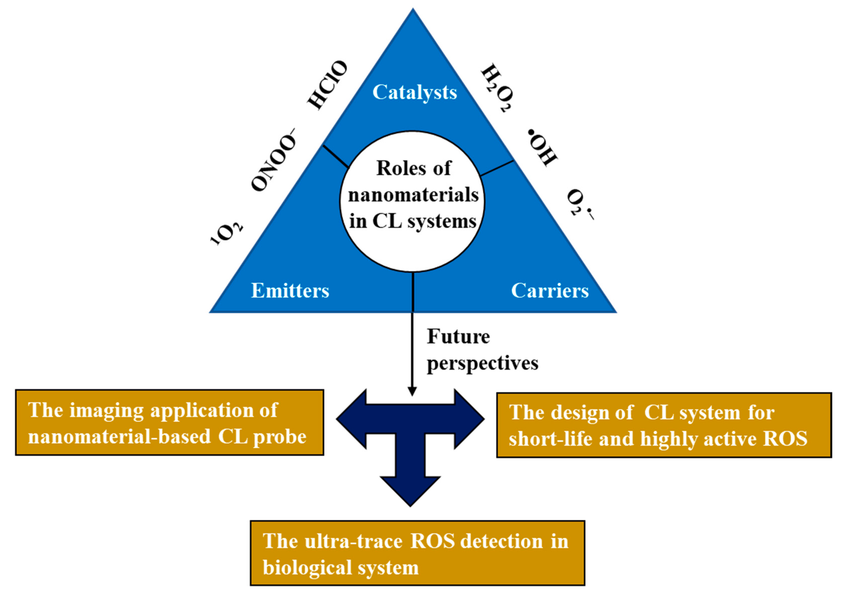
2. The Role of Nanomaterials in the CL System
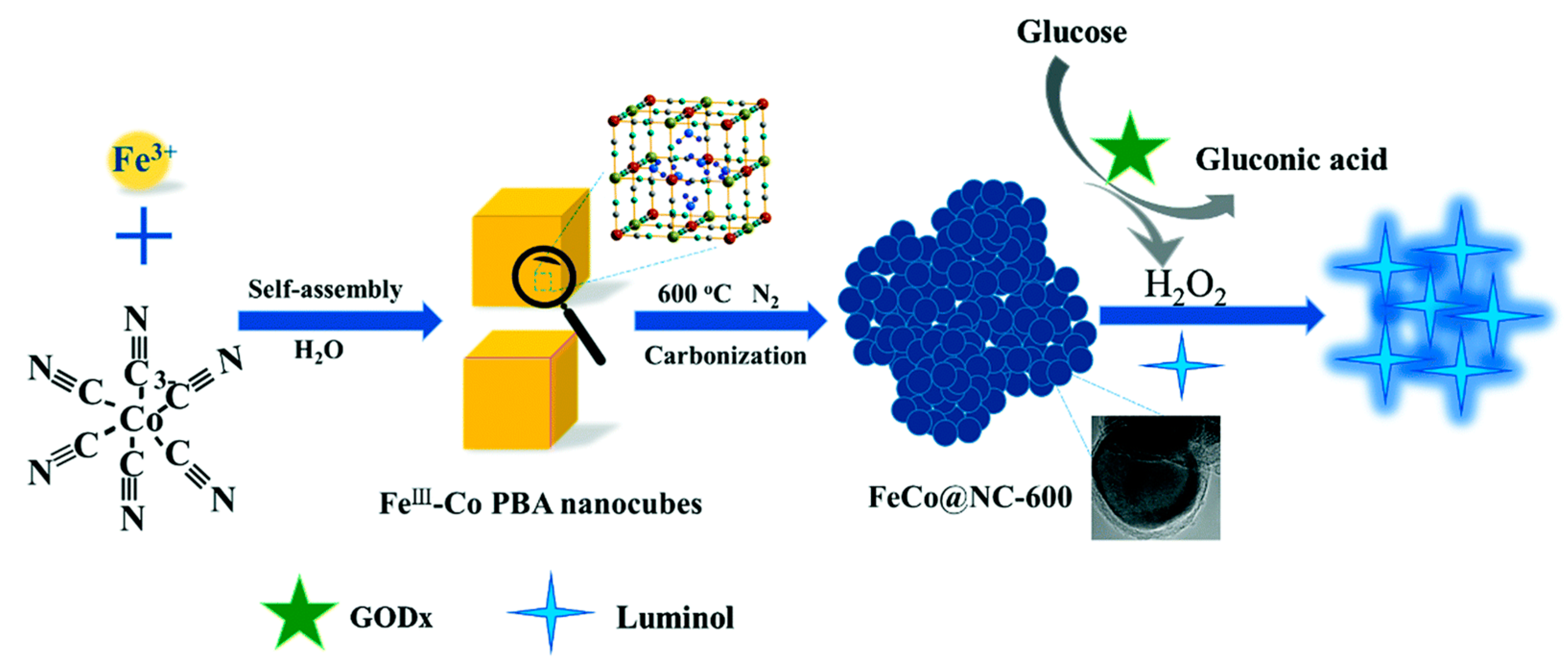
2.1. As Sensitizers and Catalysts
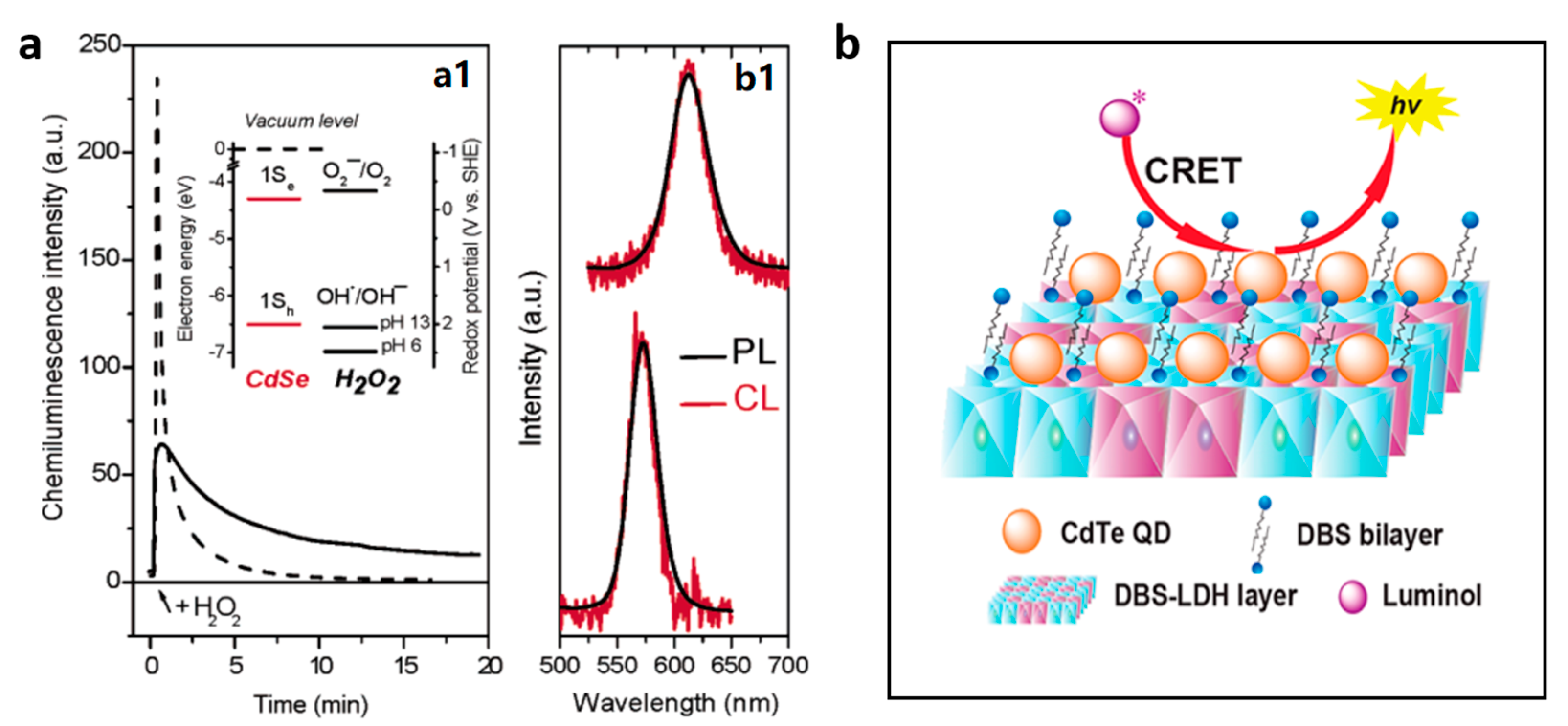
2.2. As Emitters or Energy Acceptor
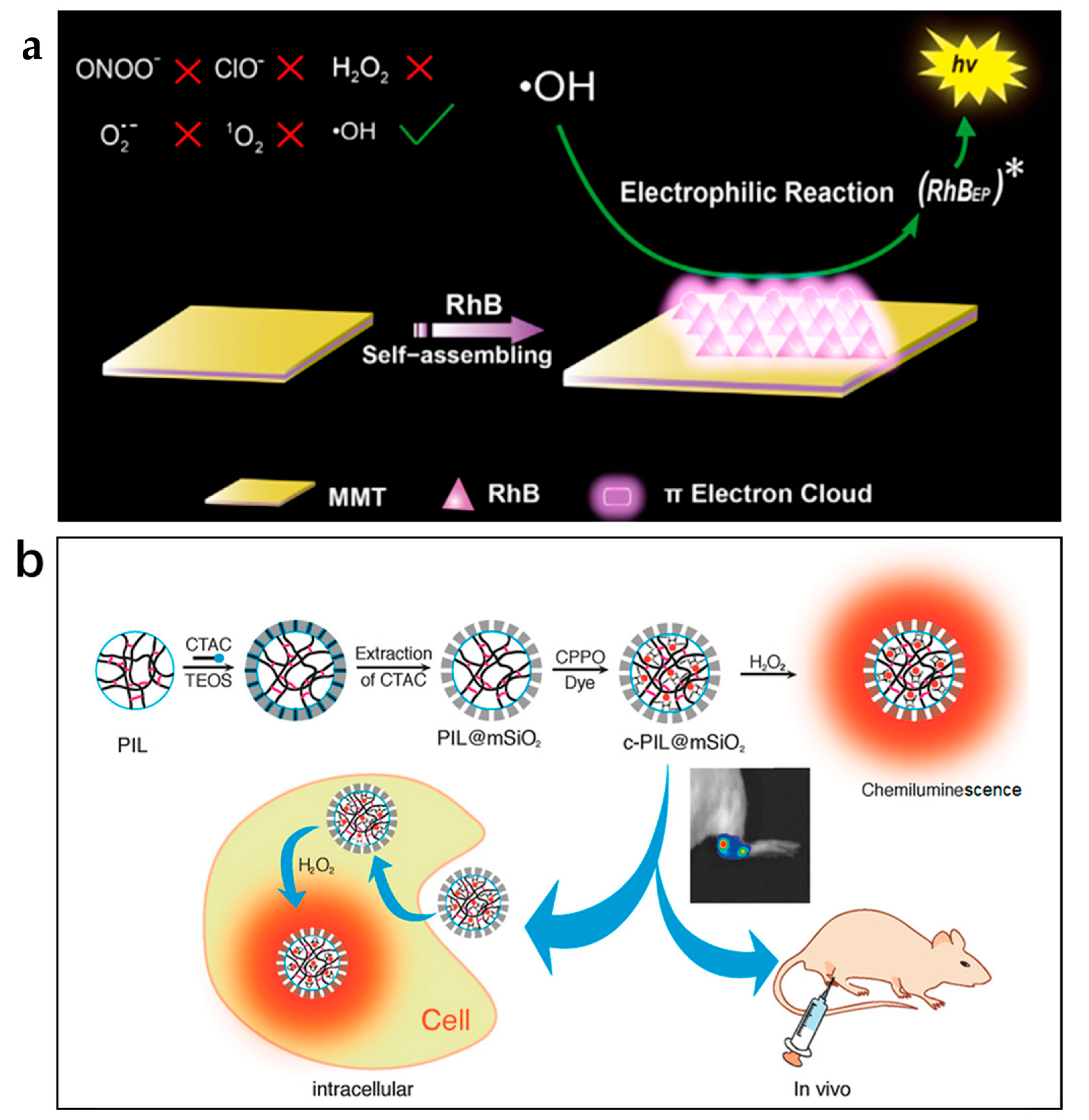
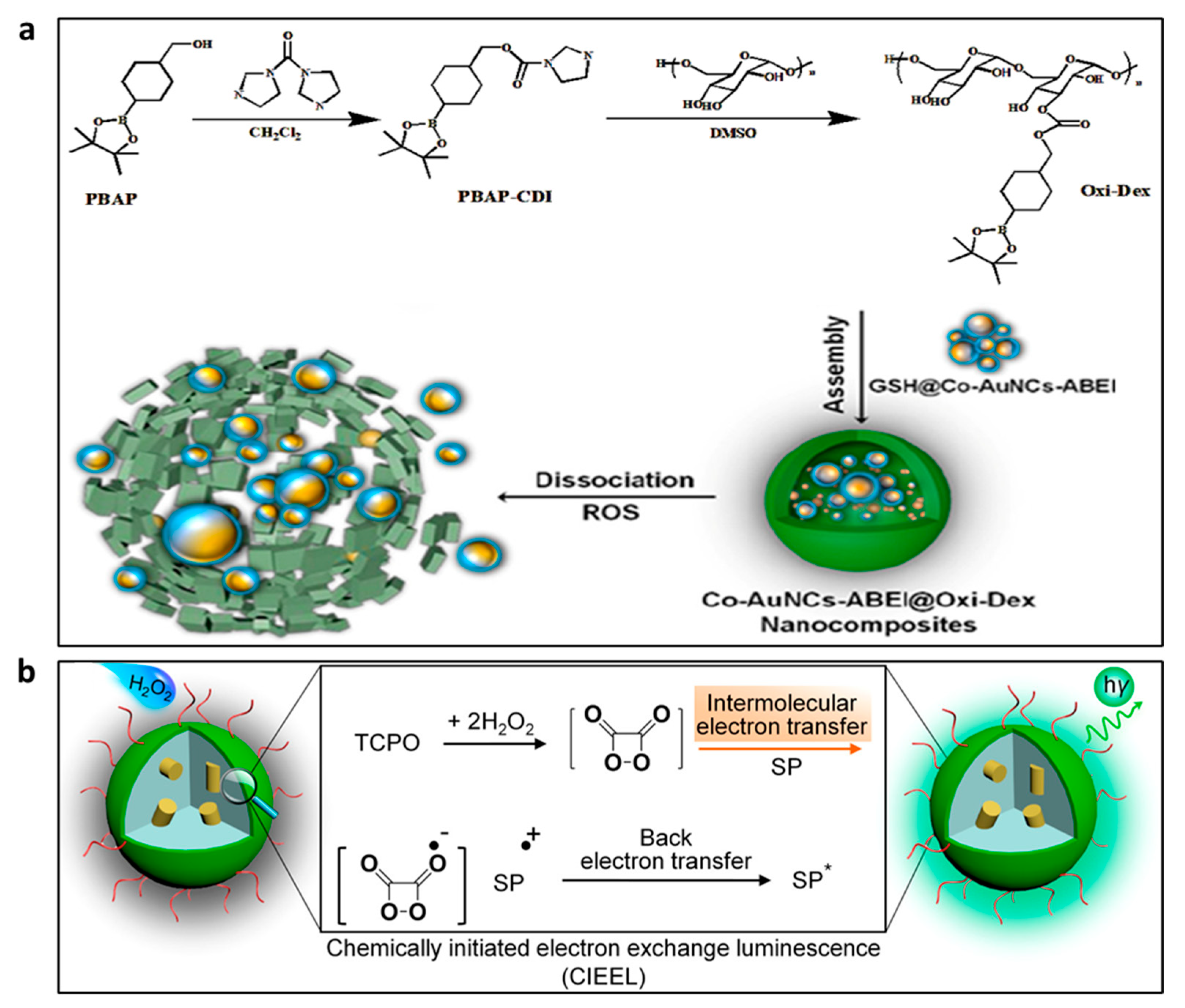
2.3. As Carriers of CL Reagents

3. Nanomaterial-Based CL Probes for Biosensing and Bioimaging of ROS
3.1. H2O2
3.2. •OH
3.3. O2•−
3.4. O2
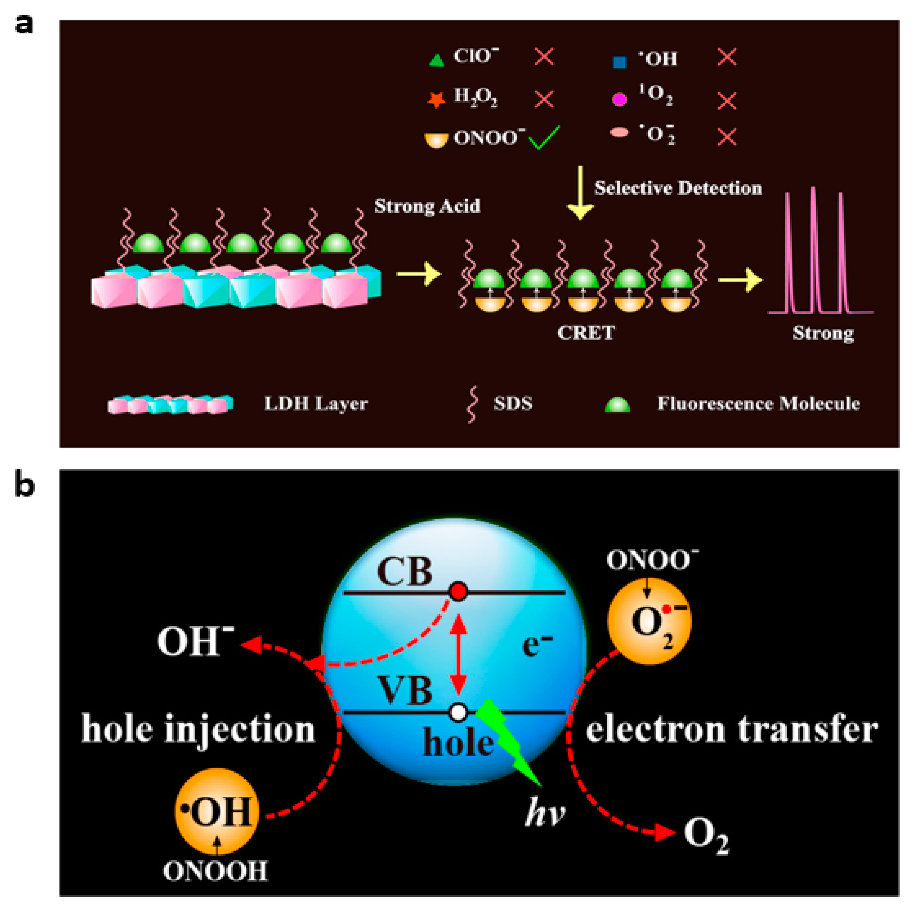
3.5. ONOO−
3.6. HClO/ClO−
4. Conclusions
Author Contributions
Funding
Data Availability Statement
Conflicts of Interest
References
- Liou, G.Y.; Storz, P. Reactive oxygen species in cancer. Free Radic. Res. 2010, 44, 479–496. [Google Scholar] [CrossRef] [PubMed]
- Kwon, N.; Kim, D.; Swamy, K.M.K.; Yoon, J. Metal-coordinated fluorescent and luminescent probes for reactive oxygen species (ROS) and reactive nitrogen species (RNS). Coord. Chem. Rev. 2021, 427, 213581. [Google Scholar] [CrossRef]
- Valko, M.; Leibfritz, D.; Moncol, J.; Cronin, M.T.; Mazur, M.; Telser, J. Free radicals and antioxidants in normal physiological functions and human disease. Int. J. Biochem. Cell Biol. 2007, 39, 44–84. [Google Scholar] [CrossRef] [PubMed]
- Nordberg, J.; Arner, E.S. Reactive oxygen species, antioxidants, and the mammalian thioredoxin system. Free Radic. Biol. Med. 2001, 31, 1287–1312. [Google Scholar] [CrossRef]
- Pacher, P.; Beckman, J.S.; Liaudet, L. Nitric oxide and peroxynitrite in health and disease. Physiol. Rev. 2007, 87, 315–424. [Google Scholar] [CrossRef]
- Stone, J.R.; Yang, S. Hydrogen peroxide: A signaling messenger. Antioxid. Redox Signal. 2006, 8, 243–270. [Google Scholar] [CrossRef]
- Winterbourn, C.C.; Hampton, M.B. Thiol chemistry and specificity in redox signaling. Free Radic. Biol. Med. 2008, 45, 549–561. [Google Scholar] [CrossRef]
- Valko, M.; Rhodes, C.J.; Moncol, J.; Izakovic, M.; Mazur, M. Free radicals, metals and antioxidants in oxidative stress-induced cancer. Chem.-Biol. Interact. 2006, 160, 1–40. [Google Scholar] [CrossRef]
- Rharass, T.; Vigo, J.; Salmon, J.M.; Ribou, A.C. Variation of 1-pyrenebutyric acid fluorescence lifetime in single living cells treated with molecules increasing or decreasing reactive oxygen species levels. Anal. Biochem. 2006, 357, 1–8. [Google Scholar] [CrossRef]
- D’Errico, G.; Vitiello, G.; De Tommaso, G.; Abdel-Gawad, F.K.; Brundo, M.V.; Ferrante, M.; De Maio, A.; Trocchia, S.; Bianchi, A.R.; Ciarcia, G.; et al. Electron spin resonance (ESR) for the study of reactive oxygen species (ROS) on the isolated frog skin (pelophylax bergeri): A non-invasive method for environmental monitoring. Environ. Res. 2018, 165, 11–18. [Google Scholar] [CrossRef]
- Nosaka, Y.; Nosaka, A.Y. Generation and detection of reactive oxygen species in photocatalysis. Chem. Rev. 2017, 117, 11302–11336. [Google Scholar] [CrossRef] [PubMed]
- Chen, X.; Wang, F.; Hyun, J.Y.; Wei, T.; Qiang, J.; Ren, X.; Shin, I.; Yoon, J. Recent progress in the development of fluorescent, luminescent and colorimetric probes for detection of reactive oxygen and nitrogen species. Chem. Soc. Rev. 2016, 45, 2976–3016. [Google Scholar] [CrossRef] [PubMed]
- Herman, J.; Zhang, Y.; Castranova, V.; Neal, S.L. Emerging technologies for optical spectral detection of reactive oxygen species. Anal. Bioanal. Chem. 2018, 410, 6079–6095. [Google Scholar] [CrossRef] [PubMed]
- Yu, W.; Zhao, L. Chemiluminescence detection of reactive oxygen species generation and potential environmental applications. TrAC Trends Anal. Chem. 2021, 136, 116197. [Google Scholar] [CrossRef]
- Arakawa, H.; Tsuruoka, K.; Ohno, K.; Tajima, N.; Nagano, H. Development of a highly sensitive chemiluminescent assay for hydrogen peroxide under neutral conditions using acridinium ester and its application to an enzyme immunoassay. Luminescence 2014, 29, 374–377. [Google Scholar] [CrossRef]
- Hananya, N.; Shabat, D. A glowing trajectory between bio- and chemiluminescence: From luciferin-based probes to triggerable dioxetanes. Angew. Chem. Int. Ed. 2017, 56, 16454–16463. [Google Scholar] [CrossRef]
- Espinoza, E.M.; Roise, J.J.; Li, I.C.; Das, R.; Murthy, N. Advances in imaging reactive oxygen species. J. Nucl. Med. 2021, 62, 457–461. [Google Scholar] [CrossRef]
- Green, O.; Gnaim, S.; Blau, R.; Eldar-Boock, A.; Satchi-Fainaro, R.; Shabat, D. Near-infrared dioxetane luminophores with direct chemiluminescence emission mode. J. Am. Chem. Soc. 2017, 139, 13243–13248. [Google Scholar] [CrossRef]
- Li, Y.-X.; Qin, H.-Y.; Hu, C.; Sun, M.-M.; Li, P.-Y.; Liu, H.; Li, J.-C.; Li, Z.-B.; Wu, L.-D.; Zhu, J. Research progress of nanomaterials-based sensors for food safety. J. Anal. Test. 2022, 6, 431–440. [Google Scholar] [CrossRef]
- Zhou, W.; Dong, S.; Lin, Y.; Lu, C. Insights into the role of nanostructure in the sensing properties of carbon nanodots for improved sensitivity to reactive oxygen species in living cells. Chem. Commun. 2017, 53, 2122–2125. [Google Scholar] [CrossRef]
- Nirala, N.R.; Pinker, N.; Desitti, C.; Shtenberg, G. Milk haptoglobin detection based on enhanced chemiluminescence of gold nanoparticles. Talanta 2019, 197, 257–263. [Google Scholar] [CrossRef] [PubMed]
- Iranifam, M.; Khodaei, S.; Saadati, M. Chemiluminescence reaction of graphene oxide–luminol–dissolved oxygen and its application for determination of isoniazid and paracetamol. Microchem. J. 2019, 146, 850–855. [Google Scholar] [CrossRef]
- Yousefzadeh, A.; Hassanzadeh, J.; Mousavi, S.M.J.; Yousefzadeh, M. Surface molecular imprinting and powerfully enhanced chemiluminescence emission by Cu nanoclusters/MOF composite for detection of tramadol. Sens. Actuators B Chem. 2019, 286, 154–162. [Google Scholar] [CrossRef]
- Gao, B.; Haghighatbin, M.A.; Cui, H. Polymer-encapsulated cobalt/gold bimetallic nanoclusters as stimuli-responsive chemiluminescent nanoprobes for reactive oxygen species. Anal. Chem. 2020, 92, 10677–10685. [Google Scholar] [CrossRef] [PubMed]
- Zhou, W.; Cao, Y.; Sui, D.; Lu, C. Radical pair-driven luminescence of quantum dots for specific detection of peroxynitrite in living cells. Anal. Chem. 2016, 88, 2659–2665. [Google Scholar] [CrossRef]
- Zhou, W.; Cao, Y.; Sui, D.; Lu, C. Turn-on luminescent probes for the real-time monitoring of endogenous hydroxyl radicals in living cells. Angew. Chem. Int. Ed. 2016, 55, 4236–4241. [Google Scholar] [CrossRef]
- Shen, C.-L.; Lou, Q.; Lv, C.-F.; Zheng, G.-S.; Zang, J.-H.; Jiang, T.-C.; Cheng, Z.; Liu, K.-K.; Niu, C.-Y.; Dong, L.; et al. Trigonal nitrogen activates high-brightness chemiluminescent carbon nanodots. ACS Mater. Lett. 2021, 3, 826–837. [Google Scholar] [CrossRef]
- Wang, Z.; Teng, X.; Lu, C. Orderly arranged fluorescence dyes as a highly efficient chemiluminescence resonance energy transfer probe for peroxynitrite. Anal. Chem. 2015, 87, 3412–3418. [Google Scholar] [CrossRef]
- Hassanzadeh, J.; Al Lawati, H.A.J.; Al Lawati, I. Metal-organic framework loaded by rhodamine B as a novel chemiluminescence system for the paper-based analytical devices and its application for total phenolic content determination in food samples. Anal. Chem. 2019, 91, 10631–10639. [Google Scholar] [CrossRef]
- Yadav, M.; Singh, G.; Lata, S. Revisiting some recently developed conducting polymer@metal oxide nanostructures for electrochemical sensing of vital biomolecules: A review. J. Anal. Test. 2022, 6, 274–295. [Google Scholar] [CrossRef]
- Zhong, J.; Yuan, Z.; Lu, C. Layered-nanomaterial-amplified chemiluminescence systems and their analytical applications. Anal. Bioanal. Chem. 2016, 408, 8731–8746. [Google Scholar] [CrossRef] [PubMed]
- Teradal, N.L.; Jelinek, R. Carbon nanomaterials in biological studies and biomedicine. Adv. Healthc. Mater. 2017, 6, 1700574. [Google Scholar] [CrossRef] [PubMed]
- Chen, J.; Qiu, H.; Zhao, S. Fabrication of chemiluminescence resonance energy transfer platform based on nanomaterial and its application in optical sensing, biological imaging and photodynamic therapy. TrAC Trends Anal. Chem. 2020, 122, 115747. [Google Scholar] [CrossRef]
- Su, Y.; Deng, D.; Zhang, L.; Song, H.; Lv, Y. Strategies in liquid-phase chemiluminescence and their applications in bioassay. TrAC Trends Anal. Chem. 2016, 82, 394–411. [Google Scholar] [CrossRef]
- Sheng, Y.; Yang, H.; Wang, Y.; Han, L.; Zhao, Y.; Fan, A. Silver nanoclusters-catalyzed luminol chemiluminescence for hydrogen peroxide and uric acid detection. Talanta 2017, 166, 268–274. [Google Scholar] [CrossRef]
- Mao, X.; Lu, Y.; Zhang, X.; Huang, Y. Beta-cyclodextrin functionalization of metal-organic framework MOF-235 with excellent chemiluminescence activity for sensitive glucose biosensing. Talanta 2018, 188, 161–167. [Google Scholar] [CrossRef]
- Tang, X.Q.; Zhang, Y.D.; Jiang, Z.W.; Wang, D.M.; Huang, C.Z.; Li, Y.F. Fe3O4 and metal-organic framework MIL-101(Fe) composites catalyze luminol chemiluminescence for sensitively sensing hydrogen peroxide and glucose. Talanta 2018, 179, 43–50. [Google Scholar] [CrossRef]
- Wang, D.M.; Zhang, Y.; Zheng, L.L.; Yang, X.X.; Wang, Y.; Huang, C.Z. Singlet oxygen involved luminol chemiluminescence catalyzed by graphene oxide. J. Phys. Chem. C 2012, 116, 21622–21628. [Google Scholar] [CrossRef]
- Bagheri, N.; Khataee, A.; Hassanzadeh, J.; Samaei, L. Highly sensitive chemiluminescence sensing system for organophosphates using mimic LDH supported ZIF-8 nanocomposite. Sens. Actuators B Chem. 2019, 284, 220–227. [Google Scholar] [CrossRef]
- Teng, Y.; Li, M.; Huang, X.; Ren, J. Singlet oxygen generation in ferriporphyrin-polymer dots catalyzed chemiluminescence system for cancer therapy. ACS Appl. Bio Mater. 2020, 3, 5020–5029. [Google Scholar] [CrossRef]
- Zhang, Z.-F.; Cui, H.; Lai, C.-Z.; Liu, L.-J. Gold nanoparticle-catalyzed luminol chemiluminescence and its analytical applications. Anal. Chem. 2005, 77, 3324–3329. [Google Scholar] [CrossRef] [PubMed]
- Chen, W.; Hong, L.; Liu, A.-L.; Liu, J.-Q.; Lin, X.-H.; Xia, X.-H. Enhanced chemiluminescence of the luminol-hydrogen peroxide system by colloidal cupric oxide nanoparticles as peroxidase mimic. Talanta 2012, 99, 643–648. [Google Scholar] [CrossRef] [PubMed]
- Li, Q.; Zhang, L.; Li, J.; Lu, C. Nanomaterial-amplified chemiluminescence systems and their applications in bioassays. TrAC Trends Anal. Chem. 2011, 30, 401–413. [Google Scholar] [CrossRef]
- Iranifam, M. Chemiluminescence reactions enhanced by silver nanoparticles and silver alloy nanoparticles: Applications in analytical chemistry. TrAC Trends Anal. Chem. 2016, 82, 126–142. [Google Scholar] [CrossRef]
- Xu, S.; Chen, F.; Deng, M.; Sui, Y. Luminol chemiluminescence enhanced by copper nanoclusters and its analytical application. RSC Adv. 2014, 4, 15664–15670. [Google Scholar] [CrossRef]
- Luo, J.; Liu, R.; Zhao, S.; Gao, Y. Bimetallic Fe-Co nanoalloy confined in porous carbon skeleton with enhanced peroxidase mimetic activity for multiple biomarkers monitoring. J. Anal. Test. 2023, 7, 53–68. [Google Scholar] [CrossRef]
- Mokhtarzadeh, E.; Abolhasani, J.; Hassanzadeh, J. Rhodamine B chemiluminescence improved by mimetic AuCu alloy nanoclusters and ultrasensitive measurement of H2O2, glucose and xanthine. Anal. Sci. 2019, 35, 543–550. [Google Scholar] [CrossRef]
- Lu, Y.; Zhang, X.; Mao, X.; Huang, Y. Engineering FeCo alloy@N-doped carbon layers by directly pyrolyzing prussian blue analogue: New peroxidase mimetic for chemiluminescence glucose biosensing. J. Mater. Chem. B 2019, 7, 4661–4668. [Google Scholar] [CrossRef]
- Li, X.; Zhang, Z.; Tao, L.; Gao, M. Sensitive and selective chemiluminescence assay for hydrogen peroxide in exhaled breath condensate using nanoparticle-based catalysis. Spectrochim. Acta Part A 2013, 107, 311–316. [Google Scholar] [CrossRef]
- Jiao, L.; Wang, Y.; Jiang, H.-L.; Xu, Q. Metal-organic frameworks as platforms for catalytic applications. Adv. Mater. 2018, 30, 1703663. [Google Scholar] [CrossRef]
- Zhu, Q.; Chen, Y.; Wang, W.; Zhang, H.; Ren, C.; Chen, H.; Chen, X. A sensitive biosensor for dopamine determination based on the unique catalytic chemiluminescence of metal–organic framework HKUST-1. Sens. Actuators B Chem. 2015, 210, 500–507. [Google Scholar] [CrossRef]
- Yang, C.P.; He, L.; Huang, C.Z.; Li, Y.F.; Zhen, S.J. Continuous singlet oxygen generation for persistent chemiluminescence in Cu-MOFs-based catalytic system. Talanta 2021, 221, 121498. [Google Scholar] [CrossRef] [PubMed]
- Liu, X.; Han, Z.; Li, F.; Gao, L.; Liang, G.; Cui, H. Highly chemiluminescent graphene oxide hybrids bifunctionalized by N-(aminobutyl)-N-(ethylisoluminol)/horseradish peroxidase and sensitive sensing of hydrogen peroxide. ACS Appl. Mater. Interfaces 2015, 7, 18283–18291. [Google Scholar] [CrossRef] [PubMed]
- Pan, F.; Zhang, Y.; Yuan, Z.; Lu, C. Determination of alizarin red s based on layered double hydroxides-improved chemiluminescence from hydrogen peroxide and luminol. Anal. Methods 2017, 9, 6468–6473. [Google Scholar] [CrossRef]
- Cheng, W.; Teng, X.; Lu, C. Structurally ordered catalyst-amplified chemiluminescence signals. Anal. Chem. 2020, 92, 5456–5463. [Google Scholar] [CrossRef]
- Li, M.; Huang, X.; Ren, J. Multicolor chemiluminescent resonance energy-transfer system for in vivo high-contrast and targeted imaging. Anal. Chem. 2021, 93, 3042–3051. [Google Scholar] [CrossRef]
- Zhou, M.; Deng, L.; Teng, Y.; Li, M.; Huang, X.; Ren, J. Polystyrene–hemin dots for chemiluminescence imaging. ACS Appl. Nano Mater. 2019, 2, 3761–3768. [Google Scholar] [CrossRef]
- Poznyak, S.K.; Talapin, D.V.; Shevchenko, E.V.; Weller, H. Quantum dot chemiluminescence. Nano Lett. 2004, 4, 693–698. [Google Scholar] [CrossRef]
- Chen, H.; Lin, L.; Lin, Z.; Guo, G.; Lin, J.-M. Chemiluminescence arising from the decomposition of peroxymonocarbonate and enhanced by CdTe quantum dots. J. Phys. Chem. A 2010, 114, 10049–10058. [Google Scholar] [CrossRef]
- Lin, Z.; Xue, W.; Chen, H.; Lin, J.-M. Peroxynitrous-acid-induced chemiluminescence of fluorescent carbon dots for nitrite sensing. Anal. Chem. 2011, 83, 8245–8251. [Google Scholar] [CrossRef]
- Dong, S.; Zhong, J.; Lu, C. Introducing confinement effects into ultraweak chemiluminescence for an improved sensitivity. Anal. Chem. 2014, 86, 7947–7953. [Google Scholar] [CrossRef] [PubMed]
- Dong, S.; Liu, F.; Lu, C. Organo-modified hydrotalcite-quantum dot nanocomposites as a novel chemiluminescence resonance energy transfer probe. Anal. Chem. 2013, 85, 3363–3368. [Google Scholar] [CrossRef]
- Huang, X.; Li, L.; Qian, H.; Dong, C.; Ren, J. A resonance energy transfer between chemiluminescent donors and luminescent quantum-dots as acceptors (CRET). Angew. Chem. Int. Ed. 2006, 45, 5140–5143. [Google Scholar] [CrossRef] [PubMed]
- Xu, S.; Li, J.; Li, X.; Su, M.; Shi, Z.; Zeng, Y.; Ni, S. A chemiluminescence resonance energy transfer system composed of cobalt(Ⅱ), luminol, hydrogen peroxide and CdTe quantum dots for highly sensitive determination of hydroquinone. Microchim. Acta 2015, 183, 667–673. [Google Scholar] [CrossRef]
- Lin, Y.; Dai, Y.; Sun, Y.; Ding, C.; Sun, W.; Zhu, X.; Liu, H.; Luo, C. A turn-on chemiluminescence biosensor for selective and sensitive detection of adenosine based on HKUST-1 and QDs-luminol-aptamer conjugates. Talanta 2018, 182, 116–124. [Google Scholar] [CrossRef]
- Liu, H.; Su, Y.; Deng, D.; Song, H.; Lv, Y. Chemiluminescence of oleic acid capped black phosphorus quantum dots for highly selective detection of sulfite in PM2.5. Anal. Chem. 2019, 91, 9174–9180. [Google Scholar] [CrossRef]
- Liu, H.; Sun, M.; Su, Y.; Deng, D.; Hu, J.; Lv, Y. Chemiluminescence of black phosphorus quantum dots induced by hypochlorite and peroxide. Chem. Commun. 2018, 54, 7987–7990. [Google Scholar] [CrossRef]
- Du, J.; Wang, Y.; Zhang, W. Gold nanoparticles-based chemiluminescence resonance energy transfer for ultrasensitive detection of melamine. Spectrochim. Acta Part A 2015, 149, 698–702. [Google Scholar] [CrossRef]
- You, X.; Li, Y.; Li, B.; Ma, J. Gold nanoclusters-based chemiluminescence resonance energy transfer method for sensitive and label-free detection of trypsin. Talanta 2016, 147, 63–68. [Google Scholar] [CrossRef]
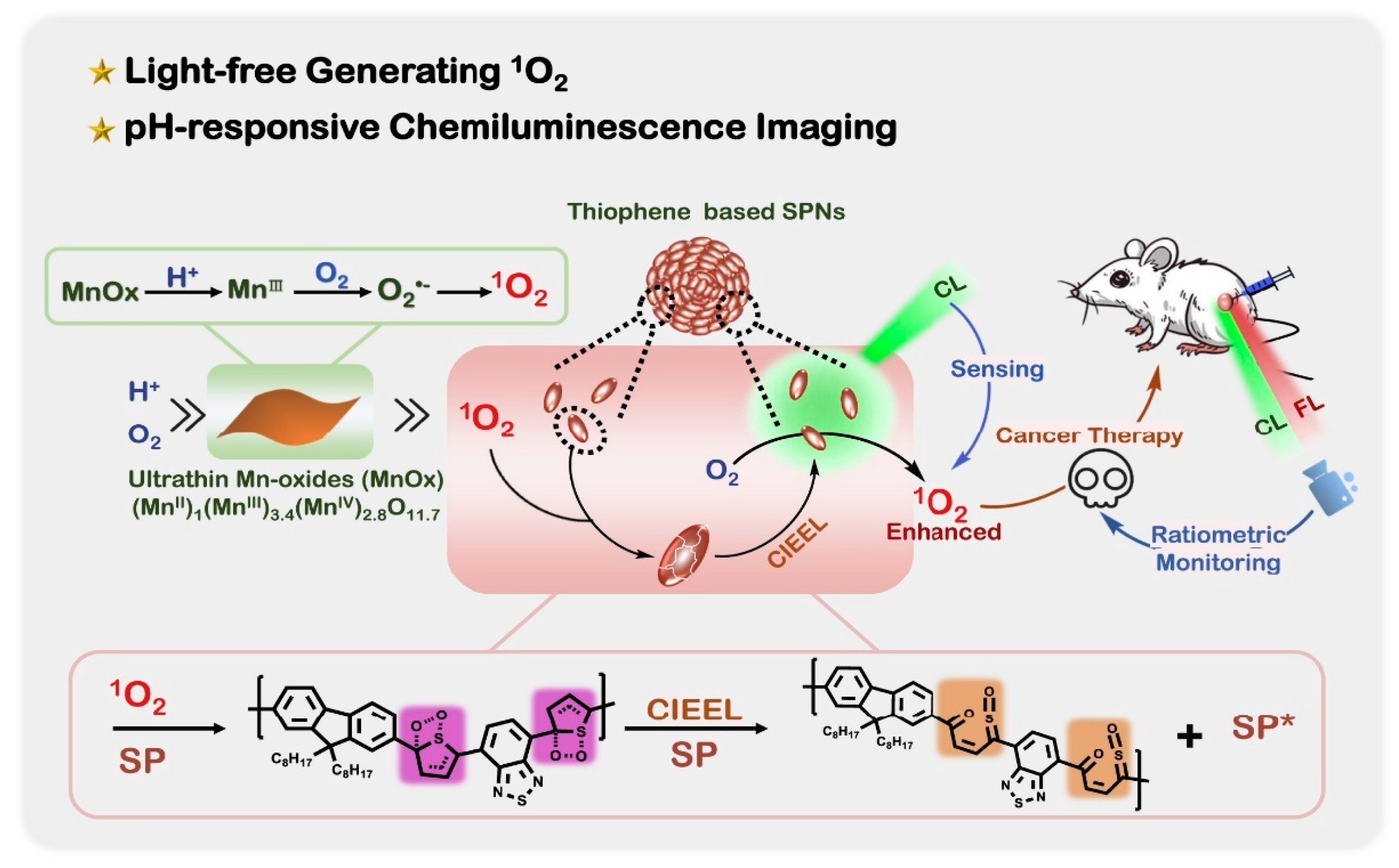
- Lu, C.; Zhang, C.; Wang, P.; Zhao, Y.; Yang, Y.; Wang, Y.; Yuan, H.; Qu, S.; Zhang, X.; Song, G.; et al. Light-free generation of singlet oxygen through manganese-thiophene nanosystems for pH-responsive chemiluminescence imaging and tumor therapy. Chem 2020, 6, 2314–2334. [Google Scholar] [CrossRef]
- Li, Z.; Deng, X.; Wu, S.; Dong, S.; Zou, G. Hydrazine hydrate and dissolved oxygen-triggered near-infrared chemiluminescence from CuInS2@ZnS nanocrystals for bioassays. Anal. Chem. 2021, 93, 8931–8936. [Google Scholar] [CrossRef] [PubMed]
- Yu, H.; Long, D. Highly chemiluminescent metal-organic framework of type MIL-101(Cr) for detection of hydrogen peroxide and pyrophosphate ions. Microchim. Acta 2016, 183, 3151–3157. [Google Scholar] [CrossRef]
- Cao, Y.; Sui, D.; Zhou, W.; Lu, C. Highly selective chemiluminescence detection of hydroxyl radical via increased π-electron densities of rhodamine B on montmorillonite matrix. Sens. Actuators B Chem. 2016, 225, 600–606. [Google Scholar] [CrossRef]
- Luo, M.; Wang, W.; Zhao, Q.; Li, M.; Chen, Y.; Lu, Z.; Liu, K.; Wang, D. Chemiluminescence biosensor for hydrogen peroxide determination by immobilizing horseradish peroxidase onto PVA-co-PE nanofiber membrane. Eur. Polym. J. 2017, 91, 307–314. [Google Scholar] [CrossRef]
- Singh, A.; Seo, Y.H.; Lim, C.K.; Koh, J.; Jang, W.D.; Kwon, I.C.; Kim, S. Biolighted nanotorch capable of systemic self-delivery and diagnostic imaging. ACS Nano 2015, 9, 9906–9911. [Google Scholar] [CrossRef] [PubMed]
- Xie, M.; Zhang, Z.; Guan, W.; Zhou, W.; Lu, C. Micelle-mediated chemiluminescence as an indicator for micellar transitions. Anal. Chem. 2019, 91, 2652–2658. [Google Scholar] [CrossRef]
- Zhou, K.; Zhang, F.; Xu, J.; He, H.; Wei, W.; Xia, Z. Core-shell poly (ionic liquid)@mesoporous silica chemiluminescent nanoprobes for sensitive intracellular hydrogen peroxide imaging. Part. Part. Syst. Charact. 2018, 35, 1700329. [Google Scholar] [CrossRef]
- Li, Y.; Zhu, B.; Han, W.; Tang, W.; Duan, X. A bright chemiluminescence conjugated polymer-mesoporous silica nanoprobe for imaging of colonic tumors in vivo. Analyst 2022, 147, 2060–2067. [Google Scholar] [CrossRef]
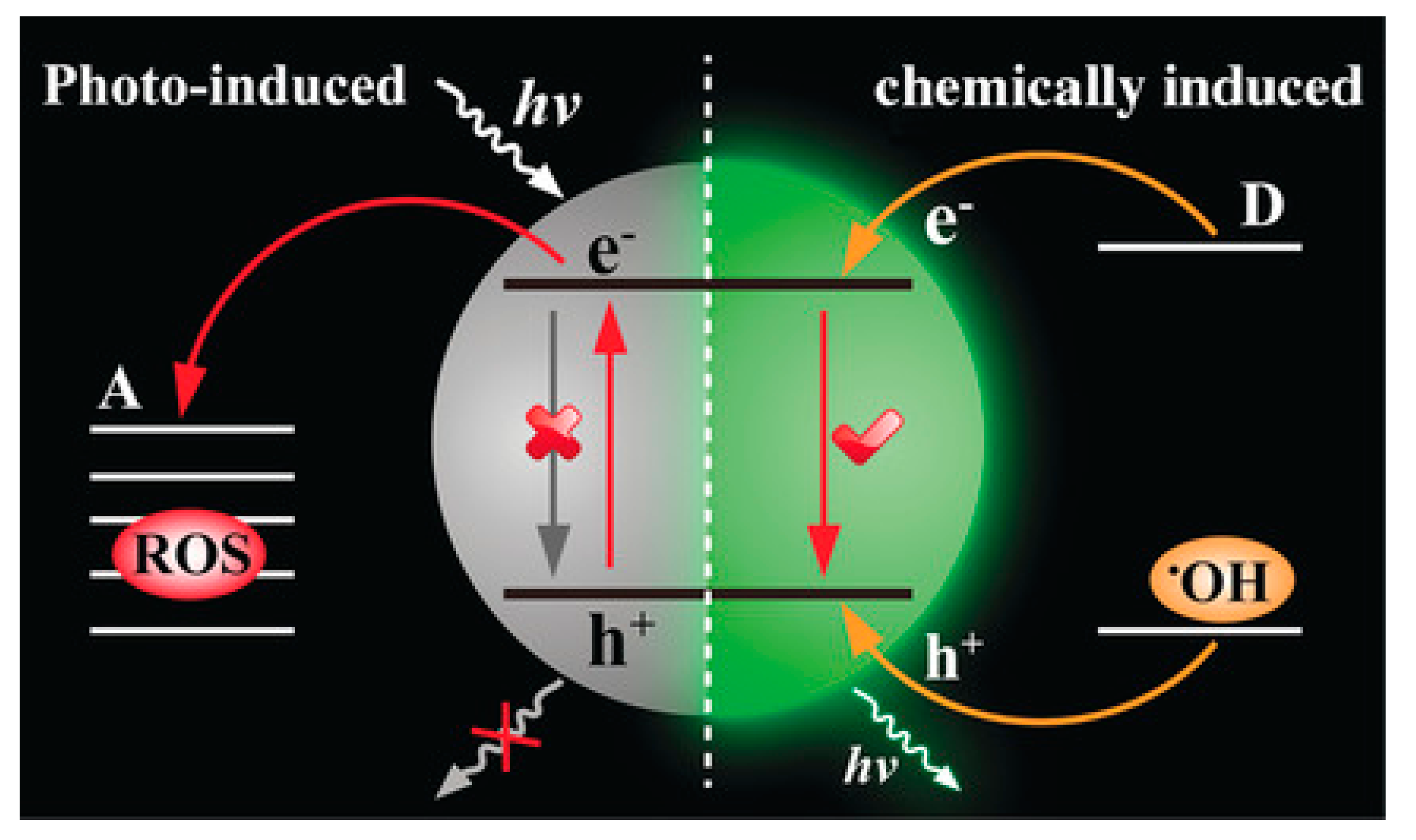
- Zhen, X.; Zhang, C.; Xie, C.; Miao, Q.; Lim, K.L.; Pu, K. Intraparticle energy level alignment of semiconducting polymer nanoparticles to amplify chemiluminescence for ultrasensitive in vivo imaging of reactive oxygen species. ACS Nano 2016, 10, 6400–6409. [Google Scholar] [CrossRef]
- Zhang, W.; Hao, L.; Huang, J.; Xia, L.; Cui, M.; Zhang, X.; Gu, Y.; Wang, P. Chemiluminescence chitosan hydrogels based on the luminol analog L-012 for highly sensitive detection of ROS. Talanta 2019, 201, 455–459. [Google Scholar] [CrossRef]
- Pu, K.; Shuhendler, A.J.; Rao, J. Semiconducting polymer nanoprobe for in vivo imaging of reactive oxygen and nitrogen species. Angew. Chem. Int. Ed. 2013, 52, 10325–10329. [Google Scholar] [CrossRef] [PubMed]
- Seo, Y.H.; Singh, A.; Cho, H.-J.; Kim, Y.; Heo, J.; Lim, C.-K.; Park, S.Y.; Jang, W.-D.; Kim, S. Rational design for enhancing inflammation-responsive in vivo chemiluminescence via nanophotonic energy relay to near-infrared AIE-active conjugated polymer. Biomaterials 2016, 84, 111–118. [Google Scholar] [CrossRef] [PubMed]
- Shen, C.-L.; Lou, Q.; Zang, J.-H.; Liu, K.-K.; Qu, S.-N.; Dong, L.; Shan, C.-X. Near-infrared chemiluminescent carbon nanodots and their application in reactive oxygen species bioimaging. Adv. Sci. 2020, 7, 1903525. [Google Scholar] [CrossRef] [PubMed]
- Mao, D.; Wu, W.; Ji, S.; Chen, C.; Hu, F.; Kong, D.; Ding, D.; Liu, B. Chemiluminescence-guided cancer therapy using a chemiexcited photosensitizer. Chem 2017, 3, 991–1007. [Google Scholar] [CrossRef]
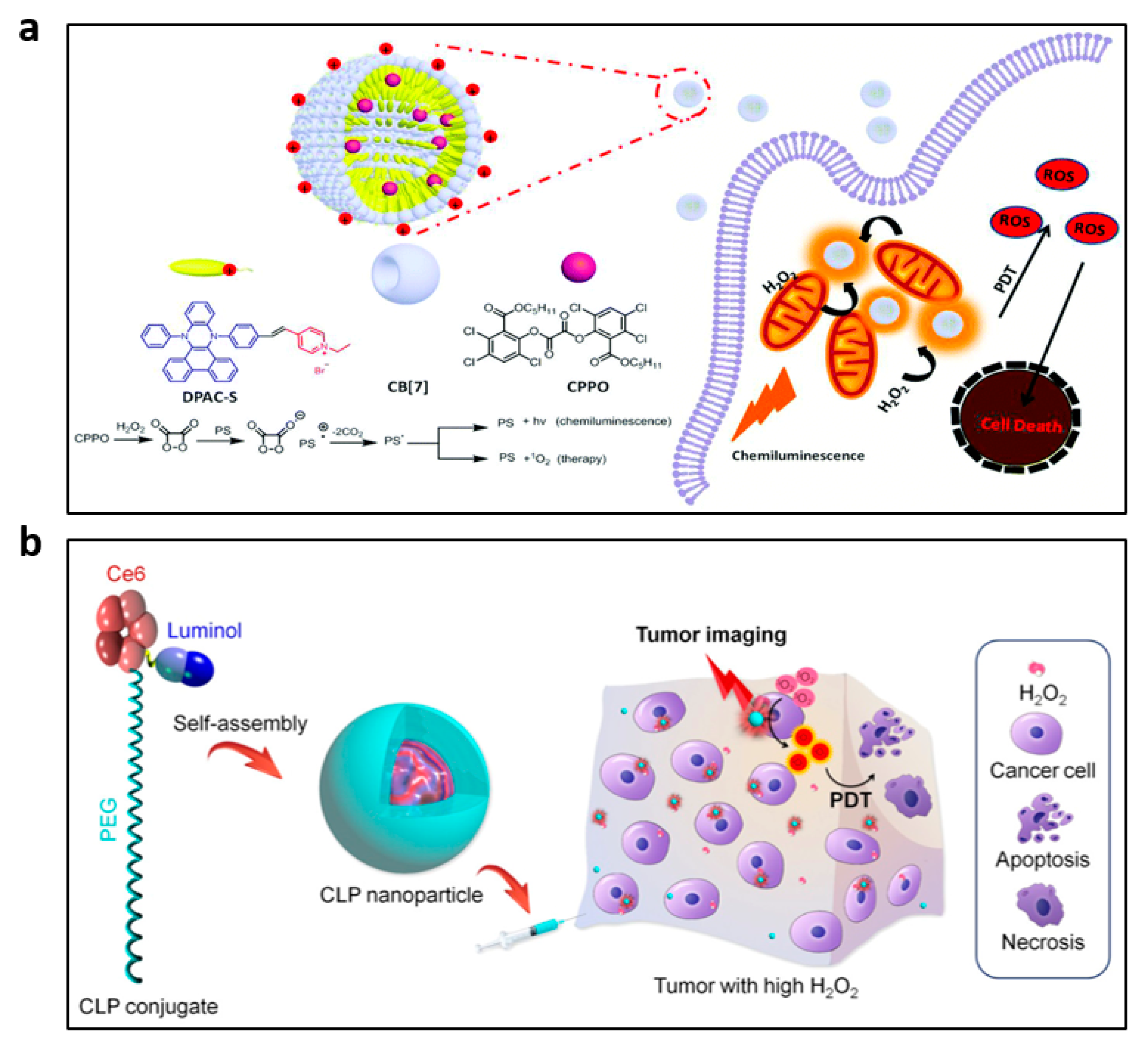
- Chen, L.; Chen, Y.; Zhou, W.; Li, J.; Zhang, Y.; Liu, Y. Mitochondrion-targeting chemiluminescent ternary supramolecular assembly for in situ photodynamic therapy. Chem. Commun. 2020, 56, 8857–8860. [Google Scholar] [CrossRef] [PubMed]
- An, H.; Guo, C.; Li, D.; Liu, R.; Xu, X.; Guo, J.; Ding, J.; Li, J.; Chen, W.; Zhang, J. Hydrogen peroxide-activatable nanoparticles for luminescence imaging and in situ triggerable photodynamic therapy of cancer. ACS Appl. Mater. Interfaces 2020, 12, 17230–17243. [Google Scholar] [CrossRef]
- Ganea, G.M.; Kolic, P.E.; El-Zahab, B.; Warner, I.M. Ratiometric coumarin-neutral red (CONER) nanoprobe for detection of hydroxyl radicals. Anal. Chem. 2011, 83, 2576–2581. [Google Scholar] [CrossRef]
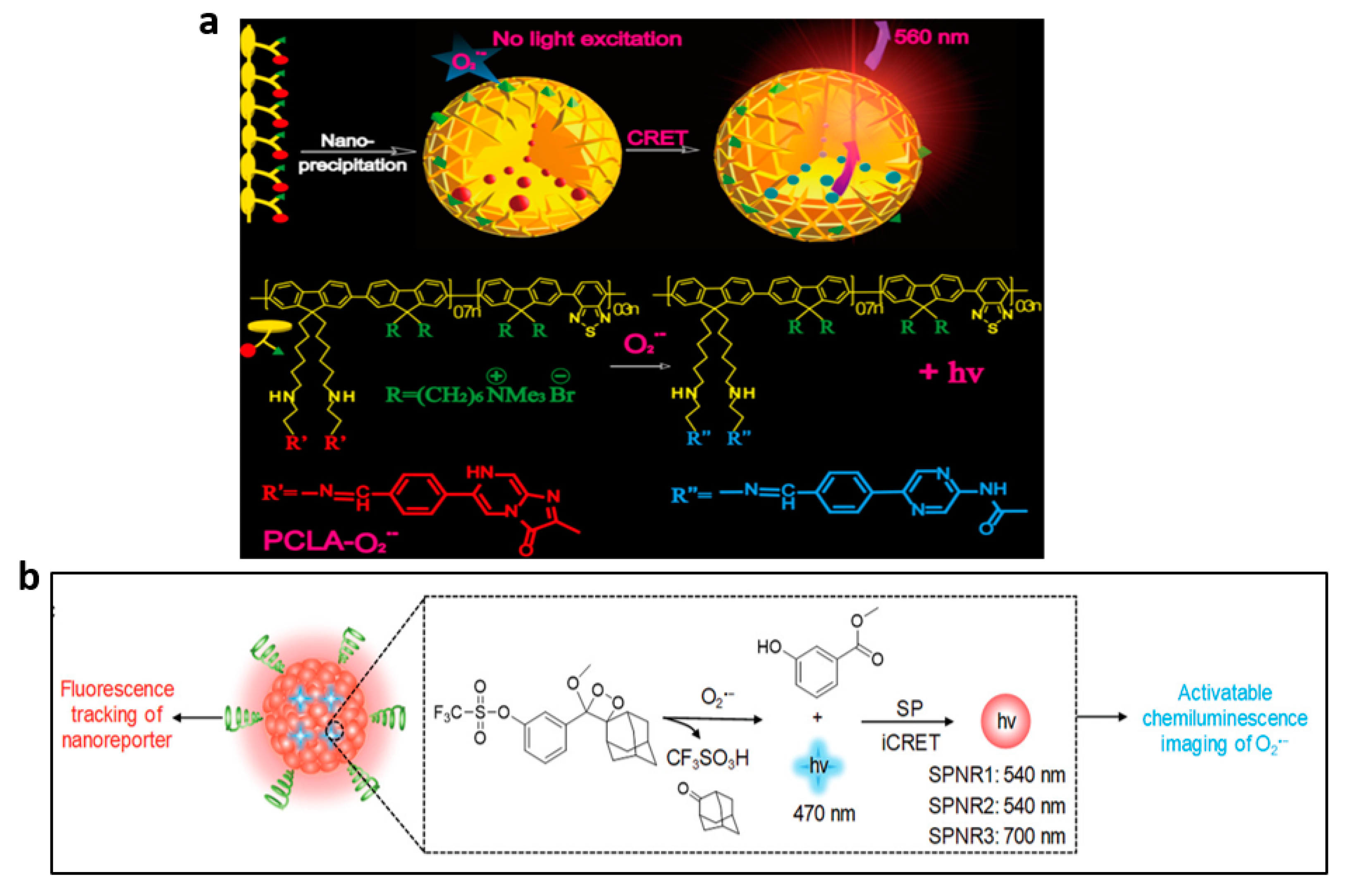
- Li, P.; Liu, L.; Xiao, H.; Zhang, W.; Wang, L.; Tang, B. A new polymer nanoprobe based on chemiluminescence resonance energy transfer for ultrasensitive imaging of intrinsic superoxide anion in mice. J. Am. Chem. Soc. 2016, 138, 2893–2896. [Google Scholar] [CrossRef]
- Cui, D.; Li, J.; Zhao, X.; Pu, K.; Zhang, R. Semiconducting polymer nanoreporters for near-infrared chemiluminescence imaging of immunoactivation. Adv. Mater. 2020, 32, 1906314. [Google Scholar] [CrossRef]
- Dickinson, B.C.; Chang, C.J. Chemistry and biology of reactive oxygen species in signaling or stress responses. Nat. Chem. Biol. 2011, 7, 504–511. [Google Scholar] [CrossRef]
- Schweitzer, C.; Schmidt, R. Physical mechanisms of generation and deactivation of singlet oxygen. Chem. Rev. 2003, 103, 1685–1757. [Google Scholar] [CrossRef]
- Klotz, L.O.; Briviba, K.; Sies, H. Mitogen-activated protein kinase activation by singlet oxygen and ultraviolet a. Methods Enzymol. 2000, 319, 130–143. [Google Scholar]
- Lucky, S.S.; Soo, K.C.; Zhang, Y. Nanoparticles in photodynamic therapy. Chem. Rev. 2015, 115, 1990–2042. [Google Scholar] [CrossRef] [PubMed]
- Ni, X.; Zhang, X.; Duan, X.; Zheng, H.-L.; Xue, X.-S.; Ding, D. Near-infrared afterglow luminescent aggregation-induced emission dots with ultrahigh tumor-to-liver signal ratio for promoted image-guided cancer surgery. Nano Lett. 2019, 19, 318–330. [Google Scholar] [CrossRef] [PubMed]
- Pedersen, S.K.; Holmehave, J.; Blaikie, F.H.; Gollmer, A.; Breitenbach, T.; Jensen, H.H.; Ogilby, P.R. Aarhus sensor green: A fluorescent probe for singlet oxygen. J. Org. Chem. 2014, 79, 3079–3087. [Google Scholar] [CrossRef] [PubMed]
- Zhao, Z.; Chen, C.; Wu, W.; Wang, F.; Du, L.; Zhang, X.; Xiong, Y.; He, X.; Cai, Y.; Kwok, R.T.K.; et al. Highly efficient photothermal nanoagent achieved by harvesting energy via excited-state intramolecular motion within nanoparticles. Nat. Commun. 2019, 10, 768. [Google Scholar] [CrossRef]
- Zhu, H.; Fang, Y.; Miao, Q.; Qi, X.; Ding, D.; Chen, P.; Pu, K. Regulating near-infrared photodynamic properties of semiconducting polymer nanotheranostics for optimized cancer therapy. ACS Nano 2017, 11, 8998–9009. [Google Scholar] [CrossRef] [PubMed]
- Zhang, S.; Cui, H.; Gu, M.; Zhao, N.; Cheng, M.; Lv, J. Real-time mapping of ultratrace singlet oxygen in rat during acute and chronic inflammations via a chemiluminescent nanosensor. Small 2019, 15, 1804662. [Google Scholar] [CrossRef]
- Wang, B.; Wang, Y.; Wang, Y.; Zhao, Y.; Yang, C.; Zeng, Z.; Huan, S.; Song, G.; Zhang, X. Oxygen-embedded pentacene based near-infrared chemiluminescent nanoprobe for highly selective and sensitive visualization of peroxynitrite in vivo. Anal. Chem. 2020, 92, 4154–4163. [Google Scholar] [CrossRef]
- Prolo, C.; Rios, N.; Piacenza, L.; Alvarez, M.N.; Radi, R. Fluorescence and chemiluminescence approaches for peroxynitrite detection. Free Radic. Biol. Med. 2018, 128, 59–68. [Google Scholar] [CrossRef]
- Cao, J.; An, W.; Reeves, A.G.; Lippert, A.R. A chemiluminescent probe for cellular peroxynitrite using a self-immolative oxidative decarbonylation reaction. Chem. Sci. 2018, 9, 2552–2558. [Google Scholar] [CrossRef]
- Dong, S.; Yuan, Z.; Zhang, L.; Lin, Y.; Lu, C. Rapid screening of oxygen states in carbon quantum dots by chemiluminescence probe. Anal. Chem. 2017, 89, 12520–12526. [Google Scholar] [CrossRef] [PubMed]
- Zhao, S.; Liu, J.; Huang, Y.; Liu, Y.M. Introducing chemiluminescence resonance energy transfer into immunoassay in a microfluidic format for an improved assay sensitivity. Chem. Commun. 2012, 48, 699–701. [Google Scholar] [CrossRef]
- Lis, S.; Kaczmarek, M. Chemiluminescent systems generating reactive oxygen species from the decomposition of hydrogen peroxide and their analytical applications. TrAC Trends Anal. Chem. 2013, 44, 1–11. [Google Scholar] [CrossRef]
- Tang, Y.; Su, Y.; Yang, N.; Zhang, L.; Lv, Y. Carbon nitride quantum dots: A novel chemiluminescence system for selective detection of free chlorine in water. Anal. Chem. 2014, 86, 4528–4535. [Google Scholar] [CrossRef] [PubMed]
- Dixon, S.J.; Stockwell, B.R. The role of iron and reactive oxygen species in cell death. Nat. Chem. Biol. 2014, 10, 9–17. [Google Scholar] [CrossRef]
- Ray, P.D.; Huang, B.W.; Tsuji, Y. Reactive oxygen species (ROS) homeostasis and redox regulation in cellular signaling. Cell Signal. 2012, 24, 981–990. [Google Scholar] [CrossRef] [PubMed]
- Zhang, R.; Song, B.; Yuan, J. Bioanalytical methods for hypochlorous acid detection: Recent advances and challenges. TrAC Trends Anal. Chem. 2018, 99, 1–33. [Google Scholar] [CrossRef]
- Andersen, J.K. Oxidative stress in neurodegeneration: Cause or consequence? Nat. Med. 2004, 10, 18–25. [Google Scholar] [CrossRef] [PubMed]
- Zhu, B.; Tang, W.; Ren, Y.; Duan, X. Chemiluminescence of conjugated-polymer nanoparticles by direct oxidation with hypochlorite. Anal. Chem. 2018, 90, 13714–13722. [Google Scholar] [CrossRef] [PubMed]


Disclaimer/Publisher’s Note: The statements, opinions and data contained in all publications are solely those of the individual author(s) and contributor(s) and not of MDPI and/or the editor(s). MDPI and/or the editor(s) disclaim responsibility for any injury to people or property resulting from any ideas, methods, instructions or products referred to in the content. |
© 2023 by the authors. Licensee MDPI, Basel, Switzerland. This article is an open access article distributed under the terms and conditions of the Creative Commons Attribution (CC BY) license (https://creativecommons.org/licenses/by/4.0/).
Share and Cite
Huang, C.; Zhou, W.; Wu, R.; Guan, W.; Ye, N. Recent Advances in Nanomaterial-Based Chemiluminescence Probes for Biosensing and Imaging of Reactive Oxygen Species. Nanomaterials 2023, 13, 1726. https://doi.org/10.3390/nano13111726
Huang C, Zhou W, Wu R, Guan W, Ye N. Recent Advances in Nanomaterial-Based Chemiluminescence Probes for Biosensing and Imaging of Reactive Oxygen Species. Nanomaterials. 2023; 13(11):1726. https://doi.org/10.3390/nano13111726
Chicago/Turabian StyleHuang, Chuanlin, Wenjuan Zhou, Riliga Wu, Weijiang Guan, and Nengsheng Ye. 2023. "Recent Advances in Nanomaterial-Based Chemiluminescence Probes for Biosensing and Imaging of Reactive Oxygen Species" Nanomaterials 13, no. 11: 1726. https://doi.org/10.3390/nano13111726