
Chemical Sensor Nanotechnology in Pharmaceutical Drug Research


1
National Laser Centre, Council for Scientific and Industrial Research, P.O. Box 395, Pretoria 0001, South Africa
2
College of Agriculture, Engineering and Science, School of Chemistry and Physics, University of Kwa-Zulu Natal, University Road, Westville, Durban 3630, South Africa
*
Author to whom correspondence should be addressed.
†
CSIR Bld 46a, 1 Meiring Naude Road, Brummeria, Pretoria 0001, South Africa.
Nanomaterials 2022, 12(15), 2688; https://doi.org/10.3390/nano12152688
Submission received: 11 July 2022
/
Revised: 26 July 2022
/
Accepted: 29 July 2022
/
Published: 5 August 2022
(This article belongs to the Topic Advanced Nanomaterials for Sensing Applications)
Abstract
:The increase in demand for pharmaceutical treatments due to pandemic-related illnesses has created a need for improved quality control in drug manufacturing. Understanding the physical, biological, and chemical properties of APIs is an important area of health-related research. As such, research into enhanced chemical sensing and analysis of pharmaceutical ingredients (APIs) for drug development, delivery and monitoring has become immensely popular in the nanotechnology space. Nanomaterial-based chemical sensors have been used to detect and analyze APIs related to the treatment of various illnesses pre and post administration. Furthermore, electrical and optical techniques are often coupled with nano-chemical sensors to produce data for various applications which relate to the efficiencies of the APIs. In this review, we focus on the latest nanotechnology applied to probing the chemical and biochemical properties of pharmaceutical drugs, placing specific interest on several types of nanomaterial-based chemical sensors, their characteristics, detection methods, and applications. This study offers insight into the progress in drug development and monitoring research for designing improved quality control methods for pharmaceutical and health-related research.
1. Introduction
With the surge of pandemic symptoms on the rise, the need for more therapeutic medication is also increasing. Secondly, it is worth noting that with the upcoming variants of the COVID-19 virus, we should expect the demand for medication to increase further. As such, pharmaceutical companies at both the research and industrial phases will experience pressure to produce more drugs in mass without compromising quality control. In the former case, research into drug design is paramount to producing new medication with better properties, less side effects, and better efficiencies. Once successful, industrial production of new and existing medication will require extensive and thorough quality screening performed in a timely and cost-effective manner. These concerns have inspired the compilation of this review article to assess the current detection, monitoring, and analysis methods used in the pharmaceutical industry. Nanomaterials, which are compounds with size dimensions from 1–100 nanometers (nm), are playing a key role in drug research as adsorption platforms [1,2,3]. These materials carry special properties such as electrical conductivity, optical transmittance, easy surface modification, thermal conductivity, and large surface areas which are all essential requirements of a good chemical and biochemical sensor [4,5]. Furthermore, the chemical modification of nanoparticles allows the design of tailored scaffolds for the recognition and adsorption of analytes. Chemical interactions such as hydrogen bonding, electrostatics, intermolecular forces, pi–pi stacking, and ligand binding allow the sensors to collect the analyte for high sensitivity and selectivity applications [6,7,8,9,10].
Another important aspect of chemical sensors is the detection method used to produce the signal. An efficient detector must be able to recognize changes in the sensing platform upon interaction with the analyte. Therefore, careful consideration needs to be applied when choosing a sensor and detector combination [11,12]. Most research in nanomaterial-based pharmaceutical investigations employs electrical and optical detection methods for signal collection [13,14]. In the former case, electrical apparatuses such as surface-modified electrodes, conductors, and electrolytes are combined to form a circuit that can produce electrical signals such as impedance, voltage, and currency as a means of analyte detection [15,16,17]. Examples of electrical methods discussed in this review are field effect transistors (FET) and electrochemical devices [18,19,20]. Optical detection is also a key feature of this publication where photonics-based methods are explored in relation to pharmaceutical research. Organic molecules such as therapeutic drugs interact with light sources to produce signals that are used in detection [21]. Nanomaterials, especially gold and silver, offer surface plasmons that produce signal-enhancing resonance effects necessary for amplification. Localized surface plasmon resonance (LSPR) is an example of a detection method where surface plasmons are used for the detection of analytes, mostly with the aid of metallic nanoparticles (MNPs) [22,23,24,25]. Another consequence of light–matter interaction is the production of inelastic scattered radiation which corresponds to the molecular bond of the analyte. Raman spectroscopy is a method that uses this scattered radiation to characterize a variety of compounds [26,27,28]. When combined with nanomaterials, signal enhancement effects occur, which provide a detection method of high sensitivity and selectivity [29,30].
In this review, the properties of polymeric, metallic, and graphene-based nanomaterials are explored as adsorption platforms for a variety of pharmaceutical drugs for a wide range of diseases. Secondly, a discussion on electrical and optical detection methods based on parameters such as limit of detection, linear range, and sensitivity is given. Lastly, we end with future prospects and shortcomings of nanomaterial-based pharmaceutical research in relation to drug monitoring and quality control.
2. Nanomaterial Scaffolds and Therapeutic Drug Applications
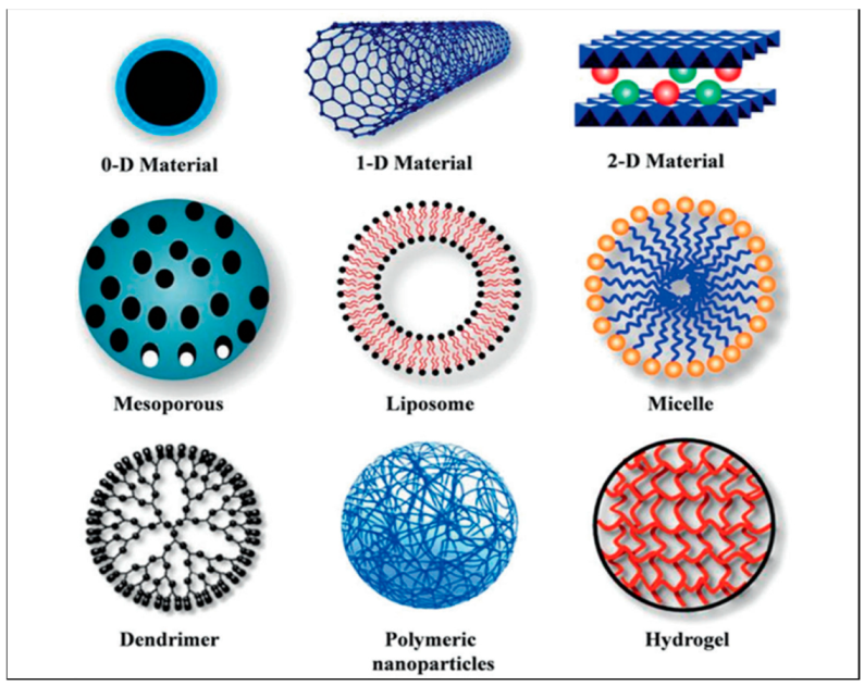
Sensor platforms play a pivotal role in chemical sensing because they interact with the target analyte to produce a detectable change in their physical and chemical properties. Such interactions occur via chemical bonding or intermolecular forces which cause the attachment of the analyte to the nanomaterial-based sensor [31,32]. This property of a chemical sensor is crucial to the signal output quality and reliability of the detection methods and as such, in this section, we explore the different nanomaterials used to produce sensing platforms for the detection of therapeutic drugs. Figure 1 below shows several types of polymeric nanomaterials used in drug delivery applications.
The nanoparticles in the image above have been used in various applications regarding therapeutic drugs and drug delivery [34,35]. In the following sections, we focus on polymer and inorganic nanoparticles as well as graphene-based sensors to cover a broader scope of an already expanding field of nanoparticles.
2.1. Polymeric Nanomaterials and Their Applications
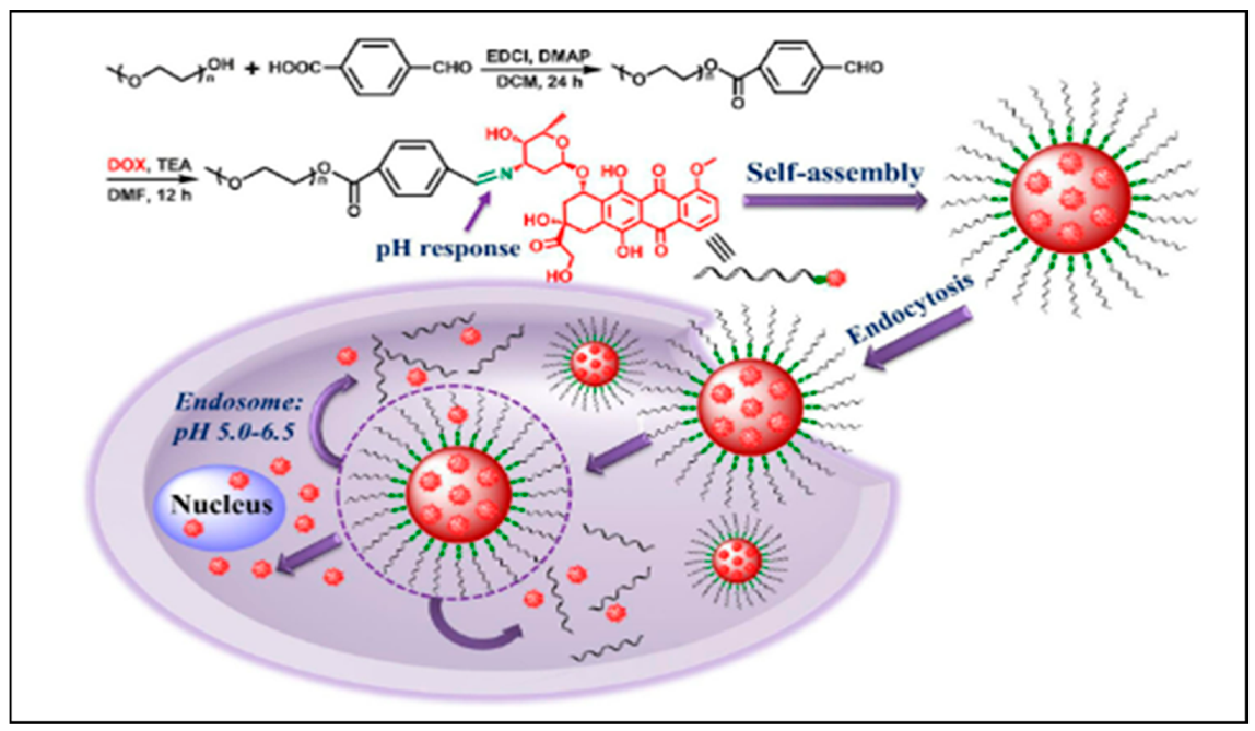
The key factors challenging the efficiency of therapeutic medicines arise from the biological barriers present in the human tissues which affect drug delivery and intracellular transport [36,37]. As a potential solution, nano-based polymers of various chemical functionalities are explored as controlled drug delivery agents for a variety of drug treatments. The interest comes from their flexibility in surface modification, biocompatibility, and loading capacity for both hydrophobic and hydrophilic drugs [38,39,40,41,42]. The current advanced methods for producing polymeric nanoparticles include sonication, emulsification, self-assembly, electro dropping, nanoprecipitation, microfluidic, and ionic gelation. The general design of polymeric nanoparticles for drug delivery applications is nanocapsules (polymeric membranes with an empty core) and nanospheres (matrix systems in solid form) [43]. Examples of nanocapsules include polymersomes, which are artificial vesicles that consist of a double membrane made from amphiphilic block copolymers. Polymersomes are known for their good stability and efficiency in drug retention during transit to the cytosol. Dendrimers are a popular example of nanocomposites that comprise hyperbranched polymers that form a three-dimensional matrix for cross-linking purposes. They have well-defined intramolecular spaces where drug encapsulation can take place [33]. The maximum amount of cargo that can be carried relies on the shape and size of the drug molecules and the number of cavities in a dendrimer. In application, the functionality or surface chemistry, as well as the size and shape, can be tailored for specific therapeutic drugs and biomolecules [44]. There are many ways that nanopolymers carry pharmaceutical drugs to the target sites. Depending on the polymer design, a drug can be encapsulated in the core of the polymer, adsorbed in the polymer matrix, or conjugated to the surface of the nanoparticle. Figure 2 below shows the synthesis and application of polymeric nanoparticles in drug delivery.
The figure shown above describes the chemical process of synthesizing PEG-Schiff-DOX conjugates for drug delivery. Anticancer drug DOX was encapsulated in a PEG-Schiff nanosphere which transports the drug into the target cell via endocytosis [45]. Using these mechanisms, a combination of modified polymers is often applied as drug carriers. For example, a pH/redox responsive stimuli sensor made from copolymers poly-ethylene-glycol (PEG) and poly-L-lysine (PLL) functionalized with platinum nanoparticles, was used to transport gemcitabine, a small molecule for the treatment of lung cancer [46]. Poly(propylene imine) (PPI) dendrimers were chemically modified with folate for targeting the anticancer drug docetaxel [47]. HIV medications efavirenz and lamivudine were also targeted using PPI dendrimers which resulted in improved drug uptake and efficacy, respectively [48,49]. In another study, an anticancer drug, oxaliplatin (IV), was cross-linked with polyethylenimine (PEI) for delivery of reactive oxygen species during chemotherapy [50]. Co-drug delivery studies have also been explored where two drugs are delivered simultaneously using polymer nanoparticles. For example, PLGA nanoparticles coated with polyvinyl alcohol (PVA) were used to transport antitumor medication paclitaxel/methotrexate complex [51]. Polymersomes of polylactic-co-glycolic acid (PLGA, inner shell) and PEG (outer shell) were loaded with anticancer therapeutics as a promising cancer drug delivery platform [52]. The survey conducted on polymeric nanoparticles shows that these nanosensors have been invaluable in pharmacology and oncology research. However, the literature has also cited a few disadvantages of polymeric nanoparticles such as toxicity and particle aggregation which weakens drug loading. As of late, only a few nanopolymer-based medicines have been FDA approved, thus this field of research is relatively new, and more experimental work and clinical trials are still required [53].
2.2. Metallic Nanomaterials and Their Applications
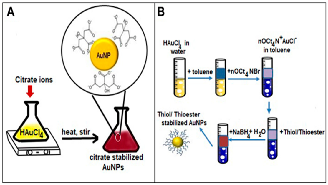
In recent decades, noble metals and some transition metals have gained interest in a variety of nano-based applications, including pharmaceutical research. The main attraction for metallic nanoparticles (MNPs) is the resonant plasmon feature that arises from the electron oscillations at the surface of metallic atoms [54]. Because of this, MNPs have played a key role in many optical detection methods as a signal enhancement platform [55]. Furthermore, the high electrical conductivity of MNPs makes them efficient electrochemical sensors in many biomedical applications. MNPs can also be tailored into various shapes and sizes as well as chemically modified with polymers and other recognition elements to suit the intended applications [56]. Nanoparticle synthesis is usually described in terms of two major groups: top down and bottom up. In the former method, bulk material is broken down into smaller fragments with sizes less than 100 nm using diverse sources of energy. Most techniques under this group are used in the fabrication of thin film sensors usually from silicon or quartz substrates [57,58]. Examples of top-down techniques include physical vapor deposition (PVD), chemical vapor deposition (CVD), and optical and electrical lithography [59,60]. For the latter case, bottom-up techniques assemble precursor reagents into nanostructures using chemical and physical methods. Examples of popular synthesis routes include self-assembly and chemical reduction [61]. Stabilizers such as sodium citrate, cellulose, thioester, dextran, gelatin, etc., are often used to prepare the MNPs and provide the starting chemical functionality for further modification [62,63]. Figure 3 below shows the basic process for the preparation of gold NPs using chemical reduction by Turkevich and Burst methods [64,65].
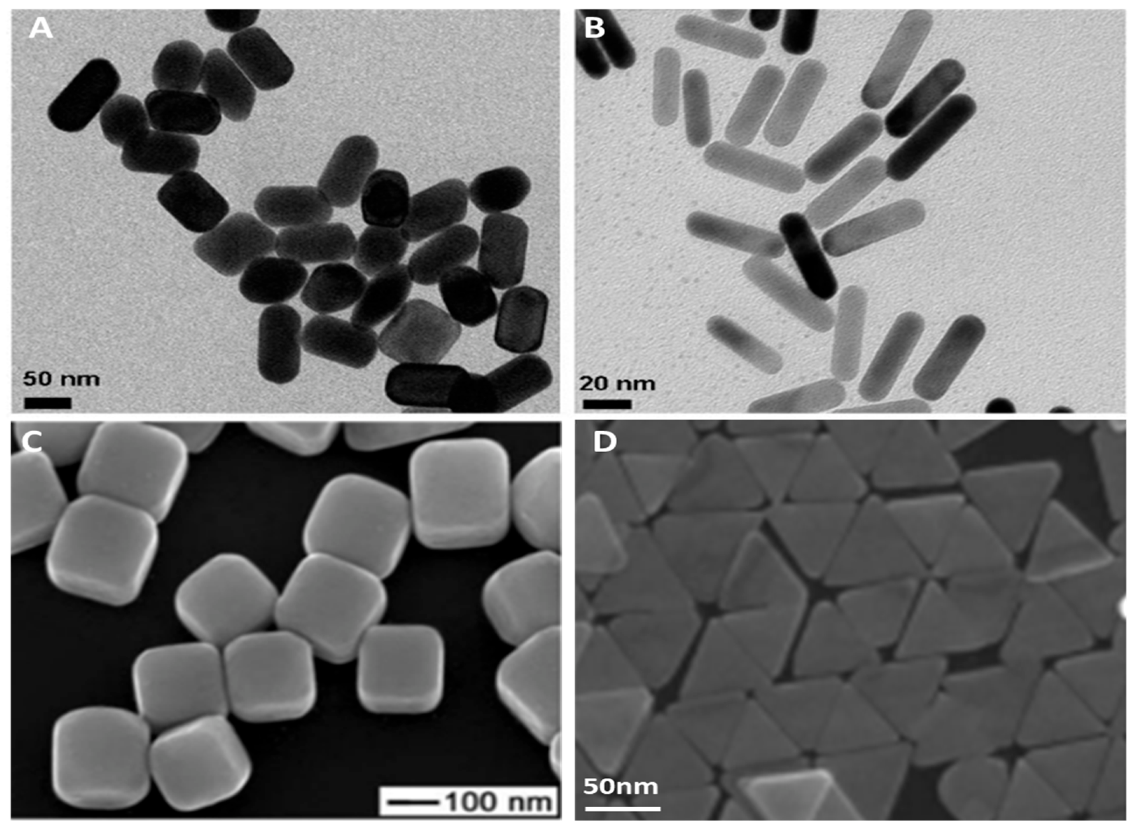
The above are two examples of how gold nanoparticles of various functionality are prepared using bottom-up methods. Popular shapes of MNPs produced using these methods include nanospheres, nanorods, nano-urchins, nano cubes, nanostars, and nanocages [66,67]. Imaging techniques such as atomic force microscopy (AFM), transmission electron microscopy (TEM), and scanning electron microscopy (SEM) have become the standard for characterizing MNPs for most synthesis methods [68]. Figure 4 below shows images of MNP acquired using different microscopy techniques.
Metals such as gold (Au), silver (Ag), titanium (Ti), platinum (Pt), and copper (Cu) have been extensively explored as sensing platforms for a wide range of therapeutic and biomedical experiments [71,72]. Amongst them, gold has received the most attention owing to its inertness, easy functionalization, and biocompatibility. For example, gold nanoparticles were modified with PEG to form a PEGAuNP composite for targeted drug delivery of pancreatic cancer medications doxorubicin and varlitinib [73]. AuNPs have also been explored as novel diagnostic tools for the management of melanoma cancer [74]. In other works, gold was shown to reduce toxicity, and improve immunogenetic activity and stability when used as a vaccine carrier in nanomedicine [75]. Glucose detection was demonstrated using AuNP in serum, showing detection limits in the nanomole region [76]. Metals such as silver have also played a role in the therapeutic drug research space as in the case where gallic acid-coated silver nanoparticles were used as drug carriers for doxorubicin [77]. Silver NPs are also capable of detecting uric acid, lidocaine hydrochloride, thiamine, lomefloxacin, and propafenone in urine samples with detection limits in the micromolar per liter range [78,79,80,81,82]. MNPs are showing increased value in pharmaceutical research and will grow our understanding of therapeutic agents as future work is published.
2.3. Graphene Based Nano-Sensors and Applications
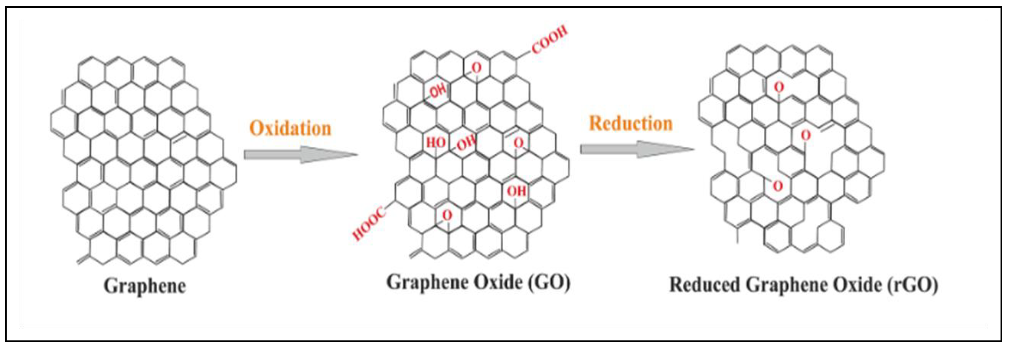
In the past decade, the fabrication of sensors and biosensors has improved with the incorporation of graphene as a scaffolding platform. The chemical and physical properties of graphene such as high surface-to-volume ratio, electrical conductivity, optical transmittance, thermal conductivity, and high mechanical strength give the nanomaterial a considerable edge over most materials in sensing applications [83]. Furthermore, due to the sp2 hybridization of carbon bonds, graphene is easily modified with various chemical and biochemical agents making it a material with versatile applicability. Graphene is usually used in its oxygenated form, known as graphene oxide (GO), as well as reduced graphene oxide (rGO) for research work that investigates hydrophilic molecules [84]. The Figure 5 below shows the molecular shapes of graphene, GO, and rGO.
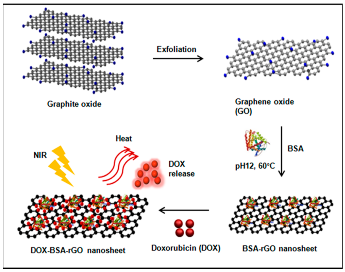
Graphene is synthesized using various methods such as the Hummers method, exfoliation or mechanical cleavage of graphite, and chemical vapor deposition [84]. To obtain GO, strong oxidizing agents such as sodium permanganate and sulphuric acid are reacted with graphite or graphene to produce hydroxyl (OH) and carboxylic acid (COOH) functional groups on the surface of the substrate. The oxygen content on GO is reduced using photochemical, microwave, and bacterial methods to produce reduced graphene oxide. GO and rGO offer extra properties suitable for chemical and biochemical sensor applications [84]. Adsorption of drug molecules, anticancer medication, polymers, proteins, genes, and other biomolecules is possible using GO and rGO because the OH and COOH groups allow easy conjugation bonding. Electrostatic interactions using the oxygen lone pair of electrons, pi–pi stacking via the aromatic rings, hydrogen bonding, and van der Waals forces are the various mechanisms by which GO and rGO adsorbs materials onto its surfaces [85]. Figure 6 below shows the adsorption of the protein BSA on rGO nanosheets.
The image above shows the synthesis of rGO from graphite oxide with the intention of delivering anti-cancer medication DOX supported by the protein BSA. The scaffolding material is then investigated using optical methods to release the DOX from the adsorption site [86]. Similar work is available in different research applications where a combination of graphene members, crosslinkers, and analyte recognition elements are combined for targeted drug delivery. For example, amino acid-functionalized iron oxide nanoparticles adsorbed on graphene sheets were used as a sensor for dopamine and ascorbic acid detection [87]. Secondly, GO functionalized with platinum nanoparticles was explored as a chemical sensor for glucose at concentrations in the millimolar range [88]. Chitosan-functionalized GO was used in the controlled release of the anti-inflammatory drug ibuprofen [89]. In the next section, more examples of graphene sensors are discussed, taking into consideration the detection methods and parameters used to classify the efficiency of a chemical sensor.
3. Detection Methods and Sensing Techniques
Adsorption of the analyte to the sensing material is a crucial element of a sensor, as explained in Section 2, and the chemical properties of the sensor and the analyte need to be compatible for the adsorption to occur. A second and equally important characteristic of a sensor is the detection method employed to produce a reliable and reproducible signal. There are currently various methods used for chemical sensing of pharmaceuticals, each of which has its own advantages and disadvantages. In this section, we explore electrical and optical detection methods, comparing their sensor performance using parameters such as limit of detection, linear range, and other additional information.
3.1. Electrical Detection Methods
The electrical potential of a chemical agent has been investigated as a means of quantifying changes in concentration or molecular bonding because of interaction with an analyte. Electrical signals such as resistance, capacitance, conductance, and impedance are the ones used to study the thermodynamic and kinetic properties of the analyte [11]. Furthermore, electron exchange between oppositely charged species in redox reactions produces a signal that can be used as a sensing mechanism. Many nanoparticle scaffolds, conjugates, and analyte recognition elements have been coupled with electrochemical detection using surface functionalized electrodes [90].
3.1.1. Field Effect Transistor (FET) Based Detection Method
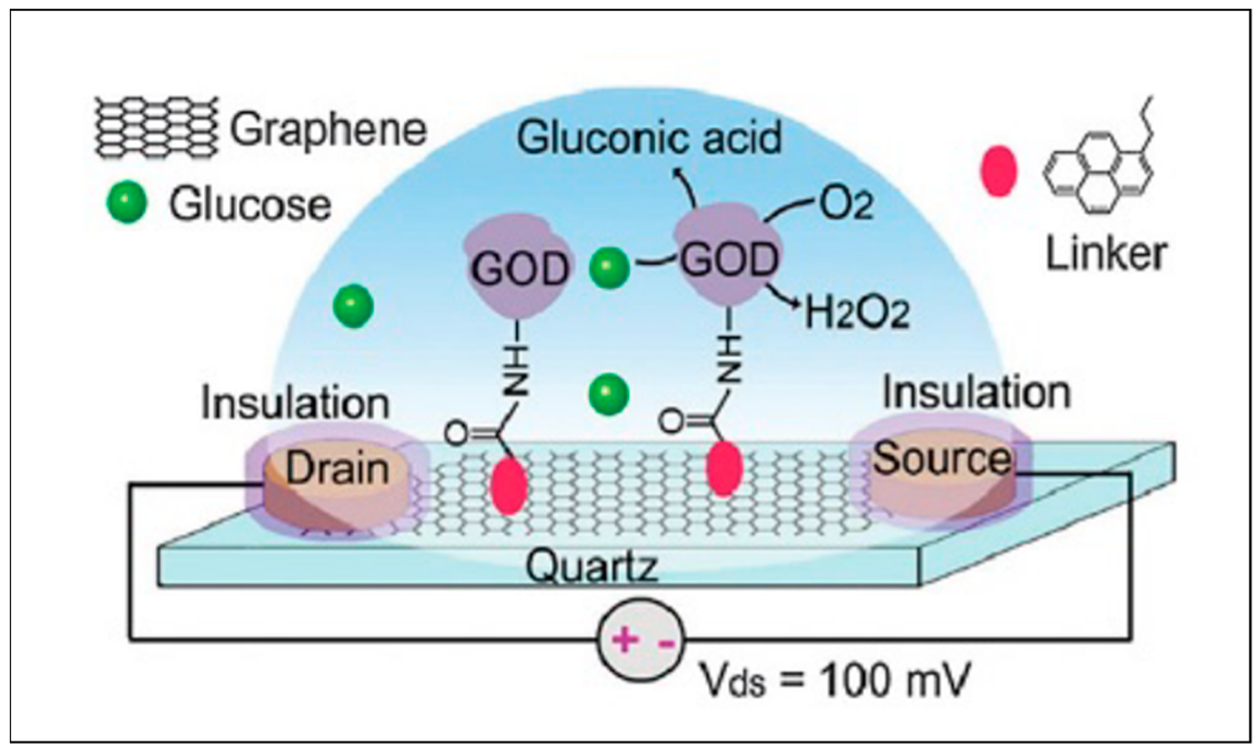
Field effect transistors are devices that use an electric field to control the flow of current in a semiconductor. In a typical FET system, source (S) and draining (D) electrodes are connected to a semiconductor path that is functionalized with sensing elements for high specificity and binding affinity. When target analytes are detected, a change in channel conductance is recorded and processed to acquire an electrical signal. There are two kinds of FETs based on the PN junction theory: n-type where electrons are the main charge carriers and p-type with holes as the primary charge carriers. In an n-type FET system, positively charged molecules are detected and charge carriers (electrons) accumulate on the sensing channels and increase the signal. However, when negatively charged targets are detected, the conductivity decreases caused by the depletion of the electrons. The second type is a p-type FET system, which binds to positive charges causing a decline in conductivity decline due to a reduction in the charge carriers (holes). The inverse occurs when capturing negative charges raises because conductivity by the holes increases. The application of this principle allows improved detection by coating nanomaterials such as carbon nanotubes, graphene, and MNPs on the electrodes and the sensing platforms [91]. In this way, biosensors can be designed for various pharmaceutical and therapeutic drug-related work. Figure 7 below shows a FET sensor used in the detection of glucose oxidase using a graphene biosensor.
In Figure 7, graphene is used as a high electrical conducting platform which changes the current on both the source and the drain electrodes when a substance adsorbs on its surface. In this case, glucose oxidase enzyme (GOD) is used together with a crosslinker molecule to detect changes in conductance when glucose binds to the enzyme [92]. Other applications of FET sensors for various pharmaceutical agents have been published. Zinc oxide nanoribbons were used as transducer materials for the detection of glucose in phosphate buffer solution (PBS) and a LOD of 70 µM and linear range of 0–80 mM were reported [93]. In another study, insulin was analyzed in PBS on graphene transducers with detection limits at 35 pM [94]. Although FET devices offer high sensitivity, selectivity, miniaturization, and low power use as advantages, dielectric membranes are sensitive to motion, which affects the accuracy of detection. Lastly, because FET uses biomarkers, it is not regarded as a label-free method, which affects the applicability of this technique due to cost implications [95].
3.1.2. Electrochemical Detection Methods
Electrochemical detection involves the use of functionalized electrodes for the detection of target analytes and signal transduction. Like FET devices, electrochemical methods translate binding affinities into readable electrical signals. The difference between the two methods is that electrochemical techniques require the use of an electrolyte solution as a conducting medium instead of solid-state transducers found on FET devices [96]. Figure 8 below shows a general schematic of an electrochemical system.
Figure 8 above shows how an electrochemical sensor is prepared and used in detection experiments. From position (a), a glass-based carbon (GCE) electrode was functionalized with gold nanoparticles followed by thioglycolic acid and lectins (b,c), while simultaneously blocking nonspecific adsorption to the electrode using BSA (d). Lectin–au-thionine bioconjugates were adsorbed on the electrode as recognition elements (e). Finally, the electrochemical apparatus is assembled and utilized to produce a signal of intensity (amperes) versus electric volts (f) [97]. Systems like these have been designed for a wide range of applications for pharmaceutical and therapeutic targets. For example, studies have shown the detection of dopamine using graphene functionalized with PVP as the sensing platform on the GCE. The study reported a LOD of 0.2 nM and a linear range up to 10−10 with an r2 of 0.99 [98]. Research on cholesterol was also undertaken using chitosan, graphene hybrid nanocomposites, reaching a LOD of 0.75 µM and a linear range of 2.2–520 µM [99]. Furthermore, extensive research into antineoplastic drugs using electrochemical sensing has been explored. Flutamide detection by silver coated GCE produced LOD (mol L−1) in the 10−6 and a linear range of 1–100 × 10−5 (mol L−1) [100]. In a similar study, gemcitabine was detected and analyzed using amino thiophenol-functionalized gold nanoparticles where LOD in the 10−15 and a linear range of 10−8 to 10−15 (mol L−1) [101]. The use of electrochemical sensing is clearly a prominent field of study, and its success can only bring more advantages for chemical sensing related to a variety of pharmaceutical drugs. Although the LOD and linear range of this method are extremely sensitive, the short lifespan is still a limitation and as such [102], other techniques have been approached to solve this issue.
3.2. Optical Detection Methods
Light/matter interaction principles have been widely explored for the investigation of organic and inorganic molecules. As such, a lot of research has been channeled towards using optical methods for detecting and collecting information on analytes adsorbed on nanomaterial scaffoldings or sensors. Nanomaterials carry the property of surface plasmons which aid in the detection method of the techniques [103]. Thus, in this section, we discuss popular plasmon-related methods used in pharmaceutical drug applications.
3.2.1. Surface Plasmon Resonance Spectroscopy
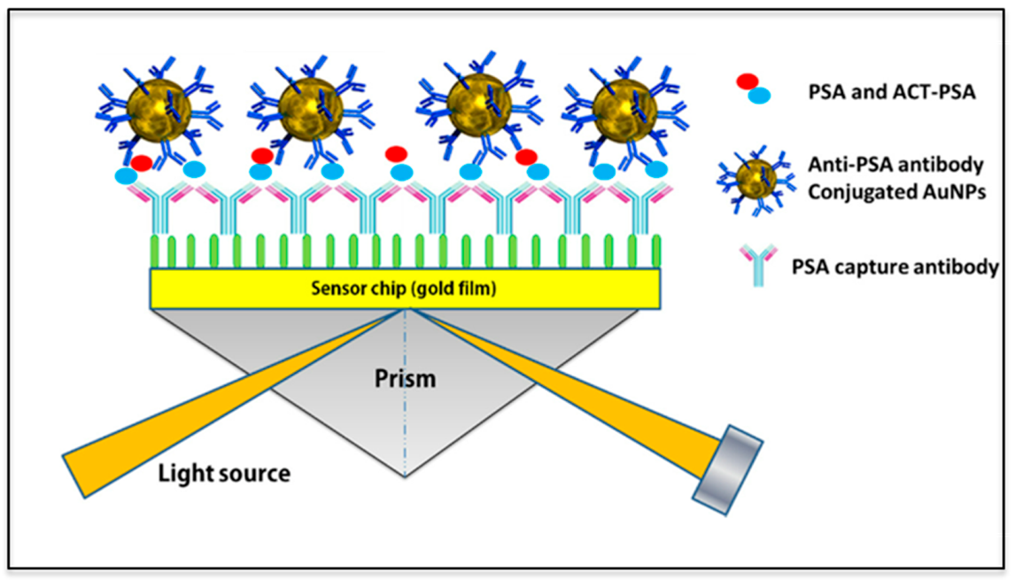
Surface plasmons found on the surface of a nanomaterial can be used for trace detection and monitoring of small molecules using changes in the refractive index as a signal in real time. In principle, when light photons approach a nanomaterial such as gold NPs at an angle, a refractive index can be obtained from the plasmon wave and set as a calibration point. When the MNP is functionalized with polymers, recognition elements, and analytes, changes in the refractive index are then recorded again and compared. Since the refractive index is related to the surface of the substrate, changes in the refractive index can also be correlated to binding affinities and used in medical diagnostics, drug detection, and virus monitoring among other applications [104]. Figure one below shows the principle of SPR and how it is used in biomedical applications.
Figure 9 shows the principle of SPR used in protein detection applications. From left to right, a glass slide is seen coated with layers of gold (Au) and a variety of antibodies. In the second step, anti-PSA is used as biorecognition elements for the detection of PSA. At each step, laser light is directed to the biosensors and changes in the refractive index in relation to the binding of molecules [105]. Examples of SPR applications can be seen in a wide range of biomedical and pharmaceutical experiments. Studies have shown the diagnosis of malignant and infectious diseases using the biomarker rhodamine 6G and SPR detection [106]. In industrial applications, aflatoxin, a toxic small molecule found in dairy products, was detected at LOD values of 76 pM using a silicon photonic biosensor [107]. Smartphone platforms are also being incorporated to SPR detection where 50 nm gold nanofilms are used for sensing immunoglobin G (IgG) producing LOD values from 15–47 nM [108]. Silver nanospheres and nanorods on titanium oxide substrates were used for the detection of streptavidin obtaining a LOD value of 0.3 µg/mL using halogen lamp technology [109]. Bromocriptine, a pharmaceutical drug used to treat menstrual problems, was investigated using laccase immobilized on a carboxymethyl dextran functionalized SPR sensor, where a detection limit of 10−2 ng/mL to 103 ng/mL was reported [110]. The values reported above show that the common feature of the SPR technique is high sensitivity and rapid analysis. However, SPR has limitations such as long lag times, sensitivity to temperature and motion as well as continuous optical alignment and maintenance [111].
3.2.2. Surface Enhanced Raman Spectroscopy (SERS)
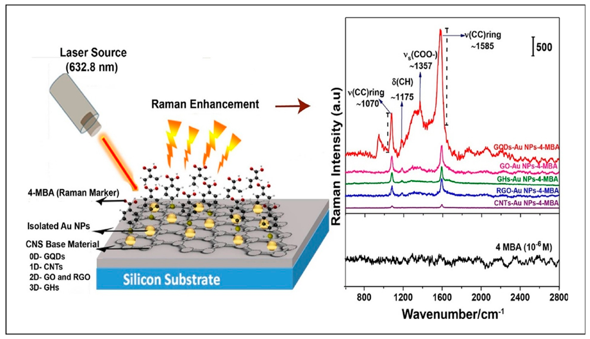
Amongst non-destructive methods of detection, Raman spectroscopy has become a favorite method for qualitative experiments because it allows molecular fingerprinting of organic and organometallic substances. Light–matter interaction produces scattered radiation which is collected at right angles to the surface. The scattered radiation produces different frequencies from the laser wavelength due to inelastic scattering caused by photon–molecule interactions. Raman shifts expressed in wavenumbers can be correlated to bond fragments of analytes as means of detection. A major limitation of this method is low signal output which is normally 0.01% of the radiation. To solve this issue, nanomaterials were incorporated into the technique by employing surface plasmonic resonance for signal enhancement purposes. Such as, surface-enhanced Raman spectroscopy (SERS) has brought renewed interest and trust in SERS as a pharmaceutical drug detection method [112]. Figure 10 below illustrates both the fabrication of SERS scaffolds and their application in qualitative analysis.
In the above image, a 632.8 nm laser source is exciting a set of carbon-based scaffolds of various dimensions and functionality. Gold quantum dots (GQDs, 0D), carbon nanotubes (CNTs, 1D), graphene oxide (GO, 2D) and reduced graphene oxide (rGO, 2D), and gold nanohybrids (GHs, 3D) were further functionalized with 4-mercaptobenzoic acid (4-MBA, 10−6 M) as the analyte. From observation of the Raman spectra, it is seen that vibrational modes are associated with 4-MBA. The spectra also show increased signal intensity amongst the scaffolds, with GQDs providing the highest intensity. Specifically, the study reports surface enhancement factors of 107 from GQDs followed by 106 from GO-Au NPs, which is a significant improvement on the Raman signals obtained, making SERS a reliable technique for small molecule detection. More examples like the one above have been reported in the literature. Levofloxacin (antibiotic) was detected from artificial urine using hydroxylamine silver nanoparticle microfluidic devices and SERS where the quantitative analysis yielded a LOD of 0.07 mM [114]. A similar pharmaceutical study quantified promethazine using the same microfluidic device and detected concentrations as lows 10−7 M [115]. Medication for the treatment of hypertension known as captopril was obtained from human blood and analyzed using SERS and citrate functionalized silver nanoparticles. It was reported that a LOD of 0,4 µM was achieved from the quantitative calculations [116]. Lastly, the common over-the-counter medication aspirin, was analyzed on silver nanoparticles supported by filter paper, the linear range reported in this study spanned from 0.1 to 1 mM [68]. The examples shown indicate that SERS is a highly versatile, sensitive, and non-destructive detection method; however, issues such as fluorescence noise, long acquisition time, and unstable lasers still hamper the full potential of this method [117]. With further research and optimization, SERS systems will surely improve knowledge on pharmaceutical drug design and quality control, which is essential for healthcare reasons.
4. Prospects and Shortcomings
The pharmaceutical industry is one of the pillars of our health systems around the world. Research and knowledge dissemination in this field is important for our global health status. This review provided a brief overview of technologies used in the detection and analysis of therapeutic drugs that are used to treat serious diseases such as cancer, hypertension, viruses, and tumors. Nanotechnology has made great strides in providing sensor applications for pharmaceutical drug design and monitoring. Properties of polymeric, metallic, and carbon-based substrates can be fabricated into sensor platforms that allow the adsorption of analytes for analysis purposes. Furthermore, electrical and optical detection methods coupled with nanomaterials have enhanced sensor application research which is a positive for the pharmaceutical industry. Parameters such as limit of detection, linear range, and sensitivity show improved responses towards analytes being investigated. With future research and design, challenges related to toxicity, shelf life, and API quality can be overcome by improved drug design, monitoring, and targeted sensing. The major limitations observed from this study start with the challenges in reproducing sensing platforms consistently using less-expensive methods. Secondly, electrical-based detection methods are sensitive to motion, and they struggle to detect multiply layered sensors, which makes calibration and label-free applications a tedious process. Thirdly, optical methods suffer from long acquisition times, laser instability, continuous alignment maintenance, and fluorescence noise, which requires optimization steps prior to acquisition. Technical challenges aside, nanomaterial-based research into pharmaceutical drugs is a very important field of science and its understanding can only be improved by more research, innovation, and application.
Author Contributions
Conceptualization, L.T. and S.O.-L.; methodology, L.T. and S.O.-L.; investigation, L.T. and S.O.-L.; resources, P.M.-K.; data curation, L.T.; writing—original draft preparation, L.T. and S.O.-L.; writing—review and editing, P.M.-K. and S.O.-L.; visualization, L.T.; supervision, P.M.-K.; project administration, P.M.-K.; funding acquisition, P.M.-K. All authors have read and agreed to the published version of the manuscript.
Funding
The authors acknowledge the financial support received from Council for Scientific and Industrial Research (CSIR), National Research Fund (NRF) and Department of Science and Innovation (DSI).
Data Availability Statement
Data are available via personal communication with proper reasons.
Conflicts of Interest
The authors declare there are no competing interests.
References
- Jeevanandam, J.; Barhoum, A.; Chan, Y.S.; Dufresne, A.; Danquah, M.K. Review on nanoparticles and nanostructured materials: History, sources, toxicity and regulations. Beilstein J. Nanotechnol. 2018, 9, 1050–1074. [Google Scholar] [CrossRef] [PubMed] [Green Version]
- Bai, Y.; Wu, F.; White, J.C.; Xing, B. 100 Nanometers: A Potentially Inappropriate Threshold for Environmental and Ecological Effects of Nanoparticles. Environ. Sci. Technol. 2014, 48, 3098–3099. [Google Scholar] [CrossRef] [PubMed]
- Murthy, S.K. Nanoparticles in modern medicine: State of the art and future challenges. Int. J. Nanomed. 2007, 2, 129–141. [Google Scholar]
- Shen, H.; Zhang, L.; Liu, M.; Zhang, Z. Biomedical Applications of Graphene. Theranostics 2012, 2, 283–294. [Google Scholar] [CrossRef] [Green Version]
- Xu, C.; Shi, X.; Ji, A.; Shi, L.; Zhou, C.; Cui, Y. Fabrication and Characteristics of Reduced Graphene Oxide Produced with Different Green Reductants. PLoS ONE 2015, 10, e0144842. [Google Scholar]
- Yadav, S.; Sharma, A.K.; Kumar, P. Nanoscale Self-Assembly for Therapeutic Delivery. Front. Bioeng. Biotechnol. 2020, 8, 127. [Google Scholar] [CrossRef]
- Bissantz, C.; Kuhn, B.; Stahl, M. A Medicinal Chemist’s Guide to Molecular Interactions. J. Med. Chem. 2010, 53, 5061–5084. [Google Scholar] [CrossRef]
- Shovsky, A.; Varga, I.; Makuška, R.; Claesson, P.M. Formation and Stability of Water-Soluble, Molecular Polyelectrolyte Complexes: Effects of Charge Density, Mixing Ratio, and Polyelectrolyte Concentration. Langmuir 2009, 25, 6113–6121. [Google Scholar] [CrossRef]
- Lombardo, D.; Kiselev, M.A.; Magazù, S.; Calandra, P. Amphiphiles Self-Assembly: Basic Concepts and Future Perspectives of Supramolecular Approaches. Adv. Condens. Matter Phys. 2015, 2015, 151683. [Google Scholar] [CrossRef] [Green Version]
- Huang, R.; Carney, R.P.; Stellacci, F.; Lau, B.L.T. Protein-nanoparticle interactions: The effects of surface compositional and structural heterogeneity are scale dependent. Nanoscale 2013, 5, 6928–6935. [Google Scholar] [CrossRef] [Green Version]
- Naresh, V.; Lee, N. A Review on Biosensors and Recent Development of Nanostructured Materials-Enabled Biosensors. Sensors 2021, 21, 1109. [Google Scholar] [CrossRef]
- Saha, K.; Agasti, S.S.; Kim, C.; Li, X.; Rotello, V.M. Gold Nanoparticles in Chemical and Biological Sensing. Chem. Rev. 2012, 112, 2739–2779. [Google Scholar] [CrossRef] [Green Version]
- Amina, S.J.; Guo, B. A Review on the Synthesis and Functionalization of Gold Nanoparticles as a Drug Delivery Vehicle. Int. J. Nanomed. 2020, 15, 9823–9857. [Google Scholar] [CrossRef]
- Bhalla, N.; Jolly, P.; Formisano, N.; Estrela, P. Introduction to biosensors. Essays Biochem. 2016, 60, 1–8. [Google Scholar] [CrossRef] [Green Version]
- Radhakrishnan, R.; Suni, I.I.; Bever, C.S.; Hammock, B.D. Impedance Biosensors: Applications to Sustainability and Remaining Technical Challenges. ACS Sustain. Chem. Eng. 2014, 2, 1649–1655. [Google Scholar] [CrossRef]
- Bharti, K.; Sadhu, K.K. Syntheses of metal oxide-gold nanocomposites for biological applications. Results Chem. 2022, 4, 100288. [Google Scholar] [CrossRef]
- Mohammad-Rezaei, R.; Golmohammadpour, M. Controlled Electrodeposition of Au-Copper Oxide Nanocomposite on a Renewable Carbon Ceramic Electrode for Sensitive Determination of NADH in Serum Samples. Electroanalysis 2020, 32, 606–612. [Google Scholar] [CrossRef]
- Gooding, J.J.; Chou, A.; Liu, J.; Losic, D.; Shapter, J.G.; Hibbert, D.B. The effects of the lengths and orientations of single-walled carbon nanotubes on the electrochemistry of nanotube-modified electrodes. Electrochem. Commun. 2007, 9, 1677–1683. [Google Scholar] [CrossRef]
- Heller, I.; Kong, J.; Heering, H.A.; Williams, K.A.; Lemay, A.S.G.; Dekker, C. Individual Single-Walled Carbon Nanotubes as Nanoelectrodes for Electrochemistry. Nano Lett. 2005, 5, 137–142. [Google Scholar] [CrossRef]
- Gao, X.P.A.; Zheng, G.; Lieber, C.M. Subthreshold Regime has the Optimal Sensitivity for Nanowire FET Biosensors. Nano Lett. 2010, 10, 547–552. [Google Scholar] [CrossRef] [Green Version]
- Tu, Q.; Chang, C. Diagnostic applications of Raman spectroscopy. Nanomed. Nanotechnol. Biol. Med. 2012, 8, 545–558. [Google Scholar] [CrossRef]
- Kazuma, E.; Tatsuma, T. Localized surface plasmon resonance sensors based on wavelength-tunable spectral dips †. Nanoscale 2014, 6, 2397–2405. [Google Scholar] [CrossRef] [Green Version]
- Pérez-Ràfols, C.P.; Serrano, N.; Díaz-Cruz, J.M.; Ariño, C.; Esteban, M. A Chemically-Bound Glutathione Sensor Bioinspired by the Defense of Organisms against Heavy Metal Contamination: Optimization of the Immobilization Conditions. Chemosensors 2017, 5, 12. [Google Scholar] [CrossRef] [Green Version]
- Li, M.; Cushing, S.K.; Wu, N.Q. Plasmon-enhanced optical sensors: A review. Analyst 2015, 140, 386–406. [Google Scholar] [CrossRef] [Green Version]
- Lakowicz, J.R.; Ray, K.; Chowdhury, M.; Szmacinski, H.; Fu, Y.; Zhang, J.; Nowaczyk, K. Plasmon-controlled fluorescence: A new paradigm in fluorescence spectroscopy. Analyst 2008, 133, 1308–1346. [Google Scholar] [CrossRef] [Green Version]
- Meyer, S.A.; Le Ru, E.C.; Etchegoin, P.G.; Champion, P.M.; Ziegler, L.D. Quantifying Resonant Raman Cross Sections with SERS. AIP Conf. Proc. 2010, 1267, 204–205. [Google Scholar]
- Cao, Y.C.; Jin, R.; Nam, J.-M.; Thaxton, C.S.; Mirkin, C.A. Raman Dye-Labeled Nanoparticle Probes for Proteins. J. Am. Chem. Soc. 2003, 125, 14676–14677. [Google Scholar] [CrossRef]
- Bertoldo Menezes, D.; Reyer, A.; Marietta, A.; Musso, M. Glass transition of polystyrene (PS) studied by Raman spectroscopic investigation of its phenyl functional groups. Mater. Res. Express 2017, 4, 015303. [Google Scholar] [CrossRef]
- Littleford, R.E.; Cunningham, D.; Matousek, P.; Towrie, M.; Parker, A.W.; Khan, I.; McComb, D.; Smith, W.E. Surface-enhanced resonance Raman scattering using pulsed and continuous-wave laser excitation. J. Raman Spectrosc. 2005, 36, 600–605. [Google Scholar] [CrossRef]
- Bumbrah, G.S.; Sharma, R.M. Raman spectroscopy–Basic principle, instrumentation and selected applications for the characterization of drugs of abuse. Egypt J. Forensic. Sci. 2016, 6, 209–215. [Google Scholar] [CrossRef] [Green Version]
- Liu, J.; Cui, L.; Losic, D. Graphene and graphene oxide as new nanocarriers for drug delivery applications. Acta Biomater. 2013, 9, 9243–9257. [Google Scholar] [CrossRef] [PubMed]
- Liu, Z.; Robinson, J.T.; Sun, X.M.; Dai, H.J. PEGylated Nanographene Oxide for Delivery of Water-Insoluble Cancer Drugs. J. Am. Chem. Soc. 2008, 130, 10876–10877. [Google Scholar] [CrossRef] [PubMed] [Green Version]
- Singh, A.P.; Biswas, A.; Shukla, A.; Maiti, P. Targeted therapy in chronic diseases using nanomaterial-based drug delivery vehicles. Signal Transduct. Target. Ther. 2019, 4, 33. [Google Scholar] [CrossRef] [PubMed] [Green Version]
- Truong, N.P.; Whittaker, M.R.; Mak, C.W.; Davis, T.P. The importance of nanoparticle shape in cancer drug delivery. Expert Opin. Drug Deliv. 2015, 12, 129–142. [Google Scholar] [CrossRef]
- Car, A.; Baumann, P.; Duskey, J.T.; Chami, M.; Bruns, N.; Meier, W. PH-responsive PDMS-b-PDMAEMA micelles for intracellular anticancer drug delivery. Biomacromolecules 2014, 15, 3235–4325. [Google Scholar] [CrossRef]
- Mitchell, M.J.; Billingsley, M.M.; Haley, R.M.; Wechsler, M.E.; Peppas, N.A.; Langer, R. Engineering precision nanoparticles for drug delivery. Nat. Rev. Drug Discov. 2020, 20, 101–124. [Google Scholar] [CrossRef]
- Taylor, J.; Huefner, A.; Li, L.; Wingfield, J.; Mahajan, S. Nanoparticles and intracellular applications of surface-enhanced Raman spectroscopy. Analyst 2016, 141, 5037–5055. [Google Scholar] [CrossRef] [Green Version]
- Zhang, L.; Beatty, A.; Lu, L.; Abdalrahman, A.; Makris, T.M.; Wang, G.; Wang, Q. Microfluidic-assisted polymer-protein assembly to fabricate homogeneous functionalnanoparticles. Mater. Sci. Eng. C 2020, 111, 110768. [Google Scholar] [CrossRef]
- Alyaudtin, R.N.; Reichel, A.; Löbenberg, R.; Ramge, P.; Kreuter, J.; Begley, D.J. Interaction ofpoly(butylcyanoacrylate) nano-particles with the blood-brain barrier in vivo and in vitro. J. Drug Target. 2001, 9, 209–221. [Google Scholar] [CrossRef]
- Yavuz, B.; Bozdag Pehlivan, S.; Sumer Bolu, B.; Nomak Sanyal, R.; Vural, I.; Unlu, N. Dexamethasone-PAMAM dendri-mer conjugates for retinal delivery: Preparation, characterization and in vivo evaluation. J. Pharm. Pharmacol. 2016, 68, 1010–1020. [Google Scholar] [CrossRef]
- Lima, T.; Bernfur, K.; Vilanova, M.; Cedervall, T. Understanding the Lipid and Protein Corona Formation on Different Sized Polymeric Nanoparticles. Sci. Rep. 2020, 10, 1129. [Google Scholar] [CrossRef]
- Osorno, L.L.; Brandley, A.N.; Maldonado, D.E.; Yiantsos, A.; Mosley, R.J.; Byrne, M.E. Review of Contemporary Self-Assembled Systems for the Controlled Delivery of Therapeutics in Medicine. Nanomaterials 2021, 11, 278. [Google Scholar] [CrossRef]
- Scarpa, E.; Bailey, J.L.; Janeczek, A.A.; Stumpf, P.S.; Johnston, A.H.; Oreffo, R.O.C.; Woo, Y.L.; Cheong, Y.C.; Evans, N.D.; Newman, T.A. Quantification of intracellular payload release from polymersome nanoparticles. Sci. Rep. 2016, 6, 29460. [Google Scholar] [CrossRef] [Green Version]
- Braun, A.C.; Gutmann, M.; Mueller, T.D.; Lühmann, T.; Meinel, L. Bioresponsive release of insulin-like growth factor-I from its PEGylated conjugate. J. Control. Release 2018, 279, 17–28. [Google Scholar] [CrossRef]
- Song, J.; Xu, B.; Yao, H.; Lu, X.; Tan, Y.; Wang, B.; Wang, X.; Yang, Z. Schiff-Linked PEGylated Doxorubicin Prodrug Forming pH-Responsive Nanoparticles with High Drug Loading and Effective Anticancer Therapy. Front. Oncol. 2021, 11, 656717. [Google Scholar] [CrossRef]
- Shi, H.; Xu, M.; Zhu, J.; Li, Y.; He, Z.; Zhang, Y.; Xu, Q.; Niu, Y.; Liu, Y. Programmed co-delivery of platinum nanodrugs and gemcitabine by a clustered nanocarrier for precision chemotherapy for NSCLC tumors. J. Mater. Chem. B 2020, 8, 332–342. [Google Scholar] [CrossRef]
- Thakur, S.; Tekade, R.K.; Kesharwani, P.; Jain, N.K. The effect of polyethylene glycol spacer chain length on the tumor-targeting potential of folate-modified PPI dendrimers. J. Nanopart. Res. 2013, 15, 1625. [Google Scholar] [CrossRef]
- Dutta, T.; Agashe, H.B.; Garg, M.; Balakrishnan, P.; Kabra, M.; Jain, N.K. Poly (propyleneimine) dendrimer based nanocontainers for targeting of efavirenz to human monocytes/macrophages in vitro. J. Drug Target. 2007, 15, 89–98. [Google Scholar] [CrossRef]
- Dutta, T.; Jain, N.K. Targeting potential and anti-HIV activity of lamivudine loaded mannosylated poly (propyleneimine) dendrimer. Biochim. Biophys. Acta 2007, 1770, 681–686. [Google Scholar] [CrossRef]
- Lim, W.Q.; Zeng, S.; Phua, F.; Zhao, Y. Redox-Responsive Polymeric Nanocomplex for Delivery of Cytotoxic Protein and Chemotherapeutics. ACS Appl. Mater. Interfaces 2019, 11, 31638–31648. [Google Scholar] [CrossRef]
- Lee, K.S.; Chung, H.C.; Im, S.A.; Park, Y.H.; Kim, C.S.; Kim, S.-B.; Rha, S.Y.; Lee, M.Y.; Ro, J. Multicenter phase II trial of Genexol-PM, a Cremophor-free, polymeric micelle formulation of paclitaxel, in patients with metastatic breast cancer. Breast Cancer Res. Treat. 2008, 108, 241–250. [Google Scholar] [CrossRef]
- Koopaei, M.N.; Khoshayand, M.R.; Mostafavi, S.H.; Amini, M.; Khorramizadeh, M.R.; Tehrani, M.J.; Atyabi, F.; Dinarvand, R. Docetaxel loaded PEG-PLGA nanoparticles: Optimized drug loading, in vitro cytotoxicity and in vivo antitumor effect. Iran. J. Pharm. Res. 2014, 13, 819–833. [Google Scholar]
- Taghipour-Sabzevar, V.; Sharifi, T.; Moghaddam, M.M. Polymeric nanoparticles as carrier for targeted and controlled delivery of anticancer agents. Ther. Deliv. 2019, 10, 527–550. [Google Scholar] [CrossRef]
- Kahraman, M.; Mullen, E.R.; Korkmaz, A.; Wachsmann-Hogiu, S. Fundamentals and applications of SERS-based bioanalytical sensing. Nanophotonics 2017, 6, 831–852. [Google Scholar] [CrossRef] [Green Version]
- Lee, C.; Robertson, C.S.; Nguyen, A.H.; Kahraman, M.; Wachsmann-Hogiu, S. Thickness of a metallic film, in addition to its roughness, plays a significant role in SERS activity. Sci. Rep. 2015, 5, 11644. [Google Scholar] [CrossRef] [Green Version]
- Justino, C.I.L.; Freitas, A.C.; Pereira, R.; Duarte, A.C.; Rocha-Santos, T.A.P. Recent developments in recognition elements for chemical sensors and biosensors. TrAC Trends Anal. Chem. 2015, 68, 2–17. [Google Scholar] [CrossRef]
- Ahmed, S.R.; Kim, J.; Tran, V.T.; Suzuki, T.; Neethirajan, S.; Lee, J.; Park, E.Y. In situ self-assembly of gold nanoparticles on hydrophilic and hydrophobic substrates for influenza virus-sensing platform. Sci. Rep. 2017, 7, 44495. [Google Scholar] [CrossRef] [Green Version]
- Bao, L.-L.; Mahurin, S.M.; Liang, C.-D.; Dai, S. Study of silver films over silica beads as a surface-enhanced Raman scattering (SERS) substrate for detection of benzoic acid. J. Raman Spectrosc. 2003, 34, 394–398. [Google Scholar] [CrossRef]
- Baptista, A.; Silva, F.J.G.; Porteiro, J.; Míguez, J.L.; Pinto, G. Sputtering Physical Vapour Deposition (PVD) Coatings: A Critical Review on Process Improvement and Market Trend Demands. Coatings 2018, 8, 402. [Google Scholar] [CrossRef] [Green Version]
- Mattevi, C.; Kim, H.; Chhowalla, M. AReviewof Chemical Vapour Deposition of Graphene on Copper. J. Mater. Chem. 2011, 21, 3324–3334. [Google Scholar] [CrossRef]
- Keeney, M.; Jiang, X.Y.; Yamane, M.; Lee, M.; Goodman, S.; Yang, F. Nanocoating for biomolecule delivery using layer-by-layer self-assembly. J. Mater. Chem. B 2015, 3, 8757–8770. [Google Scholar] [CrossRef] [Green Version]
- Gao, M.; Song, Y.; Liu, Y.; Jiang, W.; Peng, J.; Shi, L.; Jia, R.; Muhammad, Y.; Huang, L. Controlled fabrication of Au@MnO2 core/shell assembled nanosheets by localized surface plasmon resonance. Appl. Surf. Sci. 2021, 537, 147912. [Google Scholar] [CrossRef]
- Park, S.; Han, U.; Choi, D.; Hong, J. Layer-by-layer assembled polymeric thin films as prospective drug delivery carriers: Design and applications. Biomater. Res. 2018, 22, 29. [Google Scholar] [CrossRef]
- Mocan, T.; Matea, C.; Tabaran, F.; Iancu, C.; Orasan, R.; Mocan, L. In Vitro Administration of Gold Nanoparticles Functionalized with MUC-1 Protein Fragment Generates Anticancer Vaccine Response via Macrophage Activation and Polarization Mechanism. J. Cancer 2015, 6, 583–592. [Google Scholar] [CrossRef]
- Brust, M.; Walker, M.; Bethell, D.; Schiffrin, D.J.; Whyman, R. Synthesis of thiol-derivatised gold nanoparticles in a two-phase Liquid–Liquid system. J. Chem. Soc. Chem. Commun. 1994, 801–802. [Google Scholar] [CrossRef]
- Prajapati, S.; Padhan, B.; Amulyasai, B.; Sarkar, A. Nanotechnology-based sensors. In Biopolymer-Based Formulations Biomedical and Food Applications. In Biopolymer-Based Formulations Biomedical and Food Applications; Catalent: Somerset, NJ, USA, 2020; pp. 237–262. [Google Scholar]
- Vo, Q.K.; Nguyen, A.T.; Ho, H.T.; Huynh, L.T.N.; Nguyen, T.P.P.; Nguyen, T.H.-T. Environmentally Friendly Controlled Synthesis of Gold Nanostars with Collagen by One-Step Reduction Method. J. Nanomater. 2022, 2022, 4046389. [Google Scholar] [CrossRef]
- Sallum, L.F.; Soares, F.L.F.; Ardila, J.A.; Carneiro, R.L. Determination of acetylsalicylic acid in commercial tablets by SERS using silver nanoparticle-coated filter paper. Spectrochim. Acta Part A Mol. Biomol. Spectrosc. 2014, 133, 107–111. [Google Scholar] [CrossRef]
- Zhang, Z.; Shen, W.; Xue, J.; Liu, Y.; Liu, Y.; Yan, P.; Liu, J.; Tang, J. Recent advances in synthetic methods and applications of silver nanostructures. Nanoscale Res. Lett. 2018, 13, 54. [Google Scholar] [CrossRef] [Green Version]
- Huang, Y.; Ferhan, A.R.; Gao, Y.; Dandapat, A.; Kim, D.-H. High-yield synthesis of triangular gold nanoplates with improved shape uniformity, tunable edge length and thickness. Nanoscale 2014, 6, 6496–6500. [Google Scholar] [CrossRef]
- Xiao, T.; Huang, J.; Wang, D.; Meng, T.; Yang, X. Au and Au-Based nanomaterials: Synthesis and recent progress in electrochemical sensor applications. Talanta 2020, 206, 120210. [Google Scholar] [CrossRef]
- Huynh, K.-H.; Pham, X.-H.; Kim, J.; Lee, S.H.; Chang, H.; Rho, W.-Y.; Jun, B.-H. Synthesis, Properties, and Biological Applications of Metallic Alloy Nanoparticles. Int. J. Mol. Sci. 2020, 21, 5174. [Google Scholar] [CrossRef] [PubMed]
- Coelho, S.C.; Reis, D.P.; Pereira, M.C.; Coelho, M.A.N. Doxorubicin and Varlitinib Delivery by Functionalized Gold Nanoparticles against Human Pancreatic Adenocarcinoma. Pharmaceutics 2019, 11, 551. [Google Scholar] [CrossRef] [PubMed] [Green Version]
- Bagheri, S.; Yasemi, M.; Safaie-Qamsari, E.; Rashidiani, J.; Abkar, M.; Hassani, M.; Mirhosseini, S.A.; Kooshki, H. Using gold nanoparticles in diagnosis and treatment of melanoma cancer. Artif. Cells Nanomed. Biotechnol. 2018, 46, 462–471. [Google Scholar] [CrossRef] [PubMed] [Green Version]
- Dykman, L.A. Gold nanoparticles for preparation of antibodies and vaccines against infectious diseases. Expert Rev. Vaccines 2020, 19, 465–477. [Google Scholar] [CrossRef] [PubMed] [Green Version]
- Sehit, E.; Drzazgowska, J.; Buchenau, D.; Yesildag, C.; Lensen, M.; Altintas, Z. Ultrasensitive nonenzymatic electrochemical glucose sensor based on gold nanoparticles and molecularly imprinted polymers. Biosens. Bioelectron. 2020, 165, 112432. [Google Scholar] [CrossRef] [PubMed]
- Nemčeková, K.; Svitková, V.; Sochr, J.; Gemeiner, P.; Labuda, J. Gallic acid-coated silver nanoparticles as perspective drug nanocarriers: Bioanalytical study. Anal. Bioanal. Chem. 2022, 414, 5493–5505. [Google Scholar] [CrossRef] [PubMed]
- Amjadi, M.; Rahimpour, E. Silver nanoparticles plasmon resonance-based method for the determination of uric acid in human plasma and urine samples. Microchim. Acta 2012, 178, 373–379. [Google Scholar] [CrossRef]
- Dou, Y.; Yang, X.; Liu, Z.; Zhu, S. Homocysteine-functionalized silver nanoparticles for selective sensing of Cu2+ ions and Lidocaine hydrochloride. Colloids Surfaces A Physicochem. Eng. Asp. 2013, 423, 20–26. [Google Scholar] [CrossRef]
- Rajamanikandan, R.; Ilanchelian, M. Simple and visual approach for highly selective biosensing of vitamin B1 based on glutathione coated silver nanoparticles as a colorimetric probe. Sensors Actuators B Chem. 2017, 244, 380–386. [Google Scholar] [CrossRef]
- Gao, M.; Li, L.; Lu, S.; Liu, Q.; He, H. Silver nanoparticles for the visual detection of lomefloxacin in the presence of cystine. Spectrochim. Acta Part A Mol. Biomol. Spectrosc. 2018, 205, 72–78. [Google Scholar] [CrossRef]
- Qu, J.C.; Chang, Y.P.; Ma, Y.H.; Zheng, J.M.; Li, H.H.; Ou, Q.Q.; Ren, C.; Chen, X.G. A simple and sensitive colorimetric method for the determination of propafenone by silver nanoprobe. Sensors Actuators B Chem. 2012, 174, 133–139. [Google Scholar] [CrossRef]
- Priyadarsini, S.; Mohanty, S.; Mukherjee, S.; Basu, S.; Mishra, M. Graphene and graphene oxide as nanomaterials for medicine and biology application. J. Nanostruct. Chem. 2018, 8, 123–137. [Google Scholar] [CrossRef] [Green Version]
- Loh, K.P.; Bao, Q.; Eda, G.; Chhowalla, M. Graphene oxide as a chemically tunable platform for optical applications. Nat. Chem. 2010, 2, 1015–1024. [Google Scholar] [CrossRef]
- Genix, A.-C.; Oberdisse, J. Nanoparticle self-assembly: From interactions in suspension to polymer nanocomposites. Soft Matter 2018, 14, 5161–5179. [Google Scholar] [CrossRef]
- Cheon, Y.A.; Bae, J.H.; Chung, B.G. Reduced Graphene Oxide Nanosheet for Chemo-photothermal Therapy. Langmuir 2016, 32, 2731–2736. [Google Scholar] [CrossRef]
- Peik-See, T.; Pandikumar, A.; Nay-Ming, H.; Hong-Ngee, L.; Sulaiman, Y. Simultaneous Electrochemical Detection of Dopamine and Ascorbic Acid Using an Iron Oxide/Reduced Graphene Oxide Modified Glassy Carbon Electrode. Sensors 2014, 14, 15227–15243. [Google Scholar] [CrossRef]
- Hoa, L.T.; Sun, K.G.; Hur, S.H. Highly sensitive non-enzymatic glucose sensor based on Pt nanoparticle decorated graphene oxide hydrogel. Sensors Actuators B Chem. 2015, 210, 618–623. [Google Scholar] [CrossRef]
- Li, N.; Zhang, Q.; Gao, S.; Song, Q.; Huang, R.; Wang, L.; Liu, L.; Dai, J.; Tang, M.; Cheng, G. Three-dimensional graphene foam as a biocompatible and conductive scaffold for neural stem cells. Sci. Rep. 2013, 3, 1604. [Google Scholar] [CrossRef] [Green Version]
- Grieshaber, D.; MacKenzie, R.; Vörös, J.; Reimhult, E. Electrochemical Biosensors-Sensor Principles and Architectures. Sensors 2008, 8, 1400–1458. [Google Scholar] [CrossRef]
- Vu, C.-A.; Chen, W.-Y. Field-Effect Transistor Biosensors for Biomedical Applications: Recent Advances and Future Prospects. Sensors 2019, 19, 4214. [Google Scholar] [CrossRef] [Green Version]
- Huang, Y.; Dong, X.; Shi, Y.; Li, C.M.; Li, L.J.; Chen, P. Nanoelectronic biosensors based on CVD grown graphene †. Nanoscale 2010, 2, 1485–1488. [Google Scholar] [CrossRef]
- Bhat, K.S.; Ahmad, R.; Yoo, J.-Y.; Hahn, Y.-B. Nozzle-Jet Printed Flexible Field-Effect Transistor Biosensor for High Performance Glucose Detection. J. Colloid Interface Sci. 2017, 506, 188–196. [Google Scholar] [CrossRef]
- Hao, Z.; Zhu, Y.; Wang, X.; Rotti, P.G.; DiMarco, C.; Tyler, S.R.; Zhao, X.; Engelhardt, J.F.; Hone, J.; Lin, Q. Real-Time Monitoring of Insulin Using a Graphene Field-Effect Transistor Aptameric Nanosensor. ACS Appl. Mater. Interfaces 2017, 9, 27504–27511. [Google Scholar] [CrossRef]
- Martinkova, P.; Kostelnik, A.; Valek, T.; Pohanka, M. Main streams in the construction of biosensors and their applications. Int. J. Electrochem. Sci. 2017, 12, 7386–7403. [Google Scholar] [CrossRef]
- Alhadrami, H.A. Biosensors: Classifications, medical applications, and future prospective. Biotechnol. Appl. Biochem. 2018, 65, 497–508. [Google Scholar] [CrossRef] [PubMed]
- Sánchez-Pomales, G.; Zangmeister, R.A. Recent Advances in Electrochemical Glycobiosensing. Int. J. Electrochem. 2011, 2011, 825790. [Google Scholar] [CrossRef] [Green Version]
- Liu, Q.; Zhu, X.; Huo, Z.; He, X.; Liang, Y.; Xu, M. Electrochemical detection of dopamine in the presence of ascorbic acid using PVP/graphene modified electrodes. Talanta 2012, 97, 557–562. [Google Scholar] [CrossRef] [PubMed]
- Anota, E.C.; Soto, A.T.; Cocoletzi, G.H. Studies of graphene–chitosan interactions and analysis of the bioadsorption of glucose and cholesterol. Appl. Nanosci. 2013, 4, 911–918. [Google Scholar] [CrossRef] [Green Version]
- Ahmadi, F.; Raoof, J.B.; Ojani, R.; Baghayeri, M.; Lakouraj, M.M.; Tashakkorian, H. Synthesis of Ag nanoparticles for the electrochemical detection of anticancer drug flutamide. Chin. J. Catal. 2015, 36, 439–445. [Google Scholar] [CrossRef]
- Radhapyari, K.; Khan, R. Biosensor for Detection of Selective Anticancer Drug Gemcitabine Based On Polyaniline-gold Nanocomposite. Adv. Mater. Lett. 2015, 6, 13–18. [Google Scholar] [CrossRef] [Green Version]
- Monzó, J.; Insua, I.; Fernandez-Trillo, F.; Rodriguez, P. Fundamentals, achievements and challenges in the electrochemical sensing of pathogens. Analyst 2015, 140, 7116–7128. [Google Scholar] [CrossRef]
- Miyazaki, C.M.; Shimizu, F.M.; Ferreira, M. Surface Plasmon Resonance (SPR) for Sensors and Biosensors. In Nanocharacterization Techniques: A volume in Micro and Nano Technologies; Elsevier: Amsterdam, The Netherlands, 2017; pp. 183–200. [Google Scholar]
- Canovi, M.; Lucchetti, J.; Stravalaci, M.; Re, F.; Moscatelli, D.; Bigini, P.; Salmona, M.; Gobbi, M. Applications of Surface Plasmon Resonance (SPR) for the Characterization of Nanoparticles Developed for Biomedical Purposes. Sensors 2012, 12, 16420–16432. [Google Scholar] [CrossRef] [Green Version]
- Nguyen, H.H.; Park, J.; Kang, S.; Kim, M. Surface Plasmon Resonance: A Versatile Technique for Biosensor Applications. Sensors 2015, 15, 10481–10510. [Google Scholar] [CrossRef] [Green Version]
- Firdous, S.; Anwar, S.; Rafya, R. Development of surface plasmon resonance (SPR) biosensors for use in the diagnostics of malignant and infectious diseases. Laser Phys. Lett. 2018, 15, 065602. [Google Scholar] [CrossRef]
- Chalyan, T.; Potrich, C.; Schreuder, E.; Falke, F.; Pasquardini, L.; Pederzolli, C.; Heideman, R.; Pavesi, L. AFM1 Detection in Milk by Fab’ Functionalized Si3N4 Asymmetric Mach–Zehnder Interferometric Biosensors. Toxins 2019, 11, 409. [Google Scholar] [CrossRef] [Green Version]
- Liu, Y.; Liu, Q.; Chen, S.; Cheng, F.; Wang, H.; Peng, W. Surface Plasmon Resonance Biosensor Based on Smart Phone Platforms. Sci. Rep. 2015, 5, 12864. [Google Scholar] [CrossRef] [Green Version]
- Blasi, D.; Sarcina, L.; Tricase, A.; Stefanachi, A.; Leonetti, F.; Alberga, D.; Mangiatordi, G.F.; Manoli, K.; Scamarcio, G.; Picca, R.A.; et al. Enhancing the Sensitivity of Biotinylated Surfaces by Tailoring the Design of the Mixed Self-Assembled Monolayer Synthesis. ACS Omega 2020, 5, 16762–16771. [Google Scholar] [CrossRef]
- Jabbari, S.; Dabirmanesh, B.; Arab, S.S.; Amanlou, M.; Daneshjou, S.; Gholami, S.; Khajeh, K. A novel enzyme based SPR-biosensor to detect bromocriptine as an ergoline derivative drug. Sensors Actuators B Chem. 2017, 240, 519–527. [Google Scholar] [CrossRef]
- Villena Gonzales, W.; Mobashsher, A.T.; Abbosh, A. The Progress of Glucose Monitoring—A Review of Invasive to Minimally and Non-Invasive Techniques, Devices and Sensors. Sensors 2019, 19, 800. [Google Scholar] [CrossRef] [Green Version]
- Izo, J.; Jesus SDe Löbenberg, R.; Bou-chacra, N.A. Raman Spectroscopy for Quantitative Analysis in the Pharmaceutical In-dustry. J. Pharm. Pharm. Sci. 2020, 23, 24–46. [Google Scholar]
- Abraham, S.; König, M.; Srivastava, S.K.; Kumar, V.; Walkenfort, B.; Srivastava, A. Carbon nanostructure (0–3 dimensional) supported isolated gold nanoparticles as an effective SERS substrate. Sensors Actuators B Chem. 2018, 273, 455–465. [Google Scholar] [CrossRef]
- Hidi, I.J.; Jahn, M.; Pletz, M.W.; Weber, K.; Cialla-May, D.; Popp, J. Toward Levofloxacin Monitoring in Human Urine Samples by Employing the LoC-SERS Technique. J. Phys. Chem. C 2016, 120, 20613–20623. [Google Scholar] [CrossRef]
- Garrido, E.; Pla, L.; Lozano-Torres, B.; Sameh, E.S.; Martínez-Máñez, R.; Sancenón, F. Chromogenic and Fluorogenic Probes for the Detection of Illicit Drugs. ChemistryOpen 2018, 7, 401–428. [Google Scholar] [CrossRef] [PubMed]
- Long, S.-Y.; Chen, Z.-P.; Chen, Y.; Yu, R.-Q. Quantitative detection of captopril in tablet and blood plasma samples by the combination of surface-enhanced Raman spectroscopy with multiplicative effects model. J. Raman Spectrosc. 2015, 46, 605–609. [Google Scholar] [CrossRef]
- Ouyang, L.; Ren, W.; Zhu, L.; Irudayaraj, J. Prosperity to challenges: Recent approaches in SERS substrate fabrication. Rev. Anal. Chem. 2017, 36, 20160027. [Google Scholar] [CrossRef]
Figure 1.
Classes of nanoparticles. Each subclass consists of unique properties for drug delivery research. (Reproduced from Ref. [33] with permission from Nature).
Figure 1.
Classes of nanoparticles. Each subclass consists of unique properties for drug delivery research. (Reproduced from Ref. [33] with permission from Nature).

Figure 2.
Illustration of self-assembled polymer PEG-Schiff nanoparticles and DOX drug delivery using pH sensitive conjugate PEG-Schiff-DOX. (Reproduced from Ref. [45] with permission from Frontiers).
Figure 2.
Illustration of self-assembled polymer PEG-Schiff nanoparticles and DOX drug delivery using pH sensitive conjugate PEG-Schiff-DOX. (Reproduced from Ref. [45] with permission from Frontiers).

Figure 3.
Synthesis of functionalized gold nanoparticles. (A) Turkevich method, citrate stabilized. (B) Burst method, thioester stabilized. (Reproduced from Ref. [13] with permission from author).
Figure 3.
Synthesis of functionalized gold nanoparticles. (A) Turkevich method, citrate stabilized. (B) Burst method, thioester stabilized. (Reproduced from Ref. [13] with permission from author).

Figure 4.
Images of different shapes of nanoparticles used in drug detection. (A,B) TEM images of gold nanorods. (C) SEM image of silver nanocubes. (D) SEM image of triangular nanoplates (Reproduced from Ref. [69] with permission from IOP.) (Reproduced from Ref. [70] with permission from RSC).

Figure 5.
Chemical conversion of graphene-to-graphene oxide (GO) and reduced graphene oxide (rGO). (Reproduced from Ref. [83] with permission from RSC).
Figure 5.
Chemical conversion of graphene-to-graphene oxide (GO) and reduced graphene oxide (rGO). (Reproduced from Ref. [83] with permission from RSC).

Figure 6.
Synthesis and application of rGO as a DOX drug carrier. (Reproduced from Ref. [86] with permission from American Chemical Society. Copywrite 2016).
Figure 6.
Synthesis and application of rGO as a DOX drug carrier. (Reproduced from Ref. [86] with permission from American Chemical Society. Copywrite 2016).

Figure 7.
Illustration of a FET system for glucose detection using a graphene-based platform. (Reproduced from Ref. [92] with permission from RSC).
Figure 7.
Illustration of a FET system for glucose detection using a graphene-based platform. (Reproduced from Ref. [92] with permission from RSC).

Figure 8.
Schematic of synthesis and application of a glycan electrochemical sensor. (Reproduced from Ref. [97] with permission from Hindawi).
Figure 8.
Schematic of synthesis and application of a glycan electrochemical sensor. (Reproduced from Ref. [97] with permission from Hindawi).

Figure 9.
Schematic of SPR configuration used in the detection of PSA. (Reproduced from Ref. [105] with permission from MDPI).
Figure 9.
Schematic of SPR configuration used in the detection of PSA. (Reproduced from Ref. [105] with permission from MDPI).

Figure 10.
Schematic of a carbon-based SERS substrate of various functional groups. (left). SERS spectra of the analyte 4-mercaptobenzoic acid (MBA) adsorbed on the scaffolds (right). (Reproduced from Ref. [113] with permission from Springer).
Figure 10.
Schematic of a carbon-based SERS substrate of various functional groups. (left). SERS spectra of the analyte 4-mercaptobenzoic acid (MBA) adsorbed on the scaffolds (right). (Reproduced from Ref. [113] with permission from Springer).

Publisher’s Note: MDPI stays neutral with regard to jurisdictional claims in published maps and institutional affiliations. |
© 2022 by the authors. Licensee MDPI, Basel, Switzerland. This article is an open access article distributed under the terms and conditions of the Creative Commons Attribution (CC BY) license (https://creativecommons.org/licenses/by/4.0/).
Share and Cite
MDPI and ACS Style
Thobakgale, L.; Ombinda-Lemboumba, S.; Mthunzi-Kufa, P. Chemical Sensor Nanotechnology in Pharmaceutical Drug Research. Nanomaterials 2022, 12, 2688. https://doi.org/10.3390/nano12152688
AMA Style
Thobakgale L, Ombinda-Lemboumba S, Mthunzi-Kufa P. Chemical Sensor Nanotechnology in Pharmaceutical Drug Research. Nanomaterials. 2022; 12(15):2688. https://doi.org/10.3390/nano12152688
Chicago/Turabian StyleThobakgale, Lebogang, Saturnin Ombinda-Lemboumba, and Patience Mthunzi-Kufa. 2022. "Chemical Sensor Nanotechnology in Pharmaceutical Drug Research" Nanomaterials 12, no. 15: 2688. https://doi.org/10.3390/nano12152688
Note that from the first issue of 2016, this journal uses article numbers instead of page numbers. See further details here.