Article

Environmentally Safe Biosynthesis of Gold Nanoparticles Using Plant Water Extracts

by
Mohadeseh Hassanisaadi
1,
Gholam Hosein Shahidi Bonjar
1,2,*,
Abbas Rahdar
3,
Sadanand Pandey
4,*,
Akbar Hosseinipour
1 and
Roohollah Abdolshahi
5
1
Department of Plant Protection, Shahid Bahonar University of Kerman, Kerman 7618411764, Iran
2
Medical Mycology and Bacteriology Research Center, Kerman University of Medical Sciences, Kerman 7616913555, Iran
3
Department of Physics, Faculty of Science, University of Zabol, Zabol 98615-538, Iran
4
Department of Chemistry, College of Natural Science, Yeungnam University, 280 Daehak-ro, Gyeongsan 38541, Korea
5
Department of Agronomy and Plant Breeding, Shahid Bahonar University of Kerman, Kerman 7618411764, Iran
*
Authors to whom correspondence should be addressed.
Nanomaterials 2021, 11(8), 2033; https://doi.org/10.3390/nano11082033
Submission received: 9 July 2021 / Revised: 6 August 2021 / Accepted: 7 August 2021 / Published: 10 August 2021
(This article belongs to the Special Issue Green Chemistry for Nanoparticle Synthesis)

Abstract

:
Due to their simplicity of synthesis, stability, and functionalization, low toxicity, and ease of detection, gold nanoparticles (AuNPs) are a natural choice for biomedical applications. AuNPs’ unique optoelectronic features have subsequently been investigated and used in high-tech applications such as organic photovoltaics, sensory probes, therapeutic agents, the administration of drugs in biological and medical applications, electronic devices, catalysis, etc. Researchers have demonstrated the biosynthesis of AuNPs using plants. The present study evaluates 109 plant species used in the traditional medicine of Middle East countries as new sources of AuNPs in a wide variety of laboratory environments. In this study, dried samples of bark, bulb, flower, fruit, gum, leaf, petiole, rhizome, root, seed, stamen, and above-ground parts were evaluated in water extracts. About 117 plant parts were screened from 109 species in 54 plant families, with 102 extracts demonstrating a bioreduction of Au3+ to Au0, revealing 37 new plant species in this regard. The color change of biosynthesized AuNPs to gray, violet, or red was confirmed by UV-Visible spectroscopy, TEM, FSEM, DLS, and EDAX of six plants. In this study, AuNPs of various sizes were measured from 27 to 107 nm. This study also includes an evaluation of the potency of traditional East Asian medicinal plants used in this biosynthesis of AuNPs. An environmentally safe procedure such as this could act as a foundation for cosmetic industries whose quality assessment systems give a high priority to non-chemically synthesized products. It is crucial that future optimizations are adequately documented to scale up the described process.

1. Introduction

As defined by the European Commission (EC), nanomaterials are particles that have at least 50% of their number, which is measured in one or more external dimensions, with a size distribution of less than 100 nm. In other words, nanomaterials are particles of one or more dimensions of 1–100 nm [1]. Green nano-biotechnology aims to construct nanoparticles (NPs) in environmentally safe methods. This approach has attracted the interest in researchers in many related fields such as medicine, biology, and microbiology [2,3,4]. Among NPs, metallic NPs (MNPs) such as Au, Ag, Zn, Cu, etc. have been widely studied by many researchers for their phenomenal properties [5,6,7,8]. Gold nanoparticles (AuNPs), among these, are an exciting topic to research because of their stability and biocompatibility, oxidation resistance [9], Surface Plasmon Resonance (SPR) [10,11], low toxic activities [12], and application in medicine and agriculture in drug and agrochemical delivery systems [13,14], sensory probes [15], and diagnostic tools [16,17]. The SPR is a unique behavior in AuNPs that has made them valuable in the medical and biological sciences such as in cancer nanotherapy [18], X-ray Computed Tomography (CT), and magnetic resonance imaging (MRI) [19]. In addition, the electrokinetic behavior of the AuNPs has caused it to be a valuable formulation for cosmetic purposes [20]. Numerous techniques, including chemical, physical, and biological, are used to produce NPs [21]. The chemical and physical methods have been reported to have adverse environmental impacts and harmful effects on human health [22]. As a result of the non-safe and detrimental consequences of the methods mentioned above, the biosynthesis of NPs by many biological agents including plant (phytonanotechnology) [23,24], bacteria [25], fungi [26], and algae [27] as a nontoxic, simple, and cost-effective method is taken into consideration as greener syntheses [28,29]. By reducing biomolecules found in these organisms, they convert metal ions into MNPs [30]. The easy availability, cultivability, and possibility of the large-scale production potential of plants makes them the preferred candidates in nano field studies [31,32]. Various bioactive molecules found in plants, including proteins, carbohydrates, organic acids, vitamins, alkaloids, and secondary metabolites have been reported to act as bioreductive, capping, and stabilizing agents for chloride and nitrate precursors within the biosynthesis of specific NPs [33,34]. For example, several alkaloids in Areca catechu such as arecoline, arecaidine, arecolidine, guvacine, guvacoline, isoguvacine, norarecaidine, and norarecoline belong to the pyridine group and play a prominent role in this reduction process [35]. All of these bioactive molecules can cause a reduction of Au3+ in the process of biosynthesis of nanoparticles. The extraction of most plant parts, including leaves, flowers, undergrounds (root), and seeds, can act as regenerative agents [36]. Since ancient times, medicinal plants in the Middle East countries have been widely used [37]. These plants are crucial resources in folkloric medicine and provide valuable capital for modern medical science to develop natural drugs; however, investigating medicinal plants would help discover their hidden and phenomenal potentials [38,39]. Their use in ancient remedies of diseases [40] and their application to biosynthesize NPs would expand their bioactivity beyond known boundaries. In such a school of thought, green nanomaterials may manifest as new natural bioresources to be the platforms in many scopes of nanotechnology. In recent years, there has been a rising awareness of the discovery of the potential of plants as major bioresources for NPs biosynthesis and their application in phytonanotechnology as AuNPs [41,42]. The biosynthesis of AuNPs via phytonanotechnology approaches is supported as an uncomplicated, rapid, and affordable method and is considered a biocompatible, nontoxic method with a large-scale application [24].
Historically, for the first time, Gardea-Torresdey et al. [43] reported the capability of plants to produce AuNPs. They used live alfalfa (Medicago sativa) as the reducing agent for AuNPs biosynthesis. Subsequently, many scientists investigated the ability of plant extracts for bioreduction AuNPs. These assessments are documented by several reviews [44,45,46,47]. In the most recent reports, several investigators showed the biosynthesis of AuNPs using Limnophilarugosa [48], Linumusitatissimum [49], Opuntiapycnacantha [42], Nigella sativa [50], Nothapodytesfoetida [51], Persicariasalicifolia [52], Garcinia kola [53], Citrus aurantifulia [54], Salvadorapersica [55], and Hibiscus syriacus [56]. Karmous et al. 2020 studied the feasibility and advantages of plant-based synthesized NPs in the prevention, diagnosis, and therapy of cancer [57,58]. Interestingly, they revealed the mechanism by which their plant-based NPs interacted with constituents of cancerous cells. Authors concluded that green NPs can act as novel tools for prognostic biomarkers in diagnosis of cancer and targeted drug delivery in tumor cells. They also expressed that biosynthesized NPs either reach the damaged tumor cells or protect healthy cells via the antioxidative and antitumor agents found in plants.
In the present survey, we report 27 medicinal plant species (in 18 families) of the Middle East to be new bioresources in the biosynthesis of AuNPs. This assessment was performed using plant water extracts in a simple, green, and cost-effective procedure. Despite the progressive growth of reports on the introduction of potent plants in the biosynthesis of AuNPs, there is a wide range of unknown valuable medicinal plants that have not yet been evaluated for the biosynthesis of AuNPs. To minimize such a gap of information, we aimed to (1) Screen common medicinal plants, which can mediate the biosynthesis of AuNPs; (2) Evaluate 117 plant parts from 109 plant species of 54 families for their ability to reduce Au3+ to Au0; and (3) Perform an instrumental characterization of six biosynthesized samples of AuNPs by UV-Visible spectroscopy, TEM, FSEM, DLS, and EDAX.

2. Materials and Methods

2.1. Collection of Plant Materials and Preparation of Water Extracts

The 117 healthy and dried plant parts of 109 species were obtained from the Laboratory of Plant Systematics, College of Agriculture, Shahid Bahonar University of Kerman, Iran to prepare herbal water extracts. The plant samples were pulverized using a mortar and pestle. Each plant powder was soaked in distilled water at a 1:100 ratio (w/v), shaken continuously for 5 min, and kept at ambient temperature. After 12 h, plant water extracts were filtered through Whatman filter paper No. 1. For more clarity, filtrates were centrifuged using a low-speed bench centrifuge at 5000 rpm for 20 min. The supernatant samples were collected and refrigerated before use.

2.2. Biosynthesis of AuNPs

The biosynthesis of AuNPs was performed according to Guo et al. [59]. Tetrachloroauric acid (HAuCl4) was purchased from Sigma-Aldrich (Saint Louis, Missouri, USA). A 0.1 M solution of AuNPs was prepared in deionized water (DW). For the reduction of Au3+ ions to Au0, the water extracts were used as regenerative agents. The purified plant extracts were mixed with 2 mL of 0.1% HAuCl4 solution to produce Au3+ ions concentrations of 0.05 M, and the solution was incubated overnight at ambient temperature. A continuous shaking of the tubes was performed during this time in order to speed up biosynthesis and prevent aggregation. A control treatment consisted of mixing 2 mL of each water plant extract with 2 mL of deionized water and treating as described above. A biosynthesis of AuNPs was examined 20–24 h after the mixtures were formulated.

2.3. Instrumentation Analyses of AuNPs

2.3.1. Visual Color Grading

The first indicator for the biosynthesis of NPs is the color change of the mixture solutions [60]. In the process of biosynthesis of AuNPs, the regeneration of the Au3+ to Au0 causes a color change from yellow or colorless to gray, violet, or red. The color changes of reaction mixtures occur following the collective oscillations of electrons on the surface of AuNPs [61]. In order to rank each sample according to the degree of color change, colors were compared to a control and rated from 0 to 4, which indicates no, slight, moderate, intense, and very drastic color changes, respectively.

2.3.2. Selected Samples for Instrumental Analysis

Six plant samples were selected for further laboratory testing from vast samples that indicated color changes. The selected criteria included red to violet color intensity, better clarity and least turbidity, minimum sedimentation, and low aggregation. The selected plants include Rosa damascena (Rosaceae), Juglans regia (Juglandaceae), Urticadioica (Urticaceae), Areca catechu (Arecaceae), Caccinia macranthera (Boraginaceae), and Anethum graveolens (Apiaceae).

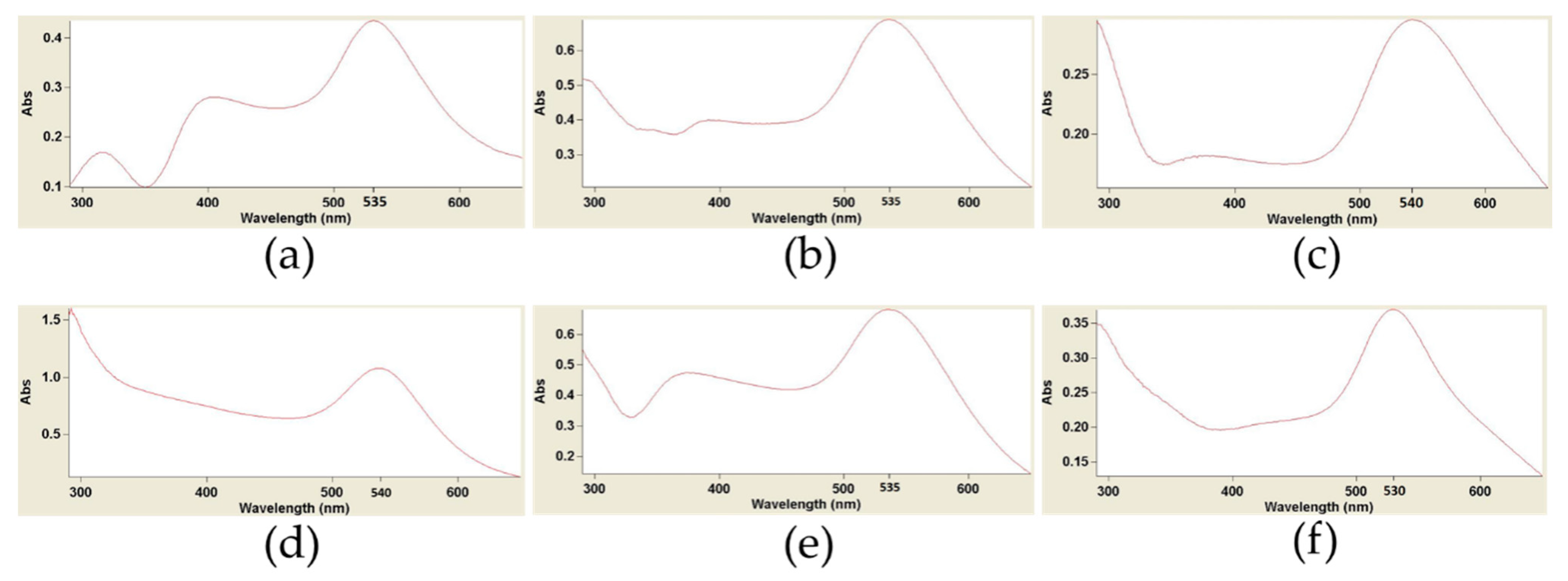
2.3.3. UV-Visible Spectroscopy
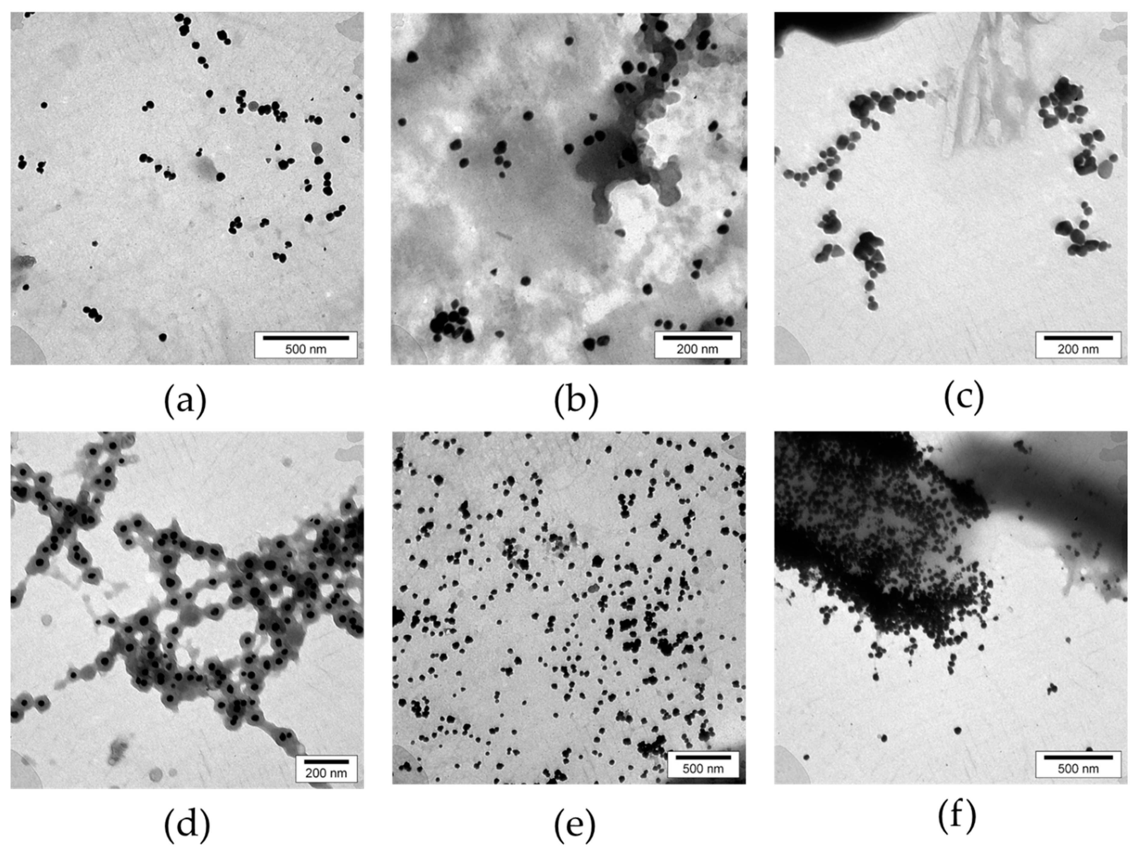
The UV-visible spectroscopic analysis can verify the bioreduction of Au3+ to Au0 accurately [23]. An AuNP’s absorbance spectrum ranges from 350 to 600 nm, which is determined by its morphological properties, distribution, and optical properties [61]. This experiment involved analyzing selected extracts at room temperature with a UV-Vis spectrophotometer (Varian Cary 50 Bio UV/Visible Spectrophotometer). A blank was prepared in all samples using water extracts of relevant plants without Au ions.

2.3.4. TEM Analyses

To understand the morphology of the biosynthesized colloidal AuNPs, transmission electron microscopy (TEM) was conducted using an LEO 912 AB TEM operating at an accelerating voltage of 30 to 100 kV. Each sample was diluted with deionized water prior to testing. Then, a drop of each was applied to the carbon-coated copper grids. After 2 min, the excess of each solution sample was removed by absorbing it to the edge of a filter paper and kept for air drying. For each sample, several electron micrographs were taken.

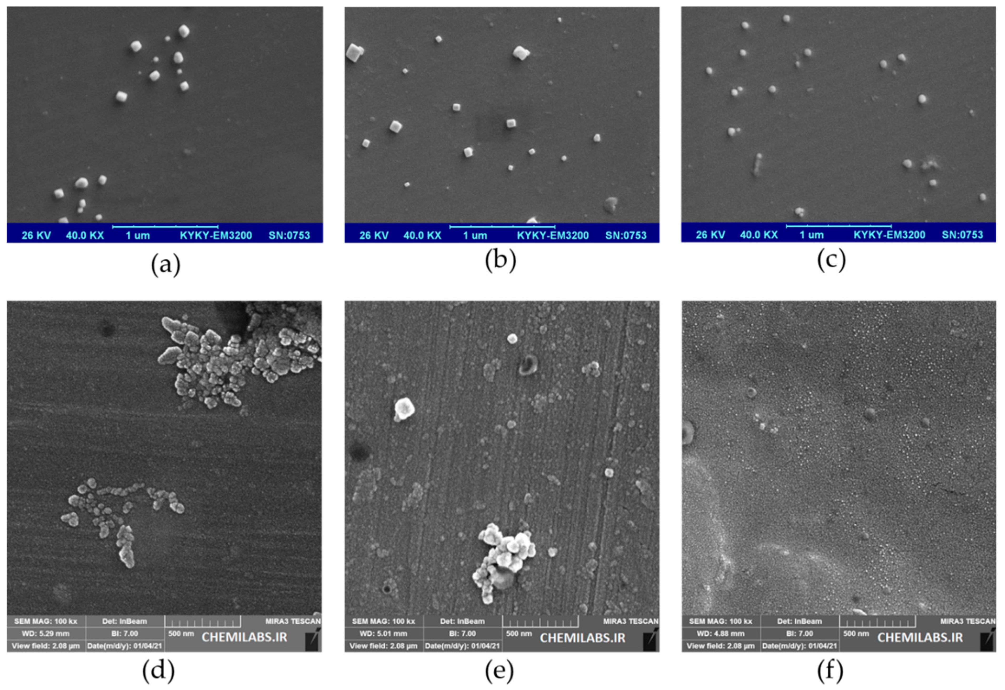
2.3.5. FSEM Analyses

Field emission scanning electron microscopy (FSEM) was adapted to facilitate the study of the topography and geometry of AuNPs. FSEM analysis is a standard method for the surface and morphological characterization of NPs [62]. Biosynthesized AuNPs were evaluated by an FSEM (FESEM TESCAN MIRA 3, Czech), and appropriate electron micrographs were prepared for all six samples. To prepare the specimens for imaging, a small quantity of each colloidal AuNPs was deposited on separate glass coverslips and allowed to dry at the ambient temperature. Then, coverslips were mounted on aluminum sputters and were examined by FSEM. Several FESEM electron micrographs were taken for each sample. Applied magnification and voltage were implanted on the corresponding electron micrographs.

2.3.6. DLS Analyses

Dynamic light scattering (DLS) is a precise, standardized technique for investigating size in NPs. It provides insight into information on the means of particle sizes and particle size distribution [63]. A DLS analyzer (HORIBA SZ-100, HORIBA, Japan) was applied to measure the size of biosynthesized AuNPs for all six samples. In addition to particle size distribution, DLS provides insight into the synthesized nanoparticles’ polydispersity index value (PI). PI is a dimensionless index that exhibits the uniformity or non-uniformity of the NPs ranging from 0 to 1. Values greater than 0.7 indicate a broad particle size distribution and their high polydispersity, while values less than 0.05 indicate the high monodispersity of NPs [64,65].

2.3.7. EDAX Analyses

Energy-dispersive X-ray analysis (EDAX) is a practical method for detecting the materials’ spectra. This technique is a key analysis in nano-based research to confirm the presence of the relevant element [66]. EDAX detects the elements based on characteristic emitted X-rays by the sample atoms via the incident beam electrons [67]. In this regard, EDAX elemental analysis of six biosynthesized AuNPs was performed using a field emission scanning electron microscope (FESEM TESCAN MIRA 3, Czech).

3. Results

3.1. Screened Plants

Relevant information for the evaluated medicinal plants, including their scientific, family names, voucher numbers, and parts of assayed plants are presented in Table S1. Furthermore, the resultant colors of reaction mixtures and the color intensity of biosynthesized AuNPs of the tested plants have been documented for each specimen. Based on visual analyses, the color intensity of reaction mixtures was rated in four scores (from 0 to 4), which represent no, slight, moderate, intense, and very intense color changes, respectively.

3.2. Instrumentation Analyses of AuNPs

3.2.1. Visual Color Grading

Visual color grading was performed by comparing biosynthesized AuNPs samples with blanks (plant extracts without Au3+). The sample plants that resulted in the biosynthesis of the AuNPs are presented in Figure S1. The color change due to the reduction of Au3+ to Au0 includes spectra of no color, pale-yellow, gray, violet, and red.

3.2.2. UV Visible Spectroscopy

UV-Vis spectra indicated preliminary confirmation of biosynthesized AuNPs. The sharp peaks were observed at 525–545 nm. A peak of absorption was observed for the six AuNPs related to bioactive samples (Figure 1), whereas there was none for the blanks.

3.2.3. TEM Analyses

TEM analysis confirmed the presence of AuNPs. As illustrated by the digital electron micrograph analysis of TEM images of six biosynthesized AuNPs via six medicinal bioactive plants, AuNPs resembled amorphous shapes (Figure 2).

3.2.4. FSEM Analyses

FSEM analysis images confirmed the formation of colloidal AuNPs. The SEM electron micrograph images of six AuNPs biosynthesized by six water extracts of medicinal plants are indicated in Figure 3.

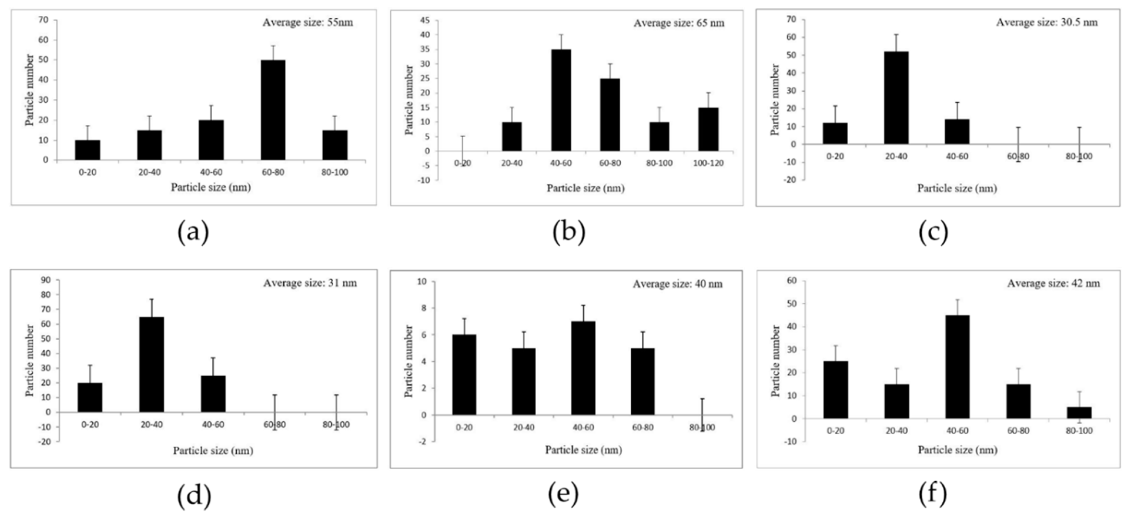
3.2.5. Particle Size Distribution
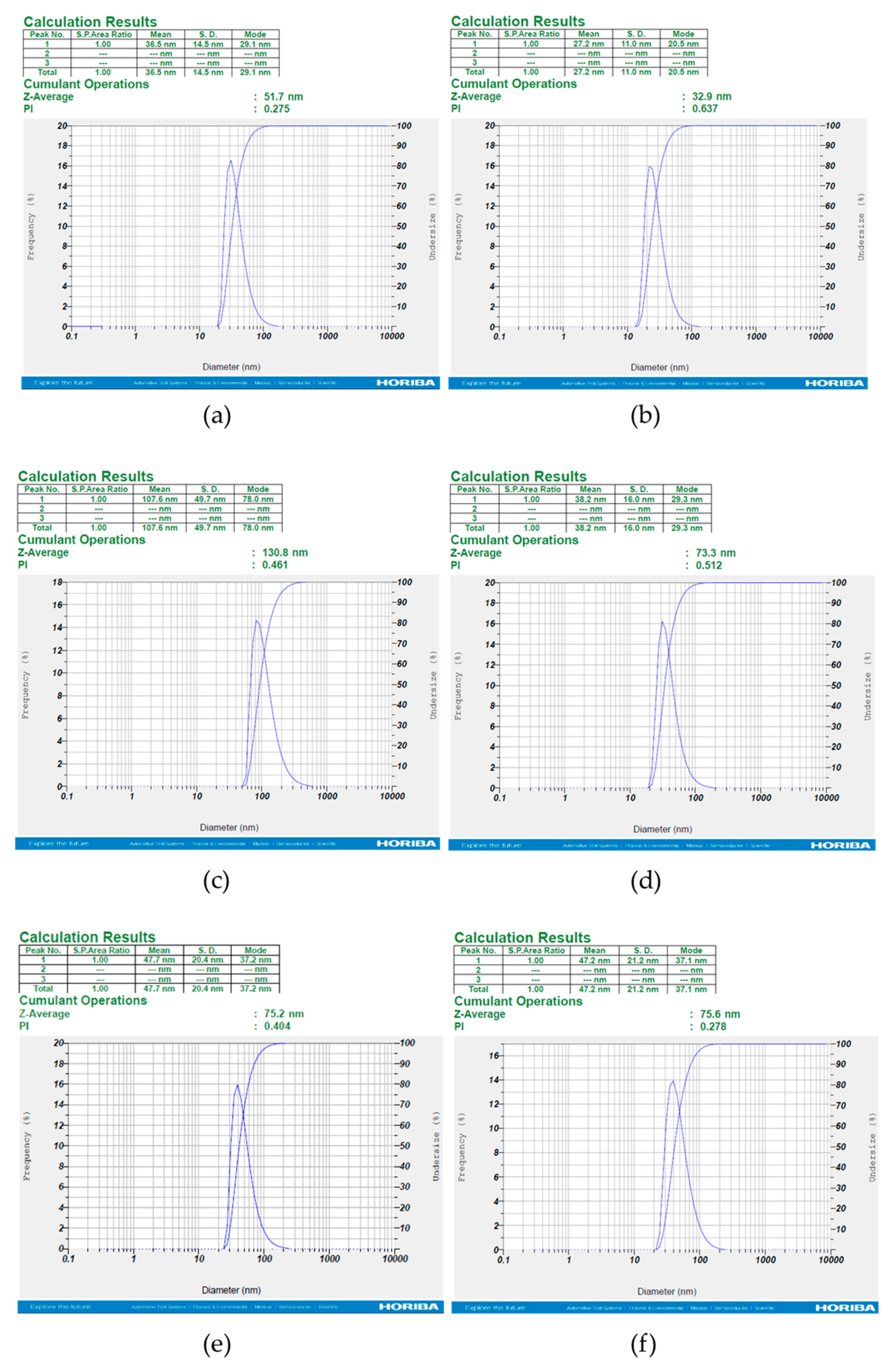
Particle size distribution histograms of the AuNPs were determined from FSEM electron micrographs with the Sigma Scan Pro software (SPSS Inc., Version 4.01.003). In the present study, the average particle size range of biosynthesized AuNPs is shown in Figure 4.

3.2.6. DLS Analyses

The DLS results of analyzed samples revealed that the diameters of distribution of particles sizes are in the range of 27.2 to 107.6 nm (Figure 5). However, the water layer surrounding NPs results in the detection of particles in a larger size [68,69]. Of the six samples analyzed, the smallest particle size belonged to Juglansregia and the largest belonged to Urticadioica with the average size of ≈27 and ≈107 nm, respectively. Additionally, PI values indicate acceptance dispersity (less than 0.7) for the samples [64].

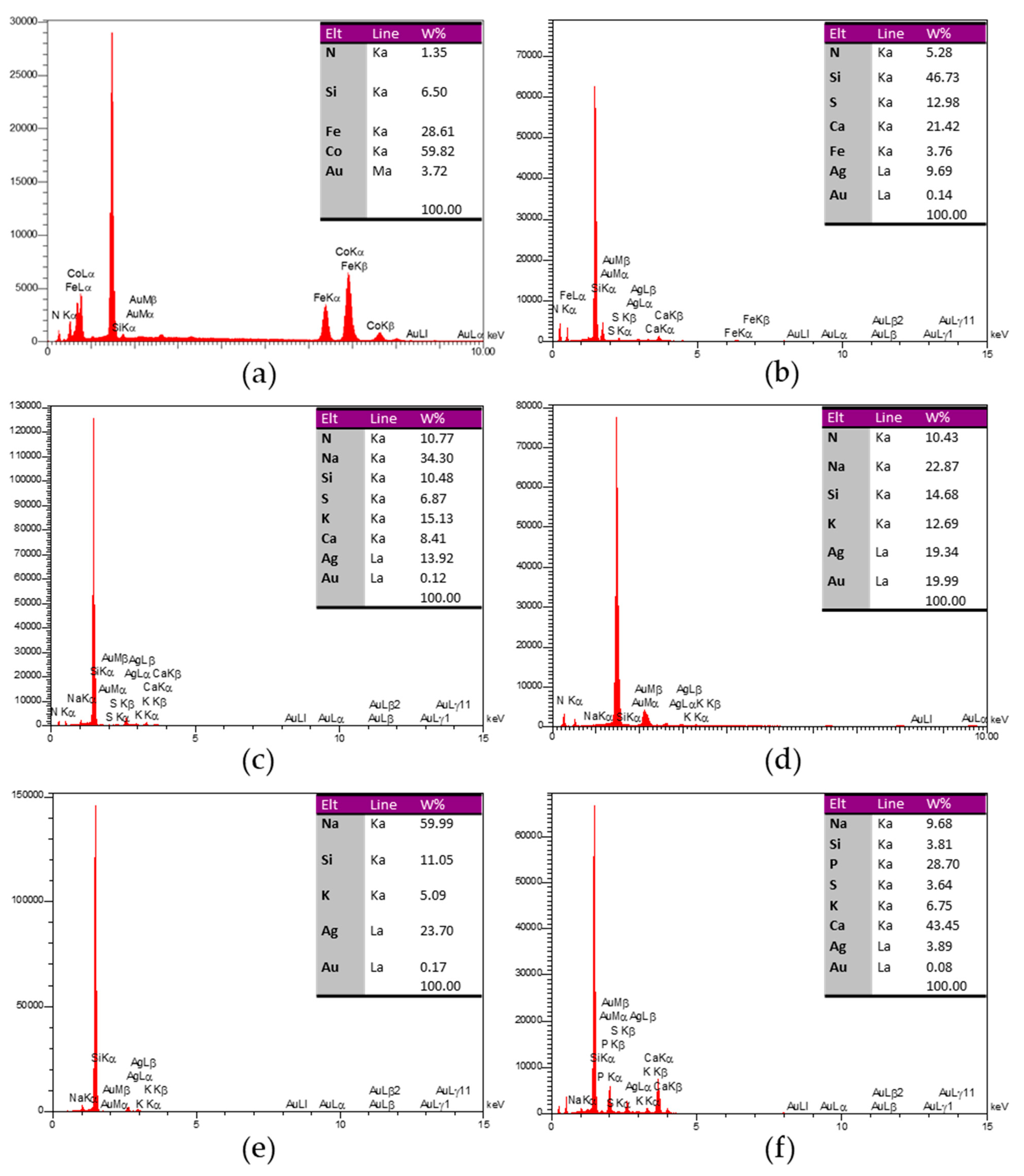
3.2.7. EDAX Analyses

Biosynthesized samples were confirmed to contain Au elements through EDAX analyses (Figure 6). Each sample is represented in a table on the related diagram indicating its detected elements. Note that some of the elements that occur in plant extracts are derived from other molecules.

3.2.8. New Bioactive Plants

Among the 109 species tested, 27 species are described for the first time according to literature surveys, as shown in Table 1.

4. Discussion

Nano-based science, nanotechnology, is a rapidly growing interdisciplinary field that has attracted the attention of numerous scientific disciplines because of its widespread use in diverse areas.
The synthesis of AuNPs is achieved through chemical, physical, and biological approaches. High energy demand, high cost, the difficulty of processing, and the use of toxic reagents have led to fundamental limitations in non-biological methods [70]. In contrast, the high speed, cost-effectiveness, and environmental compatibility of biological methods have led to greater acceptance of these methods. In addition, in biological methods, the bioreagents by capping the NPs play a central role in stabilization and non-aggregation [71]. Among these, phytonanotechnology, which is based on using plants as the regenerative agents for the biosynthesis of AuNPs, is the faster, easier, and cheaper technique compared with the biosynthesis of AuNPs using other bioresources such as bacteria, fungi, and algae [72]. In this context, medicinal plants play a valuable investment role due to their bioactive compounds [73]. Recent works have documented the phytochemical importance of medicinal plants and their bioactive compounds [74,75,76,77]. The biological characteristics of medicinal plants play a pivotal role in the characterization of NPs [78,79]. The medicinal plants have bioactive molecules for use in folkloric medicine and are considered an investment for the biosynthesis of MNPs, such as AuNPs. For instance, the Hygrophila auriculata plant as a medicinal plant in Asia and Africa has been shown to have bioactivity in the regeneration of Au3+ to Au0 [80]. Uzma et al. [81] biosynthesized AuNPs using Commiphora wightii and proved their anticancer effect on breast cancer. Similarly, Thymus vulgaris was an efficient plant source to produce AuNPs [82]. We would like to express that special attention must be devoted to the choice of plant species for the toxic phytocompounds present in some plants, as we noticed the presence of cobalt with Rosa damascena, iron with Juglans regia, and calcium with Anethum graveolens, Juglans regia, and Caccinia macranthera (Figure 6). However, special attention should be paid to the increase in metal or release of trace elements from metal oxide and metal biosynthesized NPs, which can result in elevated oxidative stress that is related to a higher risk of cancer onset. In other words, the selection of the plant species, organs, and extracts is very crucial, since some plants can be toxic to healthy cells. There are several reports on the beneficial action of AuNPs to treat cancers [83,84,85]. An interesting criterion of green NPs is that their surfaces selectively adsorb biomolecules while coming into contact with complex biological fluids, progressively forming a halo circumference or corona that reciprocally interacts with biological systems. These circumference layers bear additional efficacy over nude biological NPs [86]. Hence, biological NPs are more efficient due to the association of bioactive ingredients on the surface of biosynthesized NPs from the biological origins, such as micro-organisms and plants. Especially in folkloric medicinal plants, there exist vast metabolites with pharmacological activities that are believed to bind to the biosynthesized NPs, exhibiting further benefit by promoting the efficacies of the NPs [24]. At present, in spite of advancements and the curative action of AuNPs, implemented nanomaterial cannot be eliminated from patients’ blood. Shahidi Bonjar in 2013 [87] made an experimental recommendation by describing a ‘‘Nanogold detoxifying machine’’ for the filtration of idle AuNPs from the blood of treated cancer patients. The device similar to a ‘‘hemodialysis machine’’ would help to increase safety in AuNPs therapy of some cancers and prevent the accumulation of AuNPs in non-target tissues or organs after therapy. It has been reported that the main mechanism of biosynthesis of NPs by plants can be related to the reduction of ions by biomolecules such as organic acids, proteins, amino acids, vitamins, and secondary metabolites [24]. Through capping, these reducing agents enhance the colloidal stability of NPs and prevent aggregation [88].
An approach using phytonanotechnology to synthesize AuNPs led us to understand the bioactivity of many medicinal plants. This approach is fast, easy, environmentally friendly, and inexpensive. Through this biosynthetic process, we screened 117 plant parts of 109 species of medicinal plants for their capabilities in the biosynthesis of AuNPs.
It was determined that among the 117 plants used in folkloric medicine of Middle East countries, 102 plants were able to produce AuNPs. Twenty-seven plant species were reported as new bioresources in the biosynthesis of AuNPs. However, no inference can be drawn about these plants’ biological activity and curative behavior in traditional medicine. In the present research, 11 species of 12 Lamiaceae (11 out of 12), 8 Leguminosae (8 out of 9), 8 Asteraceae (8 out of 8), and 7 Apiaceae (7 out of 8) showed the most bioactivity in the biosynthesis of AuNPs.
According to our results, medicinal plants can be considered biological candidates for future applications in nanomaterials’ biosynthesis and more agricultural and medical applications. We should conduct more research to get a better understanding of nanomaterials’ environmental effects in order to accomplish this goal. Indeed, the assessment of antifungal and antibacterial activities of AuNPs against plant and human pathogens would further add to the knowledge on the efficiency of AuNPs in different sciences and the bioactivity of medicinal plants in biological processes. Evaluation of the therapeutic and diagnostic effectiveness of these biosynthesized AuNPs can help improve our knowledge of their future uses.

5. Conclusions

This study (a): screened 117 medicinal plant parts for the biosynthesis of AuNPs, (b) ranked plants’ performances according to their bioactivity in related families, (c) found that 102 plant parts could reduce Au3+ to Au0, (d) noticed 27 plant species are reported for the first time, and (e) revealed that the most bioactive species belong to Lamiaceae, Leguminosae, Asteraceae, and Apiaceae respectively. We believe that their mechanism for bioactivity should be thoroughly investigated. Finally, based on the spectacular achievement of nanotechnology and wide application of AuNPs in various fields, it can be concluded that the synthesis of AuNPs in line with phytonanotech goals will be a launching pad for science in the future.

Supplementary Materials

The following are available online at https://www.mdpi.com/article/10.3390/nano11082033/s1, Table S1: Screening of medicinal plants used in folkloric medicine of Middle East countries for bioreduction Au3+ to Au0, Figure S1: Pictorial results of biosynthesis of colloidal AuNPs by water extracts of 102 medicinal plant parts used in folkloric medicine of Middle East countries.

Author Contributions

M.H.: Writing, Methodology; G.H.S.B.: Project Administration, Supervision, Writing, Methodology; A.R., S.P.: Visualization, Validation; A.R., S.P., A.H., R.A.: Review and Editing. Manuscript was written through contributions of all authors. All authors have read and agreed to the published version of the manuscript.

Funding

This study was financially supported by grant No. 99/10910 of the Biotechnology Development Council of the Islamic Republic of Iran. Supports of Research Office of Kerman University of Shahid Bahonar and Research and Technology Institute of Plant Production (RTIPP) are highly appreciated.

Data Availability Statement

Not applicable.

Acknowledgments

The spirit of this research is dedicated to the souls of Alireza Afzalipour and Fakhereh Saba, the founders of Universities in Kerman, Iran.

Conflicts of Interest

The authors declare no conflict of interest.

References

  1. Rauscher, H.; Rasmussen, K.; Sokull-Klüttgen, B. Regulatory aspects of nanomaterials in the EU. Chem. Ing. Tech. 2017, 89, 224–231. [Google Scholar] [CrossRef]
  2. Chandran, S.P.; Chaudhary, M.; Pasricha, R.; Ahmad, A.; Sastry, M. Synthesis of gold nanotriangles and silver nanoparticles using Aloe vera plant extract. Biotechnol. Prog. 2006, 22, 577–583. [Google Scholar] [CrossRef]
  3. Ismail, E.H.; Saqer, A.M.A.; Assirey, E.; Naqvi, A.; Okasha, R.M. Successful Green Synthesis of Gold Nanoparticles using a Corchorus olitorius Extract and Their Antiproliferative Effect in Cancer Cells. Int. J. Mol. Sci. 2018, 19, 2612. [Google Scholar] [CrossRef] [Green Version]
  4. Singh, P.; Pandit, S.; Garnaes, J.; Tunjic, S.; Mokkapati, V.R.; Sultan, A.; Thygesen, A.; Mackevica, A.; Mateiu, R.V.; Daugaard, A.E.; et al. Green synthesis of gold and silver nanoparticles from Cannabis sativa (industrial hemp) and their capacity for biofilm inhibition. Int. J. Nanomed. 2018, 13, 3571–3591. [Google Scholar] [CrossRef] [PubMed]
  5. Alphandéry, E. Natural metallic nanoparticles for application in nano-oncology. Int. J. Mol. Sci. 2020, 21, 4412. [Google Scholar] [CrossRef]
  6. Arshad, R.; Pal, K.; Sabir, F.; Rahdar, A.; Bilal, M.; Shahnaz, G.; Kyzas, G.Z. A review of the nanomaterials use for the diagnosis and therapy of salmonella typhi. J. Mol. Struct. 2021, 1230, 129928. [Google Scholar] [CrossRef]
  7. Kheyri, N.; Norouzi, H.A.; Mobasser, H.R.; Torabi, B. Effects of silicon and zinc nanoparticles on growth, yield, and biochemical characteristics of Rice. J. Agron. 2019, 111, 3084–3090. [Google Scholar] [CrossRef]
  8. Pillai, A.M.; Sivasankarapillai, V.S.; Rahdar, A.; Joseph, J.; Sadeghfar, F.; Rajesh, K.; Kyzas, G.Z. Green synthesis and characterization of zinc oxide nanoparticles with antibacterial and antifungal activity. J. Mol. Struct. 2020, 1211, 128107. [Google Scholar] [CrossRef]
  9. Daniel, M.-C.; Astruc, D. Gold nanoparticles: Assembly, supramolecular chemistry, quantum-size-related properties, and applications toward biology, catalysis, and nanotechnology. Chem. Rev. 2004, 104, 293–346. [Google Scholar] [CrossRef] [PubMed]
  10. Del Fatti, N.; Vallée, F.; Flytzanis, C.; Hamanaka, Y.; Nakamura, A. Electron dynamics and surface plasmon resonance nonlinearities in metal nanoparticles. Chem. Phys. 2000, 251, 215–226. [Google Scholar] [CrossRef]
  11. Es-Haghi, A.; Taghavizadeh Yazdi, M.E.; Sharifalhoseini, M.; Baghani, M.; Yousefi, E.; Rahdar, A.; Baino, F. Application of Response Surface Methodology for Optimizing the Therapeutic Activity of ZnO Nanoparticles Biosynthesized from Aspergillus niger. Biomimetics 2021, 6, 34. [Google Scholar] [CrossRef]
  12. Ghosh, P.; Han, G.; De, M.; Kim, C.K.; Rotello, V.M. Gold nanoparticles in delivery applications. Adv. Drug Deliv. Rev. 2008, 60, 1307–1315. [Google Scholar] [CrossRef]
  13. Hassanen, E.I.; Korany, R.M.; Bakeer, A.M. Cisplatin-conjugated gold nanoparticles-based drug delivery system for targeting hepatic tumors. J. Biochem. Mol. Toxicol. 2021, 35, e22722. [Google Scholar] [CrossRef]
  14. Nair, R.; Varghese, S.H.; Nair, B.G.; Maekawa, T.; Yoshida, Y.; Kumar, D.S. Nanoparticulate material delivery to plants. Plant. Sci. 2010, 179, 154–163. [Google Scholar] [CrossRef]
  15. Veigas, B.; Fernandes, A.R.; Baptista, P.V. AuNPs for identification of molecular signatures of resistance. Front. Microbiol. 2014, 5, 455. [Google Scholar] [CrossRef]
  16. Gulati, S.; Singh, P.; Diwan, A.; Mongia, A.; Kumar, S. Functionalized gold nanoparticles: Promising and efficient diagnostic and therapeutic tools for HIV/AIDS. RSC Med. Chem. 2020, 11, 1252–1266. [Google Scholar] [CrossRef] [PubMed]
  17. Tripathi, R.M.; Sharma, P. Gold Nanoparticles-Based Point-of-Care Colorimetric Diagnostic for Plant Diseas-es. In Biosensors in Agriculture: Recent Trends and Future Perspectives; Pudake, R.N., Jain, U., Kole, C., Eds.; Con-cepts and Strategies in Plant Sciences; Springer: Cham, Switzerland, 2021; pp. 191–204. [Google Scholar] [CrossRef]
  18. Cai, W.; Gao, T.; Hong, H.; Sun, J. Applications of gold nanoparticles in cancer nanotechnology. Nanotechnol. Sci. Appl. 2008, 1, 17–32. [Google Scholar] [CrossRef] [PubMed] [Green Version]
  19. Alric, C.; Taleb, J.; Le Duc, G.; Mandon, C.; Billotey, C.; Le Meur-Herland, A.; Brochard, T.; Vocanson, F.; Janier, M.; Perriat, P.; et al. Gadolinium chelate coated gold nanoparticles as contrast agents for both X-ray computed tomography and magnetic resonance imaging. J. Am. Chem. Soc. 2008, 130, 5908–5915. [Google Scholar] [CrossRef]
  20. Cao, M.; Li, J.; Tang, J.; Chen, C.; Zhao, Y. Gold nanomaterials in consumer cosmetics nanoproducts: Analyses, characterization, and dermal safety assessment. Small 2016, 12, 5488–5496. [Google Scholar] [CrossRef] [PubMed]
  21. Khan, T.; Ullah, N.; Khan, M.A.; Nadhman, A. Plant-based gold nanoparticles; a comprehensive review of the decade-long research on synthesis, mechanistic aspects and diverse applications. Adv. Colloid Interface Sci. 2019, 272, 102017. [Google Scholar] [CrossRef] [PubMed]
  22. Gurunathan, S.; Kalishwaralal, K.; Vaidyanathan, R.; Venkataraman, D.; Pandian, S.R.K.; Muniyandi, J.; Hariharan, N.; Eom, S.H. Biosynthesis, purification and characterization of silver nanoparticles using Escherichia coli. Colloids Surf. B 2009, 74, 328–335. [Google Scholar] [CrossRef] [PubMed]
  23. Hassanisaadi, M.; Shahidi Bonjar, G.H. Plants used in folkloric medicine of Iran are exquisite bio-resources in production of silver nanoparticles. IET Nanobiotechnol. 2016, 11, 300–309. [Google Scholar] [CrossRef]
  24. Singh, P.; Kim, Y.-J.; Zhang, D.; Yang, D.-C. Biological synthesis of nanoparticles from plants and microorganisms. Trends Biotechnol. 2016, 34, 588–599. [Google Scholar] [CrossRef] [PubMed]
  25. Mendoza, R.M.; Baybay, Z.K.; Fernando, L.M.; Montecillo, A.D.; Ilag, L.L.; Villegas, L.C. Characterization of gold nanoparticles produced by biogenic synthesis using Serratia marcescens NBL1001. IOP Conf. Ser. Earth Environ. Sci. 2019, 230, 012103. [Google Scholar] [CrossRef]
  26. Naimi-Shamel, N.; Pourali, P.; Dolatabadi, S. Green synthesis of gold nanoparticles using Fusarium oxysporum and antibacterial activity of its tetracycline conjugant. J. Mycol. Med. 2019, 29, 7–13. [Google Scholar] [CrossRef]
  27. Khanna, P.; Kaur, A.; Goyal, D. Algae-based metallic nanoparticles: Synthesis, characterization and applications. J. Microbiol. Methods 2019, 163, 105656. [Google Scholar] [CrossRef]
  28. Francis, S.; Joseph, S.; Koshy, E.P.; Mathew, B. Green synthesis and characterization of gold and silver nanoparticles using Mussaenda glabrata leaf extract and their environmental applications to dye degradation. Sci. Pollut. Res. 2017, 24, 17347–17357. [Google Scholar] [CrossRef]
  29. Vimalraj, S.; Ashokkumar, T.; Saravanan, S. Biogenic gold nanoparticles synthesis mediated by Mangifera indica seed aqueous extracts exhibits antibacterial, anticancer and anti-angiogenic properties. Biomed. Pharmacother. 2018, 105, 440–448. [Google Scholar] [CrossRef]
  30. Keijok, W.J.; Pereira, R.H.A.; Alvarez, L.A.C.; Prado, A.R.; da Silva, A.R.; Ribeiro, J.; de Oliveira, J.P.; Guimarães, M.C.C. Controlled biosynthesis of gold nanoparticles with Coffea arabica using factorial design. Sci. Rep. 2019, 9, 1–10. [Google Scholar] [CrossRef] [PubMed]
  31. Khan, M.; Shaik, M.R.; Adil, S.F.; Khan, S.T.; Al-Warthan, A.; Siddiqui, M.R.H.; Tahir, M.N.; Tremel, W. Plant extracts as green reductants for the synthesis of silver nanoparticles: Lessons from chemical synthesis. Dalton Trans. 2018, 47, 11988–12010. [Google Scholar] [CrossRef]
  32. Ogunyemi, S.O.; Abdallah, Y.; Zhang, M.; Fouad, H.; Hong, X.; Ibrahim, E.; Masum, M.M.I.; Hossain, A.; Mo, J.; Li, B. Green synthesis of zinc oxide nanoparticles using different plant extracts and their antibacterial activity against Xanthomonas oryzae pv. oryzae. Artif. Cells Nanomed. Biotechnol. 2019, 47, 341–352. [Google Scholar] [CrossRef] [PubMed] [Green Version]
  33. Duan, H.; Wang, D.; Li, Y. Green chemistry for nanoparticle synthesis. Chem. Soc. Rev. 2015, 44, 5778–5792. [Google Scholar] [CrossRef] [PubMed]
  34. Chu, N.-S. Effects of betel chewing on the central and autonomic nervous systems. J. Biomed. Sci. 2001, 8, 229–236. [Google Scholar] [CrossRef]
  35. Peng, W.; Liu, Y.J.; Wu, N.; Sun, T.; He, X.Y.; Gao, Y.X.; Wu, C.J. Areca catechu L. (Arecaceae): A review of its traditional uses, botany, phytochemistry, pharmacology and toxicology. J. Ethnopharmacol. 2015, 164, 340–356. [Google Scholar] [CrossRef]
  36. Moradi, F.; Sedaghat, S.; Moradi, O.; Arab Salmanabadi, S. Review on green nano-biosynthesis of silver nanoparticles and their biological activities: With an emphasis on medicinal plants. Inorg. Nano Met. Chem. 2021, 51, 133–142. [Google Scholar] [CrossRef]
  37. Hosseinzadeh, S.; Jafarikukhdan, A.; Hosseini, A.; Armand, R. The application of medicinal plants in traditional and modern medicine: A review of Thymus vulgaris. Int. J. Clin. Med. 2015, 6, 635. [Google Scholar] [CrossRef] [Green Version]
  38. Shahidi Bonjar, G. Evaluation of antibacterial properties of some medicinal plants used in Iran. J. Ethnopharmacol. 2004, 94, 301–305. [Google Scholar] [CrossRef] [PubMed]
  39. Varijakzhan, D.; Chong, C.-M.; Abushelaibi, A.; Lai, K.-S.; Lim, S.-H.E. Middle Eastern plant extracts: An alternative to modern medicine problems. Molecules 2020, 25, 1126. [Google Scholar] [CrossRef] [PubMed] [Green Version]
  40. Mulat, M.; Khan, F.; Muluneh, G.; Pandita, A. Phytochemical profile and antimicrobial effects of different medicinal plant: Current knowledge and future perspectives. Curr. Tradit. Med. 2020, 6, 24–42. [Google Scholar] [CrossRef]
  41. Aguilar-Pliego, J.; Nuñez, R.Z.; Agundez, J.; de la Serna Valdés, R.; Pérez-Pariente, J. Biosynthesis of Gold Clusters and Nanoparticles by Using Extracts of Mexican Plants and Evaluation of Their Catalytic Activity in Oxidation Reactions. Catal. Lett. 2020, 1–8. [Google Scholar] [CrossRef]
  42. Soltani, R.; Baghizadeh, A.; Karimi-Maleh, H.; Farrokhi, N. Genotypic diversity of 17 cacti species and application to biosynthesis of gold nanoparticles. Spectrochim. Acta A Mol. Biomol. Spectrosc. 2021, 259, 119909. [Google Scholar] [CrossRef]
  43. Gardea-Torresdey, J.; Parsons, J.; Gomez, E.; Peralta-Videa, J.; Troiani, H.; Santiago, P.; Yacaman, M.J. Formation and growth of Au nanoparticles inside live alfalfa plants. Nano Lett. 2002, 2, 397–401. [Google Scholar] [CrossRef]
  44. Khatami, M.; Alijani, H.Q.; Nejad, M.S.; Varma, R.S. Core@ shell nanoparticles: Greener synthesis using natural plant products. Appl. Sci. 2018, 8, 411. [Google Scholar] [CrossRef] [Green Version]
  45. Lee, K.X.; Shameli, K.; Yew, Y.P.; Teow, S.-Y.; Jahangirian, H.; Rafiee-Moghaddam, R.; Webster, T.J. Recent Developments in the Facile Bio-Synthesis of Gold Nanoparticles (AuNPs) and Their Biomedical Applications. Int. J. Nanomed. 2020, 15, 275–300. [Google Scholar] [CrossRef] [PubMed]
  46. Nadeem, M.; Abbasi, B.H.; Younas, M.; Ahmad, W.; Khan, T. A review of the green syntheses and anti-microbial applications of gold nanoparticles. Green Chem. Lett. Rev. 2017, 10, 216–227. [Google Scholar] [CrossRef] [Green Version]
  47. Noruzi, M. Biosynthesis of gold nanoparticles using plant extracts. Bioprocess. Biosyst. Eng. 2015, 38, 1–14. [Google Scholar] [CrossRef] [PubMed]
  48. Le, V.T.; Ngu, N.N.Q.; Chau, T.P.; Nguyen, T.D.; Nguyen, V.T.; Nguyen, T.L.H.; Cao, X.T.; Doan, V.-D. Silver and Gold Nanoparticles from Limnophila rugosa Leaves: Biosynthesis, Characterization, and Catalytic Activity in Reduction of Nitrophenols. J. Nanomater. 2021, 2021, 5571663. [Google Scholar] [CrossRef]
  49. Al-Radadi, N.S. Green Biosynthesis of Flaxseed Gold Nanoparticles (Au-NPs) as Potent Anti-cancer Agent against Breast Cancer Cells. J. Saudi Chem. Soc. 2021, 25, 101243. [Google Scholar] [CrossRef]
  50. Sanjeevram, D.; Xu, X.; Wang, R.; Puja, A.M.; Kim, H.; Perumalsamy, H.; Balusamy, S.R.; Kim, Y.-J. Biosynthesis of gold nanoparticles using Nigella sativa and Curtobacterium proimmune K3 and evaluation of their anticancer activity. Mater. Sci. Eng. C 2021, 112214. [Google Scholar] [CrossRef]
  51. Datkhile, K.D.; Durgavale, P.P.; Patil, M.N.; Jagdale, N.J.; Deshmukh, V.N. Biosynthesis, Characterization and Evaluation of Biological Properties of Biogenic Gold Nanoparticles Synthesized Using Nothapodytes foetida Leaf Extract. Nanosci. Nanotechnol. Asia 2021, 11, 84–96. [Google Scholar] [CrossRef]
  52. Hosny, M.; Fawzy, M. Instantaneous phytosynthesis of gold nanoparticles via Persicaria salicifolia leaf extract, and their medical applications. Adv. Powder Technol. 2021, 32, 2891–2904. [Google Scholar] [CrossRef]
  53. Akintelu, S.A.; Yao, B.; Folorunso, A.S. Green synthesis, characterization, and antibacterial investigation of synthesized gold nanoparticles (AuNPs) from Garcinia kola pulp extract. Plasmonics 2021, 16, 157–165. [Google Scholar] [CrossRef]
  54. Xing, H. Citrus aurantifulia extract as a capping agent to biosynthesis of gold nanoparticles: Characterization and evaluation of cytotoxicity, antioxidant, antidiabetic, anticholinergics, and anti-bladder cancer activity. Appl. Organomet. Chem. 2021, 35, e6191. [Google Scholar] [CrossRef]
  55. Singh, R.K.; Srivastava, P.; Prakash, O. Biosynthesis of gold nanoparticles using leaf extract of Salvadora persica and its role in boosting urease performance via immobilization. J. Plant. Biochem. Biotechnol. 2021, 1–6. [Google Scholar] [CrossRef]
  56. Xu, X.Y.; Tran, T.H.M.; Perumalsamy, H.; Sanjeevram, D.; Kim, Y.-J. Biosynthetic gold nanoparticles of Hibiscus syriacus L. callus potentiates anti-inflammation efficacy via an autophagy-dependent mechanism. Mater. Sci. Eng. C 2021, 124, 112035. [Google Scholar] [CrossRef] [PubMed]
  57. Karmous, I.; Pandey, A.; Haj, K.B.; Chaoui, A. Efficiency of the Green Synthesized Nanoparticles as New Tools in Cancer Therapy: Insights on Plant-Based Bioengineered Nanoparticles, Biophysical Properties, and Anticancer Roles. Biol. Trace Elem. Res. 2020, 196, 330–342. [Google Scholar] [CrossRef]
  58. Nazaripour, E.; Mousazadeh, F.; Moghadam, M.D.; Najafi, K.; Borhani, F.; Sarani, M.; Ghasemi, M.; Rahdar, A.; Iravani, S.; Khatami, M. Biosynthesis of lead oxide and cerium oxide nanoparticles and their cytotoxic activities against colon cancer cell line. Inorg. Chem. Commun. 2021, 131, 108800. [Google Scholar] [CrossRef]
  59. Guo, M.; Li, W.; Yang, F.; Liu, H. Controllable biosynthesis of gold nanoparticles from a Eucommia ulmoides bark aqueous extract. Spectrochim. Acta Part A Mol. Biomol. Spectrosc. 2015, 142, 73–79. [Google Scholar] [CrossRef] [PubMed]
  60. Ranjani, S.; Adnan, M.; Ruckmani, K.; Hemalatha, S. Synthesis, characterization and applications of endophytic fungal nanoparticles. Inorg. Nano Met. Chem. 2020, 51, 280–287. [Google Scholar]
  61. Nachiyar, V.; Sunkar, S.; Prakash, P. Biological synthesis of gold nanoparticles using endophytic fungi. Der Pharma Chem. 2015, 7, 31–38. [Google Scholar]
  62. Schaffer, B.; Hohenester, U.; Trügler, A.; Hofer, F. High-resolution surface plasmon imaging of gold nanoparticles by energy-filtered transmission electron microscopy. Phys. Rev. B 2009, 79, 041401. [Google Scholar] [CrossRef] [Green Version]
  63. Rasmussen, K.; González, M.; Kearns, P.; Sintes, J.R.; Rossi, F.; Sayre, P. Review of achievements of the OECD Working Party on Manufactured Nanomaterials’ Testing and Assessment Programme. From exploratory testing to test guidelines. Regul. Toxicol. Pharmacol. 2016, 74, 147–160. [Google Scholar] [CrossRef]
  64. Bhatnagar, S.; Kobori, T.; Ganesh, D.; Ogawa, K.; Aoyagi, H. Biosynthesis of silver nanoparticles mediated by extracellular pigment from Talaromyces purpurogenus and their biomedical applications. Nanomaterials 2019, 9, 1042. [Google Scholar] [CrossRef] [PubMed] [Green Version]
  65. Anarjan, N.; Jafarizadeh-Malmiri, H.; Nehdi, I.A.; Sbihi, H.M.; Al-Resayes, S.I.; Tan, C.P. Effects of homogenization process parameters on physicochemical properties of astaxanthin nanodispersions prepared using a solvent-diffusion technique. Int. J. Nanomed. 2015, 10, 1109. [Google Scholar]
  66. Scimeca, M.; Bischetti, S.; Lamsira, H.K.; Bonfiglio, R.; Bonanno, E. Energy Dispersive X-ray (EDX) microanalysis: A powerful tool in biomedical research and diagnosis. Eur. J. Histochem. EJH 2018, 62, 2841. [Google Scholar] [CrossRef] [PubMed]
  67. Samuelson, D.A. Energy dispersive X-ray microanalysis. Methods Mol. Biol. 1998, 108, 413–424. [Google Scholar] [PubMed]
  68. Ahluwalia, V.; Elumalai, S.; Kumar, V.; Kumar, S.; Sangwan, R.S. Nano silver particle synthesis using Swertia paniculata herbal extract and its antimicrobial activity. Microb. Pathog. 2018, 114, 402–408. [Google Scholar] [CrossRef]
  69. Rolim, W.R.; Pelegrino, M.T.; de Araújo Lima, B.; Ferraz, L.S.; Costa, F.N.; Bernardes, J.S.; Rodigues, T.; Brocchi, M.; Seabra, A.B. Green tea extract mediated biogenic synthesis of silver nanoparticles: Characterization, cytotoxicity evaluation and antibacterial activity. Appl. Surf. Sci. 2019, 463, 66–74. [Google Scholar] [CrossRef]
  70. Usman, A.I.; Aziz, A.A.; Noqta, O.A. Application of green synthesis of gold nanoparticles: A review. J. Teknol. 2019, 81. [Google Scholar] [CrossRef] [Green Version]
  71. Makarov, V.; Love, A.; Sinitsyna, O.; Makarova, S.; Yaminsky, I.; Taliansky, M.; Kalinina, N. “Green” nanotechnologies: Synthesis of metal nanoparticles using plants. Acta Nat. 2014, 6, 20. [Google Scholar] [CrossRef] [Green Version]
  72. Akintelu, S.A.; Olugbeko, S.C.; Folorunso, A.S. A review on synthesis, optimization, characterization and antibacterial application of gold nanoparticles synthesized from plants. Int. Nano Lett. 2020, 1–12. [Google Scholar] [CrossRef]
  73. Kumar, S.; Paul, S.; Walia, Y.K.; Kumar, A.; Singhal, P. Therapeutic potential of medicinal plants: A review. J. Biol. Chem. Chron. 2015, 1, 46–54. [Google Scholar]
  74. Alzandi, A.A.; Taher, E.A.; Al-Sagheer, N.A.; Al-Khulaidi, A.W.; Azizi, M.; Naguib, D.M. Phytochemical components, antioxidant and anticancer activity of 18 major medicinal plants in Albaha region, Saudi Arabia. Biocatal. Agric. Biotechnol. 2021, 34, 102020. [Google Scholar] [CrossRef]
  75. Anand, A.V.; Balamuralikrishnan, B.; Kaviya, M.; Bharathi, K.; Parithathvi, A.; Arun, M.; Senthilkumar, N.; Velayuthaprabhu, S.; Saradhadevi, M.; Al-Dhabi, N.A. Medicinal Plants, Phytochemicals, and Herbs to Combat Viral Pathogens Including SARS-CoV-2. Molecules 2021, 26, 1775. [Google Scholar] [CrossRef]
  76. Hajdari, A.; Mustafa, B.; Hyseni, L.; Bajrami, A.; Mustafa, G.; Quave, C.L.; Nebija, D. Phytochemical study of eight medicinal plants of the lamiaceae family traditionally used as tea in the Sharri Mountains region of the Balkans. Sci. World J. 2020, 2020, 4182064. [Google Scholar] [CrossRef] [Green Version]
  77. Verma, A.K.; Singh, S. Phytochemical analysis and in vitro cytostatic potential of ethnopharmacological important medicinal plants. Toxicol. Rep. 2020, 7, 443–452. [Google Scholar] [CrossRef]
  78. Saratale, R.G.; Saratale, G.D.; Cho, S.-K.; Ghodake, G.; Kadam, A.; Kumar, S.; Mulla, S.I.; Kim, D.-S.; Jeon, B.-H.; Chang, J.S. Phyto-fabrication of silver nanoparticles by Acacia nilotica leaves: Investigating their antineoplastic, free radical scavenging potential and application in H2O2 sensing. J. Taiwan Inst. Chem. Eng. 2019, 99, 239–249. [Google Scholar] [CrossRef]
  79. Saratale, R.G.; Saratale, G.D.; Ghodake, G.; Cho, S.-K.; Kadam, A.; Kumar, G.; Jeon, B.-H.; Pant, D.; Bhatnagar, A.; Shin, H.S. Wheat straw extracted lignin in silver nanoparticles synthesis: Expanding its prophecy towards antineoplastic potency and hydrogen peroxide sensing ability. Int. J. Biol. Macromol. 2019, 128, 391–400. [Google Scholar] [CrossRef] [PubMed]
  80. Satpathy, S.; Patra, A.; Ahirwar, B.; Hussain, M.D. Process optimization for green synthesis of gold nanoparticles mediated by extract of Hygrophila spinosa T. Anders and their biological applications. Phys. E Low Dimens. Syst. Nanostruct. 2020, 121, 113830. [Google Scholar] [CrossRef]
  81. Uzma, M.; Sunayana, N.; Raghavendra, V.B.; Madhu, C.S.; Shanmuganathan, R.; Brindhadevi, K. Biogenic synthesis of gold nanoparticles using Commiphora wightii and their cytotoxic effects on breast cancer cell line (MCF-7). Process. Biochem. 2020, 92, 269–276. [Google Scholar] [CrossRef]
  82. Hemmati, S.; Joshani, Z.; Zangeneh, A.; Zangeneh, M.M. Green synthesis and chemical characterization of Thymus vulgaris leaf aqueous extract conjugated gold nanoparticles for the treatment of acute myeloid leukemia in comparison to doxorubicin in a leukemic mouse model. Appl. Organomet. Chem. 2020, 34, e5267. [Google Scholar] [CrossRef]
  83. Fan, M.; Han, Y.; Gao, S.; Yan, H.; Cao, L.; Li, Z.; Liang, X.-J.; Zhang, J. Ultrasmall gold nanoparticles in cancer diagnosis and therapy. Theranostics 2020, 10, 4944–4957. [Google Scholar] [CrossRef]
  84. Siddique, S.; Chow, J.C.L. Gold Nanoparticles for Drug Delivery and Cancer Therapy. Appl. Sci. 2020, 10, 3824. [Google Scholar] [CrossRef]
  85. Sztandera, K.; Gorzkiewicz, M.; Klajnert-Maculewicz, B. Gold Nanoparticles in Cancer Treatment. Mol. Pharm. 2019, 16, 1–23. [Google Scholar] [CrossRef] [PubMed]
  86. Monopoli, M.P.; Åberg, C.; Salvati, A.; Dawson, K.A. Biomolecular coronas provide the biological identity of nanosized materials. Nat. Nanotechnol. 2012, 7, 779–786. [Google Scholar] [CrossRef] [PubMed]
  87. Shahidi Bonjar, L. “Nanogold detoxifying machine” to remove idle nanogold particles from blood stream of cancer patients treated with antibody-nanogold therapeutics. Med. Hypotheses 2013, 80, 601–605. [Google Scholar] [CrossRef]
  88. Javed, R.; Zia, M.; Naz, S.; Aisida, S.O.; ul Ain, N.; Ao, Q. Role of capping agents in the application of nanoparticles in biomedicine and environmental remediation: Recent trends and future prospects. J. Nanobiotechnology 2020, 18, 1–15. [Google Scholar] [CrossRef] [PubMed]
Figure 1. UV-Visible spectra of biosynthesized AuNPs by water extracts of six medicinal plants used in folkloric medicine of Middle East countries. Each spectrum was presented for its related sample. (af) indicate UV-Visible spectra of Rosa damascena, Juglans regia, Urtica dioica, Areca catechu, Caccinia macranthera, and Anethum graveolens, respectively.
Nanomaterials 11 02033 g001
Figure 2. Transmission electron micrographs of six biosynthesized AuNPs by six bioactive medicinal plant extracts. Each electron micrograph is presented for its related sample. (af) indicated TEM microscopic evaluations of Rosa damascena, Juglans regia, Urtica dioica, Areca catechu, Caccinia macranthera, and Anethum graveolens, respectively.
Nanomaterials 11 02033 g002
Figure 3. Scanning electron micrographs of biosynthesized AuNPs by six bioactive medicinal plant extracts. Each image was presented for its related sample. (af) indicate SEM images mediated by water extract of Rosa damascena, Juglansregia, Caccinia macranthera, Urticadioica, Areca catechu, and Anethum graveolens, respectively.
Nanomaterials 11 02033 g003
Figure 4. Particle size distribution histograms of the biosynthesized AuNPs via six aqueous extracts of medicinal plants used in folkloric medicine in the Middle East. Each image was presented for its related sample. (af) indicated the particle size distribution of Rosa damascena, Juglansregia, Urticadioica, Areca catechu, Caccinia macranthera, and Anethum graveolens, respectively.
Nanomaterials 11 02033 g004
Figure 5. Dynamic light scattering of the biosynthesized AuNPs from water extracts of six medicinal plants. The diameters of the distribution of particles are 27.2 to 107.6 nm. The mean size distribution of AuNPs for Rosa damascena, Juglans regia, Urtica dioica, Areca catechu, Caccinia macranthera, and Anethum graveolens are shown in (af). The ranging of polydispersity index (PI) was between 0.27 and 0.63.
Nanomaterials 11 02033 g005
Figure 6. The results of energy-dispersive X-ray analysis of the biosynthesized AuNPs mediated by six water extracts of medicinal plants. Each graph and the table next to it represent its related sample. (af) indicate EDAX diagrams of reaction mixtures (Au3+, Au0, and the plant extract) of Rosa damascena, Juglans regia, Caccinia macranthera, Urtica dioica, Areca catechu, and Anethum graveolens respectively.
Nanomaterials 11 02033 g006
Table 1. Newly identified plant species for the synthesis of AuNPs used in folkloric medicine.
No.Scientific NameFamilyVN 1PP 2
1PistacialentiscusAnacardiaceaeANAC120Gu
2HeracleumpersicumApiaceaeAPIA43Fr
3Artemisia cinaAsteraceaeASTE18Se
4Pyrethrum roseumAsteraceaeASTE60Fr
5EchiumamoenumBoraginaceaeBORA23Fl
6CacciniamacrantheraBoraginaceaeCACC64Le
7Nasturtium officinalisBrassicaceaeBRAS49Ab
8LepidiumsativumBrassicaceaeBRAS66Se
9Eugenia caryophyllataCaryophyllaceaeCARY47Fl
10Fraxinus excelsiorFraxinaceaeFRAX111Fr
11Erodium sp.GeraniaceaeGERA3Ab
12TeucriumpoliumLamiaceaeLAMI24Ab
13AstragalusadscendensLeguminosaeLEGU103Gu
14Astragalus fasciculifoliusLeguminosaeLEGU108Gu
15Allium schoenoprasumLiliaceaeLILI92Se
16Allium stipitatumLiliaceaeLILI8Bu
17Allium schoenoprasumLiliaceaeLILI85Le
18Sesamum indicumPedaliaceaePEDA37Se
19Oryza sativaPoaceaePOAC82Se
20Rheum ribesPolygonaceaePOLY65Le
21RumexalpinusPolygonaceaePOLY76Fr
22Rheum palmatumPolygonaceaePOLY114Rh
23Ranunculus sp.RanunculaceaeRANU63Ab
24CydoniaoblongaRosaceaeROSA11Fr
25AmygdaluscommunisRosaceaeROSA94Se
26PrunuscerasusaviumRosaceaeROSA104Fs
27RubiaTinctorumRubiaceaeRUBI51Fr
28ValerianaofficinalisValerianaceaeVALE20Ab
1 VN: Voucher number of plants stored in Laboratory of Plant Systematic, College of Agriculture, Shahid Bahonar University of Kerman, Iran; 2 PP: Part plants used for biosynthesis of AuNPs (Ab: above-ground parts, Ba: bark, Bu: bud, Fl: flower, Fr: fruit, Fs: fruit stalk, Gl: glumes, Le: leaf, Pe: petioles, Rh: rhizome, Ro: root, Se: seeds, Sm: flower stamen and Wh: whole plant).
Publisher’s Note: MDPI stays neutral with regard to jurisdictional claims in published maps and institutional affiliations.

