

Engineered Cross-Linked Silane with Urea Polymer Thin Durable Coatings onto Polymeric Films for Controlled Antiviral Release of Activated Chlorine and Essential Oils

Abstract
:1. Introduction
2. Experimental
2.1. Materials
2.2. Methodology
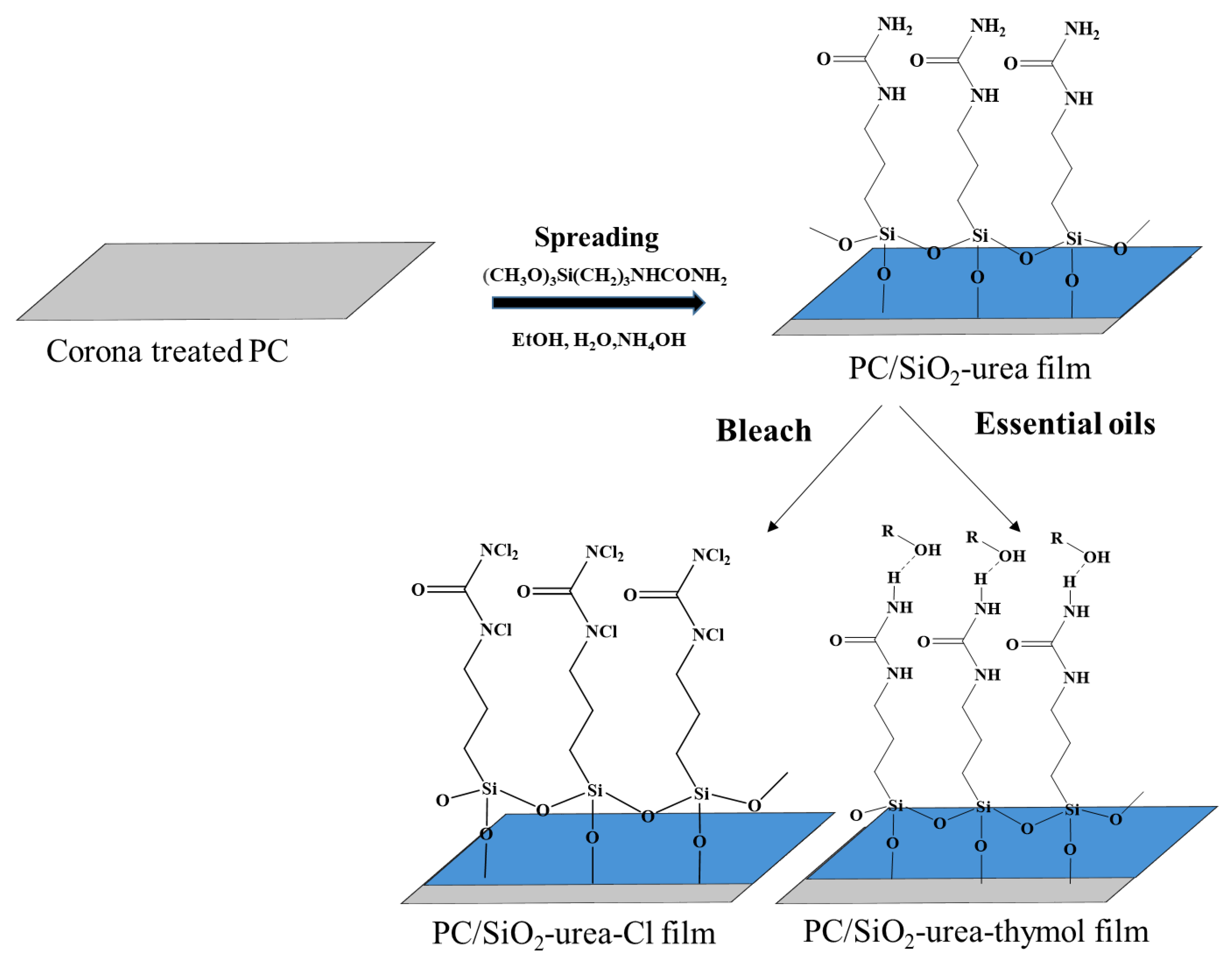
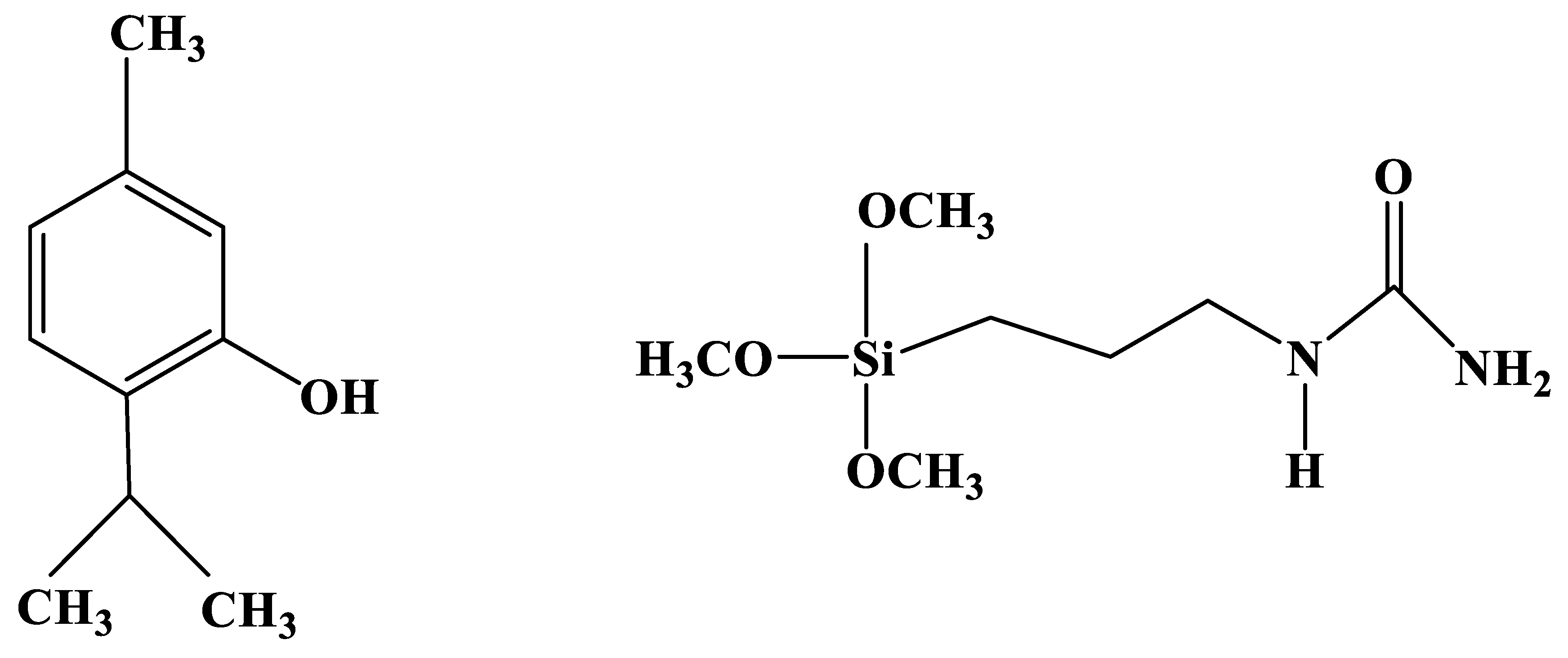
2.2.1. Preparation of SiO2-Urea and SiO2-Urea-Thymol Coatings on PC Film
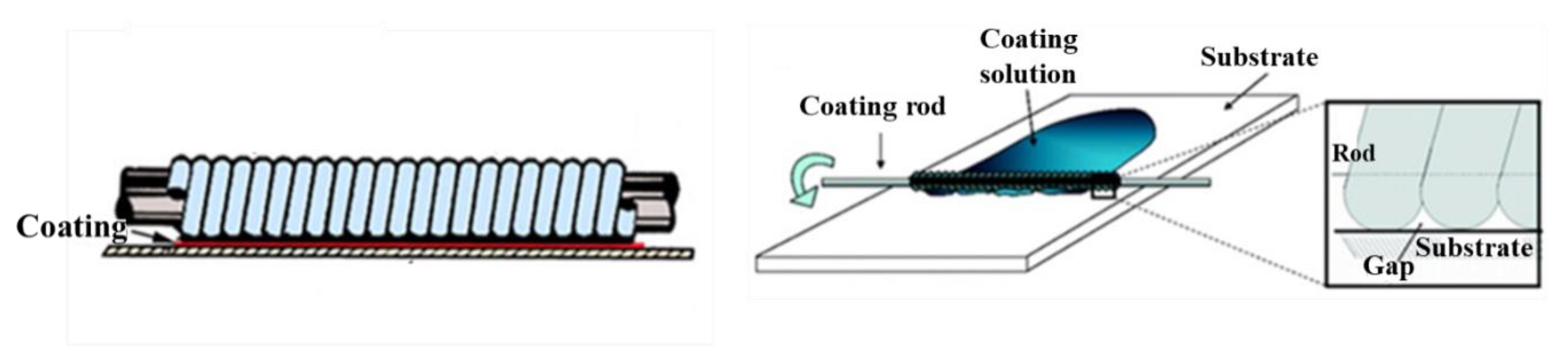
2.2.2. Rod-Coating Technique
2.2.3. SiO2-Urea-Cl Coating on PC Film
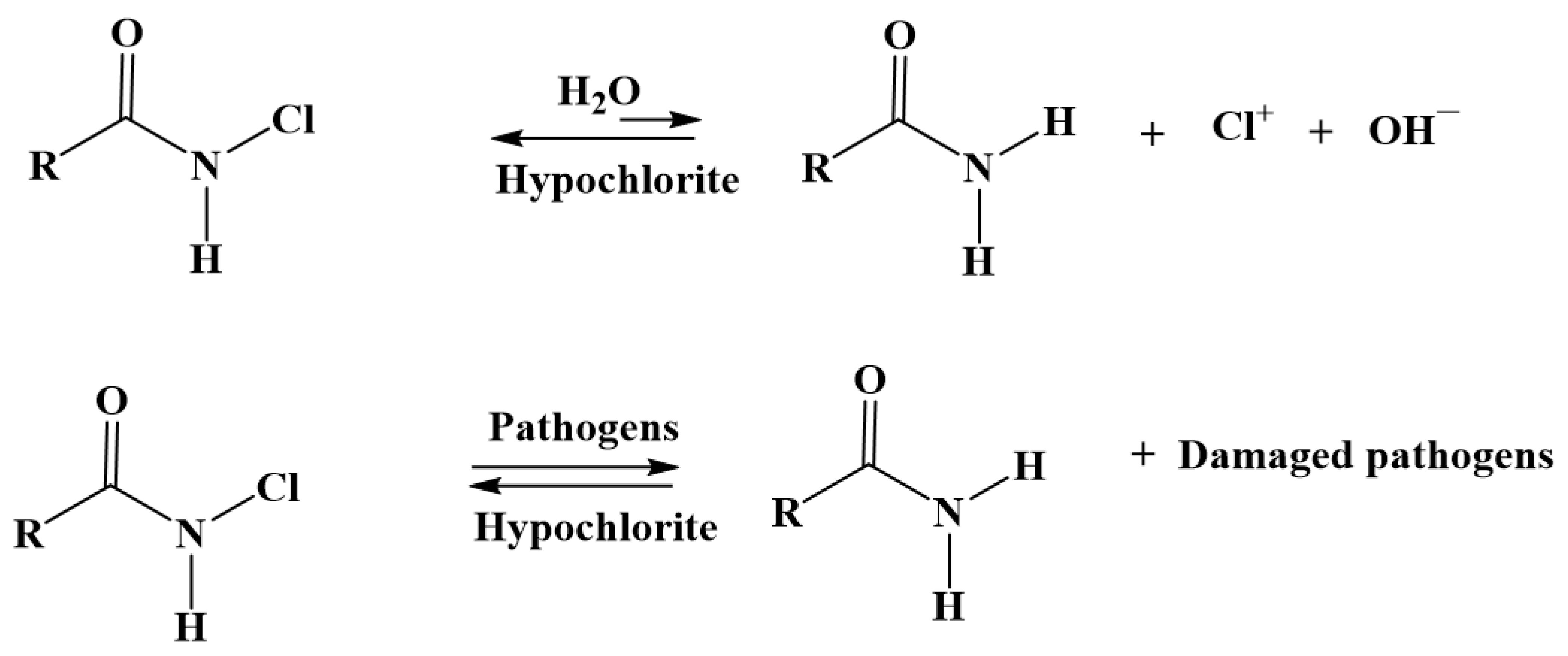
Chlorination of the SiO2-Urea Coating
Active Chlorine Determination
2.2.4. Characterization of the Coatings
Atomic Force Microscope
Fourier Transform Infrared Spectroscopy
Contact Angles
X-ray Photoelectron Spectroscopy
2.2.5. Release Rates at Different Temperatures of Activated Cl from PC/SiO2-Urea-Cl Films
2.2.6. Determination of Thymol Content in PC/SiO2-Urea Thymol-Containing Films
2.2.7. Release Rates of Thymol from PC/SiO2-Urea-Thymol and PC/SiO2-Urea-Cl-Thymol Films
2.2.8. Durability of SiO2-Urea Coating on PC Film
2.2.9. Bacteriophage Assay
2.2.10. CCV Assay
3. Results and Discussion
3.1. Characterization of the Coatings
3.1.1. Atomic Force Microscopy (AFM)
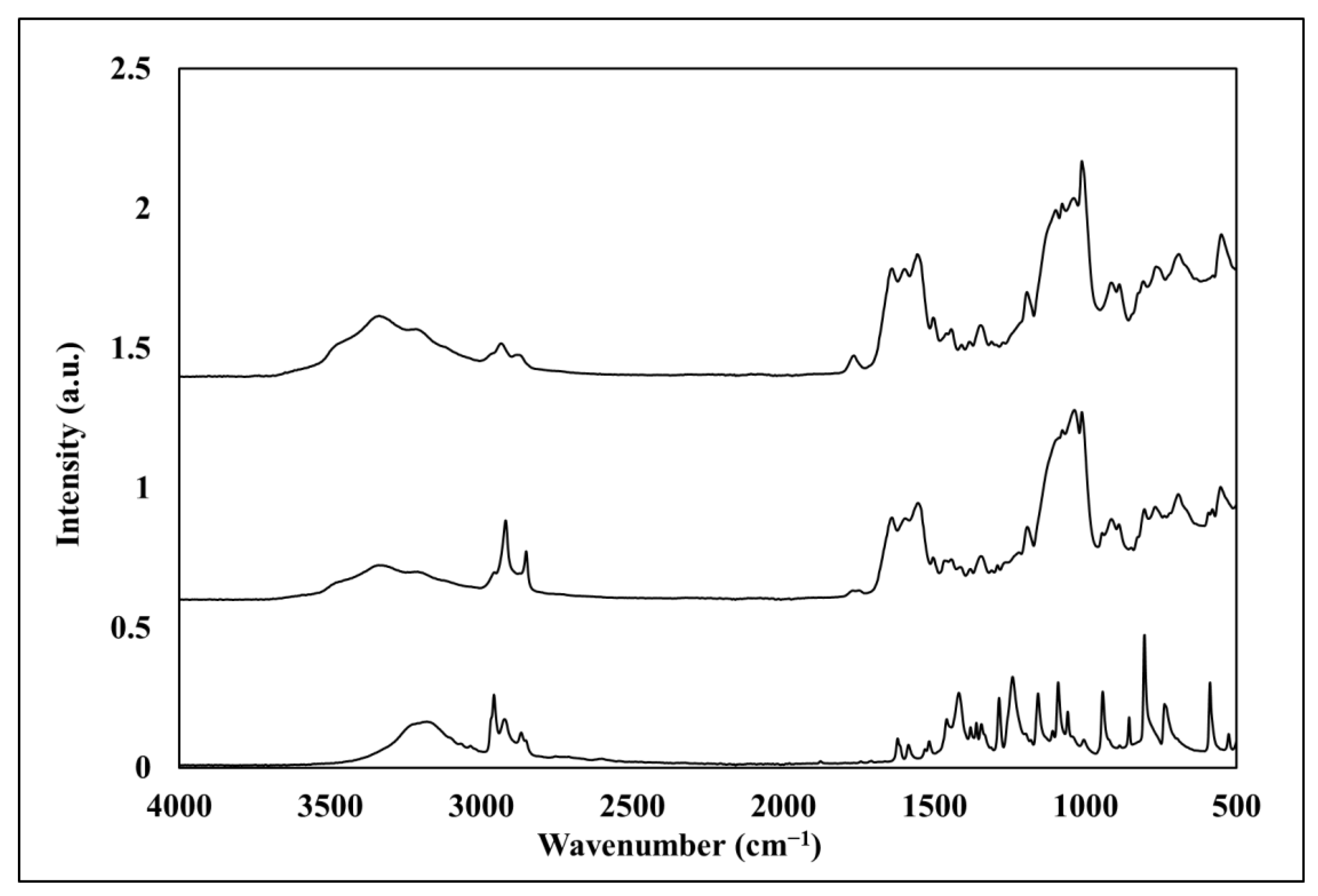
3.1.2. Fourier Transform IR/Attenuated Total Reflection
3.1.3. Contact Angle (CA)
3.2. Determination of Cl Content in PC/SiO2-Urea-Cl Film
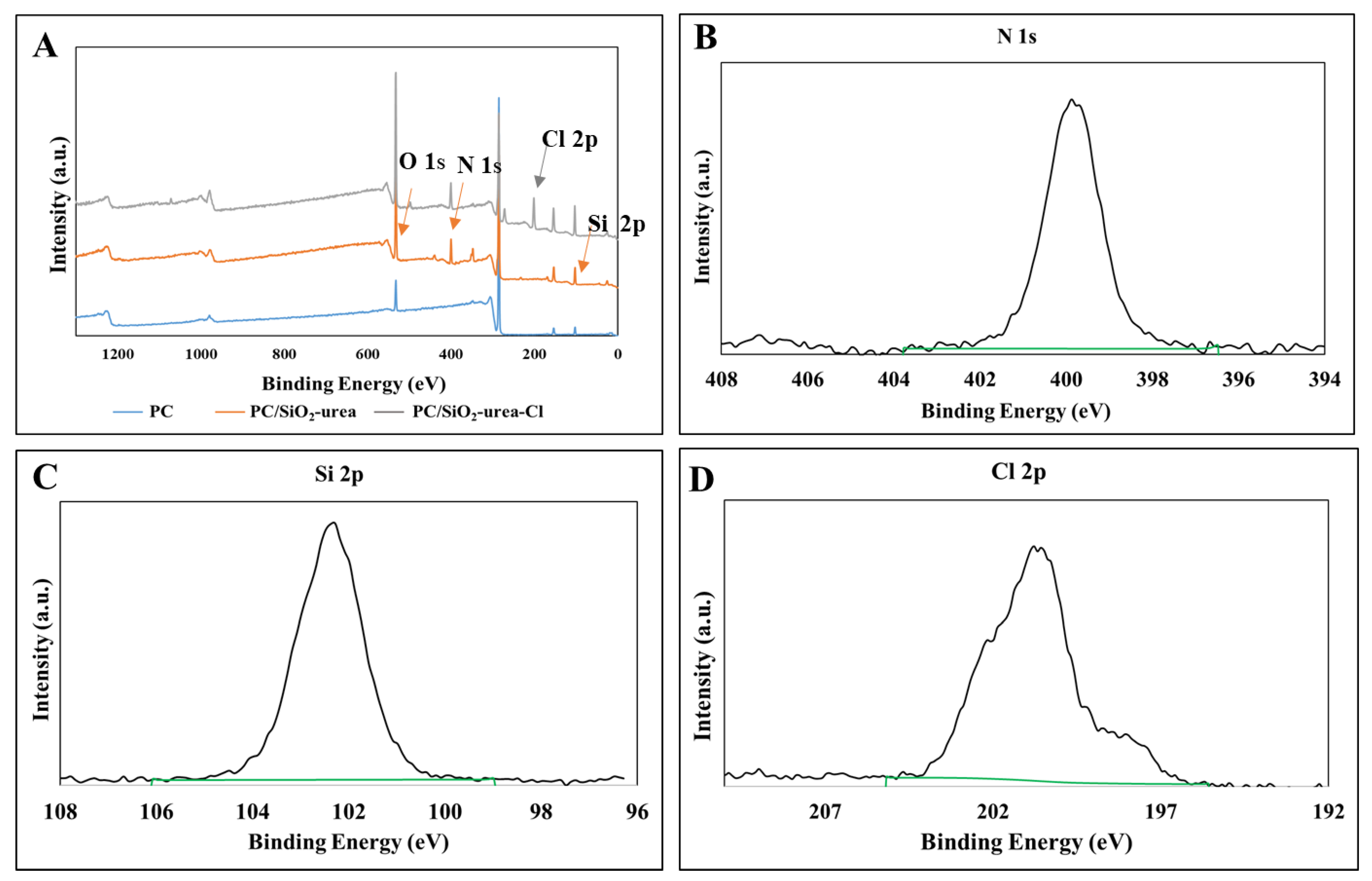
3.2.1. X-ray Photoelectron Spectroscopy
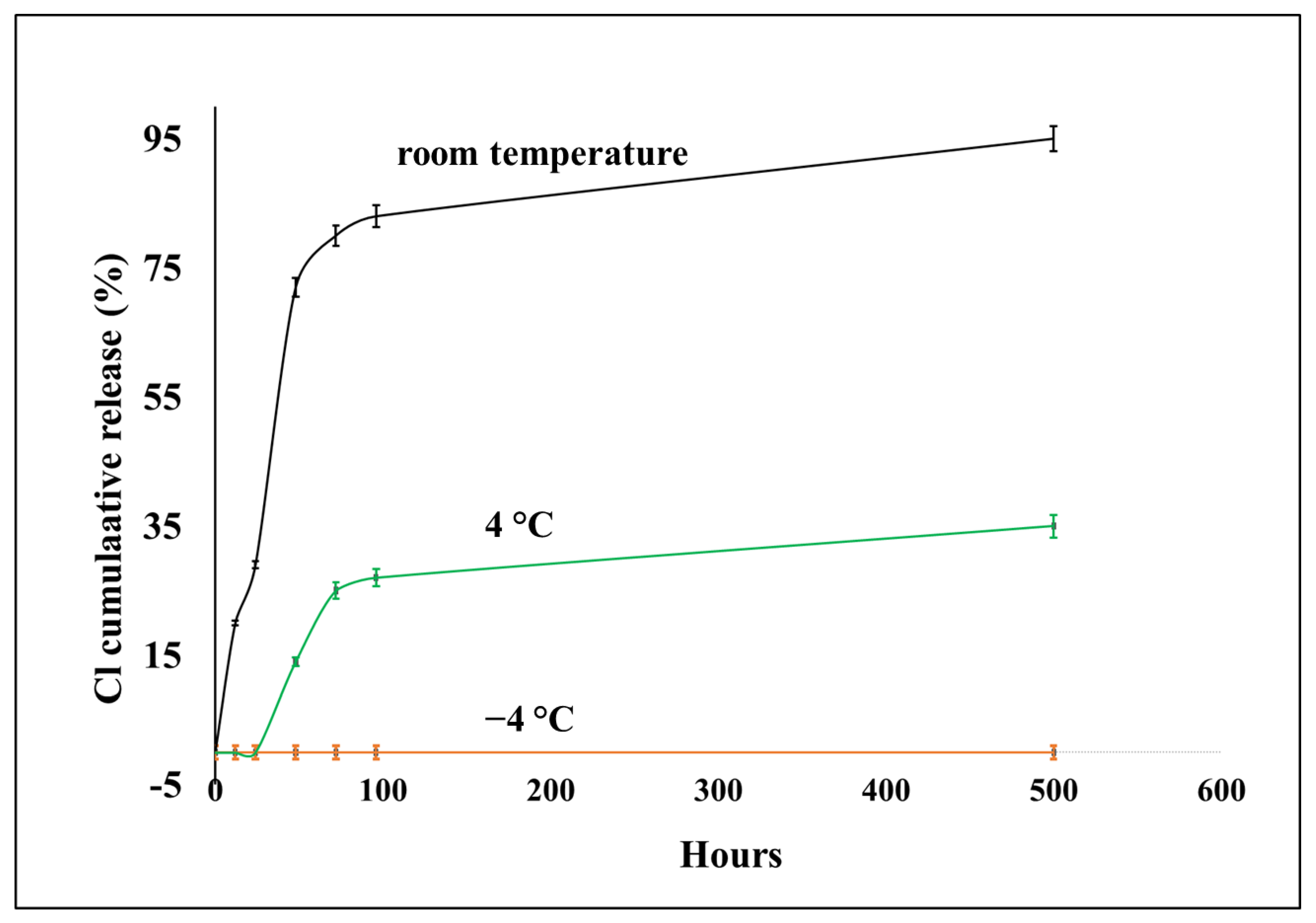
3.2.2. Release Rates of Chlorine from PC/SiO2-Urea-Cl and PC/SiO2-Urea-Cl-Thymol Films
3.3. Determination of Thymol Content in PC/SiO2-Urea Films
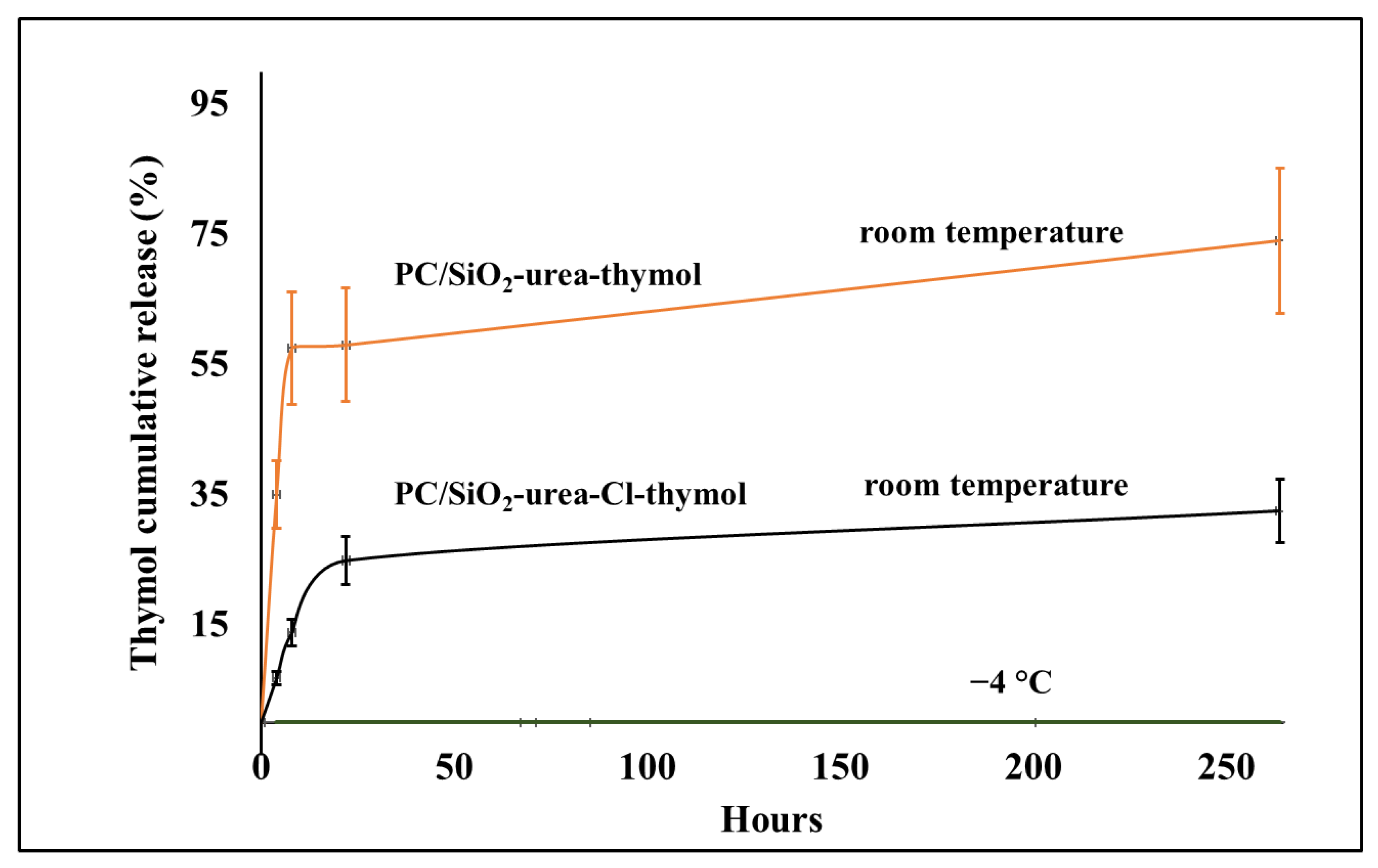
Release Rates of Thymol from PC/SiO2-Urea-Thymol and PC/SiO2-Urea-Cl-Thymol Films
3.4. Durability of SiO2-Urea Coating on PC Film
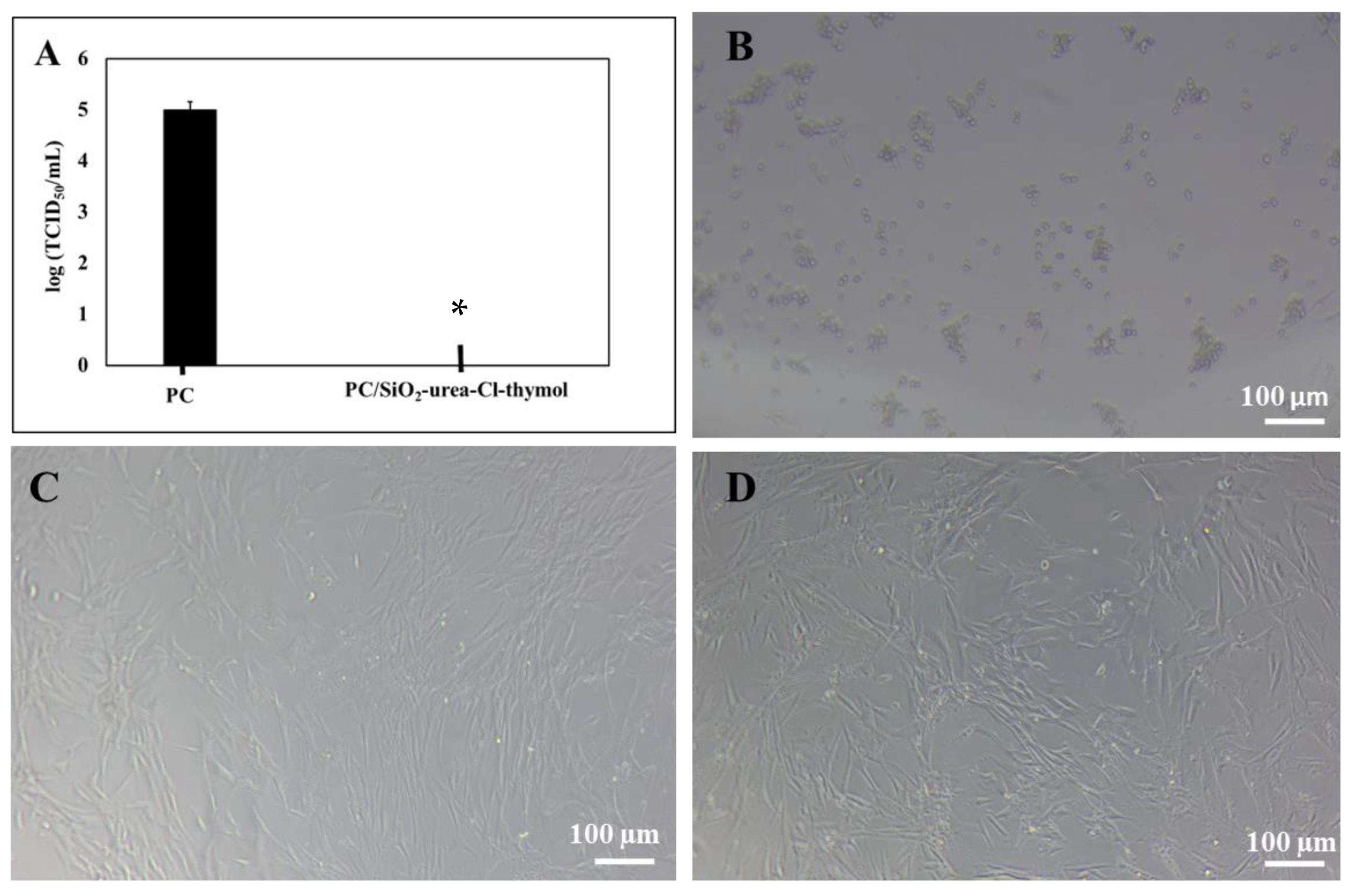
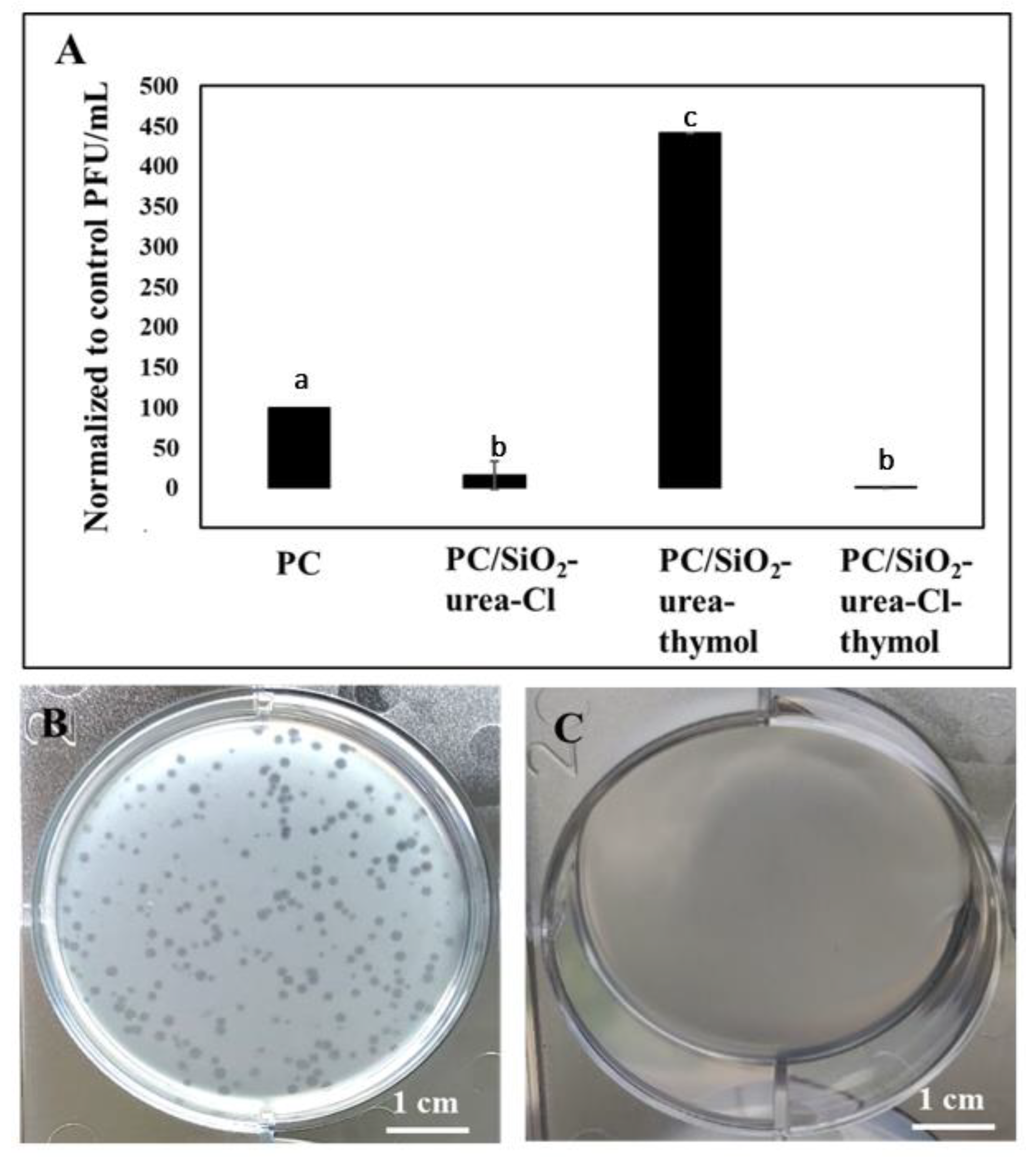
3.5. Antiviral Activity
4. Summary and Discussion
Supplementary Materials
Author Contributions
Funding
Data Availability Statement
Acknowledgments
Conflicts of Interest
References
- McIntosh, K.; Hirsch, M.S.; Bloom, A. Coronavirus disease (COVID-19). UpToDate Hirsch MS Bloom 2020, 5, 873. [Google Scholar]
- Rawlinson, S.; Ciric, L.; Cloutman-Green, E. COVID-19 pandemic—Let’s not forget surfaces. J. Hosp. Infect. 2020, 105, 790–791. [Google Scholar]
- Patients, L.; Taylor, D.; Lindsay, A.C.; Halcox, J.P. Aerosol and surface stability of SARS-CoV-2 as compared with SARS-CoV-1. N. Engl. J. Med. 2020, 382, 1564–1567. [Google Scholar]
- Kampf, G.; Todt, D.; Pfaender, S.; Steinmann, E. Persistence of coronaviruses on inanimate surfaces and their inactivation with biocidal agents. J. Hosp. Infect. 2020, 104, 246–251. [Google Scholar]
- Imani, S.M.; Ladouceur, L.; Marshall, T.; Maclachlan, R.; Soleymani, L.; Didar, T.F. Antimicrobial nanomaterials and coatings: Current mechanisms and future perspectives to control the spread of viruses including SARS-CoV-2. ACS Nano 2020, 14, 12341–12369. [Google Scholar]
- Chiari, W.; Damayanti, R.; Harapan, H.; Puspita, K.; Saiful, S.; Rahmi, R.; Rizki, D.R.; Iqhrammullah, M. Trend of polymer research related to COVID-19 pandemic: Bibliometric analysis. Polymers 2022, 14, 3297. [Google Scholar]
- Sharun, K.; Timari, R.; Yatoo, M.I.; Natesan, S.; Megawati, D.; Singh, K.P.; Michalak, I.; Dhama, K. A comprehensive review on pharmacologic agents, immunotherapies and supportive therapeutics for COVID-19. Narra J. 2022, 2, e92. [Google Scholar]
- Pirtle, E.C.; Beran, G.W. Virus survival in the environment. Rev. Sci. Tech. 1991, 10, 733–748. [Google Scholar]
- Centers for Disease Control (US). Recommendations for Prevention of HIV Transmission in Health-Care Settings; US Department of Health and Human Services, Public Health Service, Centers for Disease Control: Atlanta, GA, USA, 1987; Volume 37.
- Otter, J.A.; Donskey, C.; Yezli, S.; Douthwaite, S.; Goldenberg, S.; Weber, D.J. Transmission of SARS and MERS coronaviruses and influenza virus in healthcare settings: The possible role of dry surface contamination. J. Hosp. Infect. 2016, 92, 235–250. [Google Scholar]
- Noyce, J.O.; Michels, H.; Keevil, C.W. Inactivation of influenza A virus on copper versus stainless steel surfaces. Appl. Environ. Microbiol. 2007, 73, 2748–2750. [Google Scholar]
- Karlstrom, A.R.; Levine, R.L. Copper inhibits the protease from human immunodeficiency virus 1 by both cysteine-dependent and cysteine-independent mechanisms. Proc. Natl. Acad. Sci. USA 1991, 88, 5552–5556. [Google Scholar]
- Ingle, A.P.; Duran, N.; Rai, M. Bioactivity, mechanism of action, and cytotoxicity of copper-based nanoparticles: A review. Appl. Microbiol. Biotechnol. 2014, 98, 1001–1009. [Google Scholar]
- Rai, M.; Deshmukh, S.D.; Ingle, A.P.; Gupta, I.R.; Galdiero, M.; Galdiero, S. Metal nanoparticles: The protective nanoshield against virus infection. Crit. Rev. Microbiol. 2016, 42, 46–56. [Google Scholar]
- Lara, H.H.; Ayala-Nuñez, N.V.; Ixtepan-Turrent, L.; Rodriguez-Padilla, C. Mode of antiviral action of silver nanoparticles against HIV-1. J. Nanobiotechnol. 2010, 8, 1. [Google Scholar]
- Gupta, P.; Rapp, F. Effect of zinc ions on synthesis of herpes simplex virus type 2-induced polypeptides. Proc. Soc. Exp. Biol. Med. 1976, 152, 455–458. [Google Scholar]
- Fridlender, B.; Chejanovsky, N.; Becker, Y. Selective inhibition of herpes simplex virus type 1 DNA polymerase by zinc ions. Virology 1978, 84, 551–554. [Google Scholar]
- Arens, M.; Travis, S. Zinc salts inactivate clinical isolates of herpes simplex virus in vitro. J. Clin. Microbiol. 2000, 38, 1758–1762. [Google Scholar]
- Gelman, F.; Lewis, K.; Klibanov, A.M. Drastically lowering the titer of waterborne bacteriophage PRD1 by exposure to immobilized hydrophobic polycations. Biotechnol. Lett. 2004, 26, 1695–1700. [Google Scholar]
- Liu, H.; Kim, Y.; Mello, K.; Lovaasen, J.; Shah, A.; Rice, N.; Yim, J.H.; Pappas, D.; Klibanov, A.M. Aerosol-assisted plasma deposition of hydrophobic polycations makes surfaces highly antimicrobial. Appl. Biochem. Biotechnol. 2014, 172, 1254–1264. [Google Scholar]
- Hu, T.; Agazani, O.; Nir, S.; Cohen, M.; Pan, S.; Reches, M. Antiviral activity of peptide-based assemblies. ACS Appl. Mater. Interfaces 2021, 13, 48469–48477. [Google Scholar]
- Steinman, N.Y.; Hu, T.; Dombrovsky, A.; Reches, M.; Domb, A.J. Antiviral polymers based on N-halamine polyurea. Biomacromolecules 2021, 10, 4357–4364. [Google Scholar]
- Padmanabhuni, R.V.; Luo, J.; Cao, Z.; Sun, Y. Preparation and characterization of N-halamiine-based antimicrobial fillers. Ind. Eng. Chem. Res. 2012, 51, 5148–5156. [Google Scholar]
- Kocer, H.B.; Cerkez, I.; Worley, S.D.; Broughton, R.M.; Huang, T.S. N-halamine copolymers for use in antimicrobial paints. ACS Appl. Mater. Interfaces 2011, 3, 3189–3194. [Google Scholar]
- Lauten, S.D.; Sarvis, H.; Wheatley, W.B.; Williams, D.E.; Mora, E.C.; Worley, S.D. Efficacies of novel N-halamine disinfectants against Salmonella and Pseudomonas species. Appl. Environ. Microbiol. 1992, 58, 1240–1243. [Google Scholar]
- Eknoian, M.W.; Worley, S.D.; Bickert, J.; Williams, J.F. Novel antimicrobial N-halamine polymer coatings generated by emulsion polymerization. Polymer 1999, 40, 1367–1371. [Google Scholar]
- Zhao, N.; Liu, S. Thermoplastic semi-IPN of polypropylene (PP) and polymeric N-halamine for efficient and durable antibacterial activity. Eur. Polym. J. 2011, 47, 1654–1663. [Google Scholar]
- Liang, J.; Chen, Y.; Ren, X.; Wu, R.; Barnes, K.; Worley, S.D.; Broughton, R.M.; Cho, U.; Kocer, H.; Huang, T.S. Fabric treated with antimicrobial N-halamine epoxides. Ind. Eng. Chem. Res. 2007, 46, 6425–6429. [Google Scholar]
- Escobar, A.; Pérez, M.; Romanelli, G.; Blustein, G. Thymol bioactivity: A review focusing on practical applications. Arab. J. Chem. 2020, 13, 9243–9269. [Google Scholar]
- Kachur, K.; Suntres, Z. The antibacterial properties of phenolic isomers, carvacrol and thymol. Crit. Rev. Food Sci. Nutr. 2020, 60, 3042–3053. [Google Scholar]
- Salehi, B.; Mishra, A.P.; Shukla, I.; Sharifi-Rad, M.; Contreras, M.D.M.; Segura-Carretero, A.; Fathi, H.; Nasrabadi, N.N.; Kobarfard, F.; Sharifi-Rad, J. Thymol, thyme, and other plant sources: Health and potential uses. Phyther. Res. 2018, 32, 1688–1706. [Google Scholar]
- Marchese, A.; Orhan, I.E.; Daglia, M.; Barbieri, R.; Di Lorenzo, A.; Nabavi, S.F.; Gortzi, O.; Izadi, M.; Nabavi, S.M. Antibacterial and antifungal activities of thymol: A brief review of the literature. Food Chem. 2016, 210, 402–414. [Google Scholar]
- Kulkarni, S.A.; Nagarajan, S.K.; Ramesh, V.; Palaniyandi, V.; Selvam, S.P.; Madhavan, T. Computational evaluation of major components from plant essential oils as potent inhibitors of SARS-CoV-2 spike protein. J. Mol. Struct. 2020, 1221, 128823. [Google Scholar]
- Walczak, M.; Michalska-Sionkowska, M.; Kaczmarek, B.; Sionkowska, A. Surface and antibacterial properties of thin films based on collagen and thymol. Mater. Today Commun. 2020, 22, 100949. [Google Scholar]
- Yue, Y.; Gong, X.; Jiao, W.; Li, Y.; Yin, X.; Si, Y.; Yu, J.; Ding, B. In-situ electrospinning of thymol-loaded polyurethane fibrous membranes for waterproof, breathable, and antibacterial wound dressing application. J. Colloid Interface Sci. 2021, 592, 310–318. [Google Scholar]
- Robledo, N.; Vera, P.; López, L.; Yazdani-Pedram, M.; Tapia, C.; Abugoch, L. Thymol nanoemulsions incorporated in quinoa protein/chitosan edible films; antifungal effect in cherry tomatoes. Food Chem. 2018, 246, 211–219. [Google Scholar]
- Ribes, S.; Ruiz-Rico, M.; Pérez-Estave, É.; Fuentes, E.; Barat, J.M. Eugenol and thymol immobilised on mesoporous silica-based material as an innovative antifungal system: Application in strawberry jam. Food Control 2017, 81, 181–188. [Google Scholar]
- Lai, W.L.; Chuang, H.S.; Lee, M.H.; Wei, C.L.; Lin, C.F.; Tsai, Y.C. Inhibition of herpes simplex virus type 1 by thymol-related monoterpenoids. Planta Med. 2012, 78, 1636–1638. [Google Scholar]
- Alagawany, M.; Farag, M.R.; Abdelnour, S.A.; Elnesr, S.S. A review on the beneficial effect of thymol on health and production of fish. Rev. Aquac. 2021, 13, 632–641. [Google Scholar]
- Aranega, A.; Boulaiz, H. Susceptibility of herpes simplex virus type 1 to monoterpenes thymol, carvacrol, p-cymene and essential oils of Sinapis arvensis L., Lallemantia royleana Benth. and Pulicaria vulgaris Gaertn. Cell. Mol. Biol. 2005, 51, 1. [Google Scholar]
- Iqhrammullah, M.; Rizki, D.R.; Purnama, A.; Duta, T.F.; Harapan, H.; Idroes, R.; Gintig, B. Antiviral molecular targets of essential oils against SARS-CoV-2: A systematic review. Sci. Pharm. 2023, 91, 15. [Google Scholar]
- Lionis, C.; Karakasiliotis, I.; Petelos, E.; Linardakis, M.; Diamantakis, A.; Symvoulakis, E.; Panopoulou, M.; Kampa, M.; Pirintsos, S.A.; Sourvinos, G.; et al. A mixture of essential oils from three Cretan Aromatic Plants (thyme, Greek sage and Cretan dittany, CAPeo) inhibits SASR-CoV-2 proliferation: In vitro evidence and a Proof-of-Concept intervention study in mild ambulatory COVID-19-positive patients. MedRxiv 2021. [Google Scholar] [CrossRef]
- Torres-Neto, L.; Monteiro, M.L.; Fernández-Romero, J.; Teleshova, N.; Sailer, J.; Conte-Junior, C.A. Essential oils block cellular entry of SARS-CoV-2 delta variant. Sci. Rep. 2022, 12, 20639. [Google Scholar]
- Esharkawy, E.R.; Almalki, F.; Hadda, T.B. In vitro potential antiviral SARS-CoV-19-activity of natural product thymohydroquinone and dithymoquinone from Nigella sativa. Bioorg. Chem. 2022, 120, 105587. [Google Scholar]
- Ćavar-Zeljković, S.; Schadich, E.; Džubák, P.; Hajdúch, M.; Tarkowski, P. Antiviral activity of selected lamiaceae essential oils and their monoterpenes against SARS-Cov-2. Front. Pharmacol. 2022, 13, 1589. [Google Scholar]
- Yadav, P.K.; Jaiswal, A.; Singh, R.K. In silico study on spice-derived antiviral phytochemicals against SARS-CoV-2 TMPRSS2 target. J. Biomol. Struct. Dyn. 2021, 40, 11874–11884. [Google Scholar]
- Hayne, S.; Margel, S. In situ coatings of polymeric films with core polystyrene, core-shell polystyrene/SiO2, and hollow SiO2 micro/nanoparticles and potential applications. ACS Omega 2023, 8, 11406–11413. [Google Scholar]
- Gutman, O.; Natan, M.; Banin, E.; Margel, S. Characterization and antibacterial properties of N-halamine-derivatized cross-linked polymethacrylamide nanoparticles. Biomaterials 2014, 35, 5079–5087. [Google Scholar]
- Jonathan, N. The infrared and Raman spectra and structure of acrylamide. J. Mol. Spectrosc. 1961, 6, 205–214. [Google Scholar]
- Rickerby, D.S. A review of the methods for the measurement of coating-substrate adhesion. Surf. Coatings Technol. 1988, 36, 541–557. [Google Scholar]
- Li, J.; Chen, F.; Yang, L.; Jiang, L.; Dan, Y. FTIR analysis on aging characteristics of ABS/PC blend under UV-irradiation in air. Spectrochim. Acta-Part A Mol. Biomol. Spectrosc. 2017, 184, 361–367. [Google Scholar]
- Demir, B.; Broughton, R.M.; Qiao, M.; Huang, T.S.; Worley, S.D. N-halamine biocidal materials with superior antimicrobial efficacies for wound dressings. Molecules 2017, 22, 1582. [Google Scholar]
- Michalska-Sionkowska, M.; Walczak, M.; Sionkowska, A. Antimicrobial activity of collagen material with thymol addition for potential application as wound dressing. Polym. Test. 2017, 63, 360–366. [Google Scholar]


| Film a | Roughness (nm) b |
|---|---|
| PC | 0.40 ± 0.02 |
| Corona-treated PC | 11.7 ± 0.5 |
| PC/SiO2-urea | 0.85 ± 0.01 |
| PC/SiO2-urea-Cl | 6.7 ± 0.3 |
| PC/SiO2-urea-thymol | 1.2 ± 0.1 |
| PC/SiO2-urea-Cl-thymol | 9.1 ± 0.7 |
| Film a | CA (°) |
|---|---|
| PC | 74 ± 2 |
| Corona-treated PC | 40 ± 2 |
| PC/SiO2-urea | 69 ± 3 |
| PC/SiO2-urea-Cl | 93 ± 4 |
| PC/SiO2-urea-thymol | 110 ± 6 |
| PC/SiO2-urea-Cl-thymol | 102 ± 4 |
| Wet Film Thickness a (µm) | [Cl] (µmol/cm2) |
|---|---|
| 120 | 0.75 |
| 200 | 1.5 |
| 400 | 3.5 |
Disclaimer/Publisher’s Note: The statements, opinions and data contained in all publications are solely those of the individual author(s) and contributor(s) and not of MDPI and/or the editor(s). MDPI and/or the editor(s) disclaim responsibility for any injury to people or property resulting from any ideas, methods, instructions or products referred to in the content. |
© 2023 by the authors. Licensee MDPI, Basel, Switzerland. This article is an open access article distributed under the terms and conditions of the Creative Commons Attribution (CC BY) license (https://creativecommons.org/licenses/by/4.0/).
Share and Cite
Sasson, E.; Agazani, O.; Malka, E.; Reches, M.; Margel, S. Engineered Cross-Linked Silane with Urea Polymer Thin Durable Coatings onto Polymeric Films for Controlled Antiviral Release of Activated Chlorine and Essential Oils. J. Funct. Biomater. 2023, 14, 270. https://doi.org/10.3390/jfb14050270
Sasson E, Agazani O, Malka E, Reches M, Margel S. Engineered Cross-Linked Silane with Urea Polymer Thin Durable Coatings onto Polymeric Films for Controlled Antiviral Release of Activated Chlorine and Essential Oils. Journal of Functional Biomaterials. 2023; 14(5):270. https://doi.org/10.3390/jfb14050270
Chicago/Turabian StyleSasson, Elisheva, Omer Agazani, Eyal Malka, Meital Reches, and Shlomo Margel. 2023. "Engineered Cross-Linked Silane with Urea Polymer Thin Durable Coatings onto Polymeric Films for Controlled Antiviral Release of Activated Chlorine and Essential Oils" Journal of Functional Biomaterials 14, no. 5: 270. https://doi.org/10.3390/jfb14050270