Review

Chlamydomonas Responses to Salinity Stress and Possible Biotechnological Exploitation

1
Research Infrastructure for Marine Biological Resources Department, Stazione Zoologica Anton Dohrn, Villa Comunale, 80121 Napoli, Italy
2
Marine Biotechnology Department, Stazione Zoologica Anton Dohrn, Villa Comunale, 80121 Napoli, Italy
3
Department of Biology, University of Naples Federico II, 80126 Naples, Italy
4
Consorzio Nazionale Interuniversitario delle Scienze del Mare (CoNISMa), 00196 Rome, Italy
*
Authors to whom correspondence should be addressed.
These authors equally contributed to the work.
J. Mar. Sci. Eng. 2021, 9(11), 1242; https://doi.org/10.3390/jmse9111242
Submission received: 6 October 2021 / Revised: 2 November 2021 / Accepted: 4 November 2021 / Published: 9 November 2021

Abstract

:
Salinity is among the main drivers affecting growth and distribution of photosynthetic organisms as Chlamydomonas spp. These species can live in multiple environments, including polar regions, and have been frequently studied for their adaptation to live at different salinity gradients. Upon salinity stress (hypersalinity is the most studied), Chlamydomonas spp. were found to alter their metabolism, reduce biomass production (growth), chlorophyll content, photosynthetic activity, and simultaneously increasing radical oxygen species production as well as lipid and carotenoid contents. This review summarizes the current literature on salt stress related studies on the green algae from the genus Chlamydomonas considering physiological and molecular aspects. The overall picture emerging from the data suggests the existence of common features of the genus in response to salinity stress, as well as some differences peculiar to single Chlamydomonas species. These differences were probably linked to the different morphological characteristics of the studied algae (e.g., with or without cell wall) or different sampling locations and adaptations. On the other hand, molecular data suggest the presence of common reactions, key genes, and metabolic pathways that can be used as biomarkers of salt stress in Chlamydomonas spp., with implications for future physiological and biotechnological studies on microalgae and plants.

1. Introduction

Salinity is one of the most significant environmental factors influencing the growth and distribution of photosynthetic organisms, especially in coastal areas, where run-off, rivers, and land use have greater impact. Moreover, the global salinity patterns are expected to change as a consequence of the global warming [1], with strong implications on the distribution and composition of microalgal communities [2]. Salt stress is particularly challenging for photosynthetic organisms, as it encompasses ionic and osmotic stress. Additionally, salt stress can lead to the generation of reactive oxygen species (ROS), which in turn interfere with photosynthesis and threaten not only the growth, but also the survival of the organism.
Marine organisms have evolved several mechanisms to inhabit extreme environments, in order to maintain cellular homeostasis and survive [3,4]. Unicellular green algae from the genus Chlamydomonas, due to their ability to withstand changing and extreme conditions, have been reported to live in a variety of environments, as wet soil, deserts, temporary pools, and even snow or sea ice, where they can face extreme salinity variations. Moreover, Chlamydomonas spp. have been deeply studied because of their countless applications in both basic and applied research. The success of the genus in laboratory studies is a result of its relatively easy genetic manipulation and cultivation, and the species offers an easy-to-use and well-known option to discover possible solutions to salinity-related problems in crop cultivating areas. Moreover, due to their metabolic plasticity, many Chlamydomonas species can grow under various conditions (photoautotrophic, heterotrophic, and mixotrophic). The aim of this review is to summarize the current knowledge on salt-stress responses, focusing on the microalgae from the Chlamydomonas genus as valuable model organisms to understand the negative effects and possible reactions to this stress. Species of the genus are characterized by two anterior flagella, a cell wall, a single chloroplast, and an eyespot that perceives light [5]. It must be mentioned that the genus Chlamydomonas, although it is currently recognized as a distinct genus, has been shown to be polyphyletic [6]. Eventually, it will be restricted to a single, smaller clade, but within the context of this review, we will consider the genus in its traditional sense. At present, almost 600 different species of Chlamydomonas have been described (http://www.algaebase.org/search/genus/detail/?genus_id=43319; accessed on 28 October 2021), but scientists mostly work with only a few. C. reinhardtii, a widespread soil-dwelling alga, is the most extensively used species for both pure research and biotechnological applications. Its nuclear, mitochondrial, and chloroplast genomes are fully sequenced [7], and it is haploid during vegetative growth, allowing any mutation to be immediately detected. Additionally, a wide range of molecular tools, mutant libraries, and culture collections are available for this species (i.e., http://www.chlamycollection.org/, accessed on 28 October 2021; discussed further below). Thus, C. reinhardtii is particularly amenable to classic genetic analyses and it has been widely used as an experimental model system to study many eukaryotic processes at the molecular level [5], as well as algal responses to stressful environmental conditions [8]. Depending on the stress severity, the alga is able to adopt different strategies ranging from acclimation to the formation of multicellular structures, known as palmelloids (4–16 cells) or aggregates (100,000 cells) which can return to the unicellular state when environmental conditions improve, or end in programmed cell death or necrosis. C. eugametos and C. moewusii are strains of historical importance, but their use as model organisms has declined substantially because of their obligate photoautotrophy. Among the salt tolerant marine species, C. pulsatilla and Chlamydomonas strain W80 appear to be the most studied. Physiological and ecological studies have been conducted mainly on C. pulsatilla, showing that it is an euryhaline species, well adapted to its natural rockpool environment, and hence to a wide salinity range [9,10,11]. On the other hand, the halotolerant Chlamydomonas strain W80 [12] has been investigated mostly at the molecular level. Moreover, Yoshimura et al. [13] reported increased salt tolerance in tobacco plants after the introduction of a Chlamydomonas W80 glutathione peroxidase, emphasizing the utility of such studies for molecular breeding of stress-tolerant plants. Recently, Chlamydomonas sp. JSC4 is also emerging as a marine model system, especially for biotechnological studies [14,15]. Finally, another remarkable group of microalgae, which is receiving increasing attention lately, is constituted by the psychrophilic green algae. Chlamydomonas sp. UWO241, C. nivalis, Chlamydomonas sp. ICE-L are some of the best studied cold-adapted algae to date [16]. In their natural environments, these organisms are often exposed not only to extreme temperatures, but also to radical salinity changes. Additionally, as they often accumulate greater levels of lipids if compared to their mesophilic counterparts, they are also more attractive targets for biofuel production [17,18]. In particular, Chlamydomonas sp. ICE-L was isolated from floating ice near the Antarctic coast, where the salinity level can reach 100‰ or more and drops during the summer season when the ice melts. Chlamydomonas sp. ICE-L is becoming a well-established model organism for molecular biology of psychrophiles, whole-genome sequence is now available [19] and it is an attractive candidate for biofuel production as well [17].

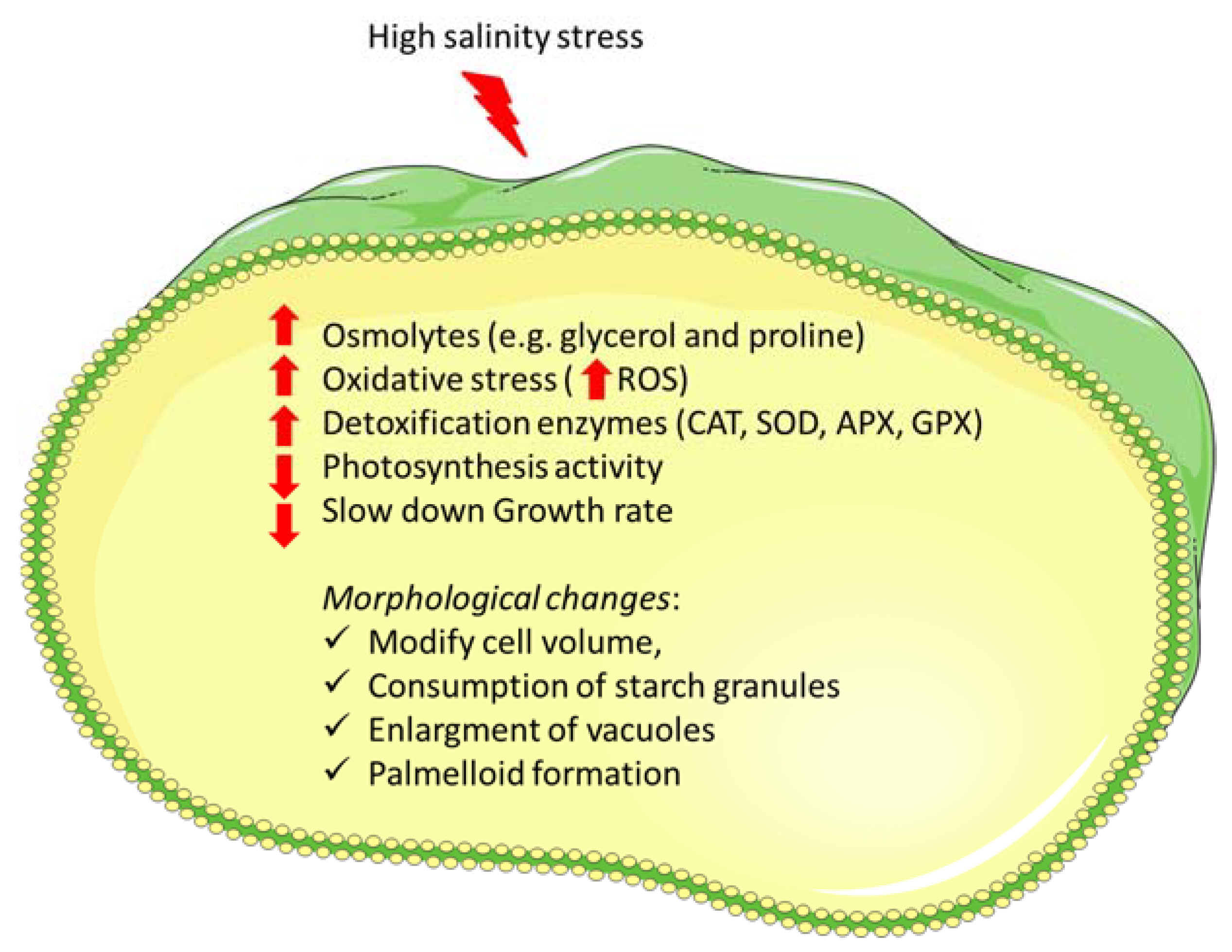
2. Main Physiological Responses to Salinity Stress Exposure

Organisms exposed to salt stress have to cope with both ionic and osmotic imbalance. Multiple morphological, physiological, and molecular adjustments occur simultaneously to improve cell survival under salt stress. Main physiological and morphological effects observed in Chlamydomonas spp. are summarized in Figure 1. High-salt stress slows down the growth rate and can damage the extracellular protective constituents of microalgae, directly influencing their morphological structures. Many studies showed that hypersalinity modifies cell size, ceases motility, and triggers the production of extracellular mucus, which favors aggregation and palmelloid formation in Chlamydomonas species [20,21]. Palmelloidy has been reported in C. reinhardtii under biotic and abiotic stress conditions, including salt stress [20,21]. A “palmelloid” is a temporary colonial-like stage triggered by unfavorable conditions, during which Chlamydomonas cells, typically free-living, undergo characteristic physiological and morphological changes [20]. Although distinct species of Chlamydomonas respond differently to stress conditions, the palmelloid state is generally characterized by clustering of cells, reduction of motility or complete loss of flagella, exopolysaccharide (EPS) secretion, cells sharing a common membrane embedded in an EPS matrix, and thickening of individual cell walls. In C. reinhardtii, motility loss and palmelloids were observed from 50 mM NaCl onwards [21], and palmelloid number increased in a time- and dose-dependent manner. At 100 mM NaCl, the palmelloid number increased until 24 h of NaCl stress and the palmelloid stage was completely reversible, but in the case of extended or excessive stress the palmelloid form remained and the cells eventually died [20].
Moreover, salinity caused a significant and immediate inhibition of nitrate and sulfate uptake rates in C. reinhardtii, at least for 24 h [22].
Then, increasing doses of stress and exposure time ultimately lead to cell death. In C. reinhardtii, high levels of NaCl caused altered mitochondrial membrane potential, but no DNA laddering nor caspase activity was observed when cells were exposed to 200 mM of NaCl. Higher NaCl concentrations, on the other hand, led to sheared genomic DNA, suggesting a necrotic-like pathway of cell death [23]. Moreover, iso-osmotic treatments with sorbitol indicated that it is the ionic rather than the osmotic component of salt stress that causes necrosis at very high NaCl concentrations in C. reinhardtii [23]. Hema et al. [24] also investigated the different impacts of osmotic and ionic stress on cell growth, and suggested that, if subjected to mild stress levels, cells are able to acclimate and modify their cellular processes to restore growth. A preliminary study on volatile organic compounds (VOCs), released when algal cells are under stress, suggested that algae communicate and signal each other to initiate the antioxidant protective pathway that will lead to improved survival [25]. The study showed that salt stress induced in C. reinhardtii cells the release of VOCs, such as hexanal and longifolene. When such compounds were collected and given to Chlamydomonas growing under optimal conditions, a slight increase in chlorophyll, a reduction in cell density, and an increase in antioxidant enzyme activity was observed. Thus, suggesting that VOCs may signal unstressed cells to prepare for an impending stressful condition, as ROS damage.
Morphological changes caused by both high and low salinities have also been studied in Antarctic extremophilic species. In the freezing–thawing cycle, sea-ice microalgae experience salinities three or even five times that of common seawater. In order to survive and grow in their extreme surroundings, Antarctic ice microalgae must have special physiological characteristics. Studies found that hyposalinity, associated with the melting of sea ice, caused an increase in cell volume [26], as did hypersalinity, in Chlamydomonas sp. ARC and Chlamydomonas sp. L4 [26,27]. High salinities also triggered in Chlamydomonas sp. L4 plasmolysis, starch granules consumption, cytoplasmic vacuolarization, appearance of different types of vacuoles, and the presence of electron-opaque deposits in vacuoles [27]. Vacuoles are known to be important for the detoxification of potentially toxic compounds in plants and appear to play a role in cation homeostasis in C. reinhardtii [28]. On the other hand, contractile vacuoles, known to eject water out of the cell, were present in Chlamydomonas sp. ARC only at low salinities (10‰, 20‰), leaving just tubular spongiome complexes at higher salinities [26].
Under hypersalinity, microalgae have been shown to synthetize and accumulate compatible solutes to prevent water loss and regulate ion transporters and pumps to maintain the internal ionic concentrations, especially K+, H+, and Na+ [29]. Compatible solutes, or osmolytes, are small organic molecules characterized by neutral charge and low toxicity at high concentration, which accumulate in organisms in reaction to osmotic stress. They help balancing the osmotic pressure between the outside medium and the cytosol, and assist in the stabilization of enzymes. A comparison between different microalgal species suggested that, despite the possible differences in the osmoregulatory mechanisms, growth at very high salinities is allowed only when glycerol and proline are used as main osmolytes [11]. Ahmad and Hellebust [10] demonstrated that, although both organic and inorganic solutes (i.e., inorganic ions) have a role in osmotic adjustment, glycerol is the main osmoregulatory solute in C. pulsatilla, a highly halotolerant strain. In fact, its growth was optimal at salinities around 10% of artificial seawater (ASW), but it maintained high division rates at salinities as high as 200% ASW. Nevertheless, only at salinities greater than 50% ASW did a substantial accumulation of glycerol take place. Thus, suggesting that the synthesis of glycerol and the uptake of ions are two connected osmoregulatory responses in C. pulsatilla. The freshwater C. reinhardtii is also known to accumulate and excrete glycerol, in a proportional manner with respect to salt concentration in the medium [30]. Glycerol excretion by Chlamydomonas offers interesting insights also for biotechnological applications in glycerol photoproduction, which could be favored by high CO2 conditions [12] or by entrapment in alginate beads (glycerol excretion was about 2-fold higher in Ca-alginate entrapped cells; [30]).
Proline is another common osmolyte, with low molecular weight, neutrally charged and highly soluble. Reynoso et al. [31] performed a preliminary study on the effects of proline (and taurine) on C. reinhardtii cells exposed to salt stress and found that exogenous application of proline reduced the harmful effects of high salinity in C. reinhardtii, increasing salt tolerance in the treated populations. The impact of taurine was similar to that of proline, although smaller. In addition, other amino acids such as leucine and lysine have been found to stimulate Chlamydomonas growth under saline conditions [9], but few studies investigated their specific roles in osmoregulation. The proline content was significantly increased under high salinity also in Chlamydomonas sp. ICE-L, which is periodically exposed to extreme salinity concentrations inside the brine channels in the Antarctic Sea ice [19]. Several molecular studies also confirmed the increase in osmolytes production upon salt stress (see paragraphs below).
Numerous studies have proven that salt stress can lead to oxidative stress, as part of its toxic effect. High salinity can indeed stimulate the production of active oxygen, which increases the permeability of cell membrane, decreases the efficiency of photosynthesis and respiration, and destroys DNA and proteins [32]. Already after 1 h at 200 mM of NaCl and 360 mM of sorbitol, a significant rise in the intracellular H2O2 level was observed in C. reinhardtii [23]. Zuo et al. [33] also observed a rapid burst in H2O2 and O2−• after only 30 min at 50, 100, and 300 mM of NaCl. The defense mechanisms against oxidative stress include both enzymatic and non-enzymatic (e.g., glutathione, thioredoxin, carotenoids, vitamin E, etc.) detoxification systems [34,35]. Enzymatic scavengers include superoxide dismutase (SOD), which is the first line of defense against O2−•, catalase (CAT), and peroxidases (APX and GPX), the main enzymes involved in H2O2 detoxification [36]. Many studies observed an increased activity of antioxidant enzymes in C. reinhardtii cells exposed to high salt concentrations [19,22,23]. Vega et al. [22] showed that a treatment with 200 mM of NaCl highly enhanced CAT activity (up to 20-fold), even to a greater extent compared to the oxidative stress induced by cadmium- and methyl viologen. Authors also found that the enhancement of CAT activity depended on the NaCl concentration. On the other hand, Vavilala et al. [23] showed that H2O2-induced oxidative stress triggered a faster stimulation of CAT activity than NaCl stress, and that, under both H2O2 and NaCl stress, the activity peak was around 10-fold compared to the control. Among the salinity tolerant species, SOD activity was observed to increase significantly in the Antarctic microalga Chlamydomonas sp. L4, which can survive to salinities up to 132. The enzymatic activity increased in a dose dependent manner when the algae were exposed to increasing salinity from 33 to 132, and is thought to have a protective role from oxidative injury to the membranes [27]. In general, studies show that activities of antioxidant enzymes, such as CAT, SOD, APX, and glutathione reductase (GR) are increased in Chlamydomonas not only when the cells are exposed to oxidative stress, but also to salt and osmotic stress. Moreover, glutamate synthase, glutamate dehydrogenase, and O-acetylserine(thioL)lyase activities were investigated under abiotic stresses in C. reinhardtii. Salt enhanced glutamate synthase and O-acetylserine(thioI)lyase activities, suggesting that an active synthesis of glutamate and cysteine was stimulated in C. reinhardtii cells treated with salt. In addition, it has been proven that salt stress can impact antioxidant genes expression [19,23], which will be further discussed in the next section.
In the freshwater alga C. reinhardtii, it was shown that the induction of these antioxidant enzyme activities was not enough to counteract the toxicity generated by ROS at high salinities. Studies suggested that the phytormone abscissic acid (ABA) can support C. reinhardtii survival under high salt conditions. In fact, externally added ABA successfully reduced the deleterious effects of salt stress-induced oxidative stress, partially releasing the growth suppression and reducing ROS generation under salinities around 100 mM of NaCl [37,38]. In a recent study, ABA addition also fully recovered photosynthesis and prevented palmelloid formation under moderately high NaCl levels, whereas it supported the growth of C. reinhardtii at their gamete phase [38]. Thus, Chlamydomonas do respond to ABA even though there are no evidence suggesting it is internally synthesized, and ABA protects Chlamydomonas cells from high salinity, possibly as a signal molecule that mitigates the oxidative stress derived from these conditions. On the other hand, ABA did not reduce the negative effects of the stress during short periods of exposure without illumination, suggesting that ABA does not induce specific reactions against the specific damages caused by osmotic and salt stresses in C. reinhardtii [37].
In general, abiotic stress reduces the rate of photosynthesis by reducing PSII efficiency and electron transport due to ROS formation. Like most photosynthetic organisms, many Chlamydomonas species showed a substantial decrease in photosynthetic activity under high salt stress. This decline in photosynthetic activity can be a consequence of deficiency in different cations, caused by excess Na+, osmotic stress, and ROS production, which affect numerous biochemical and physiological processes. In C. reinhardtii, at a concentration of 200 mM of NaCl, photosynthetic activity was immediately suppressed, then recovered 1 h later at 33% of the original activity, remaining at this level for the next 24 h. On the contrary, respiration level of the alga was less susceptible to salt stress, and it remained about 77% of the original value [22]. At 50 mM of NaCl, no photosynthetic inhibition was observed [30]. The photosynthetic performance in algae and plants is often monitored also by measuring chlorophyll fluorescence. Frequently, the Fv/Fm parameter is employed to estimate the maximum quantum yield of PSII photochemistry, and non-photochemical quenching (NPQ) is used to evaluate energy dissipation through heat loss from PSII. Decreased Fv/Fm, as an indicator of stress conditions, has been observed under short-term salt stress in C. reinhardtii, as well as higher NPQ values [33]. Mou et al. [39] and An et al. [17] also observed lower Fv/Fm and associated induction of NPQ when Chlamydomonas sp. ICE-L was subjected to salinity stress for 2 or 3 h, but notably, this species could accommodate longer periods of stress, which are common in its natural environment. Moreover, Chlamydomonas sp. ICE-L showed considerable stress only at salinities higher than 90‰ NaCl, while it was less susceptible to low salinities. Physiological and biophysical analyses demonstrated that the major effect of short-term hyperosmotic stress on photosynthesis is to inhibit P700+ reduction, blocking the electron transfer between cytochrome c6 or plastocyanin (PC) and P700 [40]. Short-term hyperosmotic stress caused by a range of osmolytes and NaCl did not induce PSI or PC destruction, but caused dehydration of the thylakoid lumen instead, which led to physical obstruction of cytochrome c6 and PC mobility and docking to PSI. Full inhibition of P700+ re-reduction occurred with 150 mM or higher concentrations of NaCl. Under these conditions, electron transport through PSII was also reduced, although to a lower degree [40]. On the other hand, long-term stress experiments in C. reinhardtii showed that from 100 mM of NaCl both photosystems are impaired and PSII is more severely damaged than PSI [21]. The electron transfer activity was reduced by 39% in isolated PSI-light harvesting complex I supercomplexes (PSI-LHCI) under high salt (100 mM of NaCl). Moreover, it was suggested that mainly the acceptor side of PSI was damaged, and pigment–pigment interactions were changed, probably due to ROS damage [41]. Regarding PSII, protein profile analysis revealed that both core and LHCII proteins were degraded in salt grown cells. However, the LHCII major subunit appears to be more prone to salt stress, possibly as a result of oxidative damage [21].
Pigment analysis in C. reinhardtii suggested that severe NaCl stress (300 mM of NaCl) induces carotenoids oxidation, and even degradation by ROS during photoprotection [33], but a lower stress level could actually increase carotenoid content after 48 h [42]. Chlorophyll content usually decreases during salt stress [18,33]. In photosynthetic organisms, chlorophylls and carotenoids are essential pigments for photosynthesis, and carotenoids also play an important role in preventing photo-oxidative damage. The excess light energy absorbed by pigments is dissipated as heat (NPQ) through xanthophyll cycle [43], suggesting that the decrease of pigment content and pigment oxidation may be one reason for the reduction of photosynthetic efficiency.
Acclimation of plant cells and microalgae to low osmotic stress levels has been associated with changes in (phospho)lipid and fatty acid compositions, which are thought to affect the biophysical properties of the membranes [17,27,42]. However, osmotic stress can also to trigger different phospholipid signaling pathways [44,45]. In particular, phosphatidic acid (PA) and its derivates, lyso-phosphatidic acid (LPA) and diacylglycerol pyrophosphate (DGPP), are thought to be involved in the early response to salt stress, and more in general hyperosmotic stress [44,45,46,47]. The unicellular green alga C. moewusii is an excellent model for studying phospholipid metabolism because it rapidly takes up and incorporates 32Pi into all phospholipids [48]. Studies showed that NaCl concentrations above 150 mM stimulate the formation of PA, LPA, and DGPP in C. moewusii. The effect is dramatic, as the increase occurs within minutes of treatment, and in a time- and dose-dependent manner [44,45]. Moreover, C. moewusii cells acclimated to 100 mM of NaCl before measuring changes in phospholipid signaling pathways tolerated higher salt concentrations, suggesting that acclimation can moderate salt stress perception and activate downstream pathways [47].
Overall, Chlamydomonas case offers a wide range of differently adapted species, useful to study both the consequences of salt stress and the characteristics of salt tolerance in salt-adapted species. When compared to other green algal models, such as Dunaliella, palmelloidy appears to be a peculiarity of the Chlamydomonas genus. Dunaliella salina is a highly halotolerant species, characterized by the lack of a cell wall, which allows the organism to rapidly change volume to adjust ion and osmolyte concentration. As for Chlamydomonas, glycolysis and starch degradation are promoted in D. salina cells exposed to high salinity, while carbon flow to the tricarboxylic acid cycle is decreased (excluding some halotolerant species like Chlamydomonas sp. JSC4), in favor of glycerol synthesis [49]. In fact, Dunaliella cells can accumulate massive amounts of glycerol, while proline is the main osmolyte in other halotolerant microalgae, as Picochlorum [50], in which starch degradation is limited. Notably, to tackle with moderate levels of salt stress, D. salina is able to increase photosynthetic activity by significantly increasing chlorophyll-a content, but at higher stress levels photosynthesis decreases similarly to Chlamydomonas species. Furthermore, it seems that an important characteristic shared by halotolerant algae is the capability to maintain the intracellular ion concentration to restore cells homeostasis [10,51]. A schematic comparison between the main responses to salinity stress exposure observed in Chlamydomonas and D. salina is reported in Table 1.

3. Molecular Studies on Salt Stress

The response to salinity stress is complex and involves many genes and biochemical-molecular mechanisms. Many stress-responsive genes have been characterized in Chlamydomonas species, and the overall molecular mechanism of salt-stress response have been deeper studied with the introduction of omics technologies [19,32].

3.1. Stress-Related and Antioxidant Genes

Hoffman and Beck [56] isolated and described the first genes with proved osmoregulation in C. reinhardtii and provided first evidence for the presence of different osmoregulated signaling pathways, which did or did not involve de novo protein synthesis. Namely, three gamete-specific (GAS) Hyp-rich pheophorin-encoding genes, GAS28, GAS30, and GAS31, were found to be induced by exposure to both hypoosmotic or hyperosmotic conditions, adding either sorbitol or NaCl (10–50 mM). These genes are likely induced as a response of the detachment of the cell-wall, originating either from gamete formation, or osmotic stress in vegetative cells. Later on, Hema et al. [24] expressed choline oxidase A (codA) from Arthrobacter globiformis, a well-characterized stress responsive gene, in transgenic C. reinhardtii cells exposed to different severe abiotic stresses, following pre-treatment with mild stress levels, called “induction stress”. CodA containing cells exhibited higher tolerance to salt stress, proving that the relevance and function of stress genes can be effectively validated overexpressing the genes of interest in the Chlamydomonas system. The genes whose involvement in salt stress response or resistance has been investigated in a Chlamydomonas spp. are listed in Table 2, and the most relevant ones are discussed in this section.
Chlamydomonas W80 is a marine species, highly tolerant to salt and oxidative stress, which has been widely used for the identification of stress genes. Chlamydomonas W80 cDNA library screenings allowed to identify several stress genes and characterize some that improved salinity tolerance in transformed Escherichia coli cells [58,64,78] or cyanobacterial cells [65]. Miyasaka et al. [57] first developed the library screening method in Chlamydomonas W80 for the isolation of stress-responsive genes, isolated several unknown genes, and sequenced 35 homologs of previously reported genes, which conferred salt- and/or oxidative-stress tolerance to the transformed E. coli colonies. Of the 35 known genes, 4 were homologs to already known stress genes: glutathione peroxidase (GPX), ascorbate peroxidase (APX), breast basic conserved (BBC1), and alternative oxidase. The alternative oxidase is usually induced by some abiotic stresses and functions as electron sink to prevent ROS formation due to energy imbalance in plant mitochondria. GPX and APX are well known antioxidant enzymes involved in ROS detoxification and both genes have been fully characterized and confirmed to be involved in the molecular mechanism of salt-resistance in Chlamydomonas W80 [58,60]. Moreover, the introduction of Chlamydomonas W80 GPX in tobacco plants succeeded in enhancing the plants salt and oxidative stress tolerances, highlighting the significance of such studies for possible plant molecular breeding [13]. Lastly, BBC1 protein is a highly hydrophilic ribosomal protein commonly found in animals and plants, screened from salt-tolerant E. coli colonies. Further studies confirmed its involvement in salt-stress tolerance and suggested it has a protective role against cellular dehydration [64]. It has been hypothesized that the bbc1 gene could either work activating specific stress genes, using the transcription-activating motif found in the BBC1 protein, or directly protecting the cell against protein and membrane damage, due to its high hydrophily, similar to what LEA (late embryogenesis abundant) proteins do in plants. Although LEA genes are spread across all kingdoms, their function in microalgae remains uncertain [80]. Another functional screening for Chlamydomonas W80 salt stress-responsive genes used the freshwater cyanobacteria Synechococcus PCC7942 as host [65] and resulted in the isolation of a gene with low but significant homology to the group 3 LEA genes (LEA3), cw80lea3. Cw80lea3 is the only LEA-like gene isolated from a Chlamydomonas species, has highly hydrophilic features, and a typical domain that usually characterizes LEA3 proteins. Transformed Synechococcus PCC7942 colonies expressing cw80lea3 showed enhanced tolerance to salt stress.
Together with LEA proteins, heat-shock proteins (Hsps) typically protect membranes and proteins from stress-induced dehydration. The involvement of a putative 70 kDa Hsp, CiHsp70, has been studied in the salt resistant Chlamydomonas sp. ICE-L [66] during a short-term incubation with 62‰ or 93‰ NaCl. Transcript levels increased 3-fold in 62‰ NaCl medium and 2.1-fold in 93‰ NaCl medium after 2 h treatment, and gradually decreased with extending treatment time, confirming its participation in short-term salt stress acclimation [66]. Suda et al. [78] also isolated a novel Chlamydomonas stress gene, WCFII, which is involved in ATP synthesis. WCFII-recombinant E. coli had an extremely strong tolerance not only to salt stress, but also to several heavy metals and oxidative stress compared to the control colonies. The gene mediates increase of intracellular ATP, indicating that the accumulation of ATP confers stress-tolerance in E. coli. Thus, considering that seven other isolated clones were ATP synthesis-related genes, it is hypothesized that the ATP synthesis system plays a crucial role in the response to environmental stress, including salt stress [78].
Enzymes involved in ROS detoxification have been shown to be essential for a proper response to salt stress and are among the most studied. SOD, CAT, and APX involvement in C. reinhardtii salt stress response was also investigated under ABA induction [37]. Expression studies revealed that osmotic stress enhanced only CAT expression, while salt stress (100 mM of NaCl) and ABA enhanced both CAT and APX expression, but not SOD. In contrast, Vavilala et al. (2015) showed that, expression of Mn-SOD, CAT and APX genes were up-regulated under both NaCl (200 mM) and osmotic stress by sorbitol. Expression levels results were confirmed by enzymatic activity as well [23]. Chen et al. [81] also confirmed that salt stress prompted APX activity in C. reinhardtii at least for three days at 100 mM of NaCl, together with other ascorbate-related antioxidant enzymes and glutathione reductase (GR), which also participates in H2O2 detoxification. A GPX homologous gene, gpxh, was isolated from C. reinhardtii as well, and confirmed as oxidative stress responding gene that encodes a GPX-like protein [59]. Gpxh was slightly overexpressed after 3 h at 200 mM of NaCl and highly overexpressed under oxidative stress, already after 30 min. GPXH protein in C. reinhardtii was found to be selenium independent, namely containing a normal cysteine residue in their catalytic site, as was the Chlamydomonas W80 GPX-like protein, resembling the GPX-like proteins typically found in plants and yeast [59]. Interestingly, C. reinhardtii also possesses a selenium-dependent GPX protein, containing a selenocysteine in its catalytic site like the typical mammalian GPX [82], the involvement of which in salt tolerance has not yet been investigated. A selenium-dependent GPX protein was also found in the Antarctic halotolerant Chlamydomonas sp. ICE-L, ICE-LGPx [32], which was overexpressed when Chlamydomonas sp. ICE-L was subjected to high salt or low salt treatment, at least for the first 24 h. After that, the mRNA level usually decreases, possibly due to the gradual detoxification of ROS in the algal cells. Other glutathione-related genes from Chlamydomonas sp. ICE-L, ICE-L glutathione reductase gene (ICE-LGR), and ICE-L glutamate cysteine ligase (ICE-LGCL) increased under salinity stress. ICE-LGR was overexpressed under low and high salinity after 12 h and decreased gradually with increasing treatment time [61]. ICE-LGCL mRNA increased in the first 48 h, but only under low salinities [62].
The ectopic expression of two antioxidant ferredoxins, PETF and FDX5, increased C. reinhardtii salt and heat stress tolerance by decreasing ROS levels in the transgenic cells [63]. Interestingly, the C. reinhardtii cells overexpressing either the FDX5 or the PETF gene showed higher starch and lipid accumulation when compared to the wild type and raised electric power density in a photo microbial fuel cell, emphasizing the potential of FDXs for biotechnological applications.
LHC proteins also play essential roles in photoprotection, other than light capture. Precisely, LhcSR (LI818) are stress-related members of the LHC protein superfamily. Three LI818 genes have been identified in the C. reinhardtii genome, LhcSR1, LhcSR2, and LhcSR3 [7] and two in the Antarctic Chlamydomonas sp. ICE-L, namely LhcSR1 and LhcSR2 [39]. The latter were shown to be quickly up-regulated during salinity stress (93‰ NaCl), reaching the highest levels after two hours. Thus, LhcSR1 and LhcSR2 probably have a photoprotective role during early hypersalinity stress in Chlamydomonas sp. ICE-L, and they may also be involved in thermal energy dissipation (NPQ) [39]. Whether this is a characteristic response of this polar microalga evolved to survive in its extreme environment remains to be elucidated.

3.2. Genes Participating in Compatible Solutes and Lipid Accumulation

Glycerol-3-phosphate dehydrogenase (GPDH) is a key enzyme for the synthesis of glycerol, a major osmolyte, but also for the synthesis of triglycerides (TAGs) [70]. GPDH, in fact, catalyzes the conversion of dihydroxyacetone phosphate (DHAP) to glycerol-3-phosphate (G3P), using NADH as an electron donor. G3P can in turn be used as a precursor for lipid synthesis, or it can be converted into glycerol by a phosphatase. Under salt stress, a GPDH has been shown to be highly expressed in the marine microalga Chlamydomonas sp. JSC4, which also showed enhanced expression of other lipid synthesis-related genes and accumulation of lipids and G3P [73]. C. reinhardtii possesses five NAD(P)+-dependent GPDH homologues, GPD1–5 [7,70]. Of these, only GPD2–4 (GPD2-like isoforms) are chimeric proteins, and cluster together with the other chlorophytes bidomain GPDHs, while GPD1 and GPD5 have a canonical GPDH structure [70]. In silico characterization and RT-PCR suggested that GPD4 (referred to as CrGPDH1) is constitutively expressed and localized in the cytosol, while GPD2 and GPD3 both possess a chloroplasts transit peptide at the N-terminus and showed inducible expression when exposed to NaCl [67,68]. Further studies confirmed GPD2 localization in the chloroplast and verified that recombinant GPD2 and GPD3 proteins have G3P dehydrogenase activity in vitro and could restore NaCl tolerance and glycerol production in a yeast double mutant-complementation experiment. Despite the similarities between the two gene sequences and gene products, the results obtained with GPD2 were significantly stronger. A recent study ultimately demonstrated that GPD2-like isoforms can synthetize glycerol directly from DHAP, suggesting a distinctive plastid pathway for fast glycerol synthesis during acclimation to hyperosmotic stress in Chlamydomonas and other core chlorophytes [70]. A transcriptome search revealed that multiple copies of similar bidomains GPDH-encoding genes are present also in two Chlamydomonas species from Antarctica [71]. At least one of them is strongly up-regulated in high salinity conditions, with the up-regulation being associated with an increase in glycerol production. Moreover, a proteomic analysis showed that GPDH was the most up-regulated enzyme in the Antarctic Chlamydomonas sp. UWO241 under salt stress [83], confirming that these enzymes participate in salt stress response in Antarctic microalgae as well, which are highly adapted to withstand high salinities and freezing temperatures.
Considering the importance of glycerol as a compatible solute in plant physiology, it is important to note that few genes participating in glycerol metabolism showed altered expression in C. reinhardtii cells subjected to salinity stress [70]. Besides GPD2, only glycerol kinase was found to be up-regulated in wild-type cells. Moreover, the kinase showed regular expression levels in GPD2/GPD3 knockdown strains, suggesting a coordinated action of antagonistic enzymes in glycerol metabolism under salt stress. Thus, bidomain GPDHs clearly are key enzymes in glycerol synthesis for salt stress acclimation, although different GPDH isoforms appear to have distinct metabolic functions [69]. On the other hand, their importance in the lipid metabolism is puzzled by other possible rate-limiting steps [69].
Glycerol itself is often not sufficient to balance the osmolarity of the external medium [10,71,83], indicating that the synthesis of other osmoregulatory solutes is necessary. Genes and enzymes involved in the synthesis of sugars such as sucrose [83] or trehalose [32], and amino acids such as proline [19,83] are usually up-regulated during salt stress, but remain less studied.

3.3. Salt Stress Signaling and Transcription Control

Although many salt stress-responsive genes have been identified and characterized to date, little is known about the osmosensing and signaling pathways involved in the upstream perception of salinity changes in Chlamydomonas spp. Recently, calmodulin (CaM) mRNA levels have been shown to respond to fluctuations in salinity levels in Chlamydomonas sp. ICE-L, even if only at very high salinity levels (96‰ and 128‰), indicating a possible involvement of calcium (Ca2+) signaling in the Antarctic microalgae in response to salt stress [75]. Several Ca2+ binding proteins were also significantly up-regulated after short-term and long-term exposure to salt stress in C. reinhardtii, such as CaM, calreticulin, and peroxygenase 3 [32,84], suggesting that Ca2+ signaling as a response to high salt may be a common pathway in Chlamydomonas. Salt treatment also strikingly induced production of nitric oxide (NO) after 1 to 3 days in C. reinhardtii, suggesting NO as another second messenger that triggers salt stress response [81].
A recent study revealed an entire set of genes in the CKIN/SnRK family in C. reinhardtii, using a combination of genome mining, protein–protein interaction databases, and real-time PCR [74]. The SnRK (Snf1-related protein Kinase) genes, CKIN in Chlamydomonas, are serine/threonine kinases, key players in energy sensing and stress response in plant systems, which can activate specific mechanisms and transcription factors to favor cell survival under critical energetic balance. Twenty-one over 22 genes in the CKIN protein family were shown to be significantly overexpressed under salt stress, suggesting their involvement in salt stress response [74]. Moreover, the MAPKs (mitogen activated protein kinases) are thought to be involved in the response to NaCl stress and lipid synthesis in C. reinhardtii, but genetic studies are still missing.
Regarding transcription factors, some evidence suggests the involvement of bZIP transcription factors in the salt stress response pathways. Specifically, a number of bZIP genes, namely CrebZIPs, were found to respond to the application of salt stress to C. reinhardtii cells, being either significantly up-regulated or down-regulated [42]. Supported by physiological observations, authors suggest that these CrebZIPs transcription factors may mediate the regulation of photosynthesis and oil accumulation, especially under salt stress, in C. reinhardtii [42]. Furthermore, the gene Femu2 encodes a transcription factor which resulted to be highly involved in C. reinhardtii hyperosmotic stress response and extremely important for C. reinhardtii salt tolerance [76]. Cells overexpressing Femu2 showed higher NaCl tolerance than the control, while Femu2-silencing transgenic cells could not survive under salt stress. Proline, chlorophyll, and soluble sugar contents are enhanced via Femu2 overexpression, while silencing Femu2 reduced the ROS scavenging capacities of the transgenic cell. RNA-sequencing analysis also showed that Femu2 silencing, or overexpression has a strong effect on global gene expression after 24 h exposure to 100 mM of NaCl, confirming that it profoundly affects the salt stress response in C. reinhardtii [76].
Altogether, these studies focused on single or few genes did not give a comprehensive overall overview on the molecular mechanisms activated by salt-stress, which were better clarified by using omics approaches.

4. Omics Studies

If gene-specific studies are helpful to validate and characterize stress-specific genes, they do not provide the overall view of the stress response mechanism, while “omics” techniques offer the possibility to observe entire cell processes and clarify the global stress responsive networks. In the current paragraph we summarize main results obtained with omics approaches studying Chlamydomonas spp. response to salinity stress (summarized in Table 3).
Recently, the whole-genome sequence of a highly halotolerant Antarctic microalga, Chlamydomonas sp. ICE-L, has been published [19]. Interestingly, the genome showed many expanded gene families, which might reflect the adaptation of Chlamydomonas sp. ICE-L to extreme conditions, such as high salinity levels. Among them, the most remarkable are gene families involved in ionic and osmotic homeostasis, such as various ion transporters, (i.e., NHX, an Na+/H+ exchanger; NCL, Na+/Ca2+ exchanger-like; KEA1, K+ efflux antiporter 1), or genes involved in the synthesis of compatible osmolytes, such as proline, starch, and raffinose. Gene families participating in ROS detoxification, such as APXS (L-ascorbate peroxidase S), katG (catalase-peroxidase), and GPX1 (glutathione peroxidase-like peroxiredoxin) are also expanded. Further RNA-sequencing (RNA-seq) analysis found 3678 transcripts to be differentially expressed in salt-stressed Chlamydomonas sp. ICE-L cells. Remarkably, several genes that were differentially expressed under abiotic stress, had also experienced gene family expansion, confirming their importance in the extreme tolerance that this polar strain exhibits to salt stress. For example, the genes involved in ‘‘antioxidant activity’’ and ‘‘inorganic ion trans-membrane transport’’, but also ‘‘calmodulin-dependent protein kinase activity,’’ were up-regulated under high salinities (64.0‰, 96.7‰). Furthermore, a preliminary proteomic study investigated the effects of salinity on the protein expression in Chlamydomonas sp. ICE-L and allowed the identification of a new peptide (51 kD) [86]. Further studies are necessary to clarify the sequence composition and function of the new peptide.
Since 2007, when the first C. reinhardtii whole-genome sequence was published [7], growing attention has been paid to omics studies of C. reinhardtii under different conditions. The molecular mechanisms underlying C. reinhardtii short-term acclimation to high salt were investigated by Perrineau et al. [85] and Wang et al. [32] by RNA-seq studies. Wang et al. [32] exposed C. reinhardtii strain GY-D55 cells to 200 mM of NaCl for 24 h and identified 10,635 dys-regulated genes under salt stress. Perrineau et al. [85] examined the gene expression profile of a C. reinhardtii CC-503 population exposed to salt stress (200 mM of NaCl) for 48 h, and also compared the results with a population raised in salt rich medium for several generations. In both studies the stress response is evident after a short-term treatment. The analyses found significant overexpression of genes involved in: (i) ROS detoxification and osmotic stress response; (ii) G3P production, and accumulation of intracellular glycerol; (iii) Ca2+ signaling and phosphatidyl inositol [85] or PA [32] signaling; (iv) membrane transport, including metal, phosphate, sugar nucleotide transporters and K+ transporters; (v) transcription and translation; (vi) use of acetate as energy source (e.g., up-regulation of acetyl-coenzyme A synthetase). Consistently, a significant down-regulation was observed in genes involved in photosynthesis and carbon fixation reactions. In addition, Wang et al. [32] observed the up-regulation of several genes participating in starch metabolism, TCA and the carbohydrates metabolism, mostly glycolysis. The authors hypothesized that glycolysis enhancement may serve to decrease the carbohydrate accumulation in cells. On the other hand, the short-term exposure to salt stress in the C. reinhardtii strain CC-503 did not cause significant changes on TCA cycle, nor suggested an accumulation of starch or lipid precursors, with many enzymes being either significantly down-regulated or showing no differences in expression [85]. However, CC-503 is a cell wall-deficient mutant, and thus lacks an important protection from environmental stresses [8,93].
To study long-term effects of salt stress, Perrineau et al. [85] used an experimental evolution approach and examined gene expression differences between a population raised in salt rich medium for several generations and the progenitor population, after exposure to salt stress. The acclimated cells could successfully handle hyperosmolarity, as they were able to grow under salinity at the same rate as the progenitor cells cultivated in salt-free medium, and at a faster rate than the progenitor cells when they were both raised in salt-free medium. Furthermore, the long-term acclimated cells showed significant down-regulation of genes involved in the stress response, enzymes involved in glycerophospholipid metabolism and many transporters when compared to the short-term stressed samples, while calmodulin was still up-regulated. Globally, the up-regulated pathways in the long-term samples were associated with amino acids and nucleotide metabolism. Nevertheless, photosynthesis was still significantly down-regulated (i.e., several LHC proteins and protein components of PSI and PSII), as well as the carbon fixation pathway. Thus, long-term and short-term acclimation responses to salt stress are fundamentally diverse from each other, and it appears that in relatively few generations gene-rich mixotrophic strains such as C. reinhardtii may be able to acclimatize rapidly and tolerate salinity stress, highlighting the importance of their metabolic flexibility and capability to use both inorganic and organic carbon sources for their survival [85].
Proteomic studies revealed post translational modifications on key proteins in response to salt stress, indicating that such modifications could be a specific consequence of NaCl stress and might provide to the cell extra stability to cope with the stress or activate new functions of the proteins [84,87,88]. Proteomic analyses by Yokthongwattana et al. [88] and Mastrobuoni et al. [87] provided the first system-wide analysis of the early response to salt stress in the salt-sensitive C. reinhardtii. Yokthongwattana et al. [88] exposed C. reinhardtii cells to a sudden strong osmotic shock (300 mM of NaCl) for a short time and the proteomic profiles were compared with cells grown in normal TAP medium. The overall changes in proteomic profiles suggested that the energy required to maintain homeostasis is substantial and could be obtained through different energy-producing metabolic pathways, such as the glycolytic pathway and carbohydrate metabolism, and confirmed that chaperones, Hsps, and antioxidant enzymes are required to renature proteins and for ROS scavenging. Moreover, some proteins could be exclusively found in one sample, but not in the other. Most of the salt-stressed exclusives were translation- and stress-related proteins, which are known to be constitutively expressed and critical for cell survival. Thus, it is suggested that short-term exposure of C. reinhardtii to 300 mM NaCl probably triggers post translational modifications on several housekeeping proteins. Post-translational modifications were also confirmed at a lower NaCl concentration in an arginine auxotrophic C. reinhardtii strain observing the dynamic changes occurring in the proteome and metabolome [87]. The combined analysis revealed that metabolic changes could precede proteomic rearrangements, which appear to be more conservative, suggesting a posttranslational activation of the up-regulated enzymes, specifically those involved in proline biosynthesis. Further proteomic studies on C. reinhardtii highlighted the role of NO signal in the regulation of C. reinhardtii tolerance to mild salt stress (100 mM of NaCl) [81]. As nitrate reductase (NR) protein content increased, an NR-dependent NO signal is suggested. Additionally, S-nitrosoglutathione reductase-mediated protein S-nitrosylation is proposed to control the activity of downstream proteins involved in cell autophagy and DNA repair, which also contribute to the early response to salt stress.
Sithtisarn et al. [84], similar to what Perrineau et al. [85] previously experimented with transcriptomic, investigated the proteomic changes in a salinity-tolerant C. reinhardtii CC-503 population which was grown for several generations under salt stress (300 mM of NaCl) and was able to proliferate in high salinity conditions. Despite the visible acclimation of the population to the high salt conditions, several proteins involved in stress and defense (i.e., ROS detoxifying enzymes) were still significantly up-regulated in the salt-tolerant population, as well as several kinases, membrane transport proteins, and proteolytic enzymes for protein turnover and amino acid recycling. Interestingly, under these conditions, the extracellular secreted proteins also differed between the salinity-tolerant and the control strain, with 203 up-regulated and 110 down-regulated proteins in the salinity-tolerant strain [92]. Many signaling proteins, channels, and membrane transport and trafficking proteins were up-regulated in the salinity-tolerant secretome, such as CaM, 1 4-3-3 homologs, matrix metalloproteases, and Fructose-1,6-bisphosphate aldolase (FBA). Moreover, many proteins lacked a signal peptide, suggesting the existence of an unconventional protein secretion pathway in Chlamydomonas, which might facilitate a rapid release of the stress-related proteins [92]. Therefore, saline stress acts also on the secreted proteins and likely communication among cells is involved in the stress response.
A detailed morphological, biochemical, and proteomic study on salt-induced multicellular states in C. reinhardtii, palmelloids, is also available [20]. Specifically, a quantitative proteomic analysis of stress and post-stress spent media was performed exposing the cells to salt stress for a maximum of 24 h, followed by 1 h of de-stress. Two categories of differentially expressed proteins in the spent media were recognized: those responding to NaCl stress, and those responding to the removal of the stress. Expansin was the most abundant protein after NaCl stress, which is probably involved in the cell wall loosening that precedes palmelloid formation. Other cell wall up-regulated proteins are pheophorins, hydroxyproline-rich glycoprotein, cathepsin-Z-like protein, and a wall stress-responsive component (WSC) domain protein. They also identified ferroxidase (FOX1), which is induced by iron deficiency, and other proteins involved in cellular metabolism as putatively implicated in palmelloid formation. After stress removal, the cells dissociate due to degradation of the exopolysaccharide matrix, which is probably facilitated by the accumulation of a protein with a PAN/APPLE-like domain, which mediates protein–protein or protein–carbohydrate interactions. Moreover, two matrix metalloproteinases (MMP13 and MMP3), endopeptidase involved in the degradation of the extracellular polysaccharide matrix, were found to be up-regulated.
A proteomic study on the polar Chlamydomonas sp. UWO 241 [83], found in the Antarctic Lake Bonney (characterized by low light, freezing temperatures, and hypersalinity ≈ 700 mM NaCl), showed that a sustained cyclic electron flow around PSI supports a robust growth and photosynthesis, providing constitutive photoprotection while also producing additional ATP for downstream metabolism [83]. Most of the up-regulated enzymes were related to the Calvin–Benson–Bassham cycle and carbon storage metabolism, such as starch metabolism, suggesting that, in contrast with salt-sensitive microalgae, the photosynthetic apparatus of Chlamydomonas sp. UWO 241 is adapted to support photosynthesis under high salinity conditions and have a strong carbon fixation potential. Moreover, Chlamydomonas sp. UWO 241 showed overexpression of several ribosomal proteins and up-regulation of two key shikimate enzymes, while the metabolome analysis also suggested a shift in the metabolism to the production of osmoprotectants and compatible solutes, such as glycerol, proline, and sucrose [83].
Several studies used metabolomics to focus on the carbohydrate and lipid metabolism [18,73]. Ho et al. [73] used dynamic metabolic profiling and transcription analysis of Chlamydomonas sp. JSC4, a salt-tolerant coastal strain, to clarify the switching mechanisms from starch to lipid synthesis at different salinities and to identify the rate-limiting steps for increasing lipid accumulation. The metabolite analysis focused on the key metabolites linked to lipid and starch synthesis and showed that the pool sizes of lipid synthesis-related metabolites, such as acetyl-CoA and G3P, significantly increased, also confirming the plausible accumulation of glycerol under salt stress. Thus, Chlamydomonas sp. JSC4 likely accumulates lipids under salinity stress utilizing newly incorporated inorganic carbon, but also by using intracellular carbon sources, such as carbohydrates. Interestingly, it has been suggested that this shift from starch to lipids as major energy storage compounds could be a specific Chlamydomonas sp. JSC4 adaptation to survive in its natural, brackish environment, since freshwater species such as C. reinhardtii did not show any salinity-induced carbon flow switching [93]. These insights also highlight the potential of this oleaginous green alga for biodiesel production, and to elucidate mechanisms for maximizing the lipid production, which is further discussed in the next paragraph. Existing studies on C. nivalis confirmed the involvement of lipid metabolism in the molecular response to salt stress. In fact, crescent NaCl concentrations could significantly affect intracellular C. nivalis lipid composition [89,91]. Several potential lipid biomarkers with up- or down-regulation in salt stressed cells were also identified, and they have been suggested to play important roles in signal transduction, cell membrane stability, and photosynthesis rate under NaCl stress [89,90]. For example, an increase in phosphatidyl inositol was registered, which is an important constituent of membranes and precursor of essential signaling molecules, as well as phosphatidyl–ethanolamine (18:1/18:1), and various sulfolipids and galactolipids. On the other hand, some lipids experienced a sharp decline, which could eventually bring to the reduction of membrane permeability to NaCl. Phosphatidyl glycerol also decreased under salt stress, and, as it plays a vital role in the aggregation of PSII LHC proteins in green algae, its decline resulted in a significant decrease in the photosynthesis rate.

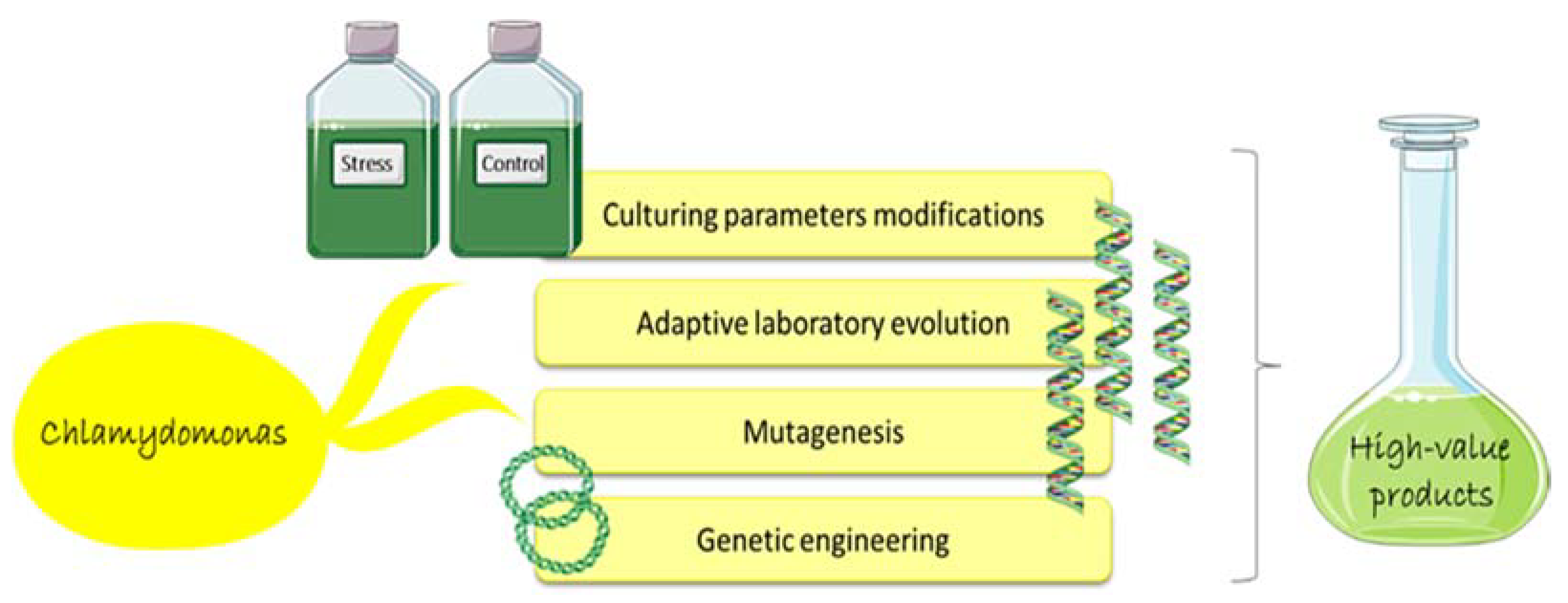
5. Chlamydomonas as Cell Factory

Microalgae, especially in recent years, have received great attention as promising source of interesting compounds with possible biotechnological applications [94,95,96,97,98,99,100,101,102,103,104,105,106]. However, different methodologies have been studied and applied for microalgae in order to implement the production of the compounds of interest, such as modifying culturing parameters, or by using adaptive laboratory evolution (ALE), mutagenesis, and genetic engineering techniques (Figure 2; [107,108,109]).
Hounslow et al. [18,110] studied the effects of salt stress (0.1, 0.2, 0.3 M of NaCl) on a C. reinhardtii low-starch strain, showing a lipid concentration increase, even after long-term exposure to 0.1 M of NaCl or short-term to 0.2 and 0.3 M of NaCl, demonstrating how salt stress can trigger lipid production and suggesting this strategy to produce lipids for biodiesel. Nevertheless, general results on Chlamydomonas spp. indicated that more “robust” and halotolerant species may be more suitable for biofuel-directed lipid production [18]. Kato et al. [111] confirmed that high salinity stress induced lipid accumulation in a halotolerant Chlamydomonas sp. The Antarctic ice microalga Chlamydomonas sp. ICE-L, known to be highly resistant to salt stress, was studied by An et al. [17] as possible alternative species for the production of microalgal oil. NaCl stress effects were evaluated on algal growth and oil yield. In addition, considering that the regulation of lipid biosynthesis in microalgae is a crucial strategy for resistance to salt stress, expression of five fatty acid desaturase genes was also evaluated: stearoyl-ACP-desaturase (Δ9ACPFAD), Δ6 fatty acid desaturase (Δ6FAD), endoplasmic reticulum ω-3 fatty acid desaturase (ω3FAD1), chloroplast ω6 fatty acid desaturase (Δ12FAD), and chloroplast ω-3 fatty acid desaturase (ω3FAD2). Authors observed that algal growth rate decreased with the gradual increase in NaCl concentration, and the highest lipid content was achieved at 16‰ NaCl (C18:3 was the dominant PUFA). At the gene level, Δ9ACPCiFAD was the most up-regulated in response to altered salt stress, while Δ12CiFAD, ω3CiFAD2, and Δ6CiFAD showed a delayed expression increase. Ho et al. [14] studied the combined effects of salinity and nitrogen depletion stress exposure on lipid accumulation in a newly isolated marine microalga, namely Chlamydomonas sp. JSC4. A new cultivation strategy (denoted salinity-gradient operation) was also used in order to increase lipid accumulation reaching an optimal lipid productivity of 223.2 mg L−1 d−1 and a lipid content of 59.4% per dry cell weight. Authors highlighted that this performance was significantly higher than those reported in most related studies and showed that the combination of biological and engineering technologies helped to effectively enhance oil production.
Another approach, named adaptive laboratory evolution (ALE), used a long-term cultivation of species under selective conditions in order to improve phenotypes by spontaneous mutations [112]. ALE aims to improve the stress tolerance of microalgae and at the same time enhance the production of high value-added products (such as lipids and carotenoids). In recent years, there have been many ALE experiments which were focused on nutrient stress, environmental stress, oxidative stress, and natural selection stress (as reviewed by Sun et al. [113]). Regarding Chlamydomonas spp. this approach was used in nitrogen limitation for 50 days (intracellular lipid bodies massively increased; [114]) and nitrogen starvation for 84 days (lipid productivity increased by 2.36 times; [115]). ALE was recently applied to salt-resistant Chlamydomonas strains, cultivated using a combined ALE (5–7% sea salt for dozens of weeks) and mutation strategy (irradiated with 50 or 100 gray (Gy) of the carbon ion beams 12C5+ 220 MeV, [111,116]). The experiments showed increased biomass production under high salinity, but delayed starch degradation and decreased lipid content.
Chemical mutagenesis by using ethyl methanesulfonate was used to enhance lipid production by C. reinhardtii [117]. Similarly, UV mutagenesis integrated with fluorescence-activated cell sorting (FACS) for selection, and confocal Raman microscopy for lipid analysis was used in Chlamydomonas to propose it as model for generating lipid-accumulating microalgae [118,119]. The experiments with Raman microscopy allowed to quantify unsaturation levels and chain lengths of microalgal lipids, important parameters for optimal production of biofuels, and the results showed stable clonal differences on saturation status of expressed lipids.
Altogether, results obtained by using the different methodologies showed that it is possible to increase biomass production or stimulate the production of specific metabolites (e.g., lipids) by Chlamydomonas. Studies have mainly focused on lipid production under high salinity conditions for biodiesel applications, but these results are of great interest also for the nutraceutical and cosmeceutical sectors. Considering its nutritional composition, Chlamydomonas has been in fact recently proposed as a potential food supplement [120] and an oil for cosmetic application is already been reported, AcquaSeal® Algae (https://activeconceptsllc.com/products/functional-actives/acquaseal-algae/, accessed on 28 October 2021).
Chlamydomonas is the first and best-studied transformation system between chlorophytes and has been recognized as suitable host for the production of high-value compounds, such as human erythropoietin, human interferon β1 and human proinsulin [121], as well as bio-hydrogen [122]. The first nuclear and chloroplast transformations of C. reinhardtii were in 1988–1989 [123,124,125]; successively, there was the first Chlamydomonas mitochondrial transformation [126], the production of the first therapeutic recombinant protein in its chloroplast [127], the sequencing of the nuclear genome [7], the application of genome editing techniques (e.g., engineered zinc-finger nucleases, transcription activator-like effectors TALE, and clustered regularly interspersed short palindromic repeat (CRISPR) system [128,129,130,131,132,133,134]), the creation of a knock-out collection [135], as well as the omics studies, discussed in the previous paragraph. These steps greatly contributed in the rise of C. reinhardtii as a model system for molecular biology studies [136]. A great challenge will be the routinely use of transgenic microalgae as cell factories for the production of drugs or other added-value compounds.

6. Conclusions

The present review gives an overview of what is currently known on the response of Chlamydomonas spp. to salinity stress. The morphological (e.g., strains with or without cell wall) and ecological (e.g., freshwater versus marine, polar versus temperate Chlamydomonas) variety of Chlamydomonas species poses some difficulties when comparing the results. Moreover, the use of different mutants (e.g., wall-less, starch-less C. reinhardtii), and experimental conditions, mainly in terms of salinity concentrations and exposure time (see Table 1 and Table 2), must be taken into account when drawing general conclusions. Overall, hyposmotic shock appears to be less studied than hyperosmotic salt stress. Physiological, morphological, molecular, and chemical analyses have been performed, which explored osmolytes accumulation, palmelloids formation, metabolism rearrangements, and the cell’s efforts toward ROS and salt detoxification.
Although there are some differences among species, common traits contributing to salt tolerance can be identified. Modification of cell size and vacuoles helps in balancing the osmotic pressure and in the detoxification process; loss of flagella, mucus production, and palmelloid formation protect the cell from the external environment. Molecular responses aid the boosting of the antioxidant system and a metabolic reorganization (e.g., glycolysis enhancement and starch consumption), to counteract the toxic effects of osmotic imbalance and ROS damage, that reflect in the physiology of the cell as reduced photosynthetic activity, reduced growth, and increased antioxidant activity. Moreover, up-regulation of membrane transport proteins helps to cope with the ionic stress. Finally, communication between cells is thought to contribute to stress resistance.
Omics studies gave a comprehensive overview of the salt stress response, while studies on specific genes helped to better characterize the functions of single genes and to understand if they could represent biomarkers for environmental health status control (e.g., to monitor global climate change). Molecular resources for microalgae, and especially those from marine environments, are still scarce [137] and the possibility of applying genetic engineering technologies is still limited. However, the advent of the omic era offered great opportunities for better understanding microalgae, their responses to main environmental drivers and the compounds they may produce [14,18,138,139,140,141,142,143]. Omics studies also allowed to demonstrate that under salt stress, key enzymes such as those related to osmolytes metabolism are largely controlled by post-translational modifications and this may be true for other pathways as well and need further investigations [70]. Researches on salt stress are very important especially for less studied species, such as polar Chlamydomonas, which experience and will experience severe salinity variations due to seasonal freezing/melting cycles, as well as melting of sea ice due to climate changes, which may affect species biodiversity and distribution [4,144]. This review also points out that, besides proteomic and transcriptomic studies, very few data are available on osmosensors and on the signaling networks/upstream components mediating salt stress responses, although some more specific studies are emerging. Moreover, some information on how cells might communicate with each other to induce an early stress response may offer interesting insights for future studies [92].
Salinity changes are strongly influencing some agricultural areas [145], resulting in considerable economic losses worldwide. Understanding both short- and long-term responses to high/low salinity will help to predict which species are more amenable to survive in harsh conditions, as well as to better direct future research in algal biology and biotechnology. Chlamydomonas spp. have been found to produce, naturally or via genetic modifications, interesting economically valuable compounds. In addition, Chlamydomonas spp. can be easily cultivated in large volumes and are characterized by high metabolic flexibility, thus attracting scientific interest not only because they represent a model for physiological and molecular studies, but also as key species for the production of high-added value products. The potential for lipid accumulation under salt stress is gaining interest for its possible application in biofuel production, but is also one of the most debated topics due to its variability among strains and among species [18,93]. Moreover, genetic engineering techniques have been developed for few microalgal species, including C. reinhardtii [146], thus more work is necessary to transform new microalgal species, especially those known to produce compounds with commercial value. Different approaches have been used until now, even if on different strains and experimental conditions. This review suggests that a multidisciplinary approach may help contribute to our understanding of microalgae with key roles in aquatic ecosystem functioning and give great insights in cell biology, evolution, metabolic adaptation strategies, and marine biotechnologies.

Author Contributions

Conceptualization, C.L. and M.S.; writing—original draft preparation, E.B., C.L. and M.S.; writing—review and editing, E.B., C.L., O.M., F.B. and M.S. All authors have read and agreed to the published version of the manuscript.

Funding

This research was funded by Italian projects: Grande Progetto “Risanamento Ambientale e Valorizzazione dei Laghi dei campi Flegrei”-POR Campania FERS 2014-2020 and by the ABBaCo project (Restauro Ambientale e Balneabilità del SIN Bagnoli-Coroglio, grant number C62F16000170001), funded by the Italian Ministry of University and Research (MIUR).

Institutional Review Board Statement

Not applicable.

Informed Consent Statement

Not applicable.

Data Availability Statement

Not applicable.

Acknowledgments

Authors thank Servier Medical Art (SMART) website (https://smart.servier.com/; accessed on 4 October 2021) by Servier for elements in Figure 1 and Figure 2.

Conflicts of Interest

The authors declare no conflict of interest.

References

  1. Durack, P.J.; Wijffels, S.E.; Matear, R.J. Ocean Salinities Reveal Strong Global Water Cycle Intensification during 1950 to 2000. Science 2012, 336, 455–458. [Google Scholar] [CrossRef] [Green Version]
  2. Beardall, J.; Raven, J.A. The Potential Effects of Global Climate Change on Microalgal Photosynthesis, Growth and Ecology. Phycologia 2004, 43, 26–40. [Google Scholar] [CrossRef]
  3. Lauritano, C.; Ianora, A. Chemical Defense in Marine Organisms. Mar. Drugs 2020, 18, 518. [Google Scholar] [CrossRef]
  4. Lauritano, C.; Rizzo, C.; Lo Giudice, A.; Saggiomo, M. Physiological and Molecular Responses to Main Environmental Stressors of Microalgae and Bacteria in Polar Marine Environments. Microorganisms 2020, 8, 1957. [Google Scholar] [CrossRef]
  5. Harris, E.H. The Chlamydomonas Sourcebook, 2nd ed.; Harris, E.H., Stern, D.B., Witman, G.B., Eds.; Academic Press: London, UK, 2009; p. iii. ISBN 978-0-12-370873-1. [Google Scholar]
  6. Pröschold, T.; Marin, B.; Schlösser, U.G.; Melkonian, M. Molecular Phylogeny and Taxonomic Revision of Chlamydomonas (Chlorophyta). I. Emendation of Chlamydomonas Ehrenberg and Chloromonas gobi, and Description of Oogamochlamys Gen. Nov. and Lobochlamys Gen. Nov. Promoter of the Systematics of Thegenus. Protist 2001, 152, 265–300. [Google Scholar] [CrossRef]
  7. Merchant, S.S.; Prochnik, S.E.; Vallon, O.; Harris, E.H.; Karpowicz, S.J.; Witman, G.B.; Terry, A.; Salamov, A.; Fritz-Laylin, L.K.; Maréchal-Drouard, L.; et al. The Chlamydomonas Genome Reveals the Evolution of Key Animal and Plant Functions. Science 2007, 318, 245–250. [Google Scholar] [CrossRef] [Green Version]
  8. de Carpentier, F.; Lemaire, S.D.; Danon, A. When Unity Is Strength: The Strategies Used by Chlamydomonas to Survive Environmental Stresses. Cells 2019, 8, 1307. [Google Scholar] [CrossRef] [Green Version]
  9. Hellebust, J.A.; Le Gresley, S.M.L. Growth Characteristics of the Marine Rock Pool Flagellate Chlamydomonas pulsatilla Wollenweber (Chlorophyta). Phycologia 1985, 24, 225–229. [Google Scholar] [CrossRef]
  10. Ahmad, I.; Hellebust, J.A. The Role of Glycerol and Inorganic Ions in Osmoregulatory Responses of the Euryhaline Flagellate Chlamydomonas pulsatilla Wollenweber 1. Plant. Physiol. 1986, 82, 406–410. [Google Scholar] [CrossRef] [Green Version]
  11. Hellebust, J.A. Mechanisms of Response to Salinity in Halotolerant Microalgae. Plant. Soil 1985, 89, 69–81. [Google Scholar] [CrossRef]
  12. Miyasaka, H.; Ohnishi, Y.; Akano, T.; Fukatsu, K.; Mizoguchi, T.; Yagi, K.; Maeda, I.; Ikuta, Y.; Matsumoto, H.; Shioji, N.; et al. Excretion of Glycerol by the Marine Chlamydomonas sp. Strain W-80 in High CO2 Cultures. J. Ferment. Bioeng. 1998, 85, 122–124. [Google Scholar] [CrossRef]
  13. Yoshimura, K.; Miyao, K.; Gaber, A.; Takeda, T.; Kanaboshi, H.; Miyasaka, H.; Shigeoka, S. Enhancement of Stress Tolerance in Transgenic Tobacco Plants Overexpressing Chlamydomonas Glutathione Peroxidase in Chloroplasts or Cytosol. Plant. J. 2004, 37, 21–33. [Google Scholar] [CrossRef]
  14. Ho, S.-H.; Nakanishi, A.; Ye, X.; Chang, J.-S.; Hara, K.; Hasunuma, T.; Kondo, A. Optimizing Biodiesel Production in Marine Chlamydomonas sp. JSC4 through Metabolic Profiling and an Innovative Salinity-Gradient Strategy. Biotechnol. Biofuels 2014, 7, 97. [Google Scholar] [CrossRef] [Green Version]
  15. Xie, Y.; Lu, K.; Zhao, X.; Ma, R.; Chen, J.; Ho, S.-H. Manipulating Nutritional Conditions and Salinity-Gradient Stress for Enhanced Lutein Production in Marine Microalga Chlamydomonas sp. Biotechnol. J. 2019, 14, 1800380. [Google Scholar] [CrossRef]
  16. Cvetkovska, M.; Hüner, N.P.A.; Smith, D.R. Chilling out: The Evolution and Diversification of Psychrophilic Algae with a Focus on Chlamydomonadales. Polar Biol. 2017, 40, 1169–1184. [Google Scholar] [CrossRef]
  17. An, M.; Mou, S.; Zhang, X.; Zheng, Z.; Ye, N.; Wang, D.; Zhang, W.; Miao, J. Expression of Fatty Acid Desaturase Genes and Fatty Acid Accumulation in Chlamydomonas sp. ICE-L under Salt Stress. Bioresour Technol. 2013, 149, 77–83. [Google Scholar] [CrossRef]
  18. Hounslow, E.; Evans, C.A.; Pandhal, J.; Sydney, T.; Couto, N.; Pham, T.K.; Gilmour, D.J.; Wright, P.C. Quantitative Proteomic Comparison of Salt Stress in Chlamydomonas reinhardtii and the Snow Alga Chlamydomonas nivalis Reveals Mechanisms for Salt-Triggered Fatty Acid Accumulation via Reallocation of Carbon Resources. Biotechnol. Biofuels 2021, 14, 121. [Google Scholar] [CrossRef]
  19. Zhang, Z.; Qu, C.; Zhang, K.; He, Y.; Zhao, X.; Yang, L.; Zheng, Z.; Ma, X.; Wang, X.; Wang, W.; et al. Adaptation to Extreme Antarctic Environments Revealed by the Genome of a Sea Ice Green Alga. Curr. Biol. 2020, 30, 3330–3341.e7. [Google Scholar] [CrossRef]
  20. Khona, D.K.; Shirolikar, S.M.; Gawde, K.K.; Hom, E.; Deodhar, M.A.; D’Souza, J.S. Characterization of Salt Stress-Induced Palmelloids in the Green Alga, Chlamydomonas reinhardtii. Algal Res. 2016, 16, 434–448. [Google Scholar] [CrossRef]
  21. Neelam, S.; Subramanyam, R. Alteration of Photochemistry and Protein Degradation of Photosystem II from Chlamydomonas reinhardtii under High Salt Grown Cells. J. Photochem. Photobiol. B Biol. 2013, 124, 63–70. [Google Scholar] [CrossRef]
  22. Vega, J.M.; Garbayo, I.; Domínguez, M.J.; Vigara, J. Effect of Abiotic Stress on Photosynthesis and Respiration in Chlamydomonas reinhardtii: Induction of Oxidative Stress. Enzyme Microb. Technol. 2006, 40, 163–167. [Google Scholar] [CrossRef]
  23. Vavilala, S.L.; Gawde, K.K.; Sinha, M.; D’Souza, J.S. Programmed Cell Death Is Induced by Hydrogen Peroxide but Not by Excessive Ionic Stress of Sodium Chloride in the Unicellular Green Alga Chlamydomonas reinhardtii. Eur. J. Phycol. 2015, 50, 422–438. [Google Scholar] [CrossRef]
  24. Hema, R.; Senthil-Kumar, M.; Shivakumar, S.; Chandrasekhara Reddy, P.; Udayakumar, M. Chlamydomonas reinhardtii, a Model System for Functional Validation of Abiotic Stress Responsive Genes. Planta 2007, 226, 655–670. [Google Scholar] [CrossRef]
  25. Zuo, Z.-J.; Zhu, Y.-R.; Bai, Y.-L.; Wang, Y. Volatile Communication between Chlamydomonas reinhardtii Cells under Salt Stress. Biochem. Syst. Ecol. 2012, 40, 19–24. [Google Scholar] [CrossRef]
  26. Eddie, B.; Krembs, C.; Neuer, S. Characterization and Growth Response to Temperature and Salinity of Psychrophilic, Halotolerant Chlamydomonas sp. ARC Isolated from Chukchi Sea Ice. Mar. Ecol. Prog. Ser. 2008, 354, 107–117. [Google Scholar] [CrossRef]
  27. Kan, G.; Shi, C.; Wang, X.; Xie, Q.; Wang, M.; Wang, X.; Miao, J. Acclimatory Responses to High-Salt Stress in Chlamydomonas (Chlorophyta, Chlorophyceae) from Antarctica. Acta Oceanol. Sin. 2012, 31, 116–124. [Google Scholar] [CrossRef]
  28. Pittman, J.K.; Edmond, C.; Sunderland, P.A.; Bray, C.M. A Cation-Regulated and Proton Gradient-Dependent Cation Transporter from Chlamydomonas reinhardtii Has a Role in Calcium and Sodium Homeostasis. J. Biol. Chem. 2009, 284, 525–533. [Google Scholar] [CrossRef] [Green Version]
  29. Shetty, P.; Gitau, M.M.; Maróti, G. Salinity Stress Responses and Adaptation Mechanisms in Eukaryotic Green Microalgae. Cells 2019, 8, 1657. [Google Scholar] [CrossRef] [Green Version]
  30. León, R.; Galván, F. Halotolerance Studies on Chlamydomonas reinhardtii: Glycerol Excretion by Free and Immobilized Cells. J. Appl. Phycol. 1994, 6, 13–20. [Google Scholar] [CrossRef]
  31. Reynoso, G.T.; de Gamboa, B.A. Salt Tolerance in the Freshwater Algae Chlamydomonas reinhardii: Effect of Proline and Taurine. Comp. Biochem. Physiol. Part. A Physiol. 1982, 73, 95–99. [Google Scholar] [CrossRef]
  32. Wang, N.; Qian, Z.; Luo, M.; Fan, S.; Zhang, X.; Zhang, L. Identification of Salt Stress Responding Genes Using Transcriptome Analysis in Green Alga Chlamydomonas reinhardtii. Int. J. Mol. Sci. 2018, 19, 3359. [Google Scholar] [CrossRef] [Green Version]
  33. Zuo, Z.; Chen, Z.; Zhu, Y.; Bai, Y.; Wang, Y. Effects of NaCl and Na2CO3 Stresses on Photosynthetic Ability of Chlamydomonas reinhardtii. Biologia 2014, 69, 1314–1322. [Google Scholar] [CrossRef]
  34. Li, Z.; Keasling, J.D.; Niyogi, K.K. Overlapping Photoprotective Function of Vitamin E and Carotenoids in Chlamydomonas. Plant. Physiol. 2012, 158, 313–323. [Google Scholar] [CrossRef] [Green Version]
  35. Çakmak, Z.E.; Ölmez, T.T.; Çakmak, T.; Menemen, Y.; Tekinay, T. Antioxidant Response of Chlamydomonas reinhardtii Grown under Different Element Regimes. Phycol. Res. 2015, 63, 202–211. [Google Scholar] [CrossRef]
  36. Lauritano, C.; Procaccini, G.; Ianora, A. Gene Expression Patterns and Stress Response in Marine Copepods. Mar. Environ. Res. 2012, 76, 22–31. [Google Scholar] [CrossRef]
  37. Yoshida, K.; Igarashi, E.; Wakatsuki, E.; Miyamoto, K.; Hirata, K. Mitigation of Osmotic and Salt Stresses by Abscisic Acid through Reduction of Stress-Derived Oxidative Damage in Chlamydomonas reinhardtii. Plant. Sci. 2004, 167, 1335–1341. [Google Scholar] [CrossRef]
  38. Abu-Ghosh, S.; Iluz, D.; Dubinsky, Z.; Miller, G. Exogenous Abscisic Acid Confers Salinity Tolerance in Chlamydomonas reinhardtii during Its Life Cycle. J. Phycol. 2021, 57, 1323–1334. [Google Scholar] [CrossRef]
  39. Mou, S.; Zhang, X.; Ye, N.; Dong, M.; Liang, C.; Liang, Q.; Miao, J.; Xu, D.; Zheng, Z. Cloning and Expression Analysis of Two Different LhcSR Genes Involved in Stress Adaptation in an Antarctic Microalga, Chlamydomonas sp. ICE-L. Extremophiles 2012, 16, 193–203. [Google Scholar] [CrossRef]
  40. Cruz, J.A.; Salbilla, B.A.; Kanazawa, A.; Kramer, D.M. Inhibition of Plastocyanin to P700 +Electron Transfer in Chlamydomonas reinhardtii by Hyperosmotic Stress. Plant. Physiol. 2001, 127, 1167–1179. [Google Scholar] [CrossRef]
  41. Subramanyam, R.; Jolley, C.; Thangaraj, B.; Nellaepalli, S.; Webber, A.N.; Fromme, P. Structural and Functional Changes of PSI-LHCI Supercomplexes of Chlamydomonas reinhardtii Cells Grown under High Salt Conditions. Planta 2010, 231, 913–922. [Google Scholar] [CrossRef]
  42. Ji, C.; Mao, X.; Hao, J.; Wang, X.; Xue, J.; Cui, H.; Li, R. Analysis of BZIP Transcription Factor Family and Their Expressions under Salt Stress in Chlamydomonas reinhardtii. Int. J. Mol. Sci. 2018, 19, 2800. [Google Scholar] [CrossRef] [Green Version]
  43. Ivanov, A.G.; Hurry, V.; Sane, P.V.; Öquist, G.; Huner, N.P.A. Reaction Centre Quenching of Excess Light Energy and Photoprotection of Photosystem II. J. Plant. Biol. 2008, 51, 85. [Google Scholar] [CrossRef]
  44. Meijer, H.J.G.; Arisz, S.A.; Van Himbergen, J.A.J.; Musgrave, A.; Munnik, T. Hyperosmotic Stress Rapidly Generates Lyso-Phosphatidic Acid in Chlamydomonas. Plant. J. 2001, 25, 541–548. [Google Scholar] [CrossRef]
  45. Munnik, T.; Meijer, H.J.G.; Ter Riet, B.; Hirt, H.; Frank, W.; Bartels, D.; Musgrave, A. Hyperosmotic Stress Stimulates Phospholipase D Activity and Elevates the Levels of Phosphatidic Acid and Diacylglycerol Pyrophosphate. Plant. J. 2000, 22, 147–154. [Google Scholar] [CrossRef]
  46. Arisz, S.A.; Munnik, T. The Salt Stress-Induced LPA Response in Chlamydomonas Is Produced via PLA₂ Hydrolysis of DGK-Generated Phosphatidic Acid. J. Lipid Res. 2011, 52, 2012–2020. [Google Scholar] [CrossRef] [Green Version]
  47. Meijer, H.J.G.; van Himbergen, J.A.J.; Musgrave, A.; Munnik, T. Acclimation to Salt Modifies the Activation of Several Osmotic Stress-Activated Lipid Signalling Pathways in Chlamydomonas. Phytochemistry 2017, 135, 64–72. [Google Scholar] [CrossRef]
  48. Arisz, S.A.; van Himbergen, J.A.J.; Musgrave, A.; van den Ende, H.; Munnik, T. Polar Glycerolipids of Chlamydomonas moewusii. Phytochemistry 2000, 53, 265–270. [Google Scholar] [CrossRef]
  49. Xia, B.-B.; Wang, S.-H.; Duan, J.-B.; Bai, L.-H. The Relationship of Glycerol and Glycolysis Metabolism Patway under Hyperosmotic Stress in Dunaliella salina. Cent. Eur. J. Biol. 2014, 9, 901–908. [Google Scholar] [CrossRef] [Green Version]
  50. Foflonker, F.; Ananyev, G.; Qiu, H.; Morrison, A.; Palenik, B.; Dismukes, G.C.; Bhattacharya, D. The Unexpected Extremophile: Tolerance to Fluctuating Salinity in the Green Alga Picochlorum. Algal Res. 2016, 16, 465–472. [Google Scholar] [CrossRef] [Green Version]
  51. Pick, U.; Karni, L.; Avron, M. Determination of Ion Content and Ion Fluxes in the Halotolerant Alga Dunaliella salina. Plant. Physiol. 1986, 81, 92–96. [Google Scholar] [CrossRef] [Green Version]
  52. Gao, F.; Nan, F.; Feng, J.; Lü, J.; Liu, Q.; Liu, X.; Xie, S. Transcriptome Profile of Dunaliella salina in Yuncheng Salt Lake Reveals Salt-Stress-Related Genes under Different Salinity Stresses. J. Oceanol. Limnol. 2021, 21, 1–27. [Google Scholar] [CrossRef]
  53. Fu, W.; Paglia, G.; Magnúsdóttir, M.; Steinarsdóttir, E.A.; Gudmundsson, S.; Palsson, B.Ø.; Andrésson, Ó.S.; Brynjólfsson, S. Effects of Abiotic Stressors on Lutein Production in the Green Microalga Dunaliella salina. Microb. Cell Factories 2014, 13, 3. [Google Scholar] [CrossRef] [Green Version]
  54. Singh, P.; Khadim, R.; Singh, A.K.; Singh, U.; Maurya, P.; Tiwari, A.; Asthana, R.K. Biochemical and Physiological Characterization of a Halotolerant Dunaliella salina Isolated from Hypersaline Sambhar Lake, India. J. Phycol. 2019, 55, 60–73. [Google Scholar] [CrossRef] [Green Version]
  55. Ye, Z.-W.; Jiang, J.-G.; Wu, G.-H. Biosynthesis and Regulation of Carotenoids in Dunaliella: Progresses and Prospects. Biotechnol. Adv. 2008, 26, 352–360. [Google Scholar] [CrossRef]
  56. Hoffmann, X.-K.; Beck, C.F. Mating-Induced Shedding of Cell Walls, Removal of Walls from Vegetative Cells, and Osmotic Stress Induce Presumed Cell Wall Genes in Chlamydomonas. Plant. Physiol. 2005, 139, 999–1014. [Google Scholar] [CrossRef] [Green Version]
  57. Miyasaka, H.; Kanaboshi, H.; Ikeda, K. Isolation of Several Anti-Stress Genes from the Halotolerant Green Alga Chlamydomonas by Simple Functional Expression Screening with Escherichia coli. World J. Microbiol. Biotechnol. 2000, 16, 23–29. [Google Scholar] [CrossRef]
  58. Takeda, T.; Yoshimura, K.; Yoshii, M.; Kanahoshi, H.; Miyasaka, H.; Shigeoka, S. Molecular Characterization and Physiological Role of Ascorbate Peroxidase from Halotolerant Chlamydomonas sp. W80 Strain. Arch. Biochem. Biophys. 2000, 376, 82–90. [Google Scholar] [CrossRef]
  59. Leisinger, U.; Rüfenacht, K.; Zehnder, A.J.B.; Eggen, R.I.L. Structure of a Glutathione Peroxidase Homologous Gene Involved in the Oxidative Stress Response in Chlamydomonas reinhardtii. Plant. Sci. 1999, 149, 139–149. [Google Scholar] [CrossRef]
  60. Takeda, T.; Miyao, K.; Tamoi, M.; Kanaboshi, H.; Miyasaka, H.; Shigeoka, S. Molecular Characterization of Glutathione Peroxidase-like Protein in Halotolerant Chlamydomonas sp. W80. Physiol. Plant. 2003, 117, 467–475. [Google Scholar] [CrossRef]
  61. Ding, Y.; Liu, Y.; Jian, J.-C.; Wu, Z.-H.; Miao, J.-L. Molecular Cloning and Expression Analysis of Glutathione Reductase Gene in Chlamydomonas sp. ICE-L from Antarctica. Mar. Genom. 2012, 5, 59–64. [Google Scholar] [CrossRef]
  62. Peng, Y.; Ding, Y.; Tang, X.; Jian, J.; Wang, J.; Wang, Q. Characteristic of Glutamate Cysteine Ligase Gene and Its Response to the Salinity and Temperature Stress in Chlamydomonas sp. ICE-L from Antarctica. Turk. J. Bot. 2018, 42, 371–381. [Google Scholar] [CrossRef]
  63. Huang, L.-F.; Lin, J.-Y.; Pan, K.-Y.; Huang, C.-K.; Chu, Y.-K. Overexpressing Ferredoxins in Chlamydomonas reinhardtii Increase Starch and Oil Yields and Enhance Electric Power Production in a Photo Microbial Fuel Cell. Int. J. Mol. Sci. 2015, 16, 19308–19325. [Google Scholar] [CrossRef] [Green Version]
  64. Tanaka, S.; Ikeda, K.; Miyasaka, H. Enhanced Tolerance Against Salt-Stress and Freezing-Stress of Escherichia coli Cells Expressing Algal Bbc1 Gene. Curr. Microbiol. 2001, 42, 173–177. [Google Scholar] [CrossRef]
  65. Tanaka, S.; Ikeda, K.; Miyasaka, H. Isolation of a New Member of Group 3 Late Embryogenesis Abundant Protein Gene from a Halotorelant Green Alga by a Functional Expression Screening with Cyanobacterial Cells. FEMS Microbiol. Lett. 2004, 236, 41–45. [Google Scholar] [CrossRef]
  66. Liu, S.; Zhang, P.; Cong, B.; Liu, C.; Lin, X.; Shen, J.; Huang, X. Molecular Cloning and Expression Analysis of a Cytosolic Hsp70 Gene from Antarctic Ice Algae Chlamydomonas sp. ICE-L. Extremophiles 2010, 14, 329–337. [Google Scholar] [CrossRef]
  67. Herrera-Valencia, V.A.; Macario-González, L.A.; Casais-Molina, M.L.; Beltran-Aguilar, A.G.; Peraza-Echeverría, S. In Silico Cloning and Characterization of the Glycerol-3-Phosphate Dehydrogenase (GPDH) Gene Family in the Green Microalga Chlamydomonas reinhardtii. Curr. Microbiol. 2012, 64, 477–485. [Google Scholar] [CrossRef]
  68. Casais-Molina, M.L.; Peraza-Echeverria, S.; Echevarría-Machado, I.; Herrera-Valencia, V.A. Expression of Chlamydomonas reinhardtii CrGPDH2 and CrGPDH3 CDNAs in Yeast Reveals That They Encode Functional Glycerol-3-Phosphate Dehydrogenases Involved in Glycerol Production and Osmotic Stress Tolerance. J. Appl. Phycol. 2016, 28, 219–226. [Google Scholar] [CrossRef]
  69. Driver, T.; Trivedi, D.K.; McIntosh, O.A.; Dean, A.P.; Goodacre, R.; Pittman, J.K. Two Glycerol-3-Phosphate Dehydrogenases from Chlamydomonas Have Distinct Roles in Lipid Metabolism. Plant. Physiol. 2017, 174, 2083–2097. [Google Scholar] [CrossRef] [Green Version]
  70. Morales-Sánchez, D.; Kim, Y.; Terng, E.L.; Peterson, L.; Cerutti, H. A Multidomain Enzyme, with Glycerol-3-Phosphate Dehydrogenase and Phosphatase Activities, Is Involved in a Chloroplastic Pathway for Glycerol Synthesis in Chlamydomonas reinhardtii. Plant. J. 2017, 90, 1079–1092. [Google Scholar] [CrossRef] [Green Version]
  71. Raymond, J.A.; Morgan-Kiss, R.; Stahl-Rommel, S. Glycerol Is an Osmoprotectant in Two Antarctic Chlamydomonas Species From an Ice-Covered Saline Lake and Is Synthesized by an Unusual Bidomain Enzyme. Front. Plant. Sci. 2020, 11, 1259. [Google Scholar] [CrossRef]
  72. Miyasaka, H.; Ogata, T.; Tanaka, S.; Ohama, T.; Kano, S.; Kazuhiro, F.; Hayashi, S.; Yamamoto, S.; Takahashi, H.; Matsuura, H.; et al. Is Chloroplastic Class IIA Aldolase a Marine Enzyme? ISME J. 2016, 10, 2767–2772. [Google Scholar] [CrossRef] [Green Version]
  73. Ho, S.-H.; Nakanishi, A.; Kato, Y.; Yamasaki, H.; Chang, J.-S.; Misawa, N.; Hirose, Y.; Minagawa, J.; Hasunuma, T.; Kondo, A. Dynamic Metabolic Profiling Together with Transcription Analysis Reveals Salinity-Induced Starch-to-Lipid Biosynthesis in Alga Chlamydomonas sp. JSC4. Sci. Rep. 2017, 7, 45471. [Google Scholar] [CrossRef]
  74. Colina, F.; Amaral, J.; Carbó, M.; Pinto, G.; Soares, A.; Cañal, M.J.; Valledor, L. Genome-Wide Identification and Characterization of CKIN/SnRK Gene Family in Chlamydomonas reinhardtii. Sci. Rep. 2019, 9, 350. [Google Scholar] [CrossRef]
  75. He, Y.; Wang, Y.; Zheng, Z.; Liu, F.; An, M.; He, X.; Qu, C.; Li, L.; Miao, J. Cloning and Stress-Induced Expression Analysis of Calmodulin in the Antarctic Alga Chlamydomonas sp. ICE-L. Curr. Microbiol. 2017, 74, 921–929. [Google Scholar] [CrossRef]
  76. Li, Y.; Fei, X.; Wu, X.; Deng, X. Iron Deficiency Response Gene Femu2 Plays a Positive Role in Protecting Chlamydomonas reinhardtii against Salt Stress. Biochim. Biophys. Acta Gen. Subj. 2017, 1861, 3345–3354. [Google Scholar] [CrossRef]
  77. Tanaka, S.; Suda, Y.; Ikeda, K.; Ono, M.; Miyasaka, H.; Watanabe, M.; Sasaki, K.; Hirata, K. A Novel Gene with Antisalt and Anticadmium Stress Activities from a Halotolerant Marine Green Alga Chlamydomonas sp. W80. FEMS Microbiol. Lett. 2007, 271, 48–52. [Google Scholar] [CrossRef] [Green Version]
  78. Suda, Y.; Yoshikawa, T.; Okuda, Y.; Tsunemoto, M.; Tanaka, S.; Ikeda, K.; Miyasaka, H.; Watanabe, M.; Sasaki, K.; Harada, K.; et al. Isolation and Characterization of a Novel Antistress Gene from Chlamydomonas sp. W80. J. Biosci. Bioeng. 2009, 107, 352–354. [Google Scholar] [CrossRef]
  79. Ishinishi, R.; Matsuura, H.; Tanaka, S.; Nozawa, S.; Tanada, K.; Kawashita, N.; Fujiyama, K.; Miyasaka, H.; Hirata, K. Isolation and Characterization of a Stress-Responsive Gene Encoding a CHRD Domain-Containing Protein from a Halotolerant Green Alga. Gene 2018, 640, 14–20. [Google Scholar] [CrossRef]
  80. Wang, Y.; Liu, X.; Gao, H.; Zhang, H.-M.; Guo, A.-Y.; Xu, J.; Xu, X. Early Stage Adaptation of a Mesophilic Green Alga to Antarctica: Systematic Increases in Abundance of Enzymes and LEA Proteins. Mol. Biol. Evol. 2020, 37, 849–863. [Google Scholar] [CrossRef]
  81. Chen, X.; Tian, D.; Kong, X.; Chen, Q.; Abd Allah, E.F.; Hu, X.; Jia, A. The Role of Nitric Oxide Signalling in Response to Salt Stress in Chlamydomonas reinhardtii. Planta 2016, 244, 651–669. [Google Scholar] [CrossRef]
  82. Shigeoka, S.; Takeda, T.; Hanaoka, T. Characterization and Immunological Properties of Selenium-Containing Glutathione Peroxidase Induced by Selenite in Chlamydomonas reinhardtii. Biochem. J. 1991, 275, 623–627. [Google Scholar] [CrossRef] [Green Version]
  83. Kalra, I.; Wang, X.; Cvetkovska, M.; Jeong, J.; McHargue, W.; Zhang, R.; Hüner, N.; Yuan, J.S.; Morgan-Kiss, R. Chlamydomonas sp. UWO 241 Exhibits High Cyclic Electron Flow and Rewired Metabolism under High Salinity. Plant. Physiol. 2020, 183, 588–601. [Google Scholar] [CrossRef]
  84. Sithtisarn, S.; Yokthongwattana, K.; Mahong, B.; Roytrakul, S.; Paemanee, A.; Phaonakrop, N.; Yokthongwattana, C. Comparative Proteomic Analysis of Chlamydomonas reinhardtii Control and a Salinity-Tolerant Strain Revealed a Differential Protein Expression Pattern. Planta 2017, 246, 843–856. [Google Scholar] [CrossRef]
  85. Perrineau, M.-M.; Zelzion, E.; Gross, J.; Price, D.C.; Boyd, J.; Bhattacharya, D. Evolution of Salt Tolerance in a Laboratory Reared Population of Chlamydomonas reinhardtii. Environ. Microbiol. 2014, 16, 1755–1766. [Google Scholar] [CrossRef]
  86. Zheng, Z.; Miao, J.; Kan, G.; Jin, Q.; Ding, Y.; Liu, F.; Wang, S.; Wang, Y. Effect of High Salinity on Cell Growth and Protein Production of Antarctic Ice Microalgae Chlamydomonas sp. ICE-L. Chin. J. Polar Sci. 2010, 21, 81–90. [Google Scholar]
  87. Mastrobuoni, G.; Irgang, S.; Pietzke, M.; Aßmus, H.E.; Wenzel, M.; Schulze, W.X.; Kempa, S. Proteome Dynamics and Early Salt Stress Response of the Photosynthetic Organism Chlamydomonas reinhardtii. BMC Genom. 2012, 13, 215. [Google Scholar] [CrossRef] [Green Version]
  88. Yokthongwattana, C.; Mahong, B.; Roytrakul, S.; Phaonaklop, N.; Narangajavana, J.; Yokthongwattana, K. Proteomic Analysis of Salinity-Stressed Chlamydomonas reinhardtii Revealed Differential Suppression and Induction of a Large Number of Important Housekeeping Proteins. Planta 2012, 235, 649–659. [Google Scholar] [CrossRef]
  89. Lu, N.; Wei, D.; Chen, F.; Yang, S.-T. Lipidomic Profiling and Discovery of Lipid Biomarkers in Snow Alga Chlamydomonas nivalis under Salt Stress. Eur. J. Lipid Sci. Technol. 2012, 114, 253–265. [Google Scholar] [CrossRef]
  90. Lu, N.; Wei, D.; Jiang, X.-L.; Chen, F.; Yang, S.-T. Regulation of Lipid Metabolism in the Snow Alga Chlamydomonas nivalis in Response to NaCl Stress: An Integrated Analysis by Cytomic and Lipidomic Approaches. Process. Biochem. 2012, 47, 1163–1170. [Google Scholar] [CrossRef]
  91. Lu, N.; Wei, D.; Jiang, X.-L.; Chen, F.; Yang, S.-T. Fatty Acids Profiling and Biomarker Identification in Snow Alga Chlamydomonas nivalis by NaCl Stress Using GC/MS and Multivariate Statistical Analysis. Anal. Lett. 2012, 45, 1172–1183. [Google Scholar] [CrossRef]
  92. Ves-urai, P.; Krobthong, S.; Thongsuk, K.; Roytrakul, S.; Yokthongwattana, C. Comparative Secretome Analysis between Salinity-Tolerant and Control Chlamydomonas reinhardtii Strains. Planta 2021, 253, 68. [Google Scholar] [CrossRef]
  93. Siaut, M.; Cuiné, S.; Cagnon, C.; Fessler, B.; Nguyen, M.; Carrier, P.; Beyly, A.; Beisson, F.; Triantaphylidès, C.; Li-Beisson, Y.; et al. Oil Accumulation in the Model Green Alga Chlamydomonas reinhardtii: Characterization, Variability between Common Laboratory Strains and Relationship with Starch Reserves. BMC Biotechnol. 2011, 11, 7. [Google Scholar] [CrossRef] [Green Version]
  94. Lauritano, C.; Ianora, A. Marine Organisms with Anti-Diabetes Properties. Mar. Drugs 2016, 14, 220. [Google Scholar] [CrossRef]
  95. Romano, G.; Costantini, M.; Sansone, C.; Lauritano, C.; Ruocco, N.; Ianora, A. Marine Microorganisms as a Promising and Sustainable Source of Bioactive Molecules. Mar. Environ. Res. 2017, 128, 58–69. [Google Scholar] [CrossRef]
  96. Lauritano, C.; Martín, J.; de la Cruz, M.; Reyes, F.; Romano, G.; Ianora, A. First Identification of Marine Diatoms with Anti-Tuberculosis Activity. Sci. Rep. 2018, 8, 2284. [Google Scholar] [CrossRef] [Green Version]
  97. Brillatz, T.; Lauritano, C.; Jacmin, M.; Khamma, S.; Marcourt, L.; Righi, D.; Romano, G.; Esposito, F.; Ianora, A.; Queiroz, E.F.; et al. Zebrafish-Based Identification of the Antiseizure Nucleoside Inosine from the Marine Diatom Skeletonema marinoi. PLoS ONE 2018, 13, e0196195. [Google Scholar] [CrossRef] [Green Version]
  98. Giordano, D.; Costantini, M.; Coppola, D.; Lauritano, C.; Núñez Pons, L.; Ruocco, N.; di Prisco, G.; Ianora, A.; Verde, C. Biotechnological Applications of Bioactive Peptides From Marine Sources. In Advances in Microbial Physiology; Elsevier: Amsterdam, The Netherlands, 2018; Volume 73, pp. 171–220. ISBN 978-0-12-815190-7. [Google Scholar]
  99. Martínez, K.A.; Lauritano, C.; Druka, D.; Romano, G.; Grohmann, T.; Jaspars, M.; Martín, J.; Díaz, C.; Cautain, B.; de la Cruz, M.; et al. Amphidinol 22, a New Cytotoxic and Antifungal Amphidinol from the Dinoflagellate Amphidinium carterae. Mar. Drugs 2019, 17, 385. [Google Scholar] [CrossRef] [Green Version]
  100. Vingiani, G.M.; De Luca, P.; Ianora, A.; Dobson, A.D.W.; Lauritano, C. Microalgal Enzymes with Biotechnological Applications. Mar. Drugs 2019, 17, 459. [Google Scholar] [CrossRef] [Green Version]
  101. Riccio, G.; Lauritano, C. Microalgae with Immunomodulatory Activities. Mar. Drugs 2020, 18, 2. [Google Scholar] [CrossRef] [Green Version]
  102. Lauritano, C.; Helland, K.; Riccio, G.; Andersen, J.H.; Ianora, A.; Hansen, E.H. Lysophosphatidylcholines and Chlorophyll-Derived Molecules from the Diatom Cylindrotheca closterium with Anti-Inflammatory Activity. Mar. Drugs 2020, 18, 166. [Google Scholar] [CrossRef] [Green Version]
  103. Saide, A.; Martínez, K.A.; Ianora, A.; Lauritano, C. Unlocking the Health Potential of Microalgae as Sustainable Sources of Bioactive Compounds. Int. J. Mol. Sci. 2021, 22, 4383. [Google Scholar] [CrossRef]
  104. Saide, A.; Lauritano, C.; Ianora, A. Pheophorbide a: State of the Art. Mar. Drugs 2020, 18, 257. [Google Scholar] [CrossRef]
  105. Riccio, G.; Ruocco, N.; Mutalipassi, M.; Costantini, M.; Zupo, V.; Coppola, D.; de Pascale, D.; Lauritano, C. Ten-Year Research Update Review: Antiviral Activities from Marine Organisms. Biomolecules 2020, 10, 1007. [Google Scholar] [CrossRef]
  106. Riccio, G.; De Luca, D.; Lauritano, C. Monogalactosyldiacylglycerol and Sulfolipid Synthesis in Microalgae. Mar. Drugs 2020, 18, 237. [Google Scholar] [CrossRef]
  107. Lauritano, C.; Andersen, J.H.; Hansen, E.; Albrigtsen, M.; Escalera, L.; Esposito, F.; Helland, K.; Hanssen, K.Ø.; Romano, G.; Ianora, A. Bioactivity Screening of Microalgae for Antioxidant, Anti-Inflammatory, Anticancer, Anti-Diabetes, and Antibacterial Activities. Front. Mar. Sci. 2016, 3, 68. [Google Scholar] [CrossRef] [Green Version]
  108. Ingebrigtsen, R.A.; Hansen, E.; Andersen, J.H.; Eilertsen, H.C. Light and Temperature Effects on Bioactivity in Diatoms. J. Appl. Phycol. 2016, 28, 939–950. [Google Scholar] [CrossRef] [Green Version]
  109. Fu, W.; Chaiboonchoe, A.; Khraiwesh, B.; Nelson, D.R.; Al-Khairy, D.; Mystikou, A.; Alzahmi, A.; Salehi-Ashtiani, K. Algal Cell Factories: Approaches, Applications, and Potentials. Mar. Drugs 2016, 14, 225. [Google Scholar] [CrossRef] [Green Version]
  110. Hounslow, E.; Kapoore, R.V.; Vaidyanathan, S.; Gilmour, D.J.; Wright, P.C. The Search for a Lipid Trigger: The Effect of Salt Stress on the Lipid Profile of the Model Microalgal Species Chlamydomonas reinhardtii for Biofuels Production. Curr. Biotechnol. 2016, 5, 305–313. [Google Scholar] [CrossRef] [Green Version]
  111. Kato, Y.; Ho, S.-H.; Vavricka, C.J.; Chang, J.-S.; Hasunuma, T.; Kondo, A. Evolutionary Engineering of Salt-Resistant Chlamydomonas Sp. Strains Reveals Salinity Stress-Activated Starch-to-Lipid Biosynthesis Switching. Bioresour. Technol. 2017, 245, 1484–1490. [Google Scholar] [CrossRef]
  112. Dragosits, M.; Mattanovich, D. Adaptive Laboratory Evolution–Principles and Applications for Biotechnology. Microb. Cell Factories 2013, 12, 64. [Google Scholar] [CrossRef] [Green Version]
  113. Sun, X.-M.; Ren, L.-J.; Zhao, Q.-Y.; Ji, X.-J.; Huang, H. Microalgae for the Production of Lipid and Carotenoids: A Review with Focus on Stress Regulation and Adaptation. Biotechnol. Biofuels 2018, 11, 272. [Google Scholar] [CrossRef] [Green Version]
  114. Velmurugan, N.; Sung, M.; Yim, S.S.; Park, M.S.; Yang, J.W.; Jeong, K.J. Systematically Programmed Adaptive Evolution Reveals Potential Role of Carbon and Nitrogen Pathways during Lipid Accumulation in Chlamydomonas reinhardtii. Biotechnol. Biofuels 2014, 7, 117. [Google Scholar] [CrossRef] [Green Version]
  115. Yu, S.; Zhao, Q.; Miao, X.; Shi, J. Enhancement of Lipid Production in Low-Starch Mutants Chlamydomonas reinhardtii by Adaptive Laboratory Evolution. Bioresour. Technol. 2013, 147, 499–507. [Google Scholar] [CrossRef]
  116. Satoh, K.; Oono, Y. Studies on Application of Ion Beam Breeding to Industrial Microorganisms at TIARA. Quantum Beam Sci. 2019, 3, 11. [Google Scholar] [CrossRef] [Green Version]
  117. Lee, B.; Choi, G.-G.; Choi, Y.-E.; Sung, M.; Park, M.S.; Yang, J.-W. Enhancement of Lipid Productivity by Ethyl Methane Sulfonate-Mediated Random Mutagenesis and Proteomic Analysis in Chlamydomonas reinhardtii. Korean J. Chem. Eng. 2014, 31, 1036–1042. [Google Scholar] [CrossRef]
  118. Abdrabu, R.; Sharma, S.; Khraiwesh, B.; Jijakli, K.; Nelson, D.; Alzahmi, A.; Koussa, J.; Sultana, M.; Khapli, S.; Jagannathan, R.; et al. Single-Cell Characterization of Microalgal Lipid Contents with Confocal Raman Microscopy; Springer: Berlin/Heidelberg, Germany, 2016; pp. 363–382. ISBN 978-3-662-49118-8. [Google Scholar]
  119. Sharma, S.K.; Nelson, D.R.; Abdrabu, R.; Khraiwesh, B.; Jijakli, K.; Arnoux, M.; O’Connor, M.J.; Bahmani, T.; Cai, H.; Khapli, S.; et al. An Integrative Raman Microscopy-Based Workflow for Rapid in Situ Analysis of Microalgal Lipid Bodies. Biotechnol. Biofuels 2015, 8, 164. [Google Scholar] [CrossRef] [Green Version]
  120. Darwish, R.; Gedi, M.A.; Akepach, P.; Assaye, H.; Zaky, A.S.; Gray, D.A. Chlamydomonas reinhardtii Is a Potential Food Supplement with the Capacity to Outperform Chlorella and Spirulina. Appl. Sci. 2020, 10, 6736. [Google Scholar] [CrossRef]
  121. Rasala, B.A.; Muto, M.; Lee, P.A.; Jager, M.; Cardoso, R.M.F.; Behnke, C.A.; Kirk, P.; Hokanson, C.A.; Crea, R.; Mendez, M.; et al. Production of Therapeutic Proteins in Algae, Analysis of Expression of Seven Human Proteins in the Chloroplast of Chlamydomonas reinhardtii. Plant. Biotechnol. J. 2010, 8, 719–733. [Google Scholar] [CrossRef] [Green Version]
  122. Baltz, A.; Dang, K.-V.; Beyly, A.; Auroy, P.; Richaud, P.; Cournac, L.; Peltier, G. Plastidial Expression of Type II NAD(P)H Dehydrogenase Increases the Reducing State of Plastoquinones and Hydrogen Photoproduction Rate by the Indirect Pathway in Chlamydomonas reinhardtii. Plant. Physiol. 2014, 165, 1344–1352. [Google Scholar] [CrossRef] [Green Version]
  123. Boynton, J.E.; Gillham, N.W.; Harris, E.H.; Hosler, J.P.; Johnson, A.M.; Jones, A.R.; Randolph-Anderson, B.L.; Robertson, D.; Klein, T.M.; Shark, K.B. Chloroplast Transformation in Chlamydomonas with High Velocity Microprojectiles. Science 1988, 240, 1534–1538. [Google Scholar] [CrossRef]
  124. Blowers, A.D.; Bogorad, L.; Shark, K.B.; Sanford, J.C. Studies on Chlamydomonas Chloroplast Transformation: Foreign DNA Can Be Stably Maintained in the Chromosome. Plant. Cell 1989, 1, 123–132. [Google Scholar] [CrossRef] [Green Version]
  125. Kindle, K.L.; Schnell, R.A.; Fernández, E.; Lefebvre, P.A. Stable Nuclear Transformation of Chlamydomonas Using the Chlamydomonas Gene for Nitrate Reductase. J. Cell Biol. 1989, 109, 2589–2601. [Google Scholar] [CrossRef]
  126. Randolph-Anderson, B.L.; Boynton, J.E.; Gillham, N.W.; Harris, E.H.; Johnson, A.M.; Dorthu, M.-P.; Matagne, R.F. Further Characterization of the Respiratory Deficient Dum-1 Mutation of Chlamydomonas reinhardtii and Its Use as a Recipient for Mitochondrial Transformation. Mol. Gen. Genet. MGG 1993, 236, 235–244. [Google Scholar] [CrossRef]
  127. Mayfield, S.P.; Franklin, S.E.; Lerner, R.A. Expression and Assembly of a Fully Active Antibody in Algae. Proc. Natl. Acad. Sci. USA 2003, 100, 438–442. [Google Scholar] [CrossRef] [Green Version]
  128. Sizova, I.; Greiner, A.; Awasthi, M.; Kateriya, S.; Hegemann, P. Nuclear Gene Targeting in Chlamydomonas Using Engineered Zinc-Finger Nucleases. Plant. J. 2013, 73, 873–882. [Google Scholar] [CrossRef]
  129. Gao, H.; Wright, D.A.; Li, T.; Wang, Y.; Horken, K.; Weeks, D.P.; Yang, B.; Spalding, M.H. TALE Activation of Endogenous Genes in Chlamydomonas reinhardtii. Algal Res. 2014, 5, 52–60. [Google Scholar] [CrossRef]
  130. Jiang, W.; Brueggeman, A.J.; Horken, K.M.; Plucinak, T.M.; Weeks, D.P. Successful Transient Expression of Cas9 and Single Guide RNA Genes in Chlamydomonas reinhardtii. Eukaryot. Cell 2014, 13, 1465–1469. [Google Scholar] [CrossRef] [Green Version]
  131. Ng, I.-S.; Tan, S.-I.; Kao, P.-H.; Chang, Y.-K.; Chang, J.-S. Recent Developments on Genetic Engineering of Microalgae for Biofuels and Bio-Based Chemicals. Biotechnol. J. 2017, 12, 1600644. [Google Scholar] [CrossRef]
  132. Kao, P.-H.; Ng, I.-S. CRISPRi Mediated Phosphoenolpyruvate Carboxylase Regulation to Enhance the Production of Lipid in Chlamydomonas reinhardtii. Bioresour. Technol. 2017, 245, 1527–1537. [Google Scholar] [CrossRef]
  133. Baek, K.; Kim, D.H.; Jeong, J.; Sim, S.J.; Melis, A.; Kim, J.-S.; Jin, E.; Bae, S. DNA-Free Two-Gene Knockout in Chlamydomonas reinhardtii via CRISPR-Cas9 Ribonucleoproteins. Sci. Rep. 2016, 6, 30620. [Google Scholar] [CrossRef]
  134. Shin, S.-E.; Lim, J.-M.; Koh, H.G.; Kim, E.K.; Kang, N.K.; Jeon, S.; Kwon, S.; Shin, W.-S.; Lee, B.; Hwangbo, K.; et al. CRISPR/Cas9-Induced Knockout and Knock-in Mutations in Chlamydomonas reinhardtii. Sci. Rep. 2016, 6, 27810. [Google Scholar] [CrossRef]
  135. Zhang, R.; Patena, W.; Armbruster, U.; Gang, S.S.; Blum, S.R.; Jonikas, M.C. High-Throughput Genotyping of Green Algal Mutants Reveals Random Distribution of Mutagenic Insertion Sites and Endonucleolytic Cleavage of Transforming DNA. Plant. Cell 2014, 26, 1398–1409. [Google Scholar] [CrossRef] [Green Version]
  136. Scaife, M.A.; Nguyen, G.T.D.T.; Rico, J.; Lambert, D.; Helliwell, K.E.; Smith, A.G. Establishing Chlamydomonas reinhardtii as an Industrial Biotechnology Host. Plant. J. 2015, 82, 532–546. [Google Scholar] [CrossRef]
  137. Lauritano, C.; Ferrante, M.I.; Rogato, A. Marine Natural Products from Microalgae: An -Omics Overview. Mar. Drugs 2019, 17, 269. [Google Scholar] [CrossRef] [Green Version]
  138. Lauritano, C.; De Luca, D.; Ferrarini, A.; Avanzato, C.; Minio, A.; Esposito, F.; Ianora, A. De Novo Transcriptome of the Cosmopolitan Dinoflagellate Amphidinium carterae to Identify Enzymes with Biotechnological Potential. Sci. Rep. 2017, 7, 11701. [Google Scholar] [CrossRef] [Green Version]
  139. Lauritano, C.; De Luca, D.; Amoroso, M.; Benfatto, S.; Maestri, S.; Racioppi, C.; Esposito, F.; Ianora, A. New Molecular Insights on the Response of the Green Alga Tetraselmis suecica to Nitrogen Starvation. Sci. Rep. 2019, 9, 3336. [Google Scholar] [CrossRef]
  140. Di Dato, V.; Di Costanzo, F.; Barbarinaldi, R.; Perna, A.; Ianora, A.; Romano, G. Unveiling the Presence of Biosynthetic Pathways for Bioactive Compounds in the Thalassiosira rotula Transcriptome. Sci. Rep. 2019, 9, 9893. [Google Scholar] [CrossRef]
  141. Elagoz, A.M.; Ambrosino, L.; Lauritano, C. De Novo Transcriptome of the Diatom Cylindrotheca closterium Identifies Genes Involved in the Metabolism of Anti-Inflammatory Compounds. Sci. Rep. 2020, 10, 1–9. [Google Scholar] [CrossRef]
  142. De Luca, D.; Lauritano, C. In Silico Identification of Type III PKS Chalcone and Stilbene Synthase Homologs in Marine Photosynthetic Organisms. Biology 2020, 9, 110. [Google Scholar] [CrossRef]
  143. Vingiani, G.M.; Štālberga, D.; De Luca, P.; Ianora, A.; De Luca, D.; Lauritano, C. De Novo Transcriptome of the Non-Saxitoxin Producing Alexandrium tamutum Reveals New Insights on Harmful Dinoflagellates. Mar. Drugs 2020, 18, 386. [Google Scholar] [CrossRef]
  144. Saggiomo, M.; Escalera, L.; Saggiomo, V.; Bolinesi, F.; Mangoni, O. Phytoplankton Blooms Below the Antarctic Landfast Ice During the Melt Season Between Late Spring and Early Summer. J. Phycol. 2021, 57, 541–550. [Google Scholar] [CrossRef]
  145. Corwin, D.L. Climate Change Impacts on Soil Salinity in Agricultural Areas. Eur. J. Soil Sci. 2021, 72, 842–862. [Google Scholar] [CrossRef]
  146. Falciatore, A.; Jaubert, M.; Bouly, J.-P.; Bailleul, B.; Mock, T. Diatom Molecular Research Comes of Age: Model Species for Studying Phytoplankton Biology and Diversity. Plant. Cell 2020, 32, 547–572. [Google Scholar] [CrossRef] [Green Version]
Figure 1. Main physiological and morphological effects observed in Chlamydomonas spp. exposed to high salinity stress.
Jmse 09 01242 g001
Figure 2. Schematic representations of possible techniques (from modification of the culturing parameters to adaptive laboratory evolution, mutagenesis, and genetic engineering) used for Chlamydomonas spp. in order to increase the production of products of interest (i.e., high-value products).
Jmse 09 01242 g002
Table 1. This table summarizes the main characteristics of salt tolerance found in the Chlamydomonas genus, compared with the other model green microalga Dunaliella salina [49,52,53,54,55].
Chlamydomonas sp.Dunaliella salina
GrowthReducedReduced
SizeVariableVariable/increased
PalmelloidyYesNo
Main osmolyteGlycerolGlycerol
Starch degradationIncreasedIncreased
Antioxidant systemEnhancedEnhanced
PhotosynthesisDecreased efficiencyDecreased efficiency at high stress levels
PigmentsIncreased carotenoid contentHighly increased carotenoid content
Table 2. This table summarizes main genes found to respond to various salinity ranges. RT-qPCR stands for reverse transcription-quantitative polymerase chain reaction.
GenesFunctionSpeciesSalinity RangeExposure TimeMethodsExpressionRef
Stress-related and detoxification genes
Anti-stress genesAnti-stressChlamydomonas W80500–1500 mM NaCl
5–6% in E. coli coltures
Growth time
3 days in E. coli
cDNA library functional expression screeningConferred salt resistance to transformed E. coli colonies[57]
Ascorbate peroxidase (APX)AntioxidantChlamydomonas W801–7% in E. coli colturesGrowth time
3 days in E. coli
Expression of the recombinant APX in E. coliEnhanced salt tolerance in transformed E. coli cells
NaCl is needed for the expression of Chlamydomonas W80 APX in E. coli
[58]
Superoxide dismutase (SOD)
Catalase (CAT)
Ascorbate peroxidase (APX)
AntioxidantC. reinhardtii IAM C-238100 mM NaCl3–24 hRT-PCREnhanced expression of CAT and SOD[37]
Super oxide dismutase (Mn-SOD)
catalase (CAT)
ascorbate peroxidase (APX)
AntioxidantC. reinhardtii200 mM NaCl 1–24 hRT-PCREnhanced expression of APX, CAT, and MnSOD[23]
Glutathione peroxidase (gpxh)AntioxidantC. reinhardtii CC-325200 mM NaCl0,5–3 hNorthern BlotWeak enhancement of gpxh expression[59]
Glutathione peroxidase-like protein (GPX-like)AntioxidantChlamydomonas W801–5% in E. coli coltures10 hExpression of the recombinant GPX-like protein in E. coliEnhanced salt tolerance in transformed E. coli cells[60]
250 mM NaCl in N. tabacum24 hExpression of the recombinant GPX-like protein in N. tabacumEnhanced salt tolerance in transgenic tobacco plants[13]
Glutathione peroxidase (ICE-LGPX)AntioxidantChlamydomonas sp. ICE-L 11‰–99‰6–72 hRT-qPCROverexpression under low and high salinity:
≈4-fold peak at 11‰ and 66‰ after 24 h
≈2-fold peak at 22‰ and 99‰ after 12 h
[32]
Glutathione reductase (ICE_LGR)AntioxidantChlamydomonas sp. ICE-L 11‰–99‰6–96 hRT-qPCRInitial underexpression, subsequent overexpression under low and high salinity:
>6-fold peak after 24 h at 11‰
≈3-fold peak after 24 h at 22‰
≈1.5-fold peak after 24 h at 66‰
≈1.5-fold peak after 12 h at 99‰
[61]
Glutamate cysteine ligase (ICE-LGCL)Reduced glutathione (GSH) synthesisChlamydomonas sp. ICE-L 11‰–99‰6–72 hRT-qPCROverexpression under low salinities: >2-fold peak after 48 h at 11‰ and 22‰
Underexpression under high salinities: <0.5-fold peak at 66‰ and 99‰
[62]
Ferredoxins (PETF, FDX5)Electron donors in the photosynthetic pathway and antioxidant systemC. reinhardtii CC125120–240 mM NaCl 12 hOverexpression in transgenic ChlamydomonasOverexpression of PETF and FDX5 enhances salt tolerance and starch and lipid production[63]
Breast basic conserved (bbc1)Protection against dehydrationChlamydomonas W801–7% in E. coli coltures3 daysExpression of the recombinant BBC1 in E. coliEnhanced salt tolerance in transformed E. coli cells[64]
Group 3 late embryogenesis abundant (cw80lea3)LEA-like protection against dehydrationChlamydomonas W80500–1500 mM NaCl
0.5–3% in S. PCC7942
6–24 h
7 days in in S. PCC7942
cDNA library functional expression screening
Northern blotting
Conferred salt resistance to transformed S. PCC7942 colonies
Overexpression after 6 h
Lowered expression after 24 h
[65]
Heat shock protein 70 (CiHsp70)Molecular chaperonChlamydomonas sp. ICE-L31‰–93‰ 2–36 hRT-qPCROverexpression:
3-fold peak after 2 h at 66‰
≈2.-fold peak after 2 h at 93‰
Expression levels gradually decreased over time
[66]
Stress-related members of the light-harvesting complex protein family (LhcSR1, LhcSR2)Potential photoprotective role during stressChlamydomonas sp. ICE-L 93‰1–24 hRT-qPCROverexpression peak of LhcSR1 and LhcSR2 at 15.68- and 12.72-fold, respectively, after 2 h.
Gradual decrease after
[39]
Osmolytes or lipid synthesis-related genes
Glycerol-3-phosphate dehydrogenase (CrGPDH1, CrGPDH2, CrGPDH3)G3P synthesis: glycerol and lipid precursorC. reinhardtii CC-125 200 mM NaCl120 minRT-PCREnhanced expression of CrGPDH2, CrGPDH3[67]
Glycerol-3-phosphate dehydrogenase (CrGPDH2, CrGPDH3)G3P synthesisC. reinhardtii CC-1255–200 mM NaCl
200–800 mM NaCl in yeast
5–120 min
4 h–4 days in yeast
RT-PCR
Functional complementation of a gpdh-lacking yeast mutant
Enhanced expression of CrGPDH2, CrGPDH3 at all conditions
Genetic complementation restored salt resistance and glycerol production
[68]
Glycerol-3-phosphate dehydrogenase (GPD1–5) G3P synthesisC. reinhardtii cw15200 mM NaCl
700–1000 mM NaCl in yeast
2 h
24 h in yeast
RT-qPCR
Functional complementation of a gpdh1-lacking yeast mutant
GPD1 and GPD5 are constitutively expressed
GPD2 is up-regulated under salt stress.
GPD3 and GPD4 are down-regulated under salt stress.
Functional complementation partly restored salt resistance and glycerol production
[69]
Genes encoding enzymes of glycerol metabolismGlycerol, G3P and DHAP synthesisC. reinhardtii CC-124 and CC-125100 mM NaCl 6 hRT-PCR and RT-qPCR
RNAi silencing of GPD2 and GPD3
Up-regulation of GPD2 and glycerol kinase in the wild-type strain
Reduced glycerol and TAGs accumulation under salinity in the RNAi strains
[70]
Glycerol-3-phosphate dehydrogenase (PSP-GPDH isoform 2)G3P synthesisChlamydomonas sp. UWO24110–1300 mM NaClGrowth until mid-exponential phaseRT-qPCROverexpression: ≈ 4-fold peak. Dose-dependent increase.[71]
Fructose-1, 6-bisphosphate aldolase (FBA)Key enzyme in glucose metabolism and Calvin–Benson cycleChlamydomonas W8050–500 mM NaCl72 hRT-qPCRUp-regulation of class I FBA at 50–75 mM NaCl
Down-regulation of class IIA FBA at 50 mM NaCl
[72]
Fatty acid desaturases (Δ9ACPCiFAD, Δ6CiFAD, ω3CiFAD1, Δ12CiFAD, ω3CiFAD2)Fatty acid desaturationChlamydomonas sp. ICE-L 16‰–128‰14 daysRT-qPCROverexpression of all genes with different patterns:
Δ6CiFAD, Δ12CiFAD, ω3CiFAD2 expression increased with time
Δ9ACPCiFAD overexpression is higher the first 2–4 days
ω3CiFAD1 overexpression under high salinities
[17]
Lipid and starch metabolism—related genes Starch synthesis, starch degradation, and lipid synthesis Chlamydomonas sp. JSC42% Sea Salt1–7 daysRT-qPCRUnderexpression of starch-synthesis-related genes
Overexpression of starch-degradation and lipid-synthesis-related genes
[73]
Signaling and transcription regulation
Sucrose nonfermenting-related kinase (CKINs/SnRK)Energy sensingC. reinhardtii250 mM NaCl48 hRT-qPCRUp-regulation of all CKIN, except for CKIN2.14[74]
Calmodulin (CaM)Calcium binding proteinChlamydomonas sp. ICE-L 32‰–128‰ NaCl 2–48 hRT-qPCROverexpression at high salinities:
≈3-fold peak at 96‰ after 24 h
≈3-fold peak at 128‰ after 12 h
[75]
Basic leucine-region zipper (CrebZIPs)Transcription factorC. reinhardtii150 mM NaCl6–48 hRT-qPCROverexpression of CrebZIP10, 11, and 16
Underexpression of CrebZIP4, 5, and 13
No obvious expression changes in 11 CrebZIP
[42]
Iron deficiency related gene (Femu2)Transcription factorC. reinhardtii CC12450–2000 mM NaCl 2–72 hRT-qPCR
Overexpression
RNAi silencing
Overexpression enhanced by ABA addition (30-fold peak at 150 mM)
Overexpression enhances salt tolerance
Silencing reduces salt tolerance
[76]
Others
Hyp-rich glycoproteins (GAS28, GAS30, GAS31)Cell-wall constituentC. reinhardtii
CC-620 (mt+) and CC-124 (mt-)
10–50 mM NaCl2 hNorthern BlotIncreased transcript levels[56]
Salt and cadmium stress related gene (scsr)UnknownChlamydomonas W801–7% NaCl in E. coli coltures20 h Expression of scsr in E. coliEnhanced salt tolerance in transformed E. coli cells[77]
WCFIIputative subunit of ATP synthaseChlamydomonas W80500–1500 mM NaCl
6% in E. coli coltures
1–2 dayscDNA library functional expression screeningConferred salt resistance to transformed E. coli colonies[78]
cluster58 (CL58)UnknownChlamydomonas W80500–1500 mM NaCl8 hRT-qPCRTranscript level almost unchanged under salt stress[79]
Table 3. This table summarizes omics studies performed on Chlamydomonas species exposed to salinity variations.
SpeciesApproachSalinityExposure TimeReference
C. reinhardtii CC-503Transcriptomic200 mM NaCl48 h: short-term
1255 generations (ca. 17 months): long-term
[85]
C. reinhardtii GY-D55Transcriptomic200 mM NaCl24 h[32]
C. reinhardtii CC124, OE-62,
RNAi-2
Transcriptomic100 mM NaCl24 h[76]
Chlamydomonas sp. ICE-LGenomic
Transcriptomic
32,7‰, 64.0‰, 96.7‰ [19]
Chlamydomonas sp. ICE-LProteomic (preliminary)33‰, 66‰, 99‰, 132‰, and 165‰2, 4, 6, 8, 10, 12, 14, 16, and 18 days[86]
C. reinhardtii CC- 1618Proteomic
Metabolomic
0, 100, 150 mM NaCl1, 3, 8, 24 h[87]
C. reinhardtii CC-503Proteomic300 Mm NaCl2 h[88]
C. reinhardtiiProteomic100 mM NaCl1, 3, 5 days[81]
C. reinhardtii CC-503Proteomic300 Mm NaClSeveral generations[84]
C. reinhardtii CC125Proteomic (on spent media)100 mM NaCl24 h
1 h de-stress
[20]
Chlamydomonas sp. UWO 241Proteomic
Metabolomic
700 mM NaClUntil midlog phase[83]
C. reinhardtii CC-4325 sta1-1 mt-[Ball I7]
C. nivalis
Proteomic200 mM NaCl11, 18 h (C. reinhardtii)
80, 168 h (C. nivalis)
[18]
C. nivalisMetabolomic (lipidomic)NaCl at 0%, 0.25%, 0.5%, and 1.0%1, 7, and 15 h, respectively[89]
C. nivalisMetabolomicNaCl at 0%, 0.25%, 0.50%, 0.75%, 1.00%, 1.25%, and 1.50%1, 3, 5, 7, 11, 15, 24, 48 h, respectively[90]
C. nivalisMetabolomicNaCl at 0%, 0.25%, 0.50%, 0.75%, 1.00%, 1.25%1, 2, 3, 5, 7, 11, 15 h, respectively[91]
Chlamydomonas sp. JSC4Metabolomic0%, 1%, 2% sea salt3, 5, 7 days[73]
C. reinhardtii CC-503Secretomic300 Mm NaClSeveral generations[92]
Publisher’s Note: MDPI stays neutral with regard to jurisdictional claims in published maps and institutional affiliations.

