Article

Immobilization on Polyethylenimine and Chitosan Sorbents Modulates the Production of Valuable Fatty Acids by the Chlorophyte Lobosphaera sp. IPPAS C-2047

by
Svetlana Vasilieva
1,2,
Karina Shibzukhova
1,
Alexei Solovchenko
1,2,*,
Olga Chivkunova
1,
Christina Antipova
3,
Alexey Morozov
4 and
Elena Lobakova
1,2
1
Faculty of Biology, Lomonosov Moscow State University, 119991 Moscow, Russia
2
Department of Biology and Biotechnology, Institute of Natural Sciences, Derzhavin Tambov State University, 392000 Tambov, Russia
3
Laboratory of Polymeric Materials, National Research Center “Kurchatov Institute”, 123098 Moscow, Russia
4
JSC “Advanced Medical Technologies”, 105318 Moscow, Russia
*
Author to whom correspondence should be addressed.
J. Mar. Sci. Eng. 2023, 11(4), 865; https://doi.org/10.3390/jmse11040865
Submission received: 14 March 2023 / Revised: 13 April 2023 / Accepted: 18 April 2023 / Published: 19 April 2023

Abstract

:
Green microalgae, including those from the genus Lobosphaera, are exploited in various fields of biotechnology to obtain valuable fatty acids (e.g., arachidonic acid (C20:4, ARA)) for the production of infant formulae, food and feed additives. In nature, microalgae frequently exist in naturally immobilized state (as biofilms) with a limited cell division rate and increased stress resilience. In the fields of biotechnology, immobilization of microalgae on artificial cell carriers simplifies biomass harvesting and increases culture robustness and productivity. The choice of a suitable cell carrier is central to biotechnology involving immobilized cultures. Cell carriers based on the natural amine-containing polymer chitosan and synthetic polyethylenimine (PEI) are promising candidates for immobilization of phototrophic microorganisms. This is the first report on the effects of immobilization on PEI and chitosan on the accumulation and composition of polyunsaturated fatty acids, including ARA, in Lobosphaera sp. IPPAS C-2047. Immobilization on PEI increased the ARA percentage in the total fatty acids and ARA accumulation by 72% and 81% compared to the suspended cells cultured in complete or nitrogen-deprived medium 14 days, respectively. Immobilization of Lobosphaera sp. on the chitosan-based carrier reduced the ARA percentage but increased oleic and α-linoleic acid percentages. The mechanisms of the effects of immobilization on the fatty acid profiles of the microalgae are discussed.

1. Introduction

Microalgae are used in various fields of biotechnology for the production of biofuels and valuable metabolites and the bioremoval of heavy metals and surplus nutrients from wastewater [1,2,3]. Green microalgae from the genus Lobosphaera (Chlorophyta, Trebouxiophyceae) are promising objects for photobiotechnology, as they are capable of accumulating high quantities of long-chain ω6-polyunsaturated arachidonic acid (C20:4, ARA). ARA is utilized in the production of infant formulae, functional food and aquaculture feed additives, as well as in pharmaceutical and cosmetology industries [4,5,6,7].
In nature, microalgae, including representatives of the genus Lobosphaera, frequently exist as part of biofilms: communities of photo- and heterotrophic microorganisms formed at interphase surfaces [8,9,10]. In biofilms, the mobility and cell division rate of the microorganisms embedded into the extracellular matrix are restricted [9]. Since the microorganisms living in biofilms are confined to a limited space, they can be thought of as naturally immobilized cells. They demonstrate increased resilience to adverse environmental conditions; thus, the formation of biofilms is among the main strategies used for the survival of microorganisms in nature [9,11,12].
Following the nature-inspired approach, microalgae immobilized through either adsorption on the surface of or entrapment within artificial cell carriers have found broad applications in biotechnology [13,14,15,16]. The main advantage of immobilized cells as compared to their suspended counterparts is simplified biomass harvesting, which is a key problem in microalgal biotechnology [17,18]. Other advantages include easy recycling of immobilized microalgal biomass, lack of the need for mixing, lower risk of contamination and higher resistance to various stresses [16,17,18,19]. Notably, the increased cell density, shorter lag phase and intensified intercellular communication characteristic of immobilized cells can increase their productivity regarding valuable metabolites as compared to the corresponding suspended cultures [18,19].
The choice of a biocompatible and inexpensive cell carrier characterized by high cell load capacity, reliable attachment of phototrophic cells and free flow of nutrients, as well as transparency to ensure adequate supply of light energy to the cells, is central to biotechnology using immobilized cultures [18]. Cell carriers based on the natural amine-containing polymer chitosan (CH) and synthetic polymer polyethylenimine (PEI) obtained by cryopolymerization have been successfully used for the immobilization of phototrophic microorganisms [16,20]. Polycationic polymers have a high affinity for microalgal cells. This is determined by the presence of negatively charged (usually carboxyl and thiol) groups of polysaccharides, proteins and polypeptides on the cell surface interacting with the positively charged amino groups of the carrier. The highly porous polymers based on PEI and CH exhibit high immobilization efficiency; they are also durable, biodegradable and non-toxic [16]. However, their effects on oleaginous microalgae, such as ARA-producing Lobosphaera, are underexplored.
To bridge this gap, at least partially, we studied the effects of immobilization of the microalga Lobosphaera sp. IPPAS C-2047. This is the first report on the responses of the fatty acid profile of an oleaginous microalga to immobilization on CH- and PEI-based polymers and their modulation by the availability of the nitrogen in the cultivation medium.

2. Materials and Methods

2.1. Cell Carrier Synthesis

The cross-linked CH-based polymeric cell carrier was synthesized from chitosan of Mw = 600 kDa (Primex ChitoClear HQG 800, Siglufjörður, Iceland). Briefly, a 2% aqueous solution of CH was mixed with 2% acetic acid and glutaraldehyde was added (1% of the amount of chitosan by weight). After 15 min stirring with a magnetic stirrer, a 1.4 mL aliquot of the mixture was transferred to 24-well plates (Eppendorf, Germany). Lyophilization of the samples was carried out with an Alpha 2–4LSC (Martin Christ, Osterode am Harz, Germany) dryer (0.250 mBar vacuum) for 24 h; before extraction, the samples were kept in a vacuum at 0.001 mBar for 2 h [21].
The PEI-based polymeric cell carrier was synthesized through cryopolymerization of highly branched PEI (MP Biomedicals, Irvine, CA, USA) with diethylene glycol diglycidyl ether (60% by weight) (“Contact”, Dzerzhinsk, Russia). The 1.4 mL aliquots of the mixture were placed in 24-well plates and lyophilized at a temperature of −24 °C for 24 h [22].
The resulting CH- and PEI-based carriers in the form of porous discs (14 mm diameter, 10 mm thickness) were used in the experiments.

2.2. Strain and Cultivation Conditions

The unialgal culture of the green microalga Lobosphaera sp. (Chlorophyta, Trebouxiophyceae) strain IPPAS C-2047 was used in this work. The pre-culture was grown for 10–12 days (before the onset of the stationary phase at 20 °C in an incubator shaker (Innova 44R, New Brunswick, NY, USA) at 120 rpm in 0.75 L flasks containing 350 mL of BG-11M medium [23] with increased phosphorus content and the following composition (g L–1): NaNO3—0.74, KNO3—0.9, K2HPO4—0.181, KH2PO4—0.089, MgSO4·7H2O—0.075, CaCl2·2H2O—0.036, citric acid—0.006, ammonium citrate—0.006, Na2EDTA·2H2O—0.001, Na2CO3—0.02, trace element solution—1 mL).
To obtain immobilized cultures for the experiment (PEI+N, PEI–N, CH+N, and CH–N in Figure 1), Lobosphaera sp. pre-culture cells were pelleted by centrifugation (5 min, 1000 rpm) from equal aliquots of the pre-culture. One group of cell pellets was washed with nitrogen-free BG-11 medium (BG-11M–N) and resuspended in the same medium; another group of cell pellets was resuspended in complete BG-11M medium to 18–20 mg Chl L–1. The 1 mL aliquots of the pre-culture cell suspension were pipetted on the surface of each PEI or CH disc (see above) to carry out the immobilization of the cells. Three PEI or CH discs with the Lobosphaera sp. cells firmly attached to them were placed in 100 mL cultivation flasks (Eppendorf, Germany) containing 45 mL of either BG-11M+N or BG-11M–N medium.
To obtain the suspended cultures serving as the control for the experiment (C+N and C–N in Figure 1), 3 mL of re-suspended cells was placed in 100 mL cultivation flasks (Eppendorf, Germany) with 42 mL of BG-11M+N or BG-11M–N medium. The initial densities, expressed as volumetric chlorophyll content, in the immobilized and suspended Lobosphaera sp. cultures were equal and amounted to 1.20–1.33 mg Chl L−1. The experimental design and the designation of the experimental treatments used in this work are shown in Figure 1. Both suspended and immobilized Lobosphaera sp. cells were cultured for 14 days in an Innova-44R incubator shaker at 120 rpm and a constant temperature of 20 °C and under constant illumination with white light (40 μM PAR quanta m–2 s–1, chosen on the basis of our previous studies of L. incisa attached cultivation [20]).

2.3. Estimation of the Condition of the Photosynthetic Apparatus

The physiological condition of the culture was assessed via analysis of chlorophyll a fluorescence induction curves using the pulse-amplitude modulation (PAM) approach [24,25]. In particular, the potential maximum photochemical quantum yield of photosystem II, Qy = (FmFo)/Fm = Fv/Fm, and the non-photochemical quenching of the electron excitation energy in the light-harvesting antenna (parameter NPQ, NPQ = Fm/Fm′ − 1) were monitored with a FluorCam FC 800-C (PSI, Drasov, Czech Republic) kinetic fluorescence imager. On days 0, 1, 3, 7 and 14 of the experiment, the PEI and CH carrier samples with the cells immobilized on them or (in the case of the C+N and C–N suspended cultures) 5 mL aliquots of Lobosphaera sp. cell suspensions were transferred from the flasks to immunological well plates and sealed aseptically. The PAM measurements were carried out after 10 min dark adaptation. Chlorophyll fluorescence was excited at 650 nm and recorded in the red region of the spectrum (λ > 680 nm). After the measurements, the samples were returned to the corresponding cultivation flasks under aseptic conditions. This procedure allowed for repeated assessments of the attached and suspended cells under our experimental conditions.

2.4. Assay of Chlorophyll and Analysis of Cell Lipid Fatty Acid Composition

On the 7th and 14th days of the experiment, 15 mL aliquots of the cell suspension (in the case of the suspended cultures) and PEI or CH carriers with microalgal cells immobilized on them were sampled and dewatered by centrifugation for 5 min at 3000× g. Total cell lipids and chlorophylls were extracted from these samples following Folch [26]: the cell pellets or the cells on the carriers were homogenized in a mixture of chloroform and methanol (2:1 by volume). Distilled water was added to the homogenate at an amount equal to 20% of the homogenate volume. The mixture was incubated in the dark overnight at 4 °C and centrifuged (10 min, 3000× g) until phase separation. Chl concentration ([Chl]) was determined spectrophotometrically in the chloroform phase of the extracts [27] with an Agilent Cary 300 spectrophotometer (Walnut Creek, CA, USA) using the equations reported in [27].
The chloroform phase of the extract was subsequently used for the assay of fatty acids. It was evaporated to dryness with a Heidolph Laborota 4000 rotary evaporator (Heidolph, Schwabach, Germany) at 30 °C. As an internal standard, 50 micrograms of margaric acid (C17:0) were added to the samples. The samples were transmethylated through incubation with 2% sulfuric acid in methanol for 1.5 h at 80 °C. Fatty acid (FA) methyl esters were extracted with 2 mL of n-hexane. In the case of immobilized cells, the carriers with the cells were ground and extracted as described above.
The FA profile of the total cell lipids was analyzed using an Agilent 7890A gas chromatograph (Agilent Technologies, Santa Clara, CA, USA) equipped with a 30 m HP-5MS UI capillary column (30 m × 0.25 mm × 0.25 μm; Agilent, USA). Helium with a flow rate of 1 mL min–1 was used as a carrier gas (for more details, see [28]).
The proportion of individual FAs among the total FAs for the Lobosphaera sp. cell lipids was inferred from the corresponding peak areas. The absolute FA contents were calculated relative to the internal standard (C17:0) peak area.

2.5. Scanning Electron Microscopy

The cells of the Lobosphaera sp. immobilized on the cell carriers were fixed in 2% (w/v) glutaraldehyde solution in 0.1 M sodium cacodylate buffer (pH 7.2) at room temperature for 0.5 h and then post-fixed for 4 h in 1% (w/v) OsO4 in the same buffer, following which they were dehydrated through graded ethanol series including anhydrous ethanol and transferred into anhydrous acetone. All samples were critical-point dried with a Dryer HCP-2 (Hitachi, Japan), coated with an Au-Pd alloy with an IB-3 Ion Coater (Eiko, Japan) and examined in a JSM-6380LA (JEOL, Tokyo, Japan) microscope at an accelerating voltage of 15 kV.

2.6. Statistical Treatment

Three independent experiments were carried out, each in triplicate (n = 9). The average values ± STD are shown in the figures, unless stated otherwise. The significance of the differences between the average values was analyzed using Student’s t test.

3. Results

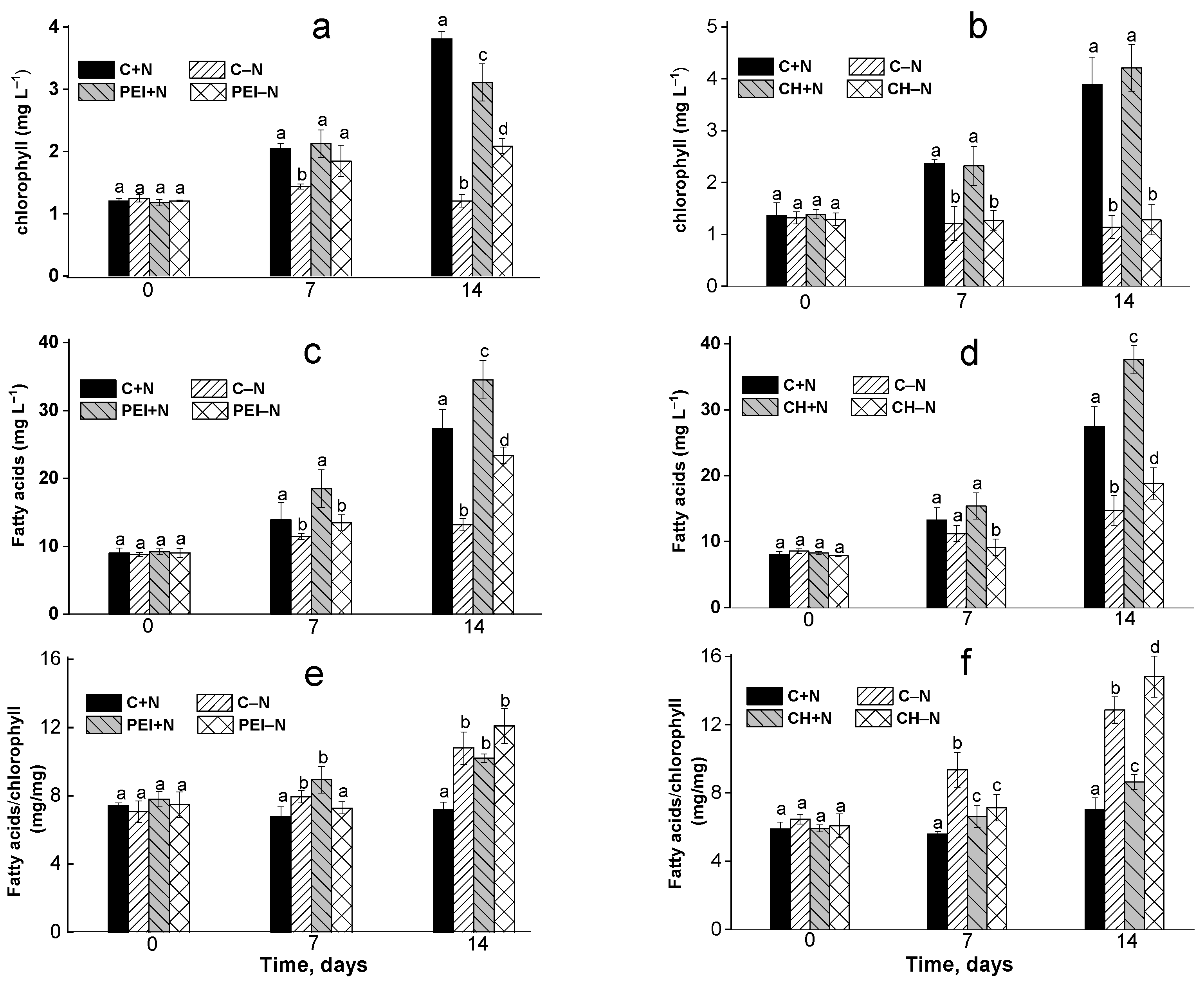
3.1. Changes in the Chlorophyll Content of the Culture

The effects of immobilization and N availability on the Lobosphaera sp. cultures were monitored in terms of the chlorophyll content since the immobilized cells were irreversibly attached to the carriers and could not be removed for counting. Moreover, chlorophyll is a marker sensitive enough to provide useful insights into the culture condition when determination of cell density is impossible [27,29].
Overall, chlorophyll content in the N-deprived cultures was 35–70% less than in N-sufficient cultures (Figure 2a,b). In turn, the chlorophyll content in the PEI+N was 15–20% lower than in the N-sufficient control (C+N), whereas in the PEI–N culture, it was 25–30% higher than in the N-deprived suspension culture C–N (Figure 2a). The difference in chlorophyll content between CH-immobilized and suspended cells was insignificant, regardless of N availability (Figure 2b).
The volumetric FA content of the suspended and immobilized N-deprived cultures increased by 1.5–2.2 times compared to the initial FA content (0 d). In the N-sufficient cultures, it increased by 3.4–4.6 times (Figure 2c,d). Importantly, the total volumetric FA content in the cells immobilized on both carrier types was 15–35% higher than that in the suspended cultures, regardless of nitrogen availability, after 14 d of incubation.
The specific FA content expressed per unit chlorophyll was 22–41% lower at the end of the experiment in N-sufficient cultures than in the N-deprived ones, regardless of the immobilization state, due to the higher chlorophyll content in the former cultures (Figure 3c,d). In the immobilized N-sufficient cultures, the specific FA content was higher in the PEI+N cultures than in the CH+N cultures; however, the opposite trend was observed for the N-deprived cultures.

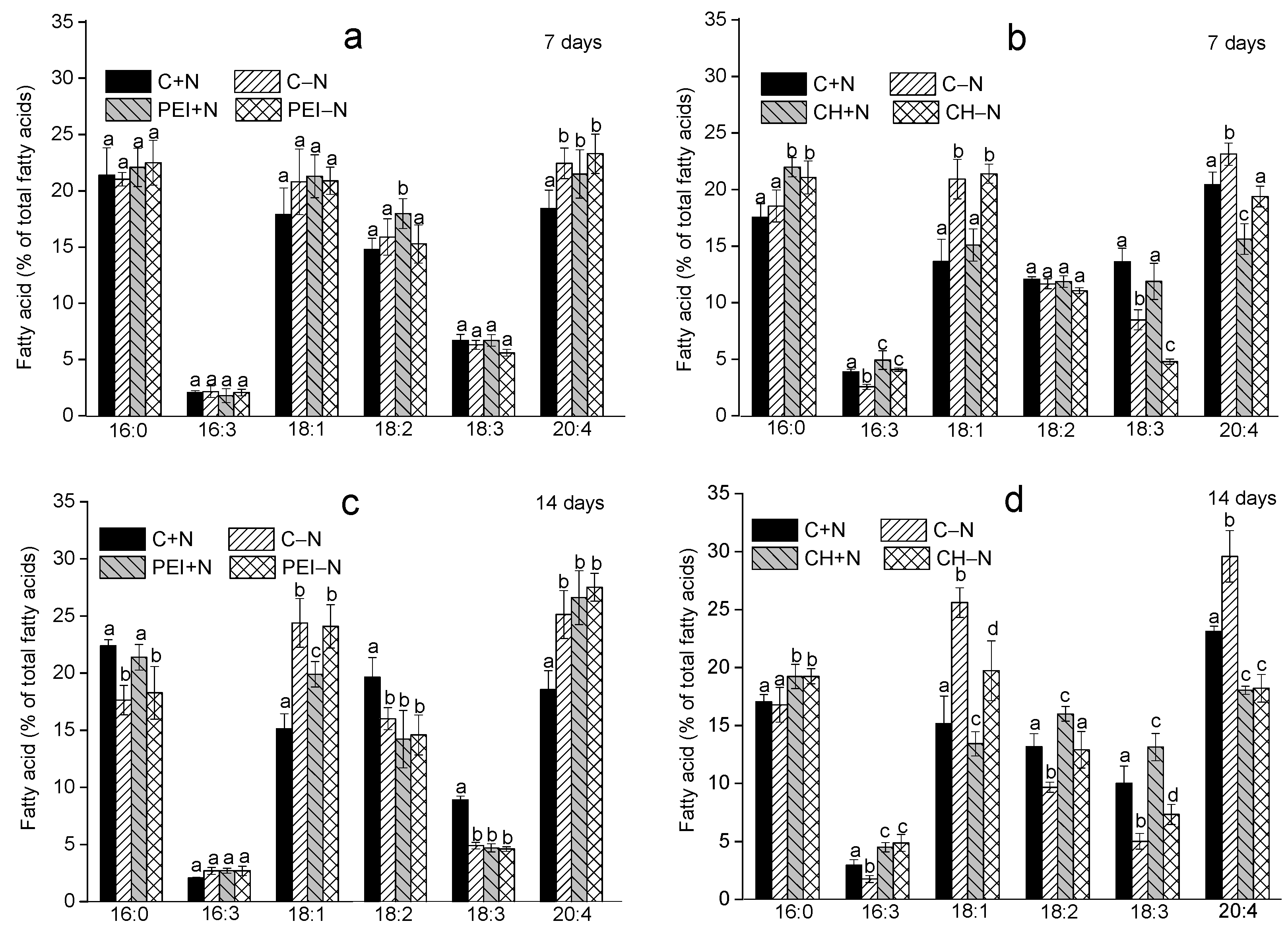
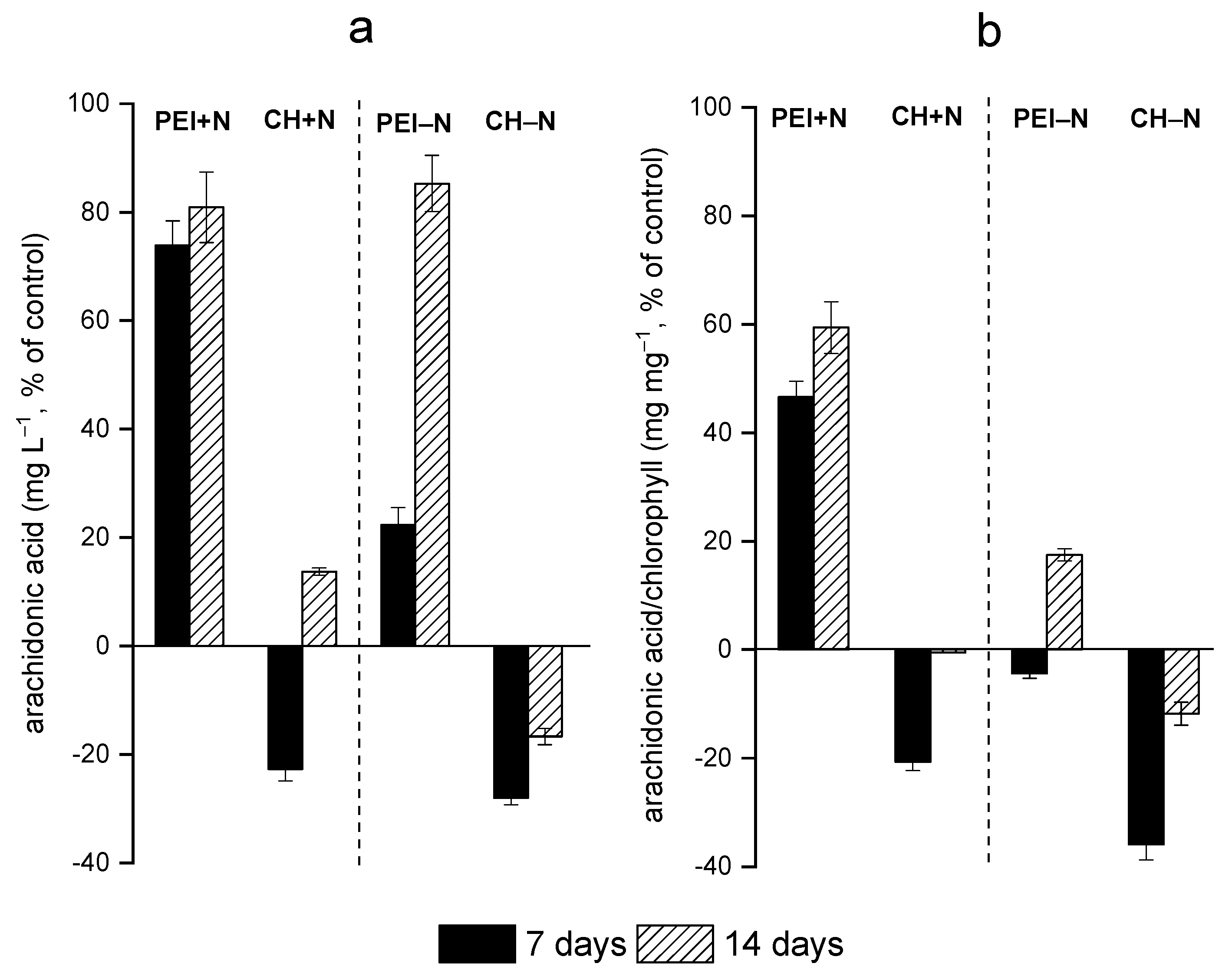
3.2. Effects of Immobilization on FA Profiles of the Cells

Analysis of the FA profiles showed that palmitic (16:0), oleic (18:1), linoleic (18:2), α-linolenic (18:3) and arachidonic (20:4, ARA) acids were the major FAs among the lipids of the Lobosphaera sp. cells suspended or immobilized on both carrier types (Figure 3). In N-replete medium, on the 14th day of cultivation, the percentages of the 16:0, 18:2, and 18:3 acids in the suspended cultures were higher, although the contents of ARA and oleic acid were lower, than in the cells cultivated in the N-free medium.
The highest ARA content was found in the cells immobilized on the carriers and deprived of nitrogen—i.e., in the PEI–N (28%; Figure 3a,c) and C–N (25-29%; Figure 3b,d) variants—after 14 d of incubation. The lowest ARA content (16–18%) was found in the cells immobilized on the CH-based carrier (Figure 3b,d). Notably, in contrast to the suspended cultures, the proportions of ARA in the total FAs of the immobilized cells did not differ significantly, regardless of nitrogen availability.
Over 14 days of incubation in the N-replete medium, the contents of the 18:2 and 18:3 fatty acids decreased in cells immobilized on PEI, whereas ARA content increased in comparison with the suspended culture with ample N availability (Figure 3a,c). In contrast, in the cells immobilized on the CH-based carrier, ARA declined but the contents of 18:2 and 18:3 fatty acids increased (Figure 3b,d).
In the N-deprived suspended or PEI-immobilized cultures, 18:1 and ARA increased, while 18:3 decreased (Figure 3a,c). In the CH-immobilized cells, N-deprivation was accompanied by increases in the 18:2, 16:3, and 18:3 acids, along with decreases in 18:1 and ARA (Figure 3b,d).
On the 7th day of the experiment, the volumetric ARA content in the cultures immobilized on PEI was higher than that in the suspended culture by 72% and 21% for N-sufficient and N-deprived cultures, respectively. In contrast, the ARA content of the cells immobilized on the CH-based carrier was 22–27% lower than that in the suspended cultures (Figure 4a).
During the next seven days of observation, the ARA content of the cells immobilized under the PEI–N conditions increased significantly in comparison to that of the suspension culture. In the case of CH-immobilized cells, a slight increase in ARA (ca. 16%) in comparison with the suspended culture was observed but only in the N-replete medium.
Overall, the N-sufficient cells immobilized on PEI displayed a significant increase in the specific ARA (expressed per unit chlorophyll) content (Figure 4b). On day 7, the specific ARA content in the CH-immobilized cells was ca. 22% lower than that in the suspended culture. Longer incubation triggered a slight increase in ARA, along with an increase in total FA content.

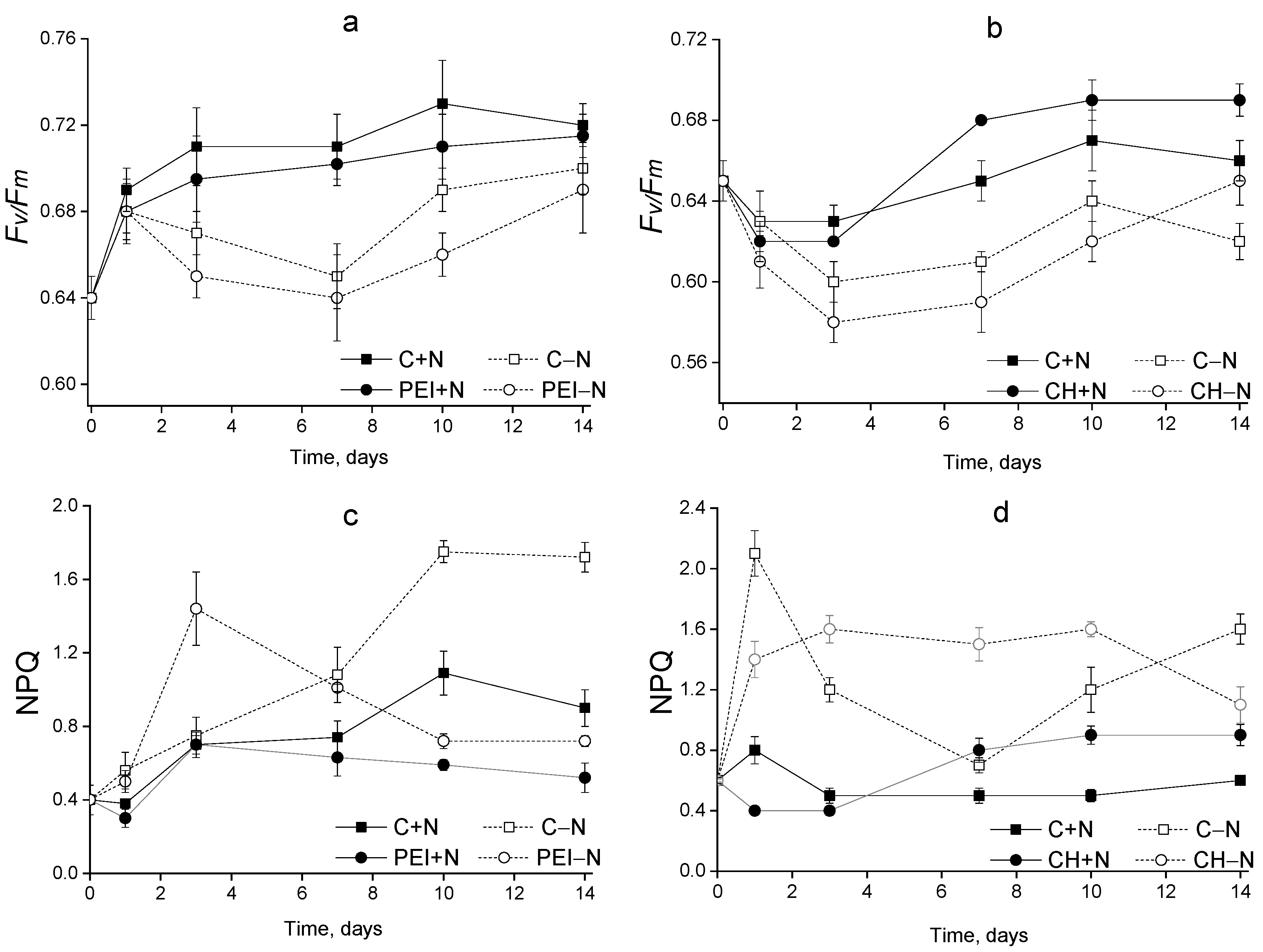
3.3. The Condition of the Photosynthetic Apparatus

The N-sufficient cells displayed a monotonous increase in Fv/Fm during 14 days of cultivation, regardless of immobilization status and cell carrier type (Figure 5a,b). At the same time, NPQ values remained low (0.4–0.9), indicating there was low stress, if any, imposed on the culture by the selected cultivation conditions (Figure 5c,d).
In the case of N-deprived cultures, a slight decline in Fv/Fm was recorded during the first seven days of cultivation, which was followed by its recovery by the end of the observation period (Figure 5a,b). The PEI-N cells displayed an increase in NPQ values during the first 1–3 days of cultivation and a gradual decrease to the initial values by the last day of the experiment. The suspended cultures deprived of N showed a pronounced increase in NPQ (by 4–4.5 times relative to the initial values). Judging from the condition of the photosynthetic apparatus, the N-shortage stress was, in effect, milder in the case of the immobilized cultures than in the case of suspended cultures.

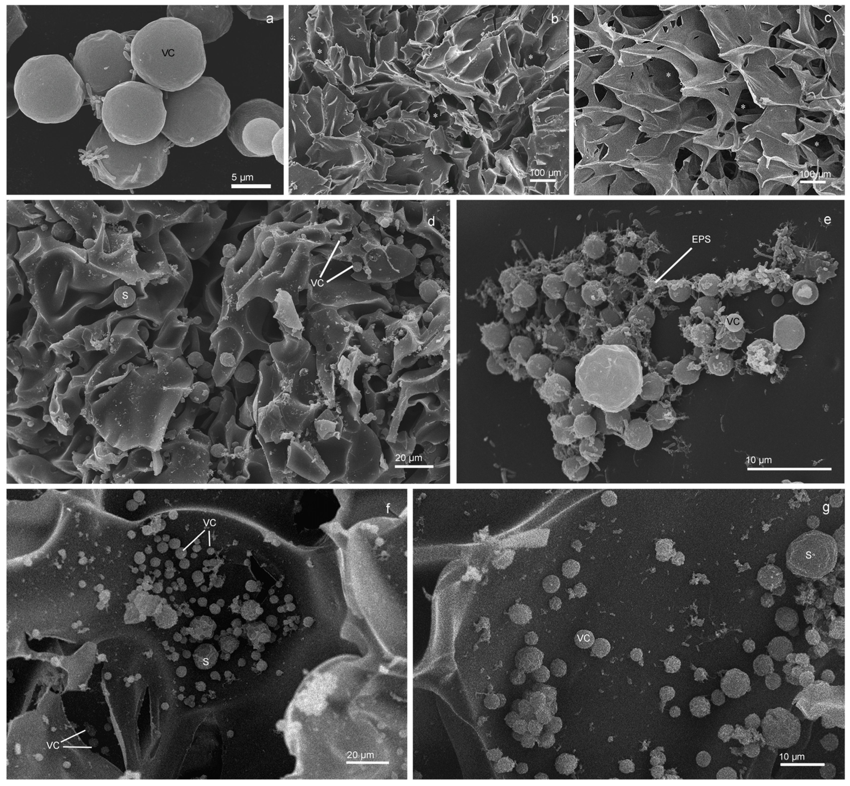
3.4. Changes in the Morphology of the Cells as Elucidated by SEM

Lobosphaera sp. IPPAS C-2047 is a unicellular green microalga with spherical cells 3.5–23 µm in diameter (Figure 6a). Numerous pores of different depths and sizes (10–300 microns) were revealed on the surface of the PEI (Figure 6b) and CH (Figure 6c). Lobosphaera sp. cells at various stages of their lifecycle (young cells, mature cells and sporangia) were detected in the pores and channels of the PEI (Figure 6d,e) and CH (Figure 6f,g). The inner parts of both types of cell carriers possessed a branched system of channels with varying diameters facilitating access to the inner bulk of the polymer for the microalgal cells (Figure 6f). The microalgal cells were strongly attached to both carrier types judging by their tight adherence to the surfaces of the polymers (Figure 6d–g). The surfaces of the suspended cells of Lobosphaera sp. were smooth (Figure 6a), while the surfaces of the immobilized cells became corrugated (Figure 6e–g). The cell and the polymeric carrier surfaces were connected by the strands of extracellular polymer substances, which likely strengthened the attachment of cells (Figure 6e) and demonstrated the onset of biofilm formation on the surface of the cell carriers.

4. Discussion

The object of this study, Lobosphaera sp. IPPAS C-2047, is an extremotolerant microalga capable of accumulating high amounts of the very-long-chain fatty acid ARA. A prominent, arguably unique feature of microalgae from this genus is the presence of ARA in the structural and reserve lipids [5,30,31]. Therefore, the biosynthesis of ARA in Lobosphaera can be upregulated under stress, but it can also be sizeable under conditions favoring rapid cell division. Nevertheless, the most significant induction of ARA is observed in relation to stressful irradiances and temperatures, nutrient shortage or combinations thereof [32]. The lipogenic response of Lobosphaera, as well as the reduction in the photosynthetic apparatus, serves as protection against the oxidative stress exacerbated by diverse environmental stresses [32,33,34]. The protection is implemented by channeling the surplus photoassimilates into the biosynthesis of triacylglycerides (TAGs) rich in oleate (18:1) and, in the case of Lobosphaera, ARA [6,33]. A similar response was observed in the N-deprived suspended culture under our experimental conditions (Figure 2c–e and Figure 3).
Under optimal conditions, the biosynthesis of structural lipids, including glycolipids of chloroplast membranes enriched with unsaturated FAs from the C18 family (18:3 and 18:2), prevails. Stresses shift the lipid metabolism towards the biosynthesis of reserve TAG harboring mostly oleate (18:1) [33,35,36,37]. Stress such as N deprivation promotes degradation of chlorophyll and the dismantling of chloroplast membranes, which manifests as reductions in 18:2 and 18:3 [38,39], as was the case in our study (Figure 3). The buildup of ARA recorded under stress might reflect the role of the lipids harboring the polyunsaturated fatty acids in protecting photosystem I against oxidative stress [40].
The similarity between the FA profile changes observed in the PEI+N cultures (increase in 18:1 and reductions in 18:2 and 18:3) and those recorded in the C–N cultures suggested that the stress responses of Lobosphaera sp. are more determined by the availability of N, the crucial nutrient, although some exceptions were noted (see below).
As has been shown previously [41,42], immobilization of cells on PEI-based carriers is itself stressful: thus, it increased the accumulation of atsaxanthin in Haematococcus pluvialis in comparison to its suspended culture. Likewise, immobilization of Lobosphaera sp. cells on PEI might augment ARA accumulation, which can be even more significant under nitrogen deprivation. This is in line with the increases in total FAs and the FA/chlorophyll ratio observed in the cultures immobilized on PEI (Figure 2c,e).
The signs of stress, such as the decrease in chlorophyll, shown by the cells immobilized on PEI when nitrogen was ample in the medium (Figure 2a) might have stemmed from the restriction imposed on cell division by spatial constraints and strong binding of the microalgal cells to the carrier. Effectively, the slowdown in cell division induced by the immobilization can shrink the metabolic sink for the photoassimilates similarly to other stresses. In turn, this would trigger general stress responses, including an increase in the biosynthesis of reserve lipids enriched in ARA.
An important outcome of this study was the pinpointing of the profound difference between the effects of immobilization on PEI and CH on the lipid metabolism of Lobosphaera sp. Unlike PEI, immobilization on CH reduced ARA but increased 18:2, 18:3 and 16:3 contents. Possible explanations of this effect include the inhibitory effect of CH on ARA biosynthesis and modulation of the lipid composition of the cell membranes. However, in contrast to the suspension culture, application of nitrogen deprivation to the cells immobilized on both carriers did not augment the ARA percentage among the total FAs. It is, therefore, likely that the nitrogen shortage in the immobilized cells was partially relieved by the supply of nitrogen-containing products from the carrier biodegradation, which was itself rich in nitrogen [16]. However, it is known that the main disadvantage of polymers such as chitosan and PEI is their low mechanical stability, which limits their application. At the same time, we did not observe significant degradation of the cell carriers under our experimental conditions. In principle, biopolymers should be biodegradable to facilitate the utilization of spent cell carriers. However, in situations requiring increased resilience in the cell carriers, their durability can be considerably enhanced by additional cross-linking with glutaraldehyde or diethylene glycol diglycidyl ether.
Interestingly, the FA content of the cells immobilized on CH was higher than that of the suspended culture (Figure 2d,f). This increase was not due to an increase in ARA but to enhancement of 18:2, 18:3 and 16:3, the valuable FAs harbored mostly by chloroplast membrane lipids. This might indicate that the cells immobilized on CH either divided more rapidly or possessed larger thylakoid membranes, allowing them to capture more light under the condition of self-shading of the cells crowded on the surface or within the volume of the carrier. The retention of the photosynthetic apparatus functionality under our experimental conditions was supported by the lack of a profound decline in photosystem II efficiency (high Fv/Fm) in the immobilized cells (Figure 5a,b). A slight decrease in Fv/Fm was noted in the suspended and immobilized cells on the first day of cultivation in N-free medium, but even there it recovered rapidly. As expected, nitrogen shortage promoted an increase in the thermal dissipation of the absorbed light energy, manifesting as an increase in NPQ [25]. However, only a moderate increase in NPQ was observed, suggesting that the stress imposed on the cultures was rather mild and the microalgal cells successfully acclimated to it (Figure 5c,d). On the other hand, the functional intactness of the photosynthetic apparatus of the immobilized cells (Figure 5) confirmed the suitability of the studied CH and PEI carriers for immobilization of microalgal cultures [16,20]. Indeed, these carriers turned out to be sufficiently transparent and biocompatible, with their porosity, determined by ice microcrystals formed during cryopolymerization, facilitating the entrapment of the cells by the carrier [21,22].
Judging by the presence of numerous sporangia and young cells released from them (Figure 6e–g), the immobilized Lobosphaera sp. cells retained the ability to grow and divide, despite their strong attachment to the surfaces of both carrier types. Nevertheless, the surfaces of mature cells and sporangia immobilized on the carriers were frequently more corrugated and deformed in comparison to those of the suspended cells (Figure 6e–g). These changes might have resulted from shear forces resulting from strong binding to the carrier, enhanced excretion of the extracellular polymeric substances (EPSs) or both processes [16]. The enhancement of EPS excretion by microalgal cells upon their immobilization on hard surfaces is well-known; it was also documented in our experimental system (Figure 6e). One might think that the EPSs make the attachment of cells to the carrier stronger or even irreversible, facilitating the formation of biofilm [16]. This hypothesis was supported by the presence of cellular aggregates consisting of microalgae and the associated heterotrophic bacteria attached to the surface of chitosan (Figure 6e). The EPS components (exopolysaccharides, proteins, amino acids) can be mobilized by the bacteria as additional nutrient sources [10,11].
The results presented in this study prove that microalgae immobilized on cross-linked chitosan and PEI polymers have great potential for “green” synthesis of polyunsaturated fatty acids, but further studies with a lab-scale biofilm photobioreactor are necessary. The effects of light intensity and nutrient concentration on fatty acid accumulation by immobilized microalgae should also be estimated in up-scaled experiments. Additionally, it is crucial to compare the cost-effectiveness of microalgae-immobilized and traditional technologies in pre-pilot cultivation experiments.

5. Conclusions

Our results confirmed the suitability of the studied CH- and PEI-based polymeric cell carriers for immobilization of microalgal cultures. Moreover, immobilization on the PEI-based carrier augmented the accumulation of ARA and total FA accumulation by 72% (cultivation in the N-replete medium) and 81% (cultivation in the N-free medium). Surprisingly, immobilization of the Lobosphaera sp. cells on a CH-based sorbent reduced the ARA percentage (likely due to the supply of nitrogen from the biodegradation of the carrier itself). At the same time, the CH-immobilized cells possessed higher amounts of other valuable FAs, such as oleic (18:2), α-linoleic (18:3) and hexadecatrienoic (16:3) acids, than cells from the suspended cultures. In view of the advantages of attached cultivation, immobilization of Lobosphaera sp. IPPAS C-2047 on CH- and PEI-based polymeric cell carriers is a promising avenue for the development of viable technology for “green” ARA production from microalgae.

Author Contributions

Conceptualization, S.V. and E.L.; methodology, C.A. and S.V.; validation, A.S.; formal analysis, O.C.; investigation, K.S. and O.C.; resources, S.V. and A.M.; data curation, A.S.; writing—original draft preparation, S.V.; writing—review and editing, A.S.; visualization, S.V.; supervision, E.L.; funding acquisition, A.S. and S.V. All authors have read and agreed to the published version of the manuscript.

Funding

The synthesis of polymers and the estimation of fatty acids and pigments in suspended and immobilized microalgae were funded by the Russian Science Foundation, grant number 23-44-00006. The analysis of the photosynthetic apparatus condition and electron microscopy studies were funded by the Russian Science Foundation, grant number 23-24-00122.

Institutional Review Board Statement

Not applicable.

Informed Consent Statement

Not applicable.

Data Availability Statement

The data are available from the corresponding author on reasonable request.

Acknowledgments

The cross-linked CH-based polymeric cell carriers were synthesized in the Laboratory of Polymer Materials of the Kurchatov Institute Research Center. The PEI-based polymeric cell carriers were synthesized by JSC “Advanced Medical Technologies”. The electron microscopy studies were carried out at the User Facilities Center of M.V. Lomonosov Moscow State University.

Conflicts of Interest

The authors declare no conflict of interest.

References

  1. Barbosa, M.J.; Janssen, M.; Sudfeld, C.; D’Adamo, S.; Wijffels, R.H. Hypes, hopes, and the way forward for microalgal biotechnology. Trends Biotechnol. 2023, 41, 452–471. [Google Scholar] [CrossRef] [PubMed]
  2. Shahi Khalaf Ansar, B.; Kavusi, E.; Dehghanian, Z.; Pandey, J.; Asgari Lajayer, B.; Price, G.W.; Astatkie, T. Removal of organic and inorganic contaminants from the air, soil, and water by algae. Environ. Sci. Pollut. Res. Int 2022. [Google Scholar] [CrossRef] [PubMed]
  3. Brodie, J.; Chan, C.X.; De Clerck, O.; Cock, J.M.; Coelho, S.M.; Gachon, C.; Grossman, A.R.; Mock, T.; Raven, J.A.; Smith, A.G.; et al. The Algal Revolution. Trends Plant Sci. 2017, 22, 726–738. [Google Scholar] [CrossRef] [PubMed]
  4. Cohen, Z.; Khozin-Goldberg, I. Searching for PUFA-rich microalgae. In Single Cell Oils, 2 ed.; Cohen, Z., Ratledge, C., Eds.; American Oil Chemists’ Society: Champaign, IL, USA, 2010; pp. 201–224. [Google Scholar]
  5. Bigogno, C.; Khozin-Goldberg, I.; Boussiba, S.; Vonshak, A.; Cohen, Z. Lipid and fatty acid composition of the green oleaginous alga Parietochloris incisa, the richest plant source of arachidonic acid. Phytochemistry 2002, 60, 497–503. [Google Scholar] [CrossRef]
  6. Khozin-Goldberg, I.; Iskandarov, U.; Cohen, Z. LC-PUFA from photosynthetic microalgae: Occurrence, biosynthesis, and prospects in biotechnology. Appl. Microbiol. Biotechnol. 2011, 91, 905–915. [Google Scholar] [CrossRef]
  7. Dumancas, G.G. Arachidonic Acid: Dietary Sources and General Functions; Nova Science Publishers, Incorporated: Hauppauge, NY, USA, 2012. [Google Scholar]
  8. Dragos, A.; Kiesewalter, H.; Martin, M.; Hsu, C.Y.; Hartmann, R.; Wechsler, T.; Eriksen, C.; Brix, S.; Drescher, K.; Stanley-Wall, N.; et al. Division of Labor during Biofilm Matrix Production. Curr. Biol. 2018, 28, 1903–1913.e5. [Google Scholar] [CrossRef]
  9. Branda, S.S.; Vik, Å.; Friedman, L.; Kolter, R. Biofilms: The matrix revisited. Trends Microbiol. 2005, 13, 20–26. [Google Scholar] [CrossRef]
  10. Nozhevnikova, A.; Botchkova, E.; Plakunov, V. Multi-species biofilms in ecology, medicine, and biotechnology. Microbiology 2015, 84, 731–750. [Google Scholar] [CrossRef]
  11. Roeselers, G.; van Loosdrecht, M.C.; Muyzer, G. Heterotrophic pioneers facilitate phototrophic biofilm development. Microb. Ecol. 2007, 54, 578–585. [Google Scholar] [CrossRef]
  12. Costerton, J.W. Overview of microbial biofilms. J. Ind. Microbiol. Biotechnol. 1995, 15, 137–140. [Google Scholar] [CrossRef]
  13. Danaee, S.; Heydarian, S.M.; Ofoghi, H.; Varzaghani, N.B. Optimization, upscaling and kinetic study of famine technique in a microalgal biofilm-based photobioreactor for nutrient removal. Environ. Technol. Innov. 2021, 24, 102043. [Google Scholar] [CrossRef]
  14. Orfanos, A.G.; Manariotis, I.D. Algal biofilm ponds for polishing secondary effluent and resource recovery. J. Appl. Phycol. 2019, 31, 1765–1772. [Google Scholar] [CrossRef]
  15. Kesaano, M.; Sims, R.C. Algal biofilm based technology for wastewater treatment. Algal Res. 2014, 5, 231–240. [Google Scholar] [CrossRef]
  16. Vasilieva, S.; Lobakova, E.; Solovchenko, A. Biotechnological Applications of Immobilized Microalgae. Environ. Biotechnol. 2021, 3, 193–220. [Google Scholar]
  17. de-Bashan, L.E.; Bashan, Y. Immobilized microalgae for removing pollutants: Review of practical aspects. Bioresour. Technol. 2010, 101, 1611–1627. [Google Scholar] [CrossRef] [PubMed]
  18. Eroglu, E.; Smith, S.M.; Raston, C.L. Application of Various Immobilization Techniques for Algal Bioprocesses. In Biomass and Biofuels from Microalgae; Springer: Berlin/Heidelberg, Germany, 2015; pp. 19–44. [Google Scholar]
  19. Moreno-Garrido, I. Microalgae immobilization: Current techniques and uses. Bioresour. Technol. 2008, 99, 3949–3964. [Google Scholar] [CrossRef]
  20. Vasilieva, S.; Shibzukhova, K.; Morozov, A.; Solovchenko, A.; Bessonov, I.; Kopitsyna, M.; Lukyanov, A.; Chekanov, K.; Lobakova, E. Immobilization of microalgae on the surface of new cross-linked polyethylenimine-based sorbents. J. Biotechnol. 2018, 281, 31–38. [Google Scholar] [CrossRef]
  21. Romanova, O.; Grigor’ev, T.; Goncharov, M.; Rudyak, S.; Solov’yova, E.; Krasheninnikov, S.; Saprykin, V.; Sytina, E.; Chvalun, S.; Pal’tsev, M. Chitosan as a modifying component of artificial scaffold for human skin tissue engineering. Bull. Exp. Biol. Med. 2015, 159, 557–566. [Google Scholar] [CrossRef]
  22. Nuzhdina, A.V.; Morozov, A.S.; Kopitsyna, M.N.; Strukova, E.N.; Shlykova, D.S.; Bessonov, I.V.; Lobakova, E.S. Simple and versatile method for creation of non-leaching antimicrobial surfaces based on cross-linked alkylated polyethyleneimine derivatives. Mater. Sci. Eng. C 2017, 70, 788–795. [Google Scholar] [CrossRef]
  23. Stanier, R.; Kunisawa, R.; Mandel, M.; Cohen-Bazire, G. Purification and properties of unicellular blue-green algae (order Chroococcales). Microbiol. Mol. Biol. Rev. 1971, 35, 171–205. [Google Scholar] [CrossRef]
  24. Strasser, R.; Tsimilli-Michael, M.; Srivastava, A. Analysis of the chlorophyll a fluorescence transient. In Chlorophyll a fluorescence: A Signature of Photosynthesis; Papageorgiou, G.G., Ed.; Springer: Berlin/Heidelberg, Germany, 2004; pp. 321–362. [Google Scholar]
  25. Maxwell, K.; Johnson, G. Chlorophyll fluorescence—A practical guide. J. Exp. Bot. 2000, 51, 659–668. [Google Scholar] [CrossRef] [PubMed]
  26. Folch, J.; Lees, M.; Sloane-Stanley, G. A simple method for the isolation and purification of total lipids from animal tissues. J. Biol. Chem. 1957, 226, 497–509. [Google Scholar] [CrossRef] [PubMed]
  27. Solovchenko, A.; Merzlyak, M.; Khozin-Goldberg, I.; Cohen, Z.; Boussiba, S. Coordinated carotenoid and lipid syntheses induced in Parietochloris incisa (Chlorophyta, Trebouxiophyceae) mutant deficient in Δ5 desaturase by nitrogen starvation and high light. J. Phycol. 2010, 46, 763–772. [Google Scholar] [CrossRef]
  28. Solovchenko, A.; Gorelova, O.; Selyakh, I.; Pogosyan, S.; Baulina, O.; Semenova, L.; Chivkunova, O.; Voronova, E.; Konyukhov, I.; Scherbakov, P. A novel CO2-tolerant symbiotic Desmodesmus (Chlorophyceae, Desmodesmaceae): Acclimation to and performance at a high carbon dioxide level. Algal Res. 2015, 11, 399–410. [Google Scholar] [CrossRef]
  29. Solovchenko, A.; Khozin-Goldberg, I.; Cohen, Z.; Merzlyak, M. Carotenoid-to-chlorophyll ratio as a proxy for assay of total fatty acids and arachidonic acid content in the green microalga Parietochloris incisa. J. Appl. Phycol. 2009, 21, 361–366. [Google Scholar] [CrossRef]
  30. Khozin-Goldberg, I.; Bigogno, C.; Shrestha, P.; Cohen, Z. Nitrogen starvation induces the accumulation of arachidonic acid in the freshwater green alga Parietochloris incisa (Trebuxiophyceae). J. Phycol. 2002, 38, 991–994. [Google Scholar] [CrossRef]
  31. Bigogno, C.; Khozin-Goldberg, I.; Cohen, Z. Accumulation of arachidonic acid-rich triacylglycerols in the microalga Parietochloris incisa (Trebuxiophyceae, Chlorophyta). Phytochemistry 2002, 60, 135–143. [Google Scholar] [CrossRef]
  32. Solovchenko, A.; Khozin-Goldberg, I.; Didi-Cohen, S.; Cohen, Z.; Merzlyak, M. Effects of light intensity and nitrogen starvation on growth, total fatty acids and arachidonic acid in the green microalga Parietochloris incisa. J. Appl. Phycol. 2008, 20, 245–251. [Google Scholar] [CrossRef]
  33. Solovchenko, A. Physiological role of neutral lipid accumulation in eukaryotic microalgae under stresses. Russ. J. Plant Physiol. 2012, 59, 167–176. [Google Scholar] [CrossRef]
  34. Solovchenko, A.E.; Merzlyak, M.N.; Chivkunova, O.B.; Reshetnikova, I.V.; Khozin-Goldberg, I.; Didi-Cohen, S.; Cohen, Z. Effects of light irradiance and nitrogen starvation on the accumulation of arachidonic acid by the microalga Parietochloris incisa. Vestn. Mosk. Univ. Seriya 16 Biol. 2008, 63, 49. [Google Scholar]
  35. Li-Beisson, Y.; Thelen, J.J.; Fedosejevs, E.; Harwood, J.L. The lipid biochemistry of eukaryotic algae. Prog. Lipid Res. 2019, 74, 31–68. [Google Scholar] [CrossRef] [PubMed]
  36. Wacker, A.; Piepho, M.; Harwood, J.L.; Guschina, I.A.; Arts, M.T. Light-induced changes in fatty acid profiles of specific lipid classes in several freshwater phytoplankton species. Front. Plant. Sci. 2016, 7, 264. [Google Scholar] [CrossRef] [PubMed]
  37. Guschina, I.A.; Harwood, J.L. Algal Lipids and Their Metabolism. In Algae for Biofuels and Energy; Borowitzka, M.A., Moheimani, N.R., Eds.; Springer: Dordrecht, The Netherlands; Heidelberg, Germany; New York, NY, USA; London, UK, 2013; pp. 17–36. [Google Scholar]
  38. Kokabi, K.; Gorelova, O.; Zorin, B.; Didi-Cohen, S.; Itkin, M.; Malitsky, S.; Solovchenko, A.; Boussiba, S.; Khozin-Goldberg, I. Lipidome Remodeling and Autophagic Respose in the Arachidonic-Acid-Rich Microalga Lobosphaera incisa Under Nitrogen and Phosphorous Deprivation. Front. Plant. Sci. 2020, 11, 614846. [Google Scholar] [CrossRef] [PubMed]
  39. Kugler, A.; Zorin, B.; Didi-Cohen, S.; Sibiryak, M.; Gorelova, O.; Ismagulova, T.; Kokabi, K.; Kumari, P.; Lukyanov, A.; Boussiba, S. Long-chain polyunsaturated fatty acids in the green microalga Lobosphaera incisa contribute to tolerance to abiotic stresses. Plant Cell Physiol. 2019, 60, 1205–1223. [Google Scholar] [CrossRef]
  40. Klyachko-Gurvich, G.; Tsoglin, L.; Doucha, J.; Kopetskii, J.; Shebalina, I.; Semenenko, V. Desaturation of fatty acids as an adaptive response to shifts in light intensity 1. Plant. Physiol. 1999, 107, 240–249. [Google Scholar] [CrossRef]
  41. Lee, J.B.; Park, Y.H.; Jeon, M.S.; Kim, S.; Choi, Y.-E. Polyethylenimine linked with chitosan improves astaxanthin production in Haematococcus pluvialis. Appl. Microbiol. Biotechnol. 2022, 107, 569–580. [Google Scholar] [CrossRef]
  42. Yoshitomi, T.; Shimada, N.; Iijima, K.; Hashizume, M.; Yoshimoto, K. Polyethyleneimine-induced astaxanthin accumulation in the green alga Haematococcus pluvialis by increased oxidative stress. J. Biosci. Bioeng. 2019, 128, 751–754. [Google Scholar] [CrossRef]
Figure 1. The experimental design and designation of the experimental variants.
Jmse 11 00865 g001
Figure 2. Total chlorophyll (a,b) and (c,d) fatty acid content, as well as (d,e) their ratios, in the Lobosphaera sp. IPPAS C–2047 cells suspended (C+N, C–N) and immobilized (PEI+N, PEI–N, CH+N, CH–N) on either PEI (a,c,e) or CH (b,d,f) and grown under either N-replete (C+N, PEI+N, CH+N) or N-free (C–N, PEI–N, CH–N) conditions. Data are presented as means ± STD (n = 9). Lowercase letters indicate significant differences between experimental variants at p < 0.05.
Jmse 11 00865 g002
Figure 3. The profiles of the most abundant FAs among the total lipids in the cells of Lobosphaera sp. IPPAS C-2047 suspended (C+N, C–N) and immobilized (PEI+N, PEI–N, CH+N, CH–N, 4) on either PEI (a,c) or CH (b,d) and incubated in complete (C+N, PEI+N, CH+N) or N-free (C–N, PEI–N, CH–N) BG11M medium for 7 (a,b) or 14 days (c,d). Data are presented as means ± STD (n = 9). Lowercase letters indicate significant differences between experimental variants at p < 0.05.
Jmse 11 00865 g003
Figure 4. The differences in arachidonic acid proportions for total fatty acids in the immobilized Lobosphaera sp. IPPAS C-2047 cells expressed (a) per unit culture volume or (b) per unit chlorophyll and calculated as percentages of the corresponding values recorded for the suspended cultures incubated under the same conditions for 7 d or 14 d.
Jmse 11 00865 g004
Figure 5. Changes in (a,b) Fv/Fm and (c,d) NPQ for Lobosphaera sp. IPPAS C-2047 cells either suspended (C+N, C–N) or immobilized (PEI+N, PEI–N, CH+N, CH–N) on PEI (a,c) or CH (b,d) cells incubated in complete (C+N, PEI+N, CH+N) or N-free (C–N, PEI–N, CH–N) BG11M medium.
Jmse 11 00865 g005
Figure 6. Scanning electron micrographs of suspended Lobosphaera sp. IPPAS C-2047 cells (a), surfaces of PEI (b) and CH (c) carriers and Lobosphaera sp. cells immobilized on PEI (d,e) and CH (f,g). VC, vegetative cells; S, sporangia; EPS, extracellular polymeric substance. Asterisks indicate the pores on the carrier surface.
Jmse 11 00865 g006
Disclaimer/Publisher’s Note: The statements, opinions and data contained in all publications are solely those of the individual author(s) and contributor(s) and not of MDPI and/or the editor(s). MDPI and/or the editor(s) disclaim responsibility for any injury to people or property resulting from any ideas, methods, instructions or products referred to in the content.

