Article

An Optimized Quantification Method for Marine Radioactivity Measurements: Application in the Southern Caspian Sea Using the KATERINA Underwater γ-Spectrometer

by
Christos Tsabaris
*,
Effrossyni G. Androulakaki
and
Stylianos Alexakis
Hellenic Center for Marine Research, Institute of Oceanography, 46.7 Km Athens-Sounio Ave, 19013 Anavyssos, Greece
*
Author to whom correspondence should be addressed.
J. Mar. Sci. Eng. 2023, 11(4), 725; https://doi.org/10.3390/jmse11040725
Submission received: 14 February 2023 / Revised: 21 March 2023 / Accepted: 21 March 2023 / Published: 27 March 2023
(This article belongs to the Special Issue Environmental Radioactivity in the Ocean)

Abstract

:
The underwater gamma-ray spectrometer KATERINA was calibrated in a special tank and then demonstrated in the Southern Caspian Sea to determine the radioactivity levels in seawater as well as in the sediment. The simulated marine efficiency of the in situ detection system was validated analyzing the high energetic (1764 keV of 214Bi and 2614 keV of 208Tl) gamma-ray peaks using their known activity concentration as determined from the low energy emissions. The analysis of the in situ gamma-ray spectra in a short acquisition time period provided quantitative data for 226Ra and 232Th progenies as well as for 40K. A satisfactory analysis was performed for quantifying the observed energy peaks in the seawater and sediment measurements acquired at the south coast of the Caspian Sea. The spectrum in the water tank was also analyzed using full spectrum analysis methods, and the reproduced spectrum was in a very good agreement with the experimental spectrum giving as an output the activity concentrations of the observed radionuclides.

1. Introduction

Recently, a lot of effort has been made to provide baseline information in terms of the naturally occurring radioactive materials of 226Ra, 232Th, their decay products, and 40K in areas of the Caspian Sea. Moreover, anthropogenic radionuclides, such as 137Cs, are of high concern, since low concentrations have been released due to nuclear weapons’ test production and testing, to the reprocessing spent fuel power plant, and to nuclear reactor accidents (such as Chernobyl and Fukushima). Seawater and sediments in marine ecosystems are sensitive markers for assessing their quality. The concentration levels can provide significant information to the responsible authorities for civil protection issues, since human exposure to natural and artificial radioactivity may damage living cells and cause chromosome aberrations and cell transformation, depending on the magnitude and exposure period. The assessment of the gamma radiation dose from natural sources is significant, since natural radiation is the largest contributor to the external dose of the world population [1]. The global average activity concentration of 226Ra, 232Th, and 40K in the soil is 32, 45, and 412 Bq⋅kg−1, respectively, while the maximum activity concentration is about 1000 Bq⋅kg−1 for 226Ra (Sweden), 360 Bq⋅kg−1 for 232Th (China), and 3200 Bq⋅kg−1 for 40K (United Kingdom) [2]. As concerns the marine environment, a lot of effort has been made to perform radiological research to determine the level of radioactivity in the marine matrices, such as sediments/seawater [3], groundwater [4], and submarine groundwater discharge [5,6,7] to evaluate/assess the radiological risk in terms of human health.
Lab-based gamma-ray spectrometry is the prominent method used to provide activity concentration levels in many matrices (seawater, sediment, beach sand, biota, etc.) of the marine environment [8,9]. In 2009, during a campaign along the southern coast of the Caspian Sea [8], a special radiological study was performed regarding the activity concentration measurements of 226Ra, 232Th, 40K, and 137Cs and their impact on humans. The activity concentrations exhibited values of (177 ± 12), (117 ± 11), (1085 ± 101), and (131 ± 4.8) Bq kg−1, for the aforementioned radionuclides, respectively, exceeding the international limits. Moreover, a recent work in 2022 [9] that was also performed on the southwestern coastline of the Caspian Sea showed interesting new results for the levels of radioactivity in sediment samples. The activity concentrations of 226Ra, 232Th, 40K, and 137Cs exhibited values in the sediment samples, with the measurement uncertainty (1σ), that varied from 22.5 to 47.5 (with an average value of 34.4 Bq kg−1 dw) for 226Ra, from 6.5 to 18.8 (with an average value of 11.4 Bq kg−1 dw) for 232Th, from 233.2 to 559.9 (with an average value of 310.6 Bq kg−1 dw) for 40K, and a maximum value of 2.7 Bq kg−1 dw (with an average value of 1.4 Bq kg−1 dw) for 137Cs [9].
The application of in situ detection systems for radioactivity monitoring in a spatial and temporal manner is required for routine and emergency monitoring. In recent decades, various tools and methods have been tested [10,11,12,13,14,15] in different areas of study, matrices, and radioactivity levels. In recent years, a lot of effort has been made to develop instruments and methods at the Marine Environmental Radioactivity Laboratory (MERL) to directly measure the activity concentration of key radionuclides in seawater [16,17,18] and sediment [19,20,21]. As concerns seawater measurements, the in situ underwater spectrometry system is calibrated in special water tanks [22] by diluting reference sources with known activity concentrations, assuming homogeneous conditions in the water column. In special cases, medium resolution spectrometers have also been developed for seawater measurements [18]. However, the intrinsic radiation of such crystals lies in energies close to 40K emissions, which is a disadvantage of applying the systems in the marine environment. 40K is a natural constituent of the seawater, and it emits at a critical energy line in continuous mode for the underwater gamma-ray spectrometry in terms of the calibration exercises of the systems [18]. The use of the 1461 keV energy line in combination with the threshold energy line at ~50 keV [23] is suitable to energy calibrate the data for nuclide identification (qualitative analysis) as well as for validating the quantitative results by intercomparison exercises with the salinity meter [24,25].
The main advantage of the methodology is that it rapidly offers to the end user the activity concentration of the gamma-ray emitters in the energy range from the threshold to 2614 keV, while its limitation is the uncertainty that is introduced in the results when the detection system is applied in nonstatic aquatic systems.
In the next section, the quantification method and its application in the specific study area at the southern Caspian Sea are described. The validation and reliability of the method was performed by theoretically reproducing the experimental data in terms of the efficiencies and response function of the detection system.

2. Material and Methods

2.1. Study Area

The experimental study is depicted in Figure 1. The study area is located in a sensitive natural ecosystem, where a comprehensive study of the radioactivity measurements or relevant studies with the atmospheric deposition (due to natural and anthropogenic effects) have not been performed. The study of radionuclides via gamma-ray spectrometry was part of this demonstration that included seawater and seabed measurements to support existing studies on deposition processes in the areas, as well as the ecosystem interaction with inland waters. The selected area of the coastline was located near the Mahmudabad.

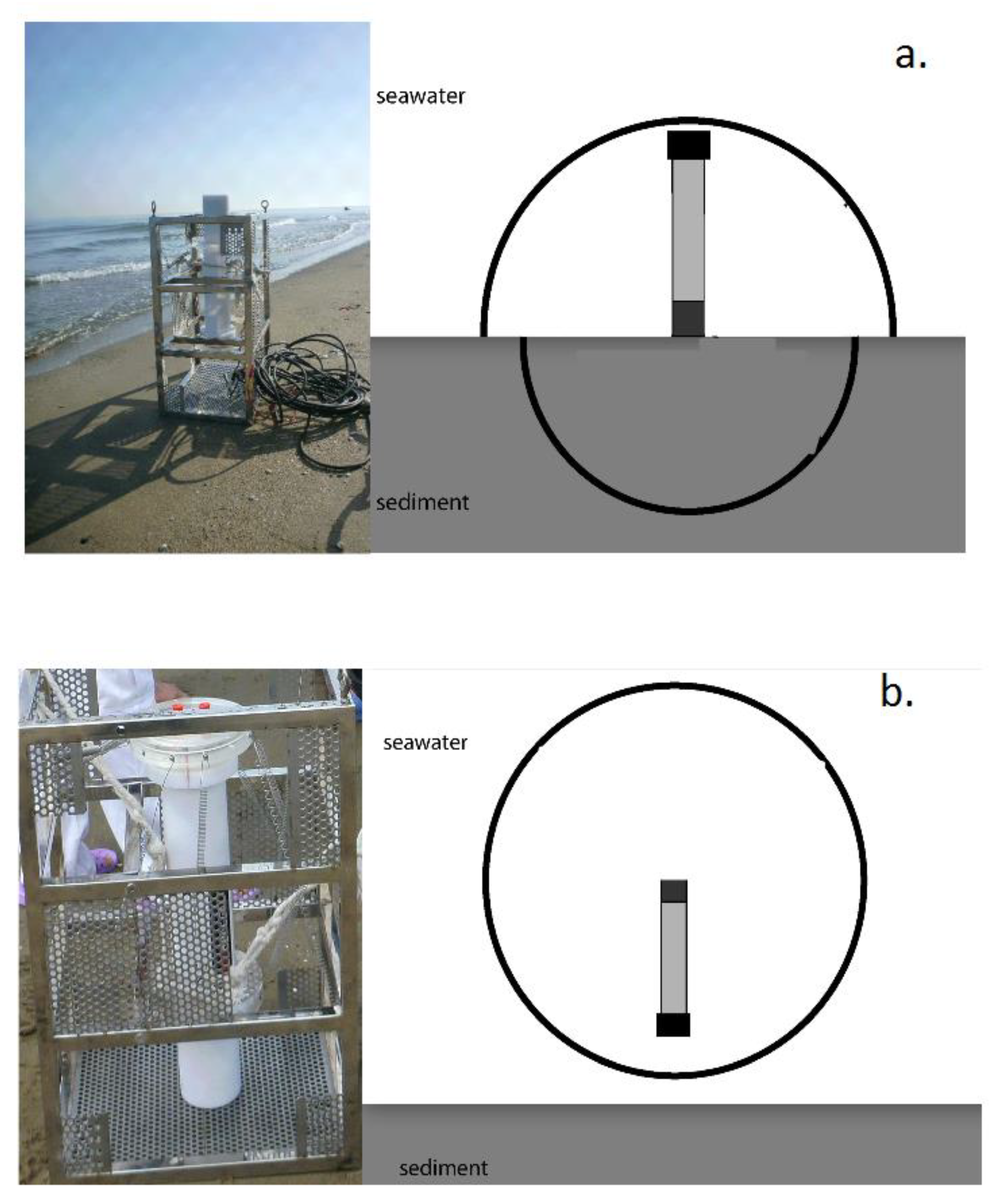
2.2. Experimental Setup

The low resolution underwater gamma-ray spectrometer, named KATERINA, developed by the Hellenic Centre for Marine Research [17,23,26], was tested and calibrated in the laboratory (using a water tank) before its deployment in the seawater. The system has a NaI crystal (3 × 3 inches) and operates in a resolution of 7% at 661 keV (137Cs). The system records all gamma-ray emitters from the threshold (50 keV) to 3000 keV, which are enriched in seawater and sediment. Special attention has been given in recent years to provide validation with theoretical methods to the activity concentration of the natural and artificial radioactivity [23,24,25,26,27] using the in situ method in the marine environment. The sensor was mounted in the middle of the water tank, 5.5 m3 in volume, and it was thus surrounded by one meter of water, to imitate the real marine environment for energies below 1000 keV (due to the attenuation of the γ-rays in the water). At the bottom of the tank, an electric pump was placed to mix the water with the appropriate radionuclides used for the calibrations to ensure homogeneous conditions. The gamma rays were utilized to perform the energy calibration of the system. The details of the calibration experiments in the water tanks are described in detail elsewhere [22,26]. The uncertainty was estimated to vary from 4 to 11%, mainly attributed to the statistics of the net area peak analysis.
In order to optimize the quantification method and marine efficiency (the volumetric full energy peak efficiency of the system when it operates in 4pi geometry in the seawater) of the detection system, the method was repeated with the same procedure with natural drinking water (without artificial calibration sources) on the premises of the IROST, where all the emission energies of the radon progenies (214Bi and 214Pb) are present. The KATERINA system was calibrated using a special tank filled with drinking water, which was highly enriched with radon gas, as natural constituent of the groundwater. The calibration tank has a cylindrical shape (2.4 m diameter and 2.4 m height). The dimensions of the tank were selected to better simulate the marine environment suppressing the total attenuation of the high energetic gamma ray in the water inside the tank (since the required distance of the energetic gamma rays is more than 1 m). The detection system acquired the gamma rays of the radon progenies, and a quantitative analysis procedure was applied. The next step was to deploy and test the detection system for a short period of time in the southern part of the Caspian Sea mainly to determine the levels of naturally occurring radioactivity in the seawater as well on the seabed area, examining the case of potential contamination due to circulation and/or accretion mechanisms.
The system was transported from the IROST premises to the northern part of Iran (south of the Caspian Sea), and a small research vessel was used to perform the experimental deployments of the KATERINA system for seawater and sediment measurements. The system was connected with a power cable. The data cable was disconnected/removed during the measurement, and a special dummy was used for the underwater application allowing the system to operate in a standalone mode according to the time lag of the measurement. The schematic diagram and the underwater photo of the experimental setup is depicted in Figure 2 for the seawater and sediment configurations (reff is the effective radius of a γ-ray in the seawater or in the sediment before impinging to the crystal).
The 137Cs activity concentration was investigated on the sediment, while in the seawater, it was below the limit of detection of the system for the specific acquisition period. The demonstration of the system proved that the KATERINA underwater gamma-ray spectrometer combined with the developed method is an effective tool to deduce quantitative data of the activity concentration applying Full Spectrum Analysis methods in seawater spectra [21], while the analysis in the sediment was performed with an existing methodology [20]. The energy peaks of the radon progenies exhibited high intensities and were clearly observed after filling the calibration tank with the water source. After some time, the gas escaped from the tank, and the concentrations were drastically reduced. First, the acquisition started on the natural drinking water observing the radon progenies peaks, and the next day, a specific quantity of KCl (2 kg) was diluted to analyze the peak of 40K at 1461 keV. Special attention was paid to subtract the 214Bi contribution in the energy range (1400–1620 keV) of the 40K emission energy using an intensity of 214Bi of ~12% according to the literature [7].

2.3. Full Spectrum Analysis (FSA) Technique

The typical analysis technique is the photopeak analysis. However, there are some critical drawbacks due to the overlapping energy peaks and the introduction of systematic uncertainty related to the background subtraction issues. The most important drawback is the inability of the method to identify unexpected radionuclides [27] that may contribute to the measured number of events in the selected energy window. Consequently, the full spectrum analysis (FSA) technique was implemented in the analysis of the acquired data in the seawater applying appropriate software. The FSA technique is based on the reproduction of standard spectra for all radionuclides of interest. Standard spectra are obtained experimentally by performing calibration measurements [27,28] or theoretically by performing Monte Carlo (MC) simulations [29,30], including the geometry and detailed characteristics of the measurement. In this work, the standard spectra were derived using the MCNP-CP simulation code [31], which can simulate the radionuclide as a whole (involved decay emissions) and take into account the true coincidence summing effects.

3. Results

3.1. Experimental Exercises in the Tank

The experiment concerning the calibration of the detection system in optimized conditions for calibrating the system experimentally (for gamma-ray energies above 1000 keV) took place in the premises of IROST and lasted several days. During the first day, the measurement of radon progenies, as natural constituents of the drinking water, was obtained. The experimental spectrum of the first day of acquisition is depicted in Figure 3. The analysis for providing quantitative data was made using the simulated efficiency curve from the calibration process of the KATERINA system. The quantification method of the detection system was performed in the spectrum acquired in the calibration tank using previously evaluated theoretical efficiency values via Monte Carlo for the energy range 100 to 2000 keV. In brief, the applied quantification methodology was validated up to the energy of 2000 keV by diluting the radioactive sources with known activity concentration into the tank. The uncertainty of the previously evaluated marine efficiency values used in the calculations was estimated to be 4%. Details are given elsewhere [22].
The evaluated marine efficiency values of the system in the energy range (351 to 1764 keV) were used to provide results in Bq/m3 for the detected radionuclides. In more detail, the activity concentration of 214Bi was analyzed and averaged at the energy lines of 1120 and 1764 keV, and the activity concentration of 214Pb was deduced by analyzing the peak at 352 keV. The activity concentration of 208Tl was deduced from the analysis of the energy peak at 583 keV, which was convoluted with the energy peak of 214Bi at 609 keV. The detected counting rate was corrected by subtracting the counting rate contribution of the 214Bi observed at 609 keV, using the SPECTRW software package V8.3 [32]. The quantitative results of the radon progenies, as well as for 208Tl, are given in Table 1. It is clearly seen that during the first day, the activity concentration of the radon progenies was enriched in the drinking water providing a homogeneous distribution of the naturally occurring radionuclides. The analysis of the gamma-ray energy lines of 214Bi provided enhanced radioactivity levels compared with the other radon progeny (214Pb). The ratio of the activity concentration of the aforementioned radon progenies was around 1.3 due to the radon loss from the tank (since the aquatic system was not closed). Similar behavior was observed in a submarine groundwater spring [33].
Radon is an inert gas, and it can move rather freely through porous media such as soil or fragmented rock. When the pores are saturated with water (as in soil and rock under the water table), radon is dissolved into the water and transported by the water flow dynamics. Water-saturated soil with a porosity of 20% and a radium concentration of 40 Bq/kg, which is the worldwide average in the earth’s crust, causes at equilibrium a radon concentration in ground water of the order of 50 Bq/L [34]. The measured data of the radon progenies (~11 and 15 Bq/L for 214Pb and 214Bi, respectively) were relatively high but below the recommended or threshold levels [34].

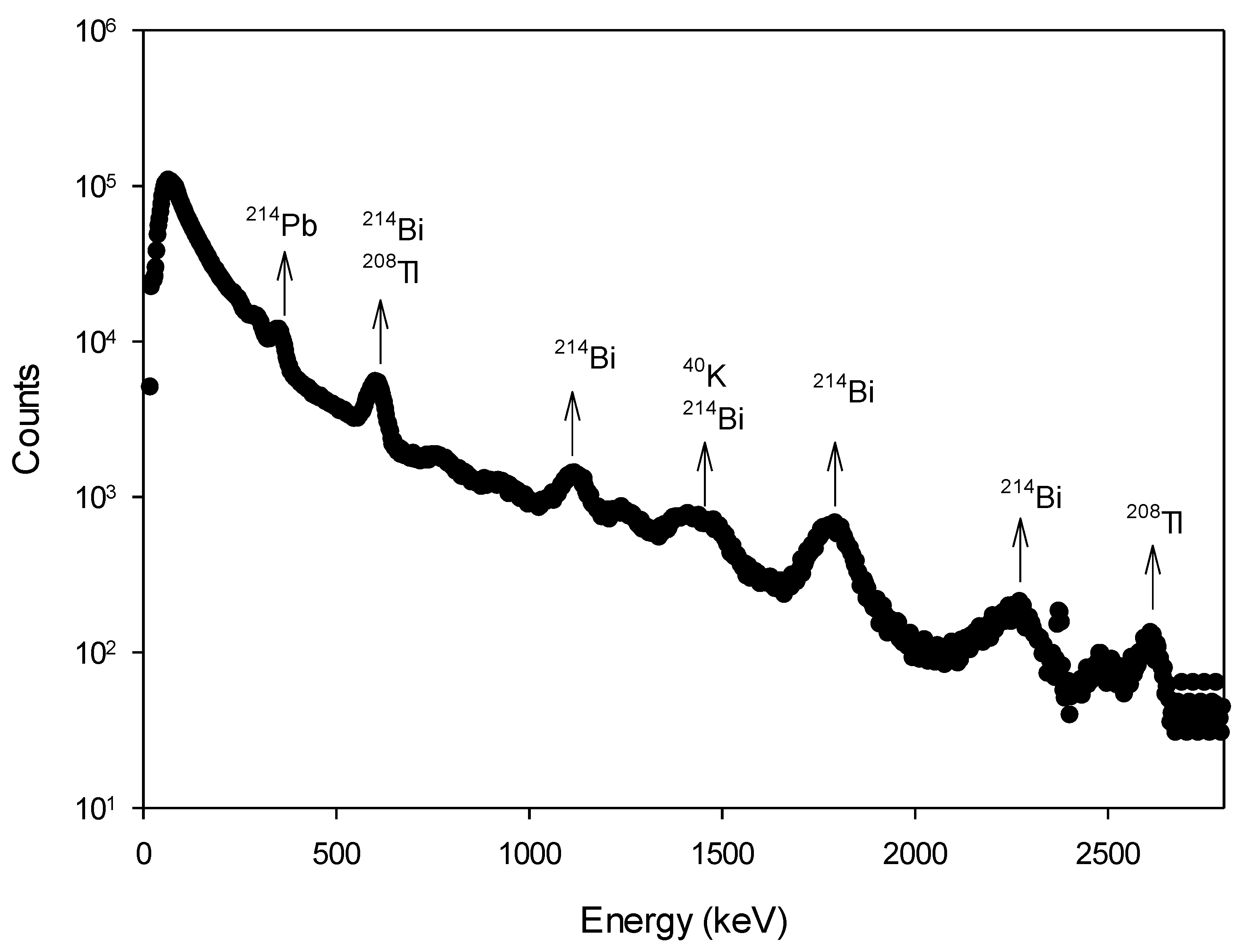
3.2. Field Measurements

The underwater in situ gamma-ray spectrometer KATERINA was deployed in an area of the Southern Caspian Sea (see Figure 1). Two spectra were acquired in a short period of time: one in seawater and another in touch geometry on the seabed. Each measurement lasted for 1 h. The main energy peak observed in the measured spectrum came from the 40K emission at 1461 keV. The analysis of seawater provided results only for the 40K contribution as a natural constituent of the seawater. The activity concentration of the 40K in the seawater was found to be 7.1 ± 0.6 Bq/L, taking into account the gamma-ray intensity and the marine efficiency of the system. All other radionuclides (artificial and natural) exhibited activity concentrations below the detection limit.
However, several radionuclides were enriched in the sediment at the specific area of study, since various contributions from natural and artificial radionuclides were identified. The acquired spectrum on the sediment is depicted in Figure 4 making a smooth operation with three channels [32].
As concerns the analysis of the sediment, the activity concentration of the enriched radionuclides is given in the Table 2 taking into account that the water content in the sediment was 32%, and the wet density of the sediment was 1.8 g/cm3. These values, as well as the activity concentrations for the detected radionuclides, were assumed to be homogeneously distributed within the sediment volume (maximum detection depth 1.5 m). The efficiency calculation for the sediment geometry was performed according to the literature [20].
Corrections for the true coincidence summing effects were not considered in the analysis. The effect was diminished due to the distance between the detector and the seabed (which is approximately 1.5 cm from the NaI crystal) and was considered to be comparable to the statistical uncertainty of the net area analysis.

4. Discussion

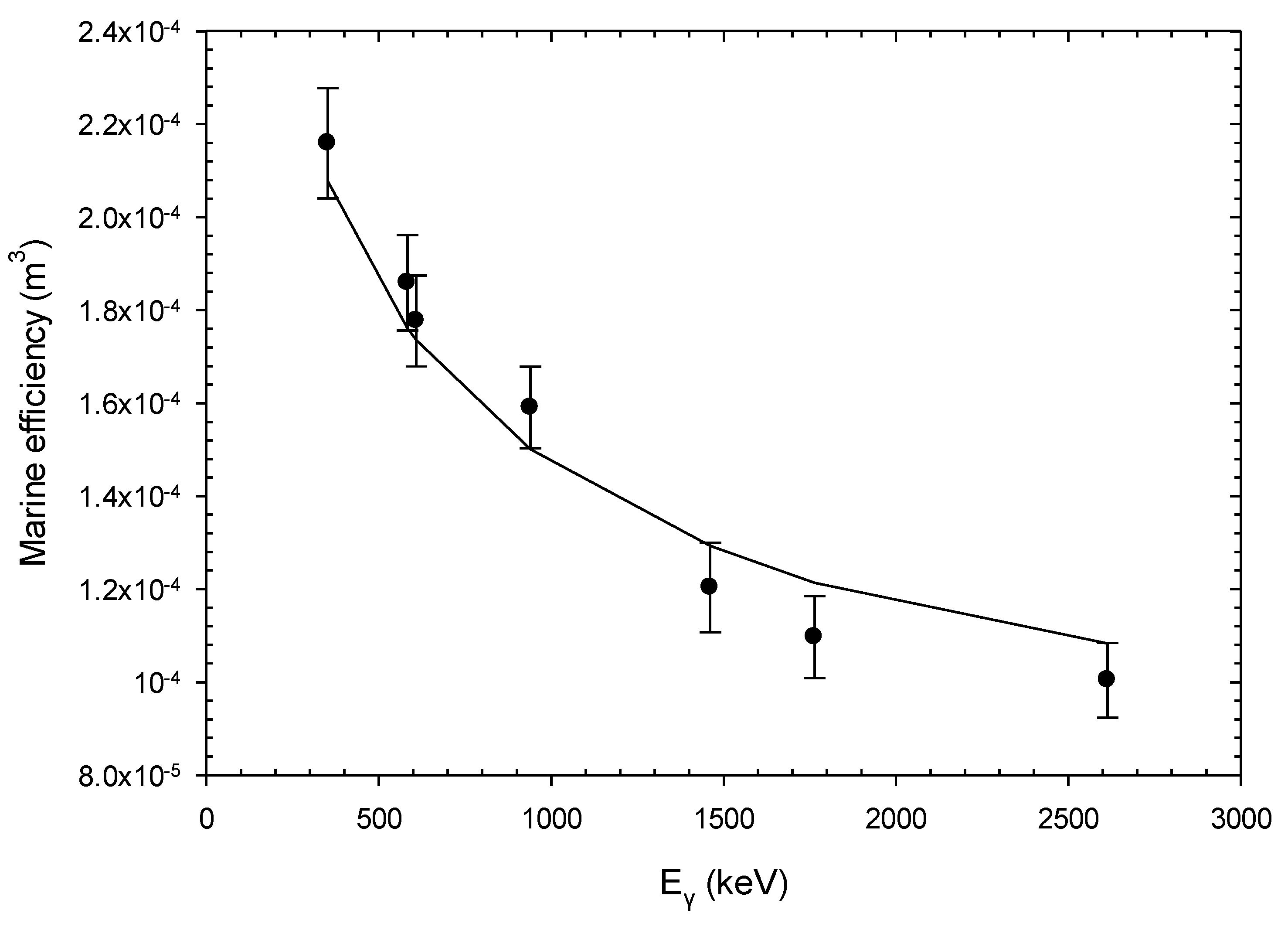
4.1. Simulated and Experimental Efficiencies

The measured data were deduced using the experimental spectrum analysis procedure for the observed peaks of radon progenies and the calculated activity concentration values using the marine efficiency values up to the gamma-ray energy of 2000 keV. The activity concentration of 214Pb, 214Bi, and 208Tl was determined applying the calibration marine efficiency data for the low energies and was evaluated in units of volume in order to deduce the counting rates of each energy photopeak in absolute units (Bq/m3 or Bq/L in seawater and Bq/kg in sediment). The activity concentration values of the natural radionuclides were used to estimate the marine efficiency for the other energy lines of the emission gamma rays of the same radionuclides. The selection of the energy lines of the gamma rays was made taking into account the intensities (the highest provide better statistics) and energy emission.
The previous data of the marine efficiency for quantifying the energy photopeaks observed in seawater at high energies (above 1461 keV) did not provide validated values due to the inadequate tank dimensions [17,22]. The marine efficiency of the system for seawater measurements was reproduced using the MCNP-CP software package ver 6.1 for the acquired spectra in the new tank (with larger dimensions compared to the previous one) to better simulate the marine environment. The simulated marine efficiency of the in situ detection system was extended up to the energy of 2614 keV. The marine efficiency was validated with the corresponding experimental data by analyzing the high energetic (1764 keV of 214Bi and 2614 keV of 208Tl) gamma-ray peaks using their known activity concentration (as determined from the low energy emissions at 1120 keV and 583 keV, respectively).
The updated results are depicted in Figure 5 together with the uncertainties of the experimental data. The experimental uncertainties were mainly attributed to the statistical uncertainty (4–6%) of the net area and the uncertainty of the estimated efficiency values (4%).
In this work, the efficiency values using the high energetic peaks of naturally occurring radionuclides were determined and validated. For instance, the experimental efficiency value of 208Tl at 2614 KeV was 0.0001004 (6%), while the reproduced simulated value was 0.000108 (5.5%). The agreement was within the uncertainties.

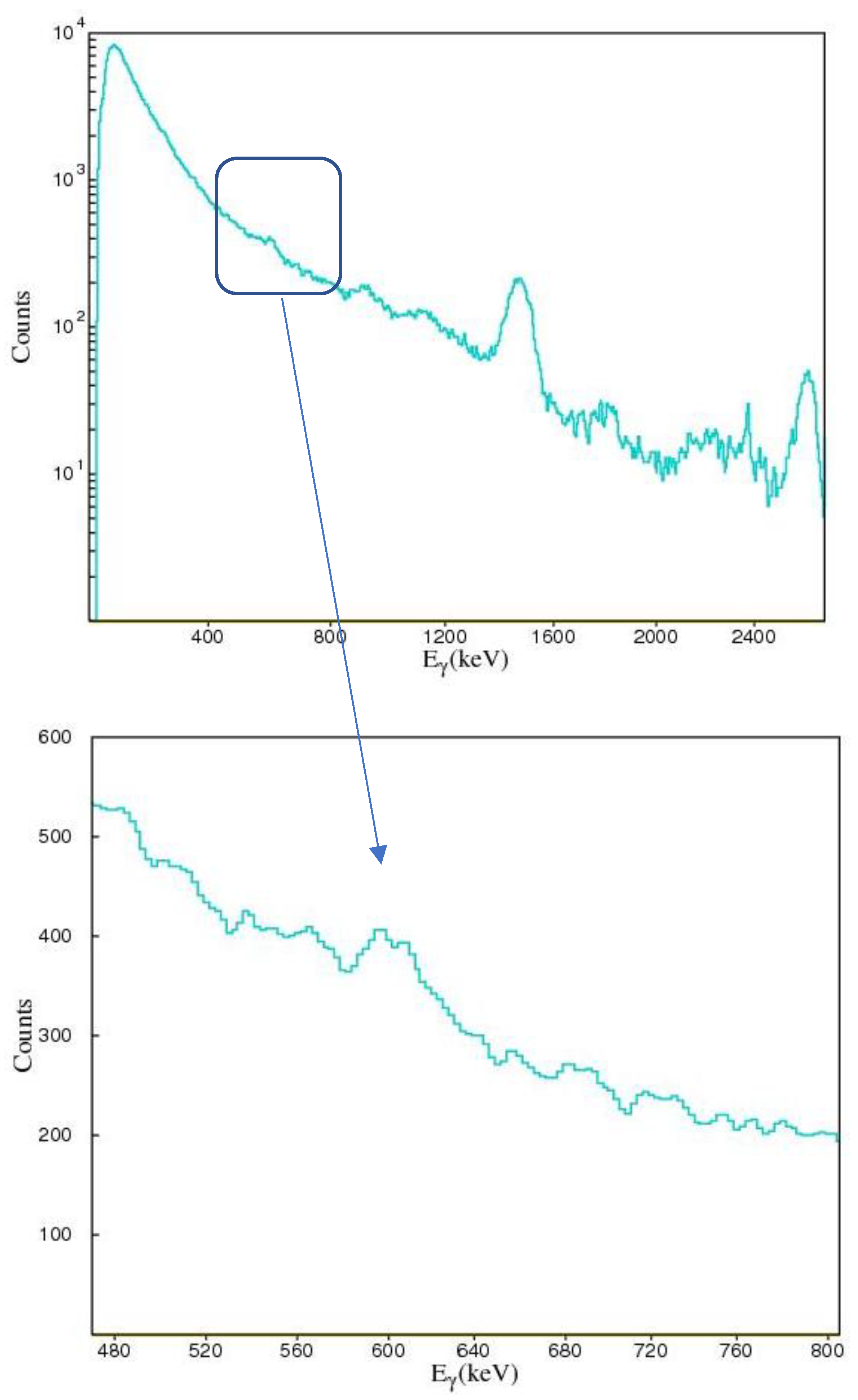
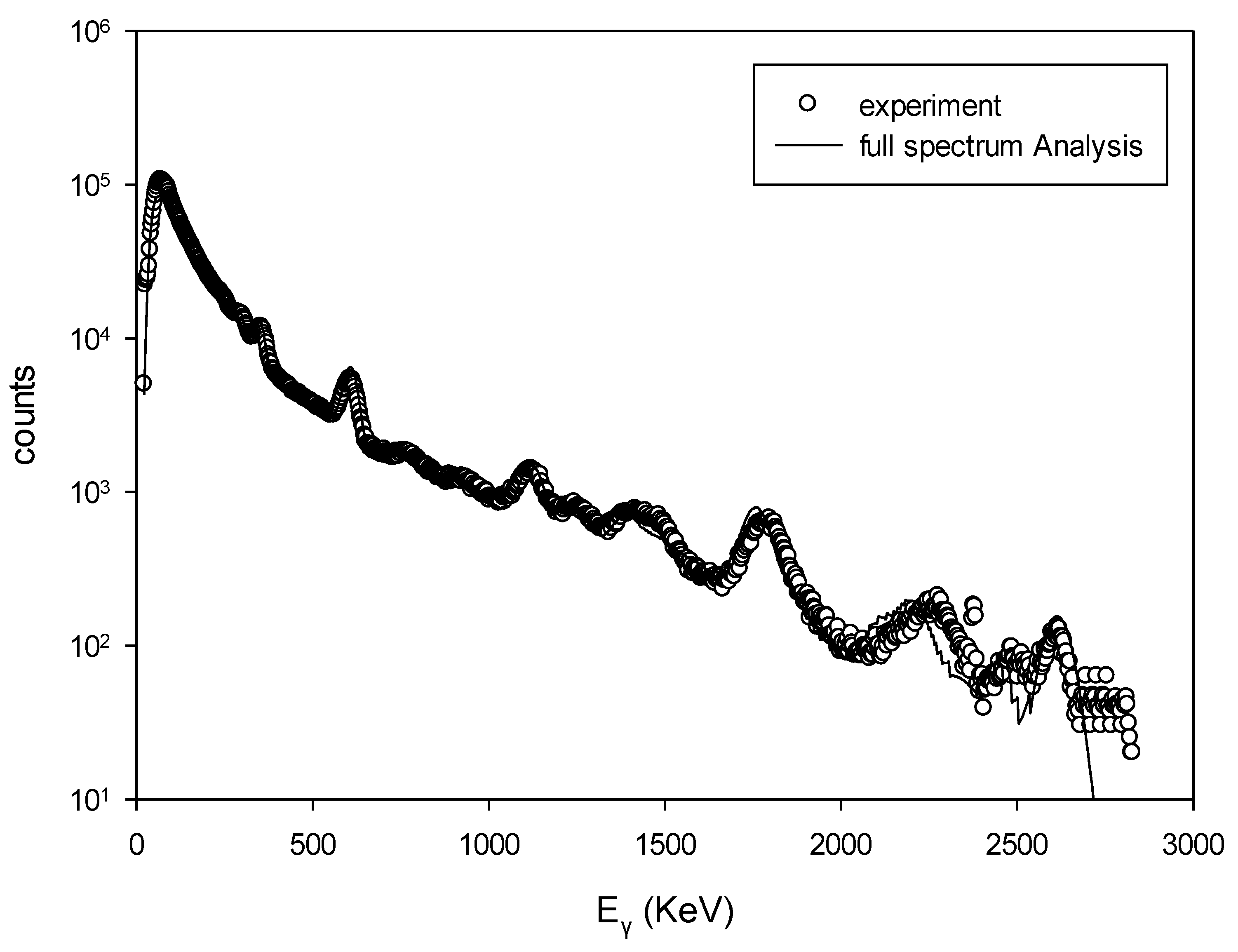
4.2. FSA in the Measured Seawater Spectrum and Response Function

The experimental spectrum acquired in the tank is depicted in Figure 6 along with the total simulated FSA spectrum (summation of 214Pb, 214Bi, 40K, 228Ac, and 208Tl), which was calculated to fit the experimental one. All the simulated spectra were normalized to the measurement period (live time). For the total simulated spectrum depicted in Figure 6, a simplified fitting procedure was applied using the derived single simulated spectra for the KATERINA system for each utilized radionuclide (214Pb, 214Bi, 40K, 228Ac, and 208Tl). For the fitting procedure, each simulated spectrum was multiplied with a free parameter αj, which was fixed at an optimum value when the best fit of the total simulated spectrum to the experimental one was obtained. These parameters were deduced from the fitting procedure of the experimental spectrum and corresponded to the activity concentration of the observed radionuclides.
The calculation was performed using as the initial value for (208Tl) the experimentally derived value (analyzed by 583 keV) and the activity concentration for the two radon progenies (214Pb and 214Bi). The incremental step of the activity concentration for the minimization process was 20 Bq/m3. The fitting process was terminated when the value of χ2 was found to be less than the statistical uncertainty of the minimization process. The calculation process provided activity concentration values of 11 Bq/L for the radon progeny 214Pb and 14 Bq/L for 214Bi. The activity concentration of the FSA calculation process for 208Tl and 228Ac was 0.9 Bq/L and 3.5 Bq/L for 40K. The agreement of the calculated spectrum using the FSA (by the minimization between the reproduced and the experimental spectrum) for all studied radionuclides (40K, 214Pb, 214Bi, 208Tl, and 228Ac) was satisfactory (see Figure 6). The 40K photopeak in the experimental and simulated spectra was not distinguished due to the low counting rate and due to the interference with the energy lines emitted from 214Bi. Furthermore, the gamma-ray energy threshold of the underwater detection system was clearly identified (~50–55 keV) using the system in the low saline seawater and/or groundwater matrix [17,18].
The discrepancy at 2400 keV may be produced from the non-intense natural radioactivity contribution. The reliability of the data was first demonstrated by the good agreement between the experimental spectrum and the theoretical one reproduced via Monte Carlo simulation. Moreover, the experimental marine efficiency values of the KATERINA detection system as calculated previously in the calibration tank were within the uncertainties for the measured values of the proposed work making the system a valuable tool for quantitative measurements in static aquatic environments in the energy range from the threshold to 2614 keV.

5. Summary and Perspectives

An optimized calibration process was performed for an underwater in situ gamma-ray spectrometer (named KATERINA) using larger tank dimensions to include and imitate the marine environment for gamma-ray emissions above 1000 keV (such as 208Tl at 2614 keV). Additionally, the FSA method was applied efficiently for the gamma-ray spectra acquired in the tank to measure the activity concentration of the natural radioactivity enriched in the natural drinking water with which the tank was filled.
As concerns the results of this work, the activity concentration of the radon progenies that were enriched in the water was enhanced compared with other radionuclides due to the mineralogy of the aquifer. The activity concentration of 40K found in the seawater in the southern Caspian Sea agreed with the salinity values. The activity concentration of the natural and artificial radionuclides that were enriched in the sediment exhibited similar values to those found in the literature [5,8,9], without any indication of a radiological issue due to the low values of the activity concentration. The new calibration method was validated using the experimental and reproduced data through Monte Carlo simulation in the energy range from the threshold to 2614 keV rendering the system capable of offering quantitative data rapidly even in the high energetic part. The detection system can support routine and emergency monitoring tasks integrating the proposed method.
In future, the quantification method will be optimized using the detected natural radionuclides enriched in the water to improve the operation of the system as a calibrationless underwater in situ detection system up to a maximum energy of 2614 keV (without the use of laborious work, which is costly due to the calibration tanks and reference radioactive sources). The FSA algorithm will be developed for supporting sediment radioactivity analysis. Both algorithms for seawater and sediment analysis will be integrated into the hardware for automated analysis in the case of using the underwater radioactivity detection system as an observation system for acquisition in a time-series mode. In this case, non-heavy software is required to enhance the rapid analysis and near real-time response in case of an emergency.

Author Contributions

Data curation, E.G.A., C.T. and S.A.; Formal analysis, E.G.A. and C.T.; Investigation, C.T. and E.G.A.; Methodology, E.G.A. and C.T., Software, S.A. and E.G.A.; Supervision, C.T.; Validation, C.T. and E.G.A.; Visualization, S.A. and E.G.A.; Writing—original draft, C.T., E.G.A. and S.A.; Writing—review & editing, C.T. and E.G.A. All authors have read and agreed to the published version of the manuscript.

Funding

This research received no external funding.

Institutional Review Board Statement

The study was conducted in the frame of a bilateral collaboration between IROST and IO/HCMR.

Informed Consent Statement

Informed consent was obtained from all subjects involved in the study.

Data Availability Statement

The data are available on request.

Acknowledgments

The authors would like to acknowledge the Iranian Research Organization for Science and Technology (IROST) for supporting all activities related to the calibration of the KATERINA sensor and its demonstration in the field. The authors would like also to acknowledge A.N. Berlizov for providing the MCNP-CP code. The authors would like to thank Rosa Vlastou for the support of the National Technical University of Athens research group for the calibration exercises that took place in their tank.

Conflicts of Interest

The authors declare no conflict of interest.

References

  1. UNSCEAR. Report of the United Nations Scientific Committee on the Effects of Atomic Radiation, Sources Effects and Risks of Ionizing Radiation: Fifty-Sixth Session; United Nation: New York, NY, USA, 2018.
  2. United Nations Scientific Committee on the Effects of Atomic Radiation; Annex, B. Exposures from Natural Radiation Sources; United Nation: New York, NY, USA, 2000.
  3. Tsabaris, C.; Eleftheriou, G.; Patiris, D.L.; Androulakaki, E.G.; Kapanadze, N.; Pappa, F.K.; Melikatze, G.; Ruzsa, G. Distribution of activity concentration and dose rates in selected coastal areas on western and eastern Black Sea. J. Radioanal. Nucl. Chem. 2019, 321, 169–181. [Google Scholar] [CrossRef]
  4. Jowzaee, S. Determination of selected natural radionuclide concentrations in southwestern Caspian groundwater using liquid scintillation counting. Radiat. Prot. Dosim. 2013, 157, 234–241. [Google Scholar] [CrossRef] [PubMed]
  5. Tsabaris, C.; Scholten, J.; Karageorgis, A.P.; Comanducci, J.F.; Georgopoulos, D.; Liong Wee Kwong, L.; Patiris, D.L.; Papathanassiou, E. Underwater in situ measurements of radionuclides in selected submarine groundwater springs, Mediterranean sea. Radiat. Prot. Dosim. 2010, 142, 273–281. [Google Scholar] [CrossRef] [PubMed]
  6. Tsabaris, C.; Patiris, D.L.; Karageorgis, A.P.; Eleftheriou, G.; Papadopoulos, V.P.; Georgopoulos, D.; Papathanassiou, E.; Povinec, P.P. In-situ radionuclide characterization of a submarine groundwater discharge site at Kalogria Bay, Stoupa, Greece. J. Environ. Radioact. 2012, 108, 50–59. [Google Scholar] [CrossRef] [PubMed]
  7. Burnett, W.C.; Aggarwal, P.K.; Aureli, A.; Bokuniewicz, H.; Cable, J.E.; Charette, M.A.; Kontar, E.; Krupa, S.; Kulkarni, K.M.; Loveless, A.; et al. Quantifying submarine groundwater discharge in the coastal zone via multiple methods. Sci. Total Environ. 2006, 367, 498–543. [Google Scholar] [CrossRef]
  8. Abdi, M.R.; Hassanzadeh, S.; Kamali, M.; Raji, H.R. 238U, 232Th, 40K and 137Cs activity concentrations along the southern coast of the Caspian Sea, Iran. Mar. Pollut. Bull. 2009, 58, 658–662. [Google Scholar] [CrossRef]
  9. Abbasia, A.; Algethamib, M.; Bawazeerc, O.; Zakaly, H.M.H. Distribution of natural and anthropogenic radionuclides and associated radiation indices in the Southwestern coastline of Caspian Sea Author links open overlay panel. Mar. Pollut. Bull. 2022, 178, 113593. [Google Scholar] [CrossRef]
  10. Aakens, U.R. Radioactivity monitored from moored Oceanographic buoys. Chem. Ecol. 1995, 10, 61–69. [Google Scholar] [CrossRef]
  11. Povinec, P.P.; Osvath, I.; Baxter, M.S. Underwater Gamma-spectrometry with HPGe and NaI(Tl) detectors. Appl. Radiat. Isot. 1996, 47, 1127–1133. [Google Scholar] [CrossRef]
  12. Wedekind, C.; Schilling, G.; Grüttmüller, M.; Becker, K. Gamma-radiation monitoring network at sea. Appl. Radiat. Isot. 1999, 50, 733–741. [Google Scholar] [CrossRef]
  13. van Put, P.; Debauche, A.; De Lellis, C.; Adam, V. Performance level of an autonomous system of continuous monitoring of radioactivity in seawater. J. Environ. Radioact. 2004, 72, 177–186. [Google Scholar] [CrossRef]
  14. Osvath, I.; Povinec, P.P.; Livingston, H.D.; Ryan, T.P.; Muslow, S.; Commanducci, J.-F. Monitoring of radioactivity in NW Irish Sea water using a stationary underwater gamma-ray spectrometer with satellite data transmission. J. Radioanal. Nucl. Chem. 2005, 263, 437–440. [Google Scholar] [CrossRef]
  15. Thornton, B.; Ohnishi, S.; Ura, T.; Odano, N.; Fujita, T. Continuous measurement of radionuclide distribution off Fukushima using a towed sea-bed gamma-ray spectrometer. Deep-Sea Res. 2013, 79, 10–19. [Google Scholar] [CrossRef]
  16. Tsabaris, C.; Patiris, D.L.; Lykousis, V. An in situ spectrometer for continuous monitoring of radon daughters in aquatic environment. Nucl. Instrum. Methods Phys. Res. Sect. A Accel. Spectrometers Detect. Assoc. Equip. 2011, 626–627, S142–S144. [Google Scholar] [CrossRef]
  17. Tsabaris, C.; Bagatelas, C.; Dakladas, T.; Papadopoulos, C.T.; Vlastou, R.; Chronis, G.T. An autonomous in situ detection system for radioactivity measurements in the marine environment. Appl. Radiat. Isot. 2008, 66, 1419–1426. [Google Scholar] [CrossRef]
  18. Tsabaris, C.; Androulakaki, E.G.; Prospathopoulos, A.; Alexakis, A.; Eleftheriou, G.; Patiris, D.L.; Pappa, F.K.; Sarantakos, K.; Kokkoris, M.; Vlastou, R. Development and optimization of an underwater in-situ cerium bromide spectrometer for radioactivity measurements in the aquatic environment. J. Environ. Radioact. 2019, 204, 12–20. [Google Scholar] [CrossRef]
  19. Androulakaki, E.G.; Kokkoris, M.; Skordis, E.; Fatsea, E.; Patiris, D.L.; Tsabaris, C.; Vlastou-Zanni, R. Implementation of FLUKA for γ-ray Applications in the Marine Environment. J. Environ. Radioact. 2016, 164, 253–257. [Google Scholar] [CrossRef]
  20. Androulakaki, E.G.; Tsabaris, C.; Eleftheriou, G.; Kokkoris, M.; Patiris, D.L.; Pappa, F.K.; Vlastou, R. Efficiency calibration for in situ γ-ray measurements on the seabed using Monte Carlo simulations: Application in two different marine environments. J. Environ. Radioact. 2016, 164, 47–59. [Google Scholar] [CrossRef]
  21. Androulakaki, E.G.; Kokkoris, M.; Tsabaris, C.; Eleftheriou, G.; Patiris, D.L.; Pappa, F.K.; Vlastou, R. In situ γ-ray spectrometry in the marine environment using full spectrum analysis for natural radionuclides. Appl. Radiat. Isot. 2016, 114, 76–86. [Google Scholar] [CrossRef]
  22. Bagatelas, C.; Tsabaris, C.; Kokkoris, M.; Papadopoulos, C.T.; Vlastou, R. Determination of marine gamma activity and study of the minimum detectable activity (MDA) in 4pi geometry based on Monte Carlo simulation. Environ. Monit. Assess. 2010, 165, 159–168. [Google Scholar] [CrossRef]
  23. Vlachos, D.S.; Tsabaris, C. Response Function Calculation of an Underwater Gamma Ray NaI(Tl) Spectrometer. Nucl. Instrum. Methods Phys. Res. A 2005, 539, 413–420. [Google Scholar] [CrossRef]
  24. Tsabaris, C.; Ballas, D.D. On line gamma-ray spectroscopy at open sea. Appl. Radiat. Isot. 2005, 62, 83–89. [Google Scholar] [CrossRef] [PubMed]
  25. Tsabaris, C. Monitoring natural and artificial radioactivity enhancement in the Aegean Sea using floating measuring systems. Appl. Radiat. Isot. 2008, 66, 1599–1603. [Google Scholar] [CrossRef] [PubMed]
  26. Tsabaris, C.; Vlachos, D.S.; Papadopoulos, C.T.; Vlastou, R.; Kalfas, C.A. Set up and application of an Underwater γ- ray spectrometer for radioactivity Measurements. Mediterr. Mar. Sci. 2005, 6, 35–40. [Google Scholar] [CrossRef] [Green Version]
  27. Caciolli, A.; Baldoncini, M.; Bezzon, G.P.; Broggini, C.; Buso, G.P.; Callegari, I.; Colonna, T.; Fiorentini, G.; Guastaldi, E.; Mantovani, F.; et al. A new FSA approach for in situ γ ray spectroscopy. Sci. Total Environ. 2012, 414, 639–645. [Google Scholar] [CrossRef] [Green Version]
  28. Hendriks, P.H.G.M.; Limburg, J.; de Meijer, R.J. Full-spectrum analysis of natural γ-ray spectra. J. Environ. Radioact. 2001, 53, 365–380. [Google Scholar] [CrossRef]
  29. van der Graaf, E.R.; Limburg, J.; Koomans, R.L.; Tijs, M. Monte Carlo based calibration of scintillation detectors for laboratory and in situ gamma ray measurements. J. Environ. Radioact. 2011, 102, 270–282. [Google Scholar] [CrossRef]
  30. Maučec, M.; de Meijer, R.J.; Rigollet, C.; Hendriks, P.H.G.M.; Jones, D.G. Detection of radioactive particles offshore by γ-ray spectrometry Part I: Monte Carlo assessment of detection depth limits. Nucl. Instrum. Methods 2004, 525, 593–609. [Google Scholar] [CrossRef]
  31. Berlizov, A.N. MCNP-CP a Correlated Particle Radiation Source Extension of a General Purpose Monte Carlo N Particle Transport Code; Semkov, T.M., Pommé, S., Jerome, S.M., Eds.; ACS Symposium Series 945; American Chemical Society: Washington, DC, USA, 2006; pp. 183–194. [Google Scholar]
  32. Kalfas, C.A.; Axiotis, M.; Tsabaris, C. SPECTRW: A software package for nuclear and atomic spectroscopy. Nucl. Instrum. Methods Phys. Res. Sect. A Accel. Spectrometers Detect. Assoc. Equip. 2016, 116, 22–33. [Google Scholar] [CrossRef]
  33. Eleftheriou, G.; Pappa, F.Κ.; Maragos, N.; Tsabaris, C. Continuous monitoring of multiple submarine springs by means of gamma-ray spectrometry. J. Environ. Radioact. 2020, 216, 106180. [Google Scholar] [CrossRef]
  34. Commission Recommendation on the Protection of the Public against Exposure to Radon in Drinking Water Supplies; Document number C (Dec 2001), (2001/928/4580), Euratom 928, L 344/85, Commission; Euratom: Maastricht, The Netherlands, 2001.
Figure 1. The area of study in the southern Caspian Sea coastal area (Muhmudabad).
Jmse 11 00725 g001
Figure 2. Schematic diagram and the underwater photo of the experimental setup in the seawater (a) and in the sediment (b).
Jmse 11 00725 g002
Figure 3. The gamma-ray spectrum acquired during the first day of the experiment in the calibration tank.
Jmse 11 00725 g003
Figure 4. The measured spectrum using the KATERINA system placed on the seabed (1 h measurement). A zoom window around 661 keV is depicted. The peaks identified in Figure 3 are clearly seen. The arrow indicates the energy window of the emission energy of 137Cs.
Jmse 11 00725 g004
Figure 5. The measured and simulated marine efficiency values from the threshold energy to 2614 keV. The filled circles represent the experimental values while the line the theoretical reproduction values of the marine efficiency.
Jmse 11 00725 g005
Figure 6. The measured and simulated spectra for estimating concentrations using the FSA method. In the Y-axis, the counts represent the detected events in each gamma-ray energy.
Jmse 11 00725 g006
Table 1. The quantitative data of the radon and thoron progenies in the water source.
IsotopeEnergy
(keV)
Activity
(Bq/m3)
Uncertainty
(Bq/m3)
214Pb35211,380450
214Bi112014,540710
176415,080600
208Tl583107045
Table 2. The measured quantitative data in the sediment using the KATERINA detection system.
IsotopeEnergy (keV)Activity Concentration (Bq/kg)Uncertainty (Bq/kg)
214Bi609152
1764202
208Tl2614355
40K146163050
137Cs6612.10.3
Disclaimer/Publisher’s Note: The statements, opinions and data contained in all publications are solely those of the individual author(s) and contributor(s) and not of MDPI and/or the editor(s). MDPI and/or the editor(s) disclaim responsibility for any injury to people or property resulting from any ideas, methods, instructions or products referred to in the content.

