Article

Microplastic Accumulation in Catfish and Its Effects on Fish Eggs from Songkhla Lagoon, Thailand

by
Siriporn Pradit
1,
Prakrit Noppradit
1,*,
Preyanuch Jitkaew
1,
Karnda Sengloyluan
2,
Mathinee Yucharoen
1,*,
Phudith Suwanno
3,
Varaporn Tanrattanakul
4,
Kittiwara Sornplang
1 and
Thongchai Nitiratsuwan
5
1
Coastal Oceanography and Climate Change Research Center, Faculty of Environmental Management, Prince of Songkla University, Songkhla 90110, Thailand
2
International College, Prince of Songkla University, Songkhla 90110, Thailand
3
Faculty of Environmental Management, Prince of Songkla University, Songkhla 90110, Thailand
4
Faculty of Science, Prince of Songkla University, Songkhla 90110, Thailand
5
Faculty of Science and Fisheries Technology, Rajamangala University of Technology Srivijaya, Trang 92150, Thailand
*
Authors to whom correspondence should be addressed.
J. Mar. Sci. Eng. 2023, 11(4), 723; https://doi.org/10.3390/jmse11040723
Submission received: 26 February 2023 / Revised: 24 March 2023 / Accepted: 25 March 2023 / Published: 27 March 2023
(This article belongs to the Special Issue Fish as Bioindicators of the Fresh and Marine Water Ecosystems)

Abstract

:
Microplastics have been found to accumulate in freshwater, marine ecosystems, and biological organisms. The frequency of studies on microplastic contamination in organs has increased recently, although there have been relatively fewer investigations on fish eggs in Thailand. To extract microplastics from catfish samples for laboratory analysis (Osteogeneiosus militaris), we used 10% potassium hydroxide in the digestion process. A needle penetrated the fish eggs to investigate microplastic contamination. We examined microplastics under a stereomicroscope and used Fourier transform infrared spectroscopy to determine the type of polymer. We found microplastic contamination in organs, most commonly in the stomach (0.91 ± 0.13 items/g), followed by tissue (0.53 ± 0.09 items/g), and gills (0.30 ± 0.03 items/g) at the level of significance p < 0.01. We found a total of 349 fish eggs with 27 items of microplastic. The dominant microplastic we found in the stomach, tissue, and gills of the fish, as well as in fish eggs, was of fiber shape. We noted that fragments were found only in the stomach and tissue of fish. The dominant color of microplastics was black in organs and blue in fish eggs. The common polymer types in organs and fish eggs were polyethylene terephthalate, polypropylene, and cellulosic fiber.

1. Introduction

Man-made waste is a significant global concern from the equator to the poles, and in a marine context, from estuaries and shorelines and from the sea surface to the ocean floor [1]. Microplastics are materials manufactured or used by humans that degrade due to various forces, such as wind and currents. According to Gordon [2], plastics are the most frequent type of marine debris, comprising over 90% of the floating particles. Marine debris is described as any manufactured or used material that is abandoned, disposed of, or discarded in the marine environment by humans—intentionally or unintentionally—through different sources [3]. Research conducted by van Sebille et al. [4] estimated the amount of floating plastic in the water at 236,000 metric tons.
Microplastics are tiny plastic fragments less than 5 mm long and come in many shapes and sizes [5,6,7]. They accumulate in the marine ecosystem and impact marine biota [8,9]. Microplastics are one of the major reasons for the extinction of 17% of the species recorded by the International Union for Conservation of Nature Red List [10,11], identified in the stomachs of marine organisms ranging from crustaceans to mammals during the last 40 years [12,13,14]. Potential effects of microplastic ingestion in the stomach include a false impression of satiation and physical injury to the gastrointestinal system of fish, leading to fish death due to malnutrition [15,16,17,18,19].
Osteogeneiosus militaris, commonly called the soldier catfish—commonly consumed by Thai people—is one marine species affected by microplastic pollution. According to Parida et al. [20], this species is distributed along the Indo-Pacific region from Singapore and the west coast of India to Bangladesh, Malacca, Pakistan, Myanmar, Brunei Darussalam, Indonesia, and Malaysia. The species lives in marine, brackish, and freshwater [20], feeding mainly on small fishes and invertebrates. Many studies have shown microplastic contamination in zooplankton [21,22,23] and larvae [24,25].
We, therefore, conducted this study to identify the abundance of microplastic in the gills, stomach, tissue, and fish eggs of O. militaris living in Songkhla Lagoon and the adjacent area of the U-Taphao canal, a natural river in Southern Thailand. Since the surrounding area has experienced tremendous economic growth and urbanization, the percentage of untreated discharged wastewater from industrial and other sectors has increased rapidly. The resulting introduction of toxic contaminants and pollutants into the freshwater environment has disrupted the ecosystem by deteriorating the water quality and ecology of the canal [26,27], with industrial effluent produced at a rate of 41,000 m3 per day by companies that produce seafood, plastic, rubber, and wood [28]. We hypothesized that microplastics may have contaminated the eggs of O. militaris. Here we focus mainly on classifying microplastics according to their size, color, and type. Our results may be used as baseline data for biota in the region.

2. Materials and Methods

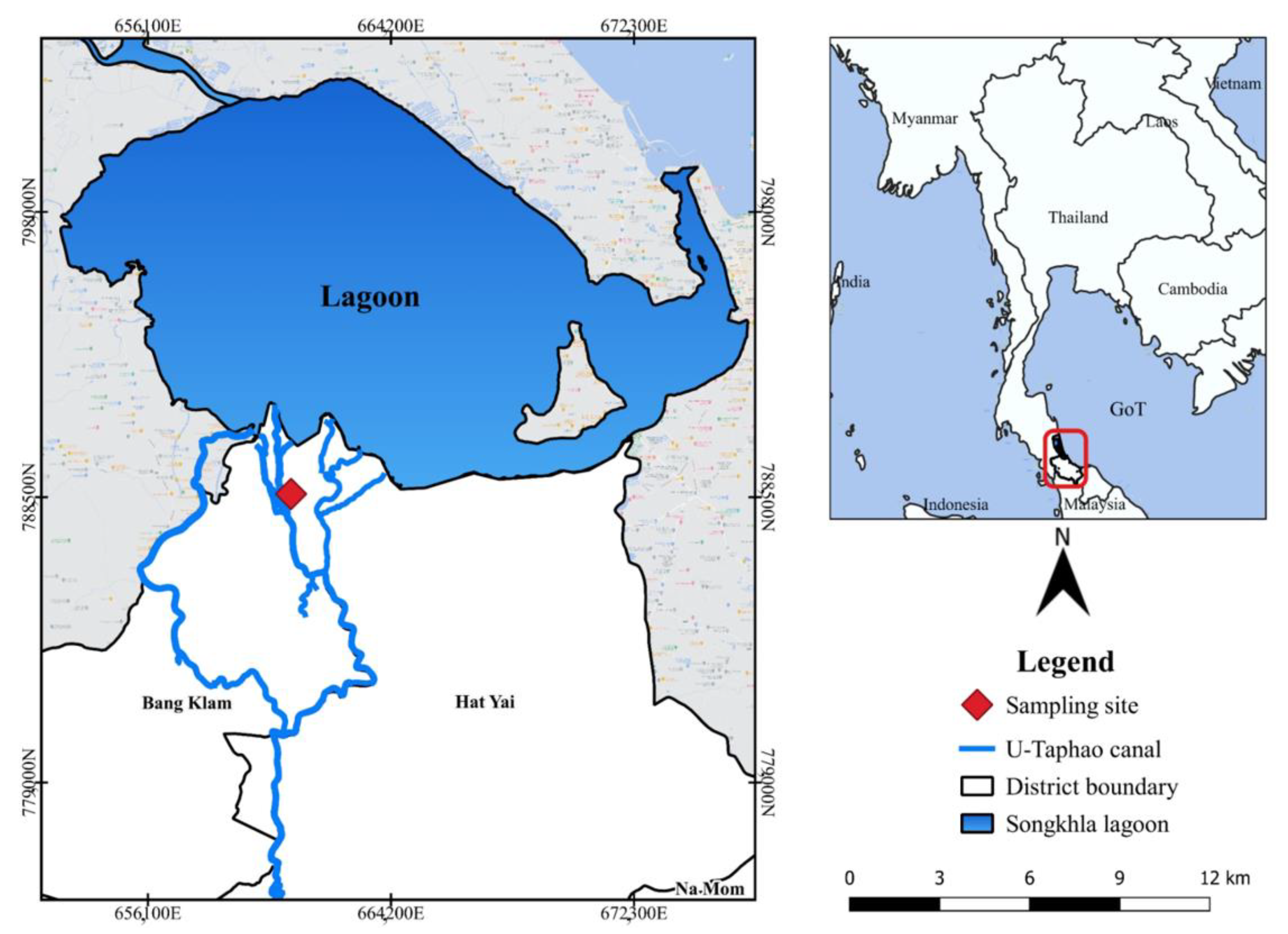
2.1. Sample Collection

We bought fish samples of O. militaris in February 2022 from the local wet market at Khlong U-Taphao, Songkhla Province (Figure 1). Fish in the local wet market are sold by local fishermen who catch fish from the Khlong U-Taphao river and Songkhla lagoon. Khlong U-Taphao is situated in Songkhla Province in Southern Thailand. It is a waterway belonging to a sub-watershed of the Songkhla Lake Basin, situated to the west of Hat Yai city at latitude 7°9′24″ North and longitude 100°27′7″ East. We sent all samples to the laboratory and stored them in a freezer at −20 °C for further analysis. We investigated catfish, O. militaris, to determine the level of microplastic contamination. In Figure 2, we show O. militaris specimens used in this study.

2.2. Laboratory Analysis

2.2.1. Part of the Observed Microplastic in the Gills, Stomach, and Tissue of Fish

In the laboratory, we defrosted the fish samples (n = 40) at room temperature before the examination. We measured and recorded the biological characteristics of O. militaris, including total length (cm), standard length (cm), and mouth length (cm). Subsequently, we dissected each fish to remove the tissue (n = 40), stomach (n = 40), and gills (n = 40) and then placed them in 250 mL glass beakers filled with distilled water. We recorded the total weight (g) of each fish before removing the samples from the stomach. We weighed the dissected parts (g) before transferring them into the glass beakers. We then filled each glass beaker with 50 mL of 10% potassium hydroxide (KOH) solution, covered them with aluminum foil, and heated them at 60 °C for 12 h. Recent studies have identified KOH as the best base for digesting fish gut materials [29,30,31,32,33]. After digestion, we filtered the solution with 20 µm filter paper and transferred the filter papers into a clean Petri dish, and dried them thoroughly for 4 to 5 h in a hot oven at 50 °C to remove moisture.

2.2.2. The Observed Microplastic in Fish Eggs

We defrosted the fish samples at room temperature in the lab before examining them. First, we removed the egg sacs from the mother catfish, then removed 349 eggs from 10 fish (out of 40 fish samples, we found only 10 females with eggs) from the egg sacs. We carefully removed each egg from the egg sac and placed them in a Petri dish without breaking any egg cells. We weighed each egg cell using an electronic balance and measured the diameter using an electronic digital Vernier caliper. We placed each egg cell in an individual Petri dish and stored them in the refrigerator for further analysis.

2.3. Microplastic Identification

2.3.1. The Observed Microplastic in the Gills, Stomach, and Tissue of Fish

We conducted microplastic identification in O. militaris by visualizing the filter papers under a Leica EZ4 W stereomicroscope (Leica, Germany). We photographed the particles with the Leica Application Suite. We categorized the particles by type: fiber or fragment. Additionally, we measured particle sizes and classified them into the following size categories: 20–100 µm; 100–300 µm; 300–500 µm; 501–700 µm; and 701 µm−1 mm. We identified and recorded the color and abundance of the microplastics. We determined the characteristics of the polymer by Fourier transform infrared spectroscopy (FTIR) (Perkin Elmer, spotlight 200i) with the resolution set at 4 cm−1 and the spectrum range fixed from 4000 to 400 cm−1 wavelength. We compared the microplastic spectrum discovered in the fish samples to the spectrum library of each type of polymer.

2.3.2. The Observed Microplastics in Fish Eggs

We conducted microplastic identification in the O. militaris fish eggs by visualizing them with an Olympus SZ61 Light-Emitting Diode stereomicroscope. We photographed the particles with a Leica Application Suite and classified them by the general properties of microplastics—namely, size (µm), color, abundance, and shape. During the microplastic identification, we ruptured the egg cells using forceps and a fine needle. To distinguish the microplastic polymers, we performed FTIR (Perkin Elmer, spotlight 200i) using the same method used in the previous section.

2.4. Data Analysis

We used Microsoft Excel (Office Professional Plus 2019, Microsoft, Washington, USA) to analyze the size, color, type, and abundance of the MPs. We present the data as mean ± standard error (items/g). We tested for a normal distribution of data. We tested for differences in the number of microplastics in fish organs using one-way ANOVA. Because the characteristics of microplastic were not normally distributed, we used the Kruskal-Wallis Test to test for significant differences among different data groups with a significant difference level of 0.05. We analyzed fish eggs using the same descriptive statistical analysis as that used in the organ analysis of soldier catfish. We analyzed the correlation between the size and weight of the fish eggs and the size of microplastics found in fish eggs using the Pearson correlation.

2.5. Contamination Prevention

Throughout the experiment, we used no plastic instruments on the samples to minimize the impact of external microplastics [34]. During dissection, we used stainless steel pans and dissection tools to reduce microplastic contamination. We covered the sample fish in the beaker with aluminum foil during the experiment to protect them from any potential air-borne pollutants. We checked the surface of the eggs for microplastic contamination before classification under a microscope and then ruptured them with a small needle to check for microplastics within. While conducting lab analysis, all lab staff wore latex/nitrile gloves and cotton lab coats [35], as well as face masks and head covers throughout the microplastic analysis. We thoroughly cleaned and rinsed all the tools and Petri dishes used for the analysis with water and detergent. To observe possible air-borne contamination, we poured distilled water into a Petri dish and left it in the laboratory for 24 h. We found no microplastics in the Petri dish at the end of the experiment.

3. Results

3.1. Abundance of Microplastics in Fish Organs

We studied 120 samples (stomachs = 40; gills = 40; tissue = 40) from 40 catfish (O. militaris). We found microplastics in 97 samples (81%). The average standard length and body weight of O. militaris were 19.50 ± 0.35 cm and 100.32 ± 5.13 g. The weights of the studied organs, including gill, stomach, and tissue, were, on average, 4.34 ± 0.19 g, 1.72 ± 0.10 g, and 3.57 ± 0.19 g, respectively. O. militaris contained an average of 4.15 ± 0.30 microplastic items/individual, with microplastics in their gills, stomachs, and tissue with an average of 0.31 ± 0.03, 0.91 ± 0.13, 0.53 ± 0.09 items/g, respectively. In addition, in the gill, stomach, and tissue, we found 1.25 ± 0.13 microplastic items/gill, 1.35 ± 0.15 items/stomach, and 1.55 ± 0.19 items/tissue. According to the one-way ANOVA analysis, we found that the number of microplastics in all three organs differed significantly at p < 0.01. In Table 1, we present data for microplastic accumulation in the gills, stomach, and tissue of soldier catfish.

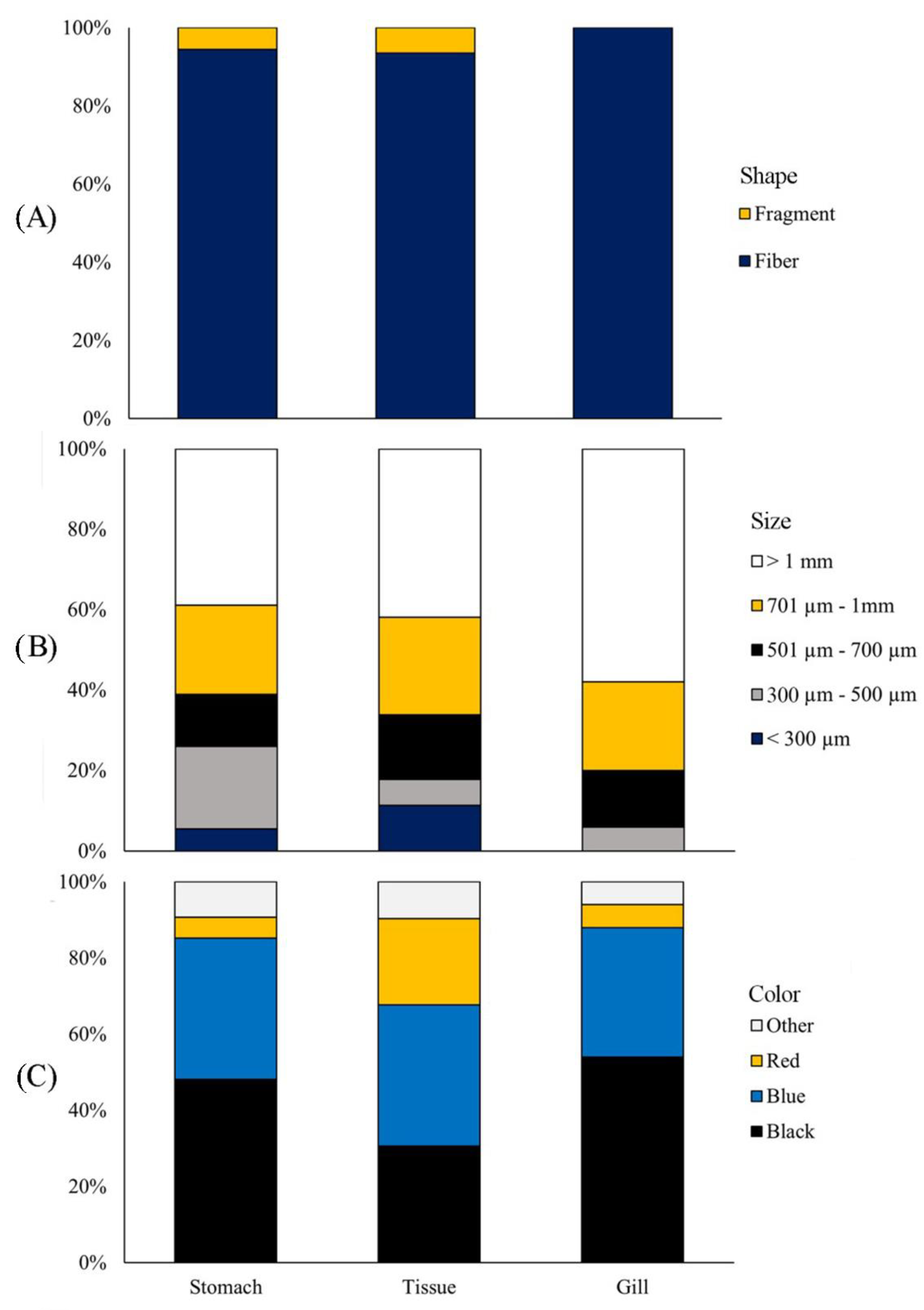
3.2. Characteristics of Microplastics in Catfish

In this study, we measured the fibers and fragments in the studied organs of O. militaris in grams. We found fiber in the stomach, tissue, and gills at 0.83 ± 0.13 items/g, 0.47 ± 0.07 items/g, and 0.30 ± 0.03 items/g, respectively. We found fragments only in the stomach and tissue in amounts of 0.08 ± 0.04 items/g and 0.06 ± 0.04 items/g, respectively, but no significant fragments in the gills when analyzed by Kruskal-Wallis-H at p < 0.01.
The size of microplastics found in the stomach was greater than that in both the tissue and the gills. The most common size of microplastics in all three organs was greater than 1 mm. In the stomach, we found 0.39 ± 0.11 items/g; in tissue, 0.24 ± 0.06 items/g; and in the gills, 0.18 ± 0.03 items/g, which all differed significantly at p < 0.01, followed by sizes 701 µm––1 mm, 501–700 µm, 300–500 µm, and <300 µm, respectively. However, we found no microplastics of <300 µm in the gills.
The colors of the microplastics in this study were black, blue, red, and others (green, transparent, and purple). The most abundant color in all three organs was black (p < 0.01)—in the stomach, at 15 items/g (9.56%), in the gills, at 5 items/g (37%), and in the tissue, at 6 items/g (3.93%). This was followed by blue in the stomach at 12 items/g (7.36%), in the gills at 6 items/g (4.07%), and in the tissue at 4 items/g (2.47%). In addition, red and others were found in amounts that did not differ significantly. In Figure 3, we show all these data.

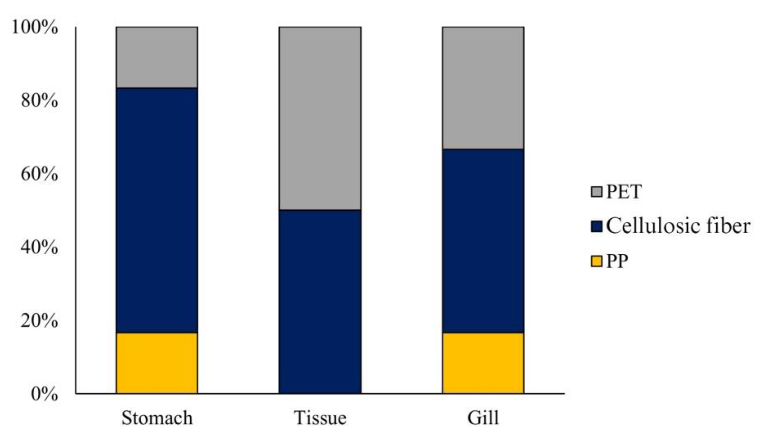
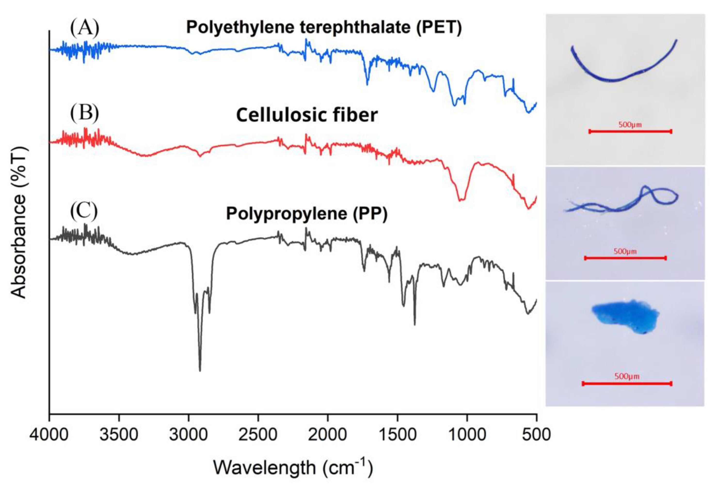
3.3. Polymer Identification in O. militaris

We found three types of polymers in all samples of organs, including polypropylene (PP), polyethylene terephthalate (PET), and cellulosic fiber. As shown in Figure 4, Cellulosic fiber was the dominant polymer found in gills at around 50%, followed by PET (33%) and PP (17%). The polymer cellulosic fiber dominated in the stomach at the highest percentage (67%), followed by PP and PET at 17%. In the tissue, we found two polymers—cellulosic fiber and PET. For all organs, cellulosic fiber was the dominant polymer, followed by PET and then PP. In Figure 5, we illustrate the spectrum absorption of polymers in O. militaris.

3.4. Abundance of Microfibers in Fish Eggs
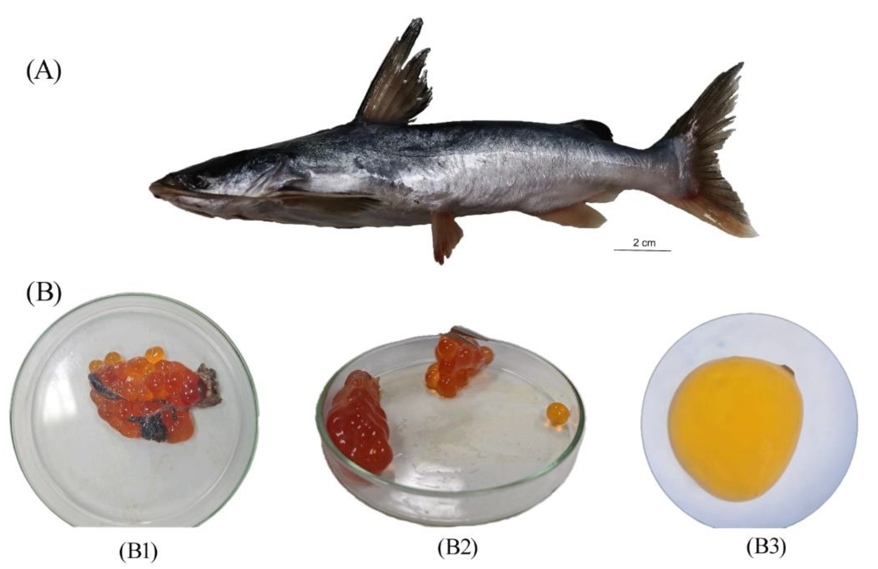
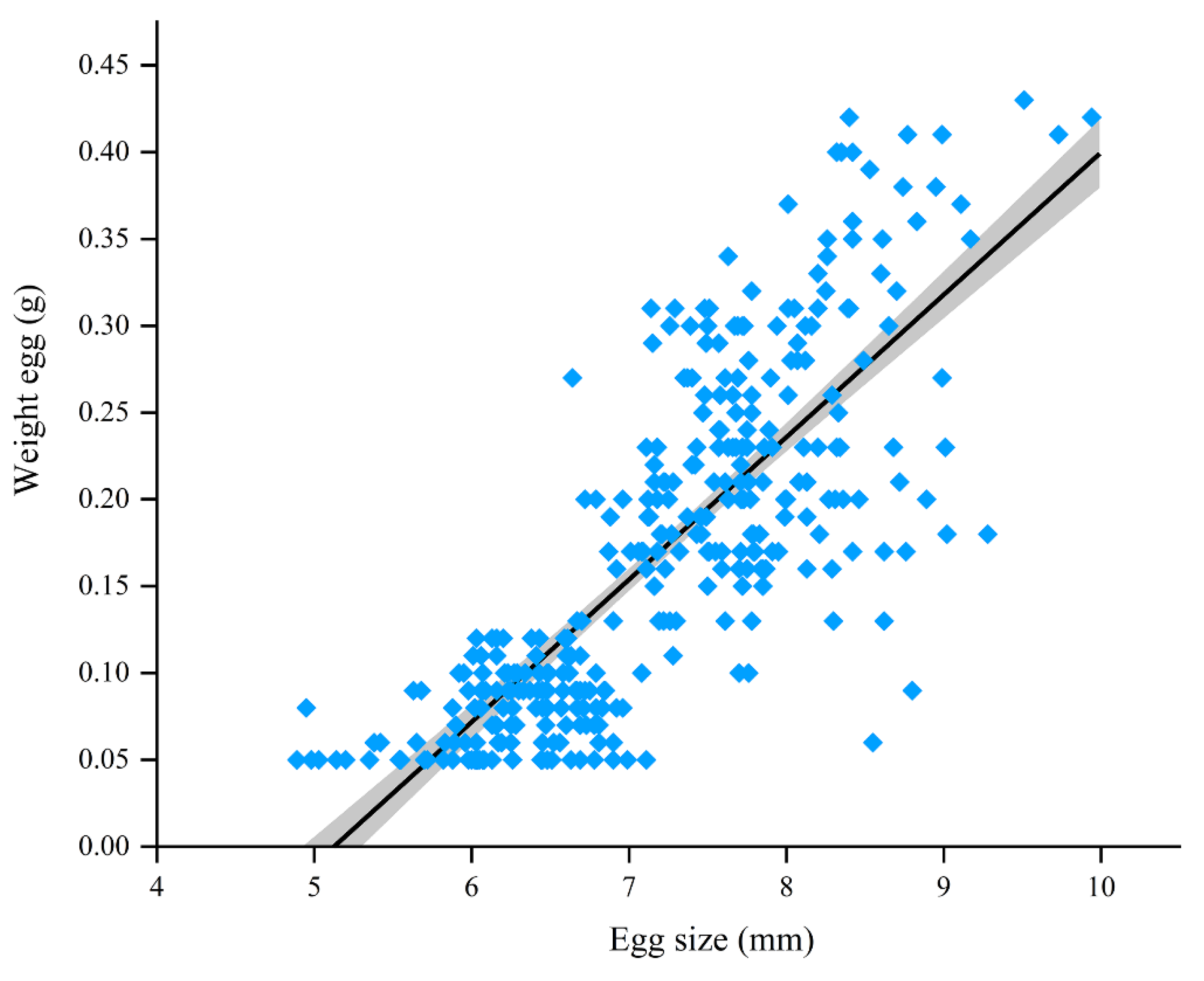
3.4.1. Fish Eggs

We extracted 349 spherical eggs in total from the mother soldier catfish. The entire bubble was pale yellow. The inside of the fish eggs was transparent, with no discernible differentiation, suggesting they were in the first stage of development. The fish eggs ranged in size (4.89–9.94 mm, average of 7.21 ± 0.05 mm) and weight (0.05–0.43 g, average of 0.17 ± 0.01 g). Increased egg size significantly increased egg weight (p < 0.05, Pearson Correlation = 0.768), as we show in Figure 6.

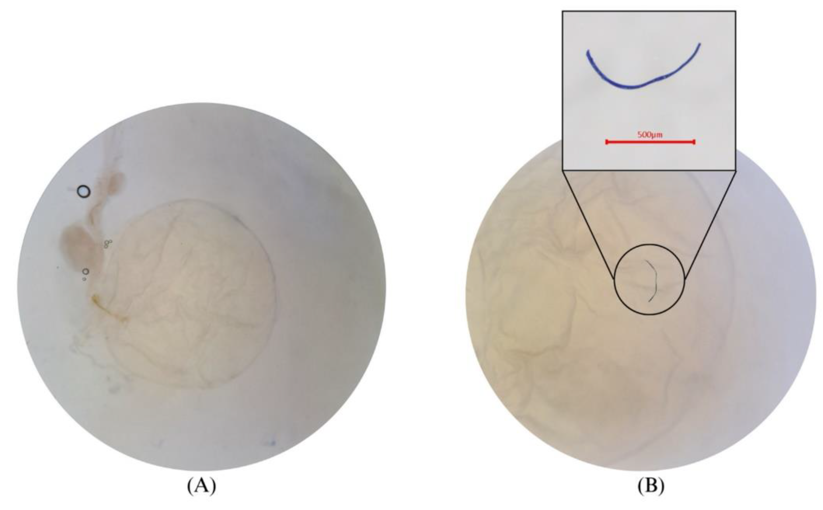
3.4.2. Microfibers in Fish Eggs of O. militaris

Microfibers appeared in this study in 25 out of 349 fish eggs. After we pierced the eggs, microfiber contamination was evident under the stereoscopic microscope, as shown in Figure 7. The size of the fish eggs found to contain microfibers was 5.82–8.95 mm in range, averaging 7.20 ± 0.17 mm, and their weight was 0.05–0.38 g in range, averaging 0.18 ± 0.02 g. This demonstrates that microfibers may enter fish eggs. We found 27 pieces of fiber ranging in size from 8–200 µm. We found 11 blue items (40.74%), 10 black items (37.04%), 3 red items (11.11%), 2 transparent items (7.41%), and 1 green item (3.70%), as we show in Figure 8. The Pearson correlation analysis showed that the relationship between egg size and microplastic size was not significant (p = 0.647, correlation coeffient −0.092).

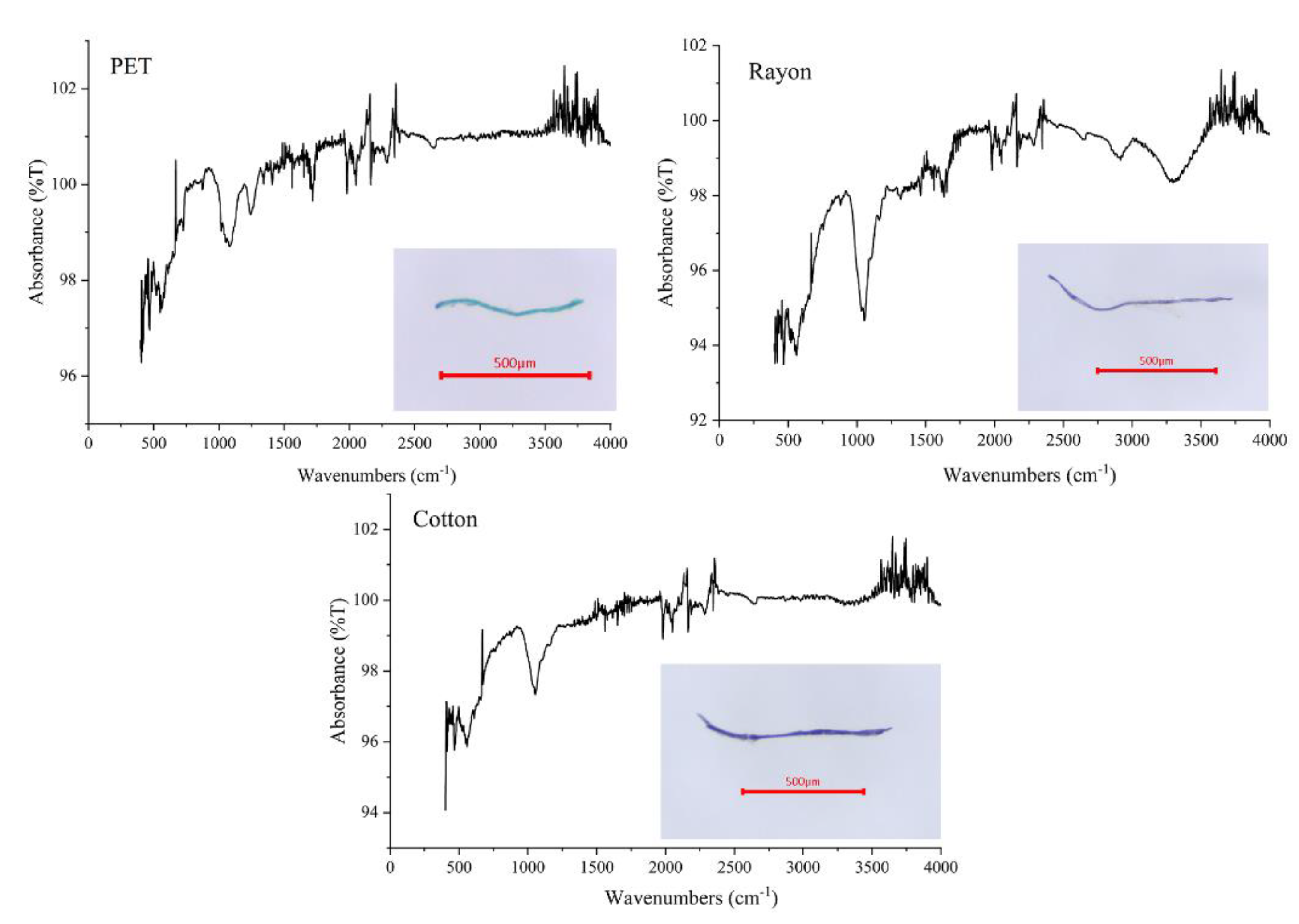
3.4.3. Polymer Identification in Fish Eggs

We found 27 contaminating microplastic pieces (from 25 fish eggs) comprising three polymers determined by FTIR, as we show in Figure 9. The predominant polymers in fish eggs were rayon (50%), PET (30%), and natural-polymer cotton (20%).

4. Discussion

4.1. Microplastics in the Stomach, Tissue, and Gills of Catfish

This study demonstrates the presence of microplastics in the appendages of O. militaris. One gram of tissue contained the most microplastics in the stomach. This suggests that the gastrointestinal tract is involved in the accumulation of microplastics because of the feeding behavior of this fish species, which eats small organisms, such as small fish and krill [36]. Microplastic particles that contaminate the water column can be unintentionally ingested by marine biota, including fish [37,38,39]. Contaminated and accumulated microplastics in prey fish may enter nutrients and be present in predator fish on a large scale, known as the transmission of microplastics into the food chain [40,41]. However, microplastics reside in the stomach only temporarily and can migrate to other organs. At the same time, the accumulation of microplastics in the stomach can increase in the future [38,40], passing this contamination on to the tissue of the fish. In a study by Have et al. [42], more microplastics were found in the intestinal mucosa than in other organ samples. It has been shown that microplastics can migrate from the stomach to the intestines and be absorbed through the intestinal mucosa into the tissue [42]. The uptake of microplastics may be related to fish protein digestion. Proteins are large organic molecules that can be digested into amino acids and absorbed through the stomach and intestinal mucosa into the circulatory system for utilization [43]. Microplastics are polymers indigestible to fish, which may contaminate organic matter being digested. However, this study is the first study on the presence of microplastics in the fish organs of O. militaris from southern Thailand. Moreover, the diameter of the microplastics found in this study was approximately 20 µm, possibly passing through the intestinal mucosa and gastric wall through intestinal adsorption [44]. When microplastics are ingested, they accumulate in the tissue. In fish tissues, there is no mechanism for the excretion of polymeric waste. The organ with the least amount of microplastic contamination was the gills. This is consistent with a study by Wei et al. [45], who found a greater accumulation of microplastics in the gastrointestinal tract than in the gums [45]. In addition, fragments were not found in the gills of the fish samples, which may depend on the gill characteristics of each fish species [46]. This study found that fiber microplastics in the stomach, tissue, and gills may be caused by fish breathing when exposed to water through the mouth. Fibers can also be sucked into the mouth through the water [39]. Thus, fibrous microplastics can represent a source of fiber from urban communities that influences accumulation in fish. The main source of fiber is fishing gear, such as nets [47], textiles [48,49], and the discharge of water from washing machines [50,51].
Most microplastics found were larger than 1 mm in size, which may not be very detrimental for fish. According to Okamoto et al. [38], fish waste may be expelled with microplastics larger than 200 µm collected in the colon [38,52]. Meanwhile, a large accumulation of microplastics, especially fibers in large quantities, may cause internal clogging due to the curling of the fibers. This may affect the process of digestion and absorption of food, causing the fish to become sick [53]. In addition, the behavior of fish species and their environment affect the color ingestion of microplastics. Okamoto et al. [38] confirmed that light and darkness have a significant influence on the color perception of fish. Under dark conditions, the ingestion of microplastic particles is reduced [38], a finding consistent with the study by Clere et al. [54]. Black is the color of microplastics predominant in fish, and because benthic environments are quite dark, these fish unwittingly ingest microplastics in their food [54].
In Table 2, we show a comparison of these findings with other studies. The amount of microplastics in the stomach of O. militaris, found to be 1.55 ± 0.19 items/stomach, was higher than that of Johnius weberi, which had 1.14 ± 1.21 items/stomach, Johnius borneensis, which had 0.90 ± 0.88 items/stomach, Dendrophysa russelli, which had 0.88 ± 1.12 items/stomach, and Panna microdon, which had 0.85 ± 1.06 items/stomach [55], but lower than that of Arius maculatus, which had 2.73 ± 0.15 items/stomach [51]. This may depend on the habitat and feeding behavior of each fish species. When comparing the number of microplastics in the units of items per individual, we found the occurrence of microplastics in this study to average 4.15 ± 0.3 items/individual, higher than that of several studies, including microplastics in the stomach of Rexea solandri, found to be 1.96 ± 1.12 items/individual [56], Scyliorhinus canicular, which had 1.20 ± 0.45 items/individual, and Mullus barbatus, which had 1.75 ± 1.14 items/individual [57]. There are few reports of microplastic studies of the gills; however, our findings were still higher than those in the studies of Mullus surmuletus, which had 3.22 items/individual [58], and Clarias gariepinus, which had 3.8 ± 2.7 items/individual [59], and lower than those of Saurida undosquamis, which had 4.65 items/individual, and Mugil cephalus, which had 7.56 items/individual [58]. In tissue studies, we found the average microplastic in demersal fish to be 4.7 ± 1.7 items/individual [60], which was greater than that in the study of microplastics in the tissues.

4.2. Contamination of Microfibers in Fish Eggs

The presence of microfiber and microplastic (PET) in the eggs suggests the transmission of microplastics through the feeding of the mother fish to the yolk sac [61]. Although microplastics were found in small amounts, it is reassuring that the microplastic fibers had not yet affected the normal development of the embryo in the early stages of fish eggs [62]. Since the mother fish may need food to feed the embryo in the egg [63], microplastics may have the potential to increase in the eggs as they develop. We foresee more detailed studies on microplastic contamination during the developmental stages of fish eggs in the future.

4.3. Polymer Type Found in Fish Organs and Fish Eggs

The occurrence of plastic waste is caused mainly by the release of waste plastic products when they are still usable. When they enter the sea, they take a long time to become microplastics and are distributed to all water columns [55,64]. The polymers found in the catfish indicate potential sources of precursors such as PP, which could be food packaging and microwave-safe containers [65], and possibly single-use masks, which can release blue polypropylene fiber or other types of fiber [66]. PET could be from packaging [67] and clothing. Rayon may be released from clothing when washed in a washing machine [51,68]. Microplastics are polymers that can be degraded to a smaller size but cannot disappear in the environment and can also absorb toxins dissolved in water, resulting in microplastic particles that have attached to organic and inorganic substances that may be harmful to the bodies of fishes [69]. These tiny plastic fragments may have a greater or lesser impact on the fish that consume them. Fish exposed to microplastics may experience the potential for a variety of negative consequences, including growth and immunity suppression, reduced feeding efficiency, and the development of neurotransmission dysfunction, endocrine disruption, genotoxicity, and oxidative stress [44], which damage cells, impair development, and cause fecundity abnormalities that decrease the abundance of fish in the ecosystem [70]. From the FTIR results in Figure 5, it is difficult to confirm whether the fibers are mainly rayon or cotton fibers because both fibers are made from natural sources using different preparation processes. Most of the chemical functional groups detected by FTIR are greatly similar [71], and microfibers produced from cotton and rayon fabrics are similar [72]. After passing through the degradation process, the two fabrics are not easy to identify. However, the spectrum was confirmed to be a rayon fiber according to the FTIR database, which delivers more than 80% precision. Cotton and rayon fibers are commonly used for non-woven fabrics—apart from polyester and nylon fibers—to produce disposable masks [73]. In practice, rayon and cotton fibers are classified as cellulose fibers. Therefore, it is not easy to differentiate between them if there is no specific change in the structure or component. The detection of cellulose fiber pollution in the environment cannot be identified between cotton and rayon fibers. However, cotton is a natural fiber, and rayon is a man-made fiber based on natural sources such as wood, plants, or agricultural waste. Both materials could be decomposed in a few months [71,74] and are classified as biodegradable. However, the degradation of these materials is also dependent on the preparation technique and degradation conditions; their degradation behavior changes under different circumstances. For instance, neat cotton fiber can completely degrade in less than two months, but after the different treatment conditions, it took longer to degrade these cotton fibers [74,75]. The degradation of cotton fiber in soil took a shorter time than that in marine conditions, and a higher temperature tended to accelerate the degradation rate of the cotton [76]. Considering the degradation behavior of neat cotton and rayon, they might not contaminate the environment as microplastics because they can be degraded in a few months; however, they can remain in the environment for several months or a few years after certain treatments.

5. Conclusions

We discovered microplastics in every organ of the studied fish, raising concerns about the impact of microplastics on the marine environment and, ultimately, human fish consumption. The most common shape discovered in this study was fibrous. Interestingly, our study uncovered evidence of microfiber in fish eggs; thus, this study should serve as a starting point for future research. Possible sources of microplastic and microfiber discovered include clothing, fishing, packaging, and single-use masks; therefore, care should be taken to avoid or reduce their leakage into the marine environment.

Author Contributions

Conceptualization, S.P., P.N., M.Y.; Investigation, P.J., S.P., K.S. (Karnda Sengloyluan), P.S., V.T., T.N.; Methodology, P.J., S.P., K.S. (Karnda Sengloyluan), K.S. (Kittiwara Sornplang); Writing—original draft, P.J., K.S. (Karnda Sengloyluan); Writing—review & editing, S.P., P.N., M.Y., P.S., V.T., T.N. All authors have read and agreed to the published version of the manuscript.

Funding

This research was funded by the National Science, Research, and Innovation Fund (NSRF) and Prince of Songkhla University (Grant No. ENV6505026M), PSU-TUYF Charitable Trust Fund under a project “Coral reef biodiversity conservation and connectivity in southern Gulf of Thai-land to support reef resilience and sustainable use” and Coastal Oceanography and Climate Change Research Center.

Institutional Review Board Statement

The study was conducted in accordance to the Animanls for Scientifuc Purposes Act, B.E 2558 (A.D. 2015).

Informed Consent Statement

Not applicable. These studies not involving humans.

Data Availability Statement

Not applicable.

Acknowledgments

We express great thanks to Hemaadarshini Krebanathan, Kivita Chandran, and Kasvinraj Murugiah (Universiti Malaysia Terengganu) for helping with the analysis of fish and fish eggs in the laboratory.

Conflicts of Interest

The authors declare no conflict of interest.

References

  1. Scientific and Technical Advisory Panel (STAP). Marine Debris as a Global Environmental Problem: Introducing Solutions Based Framework Focused on Plastic. A STAP In-Formation Document; Global Environment Facility: Washington, DC, USA, 2011. [Google Scholar]
  2. Gordon, M. Eliminating Land-Based Discharges of Marine Debris in California: A Plan of Action from the Plastic Debris Project; California Coastal Commission: San Francisco, CA, USA, 2006.
  3. Galgani, F.; Fleet, D.; Van Franeker, J.; Katsanevakis, S.; Maes, T.; Mouat, J.; Oosterbaan, L.; Poitou, I.; Hanke, G.; Thompson, R.; et al. Marine Strategy Framework Directive, Task Group 10 Report: Marine Litter. In JRC Scientific and Technical Reports; Zampoukas, N., Ed.; European Commission Joint Research Centre: Ispra, Italy, 2010. [Google Scholar]
  4. Van Sebille, E.; Wilcox, C.; Lebreton, L.; Maximenko, N.; Hardesty, B.D.; Van Franeker, J.A.; Eriksen, M.; Siegel, D.; Galgani, F.; Law, K.L. A global in-ventory of small floating plastic debris. Environ. Res. Lett. 2015, 10, 124006. [Google Scholar] [CrossRef]
  5. Group of Experts on the Scientific Aspects of Marine Environmental Protection. GESAMP: Sources, Fate and Effects of Microplastics in the Marine Environment: A Global Assessment; GESAMP: London, UK, 2015. [Google Scholar]
  6. Hidalgo-Ruz, V.; Gutow, L.; Thompson, R.C.; Thiel, M. Microplastics in the marine environment: A review of the methods used for identification and quantification. Environ. Sci. Technol. 2012, 46, 3060–3075. [Google Scholar] [CrossRef]
  7. Cole, M.; Lindeque, P.; Halsband, C.; Galloway, T.S. Microplastics as contaminants in the marine environment: A Review. Mar. Pollut. Bull. 2011, 62, 2588–2597. [Google Scholar] [CrossRef] [PubMed]
  8. Rochman, C.M.; Tahir, A.; Williams, S.L.; Baxa, D.V.; Lam, R.; Miller, J.T.; Teh, S.J. Anthropogenic debris in seafood: Plastic debris and fibers from textiles in fish and bivalves sold for human consumption. Sci. Rep. 2015, 5, 14340. [Google Scholar] [CrossRef] [PubMed] [Green Version]
  9. Kühn, S.; Van Franeker, J.A. Quantitative overview of marine debris ingested by marine megafauna. Mar. Pollut. Bull. 2020, 151, 110858. [Google Scholar] [CrossRef]
  10. Hardesty, B.D.; Good, T.P.; Wilcox, C. Novel methods, new results and science-based solutions to tackle marine debris impacts on wildlife. Ocean. Coast. Manag. 2015, 115, 4–9. [Google Scholar] [CrossRef] [Green Version]
  11. Gall, S.; Thompson, R. The impact of debris on Marine Life. Mar. Pollut. Bull. 2015, 92, 170–179. [Google Scholar] [CrossRef]
  12. Li, J.; Qu, X.; Su, L.; Zhang, W.; Yang, D.; Kolandhasamy, P.; Shi, H. Microplastics in mussels along the coastal waters of China. Environ. Pollut. 2016, 214, 177–184. [Google Scholar] [CrossRef]
  13. Jabeen, K.; Su, L.; Li, J.; Yang, D.; Tong, C.; Mu, J.; Shi, H. Microplastics and mesoplastics in fish from coastal and fresh waters of China. Environ. Pollut. 2017, 221, 141–149. [Google Scholar] [CrossRef]
  14. Pozo, K.; Gomez, V.; Torres, M.; Vera, L.; Nuñez, D.; Oyarzún, P.; Klánová, J. Presence and characterization of microplastics in fish of commercial importance from the Biobío Region in central Chile. Mar. Pollut. Bull. 2019, 140, 315–319. [Google Scholar] [CrossRef]
  15. Cole, M.; Webb, H.; Lindeque, P.K.; Fileman, E.S.; Halsband, C.; Galloway, T.S. Isolation of microplastics in biota-rich seawater samples and marine organisms. Sci. Rep. 2014, 4, 4528. [Google Scholar] [CrossRef] [Green Version]
  16. Wright, S.L.; Thompson, R.C.; Galloway, T.S. The physical impacts of microplastics on marine organisms: A Review. Environ. Pollut. 2013, 178, 483–492. [Google Scholar] [CrossRef] [PubMed]
  17. Lusher, A.L.; Hernandez-Milian, G.; O’Brien, J.; Berrow, S.; O’Connor, I.; Officer, R. Microplastic and macroplastic ingestion by a deep diving, oceanic cetacean: The true’s beaked whale Mesoplodon Mirus. Environ. Pollut. 2015, 199, 185–191. [Google Scholar] [CrossRef] [PubMed]
  18. Nadal, M.; Alomar, C.; Deudero, S. High levels of microplastic ingestion by the semipelagic fish Bogue Boops Boops (L.) around the Balearic Islands. Environ. Pollut. 2016, 214, 517–523. [Google Scholar] [CrossRef] [PubMed]
  19. Wójcik-Fudalewska, D.; Normant-Saremba, M.; Anastácio, P. Occurrence of plastic debris in the stomach of the invasive crab eriocheir sinensis. Mar. Pollut. Bull. 2016, 113, 306–311. [Google Scholar] [CrossRef]
  20. Parida, P.K.; Jaiswar, A.K.; Palaniswamy, R.; Kumar, P.; Chakraborty, S.K. Growth and mortality of Osteogeneiosus mili-taris (Linnaeus 1758) from Mumbai waters. Indian J. Fish. 2014, 61, 12–15. [Google Scholar]
  21. Fagiano, V.; Alomar, C.; Compa, M.; Soto-Navarro, J.; Jordá, G.; Deudero, S. Neustonic microplastics and zooplankton in coastal waters of Cabrera marine protected area (Western Mediterranean Sea). Sci. Total Environ. 2022, 804, 150120. [Google Scholar] [CrossRef]
  22. Sambolino, A.; Herrera, I.; Álvarez, S.; Rosa, A.; Alves, F.; Canning-Clode, J.; Kaufmann, M. Seasonal variation in micro-plastics and zooplankton abundances and characteristics: The ecological vulnerability of an oceanic island system. Mar. Pollut. Bull. 2022, 181, 113906. [Google Scholar] [CrossRef]
  23. Botterell, Z.L.; Bergmann, M.; Hildebrandt, N.; Krumpen, T.; Steinke, M.; Thompson, R.C.; Lindeque, P.K. Microplastic ingestion in zooplankton from the Fram Strait in the Arctic. Sci. Total Environ. 2022, 831, 154886. [Google Scholar] [CrossRef]
  24. Zhang, C.; Zuo, Z.; Wang, Q.; Wang, S.; Lv, L.; Zou, J. Size Effects of Microplastics on Embryos and Observation of Toxicity Kinetics in Larvae of Grass Carp (Ctenopharyngodon idella). Toxics 2022, 10, 76. [Google Scholar] [CrossRef]
  25. Uy, C.A.; Johnson, D.W. Effects of microplastics on the feeding rates of larvae of a coastal fish: Direct consumption, trophic transfer, and effects on growth and survival. Mar. Biol. 2022, 169, 27. [Google Scholar] [CrossRef] [PubMed]
  26. Chuvanich, S.; Thongnoo, K.; Chevakidagarn, P.; Phongdara, A. Evaluation of Artificial Neural Networks for Electrical Con-ductivity-based and Flow Rate-based Prediction of the Nitrate Nitrogen Concentration in the U-Tapao Canal, Hat Yai, Thailand. Environ. Asia 2017, 10, 15–24. [Google Scholar]
  27. Gyawali, S.; Techato, K.; Yuangyai, C. Effects of Industrial Waste Disposal on the Surface Water Quality of U-tapao River, Thailand. In Proceedings of the International Conference on Environmental Science Engineering IPCBEE, Bangkok, Thailand, 24 March 2012; Volume 3, pp. 109–113. [Google Scholar]
  28. Sirinawin, W.; Sompongchaiyakul, P. Nondetrital and total metal distribution in core sediments from the U-Tapao Canal, Songkhla, Thailand. Mar. Chem. 2005, 94, 5–16. [Google Scholar] [CrossRef]
  29. Foekema, E.M.; De Gruijter, C.; Mergia, M.T.; van Franeker, J.A.; Murk, A.J.; Koelmans, A.A. Plastic in North sea fish. Environ. Sci. Technol. 2013, 47, 8818–8824. [Google Scholar] [CrossRef] [PubMed]
  30. Dehaut, A.; Cassone, A.L.; Frère, L.; Hermabessiere, L.; Himber, C.; Rinnert, E.; Rivière, G.; Lambert, C.; Soudant, P.; Huvet, A.; et al. Microplastics in seafood: Benchmark protocol for their extraction and characterization. Environ. Pollut. 2016, 215, 223–233. [Google Scholar] [CrossRef] [Green Version]
  31. Hermsen, E.; Pompe, R.; Besseling, E.; Koelmans, A.A. Detection of low numbers of microplastics in North Sea fish using strict quality assurance criteria. Mar. Pollut. Bull. 2017, 122, 253–258. [Google Scholar] [CrossRef]
  32. Kühn, S.; Van Werven, B.; Van Oyen, A.; Meijboom, A.; Rebolledo, E.L.B.; Van Franeker, J.A. The use of potassium hy-droxide (KOH) solution as a suitable approach to isolate plastics ingested by marine organisms. Mar. Pollut. Bull. 2017, 115, 86–90. [Google Scholar] [CrossRef]
  33. Bessa, F.; Barría, P.; Neto, J.M.; Frias, J.P.; Otero, V.; Sobral, P.; Marques, J.C. Occurrence of microplastics in commercial fish from a natural estuarine environment. Mar. Pollut. Bull. 2018, 128, 575–584. [Google Scholar] [CrossRef]
  34. Pan, Z.; Zhang, C.; Wang, S.; Sun, D.; Zhou, A.; Xie, S.; Zou, J. Occurrence of Microplastics in the Gastrointestinal Tract and Gills of Fish from Guangdong, South China. J. Mar. Sci. Eng. 2021, 9, 981. [Google Scholar] [CrossRef]
  35. Güven, O.; Gökdağ, K.; Jovanović, B.; Kıdeyş, A.E. Microplastic litter composition of the Turkish territorial waters of the Mediterranean Sea, and its occurrence in the gastrointestinal tract of fish. Environ. Pollut. 2017, 223, 286–294. [Google Scholar] [CrossRef]
  36. Angsupanich, S.; Somsak, S.; Phrommoon, J. Stomach contents of the catfishes Osteogeneiosus militaris (Linnaeus, 1758) and Arius maculatus (Thunberg, 1792) in the Songkhla lake. Warasan Songkhla Nakharin (Sakha Witthayasat lae Technology). 2005. Available online: https://agris.fao.org/agris-search/search.do?recordID=TH2008001875 (accessed on 25 February 2023).
  37. Kalaiselvan, K.; Pandurangan, P.; Velu, R.; Robinson, J. Occurrence of Microplastics in Gastrointestinal Tracts of Planktivorous Fish from the Thoothukudi Region. Environ. Sci. Pollut. Res. 2022, 29, 44723–44731. [Google Scholar] [CrossRef] [PubMed]
  38. Okamoto, K.; Nomura, M.; Horie, Y.; Okamura, H. Color Preferences and Gastrointestinal-Tract Retention Times of Mi-croplastics by Freshwater and Marine Fishes. Environ. Pollut. 2022, 304, 119253. [Google Scholar] [CrossRef] [PubMed]
  39. Li, B.; Liang, W.; Liu, Q.X.; Fu, S.; Ma, C.; Chen, Q.; Su, L.; Craig, N.J.; Shi, H. Fish Ingest Microplastics Unintentionally. Environ. Sci. Technol. 2021, 55, 10471–10479. [Google Scholar] [CrossRef] [PubMed]
  40. Jovanović, B. Ingestion of Microplastics by Fish and Its Potential Consequences from a Physical Perspective. Integr. Environ. Assess. Manag. 2017, 13, 510–515. [Google Scholar] [CrossRef] [PubMed]
  41. Cverenkárová, K.; Valachovičová, M.; Mackuľak, T.; Žemlička, L.; Bírošová, L. Microplastics in the Food Chain. Life 2021, 11, 1349. [Google Scholar] [CrossRef]
  42. Haave, M.; Gomiero, A.; Schönheit, J.; Nilsen, H.; Olsen, A.B. Documentation of Microplastics in Tissues of Wild Coastal Animals. Front. Environ. Sci. 2021, 9, 31. [Google Scholar] [CrossRef]
  43. Phillips, S. Environmental Impacts of Marine Aquaculture Issue Paper; Pacific States Marine Fisheries Commission: Portland, OR, USA, 2005.
  44. Lu, Y.; Zhang, Y.; Deng, Y.; Jiang, W.; Zhao, Y.; Geng, J.; Ding, L.; Ren, H. Uptake and Accumulation of Polystyrene Mi-croplastics in Zebrafish (Danio Rerio) and Toxic Effects in Liver. Environ. Sci. Technol. 2016, 50, 4054–4060. [Google Scholar] [CrossRef]
  45. Wei, L.; Wang, D.; Aierken, R.; Wu, F.; Dai, Y.; Wang, X.; Zhen, Y. The prevalence and potential implications of microplastic contamination in marine fishes from Xiamen Bay, China. Mar. Pollut. Bull. 2022, 174, 113306. [Google Scholar] [CrossRef]
  46. Hughes, G.M.; Morgan, M. The Structure of Fish Gills in Relation to Their Respiratory Function. Biol. Rev. 1973, 48, 419–475. [Google Scholar] [CrossRef]
  47. Wright, L.S.; Imogen.; Napper, I.E.; Thompson, R.C. Potential Microplastic Release from Beached Fishing Gear in Great Britain’s Region of Highest Fishing Litter Density. Mar. Pollut. Bull. 2021, 173, 113115. [Google Scholar] [CrossRef]
  48. Naidoo, T.S.; Thompson, R.C.; Rajkaran, A. Quantification and Characterisation of Microplastics Ingested by Selected Juvenile Fish Species Associated with Mangroves in KwaZulu-Natal, South Africa. Environ. Pollut. 2020, 257, 113635. [Google Scholar] [CrossRef] [PubMed]
  49. Thiele, C.J.; Hudson, M.D.; Russell, A.E.; Saluveer, M.; Sidaoui-Haddad, G. Microplastics in Fish and Fishmeal: An Emerging Environmental Challenge? Sci. Rep. 2021, 11, 2045. [Google Scholar] [CrossRef]
  50. Jiwarungrueangkul, T.; Phaksopa, J.; Sompongchaiyakul, P.; Tipmanee, D. Seasonal Microplastic Variations in Estuarine Sediments from Urban Canal on the West Coast of Thailand: A Case Study in Phuket Province. Mar. Pollut. Bull. 2021, 168, 112452. [Google Scholar] [CrossRef] [PubMed]
  51. Pradit, S.; Noppradit, P.; Goh, P.B.; Sornplang, K.; Ong, M.C.; Towatana, P. Occurrence of Microplastics and Trace Metals in Fish and Shrimp from Songkhla Lake, Thailand during the COVID-19 Pandemic. Appl. Ecol. Environ. Res. 2021, 19, 1085–1106. [Google Scholar] [CrossRef]
  52. de Vries, A.N.; Sigurður, D.G.; Árnason, H.; Carlsson, P. Microplastic Ingestion by Fish: Body Size, Condition Factor and Gut Fullness Are Not Related to the Amount of Plastics Consumed. Mar. Pollut. Bull. 2020, 151, 110827. [Google Scholar] [CrossRef]
  53. Yin, L.; Chen, B.; Xia, B.; Shi, X.; Qu, K. Polystyrene Microplastics Alter the Behavior, Energy Reserve and Nutritional Composition of Marine Jacopever (Sebastes schlegelii). J. Hazard. Mater. 2018, 360, 97–105. [Google Scholar] [CrossRef] [PubMed]
  54. Clere, I.K.; Ahmmed, F.; Remoto, P.J.G., III; Fraser-Miller, S.J.; Gordon, K.C.; VKomyakova, V.; Allan, B.J.M. Quantification and Characterization of Microplastics in Commercial Fish from Southern New Zealand. Mar. Pollut. Bull. 2022, 184, 114121. [Google Scholar] [CrossRef] [PubMed]
  55. Azad, S.M.O.; Towatana, P.; Pradit, S.; Patricia, B.G.; Hue, H.T. Ingestion of Microplastics by Some Commercial Fishes in the Lower Gulf of Thailand: A Preliminary Approach to Ocean Conservation. Int. J. Agric. Technol. 2018, 14, 1017–1032. [Google Scholar]
  56. Zhu, L.; Wang, H.; Chen, B.; Sun, X.; Qu, K.; Xia, B. Microplastic Ingestion in Deep-Sea Fish from the South China Sea. Sci. Total Environ. 2019, 677, 493–501. [Google Scholar] [CrossRef] [PubMed]
  57. Bellas, J.; José Martínez-Armental, J.; Martínez-Cámara, A.; Besada, V.; Martínez-Gómez, C. Ingestion of Microplastics by Demersal Fish from the Spanish Atlantic and Mediterranean Coasts. Mar. Pollut. Bull. 2016, 109, 55–60. [Google Scholar] [CrossRef]
  58. Kılıç, E.; Yücel, N. Microplastic Occurrence in the Gastrointestinal Tract and Gill of Bioindicator Fish Species in the Northeastern Mediterranean. Mar. Pollut. Bull. 2022, 177, 113556. [Google Scholar] [CrossRef] [PubMed]
  59. Kılıç, E.; Yücel, N.; Şahutoğlu, S.M. First Record of Microplastic Occurence at the Commercial Fish from Orontes River. Environ. Pollut. 2022, 307, 119576. [Google Scholar] [CrossRef] [PubMed]
  60. Khan, F.R.; Shashoua, Y.; Crawford, A.; Drury, A.; Sheppard, K.; Stewart, K.; Sculthorp, T. ‘The Plastic Nile’: First Evidence of Microplastic Contamination in Fish from the Nile River (Cairo, Egypt). Toxics 2020, 8, 22. [Google Scholar] [CrossRef] [PubMed] [Green Version]
  61. Fyhn, H.J. First Feeding of Marine Fish Larvae: Are Free Amino Acids the Source of Energy? Aquaculture 1989, 80, 111–120. [Google Scholar] [CrossRef]
  62. Bunge, A.; Kammann, U.; Scharsack, J.P. Exposure to Microplastic Fibers Does Not Change Fish Early Life Stage Devel-opment of Three-Spined Sticklebacks (Gasterosteus aculeatus). Microplastics Nanoplastics 2021, 1, 15. [Google Scholar] [CrossRef]
  63. Rønnestad, I.; Thorsen, A.; Finn, R.N. Fish Larval Nutrition: A Review of Recent Advances in the Roles of Amino Acids. Aquaculture 1999, 177, 201–216. [Google Scholar] [CrossRef]
  64. Wu, W.N.; Yang, J.; Criddle, C.S. Microplastics Pollution and Reduction Strategies. Front. Environ. Sci. Eng. 2016, 11, 6. [Google Scholar] [CrossRef]
  65. Xu, P.; Peng, G.; Su, L.; Gao, Y.; Gao, L.; Li, D. Microplastic Risk Assessment in Surface Waters: A Case Study in the Changjiang Estuary, China. Mar. Pollut. Bull. 2018, 133, 647–654. [Google Scholar] [CrossRef]
  66. Sun, J.; Yang, S.; Zhou, G.J.; Zhang, K.; Lu, Y.; Jin, Q.; He, Y. Release of microplastics from discarded surgical masks and their adverse impacts on the marine copepod Tigriopus japonicus. Environ. Sci. Technol. Lett. 2021, 8, 1065–1070. [Google Scholar] [CrossRef]
  67. Choong, W.S.; Hadibarata, T.; Tang, D.K.H. Abundance and Distribution of Microplastics in the Water and Riverbank Sediment in Malaysia—A Review. Biointerface Res. Appl. Chem. 2021, 11, 11700–11712. [Google Scholar]
  68. Ding, J.; Li, J.; Sun, C.; Jiang, F.; Ju, P.; Qu, L.; Zheng, Y.; He, C. Detection of Microplastics in Local Marine Organisms Using a Multi-Technology System. Anal. Methods 2019, 11, 78–87. [Google Scholar] [CrossRef]
  69. Cruz, A.H. Impact of Plastic Waste Ingestion by Fish. Circ. Econ. Sustain. 2022, 3, 607–616. [Google Scholar] [CrossRef]
  70. Mao, X.; Xu, Y.; Cheng, Z.; Yang, Y.; Guan, Z.; Jiang, L.; Tang, K. The Impact of Microplastic Pollution on Ecological Environment: A Review. Front. Biosci.-Landmark 2022, 27, 46. [Google Scholar] [CrossRef] [PubMed]
  71. Sunghyun, N.; Slopek, R.; Wolf, D.; Warnock, M.; Condon, B.D.; Sawhney, P.; Gbur, E.; Reynolds, M.; Allen, C. Comparison of biodegradation of low-weight hy-droentangled raw cotton nonwoven fabric and that of commonly used disposable nonwoven fabrics in aerobic Captina silt loam soil. Text. Res. J. 2016, 86, 155–166. [Google Scholar] [CrossRef]
  72. Marielis, C.Z.; Pawlak, J.J.; Daystar, J.; Ankeny, M.; Cheng, J.J.; Venditti, R.A. Microfibers generated from the laundering of cotton, rayon and polyester based fabrics and their aquatic biodegradation. Mar. Pollut. Bull. 2019, 142, 394–407. [Google Scholar] [CrossRef]
  73. Shruti, V.C.; Perez-Guevara, F.; Elizade-Martinez, I.; Kutralam-Muniasamy, G. Re-usable masks for COVID-19: A missing piece of the microplastic problem during the global health crisis. Mar. Pollut. Bull. 2020, 161, 111777. [Google Scholar] [CrossRef] [PubMed]
  74. Milošević, M.; Krkobabić, A.; Radoičić, M.; Šaponjić, Z.; Radetić, T.; Radetić, M. Biodegradation of cotton and cotton/polyester fabrics impregnated with Ag/TiO2 nanoparticles in soil. Carbohydr. Polym. 2017, 158, 77–84. [Google Scholar] [CrossRef] [PubMed]
  75. Smith, S.; Ozturk, M.; Frey, M. Soil biodegradation of cotton treated with common finishes. Cellulose 2021, 28, 4485–4494. [Google Scholar] [CrossRef]
  76. Kim, S.; Cho, Y.; Prak, C.H. Effect of cotton fabric properties on fiber release and marine biodegradation. Text. Res. J. 2022, 92, 2121–2137. [Google Scholar] [CrossRef]
Figure 1. Sampling site at U-Taphao canal, a river flowing to Songkhla Lagoon, Southern Thailand.
Jmse 11 00723 g001
Figure 2. Fish samples in the study: (A) O. militaris or soldier catfish; (B) fish eggs randomly picked from mother catfish; (B1) fish eggs taken from mother catfish; (B2) each fish egg carefully separated; (B3) appearance of a fish egg under a microscope.
Jmse 11 00723 g002
Figure 3. Distribution of microplastic characteristics in this study: (A) shape; (B) size; (C) color in the stomach, flesh, and gills of the soldier catfish O. militaris.
Jmse 11 00723 g003
Figure 4. The percentage of polymer type found in each organ studied in O. militaris.
Jmse 11 00723 g004
Figure 5. Three types of polymers analyzed by FTIR include: (A) PET; (B) Cellulosic fiber; and (C) PP found in all three organs studied in O. militaris.
Jmse 11 00723 g005
Figure 6. A positive linear correlation between size (mm) and weight (g) of fish eggs (black line), r = 0.801, p < 0.01, n = 349.
Jmse 11 00723 g006
Figure 7. The presence of microfiber contamination in fish eggs: (A) microfiber-free pierced eggs, and (B) microfiber-contaminated fish eggs.
Jmse 11 00723 g007
Figure 8. The relative proportions of various microfiber colors and the amounts present in fish eggs.
Jmse 11 00723 g008
Figure 9. Three types of polymers were found in fish eggs: PET, rayon, and cotton.
Jmse 11 00723 g009
Table 1. Data of catfish size measurement and the quantity of microplastics found in catfish organs.
Fish Sample Data and MP DataMinimumMaximumMean ± S.E.
Weight data
Standard length (cm)15.8023.5019.50 ± 0.35
Body weight (g)51.39152.50100.32 ± 5.13
Gill weight (g)2.176.614.34 ± 0.19
Stomach weight (g)0.522.941.72 ± 0.10
Tissue weight (g)1.235.883.57 ± 0.19
MP data
MPs in gills (items/gill)0.003.001.25 ± 0.13
MPs in gills (items/g gill)0.000.92(0.30 ± 0.03) a
MPs in the stomach (items/stomach)0.004.001.35 ± 0.15
MPs in the stomach (items/g stomach)0.003.85(0.91 ± 0.13) c
MPs in tissue (items/tissue)0.004.001.55 ± 0.19
MPs in tissue (items/g tissue)0.003.25(0.53 ± 0.09) b
MPs in fish (items/ind)0.008.004.15 ± 0.30
Note: MPs = microplastics, S.E. = standard error, different letters in the same row indicate significant difference at p < 0.05 (One-way ANOVA).
Table 2. Abundance and characteristics of microplastics in the stomach, tissue, and gills from several research studies.
Fish SpeciesHabitatAbundance MPs ShapeSize (mm)Reference
Stomach
Arius maculatus, n = 11Benthic 2.73 ± 0.15 items/st *Fiber0.15–5[51]
Rexea solandriDeep waters1.53 ± 1.08 items/g or
1.96 ± 1.12 items/ind
Film-like<1[56]
Scyliorhinus canicula, n = 72Demersal1.20 ± 0.45 items/indFiber 0.5–1[57]
Merluccius merluccius, n = 12Demersal1.0 items/indFiber 0.5–1
Mullus barbatus, n = 128Demersal1.75 ± 1.14 items/indFiber 0.5–1
Panna microdonDemersal0.85 ± 1.06items/st *Fiber-[55]
Dendrophysa russelliDemersal0.88 ± 1.12 items/st *Fiber-
Johnius borneensisBenthopelagic0.90 ± 0.88 items/st *Fiber-
Johnius weberiBenthopelagic1.14 ± 1.21 items/st *Fiber-
O. militaris, n = 40Benthic 1.35 ± 0.15items/st *
0.91 ± 0.13 items/g
Fiber>1 This study
Tissue
Bagrus bayad, n = 14Demersal4.7 ± 1.7 items/indFiber>1 [60]
Mystus vittatus, n = 3DemersalFiber>1
Heteropneustes fossilis, n =2DemersalFiber>1
O. militaris, n = 40Benthic 1.55 ± 0.19 items/tis * or
0.53 ± 0.09 items/g
Fiber>1 This study
Gill
Mullus barbatus, n = 43Demersal3.54 items/indFiber<1[58]
Mullus surmuletus, n = 41Demersal3.22 items/indFiber
Saurida undosquamis, n = 39Reef-associated4.65 items/indFiber
Mugil cephalus, n = 20Benthopelagic7.56 items/indFiber
Clarias gariepinus, n = 10Benthopelagic3.8 ± 2.7 items/indFiber<0.25 to >5[59]
O. militaris, n = 40Benthic 1.25 ± 0.13 items/gill
0.30 ± 0.03 items/g
Fiber>1This study
Note: * St = Stomach, tis—Tissue.
Disclaimer/Publisher’s Note: The statements, opinions and data contained in all publications are solely those of the individual author(s) and contributor(s) and not of MDPI and/or the editor(s). MDPI and/or the editor(s) disclaim responsibility for any injury to people or property resulting from any ideas, methods, instructions or products referred to in the content.

