Article

Isolation and Identification of Luminescent Bacteria in Deep Sea Marine Organisms from Sicilian Waters (Mediterranean Sea)

1
Stazione Zoologica Anton Dohrn—Sicily Marine Centre, Integrative Marine Ecology Department, Villa Pace, Contrada Porticatello 29, 98167 Messina, Italy
2
Stazione Zoologica Anton Dohrn—Sicily Marine Centre, Ecosustainable Marine Biotechnology Department, Villa Pace, Contrada Porticatello 29, 98167 Messina, Italy
3
National Research Council, Institute of Polar Sciences (CNR-ISP), Spianata S. Raineri 86, 98122 Messina, Italy
4
National Institute for Environmental Protection and Research, ISPRA, Via dei Mille 46, 98057 Milazzo, Italy
*
Author to whom correspondence should be addressed.
J. Mar. Sci. Eng. 2022, 10(8), 1113; https://doi.org/10.3390/jmse10081113
Submission received: 11 July 2022 / Revised: 10 August 2022 / Accepted: 10 August 2022 / Published: 13 August 2022

Abstract

:
Luminescent bacteria are a fascinating component of marine microbial communities, often related to the light emissions in deep sea marine organisms. They are mainly affiliated with specific phylogenetic groups, such as Photobacterium, Vibrio, and Photorhabdus, and are sometimes involved in symbiotic relationships. However, the luminescence of some marine organisms remains a poorly understood process, and it is not always certain whether their luminescence is attributable to associated luminescent bacteria. In this study, for the first time, luminescent bacteria were isolated from two deep sea organisms, namely, the cephalopod Neorossia caroli and the teleost Chlorophthalmus agassizi. The isolation was carried out on glycerol-supplemented medium, and the search for the luxAB gene was performed on all isolates as a complementary tool to the culture-dependent techniques to detect bioluminescence by molecular approach. The optimum of salinity, temperature, and pH was evaluated by physiological tests for all isolates. The production of extracellular polymeric substances was also preliminarily screened. A total of 24 luminescent isolates were obtained, with an abundance from C. agassizi specimens. All the isolates were taxonomically characterized and were related to different species of Photobacterium, with the exception of Vibrio sp. CLD11 that was from C. agassizi. The luxAB gene was detected in about the 90% of the analysed strains.

1. Introduction

Luminescent bacteria are ubiquitous in marine environments, brackish waters, and soils [1,2], and some species can establish symbiotic relationships with other marine organisms [3,4]. This topic has attracted increasing interest during the last century [5,6,7,8]. Bioluminescence is the peculiar process of light emission from living organisms, involved in many ecological dynamics, and is so widespread in nature that 76% of marine pelagic meso- and macro-organisms are bioluminescent [9].
In a marine environment, the sunlight filters down through the water layers, gradually diminishing about 10-fold for every 75 m of descent, until all visible light disappears below 1000 m [10]. Several deep sea organisms adopted bioluminescence as an adaptive strategy for living in the lower level (in the twilight zone, i.e., at a depth of 200–1000 m) due to the absence of light in this environment. This phenomenon could be used as a sexual selection tool to attract the counterpart during the reproduction period as well as being used as a support for predation and camouflage strategies, as a defense mechanism, and for communication [10,11,12,13,14,15,16].
Light-emitting marine organisms include the members of several taxonomical groups, from bacteria to more evolved animals such as fish and cephalopods. In Eukaryotes, bioluminescence could be intrinsic (autogenic bioluminescence) if derived from chemical compounds produced in special tissues of animals, or extrinsic (bacteriogenic bioluminescence) if bacterially mediated, originating from a symbiotic relationship with luminous bacteria hosted in specialized organs [10,12,17,18]. In most cases, bioluminescent species developed organized luminescent organs and photophores that can host photocytes or luminous symbiotic bacteria. Regarding the symbiotic bioluminescence of fish, bacteria are often hosted in specific areas of the fish, such as ventral luminous organs (e.g., Macrouridae, Moridae, and Chlorophthalmidae), luminous lures (e.g., Ceratiidae and Melanocetidae), subocular organs (e.g., Anomalopidae), and esophageal pouches (e.g., Leiognathidae) [7,17,19,20].
Several studies indicated that symbiotic bacteria are also hosted in cephalopod light organs or structures [21,22,23,24,25,26]. Specific tissues are usually formed during embryonic development and are poised to receive luminous bacteria from the environment [21,27,28]. The light organ is then colonized by highly specific symbiotic bacteria, becoming fully functional [25]. The bacteria in the photophores produce wavelengths of light similar to those in downwelling moonlight. This light appears to counterilluminate the squid’s ink sac, making it more difficult for visual predators to see [29,30,31].
Different aspects of the symbiotic bioluminescence need deeper investigation and an ecological interpretation. Luminous bacteria forming specific symbioses with fishes and cephalopods provide the host with a light that could be used for several functions, and at the same time, the host provides the bacteria with an ideal growth environment [10]. The presence of symbiotic bacteria also seems to be necessary for the correct morphogenesis and development of luminous organs, and in many cases, the relationship is obligate [32,33,34]. More interestingly, the relationship was proven to have a certain grade of specificity since animals are able to select the specific symbiont bacteria [35,36] and actively eliminate non-luminous bacteria. In the association between Aliivibrio fischeri and the bobtail squid Euprymna scolopes, the host was demonstrated to be able to detect at a molecular and physiological level if its symbionts are bioluminescent or not and to reject non-luminescent strains of A. fischeri [37,38]. It is also well-known that even if luminous organs are mostly colonized by a unique bacterial species, co-existence with a more taxonomic group could occur. Indeed, the light organs of a Sepiola spp. host mixed the bacterial population of A. fischeri and A. logei [24]. The co-occurrence of different luminous bacteria in the same light organ was also observed both in some species of Perciform fish, i.e., the co-symbionts Photobacterium mandapamensis and Photobacterium leiognathi [39], and in loliginid squids [40]. Evidence for parallel speciation patterns among sepiolid squids and their luminous symbiotic bacteria were also provided by experimental studies, indicating a hierarchy of symbiont competency that reflects the phylogenetic relationships of the partners [41]. Changes in the taxonomic composition of symbiotic luminescent bacteria could also occur in response to seasonal variation, which was observed in the in-fish intestinal luminescent microflora that mainly contain P. phosphoreum members in the summer while containing more A. logei members in the winter [42].
Although several studies examined the symbiotic relationship between luminous bacteria and fish or cephalopods, they often focused on the same host species. For this reason, it is important to improve our current knowledge on other poorly investigated taxa equipped with luminescent organs. In this study, we examined two deep sea demersal Mediterranean species, the Carol bobtail squid (Neorossia caroli, Sepiolidae, Cephalopoda) and the shortnose greeneye (Chlorophthalmus agassizi, Chlorophthalmidae, Osteichthyes). The Carol bobtail squid is the most bathyal species among sepiolids, which can live at depths greater than 1700 m (but more commonly found between 200 and 700 m) and shows a preference for deep muddy bottoms colonized by Isidella elongata [43]. C. agassizi is a demersal teleost that lives on muddy and clay bottoms (up to 1000 m depth) [44]. Our study evaluated the presence of luminous bacteria in association with N. caroli and C. agassizi, examining organisms collected in the Strait of Messina and in the southern Tyrrhenian Sea. These analyses were carried out through an integrated approach, coupling molecular techniques and culture-dependent methodologies. We also screened the physiological capabilities of luminous strains and searched for the luxAB gene, which is responsible for bioluminescence production. Finally, we preliminarily investigated the biotechnological ability of isolates as potential exopolysaccharides producers.

2. Materials and Methods

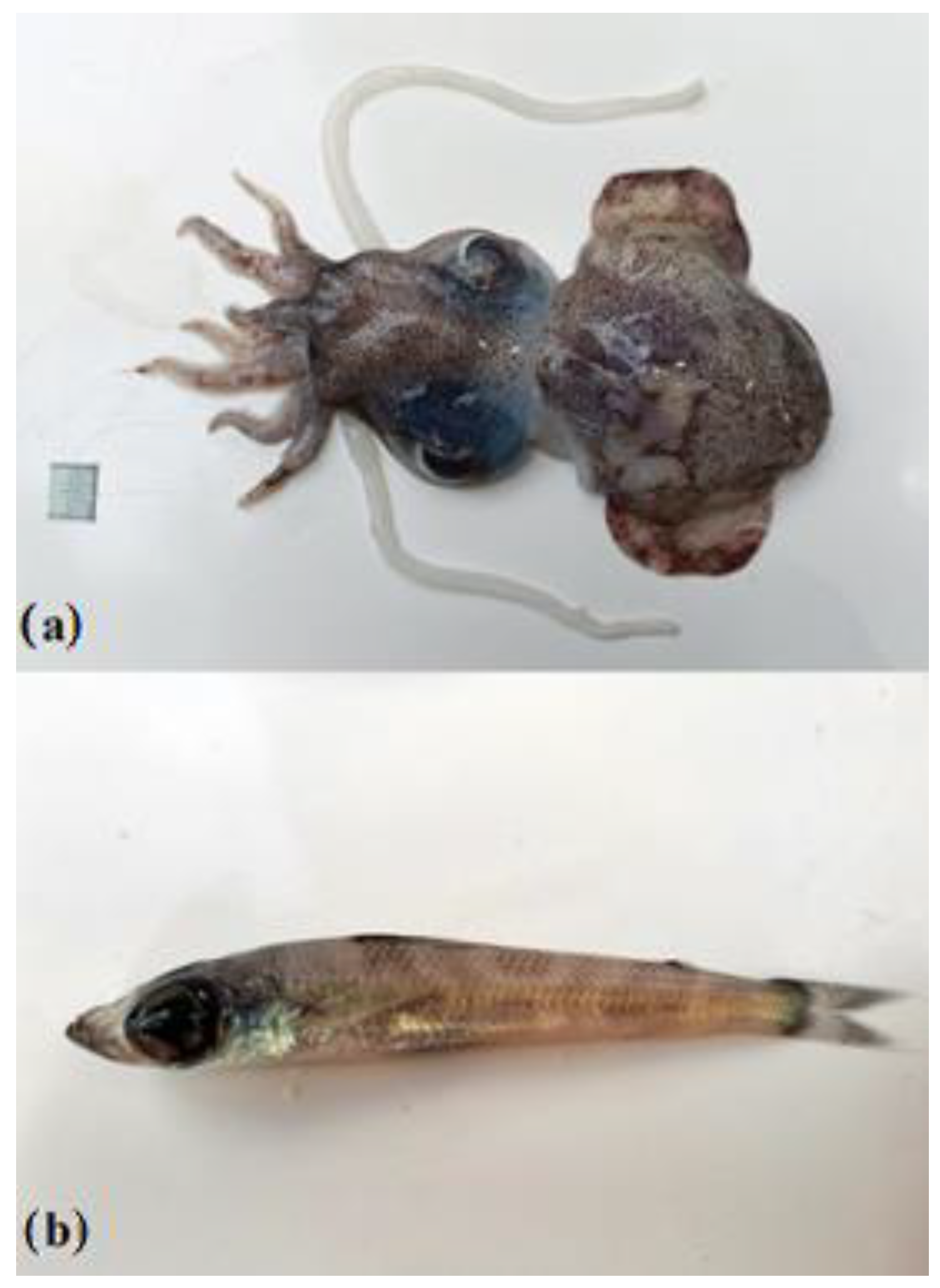
2.1. Sample Collection

Overall, fourteen N. caroli (Figure 1a) and thirteen C. agassizi (Figure 1b) specimens were collected by bottom trawling between 550 and 650 m deep in the Gulf of Patti (southern Tyrrhenian Sea) and were used for the isolation of luminescent bacteria. Additional individuals (n = 13) of C. agassizi were also collected from the nearby area of the Strait of Messina, where fresh deep sea fauna is usually found on the shore after stranding phenomena due to upwelling currents [45]. Each individual was collected and kept in a sterile tube at 4 °C until its arrival in the laboratory. We used disposable gloves while measuring each to the nearest 0.1 mm (mantle length ML for N. caroli; total length TL for C. agassizi) and weighing it (body mass W to the nearest 0.1 g). All manipulation procedures for further analysis were carried out through tweezers that were previously cleaned with absolute alcohol.

2.2. Preliminary Treatment of Samples

In laboratory, each specimen was firstly washed slightly with sterile seawater to remove transient and loosely attached bacteria and impurities, and it was then kept cold to prevent decomposition and avoid the growth of foreign bacteria.
In aseptic conditions, the following different tissues/portions were collected from the specimens using sterile scalpels and forceps:
  • N. caroli: small portions of the siphon, ventral mantle, eye, and the mantle invagination
  • C. agassizi: the ventral perirectal light organ located in the abdomen just before the anal opening

2.3. Isolation of Luminous Bacteria

All tissue portions were cut and washed with sterile seawater and then homogenized using Ultraturrax (T10 basic ULTRA-TURRAX®, IKA, Staufen, Germany). This operation was carried out in ice to avoid tissue damages due to overheating. The luminescent bacteria were isolated from the pooled homogenates of each portion of interest from all specimens. Aliquots (100 µL) of serially diluted pooled homogenates were spread plated in two replicates on Sea Water Complete (SWC) plates (composition per liter: 5 g bacteriological peptone, 3 g yeast extract, 3 mL glycerol, and 15 g agar, in a mixture of 250 mL distilled water and 750 mL seawater) [46]. Plates were incubated at RT for 24 h and observed for the appearance of luminescent colonies in a dark room. Luminescent colony-forming units (CFU/g) were counted, randomly picked up from each pool, and further re-streaked onto SWC plates three times under the same conditions.

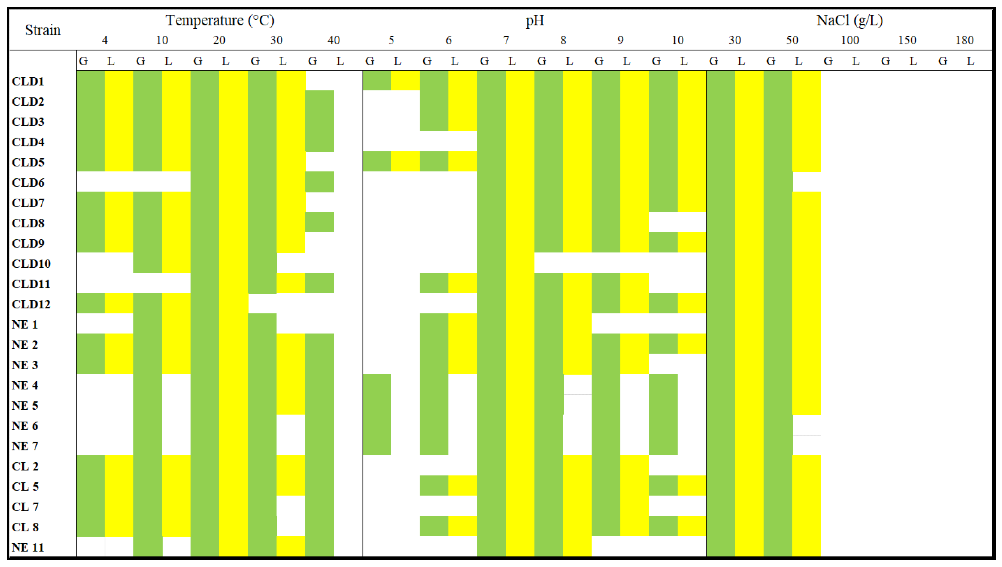
2.4. Physiological Tests

The growth of isolates was tested on a luminescence agar medium (LA, composition per liter: 30 g NaCl, 5 g yeast extract, 10 g peptone (Bacto Peptone, Life Technologies, Carlsbad, CA, USA), and 15 g agar in 1000 mL distilled water), LSW70 medium (composition per liter: 10 g tryptone, 5 g yeast extract, and 15 g agar, in a mixture of 350 mL seawater and 650 mL distilled water), and Thiosulfate-Citrate-Bile-Sucrose Agar (TCBS, Condalab, Madrid, Spain) medium.
The growths on the luminescence agar medium [47] and LSW70 medium [48] were tested to assess which medium was more optimal for growth and the presence/absence of bioluminescence in comparison to the SWC medium. TCBS is a selective medium for the isolation and detection of Vibrio cholerae and other Vibrio species, and it was used here to test the growth of all isolated luminous bacteria. All isolates were streaked onto TCBS medium plates and incubated for 24 h at 35 °C. The appearance of yellow-colored colonies was recorded as an indication of the presence of Vibrio sp. members. All isolates were grown at different temperatures, salinities, and pH values to establish the growth range and the optimum. The growth at different temperatures was first assayed on SWC broth by incubation at 4,10, 15, 20, and 30 °C for 24 h. Once the optimum temperature was identified, the bacterial growth was tested by adjusting the pH values in the range of 5 to 10 in SWC broth. Finally, once the optimum pH was also identified, different concentrations of NaCl were tested on a SWC agar plate (50–100–150–180 g/L; w/v). All tests were performed in triplicate.
As an additional preliminary screening, the potential production of extracellular polymeric substances (EPSs) was evaluated by observing the occurrence of mucous phenotypes during growth in solidified SWC that was supplemented with glucose (1%, w/v) and sucrose (1%, w/v) [49].

2.5. PCR Amplification of 16S rDNA for Bacterial Isolates

DNA was extracted from single colonies of each strain through lysis by heating at 95 °C for 10 min. Amplification of 16S rRNA gene was performed with a thermocycler (BioRad C100 touch thermocycler) using bacterial specific universal primers 27F (5′-AGAGTTTGATCCTGGCTCAG-3′) and 1492R (5′-GGTTACCTTGTTACGACTT-3′). The reaction mixtures contained for each sample: 1 μL of DNA, 1 μL of each forward and reverse primer (10 μM), 1 μL of dNTP mix (10 mM), 5 μL of reaction buffer 10×, 0.4 μL of polymerase (Hot Start DNA Polymerase), and 2 μL of MgCl2 in a final volume of 50 μL with MilliQ water. Negative controls for DNA extraction and PCR setup (reaction mixture without a DNA template) were also used in every PCR run.
The PCR program was as follows: an initial polymerase activation at 95 °C for 15 min, 35 cycles of 30 s at 95 °C for denaturation, 30 s at 51 °C for annealing phase, 30 s at 72 °C for elongation, and a final elongation for 5 min at 72 °C. The PCR products were visualized on agarose gel electrophoresis (1%, w/v) in TAE buffer (0.04 M Tris-acetate, 0.02 M acetic acid, and 0.001 M EDTA).

2.6. Sequencing and Analysis of 16S rRNA Gene

Sequencing was carried out at the Eurofins Genomics Laboratory (Milan, Italy). The sequences of isolates were compared with 16S rRNA gene sequences in the NCBI GenBank and the EMBL databases using BLAST to detect the next relatives of isolates through the ‘‘Seqmatch’’ and ‘‘Classifier’’ programs of the Ribosomal Database Project II (http://rdp.cme.msu.edu/ accessed on 25 June 2022) [50].
The program “Clustal W” was used to align sequences [51] to the most similar orthologous sequences retrieved from the database. Each alignment was checked manually, corrected, and then analyzed using the maximum likelihood method according to the model of Jukes–Cantor distances [52]. A phylogenetic tree was constructed using the MEGA 5 (Molecular Evolutionary Genetics Analysis) software [53]. The robustness of the inferred trees was evaluated by 400 bootstrap re-samplings.

2.7. LuxAB Gene Amplification

The LuxAB gene was searched for all bacterial isolates. The gene was amplified by using the specific Primers Lux AB-66F (3′-CAAATGTGRAAAGGTCGTTTTAATTTTGG-5′) and 611R (3′-AACRAAATCWYKCCATTGRCCTTTAT-5′) [54]. The reaction mixtures were carried out as described above, and the PCR program was as follows: an initial polymerase activation at 95 °C for 1 min, 30 cycles of 15 s at 95 °C for denaturation, 15 s at 37 °C for an annealing phase, 10 s at 72 °C for elongation, and a final elongation for 5 min at 72 °C. The PCR products were confirmed by visualization on agarose gel electrophoresis (1.5%, w/v) in TAE buffer (0.04 M Tris-acetate, 0.02 M acetic acid, and 0.001 M EDTA).

3. Results

The N. caroli specimens ranged between 32.1 and 68.0 mm in size (ML) and between 22.7 and 90.5 g in weight (mean ML ± s.d. = 47.9 ± 10.9 mm; mean W ± s.d. = 42.9 ± 20.7 g). The C. agassizi specimens measured between 41.7 and 184.7 mm in size (TL) and between 0.44 to 48.6 g in weight (mean ML ± s.d. = 91.9 ± 52.0 mm; mean W ± s.d. = 11.7 ± 15.5 g).

3.1. Isolation of Luminous Bacteria

A total of 24 strains were isolated from the two species. Overall, eight luminous bacterial isolates were from N. caroli specimens: five were isolated from the siphon, two from the ventral part of mantle, and one from mantle invagination. A total of 17 isolates were from C. agassizi. All strains showed growth on the LA medium, and 22 out of the 24 strains were capable of growth on LSW-40. Only one strain was able to grow on the TCBS medium. Complete data are shown in Table 1.

3.2. Physiological Tests

The results of the physiological tests are shown in Figure 2. The optimum growth temperature was identified to be 20 °C. More than half of the tested strains showed growth at 4 °C, and as many as 72% managed to grow up to 40 °C, even though at higher temperatures the bioluminescence was inhibited. The isolates CLD1, CLD5, CLD7, CLD9, CLD10, CLD12, and NE1 were not able to tolerate temperatures higher than 30 °C. The results of the different pH values tests showed that, in general, isolates tolerate acidity conditions poorly. The optimum condition was detected to be 7 pH value, and 3% of the tested bacterial strains were able to grow in a range between 6 and 10 pH values, in some cases without bioluminescence. Only two strains (CLD1 and CLD5) showed growth at pH 5 and maintained their bioluminescence. Salinity tests showed that all bacterial strains grew up to a final concentration of 50 g/L NaCl in the isolation media. However, in some cases, the bioluminescence was inhibited by the salt addition at the final NaCl concentration of 50 g/L, namely, in the case of isolates CLD6, NE6, and NE7. No strain was able to tolerate salinity concentration higher than 50 g/L.
The results from the preliminary screening for EPS production showed that 12% of the bacterial strains presented a mucous phenotype. In detail, the isolates CL2, CLD10, and CLD12 showed a more mucoid aspect in the culture medium plates supplemented separately with both glucose and sucrose (final concentration 1%, w/v). In the cultures in liquid broth with the same conditions, a very dense and milky mucus was observed.

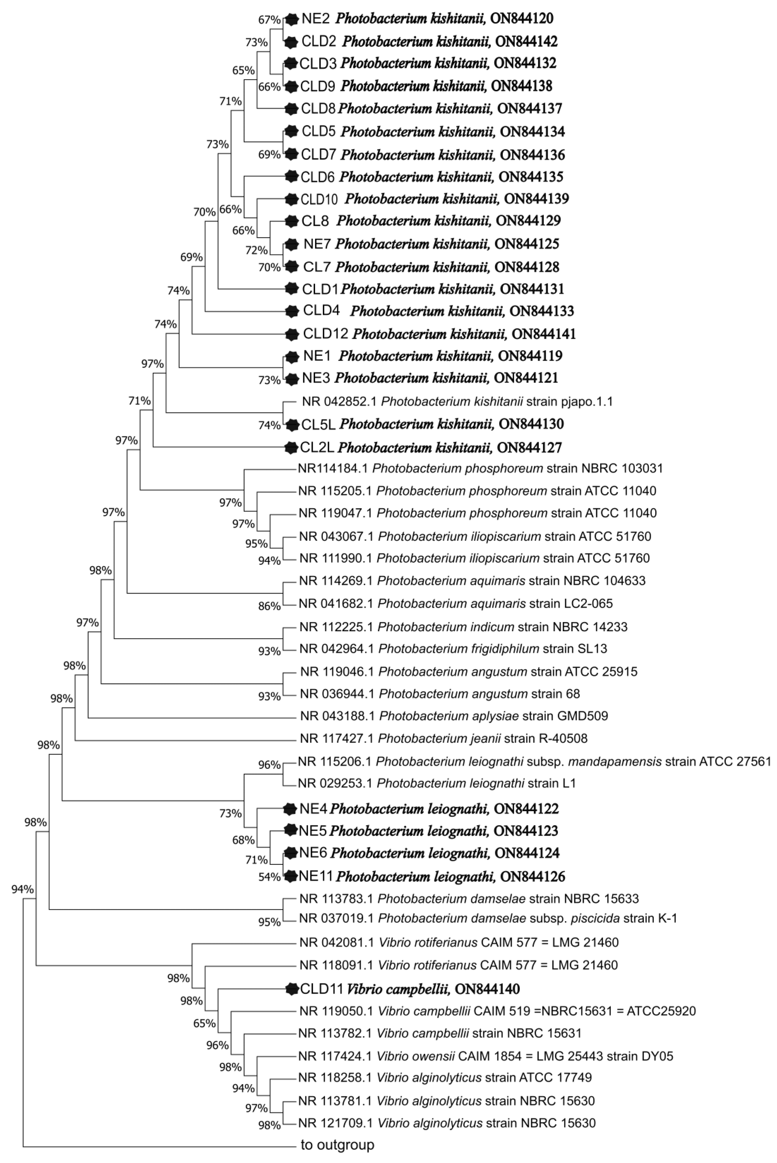
3.3. Phylogenetic Identification

The comparative sequence analysis indicated that the luminous bacteria were closely related to known bacteria with 16S rRNA gene similarity ≥98%. All isolates were affiliated to Gammaproteobacteria and were specifically identified as Photobacterium genus members, with the exception of one Vibrio member (Table 2). The phylogenetic analysis showed that a high species diversity was found on the 25 bacterial strains identified. Overall, three species of Photobacterium (P. kishitanii, P. leiognathi, and P. phosphoreum) and one species of Vibrio (V. campbelli) were identified. The genomic DNA were successfully extracted, and the luxAB gene was amplified in 22 strains of all isolates (Table 2; Figure 3).
The nucleotide sequences were deposited into the GenBank database under the accession nos. ON844119–ON844142.

4. Discussion

This study is the first to provide evidence of the presence of associated luminous bacteria in the cephalopod N. caroli and the fish C. agassizi. In our samples, on a total of 17 isolates, we found different associated bacteria belonging to three Photobacterium genera, namely, P. kishitanii, P. leiognathi, and P. phosphoreum, and one belonging to the genus Vibrio, namely, V. campbellii. In particular, P. kishitanii and P. leiognathi were associated with N. caroli, whereas P. kishitanii, P. phosphoreum, and V. campbellii were associated with C. agassizi.
To date, all known luminescent bacteria involved in symbiotic luminescence with fish and cephalopods are Gammaproteobacteria affiliated with the Vibrio, Photobacterium, and Shewanella genera.
The presence of symbiont luminous bacteria belonging to Photobacterium and Vibrio has been observed in sepiolids equipped with extrinsic bioluminescence [26,40,55,56,57,58,59,60,61,62,63]. The sepiolid Euprymna scolopes is the most-investigated species for the study of bioluminescence in cephalopods [64]; its luminous organ is a morphologically complex structure, containing crypts that house bioluminescent strains of Vibrio as well as reflective tissue, a lens, and a shutter mechanism [65,66]. According to [59], the E. scolopes–V. fischeri partnership is ecologically obligate, and juvenile squids develop a light organ with a superficial ciliated epithelium containing “appendages” that help recruit environmental bacteria. The colonization of epithelium crypts by free-living V. fischeri leads to light organ morphogenesis.
While studies on E. scolopes focused on several aspects of the association with luminous bacteria by describing the morphology of their luminous organs, the characterization of symbiont bacteria, and their selection mechanism from the environment [59], the other sepiolids are poorly investigated. The symbiont bacteria P. leiognathi was found in the light organ of Rondeletiola minor in the Mediterranean Sea [63] as well as in Sepiolina nipponensis and Uroteuthis noctiluca [67]. A recent study [26] on the phylogenomics of Sepiolida reported that N. caroli lacks luminescence. In our study, we found P. kishitanii and P. leiognathi associated with N. caroli, which was not previously observed. This finding suggests a possible association between the sepiolid and luminous bacteria, but it is not easy to establish whether these luminous bacteria are real symbionts or not. While all procedures in our study were carried out scrupulously, taking care to treat different tissues separately to avoid cross-contamination, it is not possible to exclude the seawater origin of transient luminous bacteria. A similar scenario was described by [68] for the P. leiognathi symbionts that can follow one of two paths upon encountering a host: simply transit for short periods along the specimen, or become symbiont inhabitants. In line with this, the presence of Vibrio sp. members was reported in the mantle cavity of E. scolopes from seawater [69] but then became representative of the local bacterial population and was able to colonize the host [58]. However, while the mantle cavity and/or the siphon are used for swimming propulsion by pumping water, the finding in our study of luminous bacteria in mantle invaginations is quite interesting because these anatomical parts were more preserved and thus potentially ideal to host symbionts that find these parts a good growth habitat. Given that N. caroli lacks light organs, the association between Photobacterium and this sepiolid may be a non-obligate relationship and instead facultative, as suggested by a recent study [70], demonstrating the non-species-specific association of five luminous bacteria with the non-luminous squid Sepioteuthis lessoniana. Future studies will be devoted to ascertain the occurrence of a symbiotic relationship between these bacteria and N. caroli by examining a higher number of specimens and by coupling a metagenomics approach to the traditional culture-dependent strategy.
With regard to C. agassizii, our study is the first to characterize the presence of luminous bacteria in the perianal gland of this deep sea fish. To the best of our knowledge, few studies investigated the presence of luminous symbionts in congeneric species [71] or observed the presence of non-identified luminous bacteria in the perianal gland of Chlorophthalmus albatrossis from Japanese waters through histological and morphological analyses of the light organ and simple luminescence tests. Our findings are in line with more specific studies that characterized the symbiont bacteria of Chlorophthalmidae, finding that they belonged to the genus Photobacterium. Indeed, Dunlap and Ast [7] observed the presence of Photobacterium phosphoreum and Photobacterium iliopiscarium through a phylogenetic identification of a total of 47 bioluminescent isolates obtained from the perianal gland of C. albatrossis. Later, other authors [72,73] reported P. kishitanii as the symbiotic luminous strain in four species of Chlorophthalmidae (C. albatrossis, C. acutifrons, C. borealis, and C. nigromarginatus). The finding of P. kishitanii in C. agassizii from Mediterranean waters (our study) supports the hypothesis formulated by Saito et al. [73], that all members of the genus Chlorophthalmus can also harbor the same bacterial species regardless of the distribution of the host. The presence of the V. campbelli strain is likely attributable to a temporary transit or to a relationship other than a symbiotic one. Luminous Vibrios are considered etiologic agents of luminous vibriosis in aquaculture industries worldwide [71]. V. campbelli is a Gram-negative bacterium widely distributed in marine environments [74], initially misidentified as Vibrio harveyi [75,76]. More recently, different studies reported its role as the pathogen responsible for the luminescent disease in farmed shrimp and the mortality in fish and shellfish [74,75,76,77,78]. However, to the best of our knowledge, this is the first observation of V. campbelli in C. agassizii organisms.
One important issue in the study of luminescent bacteria is their difficult maintenance in laboratory conditions since it requires many precautions and care to keep these bacteria alive and luminescent. In addition to the importance of investigation on their ecological role, different biotechnological and industrial applications of luminous bacteria have recently been highlighted. For instance, bioluminescent bacteria were proposed as indicators or biosensors for measuring the pollution level and toxicity of chemicals in environmental samples or for detecting possible DNA-tropic and oxidative stress-causing agents [79,80,81].
It is important to broaden our knowledge about the optimal growth conditions for and maintenance of these bacteria. All isolates tested in our study showed a wide growth-temperature range while better tolerating basic conditions of pH rather than acidic conditions. Salinity was proven to be the greatest factor to affect bioluminescence, with several isolates able to grow at increasing NaCl concentrations but unable to produce luminescence. Moreover, based on our results, the bacterial strains affiliated with P. kishitanii and leiognathi were strongly inhibited by pH values greater than 9. Recently, some luminous bacteria were reported to be producer of EPSs [80]. In this study, the P. kishitanii members provided promising results for the production of EPS with the addition of both glucose and sucrose to the culture medium.
The search for the luxAB gene was performed to assess if it could be used as tool for the monitoring and detection of luminous bacteria. The luciferase enzyme catalyzes the light emission reaction in luminescent bacteria, and the luxAB gene encodes the alpha subunit of luciferase [82,83,84]. Approximately 90% of luminous isolates showed the presence of the luxAB gene, confirming the luxAB gene search as suitable screening approach.
Our proposed investigative approach was proven suitable to carry out the study and could also be used in future studies aiming to improve the biotechnological potential of luminous bacteria (i.e., extracellular polymeric molecules production, pathogenicity tests, and adaptative strategies).

5. Conclusions

For many deep sea marine organisms able to produce luminescence, it is hard for scientists to identify whether this function is related to the presence of symbiotic luminescent bacteria. For the first time, we succeeded in isolating luminescent bacteria in two underexplored species, namely, the cephalopod Neorossia caroli and the teleost Chlorophthalmus agassizii. We obtained strains affiliated with the species P. kishitanii, P. phosphoreum, and P. leiognathi and only one microorganism of the genus Vibrio. Moreover, we elucidated some physiological and biotechnological aspects of luminous isolates that were previously poorly investigated as a topical biotechnological issue.
In the future, studies should continue to search for these luminescent symbionts in invertebrate and vertebrate organisms living in deep sea environments, identify their ecological role to assess if they occur in symbiotic relationships as consortia of different taxonomic group, detect the stimulation mechanisms at the base of the luminescent process and the possible quorum-sensing processes, and understand the symbiont bacteria selection by the host and its evolutive scenarios. Further studies are also necessary for improving our knowledge about the biotechnological potential of luminescent bacteria by investigating the production and the optimization of EPSs.

Author Contributions

Conceptualization, C.R., P.B. and T.R.; methodology, C.R. and P.B.; software, C.R.; validation, C.R., R.C. and P.B.; formal analysis, C.R.; investigation, R.C. and M.G.S.; data curation, C.R., P.B. and R.C.; writing—original draft preparation, R.C., E.A. and C.R.; writing—review and editing, P.B., M.G.S. and C.R.; visualization, C.R. and P.B.; supervision, T.R., P.B. and C.R.; funding acquisition, T.R., P.B., C.R. and P.C. All authors have read and agreed to the published version of the manuscript.

Funding

This research was partially funded by REsPoNSo 65/RBC/18 CUP J69E18000500007 and Misura 1.40 -PO FEAMP 2014–2020.

Institutional Review Board Statement

Ethical review and approval were waived for this study since the samples were already dead at the time of collection. They were collected from the commercial discards of trawl fishery at landings, or they were collected after having been stranded on the shore in the Strait of Messina.

Informed Consent Statement

Not applicable.

Data Availability Statement

The nucleotide sequences were deposited into the NCBI GenBank database under the accession numbers ON844119–ON844142.

Acknowledgments

The authors are grateful to the crew of the fishing vessel “Papà Carmelo”.

Conflicts of Interest

The authors declare no conflict of interest.

References

  1. Nealson, K.H.; Hastings, J.W. The Luminous Bacteria. In The Prokaryotes, 2nd ed.; Balows, A., Tru, H., Eds.; Springer: New York, NY, USA, 1992; pp. 625–639. [Google Scholar]
  2. Dunlap, P.V.; Kita-Tsukamoto, K. Luminous Bacteria. In The Prokaryotes; Dworkin, M., Falkow, S., Eds.; Academic Press: New York, NY, USA, 2001. [Google Scholar]
  3. Ramesh, A.B.; Loganathan, G. Ecological dynamics of marine luminous bacteria. J. Basic Microbiol. 1990, 30, 686–703. [Google Scholar] [CrossRef]
  4. Ruby, E.G.; Urbanowski, M. Genome sequence of Vibrio fischeri: A symbiotic bacterium with pathogenic congeners. Proc. Natl. Acad. Sci. USA 2005, 102, 3004–3009. [Google Scholar] [CrossRef]
  5. Hastings, J.W. Chemistries and colours of bioluminescent reactions: A review. Gene 1996, 173, 5–11. [Google Scholar] [CrossRef]
  6. Sukovataya, I.E.; Tyulkova, N.A. Kinetic analysis of bacterial bioluminescence in water organic media. Luminescence 2001, 16, 271–273. [Google Scholar] [CrossRef]
  7. Ast, J.C.; Dunlap, P.V. Phylogenetic resolution and habitat specificity of members of the Photobacterium phosphoreum species group. Environ. Microbiol. 2005, 7, 1641–1654. [Google Scholar] [CrossRef]
  8. Ast, J.C.; Cleenwerck, I. Photobacterium kishitanii sp. nov., a luminous marine bacterium symbiotic with deep-sea fishes. Int. J. Syst. Evol. Microbiol. 2007, 57, 2073–2078. [Google Scholar] [CrossRef]
  9. Martini, S.; Haddock, S.H. Quantification of bioluminescence from the surface to the deep sea demonstrates its predominance as an ecological trait. Sci. Rep. 2017, 7, 45750. [Google Scholar] [CrossRef]
  10. Widder, E.A. Bioluminescence in the ocean: Origins of biological, chemical, and ecological diversity. Science 2010, 328, 704–708. [Google Scholar] [CrossRef]
  11. Hastings, J.W.; Mitchell, G. Endo-symbiotic bioluminescent bacteria from the light organ of pony fish. Biol. Bull. 1971, 141, 261–268. [Google Scholar] [CrossRef]
  12. Haddock, S.H.; Moline, M.A. Bioluminescence in the sea. Ann. Rev. Mar. Sci. 2010, 2, 443–493. [Google Scholar] [CrossRef]
  13. Catul, V.; Gauns, M. A review on mesopelagic fishes belonging to family Myctophidae. Rev. Fish Biol. Fish. 2011, 21, 339–354. [Google Scholar] [CrossRef]
  14. Chakrabarty, P.; Davis, M.P. Is sexual selection driving diversification of the bioluminescent ponyfishes (Teleostei: Leiognathidae)? Mol. Ecol. 2011, 20, 2818–2834. [Google Scholar] [CrossRef]
  15. Davis, M.P.; Holcroft, N.I. Species-specific bioluminescence facilitates speciation in the deep sea. Mar. Biol. 2014, 161, 1139–1148. [Google Scholar] [CrossRef]
  16. Hoving, H.J.; Jose Angel, A. The study of Deep-Sea Cephalopods. In Advances in Marine Biology; Erica, A., Vidal, G., Eds.; Elsevier: Amsterdam, The Netherlands, 2014; Volume 67, pp. 235–359. [Google Scholar]
  17. Davis, M.P.; Sparks, J.S. Repeated and widespread evolution of bioluminescence in marine fishes. PLoS ONE 2016, 11, e0155154. [Google Scholar] [CrossRef]
  18. Lau, E.S.; Oakley, T.H. Multi-level convergence of complex traits and the evolution of bioluminescence. Biol. Rev. 2020, 96, 673–691. [Google Scholar] [CrossRef]
  19. Haygood, M.G. Light a organ symbioses in fishes. Crit. Rev. Micr. 1993, 19, 191–216. [Google Scholar] [CrossRef]
  20. Hendry, T.A.; Freed, L.L. Ongoing transposon-mediated genome reduction in the luminous bacterial symbionts of deep-sea ceratioid anglerfishes. MBio 2018, 9, e01033-18. [Google Scholar] [CrossRef]
  21. McFall-Ngai, M.J.; Ruby, E.G. Symbiont recognition and subsequent morphogenesis as early events in an animal-bacterial symbiosis. Science 1991, 254, 1491–1494. [Google Scholar] [CrossRef]
  22. Montgomery, M.K.; McFall-Ngai, M.J. The muscle-derived lens of a squid bioluminescent organ is biochemically convergent with the ocular lens. J. Biol. Chem. 1992, 267, 20999–21003. [Google Scholar] [CrossRef]
  23. Weis, V.M.; Montgomery, M.K. Enhanced production of ALDH-like proteins in the bacterial light organ of the sepiolid squid Euprymna scolopes. Biol. Bull. 1993, 184, 309–321. [Google Scholar] [CrossRef]
  24. Fidopiastis, P.M.; Boletzky, S.V. A new niche for Vibrio logei, the predominant light organ symbiont squids in the genus Sepiola. J. Bacteriol. 1998, 180, 59–64. [Google Scholar] [CrossRef]
  25. Nishiguchi, M.K.; Lopez, J.E. Enlightenment of old ideas from new investigations: More questions regarding the evolution of bacteriogenic light organs in squids. Evol. Dev. 2004, 6, 41–49. [Google Scholar] [CrossRef] [PubMed]
  26. Sanchez, G.; Fernandez Alvarez, F.A. Phylogenomics illuminates the evolution of bobtail and bottletail squid (order Sepiolida). Commun. Biol. 2021, 4, 819. [Google Scholar] [CrossRef] [PubMed]
  27. Montgomery, M.K.; McFall-Ngai, M.J. Embryonic development of the light organ of the sepiolid squid Euprymnascolopes Berry. Biol. Bull. 1993, 184, 296–308. [Google Scholar] [CrossRef]
  28. Nishiguchi, M.K. Host recognition is responsible for symbiont composition in environmentally transmitted symbiosis. Microb. Ecol. 2002, 44, 10–18. [Google Scholar] [CrossRef] [PubMed]
  29. Young, R.E. Ventral bioluminescent countershading in midwater cephalopods. Symp. Zool. Soc. Lond. 1977, 38, 161–190. [Google Scholar]
  30. Moynihan, M. Notes on the behavior of Euprymna scolopes (Cephalopoda: Sepiolidae). Behaviour 1983, 85, 25–41. [Google Scholar] [CrossRef]
  31. Jones, B.W.; Nishiguchi, M.K. Counterillumination in the hawaiian bobtail squid, Euprymna scolopes Berry (Mollusca: Cephalopoda). Mar. Biol. 2004, 144, 1151–1155. [Google Scholar] [CrossRef]
  32. Mandel, M.J.; Dunn, A.K. Impact and influence of the natural Vibrio-squid symbiosis in understanding bacterial animal interactions. Front. Microbiol. 2016, 7, 1982. [Google Scholar] [CrossRef]
  33. McFall-Ngai, M.J. The importance of microbes in animal development: Lessons from the squid-Vibrio symbiosis. Annu. Rev. Microbiol. 2014, 68, 177–194. [Google Scholar] [CrossRef]
  34. Tischler, A.H.; Hodge-Hanson, K.M.; Visick, K.L. Vibrio fischeri–squid symbiosis. Els. Oceanogr. Ser. 2019, 267, 1–9. [Google Scholar]
  35. McFall-Ngai, M.J. The Molecular Dialogue Through Ontogeny between a Squid Host and Its Luminous Symbiont. In Cellular Dialogues in the Holobiont; CRC Press: Boca Raton, FL, USA, 2020; pp. 153–171. [Google Scholar]
  36. Ramesh, C.; Bessho-Uehara, M. Acquisition of bioluminescent trait by non-luminous organisms from luminous organisms through various origins. Photochem. Photobiol. Sci. 2021, 20, 1547–1562. [Google Scholar] [CrossRef] [PubMed]
  37. Bose, J.L.; Rosenberg, C.S. Effects of luxCDABEG induction in Vibrio fischeri: Enhancement of symbiotic colonization and conditional attenuation of growth in culture. Arch. Microbiol. 2008, 190, 169–183. [Google Scholar] [CrossRef]
  38. Koch, E.J.; Miyashiro, T.; McFall-Ngai, M.J.; Ruby, E.G. Features governing symbiont persistence in the squid-vibrio association. Mol. Ecol. 2014, 23, 1624–1634. [Google Scholar]
  39. Kaeding, A.J.; Ast, J.C. Phylogenetic diversity and cosymbiosis in the bioluminescent symbioses of Photobacterium mandapamensis. Appl. Environ. Microbiol. 2007, 73, 3173–3182. [Google Scholar] [CrossRef]
  40. Guerrero-Ferreira, R.; Gorman, C. Characterization of the bacterial diversity in Indo-West Pacific loliginid and sepiolid squid light organs. Microb. Ecol. 2013, 65, 214–226. [Google Scholar] [CrossRef]
  41. Nishiguchi, M.K.; Ruby, E.G. Competitive dominance among strains of luminous bacteria provides an unusual form of evidence for parallel evolution in sepiolid squid-Vibrio symbioses. App. Environ. Microb. 1998, 64, 3209–3213. [Google Scholar] [CrossRef]
  42. Bazhenov, S.V.; Khrulnova, S.A.; Konopleva, M.N.; Manukhov, I.V. Seasonal changes in luminescent intestinal microflora of the fish inhabiting the Bering and Okhotsk seas. FEMS Microbiol. Lett. 2019, 366, 4. [Google Scholar] [CrossRef]
  43. Jereb, P.; Roper, C.F. Cephalopods of the Indian Ocean. A review. Part, I. Inshore squids (Loliginidae) collected during the international Indian Ocean expedition. Proc. Biol. Soc. Wash. 2006, 119, 91–136. [Google Scholar] [CrossRef]
  44. Sulak, K.J. Chlorophthalmidae. In Fishes of the North-Eastern Atlantic and the Mediterranean; Whitehead, P.J.P., Bauchot, M.-L., Hureau, J.-C., Nielsen, J., Tortonese, E., Eds.; UNESCO: Paris, France, 1984; Volume 1, pp. 412–420. [Google Scholar]
  45. Battaglia, P.; Ammendolia, G. Influence of lunar phases, winds and seasonality on the stranding of mesopelagic fish in the Strait of Messina (Central Mediterranean Sea). Mar. Ecol. 2017, 38, e12459. [Google Scholar] [CrossRef]
  46. Liu, P.C.; Chuang, W.H. Infectious gastroenteritis caused by Vibrio harveyi (V. carchariae) in cultured red drum, Sciaenops ocellatus. J. App. Ichthyo. 2003, 19, 59–61. [Google Scholar] [CrossRef]
  47. Malave-Orengo, E.; Rubio-Marrero, N. Isolation and characterization of bioluminescent bacteria from marine environments of Puerto Rico. Technol. Educ. Top. Appl. Microbiol. Microb. Biotechnol. 2010, 2, 103–108. [Google Scholar]
  48. Nealson, K.H. Isolation, identification and manipulation of luminous bacteria. Methods Enzymol. 1978, 57, 153–166. [Google Scholar]
  49. Caruso, C.; Rizzo, C.; Mangano, S.; Poli, A.; Di Donato, P.; Nicolaus, B.; Finore, I.; Di Marco, G.; Michaud, L.; Lo Giudice, A. Isolation, characterization and optimization of EPSs produced by acold-adapted Marinobacter isolate from Antarctic seawater. Antarct. Sci. 2019, 31, 69–79. [Google Scholar] [CrossRef]
  50. Altschul, S.F.; Madden, T.L. Gapped BLAST and PSI-BLAST: A new generation of protein database search programs. Nucleic Acids Res. 1997, 25, 3389–3402. [Google Scholar] [CrossRef]
  51. Thompson, J.D.; Higgins, D.G. CLUSTALW: Improving the sensitivity of progressive multiple sequence alignment through sequence weighting, position-specific gap penalties and weightmatrix choice. Nucleic Acids Res. 1994, 22, 4673–4680. [Google Scholar] [CrossRef]
  52. Jukes, T.H.; Cantor, C.R. Evolution of Protein Molecules. In Mammalian Protein Metabolism; Munro, H.N., Ed.; Academic Press: New York, NY, USA, 1969; pp. 21–132. [Google Scholar]
  53. Kumar, S. Cloning of a cDNA which encodes a novel ubiquitin-like protein. Biochem. Biophys. Res. Commun. 1993, 195, 393–399. [Google Scholar] [CrossRef]
  54. Gentile, G.; De Luca, M.; Denaro, R.; La Cono, V.; Smedile, F.; Scarfì, S.; De Domenico, E.; De Domenico, M.; Yakimov, M.M. PCR-based detection of bioluminescent microbial populations in Tyrrhenian Sea. Deep. Sea Res. Part II Top. Stud. Oceanogr. 2009, 56, 763–767. [Google Scholar] [CrossRef]
  55. Ruby, E.G.; Lee, K.H. The Vibrio fischeri-Euprymna scolopes light organ association: Current ecological paradigms. App. Environ. Microb. 1998, 64, 805–812. [Google Scholar] [CrossRef]
  56. Nyholm, S.V.; McFall-Ngai, M.J. Sampling the light-organ microenvironment of Euprymna scolopes: Description of a population of host cells in association with the bacterial symbiont Vibrio fischeri. Biol. Bull. 1998, 195, 89–97. [Google Scholar] [CrossRef]
  57. Nyholm, S.V.; McFall-Ngai, M.J. Dominance of Vibrio fischeri in secreted mucus outside the light organ of Euprymna scolopes: The first site of symbiont specificity. App. Environ. Microb. 2003, 69, 3932–3937. [Google Scholar] [CrossRef]
  58. Nyholm, S.V.; McFall-Ngai, M. The winnowing: Establishing the squid-vibrio symbiosis. Nat. Rev. Microb. 2004, 2, 632–642. [Google Scholar] [CrossRef]
  59. Nyholm, S.V.; McFall-Ngai, M.J. A lasting symbiosis: How the Hawaiian bobtail squid finds and keeps its bioluminescent bacterial partner. Nat. Rev. Microb. 2021, 19, 666–679. [Google Scholar] [CrossRef]
  60. Nyholm, S.V.; Stabb, E.V. Establishment of an animal–bacterial association: Recruiting symbiotic vibrios from the environment. Proc. Natl. Acad. Sci. USA 2000, 97, 10231–10235. [Google Scholar] [CrossRef]
  61. Nyholm, S.V.; Deplancke, B. Roles of Vibrio fischeri and nonsymbiotic bacteria in the dynamics of mucus secretion during symbiont colonization of the Euprymna scolopes light organ. App. Environ. Microb. 2002, 68, 5113–5122. [Google Scholar] [CrossRef]
  62. Guerrero-Ferreira, R.C.; Nishiguchi, M.K. Biodiversity among luminescent symbionts from squid of the genera Uroteuthis, Loliolus and Euprymna (Mollusca: Cephalopoda). Cladistics 2007, 23, 497–506. [Google Scholar] [CrossRef]
  63. Zamborsky, D.J.; Nishiguchi, M.K. Phylogeographical patterns among Mediterranean sepiolid squids and their Vibrio symbionts: Environment drives specificity among sympatric species. App. Environ. Microb. 2011, 77, 642–649. [Google Scholar] [CrossRef]
  64. Ruby, E.G. Lessons from a cooperative bacterial-animal association: The Vibrio fischeri-Euprymna scolopes light organ symbiosis. Annu. Rev. Microbiol. 1996, 50, 591–624. [Google Scholar] [CrossRef]
  65. McFall-Ngai, M.; Montgomery, M.K. The anatomy and morphology of the adult bacterial light organ of Euprymna scolopes Berry (Cephalopoda: Sepiolidae). Biol. Bull. 1990, 179, 332–339. [Google Scholar] [CrossRef]
  66. Montgomery, M.K.; McFall-Ngai, M.J. Bacterial symbionts induce host organ morphogenesis during early postembryonic development of the squid Euprymna scolopes. Development 1994, 120, 1719–1729. [Google Scholar] [CrossRef]
  67. Nishiguchi, M.K.; Nair, V.S. Evolution of pathogenicity and symbiosis in Vibrionaceae: A combined approach using molecules and physiology. Int. J. Syst. Evol. Microbiol. 2003, 53, 2019–2026. [Google Scholar] [CrossRef] [PubMed]
  68. Nyholm, V.; Stewart, J. Recognition between symbiotic Vibrio fischeri and the haemocytes of Euprymna scolopes. Environ. Microb. 2009, 11, 483–493. [Google Scholar] [CrossRef] [PubMed]
  69. Wei, S.L.; Young, R.E. Development of symbiotic bioluminescence in a nearshore cephalopod, Euprymna scolopes. Mar. Biol. 1989, 103, 541–546. [Google Scholar] [CrossRef]
  70. Zari, A.; Ramesh, C.H.; Mohanraju, R. Non-species specific composition of bioluminescent bacteria in non-bioluminescent squid Sepioteuthis lessoniana (Lesson, 1830). Ind. J. Geo Mar. Sci. 2020, 49, 975–981. [Google Scholar]
  71. Somya, H. Bacterial bioluminescence in chlorophthalmid deep-sea fish: A possible interrelationship between the light organ and the eyes. Experientia 1977, 33, 906–909. [Google Scholar] [CrossRef]
  72. Dunlap, P.V. Bioluminescence, Microbial. In Encyclopedia of Microbiology; Schaechter, M., Ed.; Elsevier: Amsterdam, The Netherlands, 2007; pp. 45–61. [Google Scholar]
  73. Saito, Y.; Yamaguchi, A. Contribution of platelet-derived growth factor signaling to retina regeneration in zebrafish. Neurosc. lett. 2020, 727, 134930. [Google Scholar] [CrossRef]
  74. Thompson, F.L.; Iida, T. Biodiversity of vibrios. Microbiol. Mol. Biol. Rev. 2004, 68, 403–431. [Google Scholar] [CrossRef]
  75. De la Pena, L.D.; Lavilla-Pitogo, C.R. Luminescent vibrios associated with mortality in pond-cultured shrimp Penaeus monodon in the Philippines: Species composition. Fish Pathol. 2001, 36, 133–138. [Google Scholar] [CrossRef]
  76. Lin, B.C.; Wang, Z. Comparative genomic analyses identify the Vibrio harveyi genome sequenced strains BAA-1116 and HY01 as Vibrio campbellii. Environ. Microbiol. 2010, 2, 81–89. [Google Scholar] [CrossRef]
  77. Phuoc, L.H.; Corteel, M. Increased susceptibility of white spot syndrome virus-infected Litopenaeus vannamei to Vibrio campbellii. Environ. Microbiol. 2008, 10, 2718–2727. [Google Scholar] [CrossRef]
  78. Wang, L.P.; Chen, Y.W. Isolation and identification of Vibrio campbellii as a bacterial pathogen for luminous vibriosis of Litopenaeus vannamei. Aquac. Res. 2015, 46, 395–404. [Google Scholar] [CrossRef]
  79. Ismailov, A.D.; Aleskerova, L.E. Photobiosensors containing luminescent bacteria. Biochem. Mosc. 2015, 80, 733–744. [Google Scholar] [CrossRef] [PubMed]
  80. Ramesh, C.H.; Mohanraju, R. A review on ecology, pathogenicity, genetics and applications of bioluminescent bacteria. J. Terr Mar. Res. 2019, 3, 1–32. [Google Scholar] [CrossRef]
  81. Kessenikh, A.G.; Novoyatlova, U.S.; Bazhenov, S.V.; Stepanova, E.A.; Khrulnova, S.A.; Gnuchikh, E.Y.; Kotova, V.Y.; Kudryavtseva, A.A.; Bermeshev, M.V.; Manukhov, I.V. Constructing of Bacillus subtilis-Based Lux-Biosensors with the Use of Stress-Inducible Promoters. Int. J. Mol. Sci. 2021, 22, 9571. [Google Scholar] [CrossRef] [PubMed]
  82. Burtseva, O.; Baulina, O. In vitro Biofilm Formation by Bioluminescent Bacteria Isolated from the Marine Fish Gut. Microb Ecol. 2021, 81, 932–940. [Google Scholar] [CrossRef]
  83. Wimpee, C.F.; Nadeau, T. Development of species-specific hybridization probes for marine luminous bacteria by using in vitro DNA amplification. App. Environ. Microb. 1991, 57, 1319–1324. [Google Scholar] [CrossRef]
  84. Makemson, J.C.; Fulayfil, N.R. Shewanella woodyi sp. nov., an exclusively respiratory luminous bacterium isolated from the Alboran Sea. Int. J. Syst. Bact. 1997, 47, 1034–1039. [Google Scholar]
Figure 1. The deep sea demersal species considered in this study. (a) Neorossia caroli (Sepiolidae, Cephalopoda) and (b) Chlorophthalmus agassizi (Chlorophthalmidae, Osteichthyes).
Jmse 10 01113 g001
Figure 2. Growth (green cells) and presence/absence of luminescence (yellow cells) of isolates at different temperatures, pH values, and salinities.
Jmse 10 01113 g002
Figure 3. Rooted phylogenetic tree calculated by a Jukes–Cantor distance estimation algorithm showing the affiliation of the most-representative bacterial isolates to the most closely related sequences from either cultivated or cloned bacteria. The percentage of trees in which the associated taxa clustered together is shown next to the branches. The tree was outgrouped with the 16S rRNA gene sequence of Methanocaldococcus jannaschii DSM2661. The representative isolates of this study are indicated by black polygons.
Jmse 10 01113 g003
Table 1. Number of isolates obtained from SWC plates. Growth of isolates on the LSW40 Agar, luminescent agar, and TCBS Agar plates.
SampleIsolatesGrowth Test
SWCTCBSLSWLA
Neorossia caroli
Siphon5045
Ventral mantle2022
Mantle invagination1011
Chlorophtalmus agassizii
Perianal gland1611617
Total isolates2402224
Table 2. Phylogenetic affiliation of luminescent bacteria from the deep sea organisms of the Strait of Messina and the occurrence of the luxAB gene.
StrainANNext Relative by GenBank Alignment
(AN **, Organism)
Isolation
Matrix
Hom §
(%)
luxAB
Gene
Neorossia caroli
NE1ON844119NR_042852.1, Photobacterium kishitanii strain pjapo.1.1siphon99+
NE2ON844120NR_042852.1, Photobacterium kishitanii strain pjapo.1.1ventral98+
NE3ON844121NR_042852.1, Photobacterium kishitanii strain pjapo.1.1ventral99+
NE4ON844122NR_115206.1, Photobacterium leiognathi subsp. mandapamensissiphon99-
NE5ON844123NR_029253.1, Photobacterium leiognathi strain L1siphon99+
NE6ON844124NR_029253.1, Photobacterium leiognathi strain L1siphon99+
NE7ON844125NR_042852.1, Photobacterium kishitanii strain pjapo.1.1siphon98+
NE11ON844126NR_029253.1, Photobacterium leiognathi strain L1invagination99+
Chlorophtalmus agassizi
CL2LON844127NR_042852.1, Photobacterium kishitanii strain pjapo.1.1perianal gland99+
CL7ON844128NR_042852.1, Photobacterium kishitanii strain pjapo.1.1perianal gland98+
CL8ON844129NR_115205.1, Photobacterium phosphoreum strain ATCC 11040 perianal gland98+
CL5LON844130NR_042852.1, Photobacterium kishitanii strain pjapo.1.1perianal gland99+
CLD1ON844131NR_042852.1, Photobacterium kishitanii strain pjapo.1.1perianal gland99+
CLD2ON844142NR_042852.1, Photobacterium kishitanii strain pjapo.1.1perianal gland98-
CLD3ON844132NR_042852.1, Photobacterium kishitanii strain pjapo.1.1perianal gland98+
CLD4ON844133NR_042852.1, Photobacterium kishitanii strain pjapo.1.1perianal gland98+
CLD5ON844134NR_042852.1, Photobacterium kishitanii strain pjapo.1.1perianal gland98+
CLD6ON844135NR_042852.1, Photobacterium kishitanii strain pjapo.1.1perianal gland98+
CLD7ON844136NR_042852.1, Photobacterium kishitanii strain pjapo.1.1perianal gland99+
CLD8ON844137NR_042852.1, Photobacterium kishitanii strain pjapo.1.1perianal gland99+
CLD9ON844138NR_042852.1, Photobacterium kishitanii strain pjapo.1.1perianal gland99+
CLD10ON844139NR_042852.1, Photobacterium kishitanii strain pjapo.1.1perianal gland99+
CLD11ON844140NR_113782.1, Vibrio campbellii strain NBRC 15631perianal gland99+
CLD12ON844141NR_042852.1, Photobacterium kishitanii strain pjapo.1.1perianal gland99+
** Accession number; § Homology.
Publisher’s Note: MDPI stays neutral with regard to jurisdictional claims in published maps and institutional affiliations.

