Article

Absence of Nitrergic Modulation of Starling Response in Haemoglobin-Less Antarctic Fish Chionodraco hamatus

by
Daniela Amelio
1,†,
Filippo Garofalo
1,†,
Elvira Brunelli
1,
Gianfranco Santovito
2 and
Daniela Pellegrino
1,*
1
Department of Biology, Ecology and Earth Science, University of Calabria, 87036 Arcavacata, Italy
2
Department of Biology, University of Padova, 35122 Padova, Italy
*
Author to whom correspondence should be addressed.
These authors contributed equally to this work.
J. Mar. Sci. Eng. 2022, 10(11), 1705; https://doi.org/10.3390/jmse10111705
Submission received: 27 September 2022 / Revised: 1 November 2022 / Accepted: 4 November 2022 / Published: 9 November 2022

Abstract

:
The Frank-Starling response is an intrinsic heart property that is particularly evident in the fish heart because piscine cardiomyocytes are extremely sensitive to stretch. Several mechanisms and compounds influence the Frank-Starling response, including the free radical nitric oxide produced by nitric oxide synthases in the vascular endothelium and cardiomyocytes of all vertebrates. Besides its role in scavenging nitric oxide, hemoglobin may act as a source and transporter. In this context, the hemoglobin-less Antarctic teleost Chionodraco hamatus (icefish) represents a unique opportunity to investigate the involvement of nitric oxide in the Frank-Starling response. Using an isolated perfused heart preparation, weverified a basal nitrergic tone that is not implicated in the Frank-Starling response. In addition, by comparing nitric oxide synthases expression and activation in C. hamatus and the red-blooded Antarctic teleost Trematomus bernacchii, we found the endothelial isoform of nitric oxide synthase (the primary generator of nitric oxide during shear stress) to be less expressed and activated in the former.

1. Introduction

In all vertebrates, the heart’s innate heterometric cardiac control (Frank-Starling mechanism) enables the myocardium to react to the elevated vein return by contracting more forcefully with its lengthened fibers, thus providing an optimal blood supply to vital organs in the body. This mechanism plays a dominant role in the heart of fish, which is extremely sensitive to stretching. Indeed, fish can increase their cardiac output by increasing their stroke volume rather than their heart rates [1]. Compared to mammals, the highest mechanical sensitivity of fish myocytes seems to be linked to a greater length–tension relationship of sarcomere [2]. Several physiological processes, including increased intracellular Ca2+ release, alterations in inositol triphosphate and protein kinase C activity, and the opening of stretch-activated ion channels, have been proposed in mammals to maintain the Frank-Starling response [3,4,5].
Nitric oxide (NO), a free radical produced by vascular endothelium and cardiomyocytes, promotes myocardial relaxation, ventricular diastolic distensibility, and, consequently, the Frank-Starling response in mammalian and non-mammalian vertebrates [1,6]. In the heart of the guinea pig, the basal release of NO affects how the heart responds to an increase in preload; this effect is significantly reduced by both NG-Methyl-L-arginine, an inhibitor of the endothelial isoform of nitric oxide synthase (eNOS), and free hemoglobin (Hb), which inhibit basal NO production/activity [6]. In the eel heart, the Frank-Starling reaction is influenced by endogenous NO release, increasing its sensitivity to preload [7], whereas exogenous NO regulates the Frank-Starling response via the activation of eNOS (mediated by PI3-kinase) and modulation of the SR-Ca2+ATPase pumps [1]. Cardiac NO generation by eNOS, in response to stretch, is well known [8,9]. Indeed, stimulation of endothelial cells by shear stress stimulates a calcium-independent eNOS activation through Akt-dependent phosphorylation [10,11,12]. The formation of phosphorylated eNOS, which involves several kinases, is an important step in controlling NO generation [13]. eNOS activity is influenced by various other factors, including the hypoxia-inducible factor 1α (HIF-1α), which is crucial for eNOS expression, phosphorylation, and the generation of NO in normoxic settings [14]. Besides the post-translational regulatory modifications, protein-protein interactions are a key regulatory mechanism of eNOS activity; in particular, the eNOS Trafficking Inducer (NOSTRIN) is an essential, eNOS-associated protein highly expressed in the endothelium. When NOSTRIN is overexpressed, it can promote the translocation of eNOS to intracellular vesicles, which results in a concurrent decline in enzymatic activity [15,16,17]. Although NO’s essential role in all cardio-circulatory functions is well known and deeply studied, for a long time, this free radical has been considered a paracrine signaling molecule with a limited half-life, low diffusibility, and fast interaction with heme proteins. However, many data demonstrate that NO, in the cardiovascular system, induces persistent effects indicating a role for red blood cells in the transportation and delivery of NO [18,19]. There is increasing evidence concerning the NO release from pre-formed stores in blood vessels. For instance, besides its classical role in NO inactivation, Hb may also act as a NO transporter, as reported by Allen and coworkers [20]. In addition, Hb and the other respiratory heme oxygenase myoglobin (Mb) exhibit a nitrite-reductase activity that is allosterically controlled and able to provide NO beside oxygen and pH gradients [21,22] and are involved in a variety of processes (physiological and/or pathological) in the heart and other organs [23].
In this scenario, the family Channichthyidae (icefish, sub-order Notothenioidei), endemic Antarctic teleost, represents a particularly useful experimental model to study the evolution of the NOsystem. Icefish is a natural genetic knockout for respiratory pigments being, in the adult stage, the only vertebrate lacking Hb and cardiac Mb in some species [24]. Antarctic Notothenioids, which evolved in the Southern Ocean’s geographic isolation characterized by constant ice and high oxygen concentration, exhibit physiological and biochemical adaptations [25]. Moreover, icefish developed an adaptive recovery based on compensatory reactions and gene expression reprogramming, including a different cardio-circulatory architecture, to deal with Hb-free blood and Mb-free cardiac muscle state [26]. The substantial enlargement of the mitochondrial compartment and subsequent rise in cytochrome-c oxidase activity in myotomal [27] and cardiac [28] muscles are two remarkable examples of compensation at the subcellular level [29]. At a systemic level, there are crucial cardio-circulatory compensations: vascular tree with lowresistance, great blood volume, heart-to-body mass ratio, and large cardiac output that, among teleosts, is among the highest [26]. Icefish heart can be defined as a typical volume pump due to its wide cardiac output, achieved with an extraordinarily large stroke volume [26]. Conceivably, intracardiac NO, given its modulatory activityinthe heart of mammals [30], could enhance the volume pump function of icefish in a paracrine/autocrine manner.
In previous works, we investigated the expression and localization of eNOS and inducible NOS isoform (iNOS) [31] and the functionality of NO cardiac signaling [32] in the hearts of T. bernacchii, expressing both Hb and Mb, and C. hamatus, Hb-less that expresses Mb, but with a reduced concentration and only at ventricular level. More recently [33], we included in our experiments another species of icefish, Chaenocephalus aceratus (Hb-/Mb-), and comparing the hearts of all three Antarctic teleosts (T. bernacchii, C. hamatus, and C. aceratus), we revealed a lower expression of both eNOS and iNOS in the latter species.
In addition, we reported species-specific differences regarding NO signaling and heart mechanical performance of C. aceratus and T. bernacchii [33], suggesting that the presence or absence of the natural scavengers Hb and Mb differ affects cardiac homeostasis driven by NOS/NO system.
The present study aims to investigate in C. hamatus the NO modulation of cardiac activity under basal and loading (i.e., Frank-Starling response) conditions. Using a physio-pharmacological approach, we confirmed the positive inotropic effect induced by a basal NO-release in the icefish.We revealed that the Frank-Starling mechanism is unaffected by NO. In addition, we analyzed the eNOS-systems in both T. bernacchii and C. hamatus and demonstrated that eNOS is less expressed and activated in the icefish compared to the red-blooded species. Indeed, by immuno-detection techniques, we showed in the icefish: (i) a lower eNOS and phosphorylated eNOS expression; (ii) a lower expression of phosphorylated Akt (p-Akt) and HIF-1α; (iii) a higher expression of NOSTRIN.

2. Materials and Methods

2.1. Animals

Antarctic fish specimens C. hamatus, weighing 425 ± 41 g (mean ± S.E.M.), and T. bernacchii, weighing 323 ± 15 g (mean ± S.E.M.), have been used. Animals have been caught by net in Terranova Bay, Ross Sea, Antarctica (74°42′ S, 164°06′ E) and maintained unfed for at least five days in seawater at 0–2 °C. Experiments have been conducted in the Italian Antarctic Base “Mario Zucchelli Station” laboratories. The investigation agrees with the Guide for the Care and Use of Laboratory Animals (US National Institutes of Health: NIH; Publication No. 85-23, revised 1996), and the animal study protocol was approved by Italian National Research Program in Antarctica (PNRA).

2.2. Isolated and Perfused Working Heart Preparations

Animals were anesthetized in benzocaine (0.2 g L−1), and hearts were transferred to a perfusion system (as described by Pellegrino and co-workers [32]). Perfusion was immediately started at constant input pressures (1.4 kPa with cardiac output set to 50 mL min−1 kg−1 body mass) to allow the heart to work ‘physiologically’. In the absence of extrinsic nervous and humoral regulations, such a condition is ideally suited to directlyanalyze cardiac autocrine/paracrine NO’s involvement. Ringer’s solution (pH adjusted to 7.84 at 1 °C) contained the following component concentrations (mmol L−1): MgSO4 2.0, KCl 5.0, dextrose 5.56, NaCl 252.4, CaCl2 2.3, NaH2PO4 0.2, Na2HPO4 2.3. Ventricular stroke work (SW, index of systolic functionality) was calculated as (mJ g−1) = (afterload − preload, kPa) × stroke volume (mL)/ventricle weight (g). Ventricular power output (PO, a measure of the heart’s ability to face systemic resistance) was calculated as (mW g−1) = (afterload − preload) (kPa) × cardiac output (mL min−1)/ventricle weight (g) × 60. A cold thermostatic cabinet (LKB 2021 Maxicoldlab, Malbo, Sweden) was used for the experiments, allowing the temperature of the heart and perfusion system to be kept close to 0 °C.

2.2.1. Experimental Protocols

Typical time-course heart rate and stroke volume curves indicated that for three hours, the heart function remained constant; thus, all experiments were carried out within 2 h. Hearts were stabilized at basal conditions, and cardiac variables were calculated throughout the experiment. To evaluate the NO influence on the Frank-Starling response, baseline and time-control curves were generated as follows: a first Starling curve was generated then, the input pressure was changed back to the control condition after baseline analysis, and a second Starling curve (untreated time-control) was produced. The same protocol was used in the presence of NOS inhibition by L-N5-(1-iminoethyl)-ornithine (L-NIO) or NO scavenging by 2-phenyl-4,4,5,5-tetramethylimidazoline-1-oxyl-3-oxide (PTIO).

2.2.2. Statistical Analyses

The percentage changes obtained from separate studies were analyzed as means of S.E.M. The Starling curves were compared within groups using the Student’s t-test on absolute values and across groups using a two-way analysis of variance (ANOVA). Duncan’s multiple-range test was used to find differences from the time-control group that were statistically significant.

2.2.3. Drugs and Chemicals

L-NIO and PTIO were purchased from Sigma Chemical Company (St Louis, MO, USA).

2.3. NOS Expression and Activation

2.3.1. Immunodetection

The hearts of C. hamatus and T. bernacchii were washed in Ringer’s solution and fixed in MAW solution (Methanol:Acetone:Water = 2:2:1), dehydrated in ethanol (90% and 100%), cleared in xylol, embedded in paraplast (Sigma), and serially sectioned at 8 μm. The sections were placed onto Superfrost Plus slides (Menzel-Glaser, Braunschwerg, Germany), deparaffined in xylene, rehydrated in an alcohol gradient, rinsed in Tris-buffered saline (TBS) and incubated with 1.5% bovine serum albumin (BSA) in TBS for 1 h. Sections were incubated overnight at 4 °C with polyclonal antibody (dilution 1:100) directed against phosphorylated eNOS (developed in goat) and p-AKT (developed in rabbit) (Santa Cruz Biotechnology, Inc., Heidelberg, Germany). For signal detection, sections were washed in TBS (3 × 10 min) and incubated with FITC-conjugated anti-goat IgG or anti-rabbit antibodies (1:100; Sigma, St. Louis, MO, USA). The reaction was stopped by rinsing the sections with TBS. For nuclear counterstaining, selected sections were incubated for 5 min with propidium iodide (Sigma; 1 μg/mL) at room temperature. Negative controls, which were routinely negative, were performed by omitting the secondary antibody. Immunodetection was performed using an enhanced chemiluminescence ECL PLUS kit (Amersham). Autoradiographs were obtained by exposure to X-ray films (Hyperfilm ECL, Amersham). Slides were then mounted with a mounting medium (Vectashield, Vector Laboratories; Burlingame, CA, USA) and observed under the microscope (Zeiss Axioscope; Thornwood, NY, USA). Images were digitalized by Axiocam 105 color (ZEISS; Thornwood, NY, USA).

2.3.2. Western Blotting and Densitometric Analyses

Ventricle samples of both species (T. bernacchiin = 3; C. hamatusn = 3) were flushed with Ringer solution, rapidly immersed in liquid nitrogen, and stored at −80 °C. They were prepared according to Amelio and co-workers [31]. Tissues were suspended in ice-cold Tris–HCl buffer (30 mM; pH 7.4) containing EGTA (15 μM), EDTA (10 μM), dithiothreitol (5 μM), pepstatin-A (0.01 μM), PMSF (1 μM), leupeptin-A (0.02 μM), benzamidine (0.1 μM) and tetrahydrobiopterin (BH4) (0.1 μM). They were then homogenized with an Ultra Turrax homogenizer (IKA-Werke, Staufen, Germany) at 22,000 rpm for 10 s. Homogenates were centrifuged at 10,000× g for 60 min at 4 °C, and the supernatant was used for Western blotting. According to the Bradford method, protein concentration was determined with BSA as a standard for comparison. Samples containing 100 μg of proteins were heated for 5 min in Laemmli buffer (Laemmli, 1970), separated by SDS–PAGE using 8% in a Bio-Rad Mini Protean-III apparatus (Bio-Rad Laboratories, Hercules, CA, USA), and then electro-blotted onto polyvinylidene difluoride membrane (Hybond-P, Amersham, GE Healthcare Biosciences, Pittsburgh, PA, USA) using a mini trans-blot (Bio-Rad Laboratories Hercules, CA, USA). The membrane was blocked with TTBS buffer containing 5% non-fat dry milk. They were then incubated overnight at 4 °C with either rabbit polyclonal antibodies directed against eNOS (Sigma), Akt, p-Akt (ser 473) NOSTRIN (nitric oxide synthase traffic inducer), HIF-1α, and β-actin, or goat polyclonal antibody directed against phosphorylated eNOS (Santa Cruz Biotechnology). All antibodies were diluted 1:500 in TTBS containing 5% BSA. The peroxidase-linked secondary antibodies (anti-rabbit, anti-goat; Santa Cruz Biotechnology) were diluted 1:5000 in TTBS containing 5% non-fat dry milk. Immunoblots were digitalized, and the densitometric analysis of the bands obtained was carried out using WCIF Image J based on 256 gray values (0 = white; 256 = black). Quantification of the bands was obtained by measuring (5 times on each band) the mean optical density of a square area after the background area was subtracted.

2.3.3. Statistical Analysis

Absorbance measurements and the gray values obtained from the densitometric analysis were expressed as means ± SE of 5 determinations for each sample. Differences between the groups were evaluated by non-parametric Mann–Whitney U test, in the case of phosphorylated eNOS/eNOS and p-Akt/Akt ratios, and by one-way analysis of variance (ANOVA) followed by Bonferroni multiple comparison test in the case of HIF-1α and NOSTRIN expression. Statistical significance for both statistical tests was established at * p < 0.05, ** p < 0.005, and *** p < 0.0005. The statistical analysis of the data was performed using GraphPad InStat® software, version 3.10 for Windows.

3. Results

3.1. Isolated Working Heart Preparations

3.1.1. Nitric Oxide and Basal Inotropism

Working under physiological loading conditions, our heart preparations may provide output pressure, cardiac output, stroke volume, and work (Table 1) comparable to in vivo values. The inotropic NO effect was studied before and after treatment with the NOS inhibitor L-NIO (10−5 mol L−1) or the NO scavenger PTIO (10−5 mol L−1). Treatment with L-NIO and PTIO under baseline conditions (physically unstimulated hearts) led to a considerable negative inotropism without changing heart rate (Figure 1).

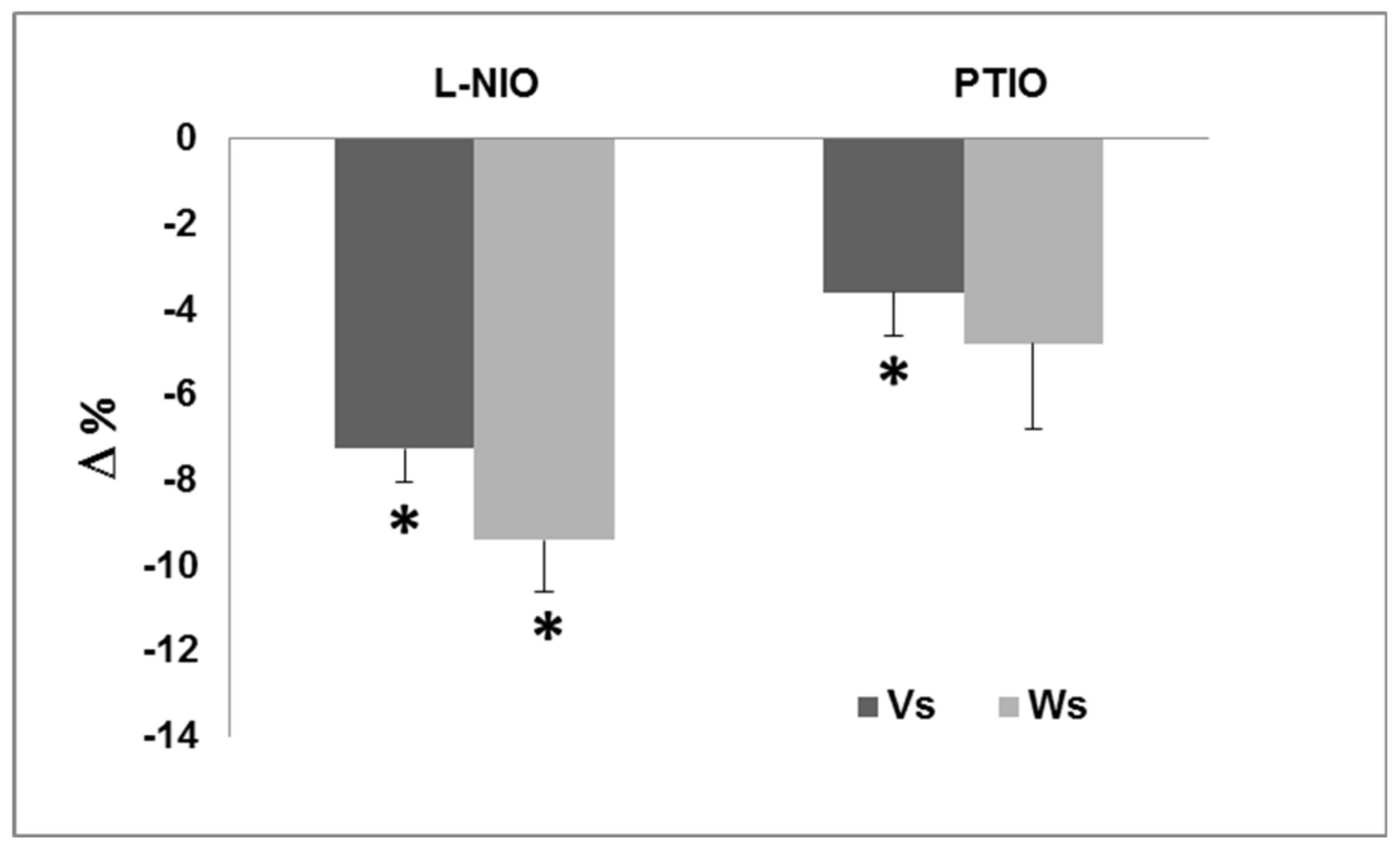
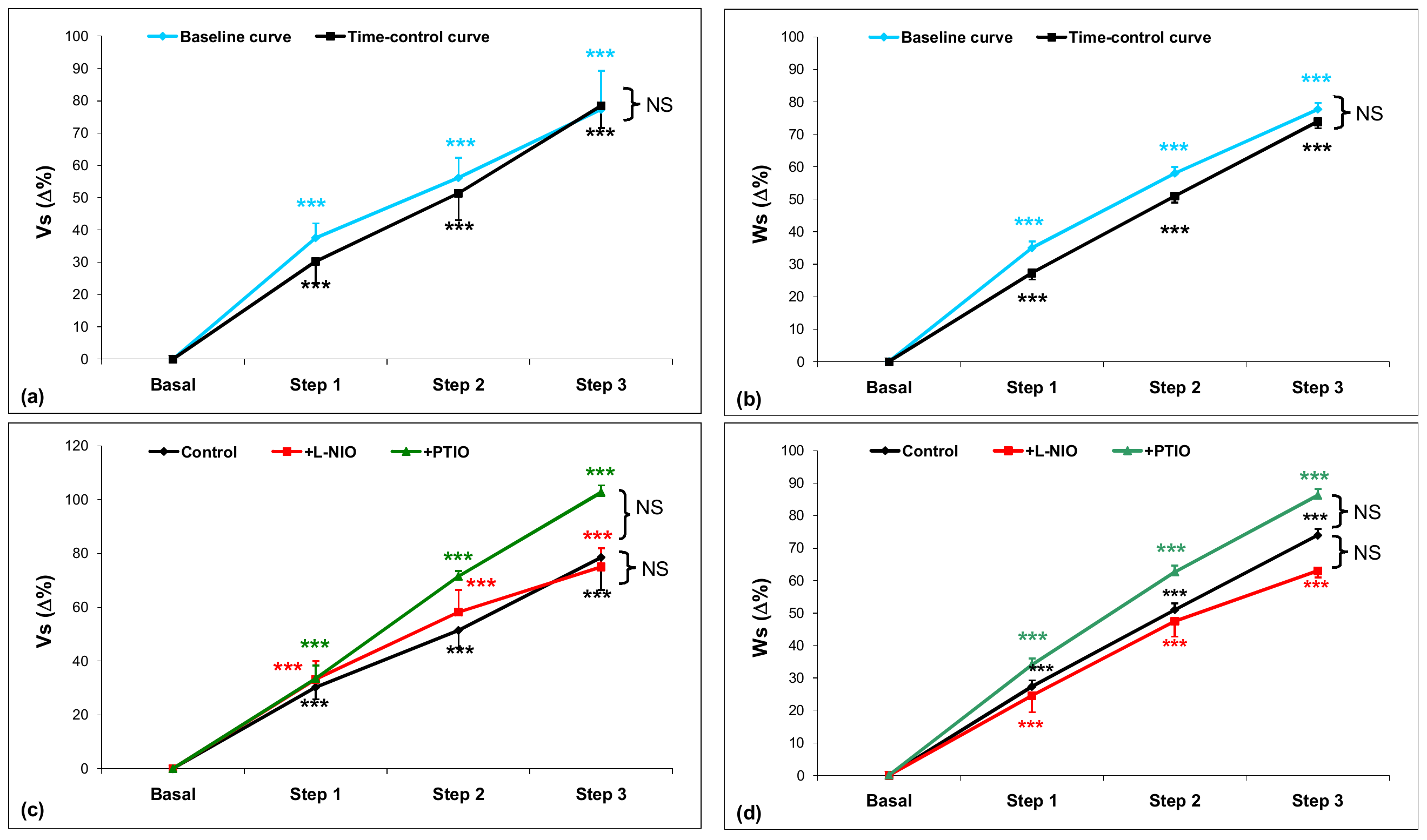
3.1.2. Nitric Oxide and the Frank–Starling Response

The hearts of C. hamatus isolated and perfused displayed a typical Frank–Starling response (Figure 2a,b baseline curves). Baseline and time-control curves were produced to isolate the time factor (i.e., the heart’s “memory”) of loading stimulation, proving the equality of the curves within the experimental error. (Figure 2a,b time-control curves). The pre-treatments with both, L-NIO and PTIO did not influence the Frank–Starling response in C. hamatus (Figure 2c,d).

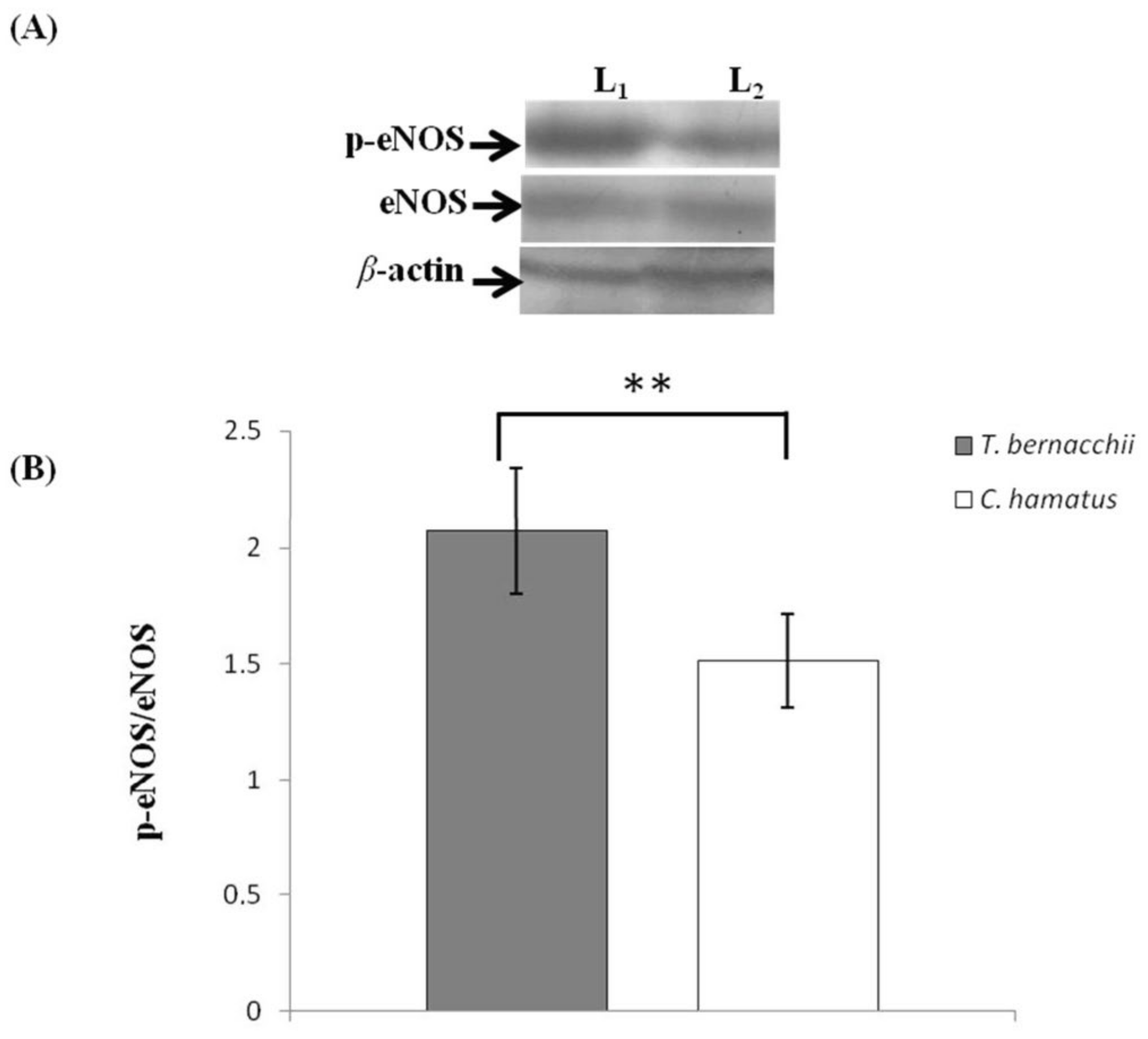
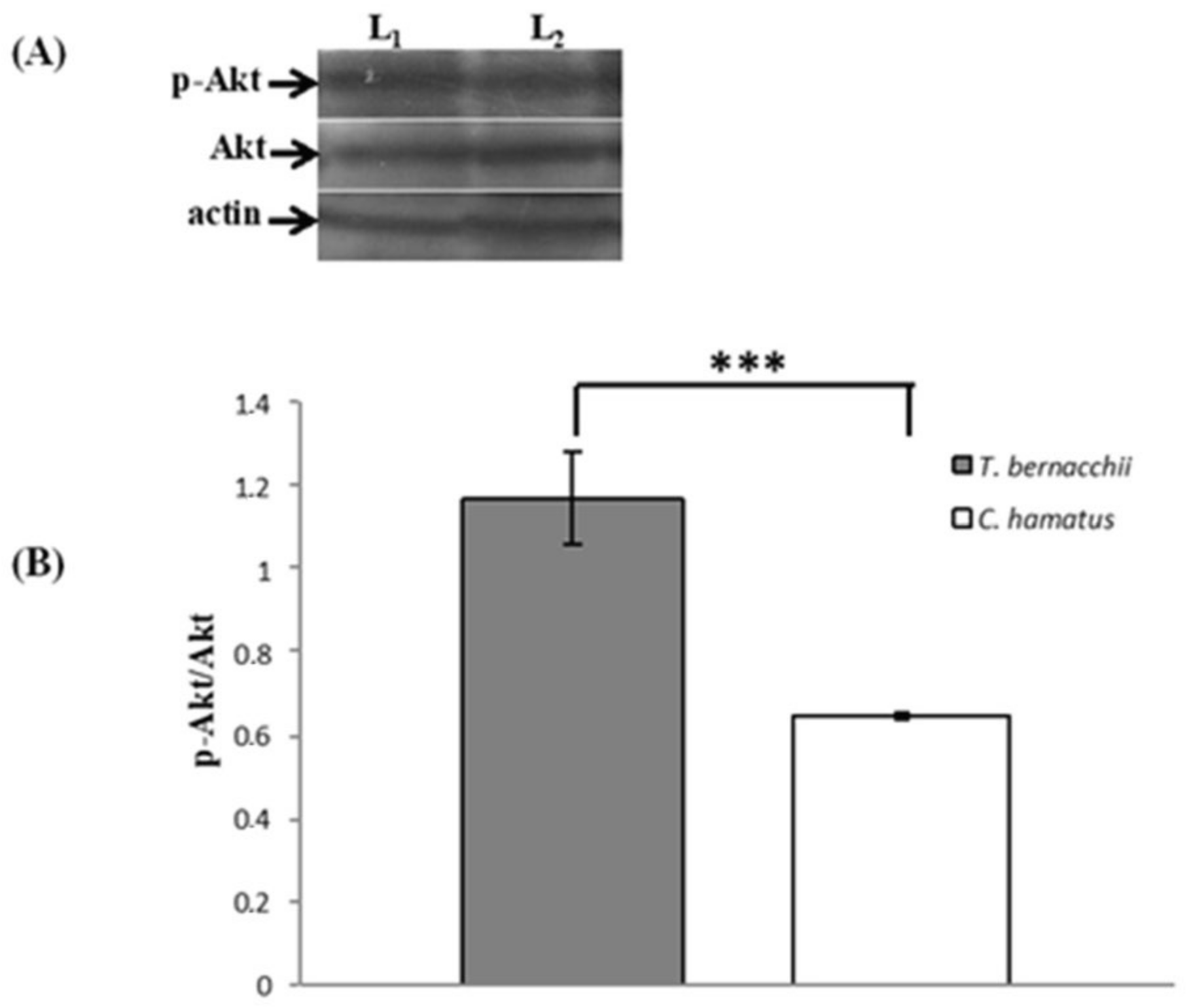
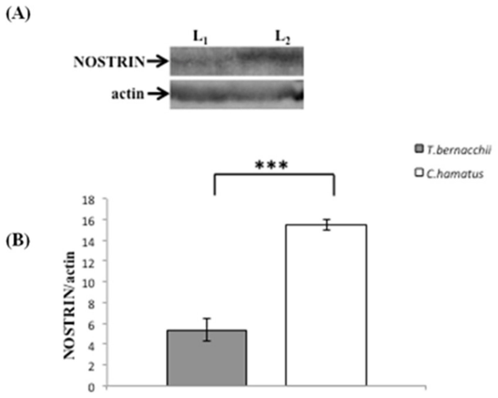
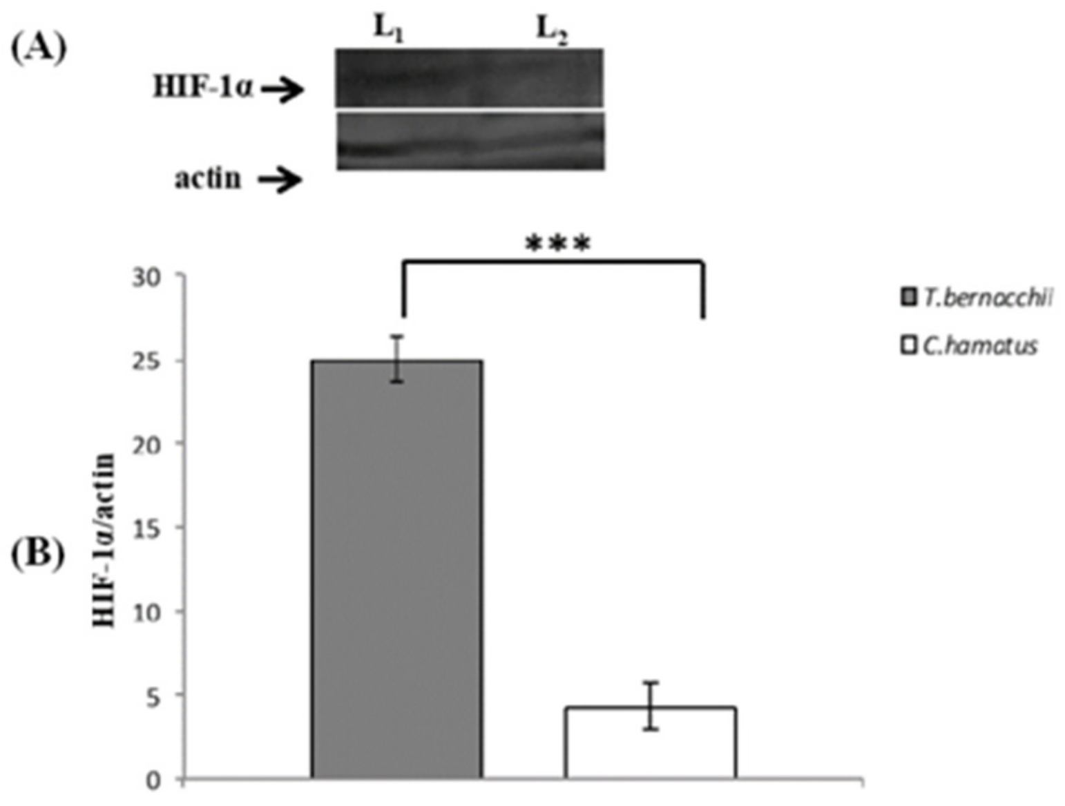
3.2. NOS Expression and Activation

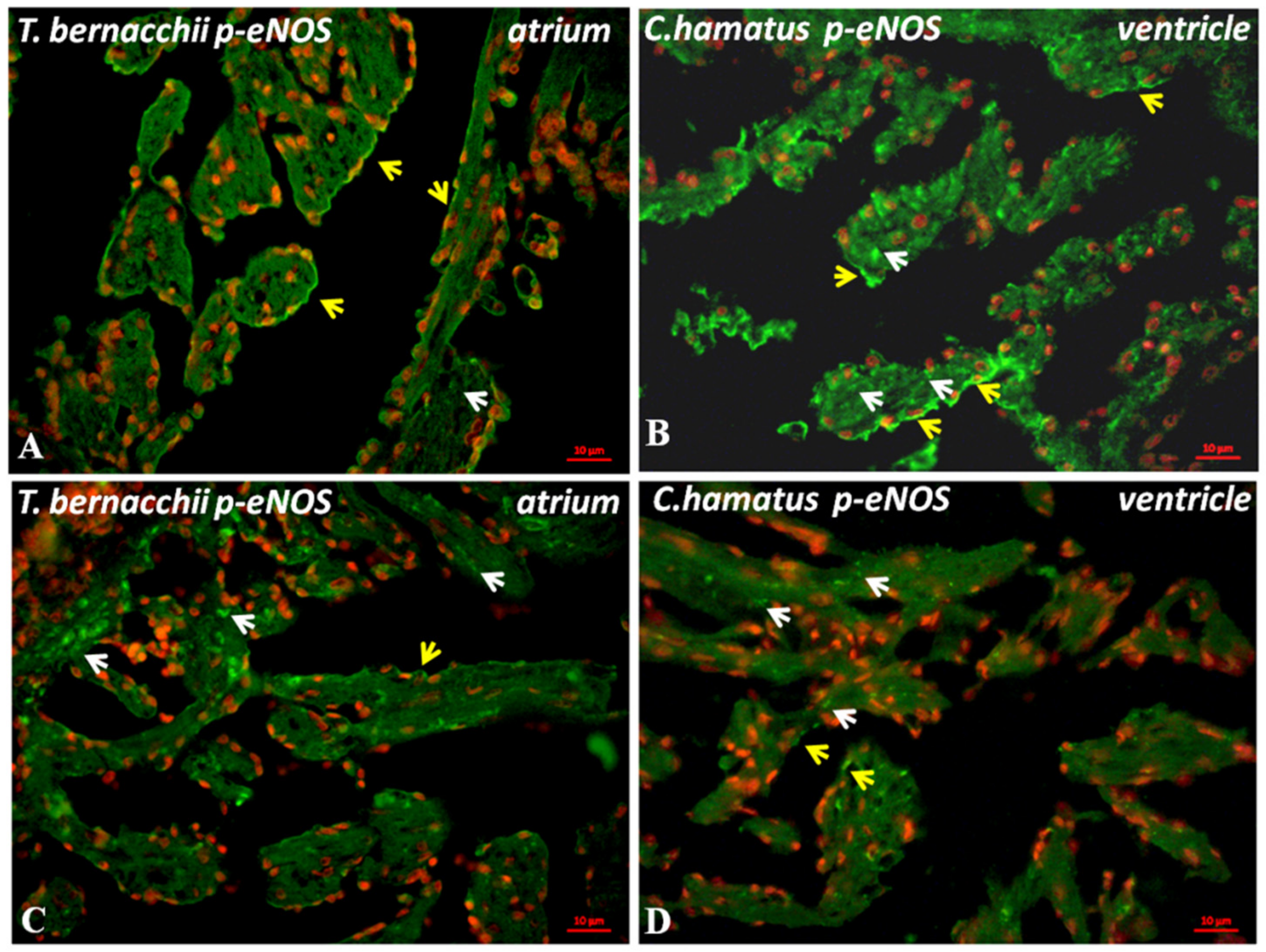
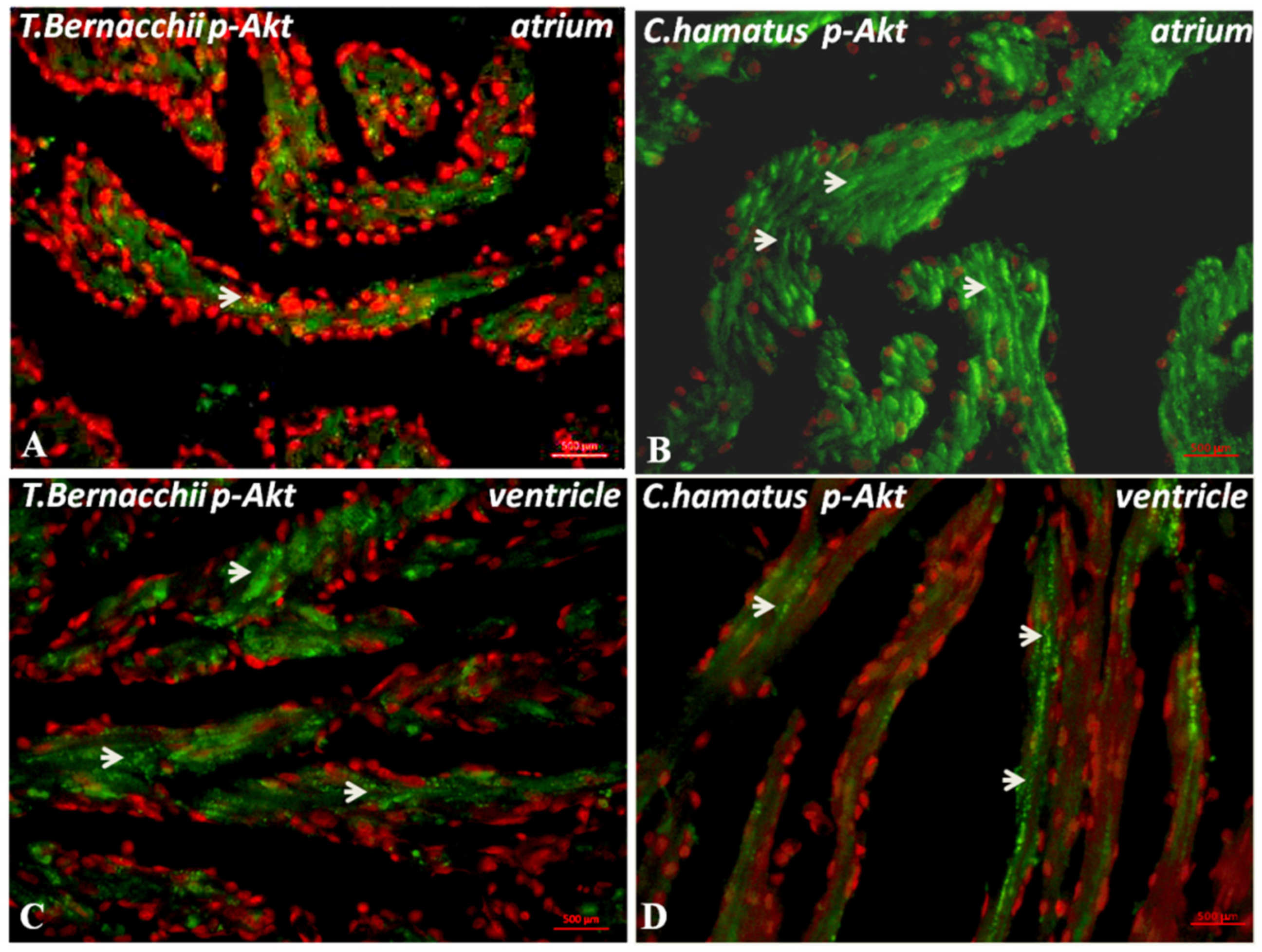
3.2.1. Immunodetection

The observation of cardiac sections immunolabeled with polyclonal anti-phosphorylated eNOS antibody (Figure 3A–D) revealed the presence of the activated enzyme in the atrium (Figure 3A,B) and in the trabeculated ventricle (Figure 3C,D) of both Antarctic teleosts (C. hamatus, Figure 3B,D; T. bernacchii Figure 3A,C). In particular, in the atrium of both species, phosphorylated eNOS signal appears confined at the endocardial level, and in myocardiocytes, the same pattern was observed in the ventricle where phosphorylated eNOS positive staining was detected at the level of endothelium endocardium (EE) enveloping trabeculae and, in a lesser extent, in myocardiocytes. When analyzing the differences between the two species, the phosphorylated eNOS expression seems stronger in the C. hamatus atrium and the ventricle of T. bernacchii. The p-Akt is localized in the atrium and the ventricle of both species in the cytoplasmic compartment of the cardiomyocytes (Figure 4A–D). However, p-Akt is more expressed in the atrium of C. hamatus (Figure 4B) than in T. bernacchii (Figure 4A). At the same time, in the ventricle of red-blooded fish (Figure 4C), the signal is more intense and uniformly diffused with respect to the icefish (Figure 4D), where the fluorescence is reduced and confined only toa few cardiomyocytes. Labeling specificity was confirmed by the lack of fluorescence in control sections treated concurrently without main antibodies.

3.2.2. Western Blotting and Densitometric Analyses

Western blotting analysis revealed the presence of immunoreactive bands corresponding to the approximate MW of eNOS (135 kDa), phosphorylated eNOS (140 kDa), Akt (60 kDa), p-Akt (60 kDa), NOSTRIN (58 kDa), HIF-1α (120 kDa) in both T. bernacchii and C. hamatus. Densitometric quantification of the blots revealed that the amount of activated eNOS, evaluated as phosphorylated eNOS/eNOS ratio, is higher in T. bernacchi than in C. hamatus (Figure 5A,B). A similar trend was also observed for p-Akt expression (Figure 6A,B). On the contrary, NOSTRIN expression is more expressed in C. hamatus (Figure 7A,B). Finally, HIF-1α expression is significantly lower in the icefish compared to the red-blooded fish (Figure 8A,B).

4. Discussion

This report showed, for the first time, that NO is not involved in the Frank-Starling response in the icefish C. hamatus. This unusual trait could be related to the peculiar deficiency of respiratory pigments, characteristic of this species. Moreover, by comparatively analyzingeNOS expression and its activation levels in C. hamatus and the red-blooded counterpart T. bernacchii, we revealed in icefish lower presence and activation levels of this enzyme.
Previously, we demonstrated the presence and functionality of the NOS/NO system in the heart of C. hamatus and T. bernacchii, revealing that both eNOS and iNOS isoforms are functionally expressed [34]. As reported by Pellegrino and co-workers [32,34] in isolated and perfused C. hamatus hearts, treatment with NO donors induced a positive inotropic effect, reversed by NO-inhibitor L-NIO. This is consistent with the significant inotropic effect mediated by NO on the unstimulated mechanical performance of C. hamatus heart [32,34]. In the present work, we also confirm, using a NO scavenging agent (PTIO), that the isolated and perfused heart of C. hamatus basal, NO release induces a positive inotropism. Indeed, under basal conditions (physically unstimulated heart), treatment with both L-NIO (10−5 mol L−1) and PTIO (10−5 mol L−1) induced a significant negative inotropism without affecting the heart rate.
According to literature data, some contrasting findings about the cardiac role of NO depend on species-specific differences, but they may also be related to the experimental designs [35,36]. In this context, our experimental setup, which also assures the reproducibility of results, validated the NO tonic modulation of mechanical performance under basal conditions. This result assumes particular significance since the icefish displays a positive NO-mediated inotropic effect, unlike other fish and mammals [26]. Further evidence for the opposite effects of NO-induced contractile stimulation on phenotypes with and without Hb expression has been furnished by Garofalo and coworkers [31] using isolated and perfused working heart of C. aceratus (Hb-/Mb-) and T. bernacchii (Hb+/Mb+). Indeed, the NO-dependent inotropism has been comparatively analyzed in three Antarctic teleost species characterized by different expression patterns for the respiratory pigments (see for review [26]), revealing that in the red-blooded species (T. bernacchii), the heart shows a small but significant negative inotropism produced by NO as temperate teleosts, frog and mammals (see for reference [36]). On the contrary, in the icefish species C. hamatus and C. aceratus, a positive inotropic effect of NO is noticed, thus suggesting that the absence of hemoglobin would be the key distinguishing element, albeit with an underlying mechanism not identified.
Additionally, the Mb- C. aceratus heart appears to be significantly more sensitive to the NOS stimulation by the genuine substrate L-arginine than the Mb+ C. hamatus heart while lacking a considerable NO-dependent basal tone and being insensitive to exogenous NO [31]. In addition, as reported by Pellegrino and co-workers [35], the NO basal inotropic response appears to be mediated only by the eNOS. Indeed, it is known that NO released by constitutive NOSs isoforms can regulate myocardium’s function by paracrine and autocrine mechanisms [37]. Given the NO shortlife and limited diffusion distance, NOSs may need to be close to their pool of effector proteins in order for them to work specifically in the heart. In mammals, eNOS-derived NO does not seem to promote relaxation of the myocardium and left ventricular compliance. However, upon stimulation of eNOS (e.g., in response to increased preload), the coronary endothelium’s release of NO may help diastolic function [35]. There are significant differences between coronary and endocardial endothelial cells, especially concerning the release of cardioactive mediators [36,38]. In the vascular heart, coronary endothelial cells are of greater relevance than endocardial endothelial cells in the paracrine crosstalk with cardiac myocytes. In the avascular heart, the fully trabeculated ventricle (spongiosa) is supplied by venous blood circulating into the myriads of intertrabecular spaces (lacunae) coated by the endocardial endothelial cells. Endocardial endothelial cells superfused by intracavitary blood mediate diffusion and exchange between the lumen and intracardiac interstitium (for reference [39]). In this case, endocardial endothelial cells influence the locally subjacent myocytes, especially by NO release.
In previous comparative studies on the NOS/NO system of Antarctic Notothenioids, we found other significant differences in fish species with different respiratory pigment expressions [32,34]. In detail, we showed that in C. hamatus (Hb-/Mb+), the inotropic effect was only elicited by selective eNOS inhibition. In comparison, a non-specific NOS inhibition did not affect inotropism while induced significant depressant effects on chronotropism [35]. This data cannot be explained by the extreme stenothermia of these organisms but rather by the hemoglobin-less condition since it is not detectable in the phylogenetically and ecologically closely related-blooded T. bernacchii.
In addition, under unstimulated conditions, hemoglobin-less and red-blooded Notothenioids show an identical isoform-specific cellular distribution and enzymatic activity pattern [31,32], similar to the mammalian heart [40]. In contrast, C. hamatus showed a lower NOS inducibility in lipopolysaccharide-stimulated hearts with respect to T. bernacchii, revealing a substantial difference in the regulation of the NO overproduction effects in the presence/absence of Hb [31].
A relevant finding concerns the non-involvement of the eNOS/NO system in the Frank-Starling response. The influence of endogenous NO on the Frank-Starling response was studied by increasing the preload in both absence (control) and the presence of L-NIO (10−5 M) or PTIO (10−5 M). The assessment of the Starling curves obtained with and without L-NIO or PTIO, evaluated by ANOVA analysis, did not display significant differences, suggesting the absence of a nitrergic modulation of the cardiac heterometric mechanism in the icefish (see Figure 2b).
It is well known that, in mammals, endogenous NO is involved in the Frank-Starling response, increasing ventricular diastolic distensibility and myocardial relaxation [41]. Our results on the heterometric mechanism in icefish also disagree with the literature data on temperate fish. For example, a significant NO modulation of the Frank-Starling mechanism, occurring via a cGMP-independent pathway and involving an Akt-mediated activation of eNOS-dependent NO production, has been well documented in eel [1]. In the icefish, the cardiac heterometric mechanism is devoid of nitrergic modulation, thus suggesting that, in this hemoglobin-less teleost, the eNOS enzymatic system is differently adapted compared to red-blooded organisms.
To better understand the mechanisms underlying this unusual Frank-Starling response in icefish, we have focused on eNOS regulatory mechanisms. It is widely known that eNOS experiences a complicated network of post-translational regulatory changes, emphasizing the dynamic regulation of enzyme activity in response to physiological stimuli and pathological situations [16]. Several protein factors that affect the catalytic efficiency of eNOS have been identified, and phosphorylation by p-Akt represents an important post-translational regulator of eNOS activity [16].
Analyzing the expression of phosphorylated eNOS and its main activator (p-Akt) in response to shear stress, we demonstrated that the amount of activated eNOS is lower in icefish than in the red-blooded T. bernacchii, and a similar trend was also observed for p-Akt/Akt. These results suggest a different modulation of enzyme activity, pointing attention to the different expressions of respiratory pigments in the two Antarctic notothenioids. Recently, it has been reported that HIF-1α contributes to the up-regulation of eNOS gene expression [42]. Although hypoxia is the predominant stimulus for its production [43,44], an increase in HIF-1α expression was also observed in cultured cells under normoxia, suggesting alternative signaling pathways in regulating this important transcription factor [14]. HIF-1α expression during normoxic conditions is particularly important for sustained expression and phosphorylation of eNOS and, therefore, for the production of NO [14]. Moreover, in this case, our results clearly showed the down-regulation of a critical stimulating factor of eNOS activation. HIF-1α expression is significantly lower in the icefish than in the red-blooded species. Further confirmation of a lower eNOS activation in icefish derives from quantifying the negative modulator of eNOS activity NOSTRIN. Indeed, quantification of NOSTRIN blots, performed on cardiac homogenates of C. hamatus and T. bernacchii, demonstrated a greater expression of this enzyme in the icefish.

5. Conclusions

In conclusion, we revealed that C. hamatus heart lacks the nitrergic modulation of Starling response and shows a slighter basal eNOS activity compared to the red-blooded counterpart.
Our findings add to our understanding of the biological and evolutionary significance of the nitrite/NO system in vertebrates, which may increase our understanding of its function in mammalian cardiac physiology. They also illustrate the potentially harmful effects of excessive NO production in the absence of hemoglobin. In addition, a deeper knowledge of the cardio-circulatory features of this Hb-less vertebrate, with its consequent and peculiar morpho-functional rearrangements, could be of interest in global warming scenario and environmental challenges, such as thermal stress and changes in oxygen availability, to better understand the mechanisms of adaptation, including antioxidant responses [45,46,47,48], of these special animals.

Author Contributions

Conceptualization, G.S. and D.P.; methodology, D.A., F.G., E.B., G.S. and D.P.; software, D.A., F.G. and D.P.; validation, D.A., F.G., E.B., G.S. and D.P.; formal analysis, D.A., F.G. and D.P.; investigation, D.A., F.G., E.B., G.S. and D.P.; resources, D.A., F.G., G.S. and D.P.; data curation, D.A., F.G., E.B., G.S. and D.P.; writing—original draft preparation, D.P.; writing—review and editing, E.B., G.S. and D.P.; visualization, D.A., F.G., E.B., G.S. and D.P.; supervision, D.P.; project administration, D.P.; funding acquisition, D.P. All authors have read and agreed to the published version of the manuscript.

Funding

All authors were supported by the Italian National Research Program in Antarctica (PNRA18_00133).

Institutional Review Board Statement

All the activities on animals performed during the Italian Antarctic Expedition are under the control of a PNRA Ethics Referent, which acts on behalf of the Italian Ministry of Foreign Affairs. In particular, the required data are the following: Project identification code: 2010/A1.08. Name of the ethics committee or institutional review board: Italian Ministry of Foreign Affairs. Name of PNRA Ethics Referent: Dr. Sandro Torcini, ENEA Antarctica, Technical Unit (UTA). Date of approval: 10 July 2012.

Informed Consent Statement

Not applicable.

Conflicts of Interest

The authors declare no conflict of interest.

References

  1. Garofalo, F.; Parisella, M.L.; Amelio, D.; Tota, B.; Imbrogno, S. Phospholamban S-nitrosylation modulates Starling response in fish heart. Proc. R. Soc. B Biol. Sci. 2009, 276, 4043–4052. [Google Scholar] [CrossRef] [Green Version]
  2. Shiels, H.A.; White, E. The Frank-Starling mechanism in vertebrate cardiac myocytes. J. Exp. Biol. 2008, 211, 2005–2013. [Google Scholar] [CrossRef] [Green Version]
  3. Gordon, A.M.; Regnier, M.; Homsher, E. Skeletal and cardiac muscle contractile activation: Tropomyosin “rocks and rolls”. News Physiol. Sci. 2001, 16, 49–55. [Google Scholar] [CrossRef]
  4. Calaghan, S.; White, E. Activation of Na+-H+ exchange and stretch-activated channels underlies the slow inotropic response to stretch in myocytes and muscle from the rat heart. J. Physiol. 2004, 559, 205–214. [Google Scholar] [CrossRef]
  5. Abraham, D.M.; Davis, R.T.; Warren, C.M.; Mao, L.; Wolska, B.M.; Solaro, R.J.; Rockman, H.A.; Semenza, G.L. β-Arrestin mediates the Frank-Starling mechanism of cardiac contractility. Proc. Natl. Acad. Sci. USA 2016, 113, 14426–14431. [Google Scholar] [CrossRef] [Green Version]
  6. Prendergast, B.D.; Sagach, V.F.; Shah, A.M. Basal release of nitric oxide augments the Frank-Starling response in the isolated heart. Circulation 1997, 96, 1320–1329. [Google Scholar] [CrossRef]
  7. Imbrogno, S.; De Iuri, L.; Mazza, R.; Tota, B. Nitric oxide modulates cardiac performance in the heart of Anguilla anguilla. J. Exp. Biol. 2001, 204, 1719–1727. [Google Scholar] [CrossRef]
  8. Petroff, M.G.V.; Kim, S.H.; Pepe, S.; Dessy, C.; Marbán, E.; Balligand, J.L.; Sollott, S.J. Endogenous nitric oxide mechanisms mediate the stretch dependence of Ca2+ release in cardiomyocytes. Nat. Cell Biol. 2001, 3, 867–873. [Google Scholar] [CrossRef]
  9. Zhang, Y.H.; Dingle, L.; Hall, R.; Casadei, B. The role of nitric oxide and reactive oxygen species in the positive inotropic response to mechanical stretch in the mammalian myocardium. Biochim. Biophys. Acta-Bioenerg. 2009, 1787, 811–817. [Google Scholar] [CrossRef] [Green Version]
  10. Devika, N.T.; Jaffar Ali, B.M. Analysing calcium dependent and independent regulation of eNOS in endothelium triggered by extracellular signalling events. Mol. Biosyst. 2013, 9, 2653–2664. [Google Scholar] [CrossRef]
  11. Dimmeler, S.; Fleming, I.; Fisslthaler, B.; Hermann, C.; Busse, R.; Zeiher, A.M. Activation of nitric oxide synthase in endothelial cells by Akt- dependent phosphorylation. Nature 1999, 399, 601–605. [Google Scholar] [CrossRef] [PubMed]
  12. Fulton, D.; Gratton, J.P.; McCabe, T.J.; Fontana, J.; Fujio, Y.; Walsh, K.; Franke, T.F.; Papapetropoulos, A.; Sessa, W.C. Regulation of endothelium-derived nitric oxide production by the protein kinase Akt. Nature 1999, 399, 597–601. [Google Scholar] [CrossRef] [PubMed] [Green Version]
  13. McCabe, T.J.; Fulton, D.; Roman, L.J.; Sessa, W.C. Enhanced electron flux and reduced calmodulin dissociation may explain “calcium-independent” eNOS activation by phosphorylation. J. Biol. Chem. 2000, 275, 6123–6128. [Google Scholar] [CrossRef] [PubMed] [Green Version]
  14. Leucker, T.M.; Bienengraeber, M.; Muravyeva, M.; Baotic, I.; Weihrauch, D.; Brzezinska, A.K.; Warltier, D.C.; Kersten, J.R.; Pratt, P.F. Endothelial-cardiomyocyte crosstalk enhances pharmacological cardioprotection. J. Mol. Cell. Cardiol. 2011, 51, 803–811. [Google Scholar] [CrossRef] [PubMed] [Green Version]
  15. Chakraborty, S.; Ain, R. Nitric-oxide synthase trafficking inducer is a pleiotropic regulator of endothelial cell function and signaling. J. Biol. Chem. 2017, 292, 6600–6620. [Google Scholar] [CrossRef] [Green Version]
  16. Michel, T.; Vanhoutte, P.M. Cellular signaling and NO production. Pflugers Arch. Eur. J. Physiol. 2010, 459, 807–816. [Google Scholar] [CrossRef] [Green Version]
  17. Dudzinski, D.M.; Michel, T. Life history of eNOS: Partners and pathways. Cardiovasc. Res. 2007, 75, 247–260. [Google Scholar] [CrossRef]
  18. Simmonds, M.J.; Detterich, J.A.; Connes, P. Nitric oxide, vasodilation and the red blood cell. Biorheology 2014, 51, 121–134. [Google Scholar] [CrossRef]
  19. Muller, B.; Kleschyov, A.L.; Alencar, J.L.; Vanin, A.; Stoclet, J.C. Nitric oxide transport and storage in the cardiovascular system. Ann. N. Y. Acad. Sci. 2002, 962, 131–139. [Google Scholar] [CrossRef]
  20. Allen, B.W.; Stamler, J.S.; Piantadosi, C.A. Hemoglobin, nitric oxide and molecular mechanisms of hypoxic vasodilation. Trends Mol. Med. 2009, 15, 452–460. [Google Scholar] [CrossRef]
  21. Gladwin, M.T.; Raat, N.J.H.; Shiva, S.; Dezfulian, C.; Hogg, N.; Kim-Shapiro, D.B.; Patel, R.P. Nitrite as a vascular endocrine nitric oxide reservoir that contributes to hypoxic signaling, cytoprotection, and vasodilation. Am. J. Physiol.-Hear. Circ. Physiol. 2006, 291, H2026–H2035. [Google Scholar] [CrossRef]
  22. Hendgen-Cotta, U.B.; Kelm, M.; Rassaf, T. A highlight of myoglobin diversity: The nitrite reductase activity during myocardial ischemia-reperfusion. Nitric Oxide-Biol. Chem. 2010, 22, 75–82. [Google Scholar] [CrossRef]
  23. Gladwin, M.T.; Schechter, A.N. NO Contest: Nitrite Versus S-Nitroso-Hemoglobin. Circ. Res. 2004, 94, 851–855. [Google Scholar] [CrossRef] [Green Version]
  24. Moylan, T.J.; Sidell, B.D. Concentrations of myoglobin and myoglobin mRNA in heart ventricles from Antarctic fishes. J. Exp. Biol. 2000, 203, 1277–1286. [Google Scholar] [CrossRef]
  25. Eastman, J.T. Antarctic Fish Biology; Academic Press: Cambridge, MA, USA, 1993; p. 274. [Google Scholar]
  26. Garofalo, F.; Pellegrino, D.; Amelio, D.; Tota, B. The Antarctic hemoglobinless icefish, fifty five years later: A unique cardiocirculatory interplay of disaptation and phenotypic plasticity. Comp. Biochem. Physiol.-A Mol. Integr. Physiol. 2009, 154, 10–28. [Google Scholar] [CrossRef]
  27. Egginton, S.; Sidell, B.D. Thermal acclimation induces adaptive changes in subcellular structure of fish skeletal muscle. Am. J. Physiol.-Regul. Integr. Comp. Physiol. 1989, 256. [Google Scholar] [CrossRef]
  28. O’Brien, K.M.; Sidell, B.D. The interplay among cardiac ultrastructure, metabolism and the expression of oxygen-binding proteins in Antarctic fishes. J. Exp. Biol. 2000, 203, 1287–1297. [Google Scholar] [CrossRef]
  29. Hardewig, I.; Van Dijk, P.L.M.; Moyes, C.D.; Pörtner, H.O. Temperature-dependent expression of cytochrome-c oxidase in Antarctic and temperate fish. Am. J. Physiol.-Regul. Integr. Comp. Physiol. 1999, 277, R508–R516. [Google Scholar] [CrossRef] [Green Version]
  30. Shah, A.M.; MacCarthy, P.A. Paracrine and autocrine effects of nitric oxide on myocardial function. Pharmacol. Ther. 2000, 86, 49–86. [Google Scholar] [CrossRef]
  31. Amelio, D.; Garofalo, F.; Pellegrino, D.; Giordano, F.; Tota, B.; Cerra, M.C. Cardiac expression and distribution of nitric oxide synthases in the ventricle of the cold-adapted Antarctic teleosts, the hemoglobinless Chionodraco hamatus and the red-blooded Trematomus bernacchii. Nitric Oxide-Biol. Chem. 2006, 15, 190–198. [Google Scholar] [CrossRef]
  32. Pellegrino, D.; Palmerini, C.A.; Tota, B. No hemoglobin but NO: The icefish (Chionodraco hamatus) heart as a paradigm. J. Exp. Biol. 2004, 207, 3855–3864. [Google Scholar] [CrossRef] [PubMed] [Green Version]
  33. Garofalo, F.; Amelio, D.; Cerra, M.C.; Tota, B.; Sidell, B.D.; Pellegrino, D. Morphological and physiological study of the cardiac NOS/NO system in the Antarctic (Hb-/Mb-) icefish Chaenocephalus aceratus and in the red-blooded Trematomus bernacchii. Nitric Oxide-Biol. Chem. 2009, 20, 69–78. [Google Scholar] [CrossRef] [PubMed]
  34. Pellegrino, D.; Acierno, R.; Tota, B. Control of cardiovascular function in the icefish Chionodraco hamatus: Involvement of serotonin and nitric oxide. Comp. Biochem. Physiol.-A Mol. Integr. Physiol. 2003, 134. [Google Scholar] [CrossRef]
  35. Pinsky, D.J.; Patton, S.; Mesaros, S.; Brovkovych, V.; Kubaszewski, E.; Grunfeld, S.; Malinski, T. Mechanical transduction of nitric oxide synthesis in the beating heart. Circ. Res. 1997, 81, 372–379. [Google Scholar] [CrossRef] [PubMed]
  36. Mebazaa, A.; Wetzel, R.; Cherian, M.; Abraham, M. Comparison between endocardial and great vessel endothelial cells: Morphology, growth, and prostaglandin release. Am. J. Physiol.-Hear. Circ. Physiol. 1995, 268, H250–H259. [Google Scholar] [CrossRef] [PubMed]
  37. Zhang, Y.H.; Casadei, B. Sub-cellular targeting of constitutive NOS in health and disease. J. Mol. Cell. Cardiol. 2012, 52, 341–350. [Google Scholar] [CrossRef]
  38. Brutsaert, D.L.; Fransen, P.; Andries, L.J.; De Keulenaer, G.W.; Sys, S.U. Cardiac endothelium and myocardial function. Cardiovasc. Res. 1998, 38, 281–290. [Google Scholar] [CrossRef] [Green Version]
  39. Tota, B.; Amelio, D.; Pellegrino, D.; Ip, Y.K.; Cerra, M.C. NO modulation of myocardial performance in fish hearts. Comp. Biochem. Physiol.-A Mol. Integr. Physiol. 2005, 142, 164–177. [Google Scholar] [CrossRef]
  40. Andries, L.J.; Brutsaert, D.L.; Sys, S.U. Nonuniformity of endothelial constitutive nitric oxide synthase distribution in cardiac endothelium. Circ. Res. 1998, 82, 195–203. [Google Scholar] [CrossRef] [Green Version]
  41. Casadei, B.; Sears, C.E. Nitric-oxide-mediated regulation of cardiac contractility and stretch responses. Prog. Biophys. Mol. Biol. 2003, 82, 67–80. [Google Scholar] [CrossRef]
  42. Rodriguez-Miguelez, P.; Lima-Cabello, E.; Martínez-Flórez, S.; Almar, M.; Cuevas, M.J.; González-Gallego, J. Hypoxia-inducible factor-1 modulates the expression of vascular endothelial growth factor and endothelial nitric oxide synthase induced by eccentric exercise. J. Appl. Physiol. 2015, 118, 1075–1083. [Google Scholar] [CrossRef] [Green Version]
  43. Richard, D.E.; Berra, E.; Pouyssegur, J. Nonhypoxic pathway mediates the induction of hypoxia-inducible factor 1α in vascular smooth muscle cells. J. Biol. Chem. 2000, 275, 26765–26771. [Google Scholar] [CrossRef]
  44. Ke, Q.; Costa, M. Hypoxia-inducible factor-1 (HIF-1). Mol. Pharmacol. 2006, 70, 1469–1480. [Google Scholar] [CrossRef]
  45. Tolomeo, A.M.; Carraro, A.; Bakiu, R.; Toppo, S.; Place, S.P.; Ferro, D.; Santovito, G. Peroxiredoxin 6 from the Antarctic emerald rockcod: Molecular characterization of its response to warming. J. Comp. Physiol. B Biochem. Syst. Environ. Physiol. 2016, 186, 59–71. [Google Scholar] [CrossRef]
  46. Garofalo, F.; Santovito, G.; Amelio, D. Morpho-functional effects of heat stress on the gills of Antarctic T. bernacchii and C. hamatus. Mar. Pollut. Bull. 2019, 141, 194–204. [Google Scholar] [CrossRef]
  47. Tolomeo, A.M.; Carraro, A.; Bakiu, R.; Toppo, S.; Garofalo, F.; Pellegrino, D.; Gerdol, M.; Ferro, D.; Place, S.P.; Santovito, G. Molecular characterization of novel mitochondrial peroxiredoxins from the Antarctic emerald rockcod and their gene expression in response to environmental warming. Comp. Biochem. Physiol. Part C Toxicol. Pharmacol. 2019, 225. [Google Scholar] [CrossRef]
  48. Chatzidimitriou, E.; Bisaccia, P.; Corrà, F.; Bonato, M.; Irato, P.; Manuto, L.; Toppo, S.; Bakiu, R.; Santovito, G. Copper/zinc superoxide dismutase from the crocodile icefish Chionodraco hamatus: Antioxidant defense at constant sub-zero temperature. Antioxidants 2020, 9, 325. [Google Scholar] [CrossRef]
Figure 1. Effects of L-NIO (10−5 mol L−1) and PTIO (10−5 mol L−1) on stroke volume (Vs) and stroke work (Ws) in isolated and perfused heart of C. hamatus. Percentage changes were evaluated as means ± S.E.M. of five experiments for each group. Asterisks indicate values significantly different from the control value (Paired Student’s t-test, * p < 0.05).
Jmse 10 01705 g001
Figure 2. (a,b) Effects of preload on stroke volume (Vs) and stroke work (Ws) in isolated and perfused heart of C. hamatus at baseline condition and at untreated time-control. (c,d) Effects of preload on stroke volume (Vs) and stroke work (Ws) under control conditions and after pretreatment with L-NIO (10−5 mol L−1) and PTIO (10−5 mol L−1) in isolated and perfused heart of C. hamatus. Percentage changes were evaluated as means ± S.E.M. of four experiments for each group. Paired Student’s t-test was used for comparisons within groups; a two-way ANOVA was used for comparison between groups (*** p < 0.005).
Jmse 10 01705 g002
Figure 3. Phosphorylated eNOS immunolocalization in the atrium (A,B) and the ventricle (C,D) of T. bernacchii (A,C) and C. hamatus (B,D). phosphorylated eNOS is localized in the endocardial endothelium (yellow arrows) and the myocardiocytes (white arrows). Nuclei counterstaining: propidium iodide.
Jmse 10 01705 g003
Figure 4. p-Akt immunolocalization in the atrium (A,B) and in the ventricle (C,D) of T. bernacchii (A,C) and C. hamatus (B,D). p-Akt is localized in the myocardiocytes (white arrows). Nuclei counterstaining: propidium iodide.
Jmse 10 01705 g004
Figure 5. (A) Western blotting of eNOS and phosphorylated eNOS in ventricle extracts from T. bernacchii (L1) and C. hamatus (L2). (B) shows the densitometric quantification of the blots. The loaded protein amount was verified using an anti-β-actin antibody. Statistical differences were evaluated by the non-parametric Mann–Whitney U test (** p < 0.005).
Jmse 10 01705 g005
Figure 6. (A) Western blotting of Akt and p-Akt in ventricle extracts from T. bernacchii (L1) and C. hamatus (L2). (B) shows the densitometric quantification of the blots. The loaded protein amount was verified using an anti-β-actin antibody. Statistical differences were evaluated by the non-parametric Mann–Whitney U test (*** p < 0.0005).
Jmse 10 01705 g006
Figure 7. (A) Western blotting of NOSTRIN in ventricle extracts from T. bernacchii (L1) and C. hamatus (L2). (B) shows the densitometric quantification of the blots. The loaded protein amount was verified using the anti-β-actin antibody. Statistical differences were evaluated by one-way ANOVA followed by Bonferroni multiple comparisons test (*** p < 0.0005).
Jmse 10 01705 g007
Figure 8. (A) Western blotting of HIF-1α in ventricle extracts from T. bernacchii (L1) and C. hamatus (L2). (B) shows the densitometric quantification of the blots. The loaded protein amount was verified using an anti-β-actin antibody. Statistical differences were evaluated by one-way ANOVA followed by Bonferroni multiple comparisons test (*** p < 0.0005).
Jmse 10 01705 g008
Table 1. Performance variables under basal conditions of isolated and perfused heart preparations of C. hamatus. Values are means ± S.E.M. of 8 experiments.
C. hamatus
Heart rate (beats/min)28.6 ± 1.09
Filling pressure (kPa)0.11 ± 0.02
Output pressure (kPa)1.43 ± 0. 09
Cardiac output (mL/min/kg) 51.3 ± 3.68
Stroke volume (mL/kg)1.8 ± 0.2
Stroke work (mJ/g)2.2 ± 0.31
Publisher’s Note: MDPI stays neutral with regard to jurisdictional claims in published maps and institutional affiliations.

