Review

Peptic Ulcer Disease: A Brief Review of Conventional Therapy and Herbal Treatment Options

1
Department of Pharmacology and Biochemistry, Faculty of Dental Medicine and Health Osijek, Josip Juraj Strossmayer University of Osijek, 31000 Osijek, Croatia
2
Department of Pathophysiology and Physiology with Immunology, Faculty of Dental Medicine and Health Osijek, Josip Juraj Strossmayer University of Osijek, 31000 Osijek, Croatia
3
Department of Internal Medicine, Faculty of Medicine Osijek, Josip Juraj Strossmayer University of Osijek, 31000 Osijek, Croatia
4
Department of Pharmacology, Faculty of Medicine Osijek, Josip Juraj Strossmayer University of Osijek, 31000 Osijek, Croatia
5
Department of Internal Medicine, University Hospital Osijek, 31000 Osijek, Croatia
*
Author to whom correspondence should be addressed.
J. Clin. Med. 2019, 8(2), 179; https://doi.org/10.3390/jcm8020179
Submission received: 31 December 2018 / Revised: 30 January 2019 / Accepted: 31 January 2019 / Published: 3 February 2019
(This article belongs to the Special Issue Helicobacter pylori Infection and Gastritis)

Abstract

:
Peptic ulcer is a chronic disease affecting up to 10% of the world’s population. The formation of peptic ulcers depends on the presence of gastric juice pH and the decrease in mucosal defenses. Non-steroidal anti-inflammatory drugs (NSAIDs) and Helicobacter pylori (H. pylori) infection are the two major factors disrupting the mucosal resistance to injury. Conventional treatments of peptic ulcers, such as proton pump inhibitors (PPIs) and histamine-2 (H2) receptor antagonists, have demonstrated adverse effects, relapses, and various drug interactions. On the other hand, medicinal plants and their chemical compounds are useful in the prevention and treatment of numerous diseases. Hence, this review presents common medicinal plants that may be used for the treatment or prevention of peptic ulcers.

1. Introduction

Peptic ulcer is an acid-induced lesion of the digestive tract that is usually located in the stomach or proximal duodenum, and is characterized by denuded mucosa with the defect extending into the submucosa or muscularis propria [1]. The estimated prevalence of peptic ulcer disease in the general population is 5–10% [2], but recent epidemiological studies have shown a decrease in the incidence, rates of hospital admissions, and mortality associated with peptic ulcer [3,4]. This is most likely secondary to the introduction of new therapies and improved hygiene, which resulted in a decline in Helicobacter pylori (H. pylori) infections.
Traditionally, mucosal disruption in patients with the acid peptic disease is considered to be a result of a hypersecretory acidic environment together with dietary factors or stress. Risk factors for developing peptic ulcer include H. pylori infection, alcohol and tobacco consumption, non-steroidal anti-inflammatory drugs (NSAIDs) use, and Zollinger–Ellison syndrome [5]. The main risk factors for both gastric and duodenal ulcers are H. pylori infection and NSAID use [6]. However, only a small proportion of people affected with H. pylori or using NSAIDs develop peptic ulcer disease, meaning that individual susceptibility is important in the beginning of mucosal damage. Functional polymorphisms in different cytokine genes are associated with peptic ulcers. For example, polymorphisms of interleukin 1 beta (IL1B) affect mucosal interleukin 1β production, causing H. pylori-associated gastroduodenal diseases [7].
On the other hand, the risk of complications of peptic ulcer is increased four times in NSAID users, and two times in aspirin users [8]. The concomitant use of NSAIDs or aspirin with anticoagulants, corticosteroids, and selective serotonin reuptake inhibitors increase the risk of upper gastrointestinal bleeding [9]. Although many people who use NSAIDs or aspirin have concurrent H. pylori infection, their interaction in the pathogenesis of peptic ulcer disease remains controversial. A meta-analysis of observational studies resulted in a conclusion that NSAIDs, aspirin use, and H. pylori infection increase the risk of peptic ulcer disease independently [10].
H. pylori-negative, NSAID-negative, and aspirin-negative peptic ulcer disease, which is classified as an idiopathic ulcer, can be diagnosed in about one-fifth of cases [11]. It is caused by the imbalance between factors that contribute to mucosal integrity and aggressive insults, but the pathogenic mechanisms behind the development of idiopathic peptic ulcer are still unknown [5]. A Danish study showed that psychological stress could increase the incidence of peptic ulcer [12]. Other etiologies include ischemia, drugs (steroids, chemotherapeutic agents) and radiotherapy, viruses, histamine, eosinophilic infiltration, gastric bypass surgery, and metabolic disturbances [13].

2. Pathogenesis of Peptic Ulcer
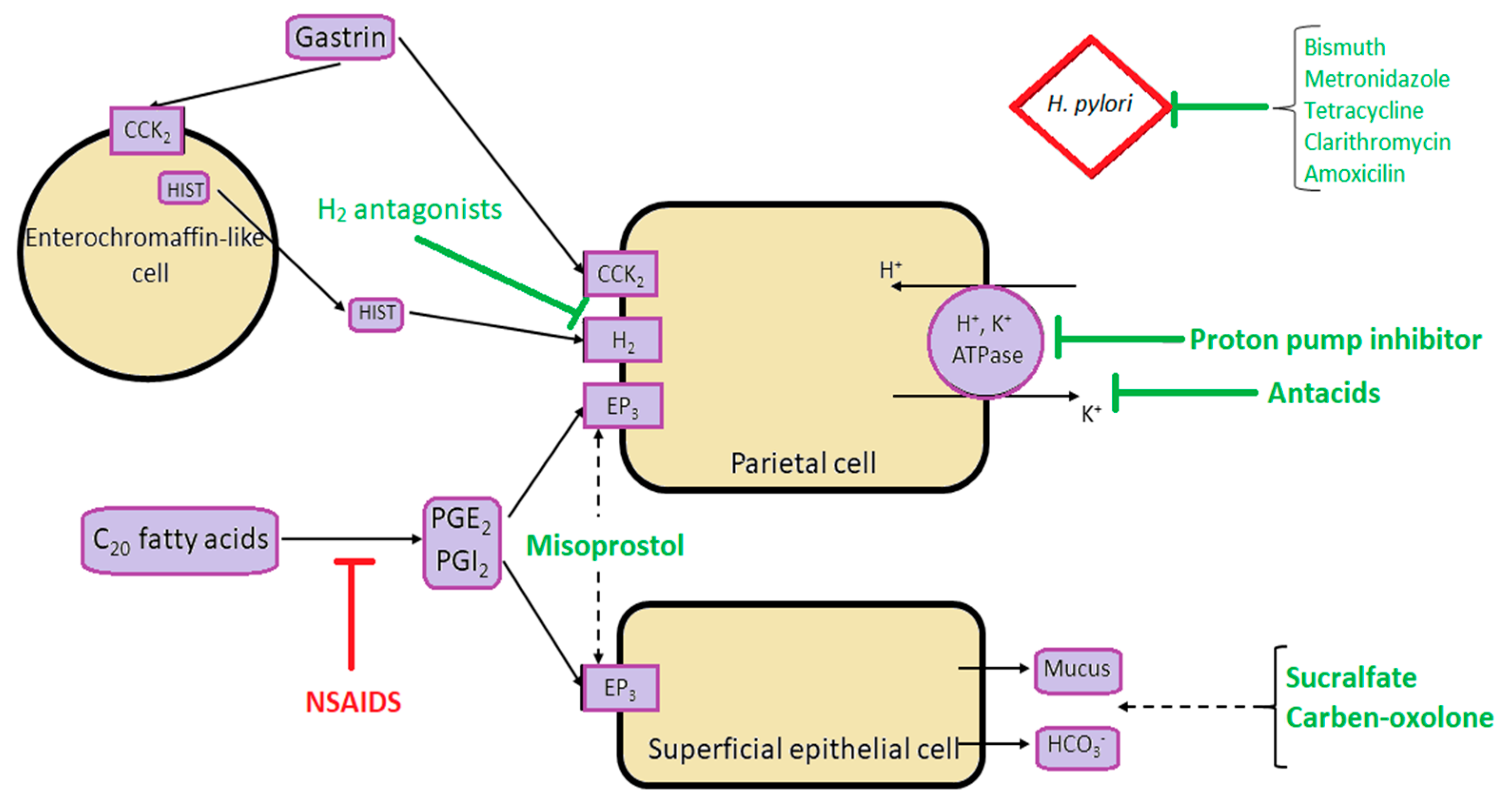
Almost half of the world’s population is colonized by H. pylori, which remains one of the most common causes of peptic ulcer disease [14]. The prevalence of H. pylori is higher in developing countries, especially in Africa, Central America, Central Asia, and Eastern Europe [15]. The organism is usually acquired in childhood in an environment of unsanitary conditions and crowding, mostly in countries with lower socioeconomic status. H. pylori causes epithelial cell degeneration and injury, which is usually more severe in the antrum, by the inflammatory response with neutrophils, lymphocytes, plasma cells, and macrophages.
The mechanism by which H. pylori induces the development of different types of lesions in the gastroduodenal mucosa is not fully explained. H. pylori infection can result in either hypochlorhydria or hyperchlorhydria, thus determining the type of peptic ulcer. The main mediators of H. pylori infection are cytokines that inhibit parietal cell secretion, but H. pylori can directly affect the H+/K+ ATPase α-subunit, activate calcitonin gene-related peptide (CGRP) sensory neurons linked to somatostatin, or inhibit the production of gastrin [16]. Although the formation of gastric ulcers is associated with hyposecretion, 10–15% of patients with H. pylori infection have increased gastric secretion caused by hypergastrinemia and reduced antral somatostatin content [17]. This leads to increased histamine secretion, and subsequently the increased secretion of acid or pepsin from parietal and gastric cells. Additionally, the eradication of H. pylori leads to a decrease in gastrin mRNA expression and an increase in somatostatin mRNA expression [18]. In the remaining majority of patients, gastric ulcers are associated with hypochlorhydria and mucosal atrophy.
The main mechanism of NSAID-associated damage of the gastroduodenal mucosa is the systemic inhibition of constitutively expressed cyclooxygenase-1 (COX-1), which is responsible for prostaglandin synthesis, and is associated with decreased mucosal blood flow, low mucus and bicarbonate secretion, and the inhibition of cell proliferation. NSAIDs inhibit the enzyme reversibly in a concentration-dependent manner. The co-administration of exogenous prostaglandins and cyclooxygenase-2 (COX-2)-selective NSAIDs use reduces mucosal damage and the risk of ulcers [19]. However, the different physicochemical properties of NSAIDs cause differences in their toxicity [20]. NSAIDs disrupt mucus phospholipids and lead to the uncoupling of mitochondrial oxidative phosphorylation, thus initiating mucosal damage. When exposed to acidic gastric juice (pH 2), NSAIDs become protonated and cross lipid membranes to enter epithelial cells (pH 7.4), where they ionize and release H+. In that form, NSAIDs cannot cross the lipid membrane, and are trapped in epithelial cells, leading to the uncoupling of oxidative phosphorylation, decreased mitochondrial energy production, increased cellular permeability, and reduced cellular integrity. Patients who have a history of peptic ulcers or hemorrhage, are over the age of 65, also use steroids or anticoagulants, and take high doses or combinations of NSAIDs are at the highest risk for acquiring NSAID-induced ulcers [1]
Main pathophysiological mechanisms and the sites of action of antiulcer treatment are shown in the Figure 1.

3. Treatment

An overview of conventional antiulcer treatment options is summarized in Table 1 and Table 2.

3.1. Helicobacter pylori Eradication

Although successful H. pylori eradication alone is paramount for healing associated peptic ulcers and preventing relapses, the growing prevalence of antibiotic resistance made it a global challenge. The first effective therapy was introduced in the 1980s, and consisted of a combination of bismuth, tetracycline, and metronidazole that was given for two weeks [14]. The standard first-line therapy is a triple therapy consisting of a proton pump inhibitor (PPI) and two antibiotics, such as clarithromycin plus amoxicillin or metronidazole given for seven to 14 days [32]. However, with an increasing prevalence of antibiotic resistance, especially for clarithromycin, there has been a marked decline in the success of triple therapy over the last 10–15 years. H. pylori eradication should be based on antimicrobial susceptibility tests. As susceptibility testing is often not available in clinical practice, the choice of first-line therapies should be based on the local prevalence of antibiotic resistance, and clarithromycin-based regimens should be abandoned in areas where the local clarithromycin resistance rate is more than 15% [36]. The rate of eradication can be increased with the use of high-dose PPI and by extending the duration to 14 days [37].
The recommended standard first-line therapy is either a bismuth-containing quadruple therapy for 14 days (PPI, a bismuth salt, tetracycline, and metronidazole) or a 14-day concomitant therapy for patients intolerant of bismuth (PPI, clarithromycin, amoxicillin, and metronidazole); both regimens yield eradication rates higher than 90% [38].
Second-line therapy is prescribed if a first-line regimen fails, and should not include metronidazole or clarithromycin [39]. Levofloxacin triple therapy (PPI, amoxicillin, and levofloxacin) for 14 days seems to be an efficacious therapy, achieving eradication rates between 74–81% [33]. If a patient received first-line treatment with a clarithromycin-based regimen, a preferred treatment option is a bismuth quadruple therapy with eradication rates of 77–93%, or a high-dose dual-therapy regimen with amoxicillin and a PPI, as H. pylori rarely develops amoxicillin resistance [34]. Despite well-developed recommendations for choosing proper treatment regimens, 5–10% of patients have persistent infection. The most common reasons for the failure of two treatments are suboptimal compliance or the resistance of H. pylori to one or more antibiotics, in which case susceptibility testing is strongly recommended.
When at least three recommended options have been unsuccessful, one of the commonly recommended salvage regimens is rifabutin-based triple therapy (PPI, rifabutin, and amoxicillin) for 10 days, with eradication rates of 66–70% [35], but rifabutin’s adverse effects such as myelotoxicity and red secretions should be taken into account [40].

3.2. NSAID-Associated Ulcer Disease and the Use of PPIs

Many strategies are available for the prevention of NSAID and aspirin-associated gastroduodenal ulcers and their complications, such as the co-therapy of NSAIDs with a PPI, H₂ receptor antagonist, or misoprostol; the use of COX-2-selective NSAIDs; or their combination with a gastroprotective agent. PPIs are the most popular and effective prophylactic agents [41]. The mechanism of action is reducing the production of gastric acid through irreversible binding to the hydrogen/potassium ATPase enzyme on gastric parietal cells. The combination of COX-2-selective NSAIDs and a PPI offers the best protection against peptic ulcer complications [42]. Standard doses of H₂ receptor antagonists cannot reduce the risk of gastric ulcers [43]. Gastrointestinal upset and abortifacient actions limit the use of misoprostol for gastric protection, despite its effective prevention of peptic ulcer complications. Ulcers heal in more than 85% of cases with six to eight weeks of PPI therapy if the offending agent is discontinued. All of the gastric ulcers require repeat endoscopy to evaluate the success of healing. If ulcers fail to heal, drug compliance should be checked. For refractory ulcers, the doubling of PPI dose for another six to eight weeks is often recommended, although the evidence supporting this is weak. After the exclusion of false-negative H. pylori status, unusual causes of peptic ulcer should be explored, such as malignancies, infections, Crohn’s disease, vasculitis, upper abdominal radiotherapy, cocaine use, and Zollinger–Ellison syndrome.
PPIs are among the most commonly used and overprescribed medications in the world [44]. The side effects of the PPIs, such as a headache, diarrhea, constipation, and abdominal discomfort, are minor and easily managed. However, recent studies have suggested an association between PPI use and several serious adverse effects, which has been a source of major concern to patients and physicians. Some of the adverse effects of PPIs are related to their suppression of gastric acid secretion, allowing ingested microbial pathogens that would have been destroyed by gastric acid to colonize the upper gastrointestinal tract and cause infections. Reports are suggesting that the use of PPIs might increase the risk of enteric infections such as Salmonella and Campylobacter, community-acquired pneumonia [45], Clostridium difficile infections [46], and spontaneous bacterial peritonitis [47].
With gastric acid suppression, there is no stimulation of endocrine D cells to produce somatostatin, and thereby no inhibition of G cells for gastrin release, resulting in hypergastrinemia. Gastrin is a growth factor that can increase proliferation in Barrett metaplasia and the colon [48]. Nonetheless, PPI-induced hypergastrinemia in humans generally is mild, and rarely causes carcinoid tumors in human patients unless they have a genetic abnormality [49]. Furthermore, PPI usage might protect against cancer in Barrett’s esophagus, since PPIs heal the chronic esophageal inflammation of reflux esophagitis, which is a risk factor for the development of malignancy.
Gastric acid inhibition by PPIs also can affect the uptake of certain vitamins, minerals, and medications. There are reports of patients on PPIs developing vitamin B12 deficiency and iron deficiency anemia [50]. Additionally, PPIs might increase the risk for osteoporosis and bone fractures by interfering with the ionization and solubilization of the calcium salts that are required for their absorption [51]. The underlying mechanism for hypomagnesemia is still not clear. PPI-induced gastric acid suppression decreases ketoconazole absorption and facilitates the absorption of digoxin [52]. Furthermore, PPIs can affect the metabolism of other drugs metabolized by the cytochrome (CYP) P450 system; for instance, they can delay the clearance of warfarin, diazepam, and phenytoin. Considerable attention has been given to the potential of PPIs to reduce the antiplatelet action of clopidogrel, since both are metabolized by the CYP2C19 enzyme [53]. The clinical importance of the interaction remains disputed, but the Food and Drug Administration (FDA) has issued warnings to avoid using omeprazole or esomeprazole with clopidogrel.
There has been a dramatic increase in reports of miscellaneous, unanticipated adverse effects of PPIs over the past several years, such as myocardial infarction, stroke, acute and chronic kidney disease, and eosinophilic esophagitis. The increased frequency of cardiovascular events in patients on clopidogrel who also use PPIs can be the result of the drugs competing for metabolism by CYP2C19, although there is a possibility that PPIs might have cardiovascular effects that are independent of their effects on clopidogrel activation, perhaps by the decreased production of nitric oxide and altered vascular homeostasis [54]. It has been proposed that PPIs might contribute to the development of eosinophilic esophagitis through their effects on peptic digestion [55]. The suppression of acid production raises gastric pH and inactivates pepsin, inhibiting peptide ingestion and degradation, and causing allergic reactions in the small intestine.

3.3. Potassium-Competitive Acid Blockers

Since up to 13% of patients treated with lansoprazole still experience ulcer recurrence, the search for alternative treatment is ongoing. Vonoprazan is a potassium-competitive acid blocker that inhibits H+, K+-ATPase in gastric parietal cells at the final stage of the acid secretory pathway [25]. The difference in the mechanism of action between vonoprazan and PPIs is that vonoprazan inhibits the enzyme in a K+-competitive and reversible manner, and does not require an acidic environment for activation. Additionally, vonoprazan shows a rapid onset of action and prolonged control of intragastric acidity [26]. Vonoprazan at doses of 10 mg and 20 mg was non-inferior to lansoprazole for the prevention of peptic ulcer recurrence in Japanese patients during NSAID therapy [25], or those who required aspirin therapy for cardiovascular or cerebrovascular protection [27], with good tolerance, a similar safety profile, and no new safety issues. Also, five weeks of treatment with vonoprazan significantly reduced post-endoscopic submucosal dissection bleeding, compared to eight weeks of treatment with PPIs [28]. Similarly, it was shown to be superior to esomeprazole [29] and rabeprazole [26] for scarring artificial ulcers, which could help make an endoscopic submucosal dissection a safer treatment.

3.4. Future Research Questions

Along with the global decline of peptic ulcer disease and in the prevalence of H. pylori, there is a rising problem of growing antimicrobial resistance, which reduces the efficiency of eradication therapy, and the overuse of PPIs, resulting in unexpected new side effects [56]. Also, the occurrence of idiopathic ulcers associated with high mortality is increasing [57], and there is a need for defining the optimum management of the idiopathic disease. There is still an open question of how H. pylori infection and NSAID or aspirin interact, leaving the best strategy to manage patients with both risks unresolved. The pathogenesis of H. pylori-related gastric lesions is still not fully understood. Its development is led by a combination of H. pylori virulent factors and the host immune response; however, the precise combination of H. pylori factors and the host genetic profile are yet to be fully enlightened. Why some patients are more susceptible than others to the gastric toxicity of NSAIDs and aspirin, and which genetic polymorphisms are associated with H. pylori-induced peptic ulcer also remain unclear.
In the absence of any possible breakthrough antimicrobial agent for H. pylori, antibiotic resistance continues to be a major challenge, and new therapies are in fact old therapies. H. pylori urease has been at the center of attention for the development of antiulcer treatment. Several potent in vitro inhibitors have been found, but with poor specificity. They usually don’t make it to the clinical setting due to the high dosage required, increased cost of treatment, and increased risk for bleeding. Recent advances in the molecular description of H. pylori pathogenesis resulted in promising candidates related to the pathogen’s persistence in the host, such as adherence. Some antivirulence agents can selectively target the pathogen’s adherence, but a high binding affinity and genetic diversity in the receptor-binding site of H. pylori complicate the finding of potent inhibitors [58].
The genetic diversity of the virulence proteome in H. pylori direct future antivirulence developments toward its more conserved assembly and secretion pathways, leaving the open question of how these inhibitors can contribute to H. pylori treatment.
Gastrointestinal bleeding as the complication of peptic ulcer disease remains life-threatening, and comorbidities are now the primary cause of death in these patients. There is an urgent need for prospective data and randomized controlled trials to define the best patient care strategy. In the meantime, appropriate diagnostics, adherence to current guidelines, and the avoidance of inferior H. pylori treatment regimens will be necessary to maintain successful treatment of peptic ulcer.

4. Alternative Therapy for Peptic Ulcer

The usage of medicinal plants in healing numerous diseases is as old as human beings, and well-known as phytotherapy. Moreover, in the past few years, there has been a rising interest in alternative therapies and the usage of herbal products, in particular, those produced from medicinal plants [59,60]. Also, due to appearance of various side effects by usage of conventional drugs for numerous diseases, medicinal plants are considered the major reservoir of potentially new drugs. Plant extracts and their crude are the most significant sources of new drugs, and have been shown to cause promising results in the treatment of gastric ulcer as well [61]. It is known that numerous pharmaceutical agents such as proton pump inhibitors, anticholinergics, antacids, antimicrobial agents, H2-receptor antagonists, sucralfate, and bismuth are not fully effective, and produce numerous adverse effects such as impotence, arrhythmia, hematopoietic alterations, hypersensitivity, and gynecomastia [62,63]. Due to that, investigations of the new pharmacologically active agents through the screening of different plant extracts led to the discovery of effective and safe drugs with gastroprotective activity. Especially, plants with antioxidant capability as the main mechanism are used as the herbal reservoir for the treatment of ulcer disease [63].
Medicinal plants have achieved their therapeutic properties from their capability to produce renewable and various secondary metabolites, which are known as phytochemical constituents. Hence, numerous plants have used these phytochemicals as a protection mechanism against pathogens [64].
On the other hand, the appearance of resistant pathogens has had a significant influence on the pharmaceutical companies to change their strategy in the development of conventional antibiotics and design new antimicrobial drugs derived from medicinal plants [65]. Nevertheless, the synthetic antibiotics are still dominant as antimicrobial drugs.
As a matter of fact, incidences of infectious diseases have enlarged within the last three decades, involving infections with different properties as well as new infections, and it has been shown that around 60% of them are of zoonotic origin (spread among human and animals). H. pylori is one of the major representatives in that group, and may cause chronic gastritis, peptic ulcer disease, and stomach cancer [66]. Therefore, one of the aims in this review was to highlight some medicinal plants that demonstrated significant antibacterial and antioxidant activity against H. pylori and peptic ulcer disease. However, some of plants lose their efficiency against H. pylori consequent to the emergence of resistant strains. Consequently, the isolation of various constituents from the most active plant extracts is encouraged [67].
It is important to emphasize that herbal products may contain numerous bioactive constituents with dangerous, but also beneficial effects. Therefore, the higher education of doctors and patients about herbal therapy is necessary, as well as legislation to control the quality of herbal products, especially for further randomized investigations to determine the effectiveness and safety of many products in digestive and other disorders [68].
Finally, the Ayurvedic knowledge and modern medicine could generate preferable antiulcer drugs derived from medicinal plants with less side effects [69].
Numerous medicinal plants with significant antibacterial activity against H. pylori and benefits for gastric ulcer disease are shown in Table 3.

4.1. The Effect on H. pylori Eradication

Several factors influence the conventional therapy failure. These include: the poor bioavailability of antibiotics, as the gastric mucus layer plays a barrier to antibiotic delivery, and therefore the drugs are unable to obtain the underlying gastric epithelium [70]; the stomach containing a pH from acidic to neutral, and only a few antibiotics are active in a wide pH range [79]; bacterial antagonism to antibiotics, where co-infection with multiple strains is quite an important feature [80]; deficiency of patient permissiveness to the therapy; patients lifestyle, and diet [46].
Numerous studies have been reported about various medicinal plants and their anti-H. pylori activity. In recent years, it has been shown that the suppression of enzymatic (dihydrofolate reductase, DNA gyrase, myeloperoxidase N-acetyltransferase, and urease) and adhesive activities, the high redox potential, and hydrophilic/hydrophobic natures of constituents have a significant role in anti-H. pylori action mechanisms. H. pylori-stimulated gastric inflammation may lead to superficial gastritis and atrophic gastritis, but also to gastric cancer. It is established that different natural products have anti-inflammation activity, and the fundamental mechanisms involve the inhibition of nuclear factor-κB and mitogen-activated protein kinase pathway activation and the suppression of oxidative stress.
Since the role of H. pylori infection regarding carcinogenesis is to ascend carcinogenesis instead to play a key role as a direct carcinogen, its eradication alone cannot inhibit H. pylori-related gastric cancers [81].
Medical plants such as Allium sativum, Zingiber officinalis, Korean red ginseng, and Cistus laurifolius are known to suppress the colonization of H. pylori, reduce gastric inflammation by chemokine release, inhibit cytokine, and suppress precancerous changes by suppressing nuclear factor-kappa B DNA binding, which suppresses mutagenesis and produces abundant levels of apoptosis. Further unresolved issues will have to be cleared out before phytoceuticals are accepted as a standard therapy for H. pylori infection [82].

4.2. Korean Red Ginseng

Korean red ginseng extract plays a significant role in inhibiting H. pylori-induced 5-LOX activity, such as inactivating c-jun, repressing NF-κB-DNA binding, inhibiting H. pylori-induced 5(S)-hydroxyeicosatetraenoic acid biosynthesis, and preventing pro-inflammatory interleukin (IL)-8 or 5-LOX mRNA. Consequently, these mechanisms decrease gastric carcinogenesis.
Moreover, Korean red ginseng has been shown to be beneficial in suppressing 5-lipoxygenase (5-LOX) mRNA and enzyme activities, and consequently the decreased synthesis of 5-hydroxy-eicosatetraenoic acid. Similarly, green tea extract may prevent the activation of multiple transcription factors and their target genes, involving COX-2 and inducible nitric oxide synthase (iNOS) mitogen-activated protein kinase activation, as well as the lipopolysaccharide of H. pylori-activated TLR-4. Due to that, these blockades increase the pro-inflammatory factors that induce gastric mucosal lesions [83,84]. Kim et al. reported on the protective effect of Korean red ginseng against H. pylori-induced cytotoxicity in vitro [83]. Meanwhile, in a previous clinical study, a supplementary administration of Korean red ginseng increased the eradication rates of H. pylori, reduced gastric inflammation, and decreased oxidative DNA damage and apoptosis [84].

4.3. Allium Sativum

Throughout history, the health benefits of garlic have been well documented, and the main use of Allium sativum was for its medicinal properties. The organosulfur components of Allium sativum, including S-allyl-L-cysteine (SAC) sulfoxides and δ-glutamyl S-allyl-L-cysteine, are known as main compounds of its bioactivity. Raw Allium sativum is easy to convert in bioinactive form. Accordingly, numerous types of its extract with different compositions of bioactive components have been developed, and their efficacy has been observed and evaluated in numerous studies [85]. The major role of Allium sativum extract has been observed in antioxidant effect by scavenging reactive oxygen species (ROS), inhibiting lipoprotein oxidation and lowering the serum glucose induction of antioxidant enzymes. Also, it showed a suppressive effect of H. pylori-induced gastric inflammation in vivo [86], and an anti-tumorigenic effect by promoting apoptosis and the induction of cell cycle arrest [87]. Allicin and allyl-methyl plus methyl-allyl thiosulfinate from acetonic Allium sativum extracts have restricted the growth of H. pylori in the in vitro investigations [88].

4.4. Cistus Laurifolius

Flavonoids are one of the most important components of the human diet with a key role in organisms and significant responsibility for numerous biological activities, in particular, antioxidant. Due to their limited availability and high cost, a rapid synthesis of polyoxygenated flavones, starting from accessible and inexpensive flavanones, has been developed. By methoxylation and bromination protocol 3′-demethoxysudachitin, a restricted flavone with antimicrobial activity against H. pylori has been designed. Numerous investigations on flavoinoids were done with an extract of Cistus laurifolius. It has been demonstrated when testing for antimicrobial activity against H. pylori that 3’-demethoxysudachitin and sudachitin were the most active compounds. A similar investigation showed that the chloroform extract of Cistus laurifolius has tremendous anti-H. pylori activity. Accordingly to these investigations, isolated flavonoids can be used as an additive component for the standard treatment of H. pylori infection [82,89].
Li HQ et al. observed diverse levels of anti-H. pylori activities in numerous isoflavones [90]. The experiment evaluated a few series of metronidazole-flavonoid extracts that have been used for antimicrobial activity against H. pylori [90]. It has been demonstrated that only one compound could remarkably achieve the enhancement in IL-8 levels in the gastric cancer cells induced with a H. pylori water extract. On the other hand, Nakagawa et al.’s experiments revealed that new flavonoid compounds 6, 7, and (2S)-4′,7-dihydroxy-8-methylflavan were discovered to be most efficacious compounds against H. pylori [91].
Similarly, Ustun et al. discovered that the chloroform extract of Cistus laurifolius holds a significant anti-H. pylori effect [42]. Accordingly, isolated flavonoids can be used as an alternative or supplement compound to the current treatment of H. pylori infection [76].

4.5. Zingiber Officinalis and Zingiber Zerumbet

Zingiber officinalis is known as ginger, which is consumed as a flavoring agent. The plant extract showed antitumor effects on colon cancer cells by inhibiting its growth, increasing DNA synthesis, and producing apoptosis [92]. Moreover, the main pungent phenolic compound of Zingiber officinalis is 6-gingerol, which has numerous pharmacological activities. Zingiber officinalis extracts containing gingerols have key role in prostaglandin E2 (PGE2) inhibition [73]. On the other side, the active phenolic compounds such as gingerol and zingerone have a significant influence in inhibiting parietal cell H+, K+-ATPase. Due to that, the activity of gingerol and zingerone plays a very important role in proton pump inhibition and the reduction of gastric acid secretion. Also, it shows a protective effect against H. pylori-induced ulcers [74].
Jiang et al. demonstrated the therapeutic effect of Zingiber officinalis as a natural antioxidant against gastric ulcers [93]. They reported free Zingiber officinalis extracts limitations such as slight solubility in gastric juices, which will reduce further as it passes to higher pH regions of duodenum or ileum in rats; numerous medicaments show a restricted transit time of less than two to four hours in the stomach; whichever part is solubilized will be instantly absorbed, because Zingiber officinalis extract indicates fast absorption, consequently, local therapeutic effect cannot be elicited adequately [93].
In addition, Sidahmed et al. showed that zerumbone from Zingiber zerumbet has a major role in gastroprotection activity against ethanol-induced gastric ulcer model in rats. They demonstrated that pretreatment with zerumbone or omeprazole in rats significantly reduced ulcer area formation compared to the ulcer control group. Moreover, pretreatment with omeprazole at 20 mg/kg body weight (b.w.) (p < 0.05) obstructed formation of ulcer by 76.77%, while pretreatment of zerumbone at five and 10 mg/kg b.w. obstructed ulcer formation by 75.59% and 88.75%, respectively. On the other hand, zerumbone and its gastroprotective mechanisms were not tested against other ulcer model; hence, other mechanisms may be implicated and their influence needs to be investigated and elucidated [94].

4.6. Camellia Sinensis (Green Tea Polyphenols)

Nowadays, Camellia sinensis is one of the most commonly used beverages. The chemopreventive effects of Camellia sinensis depend on its activity as an antioxidant, but also on its molecular regulatory functions on cellular growth, development, and apoptosis; and a selective improvement in the function of the intestinal bacterial flora. Between the numerous constituents of green tea, polyphenols and epigallocatechin gallate (EGCG) suppress tumor necrosis factor-alpha (TNF-α) gene expression [95]. On the other hand, the urease of H. pylori is crucial for its colonization, and investigations concentrated on Camellia sinensis extract demonstrated the inhibitory activity of this enzyme. That results in the inhibition of bacterial colonization [96]. Numerous similar studies demonstrated the inhibitory effect of Camellia sinensis extract by increasing cell vacuolation by vacuolating cytotoxin A (vacA) and urea conduction in H. pylori infection. Consequently, it could pursue anti-H. pylori activity in vivo [97].
In 2008, Rao et al. reported on the gastroprotective activity of 50% ethanolic extract of Ficus glomerata fruit (FGE) in gastric ulcer models in rats [98]. FGE was administered per mouth (50, 100, and 200 mg/kg body weight), twice daily for five days for prevention from ethanol (EtOH), pylorus ligation (PL), and cold restraint stress (CRS), which induced ulcer formation. It demonstrated a dose-dependent suppression of ulcer, and it had a significant role in preventing the oxidative damage of gastric mucosa by preventing lipid peroxidation and significantly reducing in H+/K+-ATPase and superoxide dismutase. Their results showed that F. glomerate has an important gastroprotective effect that might be consequent to the gastric defense factors [98].

4.7. Curcuma Longa and Artemisia Asiatica

Medicinal plants with antioxidant and anti-inflammatory activity have had a demonstrated effect on gastroesophageal reflux disease (GERD). The medicinal plants and herbal preparations with antioxidant and anti-inflammatory mechanisms include Curcuma longa, Panax quinquefolium, Artemisia asiatica, and Lonicera japonica. Moreover, other mechanisms include: the down-regulation of the genes encoding proteins that have key role in acute inflammation, including 1 intercellular adhesion molecule-1 (ICAM-1) and cytokine-induced neutrophil chemoattractant-2-beta (CINC-2-2 beta) (Panax quinquefolium); ameliorating the function and gastric mucus (Morus alba, Curcuma longa); reducing gastric acid, such as for instance Curcuma longa, Morus alba, and acidinol syrup, increasing tonic contractions of the lower esophageal sphincter (LES) (Salvia miltiorrhiza, STW 5), and preventing the pro-inflammatory cytokines IL-1 b and TNF-a (STW 5) [99].
It is important to mention investigation on rats where pretreatment with compounds of Artemisia asiatica (DA-9601) reduced the overall density of the esophageal wall and volume of ulceration beyond the ranitidine group [100].
Mahattanadul showed in his study on rats that the rhizome of Curcuma longa plays a protective role in the formation of acute acid reflux esophagitis (RE), but it was not effective in the prevention of chronic acid RE [101]. However, its combination with dimethyl sulfoxide as an antioxidant compound reduced the severity of the esophagitis ulcer index to around that of lansoprazole. In contrast, lansoprazole inclined to elevate the severity of all histopathological changes above the control and curcumin-treated groups. Hence, it seemed that the antioxidant and anti-inflammatory activity of curcumin plays a major role in its beneficial effects on GERD [101].
Herbal medicine can be a mighty weapon for suppressing or modulating the disease-associated footprints of H. pylori infection and eradication. Finally, those plant products have shown strong potential as pharmaceutical candidates in gastric disease prevention [68].

5. Herb–Drug Interactions

Together with increasing use of herbal supplements worldwide, the number of adverse events and drug interactions is rising. Interactions between an herbal supplement and a drug can manifest as a pharmacokinetic or pharmacodynamic interaction. Pharmacokinetic interaction is a result of using the same mechanism of absorption, distribution, metabolism, or excretion between an herbal supplement and a co-administered drug, leading to the change of the drug’s concentration in the blood and pharmacologic action. Pharmacodynamic interactions involve a direct effect on the mechanism of action of a co-administered drug without changing the drug’s concentration, only by antagonizing or exacerbating the drug’s clinical effects [77].
Allium sativum extract decreases concentrations of drugs transported by P-gp, such as digoxin, doxorubicin, rosuvastatin, and verapamil [102]. The most studied Allium sativum interactions is the one with warfarin, although this has not yet been confirmed by controlled clinical trials. Also, it inhibits platelet aggregation, so it should be used with caution in patients with clotting disorders or those with anticoagulant therapy [103]. Zingiber officinalis prolongs bleeding time by the inhibition of thromboxane synthetase, but this has not been confirmed in a clinical trial [104]. Ginkgo biloba could increase bleeding risk, especially in combination with anticoagulant drugs, due to the inhibition of platelet aggregation. Flavonoids in Ginkgo biloba have antiplatelet activity, but do not affect blood coagulation or platelet function in humans [103]. In combination with NSAIDs, it can cause severe bleeding [105].
Panax ginseng induces cytochrome P450 3A4 (CYP3A4), which decreases the effectiveness of calcium channel blockers, certain antihypertensive and statin medications, and some antidepressants [106]. Panax ginseng has hypoglycemic activity in patients with diabetes, and may cause headache, trembling, and manic behavior in patients treated with phenelzine [107].
Green tea extract has been shown to increase simvastatin concentrations [108], or inhibit the drug transporters organic anion transporting protein 1a1 (OATP1A1) and anion transporting protein 1a12 (OATP1A2), which are responsible for the transport of fluoroquinolones, beta blockers, and imatinib [77].
Of the conventional antiulcer treatment, it is important to emphasize the many drug interactions of cimetidine [109]. Studies have reported clinically important interactions with warfarin, phenytoin, diazepam, chlormethiazole, propranolol, lidocaine, and a number of other drugs [110]. Also, cimetidine can increase the level or effect of green tea due to CYP1A2 inhibition, which consequently inhibits the hepatic oxidative metabolism of caffeine [111].

6. Conclusions

The combination of herbal products and standard anti-gastric ulcer drugs might present a synergistic effect against H. pylori and gastric ulcer disease and improve the outcome for patients with gastric ulcer. With only a few human studies, it is suggested to conduct further clinical studies with larger sample sizes on the efficacy and safety of medicinal plants with antiulcer activity. Also, it would be beneficial to design studies to investigate and further elucidate the mechanisms of action of medicinal plants used for the treatment or prevention of peptic ulcer.
Finally, herbal products used for medicinal purposes require licensing in order to ameliorate their safety and quality, and ensure that randomized controlled investigations validate demands of its possible efficacy. With increased reports of herb–drug interactions, there is still a problem of deficient research in this field, with no measures taken to address this problem. Hence, pharmacists and doctors should be aware especially of the risks associated with the usage of herbal preparations, whether on their own or in combination with other herbal or standard conventional therapy.

Author Contributions

Writing the manuscript (L.K., J.J.), updating text (L.K., J.J., M.S.), literature searches (N.R.-L.), figure and table drawings (N.R.-L.), critical reviewing of the manuscript (R.S., A.V.), acquisition of funding (M.S., A.V.), organization and editing of the manuscript (M.S.).

Funding

The study was funded by grants from Croatian Ministry of Science, Education and Sports dedicated to multi-year institutional funding of scientific activity at the Josip Juraj Strossmayer University of Osijek, Osijek, Croatia –grant’s numbers: VIF-2017-MEFOS-5 (to Martina Smolic) and VIF-2017-MEFOS-2 (to Aleksandar Vcev).

Conflicts of Interest

The authors declare no conflict of interest. The funders had no role in the design of the study; in the collection, analyses, or interpretation of data; in the writing of the manuscript, or in the decision to publish the results.

Abbreviations

IL1Binterleukin 1 beta
COX-1cyclooxygenase 1
COX-2cyclooxygenase 2
CYPcytochrome
FDAFood and drug administration
H. pyloriHelicobacter pylori
CCK2cholecystokinin receptor
PGE2prostaglandin E2
PGI2prostaglandin I2
EP3prostaglandin E receptor 3
HISThistamine
H2 receptor agonistshistamine-2 receptor agonists
NSAIDsnon-steroidal anti-inflammatory drugs
PPIsproton pump inhibtors
5-LOX5-lypoxigenase
iNOSinducible nitric oxide synthase
SACS-allyl-L-cysteine
EGCGepigallocatechin gallate
vacAvacuolating cytotoxin A
CRScold restraint stress
PLpylorus ligation
GERDgastroesophageal reflux disease
LESlower esophageal sphincter
ILinterleukin
ROSreactive oxygen species
TNF-αtumor necrosis factor-alpha
ICAM 1intercellular adhesion molecule-1
CINC 2-betacytokine-induced neutrophil chemoattractant-2-beta
OATP1A1organic anion transporting protein 1a1
OATP1A2anion transporting protein 1a2
STW 5a complex herbal combination preparation composed of 9 different herbal extracts
CYP3A4cytochrome P450 3A4

References

  1. Narayanan, M.; Reddy, K.M.; Marsicano, E. Peptic ulcer disease and Helicobacter pylori infection. Mo. Med. 2018, 115, 219–224. [Google Scholar] [PubMed]
  2. Lanas, A.; Chan, F.K.L. Peptic ulcer disease. Lancet 2017, 390, 613–624. [Google Scholar] [CrossRef]
  3. Lanas, A.; García-Rodríguez, L.A.; Polo-Tomás, M.; Ponce, M.; Quintero, E.; Perez-Aisa, M.A.; Gisbert, J.P.; Bujanda, L.; Castro, M.; Muñoz, M.; et al. The changing face of hospitalisation due to gastrointestinal bleeding and perforation. Aliment. Pharmacol. Ther. 2011, 33, 585–591. [Google Scholar] [CrossRef] [PubMed] [Green Version]
  4. Sonnenberg, A. Review article: Historic changes of helicobacter pylori-associated diseases. Aliment. Pharmacol. Ther. 2013, 38, 329–342. [Google Scholar] [CrossRef] [PubMed]
  5. Søreide, K.; Thorsen, K.; Harrison, E.M.; Bingener, J.; Møller, M.H.; Ohene-Yeboah, M.; Søreide, J.A. Perforated peptic ulcer. Lancet 2015, 386, 1288–1298. [Google Scholar] [CrossRef] [Green Version]
  6. Zhang, B.B.; Li, Y.; Liu, X.Q.; Wang, P.J.; Yang, B.; Bian, D.L. Association between vacA genotypes and the risk of duodenal ulcer: A meta-analysis. Mol. Biol. Rep. 2014, 41, 7241–7254. [Google Scholar] [CrossRef] [PubMed]
  7. Datta De, D.; Roychoudhury, S. To be or not to be: The host genetic factor and beyond in Helicobacter pylori mediated gastro-duodenal diseases. World J. Gastroenterol. 2015, 21, 2883–2895. [Google Scholar] [CrossRef]
  8. Lanas, Á.; Carrera-Lasfuentes, P.; Arguedas, Y.; García, S.; Bujanda, L.; Calvet, X.; Ponce, J.; Perez-Aísa, Á.; Castro, M.; Muñoz, M.; et al. Risk of upper and lower gastrointestinal bleeding in patients taking nonsteroidal anti-inflammatory drugs, antiplatelet agents, or anticoagulants. Clin. Gastroenterol. Hepatol. 2015, 13, 906–912.e2. [Google Scholar] [CrossRef]
  9. Masclee, G.M.; Valkhoff, V.E.; Coloma, P.M.; de Ridder, M.; Romio, S.; Schuemie, M.J.; Herings, R.; Gini, R.; Mazzaglia, G.; Picelli, G.; et al. Risk of upper gastrointestinal bleeding from different drug combinations. Gastroenterology 2014, 147, 784–792. [Google Scholar] [CrossRef]
  10. Huang, J.Q.; Sridhar, S.; Hunt, R.H. Role of helicobacter pylori infection and non-steroidal anti-inflammatory drugs in peptic-ulcer disease: A meta-analysis. Lancet 2002, 359, 14–22. [Google Scholar] [CrossRef]
  11. Charpignon, C.; Lesgourgues, B.; Pariente, A.; Nahon, S.; Pelaquier, A.; Gatineau-Sailliant, G.; Roucayrol, A.M.; Courillon-Mallet, A.; Group de l’Observatoire National des Ulcères de l’Association Nationale des HépatoGastroentérologues des Hôpitaux Généraux (ANGH). Peptic ulcer disease: One in five is related to neither Helicobacter pylori nor aspirin/NSAID intake. Aliment. Pharmacol. Ther. 2013, 38, 946–954. [Google Scholar] [CrossRef] [PubMed]
  12. Levenstein, S.; Rosenstock, S.; Jacobsen, R.K.; Jorgensen, T. Psychological stress increases risk for peptic ulcer, regardless of Helicobacter pylori infection or use of nonsteroidal anti-inflammatory drugs. Clin. Gastroenterol. Hepatol. 2015, 13, 498–506.e1. [Google Scholar] [CrossRef] [PubMed]
  13. McColl, K.E. Helicobacter pylori-negative nonsteroidal anti-inflammatory drug-negative ulcer. Gastroenterol. Clin. N. Am. 2009, 38, 353–361. [Google Scholar] [CrossRef] [PubMed]
  14. Siddique, O.; Ovalle, A.; Siddique, A.S.; Moss, S.F. Helicobacter pylori infection: An update for the internist in the age of increasing global antibiotic resistance. Am. J. Med. 2018, 131, 473–479. [Google Scholar] [CrossRef] [PubMed]
  15. Hooi, J.K.Y.; Lai, W.Y.; Ng, W.K.; Suen, M.M.Y.; Underwood, F.E.; Tanyingoh, D.; Malfertheiner, P.; Graham, D.Y.; Wong, V.W.S.; Wu, J.C.Y.; et al. Global prevalence of Helicobacter pylori infection: Systematic review and meta-analysis. Gastroenterology 2017, 153, 420–429. [Google Scholar] [CrossRef] [PubMed]
  16. Zaki, M.; Coudron, P.E.; McCuen, R.W.; Harrington, L.; Chu, S.; Schubert, M.L. H. Pylori acutely inhibits gastric secretion by activating CGRP sensory neurons coupled to stimulation of somatostatin and inhibition of histamine secretion. Am. J. Physiol. Gastrointest. Liver Physiol. 2013, 304, G715–G722. [Google Scholar] [CrossRef] [PubMed]
  17. El-Omar, E.M.; Oien, K.; El-Nujumi, A.; Gillen, D.; Wirz, A.; Dahill, S.; Williams, C.; Ardill, J.E.; McColl, K.E. Helicobacter pylori infection and chronic gastric acid hyposecretion. Gastroenterology 1997, 113, 15–24. [Google Scholar] [CrossRef]
  18. Moss, S.F.; Legon, S.; Bishop, A.E.; Polak, J.M.; Calam, J. Effect of helicobacter pylori on gastric somatostatin in duodenal ulcer disease. Lancet 1992, 340, 930–932. [Google Scholar] [CrossRef]
  19. Bhala, N.; Emberson, J.; Merhi, A.; Abramson, S.; Arber, N.; Baron, J.A.; Bombardier, C.; Cannon, C.; Farkouh, M.E.; FitzGerald, G.A.; et al. Vascular and upper gastrointestinal effects of non-steroidal anti-inflammatory drugs: Meta-analyses of individual participant data from randomised trials. Lancet 2013, 382, 769–779. [Google Scholar]
  20. Bjarnason, I.; Scarpignato, C.; Takeuchi, K.; Rainsford, K.D. Determinants of the short-term gastric damage caused by NSAIDs in man. Aliment. Pharmacol. Ther. 2007, 26, 95–106. [Google Scholar] [CrossRef] [Green Version]
  21. Mössner, J. The indications, applications, and risks of proton pump inhibitors. Dtsch. Arztebl. Int. 2016, 113, 477–483. [Google Scholar] [CrossRef] [PubMed]
  22. Maes, M.L.; Fixen, D.R.; Linnebur, S.A. Adverse effects of proton-pump inhibitor use in older adults: A review of the evidence. Ther. Adv. Drug Saf. 2017, 8, 273–297. [Google Scholar] [CrossRef] [PubMed]
  23. Pension, J.; Wormsley, K.G. Adverse reactions and interactions with H2-receptor antagonists. Med. Toxicol. 1986, 1, 192–216. [Google Scholar] [CrossRef]
  24. Maton, P.N.; Burton, M.E. Antacids revisited: A review of their clinical pharmacology and recommended therapeutic use. Drugs 1999, 57, 855–870. [Google Scholar] [CrossRef] [PubMed]
  25. Mizokami, Y.; Oda, K.; Funao, N.; Nishimura, A.; Soen, S.; Kawai, T.; Ashida, K.; Sugano, K. Vonoprazan prevents ulcer recurrence during long-term NSAID therapy: Randomised, lansoprazole-controlled non-inferiority and single-blind extension study. Gut 2018, 67, 1042–1051. [Google Scholar] [CrossRef] [PubMed]
  26. Yamasaki, A.; Yoshio, T.; Muramatsu, Y.; Horiuchi, Y.; Ishiyama, A.; Hirasawa, T.; Tsuchida, T.; Sasaki, Y.; Fujisaki, J. Vonoprazan is superior to rabeprazole for healing endoscopic submucosal dissection: Induced ulcers. Digestion 2018, 97, 170–176. [Google Scholar] [CrossRef] [PubMed]
  27. Kawai, T.; Oda, K.; Funao, N.; Nishimura, A.; Matsumoto, Y.; Mizokami, Y.; Ashida, K.; Sugano, K. Vonoprazan prevents low-dose aspirin-associated ulcer recurrence: Randomised phase 3 study. Gut 2018, 67, 1033–1041. [Google Scholar] [CrossRef]
  28. Kagawa, T.; Iwamuro, M.; Ishikawa, S.; Ishida, M.; Kuraoka, S.; Sasaki, K.; Sakakihara, I.; Izumikawa, K.; Yamamoto, K.; Takahashi, S.; et al. Vonoprazan prevents bleeding from endoscopic submucosal dissection-induced gastric ulcers. Aliment. Pharmacol. Ther. 2016, 44, 583–591. [Google Scholar] [CrossRef] [Green Version]
  29. Tsuchiya, I.; Kato, Y.; Tanida, E.; Masui, Y.; Kato, S.; Nakajima, A.; Izumi, M. Effect of vonoprazan on the treatment of artificial gastric ulcers after endoscopic submucosal dissection: Prospective randomized controlled trial. Dig. Endosc. 2017, 29, 576–583. [Google Scholar] [CrossRef] [Green Version]
  30. Marks, I.N. Sucralfate-safety and side effects. Scand. J. Gastroenterol. Suppl. 1991, 26, 36–42. [Google Scholar] [CrossRef]
  31. Aubert, J.; Bejan-Angoulvant, T.; Jonville-Bera, A.P. [pharmacology of misoprostol (pharmacokinetic data, adverse effects and teratogenic effects)]. J. Gynecol. Obstet. Biol. Reprod. (Paris) 2014, 43, 114–122. [Google Scholar] [CrossRef] [PubMed]
  32. Malfertheiner, P.; Megraud, F.; O’Morain, C.A.; Gisbert, J.P.; Kuipers, E.J.; Axon, A.T.; Bazzoli, F.; Gasbarrini, A.; Atherton, J.; Graham, D.Y.; et al. Management of Helicobacter pylori infection-the maastricht V/Florence consensus report. Gut 2017, 66, 6–30. [Google Scholar] [CrossRef] [PubMed]
  33. Chen, P.Y.; Wu, M.S.; Chen, C.Y.; Bair, M.J.; Chou, C.K.; Lin, J.T.; Liou, J.M.; Taiwan Gastrointestinal Disease and Helicobacter Consortium. Systematic review with meta-analysis: The efficacy of levofloxacin triple therapy as the first- or second-line treatments of Helicobacter pylori infection. Aliment. Pharmacol. Ther. 2016, 44, 427–437. [Google Scholar] [CrossRef] [PubMed]
  34. Shiota, S.; Reddy, R.; Alsarraj, A.; El-Serag, H.B.; Graham, D.Y. Antibiotic resistance of Helicobacter pylori among male united states veterans. Clin. Gastroenterol. Hepatol. 2015, 13, 1616–1624. [Google Scholar] [CrossRef] [PubMed]
  35. Graham, D.Y.; Lee, Y.C.; Wu, M.S. Rational Helicobacter pylori therapy: Evidence-based medicine rather than medicine-based evidence. Clin. Gastroenterol. Hepatol. 2014, 12, 177–186. [Google Scholar] [CrossRef] [PubMed]
  36. Fallone, C.A.; Chiba, N.; van Zanten, S.V.; Fischbach, L.; Gisbert, J.P.; Hunt, R.H.; Jones, N.L.; Render, C.; Leontiadis, G.I.; Moayyedi, P.; et al. The toronto consensus for the treatment of Helicobacter pylori infection in adults. Gastroenterology 2016, 151, 51–69. [Google Scholar] [CrossRef] [PubMed]
  37. Dore, M.P.; Lu, H.; Graham, D.Y. Role of bismuth in improving Helicobacter pylori eradication with triple therapy. Gut 2016, 65, 870–878. [Google Scholar] [CrossRef]
  38. Sun, Q.; Liang, X.; Zheng, Q.; Liu, W.; Xiao, S.; Gu, W.; Lu, H. High efficacy of 14-day triple therapy-based, bismuth-containing quadruple therapy for initial Helicobacter pylori eradication. Helicobacter 2010, 15, 233–238. [Google Scholar] [CrossRef]
  39. Chey, W.D.; Leontiadis, G.I.; Howden, C.W.; Moss, S.F. Acg clinical guideline: Treatment of Helicobacter pylori infection. Am. J. Gastroenterol. 2017, 112, 212–239. [Google Scholar] [CrossRef]
  40. Gisbert, J.P.; Calvet, X. Review article: Rifabutin in the treatment of refractory Helicobacter pylori infection. Aliment. Pharmacol. Ther. 2012, 35, 209–221. [Google Scholar] [CrossRef]
  41. Strand, D.S.; Kim, D.; Peura, D.A. 25 years of proton pump inhibitors: A comprehensive review. Gut Liver 2017, 11, 27–37. [Google Scholar] [CrossRef] [PubMed]
  42. DaCosta DiBonaventura, M.; Yuan, Y.; Wagner, J.S.; L’Italien, G.J.; Lescrauwaet, B.; Langley, P. The burden of viral hepatitis C in Europe: A propensity analysis of patient outcomes. Eur. J. Gastroenterol. Hepatol. 2012, 24, 869–877. [Google Scholar] [CrossRef] [PubMed]
  43. Rostom, A.; Muir, K.; Dube, C.; Lanas, A.; Jolicoeur, E.; Tugwell, P. Prevention of NSAID-related upper gastrointestinal toxicity: A meta-analysis of traditional NSAIDs with gastroprotection and COX-2 inhibitors. Drug Healthc. Patient Saf. 2009, 1, 47–71. [Google Scholar] [CrossRef] [PubMed]
  44. Spechler, S.J. Proton pump inhibitors: What the internist needs to know. Med. Clin. N. Am. 2019, 103, 1–14. [Google Scholar] [CrossRef]
  45. Lambert, A.A.; Lam, J.O.; Paik, J.J.; Ugarte-Gil, C.; Drummond, M.B.; Crowell, T.A. Risk of community-acquired pneumonia with outpatient proton-pump inhibitor therapy: A systematic review and meta-analysis. PLoS ONE 2015, 10, e0128004. [Google Scholar] [CrossRef]
  46. Kwok, C.S.; Arthur, A.K.; Anibueze, C.I.; Singh, S.; Cavallazzi, R.; Loke, Y.K. Risk of clostridium difficile infection with acid suppressing drugs and antibiotics: Meta-analysis. Am. J. Gastroenterol. 2012, 107, 1011–1019. [Google Scholar] [CrossRef]
  47. Deshpande, A.; Pasupuleti, V.; Thota, P.; Pant, C.; Mapara, S.; Hassan, S.; Rolston, D.D.; Sferra, T.J.; Hernandez, A.V. Acid-suppressive therapy is associated with spontaneous bacterial peritonitis in cirrhotic patients: A meta-analysis. J. Gastroenterol. Hepatol. 2013, 28, 235–242. [Google Scholar] [CrossRef]
  48. Haigh, C.R.; Attwood, S.E.; Thompson, D.G.; Jankowski, J.A.; Kirton, C.M.; Pritchard, D.M.; Varro, A.; Dimaline, R. Gastrin induces proliferation in Barrett’s metaplasia through activation of the CCK2 receptor. Gastroenterology 2003, 124, 615–625. [Google Scholar] [CrossRef] [PubMed]
  49. Laine, L.; Ahnen, D.; McClain, C.; Solcia, E.; Walsh, J.H. Review article: Potential gastrointestinal effects of long-term acid suppression with proton pump inhibitors. Aliment. Pharmacol. Ther. 2000, 14, 651–668. [Google Scholar] [CrossRef]
  50. Lam, J.R.; Schneider, J.L.; Zhao, W.; Corley, D.A. Proton pump inhibitor and histamine 2 receptor antagonist use and vitamin B12 deficiency. JAMA 2013, 310, 2435–2442. [Google Scholar] [CrossRef] [PubMed]
  51. Koivisto, T.T.; Rautelin, H.I.; Voutilainen, M.E.; Heikkinen, M.T.; Koskenpato, J.P.; Färkkilä, M.A. First-line eradication therapy for Helicobacter pylori in primary health care based on antibiotic resistance: Results of three eradication regimens. Aliment. Pharmacol. Ther. 2005, 21, 773–782. [Google Scholar] [CrossRef] [PubMed]
  52. Lew, E.A. Review article: Pharmacokinetic concerns in the selection of anti-ulcer therapy. Aliment. Pharmacol. Ther. 1999, 13 (Suppl. S5), 11–16. [Google Scholar] [CrossRef] [PubMed]
  53. Gilard, M.; Arnaud, B.; Le Gal, G.; Abgrall, J.F.; Boschat, J. Influence of omeprazol on the antiplatelet action of clopidogrel associated to aspirin. J. Thromb. Haemost. 2006, 4, 2508–2509. [Google Scholar] [CrossRef] [PubMed] [Green Version]
  54. Ghebremariam, Y.T.; Lee, J.C.; LePendu, P.; Erlanson, D.A.; Slaviero, A.; Shah, N.H.; Leiper, J.M.; Cooke, J.P. Response to letters regarding article, “unexpected effect of proton pump inhibitors: Elevation of the cardiovascular risk factor asymmetric dimethylarginine”. Circulation 2014, 129, e428. [Google Scholar] [CrossRef] [PubMed]
  55. Merwat, S.N.; Spechler, S.J. Might the use of acid-suppressive medications predispose to the development of eosinophilic esophagitis? Am. J. Gastroenterol. 2009, 104, 1897–1902. [Google Scholar] [CrossRef] [PubMed]
  56. Lanas, A. We are using too many PPIs, and we need to stop: A European perspective. Am. J. Gastroenterol. 2016, 111, 1085–1086. [Google Scholar] [CrossRef] [PubMed]
  57. Wong, G.L.; Wong, V.W.; Chan, Y.; Ching, J.Y.; Au, K.; Hui, A.J.; Lai, L.H.; Chow, D.K.; Siu, D.K.; Lui, Y.N.; et al. High incidence of mortality and recurrent bleeding in patients with helicobacter pylori-negative idiopathic bleeding ulcers. Gastroenterology 2009, 137, 525–531. [Google Scholar] [CrossRef] [PubMed]
  58. Debraekeleer, A.; Remaut, H. Future perspective for potential helicobacter pylori eradication therapies. Future Microbiol. 2018, 13, 671–687. [Google Scholar] [CrossRef] [PubMed]
  59. Rates, S.M. Plants as source of drugs. Toxicon 2001, 39, 603–613. [Google Scholar] [CrossRef]
  60. Yesilada, E.; Gürbüz, I.; Shibata, H. Screening of Turkish antiulcerogenic folk remedies for anti-Helicobacter pylori activity. J. Ethnopharmacol. 1999, 66, 289–293. [Google Scholar] [CrossRef]
  61. Falcão, H.S.; Mariath, I.R.; Diniz, M.F.; Batista, L.M.; Barbosa-Filho, J.M. Plants of the American continent with antiulcer activity. Phytomedicine 2008, 15, 132–146. [Google Scholar] [CrossRef] [PubMed]
  62. Chanda, S.; Baravalia, Y.; Kaneria, M. Protective effect of Polyalthia longifolia var. Pendula leaves on ethanol and ethanol/HCL induced ulcer in rats and its antimicrobial potency. Asian Pac. J. Trop. Med. 2011, 4, 673–679. [Google Scholar] [CrossRef]
  63. Palle, S.; Kanakalatha, A.; Kavitha, C.N. Gastroprotective and antiulcer effects of Celastrus paniculatus seed oil against several gastric ulcer models in rats. J. Diet. Suppl. 2018, 15, 373–385. [Google Scholar] [CrossRef] [PubMed]
  64. Abdallah, E.M. Plants: An alternative source for antimicrobials. J. Appl. Pharm. Sci. 2011, 1, 16–20. [Google Scholar]
  65. Silva, N.C.C.; Fernandes Júnior, A. Biological properties of medicinal plants: A review of their antimicrobial activity. J. Venom. Anim. Toxins Include. Trop. Dis. 2010, 16, 402–413. [Google Scholar] [CrossRef]
  66. Dikid, T.; Jain, S.K.; Sharma, A.; Kumar, A.; Narain, J.P. Emerging & re-emerging infections in India: An overview. Indian J. Med. Res. 2013, 138, 19–31. [Google Scholar] [PubMed]
  67. Abdallah, E.M. Medicinal plants with antibacterial properties against helicobacter pylori: A brief review. Curr. Trends Nutraceuticals 2016, 1, 3. [Google Scholar]
  68. Langmead, L.; Rampton, D.S. Review article: Herbal treatment in gastrointestinal and liver disease—Benefits and dangers. Aliment. Pharmacol. Ther. 2001, 15, 1239–1252. [Google Scholar] [CrossRef] [PubMed]
  69. Meshram, N.; Ojha, M.; Singh, A.; Alexander, A.; Sharma, M. Significance of medicinal plant used for the treatment of peptic ulcer. Asian J. Pharm. Technol. 2015, 5, 32–37. [Google Scholar] [CrossRef]
  70. Ricci, V.; Zarrilli, R.; Romano, M. Voyage of helicobacter pylori in human stomach: Odyssey of a bacterium. Dig. Liver Dis. 2002, 34, 2–8. [Google Scholar] [CrossRef]
  71. Mital, B.; Kansara, A.J.J. Possible interactions between garlic and conventional drugs: A review. Pharm. Biol. Eval. 2017, 4, 73–81. [Google Scholar]
  72. Tuorkey, M.; Karolin, K. Anti-ulcer activity of curcumin one experimental gastric ulcer in rats and its effect on oxidative stress/antioxidant, IL-6 and enzyme activities. Biomed. Environ. Sci. 2009, 22, 488–495. [Google Scholar] [CrossRef]
  73. Pan, M.H.; Hsieh, M.C.; Hsu, P.C.; Ho, S.Y.; Lai, C.S.; Wu, H.; Sang, S.; Ho, C.T. 6-shogaol suppressed lipopolysaccharide-induced up-expression of inos and cox-2 in murine macrophages. Mol. Nutr. Food Res. 2008, 52, 1467–1477. [Google Scholar] [CrossRef] [PubMed]
  74. Siddaraju, M.N.; Dharmesh, S.M. Inhibition of gastric H+, K+-ATPase and helicobacter pylori growth by phenolic antioxidants of Zingiber officinale. Mol. Nutr. Food Res. 2007, 51, 324–332. [Google Scholar] [CrossRef] [PubMed]
  75. Sripramote, M.; Lekhyananda, N. A randomized comparison of ginger and vitamin B6 in the treatment of nausea and vomiting of pregnancy. J. Med. Assoc. Thail. 2003, 86, 846–853. [Google Scholar]
  76. Ustün, O.; Ozçelik, B.; Akyön, Y.; Abbasoglu, U.; Yesilada, E. Flavonoids with anti-Helicobacter pylori activity from Cistus laurifolius leaves. J. Ethnopharmacol. 2006, 108, 457–461. [Google Scholar] [CrossRef] [PubMed]
  77. Asher, G.N.; Corbett, A.H.; Hawke, R.L. Common herbal dietary supplement-drug interactions. Am. Fam. Physician 2017, 96, 101–107. [Google Scholar] [PubMed]
  78. Amber Nawab, N.F. Review on green tea constituents and its negative effects. Pharm. Innov. J. 2015, 4, 21–24. [Google Scholar]
  79. Vakil, N. Helicobacter pylori treatment: A practical approach. Am. J. Gastroenterol. 2006, 101, 497–499. [Google Scholar] [CrossRef] [PubMed]
  80. Campo, S.M.; Zullo, A.; Hassan, C.; Morini, S. Antibiotic treatment strategies for Helicobacter pylori infection. Recent Pat. Antiinfect. Drug Discov. 2007, 2, 11–17. [Google Scholar] [CrossRef] [PubMed]
  81. Han, S.U.; Kim, Y.B.; Joo, H.J.; Hahm, K.B.; Lee, W.H.; Cho, Y.K.; Kim, D.Y.; Kim, M.W. Helicobacter pylori infection promotes gastric carcinogenesis in a mice model. J. Gastroenterol. Hepatol. 2002, 17, 253–261. [Google Scholar] [CrossRef] [PubMed]
  82. Lee, S.Y.; Shin, Y.W.; Hahm, K.B. Phytoceuticals: Mighty but ignored weapons against Helicobacter pylori infection. J. Dig. Dis. 2008, 9, 129–139. [Google Scholar] [CrossRef] [PubMed]
  83. Kim, D.K.; Lee, J.A.; Kim, Y.B.; Lee, K.M.; Hahm, K.B. A randomized controlled trial assessing Korea red ginseng treatment of Helicobacter pylori-associated chronic gastritis. Korean J. Med. 2007, 72, 20–28. [Google Scholar]
  84. Park, S.; Yeo, M.; Jin, J.H.; Lee, K.M.; Jung, J.Y.; Choue, R.; Cho, S.W.; Hahm, K.B. Rescue of Helicobacter pylori-induced cytotoxicity by red ginseng. Dig. Dis. Sci. 2005, 50, 1218–1227. [Google Scholar] [CrossRef] [PubMed]
  85. Park, J.M.; Han, Y.M.; Kangwan, N.; Lee, S.Y.; Jung, M.K.; Kim, E.H.; Hahm, K.B. S-allyl cysteine alleviates nonsteroidal anti-inflammatory drug-induced gastric mucosal damages by increasing cyclooxygenase-2 inhibition, heme oxygenase-1 induction, and histone deacetylation inhibition. J. Gastroenterol. Hepatol. 2014, 29 (Suppl. S4), 80–92. [Google Scholar] [CrossRef] [PubMed] [Green Version]
  86. Iimuro, M.; Shibata, H.; Kawamori, T.; Matsumoto, T.; Arakawa, T.; Sugimura, T.; Wakabayashi, K. Suppressive effects of garlic extract on Helicobacter pylori-induced gastritis in Mongolian gerbils. Cancer Lett. 2002, 187, 61–68. [Google Scholar] [CrossRef]
  87. Trio, P.Z.; You, S.; He, X.; He, J.; Sakao, K.; Hou, D.X. Chemopreventive functions and molecular mechanisms of garlic organosulfur compounds. Food Funct. 2014, 5, 833–844. [Google Scholar] [CrossRef]
  88. Cañizares, P.; Gracia, I.; Gómez, L.A.; Martín de Argila, C.; Boixeda, D.; García, A.; de Rafael, L. Allyl-thiosulfinates, the bacteriostatic compounds of garlic against Helicobacter pylori. Biotechnol. Prog. 2004, 20, 397–401. [Google Scholar] [CrossRef]
  89. Bovicelli, P.; D’Angelo, V.; Collalto, D.; Verzina, A.; D’Antona, N.; Lambusta, D. Efficient synthesis of polyoxygenated flavones from naturally occurring flavanones. J. Pharm. Pharmacol. 2007, 59, 1697–1701. [Google Scholar] [CrossRef]
  90. Li, H.Q.; Xu, C.; Li, H.S.; Xiao, Z.P.; Shi, L.; Zhu, H.L. Metronidazole-flavonoid derivatives as anti-Helicobacter pylori agents with potent inhibitory activity against HPE-induced interleukin-8 production by AGS cells. ChemMedChem 2007, 2, 1361–1369. [Google Scholar] [CrossRef]
  91. Afdhal, N.; Reddy, K.R.; Nelson, D.R.; Lawitz, E.; Gordon, S.C.; Schiff, E.; Nahass, R.; Ghalib, R.; Gitlin, N.; Herring, R.; et al. Ledipasvir and sofosbuvir for previously treated HCV genotype 1 infection. N. Engl. J. Med. 2014, 370, 1483–1493. [Google Scholar] [CrossRef] [PubMed]
  92. Banerjee, S.; Mullick, H.I.; Banerjee, J.; Ghosh, A. Zingiber officinale: A natural gold. Int. J. Pharm. Bio-Sci. 2011, 2, 283–294. [Google Scholar]
  93. Jiang, S.Z.; Wang, N.S.; Mi, S.Q. Plasma pharmacokinetics and tissue distribution of [6]-gingerol in rats. Biopharm. Drug Dispos. 2008, 29, 529–537. [Google Scholar] [CrossRef] [PubMed]
  94. Sidahmed, H.M.; Hashim, N.M.; Abdulla, M.A.; Ali, H.M.; Mohan, S.; Abdelwahab, S.I.; Taha, M.M.; Fai, L.M.; Vadivelu, J. Antisecretory, gastroprotective, antioxidant and anti-Helicobcter pylori activity of Zerumbone from Zingiber zerumbet (l.) smith. PLoS ONE 2015, 10, e0121060. [Google Scholar] [CrossRef] [PubMed]
  95. Fujiki, H.; Suganuma, M.; Okabe, S.; Kurusu, M.; Imai, K.; Nakachi, K. Involvement of TNF-alpha changes in human cancer development, prevention and palliative care. Mech. Ageing Dev. 2002, 123, 1655–1663. [Google Scholar] [CrossRef]
  96. Matsubara, S.; Shibata, H.; Ishikawa, F.; Yokokura, T.; Takahashi, M.; Sugimura, T.; Wakabayashi, K. Suppression of helicobacter pylori-induced gastritis by green tea extract in Mongolian gerbils. Biochem. Biophys. Res. Commun. 2003, 310, 715–719. [Google Scholar] [CrossRef] [PubMed]
  97. Ruggiero, P.; Rossi, G.; Tombola, F.; Pancotto, L.; Lauretti, L.; Del Giudice, G.; Zoratti, M. Red wine and green tea reduce H pylori- or VacA-induced gastritis in a mouse model. World J. Gastroenterol. 2007, 13, 349–354. [Google Scholar] [CrossRef]
  98. Rao, C.V.; Verma, A.R.; Vijayakumar, M.; Rastogi, S. Gastroprotective effect of standardized extract of Ficus glomerata fruit on experimental gastric ulcers in rats. J. Ethnopharmacol. 2008, 115, 323–326. [Google Scholar] [CrossRef]
  99. Salehi, M. Medicinal plants for management of gastroesophageal reflux disease: A review of animal and human studies. J. Altern. Complement. Med. 2010, 23, 82–95. [Google Scholar] [CrossRef]
  100. Oh, T.Y.; Lee, J.S.; Ahn, B.O.; Cho, H.; Kim, W.B.; Kim, Y.B.; Surh, Y.J.; Cho, S.W.; Hahm, K.B. Oxidative damages are critical in pathogenesis of reflux esophagitis: Implication of antioxidants in its treatment. Free Radic. Biol. Med. 2001, 30, 905–915. [Google Scholar] [CrossRef]
  101. Mahattanadul, S.; Radenahmad, N.; Phadoongsombut, N.; Chuchom, T.; Panichayupakaranant, P.; Yano, S.; Reanmongkol, W. Effects of curcumin on reflux esophagitis in rats. J. Nat. Med. 2006, 60, 198–205. [Google Scholar] [CrossRef] [PubMed]
  102. Hajda, J.; Rentsch, K.M.; Gubler, C.; Steinert, H.; Stieger, B.; Fattinger, K. Garlic extract induces intestinal P-glycoprotein, but exhibits no effect on intestinal and hepatic CYP3A4 in humans. Eur. J. Pharm. Sci. 2010, 41, 729–735. [Google Scholar] [CrossRef] [PubMed] [Green Version]
  103. Alissa, E.M. Medicinal herbs and therapeutic drugs interactions. Ther. Drug Monit. 2014, 36, 413–422. [Google Scholar] [CrossRef] [PubMed]
  104. Jiang, X.; Williams, K.M.; Liauw, W.S.; Ammit, A.J.; Roufogalis, B.D.; Duke, C.C.; Day, R.O.; McLachlan, A.J. Effect of ginkgo and ginger on the pharmacokinetics and pharmacodynamics of warfarin in healthy subjects. Br. J. Clin. Pharmacol. 2005, 59, 425–432. [Google Scholar] [CrossRef] [Green Version]
  105. Abebe, W. Herbal medication: Potential for adverse interactions with analgesic drugs. J. Clin. Pharm. Ther. 2002, 27, 391–401. [Google Scholar] [CrossRef]
  106. Malati, C.Y.; Robertson, S.M.; Hunt, J.D.; Chairez, C.; Alfaro, R.M.; Kovacs, J.A.; Penzak, S.R. Influence of panax ginseng on cytochrome p450 (CYP)3a and p-glycoprotein (P-gp) activity in healthy participants. J. Clin. Pharmacol. 2012, 52, 932–939. [Google Scholar] [CrossRef]
  107. Zhou, S.; Lim, L.Y.; Chowbay, B. Herbal modulation of p-glycoprotein. Drug Metab. Rev. 2004, 36, 57–104. [Google Scholar] [CrossRef]
  108. Werba, J.P.; Giroli, M.; Cavalca, V.; Nava, M.C.; Tremoli, E.; Dal Bo, L. The effect of green tea on simvastatin tolerability. Ann. Int. Med. 2008, 149, 286–287. [Google Scholar] [CrossRef]
  109. Sorkin, E.M.; Darvey, D.L. Review of cimetidine drug interactions. Drug Intell. Clin. Pharm. 1983, 17, 110–120. [Google Scholar] [CrossRef]
  110. Feely, J. Interaction of cimetidine with other drugs. S. Med. J. 1983, 76, 753–758. [Google Scholar] [CrossRef]
  111. Broughton, L.J.; Rogers, H.J. Decreased systemic clearance of caffeine due to cimetidine. Br. J. Clin. Pharmacol. 1981, 12, 155–159. [Google Scholar] [CrossRef] [PubMed]
Figure 1. Schematic presentation of main pathophysiological mechanisms involved in the development of peptic ulcer disease, and the sites of action of the most commonly used pharmacological options in the treatment of peptic ulcer disease. CCK2 = Cholecystokinin Receptor; PGE2 = Prostaglandin E2; PGI2 = Prostaglandin I2; EP3 = Prostaglandin E receptor 3; HIST = Histamine.
Jcm 08 00179 g001
Table 1. Mechanisms of action and adverse effects of the most commonly used antiulcer treatment options.
MedicineMechanism of ActionAdverse EffectsReferences
Proton Pump Inhibitors (PPIs)OmeprazoleInhibition of the gastric H+/K+-ATPase (proton pump) enzyme systemHeadache
Abdominal pain
Diarrhea
Nausea
Vomiting
Constipation
Flatulence
Vitamin B12 deficiency
Osteoporosis
[21,22]
Lansoprazole
Rabeprazole
Esomeprazole
Pantoprazole
H2 Receptor BlockersCimetidineBlocking the action of histamine at the histamine H2 receptors of parietal cellsHeadache
Anxiety
Depression
Dizziness
Cardiovascular events
Thrombocytopenia
[23]
Famotidine
Nizatidine
Ranitidine
AntacidsAluminum hydroxideIncreases gastric pH to greater than four, and inhibits the proteolytic activity of pepsinFrequency not defined: Nausea
Vomiting
Hypophosphatemia
Chalky taste
Constipation
Abdominal cramping
Diarrhea
Electrolyte imbalance
[24]
Magnesium hydroxideCauses osmotic retention of fluid
Potassium-Competitive Acid BlockerVonoprazanInhibits H+, K+-ATPase in gastric parietal cells at the final stage of the acid secretory pathwayNasopharyngitis
Fall
Contusion
Diarrhea
Upper respiratory tract inflammation
Eczema
Constipation
Back pain
[25,26,27,28,29]
Cytoprotective AgentsMisoprostolStimulate mucus production and enhance blood flow throughout the lining of the gastrointestinal tractDiarrhea
Abdominal pain
Headache
Constipation
[30,31]
Sucralfate
Table 2. Types and efficiency of Helicobacter pylori (H. pylori) eradication treatment options.
TypeDurationEfficiencyReferences
First line
Standard triple therapy:
PPI + two antibiotics (clarithromycin + metronidazile or amoxicilin)7–14 days70–85%[32]
Second line
Bismuth-containing quadruple therapy:
PPI + bismuth salt + tetracycline + metronidazole14 days77–93%[33,34]
Non-bismuth based concomitant therapy:
PPI + clarithromycin + amoxicillin + metronidazole14 days75–90%
Levofloxacin triple therapy:
PPI + amoxicillin + levofloxacin14 days74–81%
Salvage regimens
Rifabutin-based triple therapy:
PPI + rifabutin + amoxicillin10 days66–70%[35]
PPI: proton pump inhibitors.
Table 3. Overview of herbal antiulcer treatment and H. pylori eradication.
Medicinal PlantPossible MechanismsEffectAdverse EffectsReferences
Korean red ginsengInhibition of H. pylori-induced 5-lipoxygenase (5-LOX) activity; preventing pro-inflammatory interleukin (IL)-8 or 5-LOX mRNAAnti-inflammatory effect; increase eradication rates of H. pylori; reduction of gastric inflammation and oxidative DNA damageInteraction with conventional drugs[69,70]
Allium sativumInhibition of lipoprotein oxidation and lower serum glucose induction of antioxidant enzymes; mechanisms need to be more investigatedAntioxidant; suppressive effect of H. pylori-induced gastric inflammation in vivo and in vitroInteraction with conventional drugs[71]
Curcuma logaInhibition of H. pylori-induced 5-LOX activityAnti-inflammatory; antioxidantNot determined[72]
Zingiber officinalisInhibition of PGE2 and parietal cell H+, K+-ATPaseAnti-inflammatory effect; antioxidantNausea and vomiting in pregnant women; restless, heartburn; interaction with conventional drugs (anticoagulants, analgesics)[73,74,75]
Zingiber zerumbetGastroprotective mechanism of zerumbone (significant increased in the endogenous antioxidant GSH, reduction of lipid peroxidation level); other mechanism need to be investigatedAntioxidant, antiproliferative, anti-inflammatory, antisecretory effect; reduction of ulcer area formationNausea and vomiting in pregnant women; restless, heartburn; interaction with conventional drugs (anticoagulants, analgesics)[75,76]
Camellia sinensis (Green tea polyphenols)Suppression of tumor necrosis factor-alpha (TNF-α) gene expression; inhibition of ureaseAntioxidant; improvement in the function of intestinal bacterial floraInteraction with conventional drugs; dizziness, diarrhea, headaches, insomnia, heartbeat, may cause deficiency of iron[77,78]

