

Is COVID-19 All That Glitters?
 ,
,  , , , and
Abstract
:1. Introduction
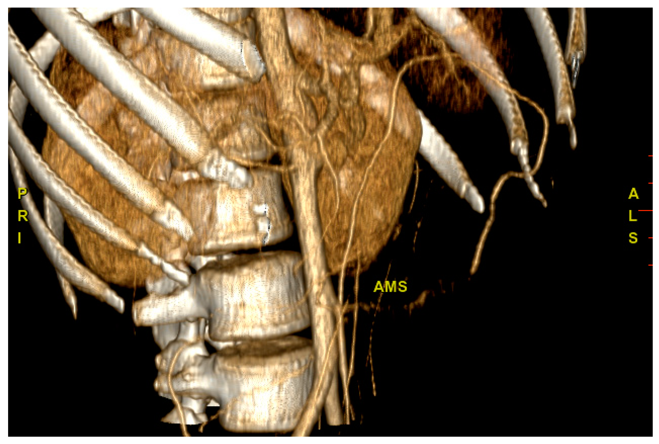
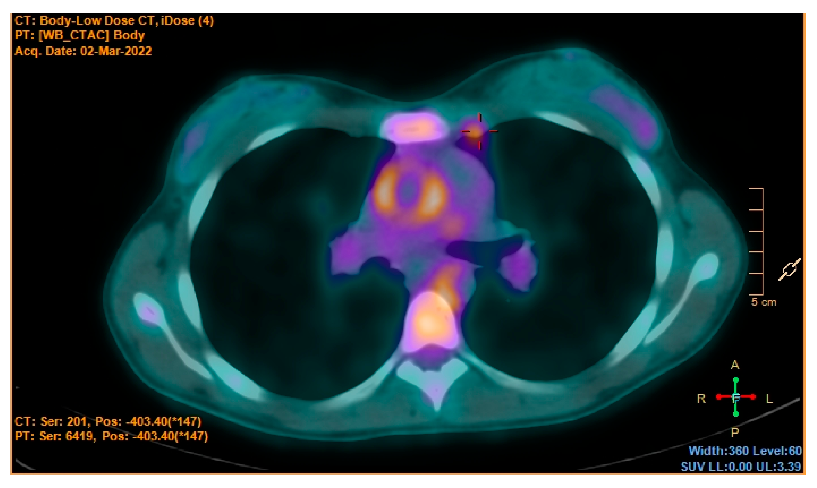
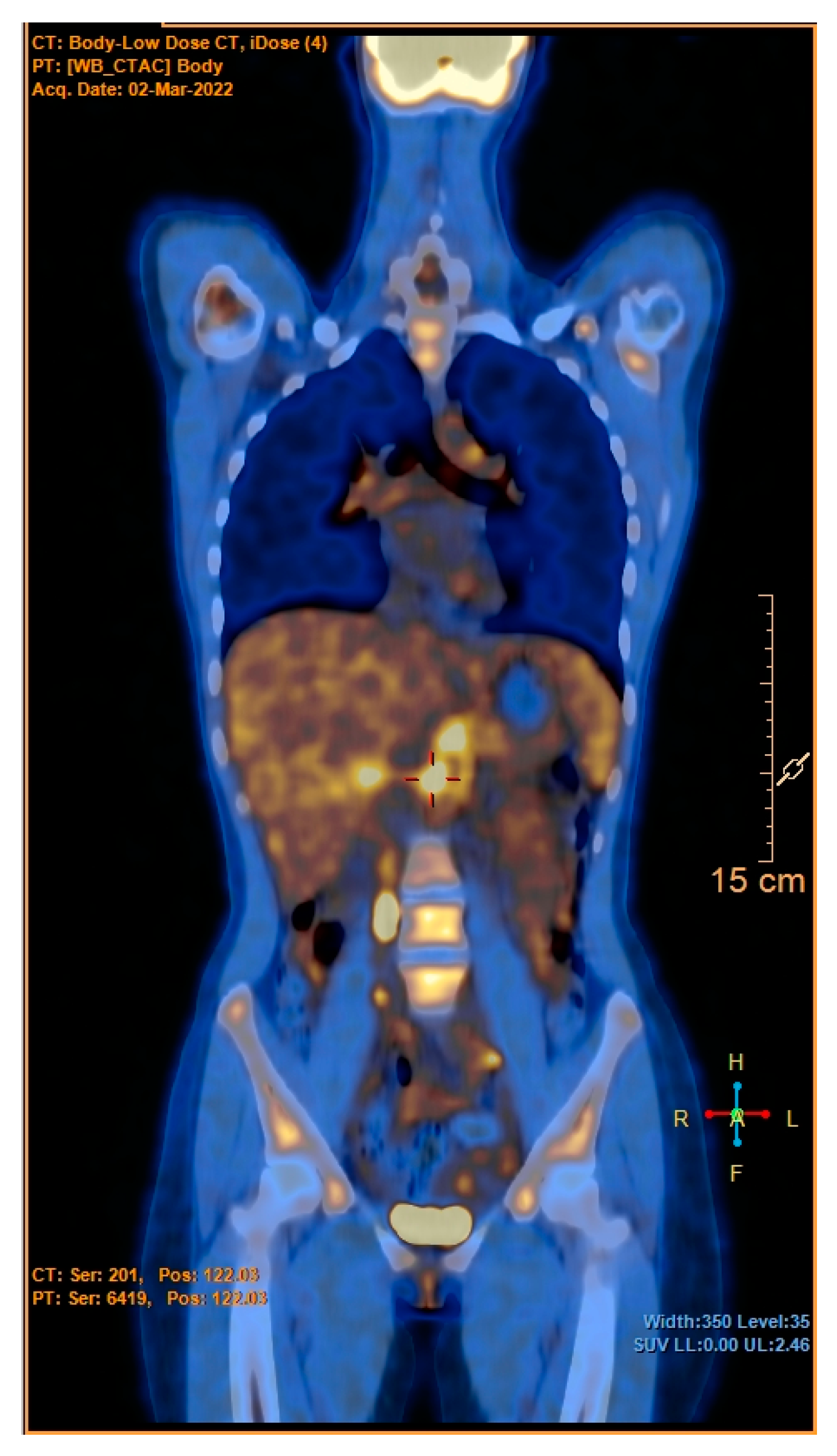
2. Case Description
- Travel abroad, contact with animals, and in-depth laboratory tests excluded any specific infectious etiology such as endocarditis, zoonoses (e.g., brucellosis, Lyme disease, and rickettsioses), typhoidal and nontyphoidal salmonella, chlamydia pneumoniae, mycoplasma pneumoniae, cytomegalovirus (CMV), Epstein–Barr virus (EBV), human immunodeficiency virus (HIV), syphilis, and mycobacterial infection;
- Lymphoproliferative disorders were excluded (no lymphadenopathy, and the peripheral blood smear, white blood cell count, and lactate dehydrogenase levels were normal);
- At the liver and spleen ultrasound examinations, no pathological findings were found;
- Rheumatological diseases were excluded (skin rash, mucous involvement, and redness and swelling of the joints were not observed in our patient; ANA, ENA, p-ANCA, c-ANCA, and antiphospholipid antibody testing were negative).
3. Discussion
4. Conclusions
Author Contributions
Funding
Institutional Review Board Statement
Informed Consent Statement
Data Availability Statement
Acknowledgments
Conflicts of Interest
References
- WHO Coronavirus (COVID-19) Dashboard. Available online: https://covid19.who.int (accessed on 22 January 2023).
- Henderson, L.A.; Canna, S.W.; Friedman, K.G.; Gorelik, M.; Lapidus, S.K.; Bassiri, H.; Behrens, E.M.; Ferris, A.; Kernan, K.F.; Schulert, G.S.; et al. American College of Rheumatology Clinical Guidance for Multisystem Inflammatory Syndrome in Children Associated with SARS-CoV-2 and Hyperinflammation in Pediatric COVID-19: Version 3. Arthritis Rheumatol. 2022, 74, e1–e20. [Google Scholar] [CrossRef] [PubMed]
- Kunal, S.; Ish, P.; Sakthivel, P.; Malhotra, N.; Gupta, K. The emerging threat of multisystem inflammatory syndrome in adults (MIS-A) in COVID-19: A systematic review. Heart Lung 2022, 54, 7–18. [Google Scholar] [CrossRef] [PubMed]
- Patel, J.M. Multisystem Inflammatory Syndrome in Children (MIS-C). Curr. Allergy Asthma Rep. 2022, 22, 53–60. [Google Scholar] [CrossRef] [PubMed]
- HAN Archive—00432. Health Alert Network (HAN). 2021. Available online: https://emergency.cdc.gov/han/2020/han00432.asp (accessed on 15 September 2022).
- Multisystem Inflammatory Syndrome in Children and Adolescents with COVID-19. Available online: https://www.who.int/publications-detail-redirect/multisystem-inflammatory-syndrome-in-children-and-adolescents-with-covid-19 (accessed on 15 September 2022).
- Haidar, G.; Singh, N. Fever of Unknown Origin. N. Engl. J. Med. 2022, 386, 463–477. [Google Scholar] [CrossRef] [PubMed]
- Ozen, S.; Pistorio, A.; Iusan, S.M.; Bakkaloglu, A.; Herlin, T.; Brik, R.; Buoncompagni, A.; Lazar, C.; Bilge, I.; Uziel, Y.; et al. EULAR/PRINTO/PRES criteria for Henoch-Schönlein purpura, childhood polyarteritis nodosa, childhood Wegener granulomatosis and childhood Takayasu arteritis: Ankara 2008. Part II: Final classification criteria. Ann. Rheum. Dis. 2010, 69, 798–806. [Google Scholar] [CrossRef] [PubMed] [Green Version]
- De Souza, A.W.S.; de Carvalho, J.F. Diagnostic and classification criteria of Takayasu arteritis. J Autoimmun. 2014, 48–49, 79–83. [Google Scholar] [CrossRef] [PubMed]
- Mason, J.C. Takayasu arteritis—Advances in diagnosis and management. Nat. Rev. Rheumatol. 2010, 6, 406–415. [Google Scholar] [CrossRef] [PubMed]
- Rácz, Á.O.; Szabó, G.T.; Erdei, N.; Győry, F.; Kolozsvári, R.V. Heart failure caused by Takayasu’s arteritis in the time of COVID-19: A case report. ESC Heart Fail. 2022, 9, 3602–3607. [Google Scholar] [CrossRef] [PubMed]
- Santi, L.; Golinelli, D.; Tampieri, A.; Farina, G.; Greco, M.; Rosa, S.; Beleffi, M.; Biavati, B.; Campinoti, F.; Guerrini, S.; et al. Non-COVID-19 patients in times of pandemic: Emergency department visits, hospitalizations and cause-specific mortality in Northern Italy. PLoS ONE 2021, 16, e0248995. [Google Scholar] [CrossRef] [PubMed]
- Smatti, M.K.; Cyprian, F.S.; Nasrallah, G.K.; Al Thani, A.A.; Almishal, R.O.; Yassine, H.M. Viruses and Autoimmunity: A Review on the Potential Interaction and Molecular Mechanisms. Viruses 2019, 11, 762. [Google Scholar] [CrossRef] [PubMed] [Green Version]
- Nikolaishvili, M.; Pazhava, A.; Di Lernia, V. Viral Infections May Be Associated with Henoch-Schönlein Purpura. J. Clin. Med. 2023, 12, 697. [Google Scholar] [CrossRef] [PubMed]
- Boleto, G.; Vieira, M.; Saadoun, D.; Cacoub, P. Hepatitis C virus-related vasculitis. Clin. Res. Hepatol. Gastroenterol. 2021, 45, 101575. [Google Scholar] [CrossRef] [PubMed]
- Sharlala, H.; Adebajo, A. Virus-induced vasculitis. Curr. Rheumatol. Rep. 2008, 10, 449. [Google Scholar] [CrossRef] [PubMed]
- Halpert, G.; Shoenfeld, Y. SARS-CoV-2, the autoimmune virus. Autoimmun. Rev. 2020, 19, 102695. [Google Scholar] [CrossRef] [PubMed]
- Mendes, J.L.; Venade, G.; Manuel, P.; Costa Matos, L.; Nascimento, E. Virus and Autoimmunity: Can SARS-CoV-2 Trigger Large Vessel Vasculitis? Eur. J. Case Rep. Intern. Med. 2022, 9, 003486. [Google Scholar] [PubMed]
- Guliyeva, V. 69 Large vessel vasculitis occurring after COVID-19 infection: 2 Takayasu cases. Rheumatology 2022, 61 (Suppl. S2), keac496.065. [Google Scholar] [CrossRef]
- Salman, R.; Masand, P.; Huisman, T.A.G.M.; Pereira, M.; Kearney, D.L.; Guillerman, R.P.; Jadhav, S. A Large-Vessel Arteritis in SARS-CoV-2–related Multisystem Inflammatory Syndrome in Children. Radiol. Cardiothorac. Imaging 2021, 3, e200535. [Google Scholar] [CrossRef] [PubMed]


| Clinical and Laboratory Findings | Value | Normal Range |
|---|---|---|
| Pulse | 110 beats/min | 60–100 |
| Blood pressure | 128/80 mmHg | 110–131/64–83 |
| Temperature | 38.5 Celsius | 36.3–37.6 |
| Respiratory rate | 18 breaths/minute | 12–20 |
| BMI | 18.6 Kg/m2 | 18.5–24.9 |
| Hemoglobin | 9.5 g/dL | 12.0–15.5 |
| Mean corpuscular volume | 73.7 fL | 80.4–95.9 |
| White blood cells | 7.60 × 10³/µL | 4.10–11–20 |
| Platelets | 551 × 103/µL | 159–388 |
| Creatinine | 0.51 mg/dL | 0.60–1.30 |
| Aspartate aminotransferase | 19 UI/L | 5–34 |
| Alanine aminotransferase | 6 UI/L | 0–55 |
| Creatine phosphokinase | 29 UI/L | 29.0–200.0 |
| Iron | 15 µg/dL | 50–170 |
| Ferritin | 338 ng/mL | 5–204 |
| C-reactive protein | 12.59 mg/dL | 0.01–0–50 |
| Procalcitonin | 0.10 µg/L | <0.05 |
| ESR | 113 mm | 0–15 |
| Fibrinogen | 671 mg/dL | 200–450 |
| Prothrombin activity | 45% | 80–120 |
| Partial thromboplastin time | 39 SEC | 25–35 |
| INR | 1.70 | 0.90–1.10 |
| TSH | 1.70 microµ/mL | 0.35–4.94 |
| Thyroxine | 1.02 ng/dL | 0.70–1.48 |
Disclaimer/Publisher’s Note: The statements, opinions and data contained in all publications are solely those of the individual author(s) and contributor(s) and not of MDPI and/or the editor(s). MDPI and/or the editor(s) disclaim responsibility for any injury to people or property resulting from any ideas, methods, instructions or products referred to in the content. |
© 2023 by the authors. Licensee MDPI, Basel, Switzerland. This article is an open access article distributed under the terms and conditions of the Creative Commons Attribution (CC BY) license (https://creativecommons.org/licenses/by/4.0/).
Share and Cite
Spampinato, S.; Di Marco, M.; Mammolito, L.; Scarfia, A.; Valastro, M.; Di Mauro, S.; Bosco, G.; Purrello, F.; Piro, S. Is COVID-19 All That Glitters? J. Clin. Med. 2023, 12, 2552. https://doi.org/10.3390/jcm12072552
Spampinato S, Di Marco M, Mammolito L, Scarfia A, Valastro M, Di Mauro S, Bosco G, Purrello F, Piro S. Is COVID-19 All That Glitters? Journal of Clinical Medicine. 2023; 12(7):2552. https://doi.org/10.3390/jcm12072552
Chicago/Turabian StyleSpampinato, Salvatore, Maurizio Di Marco, Luciano Mammolito, Alessia Scarfia, Maurizio Valastro, Stefania Di Mauro, Giosiana Bosco, Francesco Purrello, and Salvatore Piro. 2023. "Is COVID-19 All That Glitters?" Journal of Clinical Medicine 12, no. 7: 2552. https://doi.org/10.3390/jcm12072552