Article

Evaluation of Event-Related Potentials in Assessing Cognitive Functions of Adult Patients with Epilepsy of Unknown Etiology

by
Klaudia Jeżowska-Jurczyk
1,*,
Piotr Jurczyk
2,
Sławomir Budrewicz
1 and
Anna Pokryszko-Dragan
1
1
Department of Neurology, Wroclaw Medical University, 50-556 Wroclaw, Poland
2
Private Medical Practice, 52-207 Wroclaw, Poland
*
Author to whom correspondence should be addressed.
J. Clin. Med. 2023, 12(7), 2500; https://doi.org/10.3390/jcm12072500
Submission received: 10 February 2023 / Revised: 21 March 2023 / Accepted: 23 March 2023 / Published: 25 March 2023
(This article belongs to the Special Issue Epilepsy: Causes, Symptoms, Diagnosis, and Treatment)

Abstract

:
Background: Cognitive impairment (CI) is an important consequence of epilepsy. The aim of the study was to assess cognitive performance in patients with epilepsy, using neuropsychological tests (NT) and event-related potentials (ERPs), with regard to demographic and clinical data. Methods: The study comprised 50 patients with epilepsy of unknown etiology and 46 healthy controls. Based on the NT results, the patients were divided into subgroups with/without CI. Parameters of P300 potential were compared between the patients and controls. P300 parameters and NT results were referred to demographics and clinical characteristics of epilepsy. Results: Based on the NT, 66% of patients were assigned as cognitively impaired. Median P300 latency was significantly (p < 0.0002) prolonged in the study group. Subgroups of patients with and without CI significantly (p < 0.034) differed in education level and vocational activity, duration of epilepsy, age at its onset and frequency of polytherapy. P300 parameters showed significant (p < 0.03) relationships with duration of epilepsy, type and frequency of seizures and polytherapy. Conclusions: Cognitive impairment and ERPs abnormalities occur in a majority of patients with epilepsy of unknown etiology. Characteristics of epilepsy and socioeconomic status are related to cognitive performance. ERPs may complement neuropsychological methods in the assessment of cognition in patients with epilepsy.

1. Introduction

Epilepsy—a chronic condition characterized by a permanent predisposition towards developing seizures—constitutes a substantial clinical and social problem, due to its prevalence and specificity. Apart from the disease itself, somatic and mental consequences of epilepsy may significantly affect the patients’ quality of life [1]. Among these consequences, impaired cognitive performance deserves particular attention.
Cognitive impairment (CI) occurs in more than a half of patients with epilepsy. It is usually mild, although epilepsy is associated with double-increased risk of dementia [2]. The decline may be global or affect particular cognitive domains, most frequently affecting memory, verbal skills, attention, executive functions and visuospatial skills. CI is occasionally observed in the patients with newly diagnosed epilepsy; it may remain stable or gradually progress in the further course of the disease [3,4,5,6,7,8,9,10].
With recent progress in clinical research in epilepsy, a novel complex approach to its neurobehavioral phenotypes, including cognitive performance, has been developed [11]. This approach integrates biological factors (related to epilepsy, comorbidities and general health issues) with psychosocial and environmental ones. Depending on the background and type of disease, relevant epilepsy-related factors, potentially contributing to cognitive impairment, may include brain injury due to a range of disorders (e.g., vascular, degenerative, inflammatory), genetic mutations and variants, systemic metabolic alterations, as well as recurrent disturbances in cerebral bioelectrical activity and adverse effects of anti-epileptic treatment [3,9,11,12,13,14,15,16,17,18,19]. In the patients with epilepsy of unknown etiology, the majority of these factors have been eliminated, so the investigation of the background for CI may be focused on the functional impairment of neuronal networks. Thus, epilepsy of unknown etiology seems to provide a good model for the consistent and reliable analysis of epilepsy-related factors contributing to CI.
Better insight into these relationships and precise evaluation of cognitive performance might allow for identification of the patients with greater risk of CI and provide them with adequate support. According to the mentioned multidimensional neurobehavioral paradigm, resilience factors and cognitive reserve of these patients should be recognized, as well as socioeconomic status and resources. Based on these factors, complex therapeutic approaches might be elaborated, including optimal pharmacological treatment, modification of lifestyle factors and cognitive rehabilitation [11].
In the evaluation of cognitive performance, event-related potentials (ERPs) can be used alongside neuropsychological tests as its electrophysiological measure. ERPs represent an averaged response of cerebral bioelectrical activity to the series of stimuli, which are associated with a task requiring cognitive and emotional engagement of the subject. The most frequently assessed ERP component, P300 potential, reflects conscious information processing and decision making [20,21]. ERPs have been widely used in the evaluation of cognitive impairment in the course of various CNS disorders [22,23,24,25,26,27,28].
The aim of this study was to assess cognitive performance in the patients with epilepsy of unknown etiology, with the use of neuropsychological tests and event-related potentials (ERPs). We also aimed to analyze relationships between measures of cognitive performance and demographics and clinical characteristics of the disease.

2. Materials and Methods

Participants to the study were recruited from the patients diagnosed with epilepsy of unknown etiology, according to the International League Against Epilepsy criteria [29], with a documented course of the disease, who were hospitalized or consulted at the Department of Neurology, Wroclaw Medical University, in the years 2016–2019. Exclusion criteria included neuroimaging evidence of cerebral lesions corresponding with epileptic seizures or located in the areas potentially involved in cognitive functions, chronic and decompensated comorbidities which might affect cognitive performance, and mental disorders and/or severe cognitive impairment which would prevent providing an informed consent to participate in the study and performing neuropsychological tests involved in the study protocol.
Finally, the study group comprised 50 individuals: 44 women and 6 men, aged 20–68 years (mean—35.6). The control group consisted of 46 healthy volunteers, matched for age (23–57 years, mean—35.9) and sex (39 women and 7 men) to the study group.
The demographic data (age, sex, educational level, vocational activity) and information concerning epilepsy and coexisting diseases were initially obtained from a self-assessment questionnaire. The clinical data concerning epilepsy (age at onset, duration of the disease, type and frequency of seizures, current treatment) were then verified and completed on the basis of medical records, including the results of neuroimaging (magnetic resonance imaging—MRI or computed tomography—CT) performed within a previous year.
For the assessment of cognitive performance, the following tests were used: Rey Auditory Verbal Learning Test (AVLT), Rey–Osterrieth Complex Figure (ROCF), Trail Making Test (TMT), Digit Span Test from Wechsler Memory Scale (WMS-R), Similarities Test from Wechsler Adult Intelligence Scale (WAIS-R), Verbal Fluency Test (VFT)—for which utility and reliability in the assessment of patients with epilepsy had been proved in experiences of epileptological centers [30,31] (for details of the tests content, scoring and interpretation of results, see Appendix A). Based on the results of these tests, the study group was divided into two subgroups: with or without cognitive impairment (≥2 different tests failed vs. abnormal result of ≤1 test).
The examination of auditory event-related potentials was performed in the study group and with the healthy controls using a Nicolet 1000 Viking device, according to the guidelines from International Federation of Clinical Neurophysiology [32]. Surface electrodes were placed on the scalp at midline points Fz, Cz and Pz, according to a 10–20 system, with the reference electrode attached to linked earlobes and the ground one—to the forearm. Auditory stimuli (duration 200 ms, intensity 70 dB) were emitted binaurally through headphones using the ‘oddball paradigm’: 20% of target tones (frequency 2000 Hz) were randomly scattered among the remaining non-target ones (80%, frequency 1000 Hz). The subject’s task was to focus on target tones and count them silently in each series. The recording was performed with a bandpass filter of 0.3–70 Hz and sweep time of 1000 ms. With the use of dedicated software, the responses to target and non-target stimuli were averaged separately for each of the references, up to receiving at least 30 target responses. This procedure was performed twice for each subject. A P300 potential was identified in the target response curve for each reference as the highest positive component in range 250–500 ms. P300 latency (time since the occurrence of target stimulus to the peak of an identified potential) and amplitude (peak-to peak) were measured after each of the two recording series, and their final values were averaged from these.
In addition, patients with epilepsy had electroencephalography (EEG) performed, using a 24-channel Nicolet One device. Surface electrodes were placed on the scalp in standardized sites according to the international 10–20 system. EEG was recorded with a bandpass filter of 0.5–100 Hz, with a sampling frequency of 256 Hz and an analogue of 50 Hz filter. The subjects lied down and stayed awake with their eyes closed. During a 20 min recording, they were asked to breathe deeply for 3 min and, subsequently, photostimulation (flashes of stroboscopic light emitted with fluctuating frequency 4–30 Hz) was applied for 3 min. After the automatic rejection of artifacts, the EEG recording was visually inspected for any abnormal graphoelements, especially including paroxysmal epileptiform activity.
All the tests were performed during one session, in the morning hours. The date of session was established after at least 2 months since the introduction or recent modification of antiepileptic treatment, and at least 48 h after a recent seizure.
The study was conducted in accordance with the Helsinki Declaration. The design of the study and all the involved procedures were accepted by the Bioethics Committee at Wroclaw Medical University. All the participants had given their written, informed consent before they were included in the study.
The subgroups of patients with and without cognitive impairment were compared with regard to the demographics and characteristics of epilepsy. Results of ERPs were compared between the study group and controls. In the study group, ERP parameters were referred to demographic, clinical and neuropsychological data.
Mean averages (x), medians (M), ranges (min-max), lower and upper quartiles (25–75Q) and standard deviations (SD) of the recorded continuous parameters were calculated. Verification of the hypothesis of equal means parameters in independent groups was performed using the ANOVA variance analysis or with the Mann–Whitney nonparametric U test (for 2 groups) in two groups of heterogeneous variance or the Kruskal-Wallis test by ranks (for 3 and more groups)—homogeneity of variance was verified by Bartlett’s test. For discrete parameters, the incidence of characteristics in groups was analyzed with the χ2df test with an appropriate number of degrees of freedom df (df = (m − 1) × (n − 1), where m—number of lines, n—number of columns). For chosen pairs of parameters, a correlation analysis using Spearman’s rank correlation coefficient was conducted. A multivariate analysis was performed, using logistic regression (quasi-Newton method) or backward multiple regression. p ≤ 0.05 was considered significant. Statistical analysis was performed using a digital packet of statistical software EPIINFO Ver. 7.2.3.1, developed by Centers for Disease Control and Prevention (CDC) in Atlanta, Georgia (US).

3. Results

3.1. Demographic Data

The study group and controls did not differ significantly in age (p = 0.632) or sex (χ2 = 0.212, p = 0.645). Among the patients with epilepsy, 13 persons (26%) had completed primary or vocational school, 18 (36%)—secondary school and 19 (38%)—university degree. A total of 31 patients (62%) were full-time or part-time employed.

3.2. Clinical Data

3.2.1. Course of Epilepsy

Disease duration in the study group ranged between 0.4 and 41 years (mean: 15 years) and age at onset—from 2 to 52 years (mean 21). Types and frequency of epileptic seizures are listed in Table 1. Family history of epilepsy was positive in six patients (12%). All the subjects were receiving antiepileptic treatment: 18 (36%)—monotherapy, 32 (64%)—polytherapy (most frequently with levetiracetam (n = 21) and lamotrigine (n = 18)) (Table 1).

3.2.2. Neuroimaging

Within the previous year, an MRI was performed in 46 patients (92%), and a CT in 4 patients (due to contraindications or lack of the subjects’ consent to perform MRI). All CT scans and 38 MRI scans showed no structural brain abnormalities. In eight cases, a few small hyperintensive lesions were revealed in cerebral deep white matter, which did not correspond with type of seizures, and were outside of areas strategic for cognitive functions, and were considered as clinically non-significant.

3.2.3. EEG

EEG recording was normal in 26 patients (52%), while in 24 patients interictal paroxysmal epileptiform activity was found (in 8 cases accompanied by the slowing of background activity) (Table 1). Out of these, paroxysmal epileptiform activity was recorded in 14 patients only during activation procedures (hyperventilation and/or photostimulation). None of the subjects experienced an epileptic seizure during or after EEG recording.

3.2.4. Coexisting Disease

Concomitant somatic disorders were documented in 13 (26%) patients—arterial hypertension (n = 5), dyslipidemia (n = 5), diabetes, polycystic ovary syndrome and psoriasis (in a single person each).
Twelve (24%) patients had been diagnosed with mental disorders: in five cases (10%)—depression, in two (4%)—anxiety disorder and in five (10%)—dissociative disorder. Those diagnosed with depression were undergoing pharmacological treatment and others—psychotherapy.

3.3. Neuropsychological Assessment

The results of all neuropsychological tests conducted in the study group are listed in Table 2. The highest percentage of abnormal results was received for AVLT (verbal memory, learning ability) and TMT (attention, executive and visuospatial functions, psychomotor speed) and the lowest—for WAIS-R Similarities Test (abstract and associative thinking) (Table 2). The number of abnormal results scored by each individual in the study group ranged from 0 to 10 (mean 3.1 ± 2.5).
On the basis of these results, the study group was divided into subgroups without (abnormal result of ≤1 test) or with (≥2 different tests failed) CI.

Neuropsychological Assessment vs. Demographic and Clinical Data

The subgroups without CI and with CI did not differ significantly for age and sex. The subgroups without CI presented with a higher frequency of professional activity, and even more significantly, a higher level of education (Table 3).
As for clinical data, the subgroup without CI showed (with high significance level) a later age of onset and shorter duration of epilepsy, and a lower proportion of patients on polytherapy (Table 3). No differences were found between the subgroups for type and frequency of epileptic seizures, or the presence of abnormalities in EEG recording.
After a multivariate analysis using logistic regression, an independent significant relationship was found between cognitive impairment and duration of epilepsy (lasting more than 7 years) (χ2 = 14,6; p = 0.00013).

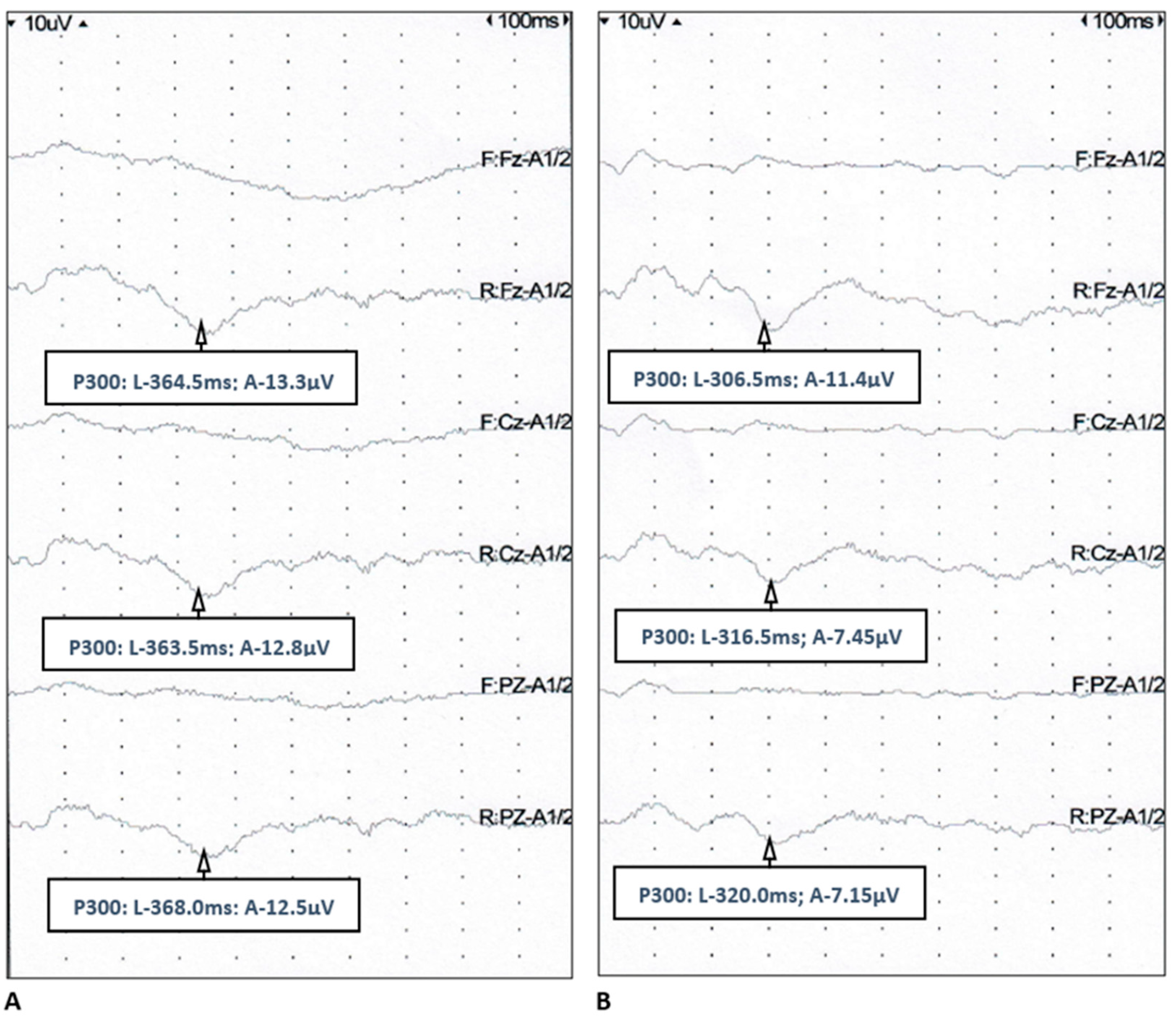
3.4. Event-Related Potentials

The P300 component was identified in ERPs recorded from 44 patients with epilepsy (88%) and all healthy controls. Median latency of P300 in all the references was significantly longer (with high significance) in the study group than in the controls, whereas median amplitude of P300 did not differ between the groups (Table 4, Figure 1).

3.4.1. Event-Related Potentials vs. Demographic and Clinical Data

In the study group, a significant positive correlation was observed between age and P300 latency but only for the Pz reference (R = 0.32; p = 0.0363). Because of a small proportion of male subjects in the study group, no relationships were analyzed between P300 parameters and sex.
With regard to clinical characteristics of epilepsy, a moderately significant positive correlation was found among P300 latency in all the references (especially in Cz and Pz) and duration of disease (Table 5). Median P300 latency was longer (moderate significance) in the patients with focal seizures (in comparison with generalized ones) and—even more significantly—in those on polytherapy (Table 6 and Table 7). Median P300 amplitude in Fz was the highest in the patients with monthly occurrence of seizures and the lowest—in those with weekly occurrence (M = 7.2 vs. M = 3.65, p = 0.0177). Median P300 amplitude in Cz was lower in the patients with paroxysmal activity in EEG than in those with normal EEG (M = 5.78 vs. M = 9.33, p = 0.00101).

3.4.2. Event-Related Potentials vs. Neuropsychological Assessment

In the overall comparative analysis, no significant differences in P300 parameters were found between the subgroups of patients with and without cognitive impairment (Table 8). In the search for relationships between the P300 parameters and particular test results, we demonstrated the most consistent significant negative correlations between P300 latency and the results of AVLT (total and after delay), WAIS-R Similarities Subscale and (with lower significance level) semantic VFT. Negative correlations were also found between P300 amplitude and the results of TMT A and B, but only for Cz reference (Table 9).

4. Discussion

Neuropsychological assessment revealed cognitive impairment in more than 60% of the study group of patients with epilepsy. The most commonly affected domains included verbal memory and learning abilities, attention, psychomotor speed, visuospatial and executive functions, while performance in nonverbal memory and abstract thinking often remained preserved. Other reports in this field, though diverse in methodology, show similar incidence and profile of cognitive impairment in the patients with various types of epilepsy [3,4,9,10,14,33,34].
The P300 component of ERPs is generated in cortico–subcortical structures (including hippocampus, thalamus and (pre)frontal areas), which are involved in cognitive processes [35,36,37]. P300 latency is interpreted as the index of time necessary for information processing and problem solving, whereas the amplitude reflects the subjects’ engagement and focusing on the task. In our study, P300 latency was significantly prolonged in the patients with epilepsy in comparison with the healthy controls, while no differences were found for P300 amplitude. Increases in P300 latency and—less frequently—lowering of P300 amplitude were reported in several studies on epilepsy [38,39,40,41,42,43,44,45,46,47,48,49,50,51,52,53,54,55,56,57,58].
Although P300 parameters are usually considered as indices of global cognitive performance, the main processes involved in generating P300 comprise attention, executive functions, psychomotor speed and (to a lesser extent) memory [59,60]. In the current study significant relationships were found mostly between P300 latency and results of the tests evaluating verbal memory, abstractive/associative thinking and executive functions with semantic verbal fluency. These correlations seem particularly interesting, considering the type of task used to evoke auditory P300 (‘oddball paradigm’ with tones of different frequency). Furthermore, P300 parameters did not differ between the subgroups of patients with or without CI. It seems that P300 does not correspond with performance in specific cognitive domains, but rather reflects more complex interactions within cognitive skills. Thus, electrophysiological and neuropsychological measures of cognitive performance should not be considered as convertible but rather as complementary ones [61].
Except for correlation of P300 latency with age, no relationships were found in the study group between age and cognitive performance. Indeed, P300 latency increases with age [62]. However, this correlation was limited only to one reference, and the patients did not differ in age from the control group, so the prolongation of P300 latency in the study group remains relevant. Subgroups with and without CI did not differ in sex structure. However, a marked sex imbalance with underrepresentation of men (6/44) in the study group prevented the reliable analysis of relationships between sex and cognitive performance.
Our patients with cognitive impairment had a lower level of education and were less frequently employed. These findings, consistent with the results from the other studies [15,63,64,65], highlight the importance of cognitive performance in social functioning of the patients with epilepsy. Low level of education and limited cognitive reserve may predispose individuals to the development of cognitive impairment in the course of the disease, but also early cognitive deficit, emerging from epilepsy onset at a young age, may impede continuing education [66]. The same perspective, affected by the role of socioeconomic and cultural background, can be referred to the vocational activity of patients [67]. In our study, a percentage of the subjects who were not employed was relatively high, in comparison to the data from other European countries [16,68]. Educational and vocational issues, socioeconomic status and availability of social support are substantially linked with neurobehavioral aspects of epilepsy, as well as the patients’ quality of life [11].
Among epilepsy-related clinical data, the duration of the disease showed most significant relationships both with P300 parameters and findings from neuropsychological assessment, with the latter also associated with age at onset of epilepsy. An impact of these factors upon cognitive performance in epilepsy has been already reported [4,14,34,65]. Long-term dysfunction of neuronal network plasticity, caused by recurring seizure activity, is supposed to be the major contributor to cognitive impairment. According to a cascade model, onset of these disturbances at the early stage of intellectual development plays a crucial role in further cognitive decline [12,15,69]. Some links were noted in the study group between P300 amplitude and frequency of seizures, but only for the single reference, which limits their significance. We also found increased P300 latency in the patients with focal seizures in comparison with those with generalized ones. Focal seizures are associated with the local disintegration of neuronal network activity which may be relevant for the structures that are strategic for cognitive processes. Considering the moderate sample size, we did not distinguish subjects with particular localization of focal seizures (e.g., frontal or temporal ones) for further analysis. Moreover, P300 parameters are considered as indices of global bioelectrical event-related activity [35,36,37]. It should be highlighted that a diversity of epilepsy-related data in the study group might have affected the findings. Other studies investigating P300 parameters with regard to clinical characteristics of epilepsy showed inconsistent results [38,39,40,43,44,46,47,48,51,52,54,55,58,70].
Polytherapy was another factor significantly related to neuropsychological test results and P300 parameters in the study group. Basically, treatment reduces seizure frequency and stabilizes the bioelectrical activity of the brain, potentially preventing cognitive decline; but on the other hand—adverse effects of antiepileptics (enhanced by their interactions) may include cognitive dysfunction. The majority of studies reveal mild or moderate adverse impact of polytherapy on cognition [14,15,34,64,71], and on alterations of evoked potentials’ parameters (ERP, EP—high frequency oscillations) [39,43,46,55,72]. In the analysis of the particular antiepileptic medication effects in this field, one should consider their mode of action (modulation of synapses excitability via ion channels), as well as extent of functionally impaired cortico–subcortical circuits which underlie specific types of seizures and are involved in the generation of evoked potentials’ components [73,74].
On the analysis of relationships between cognitive performance and epilepsy characteristics, the interrelations among the latter and their accumulated effect have to be considered. In the study group, the patients with focal epilepsy had longer disease duration and higher frequency of seizures, and more often were being treated with polytherapy.
Overall, identification of these patients burdened with the factors predisposing to CI should encourage thorough assessment of their cognitive performance (e.g., with the use of neuropsychological and electrophysiological measures), followed by appropriate interventions (cognitive rehabilitation, optimization of treatment, psychological support).

Strengths and Limitations of the Study

Our study attempted a comprehensive analysis of cognitive performance in the patients with epilepsy, with regard to demographic and clinical data. Integration of neuropsychological and electrophysiological findings provided more detailed insight into the prevalence and profile of cognitive impairment in the study group. The focus on patients with epilepsy of unknown etiology, due to the limited range of factors able to affect cognitive functions, allowed a more consistent and reliable analysis of relationships between them.
The limitations of the study included its cross-sectional mode, relatively small sample size and diversity of epilepsy characteristics, despite the aforementioned exclusion criteria. Considering the complex relationships between CI and education in epileptic patients, the differences in level of education between the study subgroups might be an additional source of bias. Nevertheless, the obtained results highlight a need for the precise evaluation of cognitive performance and considering this aspect in the complex approach to the management of epilepsy. There is a need for future studies on this topic require a comprehensive model to account for the multidimensional nature of the problem, as it is presently difficult to characterize how different factors may affect one another. Further investigation in this field seems necessary, comprising patients with different types of epilepsy, and monitoring cognitive deficit throughout the course of disease, to determine therapeutic implications of neuropsychological and electrophysiological findings.

5. Conclusions

Cognitive impairment and abnormalities of ERPs occur in a vast majority of patients with epilepsy of unknown etiology. Characteristics of epilepsy and socioeconomic status are potential factors related to cognitive performance. Event-related potentials may complement neuropsychological methods in the assessment of cognition, which constitute important elements in a multidimensional approach to epilepsy management.

Author Contributions

Conceptualization, K.J.-J. and A.P.-D.; methodology, K.J.-J. and A.P.-D.; validation, S.B.; investigation, K.J.-J.; resources, K.J.-J. and A.P.-D.; data curation, K.J.-J.; writing—original draft preparation, K.J.-J. and P.J.; writing—review and editing, A.P.-D.; visualization, K.J.-J.; supervision, S.B.; project administration, K.J.-J. All authors have read and agreed to the published version of the manuscript.

Funding

This research received no external funding.

Institutional Review Board Statement

The study was conducted in accordance with Helsinki Declaration. The design of the study and all the involved procedures were accepted by the Bioethics Committee at Wroclaw Medical University (nr 187/2016).

Informed Consent Statement

Informed consent was obtained from all subjects involved in the study.

Data Availability Statement

Publicly available datasets were analyzed in this study. This data can be found here: https://ppm.umed.wroc.pl/info/achievement/UMWb9153c4b510e4a0e95b9aeee01b61cf1/ (accessed on 17 June 2021).

Conflicts of Interest

The authors declare no conflict of interest.

Appendix A. Neuropsychological Test Used for Assessment of Cognitive Performance

• Rey Auditory Verbal Learning Test—AVLT
The test consists of five presentations of a list of 15 words (list A). After each reading, the participant is asked to recall as many words as possible. Next, the investigator presents another list of 15 words (list B). The participant is asked to recall the words from list B, then again from list A. After 20 min, the participant recalls words from list A once again. The results include the number of words from list A remembered in the first part of the test, after a distraction (list B) and after a delay. The test evaluates verbal memory and learning ability [75].
• Rey–Osterrieth Complex Figure—ROCF
The participant’s task is to copy a figure from a provided pattern, and then recreate it after 3 min in an Immediate Recall trial. Accuracy in copying and correct placement of all 18 elements of the figure are graded in both trials. The test assesses nonverbal memory, attention, executive and visuospatial skills [76].
• Trail Making Test—TMT
Part A of the test includes drawing lines linking 25 circles labeled with subsequent numbers. In part B, the subject is asked to alternately link circles labeled with numbers and letters (1-A-2-B-3-C, etc.). The result is the time needed to complete each part. The test assesses attention, executive and visuospatial skills and psychomotor speed [77].
• Digit Span Test from Wechsler Memory Scale—WMS-R
In the first part of the test, the participant repeats a series of numbers in an order given by the investigator, whereas in the second part—repeats them backwards. The result is a sum of correct answers. The test evaluates verbal memory and attention [78].
• Similarities Test from Wechsler Adult Intelligence Scale—WAIS-R
The participant is asked to find similarities/common features of sequentially presented pairs of words and can receive 0–2 points, depending on the correctness and precision of the answer. The result is a sum of received points. Abstract and associative thinking are assessed [78].
• Verbal Fluency Test—VFT
The task includes listing possibly many words beginning with the letter ‘k’ (phonetic fluency) and then names of animals (semantic fluency) within 60 s each. The result equals the number of words fulfilling the criteria of each task. The test assesses verbal fluency [79].
The results of the tests were then referred to standardized norms and considered abnormal if they exceeded 1 standard deviation (SD) (AVLT, ROCF, TMT, VFT) or 2 SDs (WAIS-R) with regard to age-adjusted normative values [78,80,81].

References

  1. Fisher, R.S.; Boas, W.; Blume, W.; Elger, C.; Genton, P.; Lee, P.; Engel, J. Epileptic Seizures and Epilepsy: Definitions Proposed by the International League Against Epilepsy (ILAE) and the International Bureau for Epilepsy (IBE). Epilepsia 2005, 46, 470–472. [Google Scholar] [CrossRef]
  2. Subota, A.; Pham, T.; Jetté, N.; Sauro, K.; Lorenzetti, D.; Holroyd-Leduc, J. The Association between Dementia and Epilepsy: A Systematic Review and Meta-Analysis. Epilepsia 2017, 58, 962–972. [Google Scholar] [CrossRef] [Green Version]
  3. Helmstaedter, C.; Kurthen, M.; Lux, S.; Reuber, M.; Elger, C.E. Chronic Epilepsy and Cognition: A Longitudinal Study in Temporal Lobe Epilepsy. Ann. Neurol. 2003, 54, 425–432. [Google Scholar] [CrossRef]
  4. Hermann, B.P.; Seidenberg, M.; Dow, C.; Jones, J.; Rutecki, P.; Bhattacharya, A.; Bell, B. Cognitive Prognosis in Chronic Temporal Lobe Epilepsy. Ann. Neurol. 2006, 60, 80–87. [Google Scholar] [CrossRef] [PubMed]
  5. Taylor, J.; Baker, G.A. Newly Diagnosed Epilepsy: Cognitive Outcome at 5 Years. Epilepsy Behav. 2010, 18, 397–403. [Google Scholar] [CrossRef] [PubMed]
  6. Baker, G.A.; Taylor, J.; Aldenkamp, A.P. Newly Diagnosed Epilepsy: Cognitive Outcome after 12 Months. Epilepsia 2011, 52, 1084–1091. [Google Scholar] [CrossRef]
  7. Bilo, L.; Santangelo, G.; Improta, I.; Vitale, C.; Meo, R.; Trojano, L. Neuropsychological Profile of Adult Patients with Nonsymptomatic Occipital Lobe Epilepsies. J. Neurol. 2013, 260, 445–453. [Google Scholar] [CrossRef]
  8. Kleen, J.K.; Scott, R.C.; Holmes, G.L.; Roberts, D.W.; Rundle, M.M.; Testorf, M.; Lenck-Santini, P.-P.; Jobst, B.C. Hippocampal Interictal Epileptiform Activity Disrupts Cognition in Humans. Neurology 2013, 81, 18. [Google Scholar] [CrossRef] [Green Version]
  9. Tang, V.; Kwan, P.; Poon, W.S. Neurocognitive and Psychological Profiles of Adult Patients with Epilepsy in Hong Kong. Epilepsy Behav. 2013, 29, 337–343. [Google Scholar] [CrossRef] [PubMed]
  10. Rai, V.K.; Shukla, G.; Afsar, M.; Poornima, S.; Pandey, R.M.; Rai, N.; Goyal, V.; Srivastava, A.; Vibha, D.; Behari, M. Memory, Executive Function and Language Function Are Similarly Impaired in Both Temporal and Extra Temporal Refractory Epilepsy—A Prospective Study. Epilepsy Res. 2015, 109, 72–80. [Google Scholar] [CrossRef]
  11. Hermann, B.P.; Struck, A.F.; Busch, R.M.; Reyes, A.; Kaestner, E.; McDonald, C.R. Neurobehavioural Comorbidities of Epilepsy: Towards a Network-Based Precision Taxonomy. Nat. Rev. Neurol. 2021, 17, 731–746. [Google Scholar] [CrossRef]
  12. Stefan, H.; Pauli, E. Progressive Cognitive Decline in Epilepsy: An Indication of Ongoing Plasticity. Prog. Brain Res. 2002, 135, 409–417. [Google Scholar]
  13. Lossius, M.I.; Hessen, E.; Mowinckel, P.; Stavem, K.; Erikssen, J.; Gulbrandsen, P.; Gjerstad, L. Consequences of Antiepileptic Drug Withdrawal: A Randomized, Double-Blind Study (Akershus Study). Epilepsia 2008, 49, 455–463. [Google Scholar] [CrossRef] [PubMed]
  14. Wang, W.H.; Liou, H.H.; Chen, C.C.; Chiu, M.J.; Chen, T.F.; Cheng, T.W.; Hua, M.S. Neuropsychological Performance and Seizure-Related Risk Factors in Patients with Temporal Lobe Epilepsy: A Retrospective Cross-Sectional Study. Epilepsy Behav. 2011, 22, 728–734. [Google Scholar] [CrossRef] [PubMed]
  15. Wang, L.; Chen, S.; Liu, C.; Lin, W.; Huang, H. Factors for Cognitive Impairment in Adult Epileptic Patients. Brain Behav. 2020, 10, e01475. [Google Scholar] [CrossRef] [PubMed] [Green Version]
  16. Hessen, E.; Lossius, M.I.; Gjerstad, L. Repeated Neuropsychological Assessment in Well-Controlled Epilepsy. Acta Neurol. Scand. 2013, 127, 53–60. [Google Scholar] [CrossRef] [PubMed]
  17. Xu, S.; Xi, J.; Lin, C.; Wang, X.; Fu, L.; Kralik, S.F.; Chen, Z. Cognitive Decline and White Matter Changes in Mesial Temporal Lobe Epilepsy. Medicine 2018, 97, e11803. [Google Scholar] [CrossRef]
  18. Babiloni, C.; Noce, G.; Di Bonaventura, C.; Lizio, R.; Pascarelli, M.T.; Tucci, F.; Soricelli, A.; Ferri, R.; Nobili, F.; Famà, F.; et al. Abnormalities of Cortical Sources of Resting State Delta Electroencephalographic Rhythms Are Related to Epileptiform Activity in Patients With Amnesic Mild Cognitive Impairment Not Due to Alzheimer’s Disease. Front. Neurol. 2020, 11, 514136. [Google Scholar] [CrossRef]
  19. Tedrus, G.M.; Negreiros, L.M.; Ballarim, R.S.; Marques, T.A.; Fonseca, L.C. Correlations Between Cognitive Aspects and Quantitative EEG in Adults With Epilepsy. Clin. EEG Neurosci. 2019, 50, 348–353. [Google Scholar] [CrossRef]
  20. Szelenberger, W. Potencjały Wywołane, 1st ed.; Elmiko: Warszawa, Poland, 2000; pp. 1–88. [Google Scholar]
  21. Słotwiński, K.; Zagrajek, M. Endogenne Potencjały Wywołane w Zaburzeniach Poznawczych. Pol. Przegląd Neurol. 2012, 8, 114–119. [Google Scholar]
  22. Xu, H.; Gu, L.; Zhang, S.; Wu, Y.; Wei, X.; Wang, C.; Xu, Y.; Guo, Y. N200 and P300 Component Changes in Parkinson’s Disease: A Meta-Analysis. Neurol. Sci. 2022, 43, 6719–6730. [Google Scholar] [CrossRef]
  23. Babiloni, C.; Arakaki, X.; Bonanni, L.; Bujan, A.; Carrillo, M.C.; del Percio, C.; Edelmayer, R.M.; Egan, G.; Elahh, F.M.; Evans, A.; et al. EEG Measures for Clinical Research in Major Vascular Cognitive Impairment: Recommendations by an Expert Panel. Neurobiol. Aging 2021, 103, 78–97. [Google Scholar] [CrossRef]
  24. Tarawneh, H.Y.; Mulders, W.H.A.M.; Sohrabi, H.R.; Martins, R.N.; Jayakody, D.M.P. Investigating Auditory Electrophysiological Measures of Participants with Mild Cognitive Impairment and Alzheimer’s Disease: A Systematic Review and Meta-Analysis of Event-Related Potential Studies. J. Alzheimer’s Dis. 2021, 84, 419–448. [Google Scholar] [CrossRef]
  25. Waliszewska-Prosół, M.; Bladowska, J.; Budrewicz, S.; Sąsiadek, M.; Dziadkowiak, E.; Ejma, M. The Evaluation of Hashimoto’s Thyroiditis with Event-Related Potentials and Magnetic Resonance Spectroscopy and Its Relation to Cognitive Function. Sci. Rep. 2021, 11, 2480. [Google Scholar] [CrossRef] [PubMed]
  26. Golshan, F.; Moss, D.; Sun, G.; Krigolson, O.; Cruz, M.T.; Loehr, J.; Mickleborough, M. ERP Evidence of Heightened Attentional Response to Visual Stimuli in Migraine Headache Disorders. Exp. Brain Res. 2022, 240, 2499–2511. [Google Scholar] [CrossRef] [PubMed]
  27. Dziadkowiak, E.; Wieczorek, M.; Zagrajek, M.; Chojdak-Łukasiewicz, J.; Gruszka, E.; Budrewicz, S.; Pokryszko-Dragan, A. Multimodal Evoked Potentials as Potential Biomarkers of Disease Activity in Patients With Clinically Isolated Syndrome. Front. Neurol. 2022, 12, 2570. [Google Scholar] [CrossRef]
  28. Szmyrka, M.; Pokryszko-Dragan, A.; Słotwiński, K.; Gruszka, E.; Korman, L.; Podemski, R.; Wiland, P. Cognitive Impairment, Event-Related Potentials and Immunological Status in Patients with Systemic Lupus Erythematosus. Adv. Clin. Exp. Med. 2019, 28, 185–192. [Google Scholar] [CrossRef]
  29. Fisher, R.S.; Acevedo, C.; Arzimanoglou, A.; Bogacz, A.; Cross, J.H.; Elger, C.E.; Engel, J.; Forsgren, L.; French, J.A.; Glynn, M.; et al. ILAE Official Report: A Practical Clinical Definition of Epilepsy. Epilepsia 2014, 55, 475–482. [Google Scholar] [CrossRef] [Green Version]
  30. Bondi, M.W.; Edmonds, E.C.; Jak, A.J.; Clark, L.R.; Delano-Wood, L.; McDonald, C.R.; Nation, D.A.; Libon, D.J.; Au, R.; Galasko, D.; et al. Neuropsychological Criteria for Mild Cognitive Impairment Improves Diagnostic Precision, Biomarker Associations, and Progression Rates. J. Alzheimer’s Dis. 2014, 42, 275–289. [Google Scholar] [CrossRef] [PubMed] [Green Version]
  31. Vogt, V.L.; Äikiä, M.; del Barrio, A.; Boon, P.; Borbély, C.; Bran, E.; Braun, K.; Carette, E.; Clark, M.; Cross, J.H.; et al. Current Standards of Neuropsychological Assessment in Epilepsy Surgery Centers across Europe. Epilepsia 2017, 58, 343–355. [Google Scholar] [CrossRef] [Green Version]
  32. Goodin, D.; Desmedt, J.; Maurer, K.; Nuwer, M.R. IFCN Recommended Standards for Long-Latency Auditory Event-Related Potentials. Report of an IFCN Committee. International Federation of Clinical Neurophysiology. Electroencephalogr. Clin. Neurophysiol. 1994, 91, 18–20. [Google Scholar] [CrossRef]
  33. Witt, J.A.; Helmstaedter, C. Should Cognition Be Screened in New-Onset Epilepsies? A Study in 247 Untreated Patients. J. Neurol. 2012, 259, 1727–1731. [Google Scholar] [CrossRef] [PubMed]
  34. Thomas, R.H.; Walsh, J.; Church, C.; Sills, G.J.; Marson, A.G.; Baker, G.A.; Rees, M.I. A Comprehensive Neuropsychological Description of Cognition in Drug-Refractory Juvenile Myoclonic Epilepsy. Epilepsy Behav. 2014, 36, 124–129. [Google Scholar] [CrossRef]
  35. Yingling, C.D.; Hosobuchi, Y. A Subcortical Correlate of P300 in Man. Electroencephalogr. Clin. Neurophysiol./Evoked Potentials 1984, 59, 72–76. [Google Scholar] [CrossRef]
  36. McCarthy, G.; Wood, C. Intracranial Recordings of Endogenous ERPs in Humans. Electroencephalogr. Clin. Neurophysiol. Suppl. 1987, 39, 331–337. [Google Scholar] [CrossRef] [PubMed]
  37. Smith, M.E.; Halgren, E.; Sokolik, M.; Baudena, P.; Musolino, A.; Liegeois-Chauvel, C.; Chauvel, P. The Intracranial Topography of the P3 Event-Related Potential Elicited during Auditory Oddball. Electroencephalogr. Clin. Neurophysiol. 1990, 76, 235–248. [Google Scholar] [CrossRef]
  38. Fukai, M.; Motomura, N.; Kobayashi, S.; Asaba, H.; Sakai, T. Event-Related Potential (P300) in Epilepsy. Acta Neurol. Scand. 1990, 82, 197–202. [Google Scholar] [CrossRef]
  39. Triantafyllou, N.I.; Zalonis, I.; Kokotis, P.; Anthracopoulos, M.; Siafacas, A.; Malliara, S.; Hamburger, H.L.; Papageorgiou, C. Cognition in Epilepsy: A Multichannel Event Related Potential (P300) Study. Acta Neurol. Scand. 1992, 86, 462–465. [Google Scholar] [CrossRef]
  40. Wu, X.; Sun, J.L.; Rou, B.Y. Event-Related Potential and Intelligence Test Performance of 50 Patients with Epilepsy. Clin. Electroencephalogr. 1997, 28, 32–35. [Google Scholar] [CrossRef]
  41. Soysal, A.; Atakli, D.; Atay, T.; Altintas, H.; Baybas, S.; ArpacI, B. Auditory Event-Related Potentials (P300) in Partial and Generalized Epileptic Patients. Seizure 1999, 8, 107–110. [Google Scholar] [CrossRef] [PubMed] [Green Version]
  42. Tandon, O.P.; Duhan, P. Event Related Evoked Potential Responses in Epileptic Patients. Indian J. Physiol. Pharmacol. 2000, 44, 461–466. [Google Scholar]
  43. Caravaglios, G.; Natalè, E.; Ferraro, G.; Fierro, B.; Raspanti, G.; Daniele, O. Auditory Event-Related Potentials (P300) in Epileptic Patients. Neurophysiol. Clin. 2001, 31, 121–129. [Google Scholar] [CrossRef]
  44. Chen, R.-C.; Tsai, S.-Y.; Chang, Y.-C.; Liou, H.-H. Seizure Frequency Affects Event-Related Potentials(P300) in Epilepsy. J. Clin. Neurosci. 2001, 8, 442–446. [Google Scholar] [CrossRef]
  45. Ford, J.M.; Mathalon, D.H.; Kalba, S.; Marsh, L.; Pfefferbaum, A. N1 and P300 Abnormalities in Patients with Schizophrenia, Epilepsy, and Epilepsy with Schizophrenialike Features. Biol. Psychiatry 2001, 49, 848–860. [Google Scholar] [CrossRef] [PubMed]
  46. Soyuer, F.; Erdoğan, F.; Şenol, V.; Arman, F. The Relationship between Fatigue and Depression, and Event-Related Potentials in Epileptics. Epilepsy Behav. 2006, 8, 581–587. [Google Scholar] [CrossRef] [PubMed]
  47. Ozmenek, O.A.; Nazliel, B.; Leventoğlu, A.; Bilir, E. The Role of Event Related Potentials in Evaluation of Subclinical Cognitive Dysfunction in Epileptic Patients. Acta Neurol. Belg. 2008, 108, 58–63. [Google Scholar]
  48. Ivetic, V.; Vasic, V.; Naumovic, N.; Bisevac, B. The Event Related Potential P300 and Reaction Time in Epilepsy. Epilepsy Behav. 2010, 17, 599. [Google Scholar] [CrossRef]
  49. Rocha, C.N.; Miziara, C.S.M.G.; de Manreza, M.L.G.; Schochat, E. Avaliação Eletrofisiológica e Comportamental da Audição em Individuos com Epilepsia em Lobo Temporal Esquerdo. Arq. Neuropsiquiatr. 2010, 68, 18–24. [Google Scholar] [CrossRef] [PubMed] [Green Version]
  50. Tumay, Y.; Altun, Y.; Ekmekci, K.; Ozkul, Y. The Effects of Levetiracetam, Carbamazepine, and Sodium Valproate on P100 and P300 in Epileptic Patients. Clin. Neuropharmacol. 2013, 36, 55–58. [Google Scholar] [CrossRef]
  51. Takhirovna, M.; Gafurovich, G. Peculiarities of Cognitive Disorders in Adult Patients with Epilepsy. Br. J. Med. Med. Res. 2016, 13, 1–7. [Google Scholar] [CrossRef]
  52. Rodin, E.; Khabbazeh, Z.; Twitty, G.; Schmaltz, S. The Cognitive Evoked Potential in Epilepsy Patients. Clin. Electroencephalogr. 1989, 20, 176–182. [Google Scholar] [CrossRef] [PubMed]
  53. Yao, X.; Yu, Q.; Yang, E.; Ouyang, H.; Chen, Y.; Yang, W.; Chen, Z.; Wang, Z. Executive Dysfunction in Patients with Temporal Lobe Epilepsy and Its Correlation with P300. Natl. Med. J. China 2014, 94, 521–524. [Google Scholar] [CrossRef]
  54. Łabuz-Roszak, B.; Pyrtek, S.; Adamczyk, K.; Janiszewska, J.; Kazimierczak, A.; Pawłowski, M.; Adamczyk-Sowa, M.; Machowska-Majchrzak, A.; Mańka-Gaca, I.; Kubicka-Bączyk, K.; et al. Psychometric and Neurophysiological Assessment of Cognitive Functions in Patients with Epilepsy. Wiad. Lek. 2015, 68, 341–346. [Google Scholar]
  55. Kubota, F.; Kifune, A.; Shibata, N.; Akata, T.; Takeuchi, K.; Takahashi, S. Study on the P300 of Adult Epileptic Patients (Unmedicated and Medicated Patients). J. Epilepsy 1998, 11, 325–331. [Google Scholar] [CrossRef]
  56. Gupta, S.; Prasad, A.; Singh, R.; Gupta, G. Auditory and Visual P300 Responses in Early Cognitive Assessment of Children and Adolescents with Epilepsy. J. Pediatr. Neurosci. 2020, 15, 9–14. [Google Scholar] [CrossRef]
  57. Zhong, R.; Li, M.; Chen, Q.; Li, J.; Li, G.; Lin, W. The P300 Event-Related Potential Component and Cognitive Impairment in Epilepsy: A Systematic Review and Meta-Analysis. Front. Neurol. 2019, 10, 943. [Google Scholar] [CrossRef] [PubMed] [Green Version]
  58. Artemiadis, A.K.; Fili, M.; Papadopoulos, G.; Christidi, F.; Gatzonis, S.; Zalonis, I.; Nikolaou, G.; Triantafyllou, N. Auditory Event-Related Potentials (P300) and Mesial Temporal Sclerosis in Temporal Lobe Epilepsy Patients. Epileptic Disord. 2014, 16, 67–73. [Google Scholar] [CrossRef]
  59. Polich, J. Updating P300: An Integrative Theory of P3a and P3b. Clin. Neurophysiol. 2007, 118, 2128–2148. [Google Scholar] [CrossRef] [PubMed] [Green Version]
  60. Sowndhararajan, K.; Kim, M.; Deepa, P.; Park, S.; Kim, S. Application of the P300 Event-Related Potential in the Diagnosis of Epilepsy Disorder: A Review. Sci. Pharm. 2018, 86, 10. [Google Scholar] [CrossRef] [Green Version]
  61. Pavarini, S.C.I.; Brigola, A.G.; Luchesi, B.M.; Souza, É.N.; Rossetti, E.S.; Fraga, F.J.; Guarisco, L.P.C.; Terassi, M.; Oliveira, N.A.; Hortense, P.; et al. On the Use of the P300 as a Tool for Cognitive Processing Assessment in Healthy Aging: A Review. Dement. Neuropsychol. 2018, 12, 1. [Google Scholar] [CrossRef] [Green Version]
  62. Bourisly, A.K. Effects of Aging on P300 between Late Young-Age and Early Middle-Age Adulthood: An Electroencephalogram Event-Related Potential Study. Neuroreport 2016, 27, 999–1003. [Google Scholar] [CrossRef]
  63. Giovagnoli, A.R.; Parente, A.; Tarallo, A.; Casazza, M.; Franceschetti, S.; Avanzini, G. Self-Rated and Assessed Cognitive Functions in Epilepsy: Impact on Quality of Life. Epilepsy Res. 2014, 108, 1461–1468. [Google Scholar] [CrossRef]
  64. Liik, M.; Vahter, L.; Gross-Paju, K.; Haldre, S. Cognitive Profile and Depressive Symptoms in Patients With Epilepsy. Medicina 2013, 49, 41. [Google Scholar] [CrossRef]
  65. Piazzini, A.; Turner, K.; Chifari, R.; Morabito, A.; Canger, R.; Canevini, M.P. Attention and Psychomotor Speed Decline in Patients with Temporal Lobe Epilepsy: A Longitudinal Study. Epilepsy Res. 2006, 72, 89–96. [Google Scholar] [CrossRef]
  66. Fastenau, P.S.; Shen, J.; Dunn, D.W.; Austin, J.K. Academic Underachievement among Children with Epilepsy: Proportion Exceeding Psychometric Criteria for Learning Disability and Associated Risk Factors. J. Learn. Disabil. 2008, 41, 195–207. [Google Scholar] [CrossRef]
  67. Gauffin, H.; Flensner, G.; Landtblom, A.M. Living with Epilepsy Accompanied by Cognitive Difficulties: Young Adults’ Experiences. Epilepsy Behav. 2011, 22, 750–758. [Google Scholar] [CrossRef]
  68. Hessen, E.; Lossius, M.I.; Reinvang, I.; Gjerstad, L. Predictors of Neuropsychological Impairment in Seizure-Free Epilepsy Patients. Epilepsia 2006, 47, 1870–1878. [Google Scholar] [CrossRef] [PubMed]
  69. Buchwald, J.S. Comparison of Plasticity in Sensory and Cognitive Processing Systems. Clin. Perinatol. 1990, 17, 57–66. [Google Scholar] [CrossRef] [PubMed]
  70. Duncan, C.C.; Mirsky, A.F.; Lovelace, C.T.; Theodore, W.H. Assessment of the Attention Impairment in Absence Epilepsy: Comparison of Visual and Auditory P300. Int. J. Psychophysiol. 2009, 73, 118–122. [Google Scholar] [CrossRef] [PubMed] [Green Version]
  71. Miller, L.A.; Galioto, R.; Tremont, G.; Davis, J.; Bryant, K.; Roth, J.; LaFrance, W.C.; Blum, A.S. Cognitive Impairment in Older Adults with Epilepsy: Characterization and Risk Factor Analysis. Epilepsy Behav. 2016, 56, 113–117. [Google Scholar] [CrossRef] [Green Version]
  72. Köseoğlu, E.; Karaman, Y. Auditory Event Related Potentials Relate with the Clinical Progression in Idiopathic Generalized Epilepsy. Electromyogr. Clin. Neurophysiol. 2001, 41, 53–57. [Google Scholar]
  73. Assenza, G.; Lanzone, J.; Insola, A.; Amatori, G.; Ricci, L.; Tombini, M.; Di Lazzaro, V. Thalamo-Cortical Network Dysfunction in Temporal Lobe Epilepsy. Clin. Neurophysiol. 2020, 131, 548–554. [Google Scholar] [CrossRef] [PubMed]
  74. Lanzone, J.; Boscarino, M.; Ricci, L.; Insola, A.; Tombini, M.; Di Lazzaro, V.; Assenza, G. The Effects of Antiepileptic Drugs on High-Frequency Oscillations in Somatosensory Evoked Potentials. Clin. Neurophysiol. 2020, 131, 1917–1924. [Google Scholar] [CrossRef]
  75. Lezak, M.D. Neuropsychological Assessment, 2nd ed.; Oxford University Press: New York, NY, USA, 1983. [Google Scholar]
  76. Boone, K.; Lesser, I.M.; Hill-Gutierrez, E.; Berman, N.G.; D’Elia, L.F. Rey-Osterrieth Complex Figure Performance in Healthy, Older Adults: Relationship to Age, Education, Sex, and IQ. Clin. Neuropsychol. 1993, 7, 22–28. [Google Scholar] [CrossRef]
  77. Reitan, R.; Wolfson, D. The Halstead-Reitan Neuropsychological Test Battery: Theory and Clinical Interpretation, 2nd ed.; Neuropsychology Press: Cambridge, MA, USA, 1993. [Google Scholar]
  78. Brzeziński, J.; Gaul, M.; Hornowska, E.; Jaworowska, A.; Machowski, A.; Zakrzewska, M. Skala Inteligencji D. Wechslera Dla Dorosłych: Wersja Zrewidowana—Renormalizacja WAIS-R(PL): Podręcznik.; Pracownia Testów Psychologicznych Polskiego Towarzystwa Psychologicznego: Warszawa, Poland, 2004. [Google Scholar]
  79. Ponichtera-Kasprzykowska, M.; Sobów, T. Adaptacja i Wykorzystanie Testu Fluencji Słownej Na Świecie. Psychiatr. Psychol. Klin. 2014, 4, 178–187. [Google Scholar] [CrossRef]
  80. Spreen, O.; Strauss, E. A Compendium of Neuropsychological Tests: Administration, Norms, and Commentary, 2nd ed.; Oxford University Press: New York, NY, USA, 1998. [Google Scholar]
  81. Tombaugh, T.N.; Kozak, J.; Rees, L. Normative Data Stratified by Age and Education for Two Measures of Verbal Fluency: FAS and Animal Naming. Arch. Clin. Neuropsychol. 1999, 14, 167–177. [Google Scholar] [CrossRef]
Figure 1. P300 event-related potential: in a 28-year-old woman with epilepsy (A) and a 30-year-old woman from the control group (B) (L—latency, A—amplitude).
Jcm 12 02500 g001
Table 1. Characteristics of epilepsy in the study group.
Number of Patients (%)
Type of seizuresGeneralized onset (tonic–clonic)23 (46%)
Focal onset27 (54%)
Impaired awareness24 (89%)
To bilateral tonic–clonic20 (74%)
Frequency of seizures>1/week17 (34%)
1/week- 1/month15 (30%)
1/month- 1/year8 (16%)
<1/year10 (20%)
Antiepileptic medicationsMonotherapy:18 (36%)
LTG 110 (56%)
CBZ 23 (17%)
LEV 33 (17%)
TPM 41 (6%)
VPA 51 (6%)
Polytherapy 2 medications:21 (42%)
LEV 3 +LTG 15 (24%)
LTG 1 +VPA 54 (19%)
LEV 3 +TPM 43 (14%)
LEV 3 +VPA 53 (14%)
CBZ 2 +LEV 32 (10%)
CBZ 2 +LTG 11 (5%)
CBZ 2 +VPA 51 (5%)
LEV 3 +PGB 61 (5%)
LTG 1 +TPM 41 (5%)
Polytherapy 3 medications:11 (22%)
LCM 7 +LEV 3 +TPM 42 (18%)
CBZ 2 +GBP 8 +LCM 71 (9%)
CBZ 2 +LEV 3 +LTG 11 (9%)
CBZ 2 +LTG 1 +TPM 41 (9%)
CZP 9 +LEV 3 +LTG 11 (9%)
LCM 7 +LTG 1 +TPM 41 (9%)
LEV 3 +LTG 1 +TPM 41 (9%)
LEV 3 +LTG 1 +VPA 51 (9%)
LEV 3 +TGB 10 +VPA 51 (9%)
LTG 1 +OXC 11 +VGB 121 (9%)
EEGNormal26 (52%)
Epileptiform activity24 (48%)
1 LTG—lamotrigine; 2 CBZ—carbamazepine; 3 LEV—levetiracetam; 4 TPM—topiramate; 5 VPA—valproic acid; 6 PGB—pregabalin; 7 LCM—lacosamide; 8 GBP—gabapentin; 9 CZP—clonazepam; 10 TGB—tiagabine; 11 OXC—oxcarbazepine; 12 VGB—vigabatrin.
Table 2. Results of neuropsychological tests in the study group (SD—standard deviation).
TESTMean ± SDNumber (Percentage) of Abnormal Results
AVLT
AVLT total45.4 ± 10.522 (44%)
AVLT after distraction8.6 ± 3.523 (46%)
AVLT delayed8.0 ± 3.424 (48%)
ROCF
ROCF copying33.3 ± 5.67 (14%)
ROCF recall16.1 ± 7.113 (26%)
TMT
TMT A51.3 ± 60.520 (40%)
TMT B110.2 ± 104.021 (42%)
WAIS-R
WMS-R Digit Span Test10.8 ± 4.37 (14%)
WAIS-R Similarities Test14.1 ± 5.41 (2%)
VFT
Phonetic13.0 ± 5.6-
Semantic 17.7 ± 6.6(38%)
Table 3. Comparison of subgroups without cognitive impairment (1) and with cognitive impairment (2) with regard to demographic and clinical factors (25Q–25th quartile; M—median; 75Q–75th quartile).
SubgroupsWithout CI ZWith CI
Sample Size1733
Age p = 0.229 *
25Q2629
M3336
75Q3743
Sex p = 0.378
χ2 = 0.778
Women14 (82%)30 (91%)
Men3 (18%)3 (9%)
Education p = 0.00300
χ22 = 11.6
Primary/vocational2 (12%)11 (33%)
Secondary3 (18%)15 (46%)
Higher12 (70%)7 (21%)
Professional activity p = 0.0333
χ2 = 4.53
Active14 (82%)17 (52%)
Non-active3 (18%)16 (48%)
Duration of epilepsy (years) p = 0.00019 *
25Q29
M717
75Q1025
Age of onset (years) p = 0.00163 *
25Q1912
M2516
75Q2922
Polytherapy 6 (35%)26 (79%)p = 0.00240
χ2 = 9.21
* a Mann–Whitney nonparametric U test was used.
Table 4. Comparison between P300 parameters in the study group and control group (25Q—25th quartile; M—median; 75Q—75th quartile).
Study Group
(n = 44)
Control Group
(n = 46)
25QM75Q25QM75Q
P300 latency (ms)
Fz316.8356.8370.0305.0321.0335.0p = 0.00005
Cz315.0355.3367.8304.0324.0335.0p = 0.00019
Pz322.5354.0369.0305.0322.5339.0p = 0.00004
P300 amplitude (mV)
Fz3.685.408.354.006.7310.00p = 0.218
Cz5.186.939.834.957.3510.00p = 0.449
Pz6.558.2011.255.808.1511.60p = 0.882
Table 5. Analysis of correlation between P300 parameters and duration of epilepsy in the study group (R—Spearman’s rank correlation coefficient).
Duration of Epilepsy
P300 latency (ms)
Fzp = 0.0116R = 0.38
Czp = 0.00627R = 0.41
Pzp = 0.00708R = 0.40
P300 amplitude (mV)
Fzp = 0.290R = −0.16
Czp = 0.196R = −0.20
Pzp = 0.519R = −0.10
Table 6. Analysis of correlation between P300 parameters in the study group and the type of epilepsy (25Q–25th quartile; M—median; 75Q–75th quartile).
Type of EpilepsyFocalGeneralized
25QM75Q25QM75Q
P300 latency (ms)
Fz322.5362.3391.5306.5349360p = 0.0255
Cz327359.5391.5310.5345.8359.5p = 0.0272
Pz337358389.5313.5347.8357p = 0.00427
P300 amplitude (mV)
Fz3.655.037.6545.5810.1p = 0.357
Cz5.77.49.44.86.7511.25p = 0.917
Pz6.78.6810.76.4812.85p = 0.789
Table 7. Analysis of correlation between P300 parameters in the study group and type of therapy (25Q–25th quartile; M—median; 75Q–75th quartile).
Type of TherapyMonotherapyPolytherapy
25QM75Q25QM75Q
P300 latency (ms)
Fz306.5320.3355353362.3377p = 0.00997
Cz310.5319.5349355360373.5p = 0.00498
Pz316327352353358370.5p = 0.00796
P300 amplitude (mV)
Fz4.96.3510.253.45.187.65p = 0.0880
Cz5.37.7811.255.056.59.25p = 0.204
Pz5.58.8513.66.77.9510.2p = 0.401
Table 8. Comparison of P300 parameters between the subgroups without (1) and with (2) cognitive impairment (25Q–25th quartile; M—median; 75Q–75th quartile).
Subgroup 1
(n = 16)
Subgroup 2
(n = 28)
25QM75Q25QM75Q
P300 latency (ms)
Fz308.0333.8360.3325.3359.5373.8p = 0.0651
Cz308.8332.0357.8325.0359.0371.8p = 0.0651
Pz318.3338.0354.0329.5358.0370.3p = 0.0810
P300 amplitude (mV)
Fz4.957.259.833.535.187.90p = 0.211
Cz5.739.6511.384.956.507.85p = 0.0810
Pz5.4010.5014.856.907.759.53p = 0.186
Table 9. Correlations between P300 parameters and neuropsychological test results in the study group (R—Spearman’s rank correlation coefficient).
P300 LatencyP300 Amplitude
FzCzPzFzCzPz
AVLT
AVLT totalp = 0.00667p = 0.0152p = 0.0179p = 0.526p = 0.109p = 0.222
R = −0.40R = −0.36R = −0.36R = 0.10R = 0.24R = 0.19
AVLT after distractionp = 0.0594p = 0.134p = 0.129 p = 0.332 p = 0.172 p = 0.452
R = −0.29R = −0.23R = −0.23R = 0.15R = 0.21R = 0.12
AVLT after delayp = 0.00736p = 0.0215p = 0.0361p = 0.397 p = 0.0886 p = 0.563
R = −0.40R = −0.35R = −0.32R = 0.13R = 0.26R = 0.09
ROCF
ROCF copyingp = 0.268 p = 0.261 p = 0.419 p = 0.960 p = 0.128p = 0.351
R = −0.17R = −0.17R = −0.13R = −0.01 R = 0.23R = 0.14
ROCF drawingp = 0.564 p = 0.633 p = 0.504 p = 0.646 p = 0.545 p = 0.568
R = −0.09R = −0.07R = −0.10R = 0.07R = 0.09R = 0.09
TMT
TMT Ap = 0.188 p = 0.107 p = 0.160 p = 0.345 p = 0.0239 p = 0.124
R = 0.20R = 0.25R = 0.22R = −0.15R = −0.34R = −0.24
TMT Bp = 0.106 p = 0.114 p = 0.135 p = 0.268 p = 0.0120 p = 0.169
R = 0.25R = 0.24R = 0.23R = −0.17R = −0.38R = −0.21
WAIS-R
WMS-R Digit Span Subtestp = 0.558 p = 0.748 p = 0.707 p = 0.411 p = 0.105 p = 0.251
R = −0.09R = −0.05R = −0.06R = 0.13R = 0.25R = 0.18
WAIS-R Similarities Subscalep = 0.0211 p = 0.0306 p = 0.0353 p = 0.958 p = 0.306 p = 0.682
R = −0.35R = −0.33R = −0.32R = −0.01R = 0.16R = 0.06
VFT
Phonetic fluencyp = 0.0867 p = 0.141 p = 0.116 p = 0.967 p = 0.677 p = 0.562
R = −0.26R = −0.23R = −0.24R = 0.01R = 0.06R = −0.09
Semantic fluencyp = 0.0437 p = 0.0660 p = 0.0449 p = 0.637 p = 0.259 p = 0.596
R = −0.31R = −0.28R = −0.30R = 0.07R = 0.17R = 0.08
Disclaimer/Publisher’s Note: The statements, opinions and data contained in all publications are solely those of the individual author(s) and contributor(s) and not of MDPI and/or the editor(s). MDPI and/or the editor(s) disclaim responsibility for any injury to people or property resulting from any ideas, methods, instructions or products referred to in the content.

