
Comparison of Needle Knife versus Scissors Forceps for Colorectal Endoscopic Submucosal Dissection: A Prospective Randomized Study

 ,
,
Abstract
:1. Introduction
2. Methods
2.1. Patient Selection
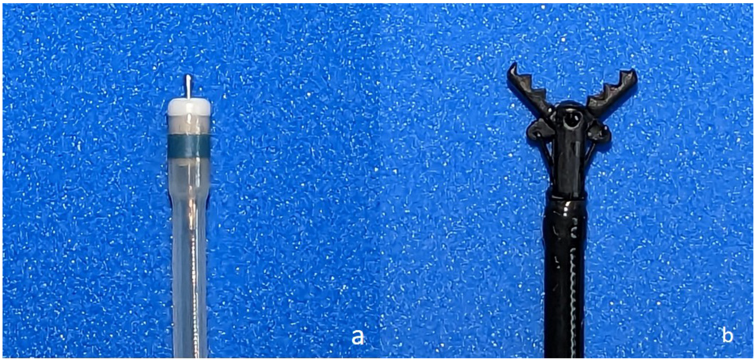
2.2. ESD Procedures
2.3. Examination Protocol
2.4. Data Analysis and Outcome Measures
2.5. Sample Size Calculation
2.6. Statistical Analysis
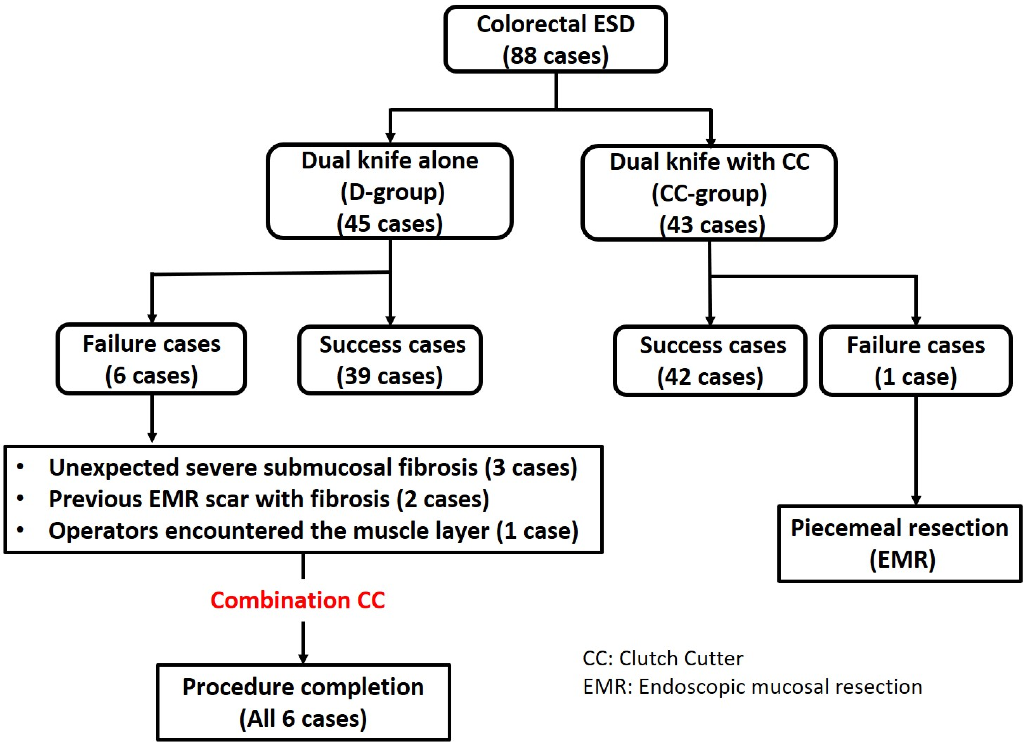
3. Results
Outcome Results
4. Discussion
5. Limitations
6. Conclusions
Author Contributions
Funding
Institutional Review Board Statement
Informed Consent Statement
Data Availability Statement
Acknowledgments
Conflicts of Interest
References
- Nakajima, T.; Saito, Y.; Tanaka, S.; Iishi, H.; Kudo, S.E.; Ikematsu, H.; Igarashi, M.; Saitoh, Y.; Inoue, Y.; Kobayashi, K.; et al. Current status of endoscopic resection strategy for large, early colorectal neoplasia in Japan. Surg. Endosc. 2013, 27, 3262–3270. [Google Scholar] [CrossRef] [PubMed]
- Tanaka, S.; Kashida, H.; Saito, Y.; Yahagi, N.; Yamano, H.; Saito, S.; Hisabe, T.; Yao, T.; Watanabe, M.; Yoshida, M.; et al. JGES guidelines for colorectal endoscopic submucosal dissection/endoscopic mucosal resection. Dig. Endosc. 2015, 27, 417–434. [Google Scholar] [CrossRef]
- Yamamoto, K.; Michida, T.; Nishida, T.; Hayashi, S.; Naito, M.; Ito, T. Colorectal endoscopic submucosal dissection: Recent technical advances for safe and successful procedures. World J. Gastrointest. Endosc. 2015, 7, 1114–1128. [Google Scholar] [CrossRef] [PubMed]
- Fuccio, L.; Hassan, C.; Ponchon, T.; Mandolesi, D.; Farioli, A.; Cucchetti, A.; Frazzoni, L.; Bhandari, P.; Bellisario, C.; Bazzoli, F.; et al. Clinical outcomes after endoscopic submucosal dissection for colorectal neoplasia: A systematic review and meta-analysis. Gastrointest. Endosc. 2017, 86, 74–86.e17. [Google Scholar] [CrossRef]
- Akahoshi, K.; Akahane, H. A new breakthrough: ESD using a newly developed grasping type scissor forceps for early gastrointestinal tract neoplasms. World J. Gastrointest. Endosc. 2010, 2, 90–96. [Google Scholar] [CrossRef]
- Akahoshi, K.; Akahane, H.; Murata, A.; Akiba, H.; Oya, M. Endoscopic submucosal dissection using a novel grasping type scissors forceps. Endoscopy 2007, 39, 1103–1105. [Google Scholar] [CrossRef] [PubMed]
- Akahoshi, K.; Akahane, H.; Motomura, Y.; Kubokawa, M.; Itaba, S.; Komori, K.; Nakama, N.; Oya, M.; Nakamura, K. A new approach: Endoscopic submucosal dissection using the Clutch Cutter® for early stage digestive tract tumors. Digestion 2012, 85, 80–84. [Google Scholar] [CrossRef] [PubMed]
- Akahoshi, K.; Motomura, Y.; Kubokawa, M.; Gibo, J.; Kinoshita, N.; Osada, S.; Tokumaru, K.; Hosokawa, T.; Tomoeda, N.; Otsuka, Y.; et al. Endoscopic Submucosal Dissection for Early Gastric Cancer using the Clutch Cutter: A large single-center experience. Endosc. Int. Open 2015, 3, E432–E438. [Google Scholar] [CrossRef] [Green Version]
- Dohi, O.; Yoshida, N.; Terasaki, K.; Azuma, Y.; Ishida, T.; Kitae, H.; Matsumura, S.; Ogita, K.; Takayama, S.; Mizuno, N.; et al. Efficacy of Clutch Cutter for Standardizing Endoscopic Submucosal Dissection for Early Gastric Cancer: A Propensity Score-Matched Analysis. Digestion 2019, 100, 201–209. [Google Scholar] [CrossRef]
- Akahoshi, K.; Kubokawa, M.; Gibo, J.; Osada, S.; Tokumaru, K.; Yamaguchi, E.; Ikeda, H.; Sato, T.; Miyamoto, K.; Kimura, Y.; et al. Endoscopic submucosal dissection of gastric adenomas using the clutch cutter. World J. Gastrointest. Endosc. 2017, 9, 334–340. [Google Scholar] [CrossRef] [PubMed]
- Nagai, K.; Uedo, N.; Yamashina, T.; Matsui, F.; Matsuura, N.; Ito, T.; Yamamoto, S.; Hanaoka, N.; Takeuchi, Y.; Higashino, K.; et al. A comparative study of grasping-type scissors forceps and insulated-tip knife for endoscopic submucosal dissection of early gastric cancer: A randomized controlled trial. Endosc. Int. Open 2016, 4, E654–E660. [Google Scholar] [CrossRef] [PubMed] [Green Version]
- Inoue, K.; Yoshida, N.; Dohi, O.; Sugino, S.; Matsumura, S.; Kitae, H.; Yasuda, R.; Nakano, T.; Terasaki, K.; Hirose, R.; et al. Effects of the combined use of a scissor-type knife and traction clip on endoscopic submucosal dissection of colorectal tumors: A propensity score-matched analysis. Endosc. Int. Open 2021, 9, E1617–E1626. [Google Scholar] [CrossRef] [PubMed]
- Oyama, T.; Tomori, A.; Hotta, K.; Morita, S.; Kominato, K.; Tanaka, M.; Miyata, Y. Endoscopic submucosal dissection of early esophageal cancer. Clin. Gastroenterol. Hepatol. 2005, 3, S67–S70. [Google Scholar] [CrossRef] [PubMed]
- Ono, H.; Kondo, H.; Gotoda, T.; Shirao, K.; Yamaguchi, H.; Saito, D.; Hosokawa, K.; Shimoda, T.; Yoshida, S. Endoscopic mucosal resection for treatment of early gastric cancer. Gut 2001, 48, 225–229. [Google Scholar] [CrossRef] [Green Version]
- Imai, K.; Hotta, K.; Yamaguchi, Y.; Ito, S.; Ono, H. Endoscopic submucosal dissection for large colorectal neoplasms. Dig. Endosc. 2017, 29 (Suppl. S2), 53–57. [Google Scholar] [CrossRef] [PubMed] [Green Version]
- Toyonaga, T.; Nishino, E.; Hirooka, T.; Dozaiku, T.; Sugiyama, T.; Iwata, Y.; Ono, W.; Ueda, C.; Tomita, M.; Hirooka, T.; et al. Use of Short Needle Knife for Esophageal Endoscopic Submucosal Dissection. Dig. Endosc. 2005, 17, 246–252. [Google Scholar] [CrossRef]
- Yahagi, N.; Fujishiro, M.; Kakushima, N.; Kobayashi, K.; Hashimoto, T.; Oka, M.; Iguchi, M.; Enomoto, S.; Ichinose, M.; Niwa, H.; et al. Endoscopic submucosal dissection for early gastric cancer using the tip of an electrosurgical snare (thin type). Dig. Endosc. 2004, 16, 34–38. [Google Scholar] [CrossRef]
- Sano, Y.; Fu, K.I.; Saito, Y.; Doi, T.; Hanafusa, M.; Fujii, S.; Fujimori, T.; Ohtsu, A. A newly developed bipolar-current needle-knife for endoscopic submucosal dissection of large colorectal tumors. Endoscopy 2006, 38 (Suppl. S2), E95. [Google Scholar] [CrossRef] [PubMed] [Green Version]
- Honma, K.; Kobayashi, M.; Watanabe, H.; Suga, T.; Tominaga, K.; Yamagata, M.; Hiraishi, H. Endoscopic submucosal dissection for colorectal neoplasia. Dig. Endosc. 2010, 22, 307–311. [Google Scholar] [CrossRef] [PubMed]
- Yamashina, T.; Takeuchi, Y.; Nagai, K.; Matsuura, N.; Ito, T.; Fujii, M.; Hanaoka, N.; Higashino, K.; Uedo, N.; Ishihara, R.; et al. Scissor-type knife significantly improves self-completion rate of colorectal endoscopic submucosal dissection: Single-center prospective randomized trial. Dig. Endosc. 2017, 29, 322–329. [Google Scholar] [CrossRef] [Green Version]
- Akahoshi, K.; Okamoto, R.; Akahane, H.; Motomura, Y.; Kubokawa, M.; Osoegawa, T.; Nakama, N.; Chaen, T.; Oya, M.; Nakamura, K. Endoscopic submucosal dissection of early colorectal tumors using a grasping-type scissors forceps: A preliminary clinical study. Endoscopy 2010, 42, 419–422. [Google Scholar] [CrossRef] [PubMed]
- Fujishiro, M.; Yahagi, N.; Kakushima, N.; Kodashima, S.; Muraki, Y.; Ono, S.; Yamamichi, N.; Tateishi, A.; Oka, M.; Ogura, K.; et al. Outcomes of endoscopic submucosal dissection for colorectal epithelial neoplasms in 200 consecutive cases. Clin. Gastroenterol. Hepatol. 2007, 5, 678–683. [Google Scholar] [CrossRef]
- Saito, Y.; Uraoka, T.; Matsuda, T.; Emura, F.; Ikehara, H.; Mashimo, Y.; Kikuchi, T.; Fu, K.I.; Sano, Y.; Saito, D. Endoscopic treatment of large superficial colorectal tumors: A case series of 200 endoscopic submucosal dissections (with video). Gastrointest. Endosc. 2007, 66, 966–973. [Google Scholar] [CrossRef] [PubMed]
- Tanaka, S.; Oka, S.; Kaneko, I.; Hirata, M.; Mouri, R.; Kanao, H.; Yoshida, S.; Chayama, K. Endoscopic submucosal dissection for colorectal neoplasia: Possibility of standardization. Gastrointest. Endosc. 2007, 66, 100–107. [Google Scholar] [CrossRef] [PubMed]
- Kakushima, N.; Fujishiro, M. Endoscopic submucosal dissection for gastrointestinal neoplasms. World J. Gastroenterol. 2008, 14, 2962–2967. [Google Scholar] [CrossRef] [PubMed]
- Yoshida, N.; Wakabayashi, N.; Kanemasa, K.; Sumida, Y.; Hasegawa, D.; Inoue, K.; Morimoto, Y.; Kashiwa, A.; Konishi, H.; Yagi, N.; et al. Endoscopic submucosal dissection for colorectal tumors: Technical difficulties and rate of perforation. Endoscopy 2009, 41, 758–761. [Google Scholar] [CrossRef] [PubMed]
- Takeuchi, Y.; Uedo, N.; Ishihara, R.; Iishi, H.; Kizu, T.; Inoue, T.; Chatani, R.; Hanaoka, N.; Taniguchi, T.; Kawada, N.; et al. Efficacy of an endo-knife with a water-jet function (Flushknife) for endoscopic submucosal dissection of superficial colorectal neoplasms. Am. J. Gastroenterol. 2010, 105, 314–322. [Google Scholar] [CrossRef]


| D-Group (45 Cases) | CC-Group (43 Cases) | p | |
|---|---|---|---|
| Sex (male/female) | 28/17 | 27/16 | p = 0.96 * |
| Median age, years (range) | 70 (41–84) | 68 (26–88) | p = 0.79 ** |
| Macroscopic type | NS | ||
| Protruded lesion | 7 | 8 | |
| LST-G (HG) | 4 | 6 | |
| LST-G (NM) | 17 | 18 | |
| LST-NG (FE) | 12 | 6 | |
| LST-NG (PD) | 5 | 5 | |
| Distribution | NS | ||
| Cecum | 4 | 5 | |
| Ascending colon | 7 | 13 | |
| Transvers colon | 11 | 7 | |
| Descending colon | 3 | 1 | |
| Sigmoid colon | 11 | 7 | |
| Rectum | 9 | 10 | |
| Median resected size (range, mm) | 35 (21–60) | 35 (18–60) | p = 0.36 ** |
| Median tumor size (range, mm) | 25 (20–49) | 25 (20–50) | p = 0.07 ** |
| D-Group (n = 45) | CC-Group (n = 43) | p | |
|---|---|---|---|
| Self-completion rate | 87% (39) | 98% (42) | p = 0.11 * |
| Complication rate | |||
| Perforation | 2% (1) | 0% (0) | p = 1.00 * |
| Delayed bleeding | 0% (0) | 5% (2) | p = 0.24 * |
| Mean procedure time, min (±SD) | 62.0 (±4.4) | 81.1 (±4.6) | p = 0.0036 ** |
| (excluding failure cases) | (39 cases) | (42 cases) | |
| En bloc resection rate | 100% (45) | 98% (42) | p = 1.00 * |
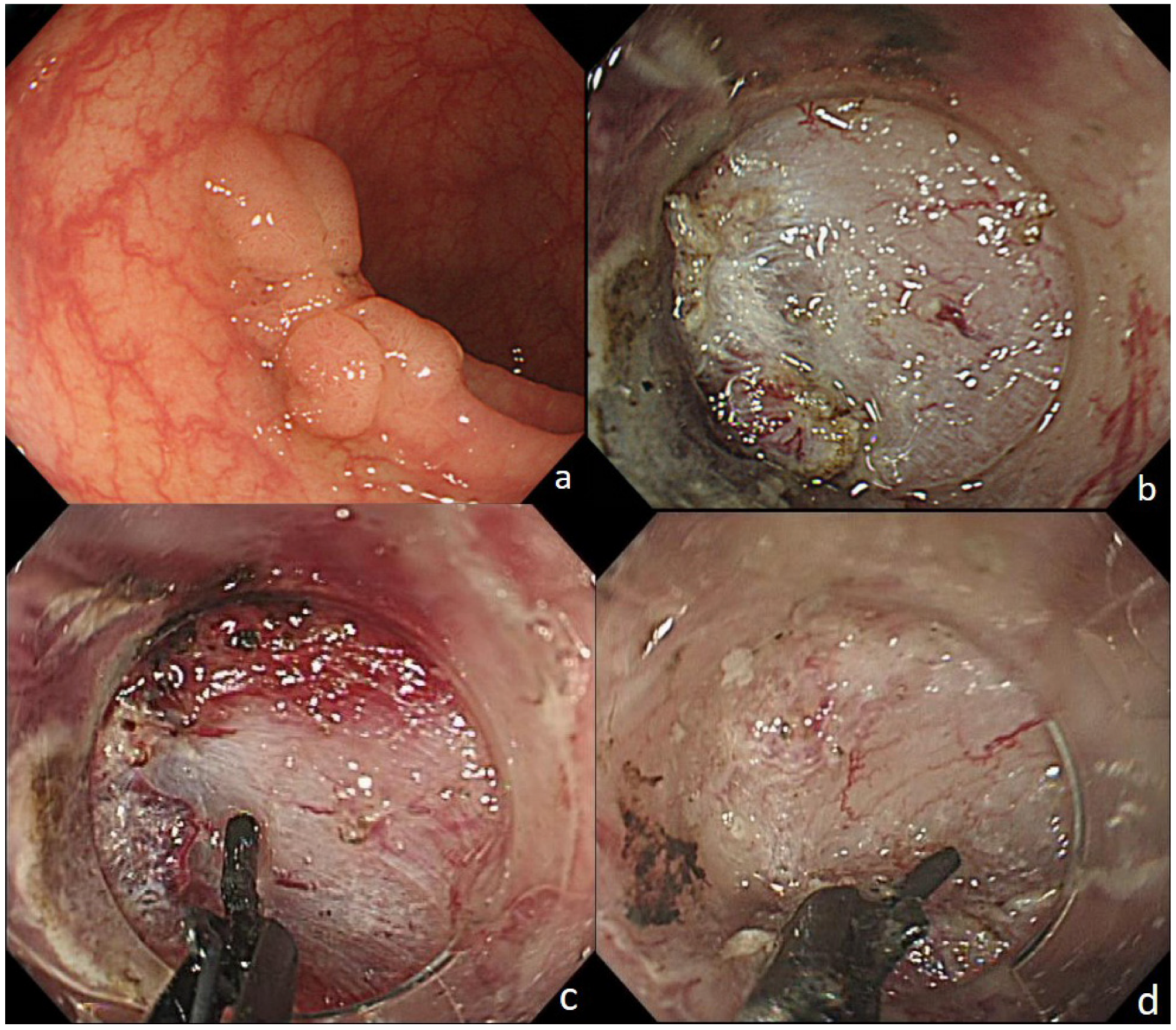
| Case1 (71 y.o. M) | Case2 (81 y.o. M) | Case3 (43 y.o. M) | Case4 (70 y.o. F) | Case5 (69 y.o. M) | Case6 (76 y.o. F) | |
|---|---|---|---|---|---|---|
| Macroscopic type | LST-NG (FE) | LST-G (NM) | 0-Isp | LST-G (NM) | 0-Is | LST-G (NM) |
| Procedure time (min) | 140 | 100 | 117 | 70 | 100 | 111 |
| Resected size (mm) | 35 | 25 | 35 | 45 | 21 | 35 |
| Tumor size (mm) | 28 | 20 | 30 | 40 | 20 | 30 |
| Distribution | Sigmoid | Sigmoid | Rs | Rb-P | Transverse | Transverse |
| Characteristic | Fibrosis | Fibrosis | Fibrosis | EMR scar | EMR scar | Encountered the muscle layer |
| Depth | Adenoma | Adenoma | sm2 (1000 μm) | m | Adenoma | Adenoma |
| Retroflex view | - | - | + | + | - | - |
| Procedure completion | + | + | + | + | + | + |
Disclaimer/Publisher’s Note: The statements, opinions and data contained in all publications are solely those of the individual author(s) and contributor(s) and not of MDPI and/or the editor(s). MDPI and/or the editor(s) disclaim responsibility for any injury to people or property resulting from any ideas, methods, instructions or products referred to in the content. |
© 2023 by the authors. Licensee MDPI, Basel, Switzerland. This article is an open access article distributed under the terms and conditions of the Creative Commons Attribution (CC BY) license (https://creativecommons.org/licenses/by/4.0/).
Share and Cite
Yachida, T.; Kobara, H.; Kozuka, K.; Nakatani, K.; Tada, N.; Matsui, T.; Chiyo, T.; Kobayashi, N.; Fujihara, S.; Nishiyama, N.; et al. Comparison of Needle Knife versus Scissors Forceps for Colorectal Endoscopic Submucosal Dissection: A Prospective Randomized Study. J. Clin. Med. 2023, 12, 2329. https://doi.org/10.3390/jcm12062329
Yachida T, Kobara H, Kozuka K, Nakatani K, Tada N, Matsui T, Chiyo T, Kobayashi N, Fujihara S, Nishiyama N, et al. Comparison of Needle Knife versus Scissors Forceps for Colorectal Endoscopic Submucosal Dissection: A Prospective Randomized Study. Journal of Clinical Medicine. 2023; 12(6):2329. https://doi.org/10.3390/jcm12062329
Chicago/Turabian StyleYachida, Tatsuo, Hideki Kobara, Kazuhiro Kozuka, Kaho Nakatani, Naoya Tada, Takanori Matsui, Taiga Chiyo, Nobuya Kobayashi, Shintaro Fujihara, Noriko Nishiyama, and et al. 2023. "Comparison of Needle Knife versus Scissors Forceps for Colorectal Endoscopic Submucosal Dissection: A Prospective Randomized Study" Journal of Clinical Medicine 12, no. 6: 2329. https://doi.org/10.3390/jcm12062329