Review

Inferior Vena Cava Ultrasonography for Volume Status Evaluation: An Intriguing Promise Never Fulfilled

1
Nephrology and Dialysis Unit, S. Maria della Scaletta Hospital, 40026 Imola, Italy
2
Department of Clinical Surgical, Diagnostic and Pediatric Sciences, University of Pavia, 27100 Pavia, Italy
3
Anaesthesia, Intensive Care and Pain Therapy, Fondazione IRCCS Policlinico San Matteo, 27100 Pavia, Italy
4
Radiology Unit, S. Maria della Scaletta Hospital, 40026 Imola, Italy
5
Department of Surgical, Medical, Molecular Pathology and Critical Care Medicine, University of Pisa, 56124 Pisa, Italy
6
Azienda Ospedaliero Universitaria Pisana Via Paradisa, 2, 56126 Pisa, Italy
*
Author to whom correspondence should be addressed.
J. Clin. Med. 2023, 12(6), 2217; https://doi.org/10.3390/jcm12062217
Submission received: 7 February 2023 / Revised: 2 March 2023 / Accepted: 7 March 2023 / Published: 13 March 2023

Abstract

:
The correct determination of volume status is a fundamental component of clinical evaluation as both hypovolaemia (with hypoperfusion) and hypervolaemia (with fluid overload) increase morbidity and mortality in critically ill patients. As inferior vena cava (IVC) accounts for two-thirds of systemic venous return, it has been proposed as a marker of volaemic status by indirect assessment of central venous pressure or fluid responsiveness. Although ultrasonographic evaluation of IVC is relatively easy to perform, correct interpretation of the results may not be that simple and multiple pitfalls hamper its wider application in the clinical setting. In the present review, the basic elements of the pathophysiology of IVC behaviour, potential applications and limitations of its evaluation are discussed.

1. Introduction

Correct determination of the volume status of the patient represents a fundamental step in clinical evaluation. In fact, in many critically ill patients hypovolaemia might result in reduced tissue perfusion, while fluid overload can lead to organ congestion with associated morbidity and mortality [1]. Thus, volaemic assessment is essential to guide clinicians in treatment and might have a potential prognostic implication.
Right atrial pressure (RAP) is a cornerstone of evaluation of the intravascular volume status that predicts overall survival in patients with heart failure [2]; the terms ‘‘central venous pressure’’ (CVP) and ‘‘right atrial pressure’’ (RAP) are synonymous as long as there is no vena cava obstruction, and they will be used interchangeably in this manuscript.
The standard monitoring tool for assessing CVP is the central venous catheter. However, due to its invasiveness, infectious and thrombotic complications are considered major concerns. In this context, point-of-care ultrasonography (POCUS) might be a reliable alternative for volume estimation.
Ultrasonographic evaluation of the IVC (US-IVC) has been proposed as the non-invasive technique of choice for CVP assessment due to its wide availability, low costs, and ease of use. Estimation of volume status by measuring the static diameter of the IVC and dynamic respiratory fluctuations has been investigated in various clinical settings; nevertheless, results are often conflicting and uncertain, as hypervolaemia and hypovolaemia are not the only factors affecting IVC diameter. Hence, although US-IVC is relatively easy to apply, its use in clinical decision-making requires a deep knowledge of pathophysiology, limitations and pitfalls to avoid perceptual and interpretive errors.
The aim of this paper is thus to review the main physiopathology about venous return and right atrial filling, explore the current evidence (accuracy and pitfalls) of US-IVC in clinical practice, and give clinicians the instruments for its correct interpretation.

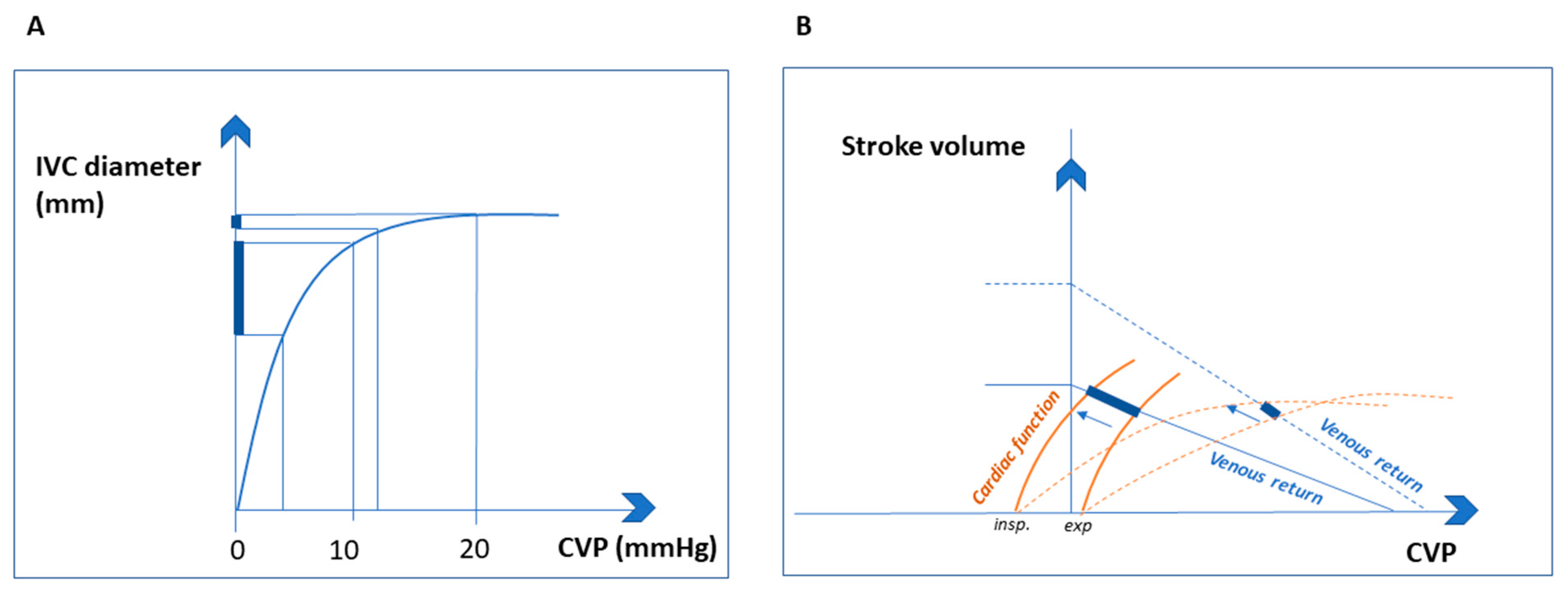
2. Pathophysiological Assumptions

The IVC has the largest diameter of the entire venous system; it is a thin-walled, valveless, retroperitoneal vessel, responsible for returning large volumes of deoxygenated blood from the lower extremities and abdomen to the right atrium. With 85% of total plasma volume in the venous circulation, the IVC is an important blood reservoir, and modifications of circulating volume result in IVC calibre variations.
Indeed, evidence of a ‘’flat vena cava” (e.g., an IVC with an anteroposterior diameter of less than 9 mm) at multiple levels is associated with significant hypovolaemia in trauma patients [3].
Patient position and decubitus can influence circulating blood volume and IVC diameter by gravity: the IVC is smaller when the patient is in the left lateral position and larger when the patient is in the right lateral position [4].
In addition to circulating volume, other important factors can lead to variations in IVC diameter during the respiratory cycle, such as right heart function and the gradient between intrathoracic and intra-abdominal pressure (Figure 1).

2.1. Intrathoracic Pressure

In spontaneously breathing subjects, during inspiration abdominal pressure increases while intrathoracic pressure (ITP) increases its negativity, lowering right atrial pressure. The haemodynamic counterpart to the aforementioned thoraco–abdominal interaction is the increase in blood return from the IVC to the RA, leading to a secondary reduction in the size of the IVC [5] and a transient increase in stroke volume. Conversely, venous return decreases during exhalation, while the calibre of the IVC increases [6,7].
IVC collapsibility may be exaggerated if ITP becomes markedly negative with forced inspiratory efforts, respiratory distress, or exacerbation of chronic obstructive pulmonary disease, causing an increased venous return to the right atrium.
In ventilated patients, positive end-expiratory pressure (PEEP) may hamper the venous return during inspiration by increasing the ITP and reducing the pressure gradient between the abdominal and thoracic compartments [8]. This issue may be critical in cases of pre-load dependence (e.g., right ventricular infarction, pulmonary embolism, tamponade, severe hypovolaemia), leading to the abrupt reduction in venous return that triggers haemodynamic instability [9].

2.2. Intra-Abdominal Pressure

Intra-abdominal pressure (IAP) may influence IVC physiology even more than ITP, as it affects both venous return and IVC diameter. In the subdiaphragmatic region, when the transmural pressure of the IVC exceeds the critical closing pressure, the IVC is pervious; in this situation, an increase in abdominal pressure reduces IVC diameter and increases transitorily the venous return, with the liver serving as the immediate blood source [7]. Conversely, when the IVC pressure is below the critical closing pressure, the increased intrabdominal pressure causes the IVC collapse with a dramatic drop in venous return [10]. Therefore, increased intra-abdominal pressure during inspiration might have opposite effects on total and regional IVC venous return.
Not only IAP but also the volaemic status can influence venous return during the respiratory cycle. In case of hypervolaemia, the active diaphragmatic descent causes a significant increase in total IVC flow by enhancing splanchnic venous return through the IVC. On the other hand, in case of hypovolaemia the possibly increased abdominal pressure reduces venous return, leading to a decrease in IVC flow at the level of the diaphragm (e.g., vascular waterfall effect) [11].
Therefore, two important factors may affect IVC flow and diameter and thus venous return: intra-abdominal pressure and volaemia. Regardless of the volaemic state, severe abdominal hypertension always causes a drop in the IVC venous return and a consequent decrease in cardiac output [12].

2.3. Cardiac Function

Because of the close relationship between venous return and right atrial pressure, the wall movements of the IVC reflect the haemodynamic behaviour of the right atrium (RA) under both physiological and pathological conditions [13], which are influenced by the cardiac cycle, right heart function, and rhythm.
In sinus rhythm, the IVC has its maximum diameter during the presystolic and systolic phases [5] while atrial fibrillation alters the filling of the IVC, making the relationship between cardiac cycle and IVC dimension difficult to assess. IVC diameter should be interpreted in the light of the physiology of venous return, right heart function and heart-lung interaction.
Indeed, the inspiratory collapsibility of the IVC with normal ITP is an expression of the adequacy of the right heart to reduce RAP (Figure 2B) [14]. In response to increased ITP, the curvilinear relationship between IVC diameter and CVP is clearly evident (Figure 2A), with an initial steep part (i.e., a minimal increase in CVP determines a large increase in IVC diameter) and a flat part (a larger increase in CVP causes minimal or no IVC dilation [9].
Under pathological conditions, such as acute circulatory failure, the change in IVC diameter is thus a function of residual venous compliance and cardiac functional reserve (Figure 2A,B).
Therefore, the behaviour of the IVC is the result of a complex interplay between the heart, volaemia, and respiratory mechanics acting simultaneously in different clinical contexts (Figure 1).

3. Anatomical Variations of Inferior Vena Cava and Their Clinical Significance

Due to a complex embryogenesis process, congenital anomalies of the IVC and its tributaries are not uncommon, with a reported prevalence of 0.3% to 10.14% of the population and a total of 14 observed variations; knowledge of these clinical entities may avoid severe consequences during retroperitoneal surgical procedures (especially laparoscopic procedures) [16,17].
The most common IVC anomalies are duplication, with a left IVC terminating below or at the level of the left renal vein, a left-sided IVC, and interruption or agenesia of the IVC [18,19].
From a clinical point of view, IVC duplication together with left-sided vena cava and megacava are generally asymptomatic, whereas aplasia and hypoplasia may be associated with iliofemoral deep venous thrombosis [16].
In all these conditions, both the calibre of the vessel (or vessels), the venous return and the behaviour of the IVC in relation to various volaemic conditions may be severely altered.

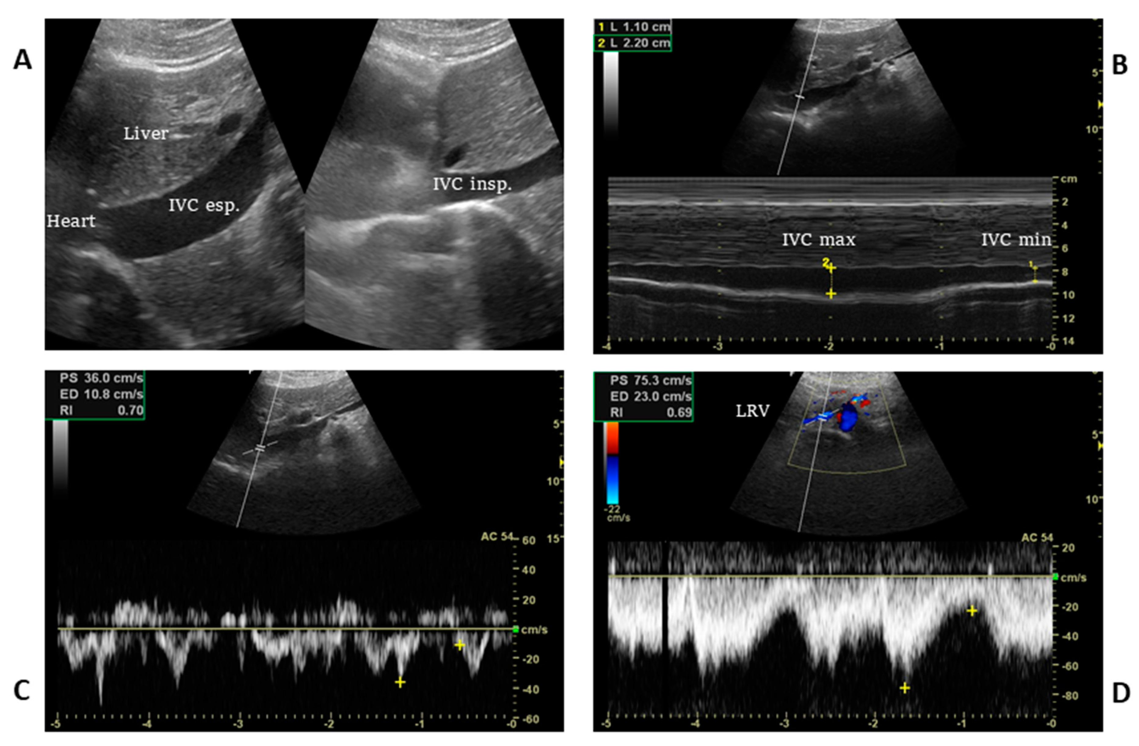
4. Ultrasound Technique, Static and Dynamic IVC Indexes

Either a low-frequency convex probe (2–5.5 MHz) for the abdomen or a phased array transducer (2–8 MHz) for the heart can be used to assess the IVC.
The IVC is usually visualized from a subcostal view by a longitudinal scan, including the veno–atrial junction and the right atrium with inner walls clearly visible. In case of a suboptimal or unavailable subcostal window, a coronal transhepatic scan along the posterior right axillary line may be an effective alternative (Figure 3).
The exact position at which the IVC diameter should be measured is crucial, although it is not universally standardized: in spontaneously breathing patients, variations in the IVC diameter are smaller near the right atrium and larger 2 cm caudal to the hepatic vein inlet or at the level of the left renal vein [20]. Most authors suggest that measurements should be acquired within 1.5 cm from the IVC-to-right atrial junction [21].
B-mode is used for the identification of the IVC and then the M-mode Doppler is applied with a sweep velocity set at 25 to 50 mm/s, depending on the respiratory rate, to capture at least three respiratory cycles.
Since the minimum venous diameter in spontaneously breathing patients may be influenced by inspiratory effort, maximal inspiration (sniffing manoeuvre) could be evoked while maximal IVC diameter is measured at the end of expiration [22].
Sample accuracy can be improved by using indexed IVC size (iIVC), which is calculated by dividing IVCmax by body surface area [23].
In addition to these static parameters, it is always useful to make a dynamic assessment using the IVC collapsibility index (cIVC), which is calculated according to the following formula: cIVC = (IVCmax − IVCmin)/IVCmax [24,25].
In mechanically ventilated patients, the IVC distensibility index, described as
dIVC = (IVCmax − IVCmin)/IVCmin, [21] the respiratory variations in inferior vena cava diameter as ΔDIVC = (IVCmax − IVCmin)/(IVCmax + IVC min)/2) [26] or the IVC Area Distensibility Index (IVC-ADI) defined as maximum IVC area-minimum IVC area)/minimum IVC area × 100% can be applied [27].
Echocolour and pulsed wave Doppler assessment of the venous spectrometric wave, although not commonly performed in IVC, can be helpful in certain clinical contexts such as stenosis/thrombosis or congenital anomalies [28,29].
The technical limitations of US-IVC are obesity or pregnancy, chest or gastric tubes, and non-negligible inter/intra-observer variability often caused by lateral displacement of the IVC during respiration, so that both the true centre of the vein and the accuracy of the measurement are lost in M-mode imaging [30,31].

5. Evidence for Volaemic Status Evaluation with IVC Ultrasonography

IVC diameter and its temporal changes in the respiratory cycle have long been studied to correlate with CVP and predict fluid responsiveness.
These two aspects are addressed here separately according to the pathophysiological premises.

5.1. Volaemic Status Evaluation in Spontaneously Breathing Patient

Overall, a statistically significant non-linear correlation was described between the sonographic dimensional parameters of IVC and CVP [32]. Most studies demonstrated a moderate correlation between measurements of IVC diameter or collapsibility and CVP or RAP [33]. Cut-off values of 2 cm diameter and cIVC of 40% provided the best diagnostic accuracy in predicting a RAP above or below 10 mmHg [34,35,36].
According to the current updated American and European guidelines, an IVC diameter ≤2.1 cm and collapsibility >50% during inspiration suggest a RAP between 0–5 mm Hg while a diameter >2.1 cm with <50% inspiratory collapse indicates a high RAP of 10–20 mmHg; a mean pressure value of 8 mmHg is used if the clinical picture does not follow the proposed pattern [37].
In outpatients undergoing haemodialysis, standardization of IVC diameter to body surface area (BSA) was recommended (i.e., IVC diameter 2.1 cm if BSA > 1.61 m2, IVC diameter 1.7 cm when BSA < 1.61 m2) [38] and an indexed IVC size (iIVC) ≥ 8 and ≤11.5 mm/m2 is considered safe to rule out severe hyper or hypovolaemia in this setting [23,36].
The addition of pulsed wave Doppler applied to the IVC may provide additional information to estimate CVP (Figure 3). The presence of continuous flow from the IVC to the RA corresponds to a low to normal CVP; on the contrary, an interrupted waveform indicates a high RAP only if it is associated with other ultrasound indices such as the IVC size and cIVC [29].
It has also been described that the IVC diameter and cIVC correlate with plasma volume removal by ultrafiltration in continuous and intermittent haemodialysis or blood donation [39,40].
For the assessment of fluid responsiveness, the US-IVC diameters are useless [41,42]. Hence “dynamic” measurements have been developed to predict the response to volume infusion and guide fluid resuscitation (Table 1). To this end, cIVC measurements are taken before intravenous fluid administration or passive leg raising and then the cardiac output response is observed.
From all these studies it emerged that only extreme cIVC values (i.e., a cIVC value > 40%) may be useful in predicting the haemodynamic response to volume expansion [30,43], due to multiple pitfalls that can affect respiratory mechanics and cardiopulmonary interactions.
Considering that even a reduced cIVC may not rule out a fluid responsiveness or the need for fluid therapy, a cIVC threshold < 15% with the addition of a standardized inspiratory effort was proposed as an attempt to improve cIVC diagnostic accuracy [44].
Therefore, in spontaneously breathing patients, a cIVC-guided fluid infusion can be considered a logical, but rarely decisive, option before the administration of infusion therapy.

5.2. Volaemic Status Evaluation in Mechanically Ventilated Patients

Regarding the non-invasive estimation of CVP, current evidence does not support the use of the IVC diameter in mechanically ventilated patients. In a recent meta-analysis that included 16 studies, the correlation between CVP and IVC diameter was not statistically significant in 8 studies and was weak to moderate in the others [33]. These results may be due to the complex interplay between intrathoracic pressure, right atrial pressure and venous return, so that a unique interpretation can be challenging in most cases (Figure 1) [37].
Evaluation of fluid responsiveness relies on the IVC’s potential to dilate by increasing its diameter during positive pressure ventilation, shifting from the steep to the flat part of the IVC-to-CVP curve (Figure 2A) [9,15]. However, in 540 subjects with acute circulatory failure of any cause (the largest adult population ever studied on this topic), respiratory variations in the IVC diameter provided only weak to moderate diagnostic accuracy in predicting fluid responsiveness; this result was likely due to concurrent abdominal hypertension and/or low level of mechanical power during protective mechanical ventilation (e.g., low tidal volume of less than 8 mL/kg predicted body weight, moderate to low positive end expiratory pressure, low respiratory rate, low driving pressures) [45].
Regarding the paediatric population, the evidence for patients on mechanical ventilation is quite sparse and contrasting. In 21 children who had undergone cardiac surgery, the ΔDIVC accurately predicted fluid responsiveness, whereas it did not in 33 neurosurgical patients [46,47].
Likewise, in the work by Weber et al. on 31 subjects (aged 1 day to 13 years), respiratory cycle-induced changes in IVC diameter were useless for predicting fluid responsiveness [48,61]. These results may be explained by the higher IVC elasticity and chest wall compliance in the paediatric population compared with the adults, resulting in dampened transpulmonary pressure [48].
Regardless, we must note that in most of the above-mentioned adult and paediatric studies, cardiac function data were not available, which is a significant limitation in the assessment reliability of IVC US.
In mechanically ventilated subjects, a new distensibility index based on IVC area was recently proposed by Yao and coworkers (VCI ADI, cut-off value 10.2%), which revealed higher sensitivity in predicting fluid responsiveness than dIVC, even though it was loaded with very low specificity (97.3% sensitivity and 40.0% specificity, respectively) [23].
In summary, for fluid responsiveness assessment, dIVC has a better diagnostic performance than cIVC in spontaneously breathing patients [26]; its clinical utility in patients receiving mechanical ventilation is questionable and can be only applied in the context of a preserved biventricular heart function. Moreover, dIVC is not adequately supported by the currently available evidence [62] in abdominal surgery, concurrent abdominal hypertension, patients ventilated with protective mechanical ventilation [63] and the paediatric population, as it is burdened by poor diagnostic accuracy.

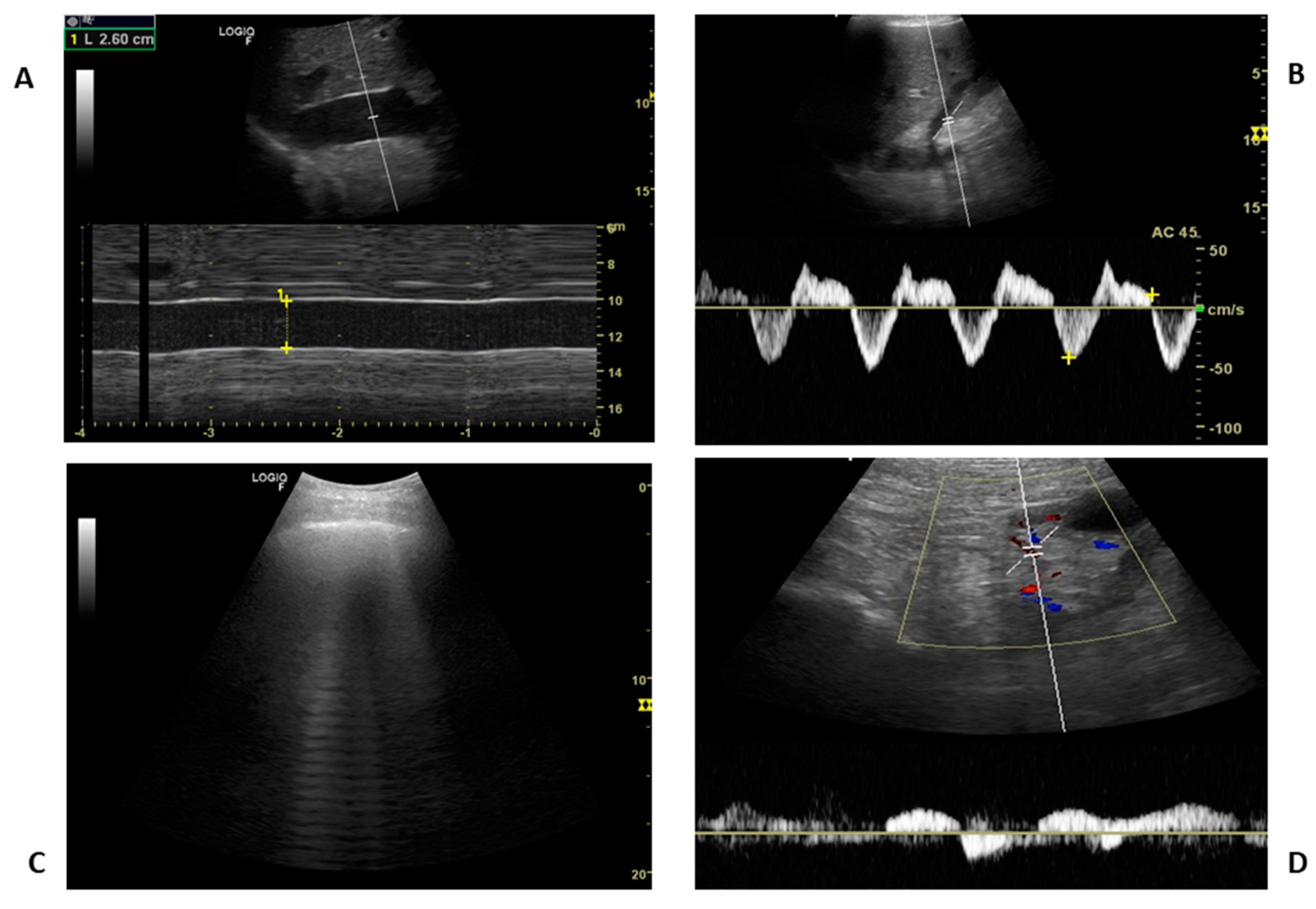
6. Pitfalls That May Lead to Misinterpretation of Volume Status

IVC dilatation had been suggested as a potential predictor of outcome in many different clinical contexts [64,65,66]. However, numerous physiological and pathophysiological conditions, as well as several interpretive pitfalls, have limited its wide applicability [24].
IVC diameter interpretation in relation to volume status includes several confounding conditions, such as: chronically dilated IVC in young trained athletes due to adaptation to chronic strenuous exercise, young patients with vasovagal syncope that present increased venous pooling, children with increased venous compliance, direct vasoplegic effect of drugs or sepsis, [67,68] severe tricuspid regurgitation, right ventricular failure, pericardial effusion or tamponade, acute pulmonary embolism, intra-abdominal hypertension including pregnancy, [69,70,71] (Figure 1), COPD exacerbations with hyperinflation and increased intrathoracic pressure [72], and patients undergoing mechanical ventilation with high positive end-expiratory pressure [73]. Similarly, local mechanical factors such as IVC stenosis and thrombosis or the presence of devices such as cava filters and catheters can reduce venous return. In all the above-mentioned conditions, chronically strained IVC can be dilated without an underlying hypervolaemic state.
On the contrary, the presence of masses compressing the IVC and COPD exacerbations with forced expiration may mimic IVC collapsibility [71]; when circulating volume depletion occurs due to severe hypoproteinaemia such as in liver cirrhosis, malnutrition, cancer or sepsis, the IVC reduces in size, but the patient can be hyper-hydrated due to the massive shift of fluid into the third space, leading to splanchnic congestion regardless of the behaviour of the IVC.

7. IVC Ultrasonography: Current Knowledge and Future Directions

The reliability of IVC US as an index of volaemic status varies greatly in different clinical contexts and this aspect must be considered before drawing therapeutic conclusions.
For all the above reasons, and because of its complex pathophysiology, the inferior vena cava evaluation has numerous interpretive pitfalls that make it useless for inferring volume status when considered as a sole measure. Recently, a combined evaluation of IVC and other body districts’ veins has been proposed to minimize its interpretative drawbacks and have a more comprehensive view of the congestion [74]. However, even in this case, IVC-US cannot be considered as a gateway to decide whether to proceed with the investigation of the splanchnic compartment, as splanchnic congestion can be present independently from IVC’s behaviour. Even if it is time-consuming, only a combined US assessment of IVC, heart, lung, portal, splenic and renal veins, (Figure 4) may provide additional insights to explain a complex pathophysiology [75].

8. Conclusions

Assessment of volume status is a cornerstone of clinical evaluation, and for this purpose we cannot ignore the major advantages of IVC ultrasonography such as non-invasivity, wide availability, low cost, relative ease of use and repeatability.
On the other hand, its use is burdened by technical limitations, errors in interpretation and limited areas of clinical application.
For these reasons, IVC-US cannot be considered a stand-alone method suitable for all patients, and a comprehensive assessment of organ perfusion in the critically ill patients requires a clinical physiopathological perspective in conjunction with an integrated ultrasonographic approach, combining multiple sites of investigation.

Author Contributions

Concept and design: P.D.N. and F.C. Analysis and interpretation of data: P.D.N., G.T., L.N. and F.C. Drafting of the manuscript: P.D.N., F.C. and G.T. Critical revision of the manuscript for important intellectual content and approval of the final draft: P.D.N., G.T., L.N. and F.C. All authors have read and agreed to the published version of the manuscript.

Funding

This research received no external funding.

Institutional Review Board Statement

Not applicable.

Informed Consent Statement

Not applicable.

Data Availability Statement

Not applicable.

Conflicts of Interest

The authors declare that they have no conflict of interests with the subject of the article.

Abbreviations

Inferior vena cava (IVC), Intensive care unit (ICU), Emergency department (ED), Right atrial pressure (RAP), Central venous pressure (CVP), Point of care ultrasonography (POCUS), Intrathoracic pressures (ITP), Intra-abdominal pressure (IAP), Positive end-expiratory pressure (PEEP), IVC collapsibility index (cIVC), IVC distensibility index (dIVC), IVC Area Distensibility Index (IVC-ADI).

References

  1. Marik, P.E.; Linde-Zwirble, W.T.; Bittner, E.A.; Sahatjian, J.; Hansell, D. Fluid Administration in Severe Sepsis and Septic Shock, Patterns and Outcomes: An Analysis of a Large National Database. Intensive Care Med. 2017, 43, 625–632. [Google Scholar] [CrossRef] [PubMed]
  2. Damman, K.; van Deursen, V.M.; Navis, G.; Voors, A.A.; van Veldhuisen, D.J.; Hillege, H.L. Increased Central Venous Pressure Is Associated with Impaired Renal Function and Mortality in a Broad Spectrum of Patients with Cardiovascular Disease. J. Am. Coll. Cardiol. 2009, 53, 582–588. [Google Scholar] [CrossRef] [Green Version]
  3. Jeffrey, R.B.; Federle, M.P. The Collapsed Inferior Vena Cava: CT Evidence of Hypovolemia. AJR Am. J. Roentgenol. 1988, 150, 431–432. [Google Scholar] [CrossRef] [PubMed]
  4. Nakao, S.; Come, P.C.; McKay, R.G.; Ransil, B.J. Effects of Positional Changes on Inferior Vena Caval Size and Dynamics and Correlations with Right-Sided Cardiac Pressure. Am. J. Cardiol. 1987, 59, 125–132. [Google Scholar] [CrossRef] [PubMed]
  5. Wexler, L.; Bergel, D.H.; Gabe, I.T.; Makin, G.S.; Mills, C.J. Velocity of Blood Flow in Normal Human Venae Cavae. Circ. Res. 1968, 23, 349–359. [Google Scholar] [CrossRef] [Green Version]
  6. Kadoya, Y.; Miyati, T.; Kobayashi, S.; Ohno, N.; Gabata, T. Evaluation of Gravity Effect on Inferior Vena Cava and Abdominal Aortic Flow Using Multi-Posture MRI. Acta Radiol. 2021, 62, 1122–1128. [Google Scholar] [CrossRef]
  7. Lloyd, T.C. Effect of Inspiration on Inferior Vena Caval Blood Flow in Dogs. J. Appl. Physiol. Respir. Environ. Exerc. Physiol. 1983, 55, 1701–1708. [Google Scholar] [CrossRef]
  8. Fessler, H.E.; Brower, R.G.; Shapiro, E.P.; Permutt, S. Effects of Positive End-Expiratory Pressure and Body Position on Pressure in the Thoracic Great Veins. Am. Rev. Respir. Dis. 1993, 148, 1657–1664. [Google Scholar] [CrossRef] [PubMed]
  9. Bodson, L.; Vieillard-Baron, A. Respiratory Variation in Inferior Vena Cava Diameter: Surrogate of Central Venous Pressure or Parameter of Fluid Responsiveness? Let the Physiology Reply. Crit. Care 2012, 16, 181. [Google Scholar] [CrossRef] [Green Version]
  10. Takata, M.; Wise, R.A.; Robotham, J.L. Effects of Abdominal Pressure on Venous Return: Abdominal Vascular Zone Conditions. J. Appl. Physiol. (1985) 1990, 69, 1961–1972. [Google Scholar] [CrossRef] [PubMed]
  11. Takata, M.; Robotham, J.L. Effects of Inspiratory Diaphragmatic Descent on Inferior Vena Caval Venous Return. J. Appl. Physiol. (1985) 1992, 72, 597–607. [Google Scholar] [CrossRef]
  12. Dagar, G.; Taneja, A.; Nanchal, R.S. Abdominal Circulatory Interactions. Crit. Care Clin. 2016, 32, 265–277. [Google Scholar] [CrossRef]
  13. Gullace, G.; Savoia, M.T. Echocardiographic Assessment of the Inferior Vena Cava Wall Motion for Studies of Right Heart Dynamics and Function. Clin. Cardiol. 1984, 7, 393–404. [Google Scholar] [CrossRef]
  14. Schmidt, G.A. Rebuttal from Dr Schmidt. Chest 2017, 151, 536–537. [Google Scholar] [CrossRef] [Green Version]
  15. Barbier, C.; Loubières, Y.; Schmit, C.; Hayon, J.; Ricôme, J.-L.; Jardin, F.; Vieillard-Baron, A. Respiratory Changes in Inferior Vena Cava Diameter Are Helpful in Predicting Fluid Responsiveness in Ventilated Septic Patients. Intensive Care Med. 2004, 30, 1740–1746. [Google Scholar] [CrossRef] [PubMed]
  16. Kim, H.; Labropoulos, N.; Blake, A.M.; Desai, K. Prevalence of Inferior Vena Cava Anomalies and Their Significance and Impact in Clinical Practice. Eur. J. Vasc. Endovasc. Surg. 2022, 64, 388–394. [Google Scholar] [CrossRef] [PubMed]
  17. Bass, J.E.; Redwine, M.D.; Kramer, L.A.; Huynh, P.T.; Harris, J.H., Jr. Spectrum of congenital anomalies of the inferior vena cava: Cross-sectional imaging findings. Radiographics 2000, 20, 639–652. [Google Scholar] [CrossRef] [PubMed] [Green Version]
  18. Li, S.J.; Lee, J.; Hall, J.; Sutherland, T.R. The inferior vena cava: Anatomical variants and acquired pathologies. Insights Imaging 2021, 12, 123. [Google Scholar] [CrossRef]
  19. Li, W.R.; Feng, H.; Jin, L.; Chen, X.M.; Zhang, Z.W. Duplication of the inferior vena cava: A case series. J. Int. Med. Res. 2022, 50, 3000605221100771. [Google Scholar] [CrossRef] [PubMed]
  20. Wallace, D.J.; Allison, M.; Stone, M.B. Inferior Vena Cava Percentage Collapse during Respiration Is Affected by the Sampling Location: An Ultrasound Study in Healthy Volunteers. Acad. Emerg. Med. 2010, 17, 96–99. [Google Scholar] [CrossRef]
  21. Denault, A.Y.; Langevin, S.; Lessard, M.R.; Courval, J.F.; Desjardins, G. Transthoracic Echocardiographic Evaluation of the Heart and Great Vessels. Can. J. Anaesth. 2018, 65, 449–472. [Google Scholar] [CrossRef] [PubMed] [Green Version]
  22. de Valk, S.; Olgers, T.J.; Holman, M.; Ismael, F.; Ligtenberg, J.J.M.; Ter Maaten, J.C. The Caval Index: An Adequate Non-Invasive Ultrasound Parameter to Predict Fluid Responsiveness in the Emergency Department? BMC Anesthesiol. 2014, 14, 114. [Google Scholar] [CrossRef] [PubMed] [Green Version]
  23. Cheriex, E.C.; Leunissen, K.M.; Janssen, J.H.; Mooy, J.M.; van Hooff, J.P. Echography of the Inferior Vena Cava Is a Simple and Reliable Tool for Estimation of “dry Weight” in Haemodialysis Patients. Nephrol. Dial. Transplant. 1989, 4, 563–568. [Google Scholar] [PubMed]
  24. Millington, S.J. Ultrasound Assessment of the Inferior Vena Cava for Fluid Responsiveness: Easy, Fun, but Unlikely to Be Helpful. Can. J. Anaesth. 2019, 66, 633–638. [Google Scholar] [CrossRef] [Green Version]
  25. Kircher, B.J.; Himelman, R.B.; Schiller, N.B. Noninvasive Estimation of Right Atrial Pressure from the Inspiratory Collapse of the Inferior Vena Cava. Am. J. Cardiol. 1990, 66, 493–496. [Google Scholar] [CrossRef]
  26. Feissel, M.; Michard, F.; Faller, J.-P.; Teboul, J.-L. The Respiratory Variation in Inferior Vena Cava Diameter as a Guide to Fluid Therapy. Intensive Care Med. 2004, 30, 1834–1837. [Google Scholar] [CrossRef]
  27. Yao, B.; Liu, J.-Y.; Sun, Y.-B.; Zhao, Y.-X.; Li, L. The Value of the Inferior Vena Cava Area Distensibility Index and Its Diameter Ratio for Predicting Fluid Responsiveness in Mechanically Ventilated Patients. Shock 2019, 52, 37–42. [Google Scholar] [CrossRef]
  28. Di Nicolò, P.; Zanoli, L.; Figuera, M.; Granata, A. Study Group of Ultrasonography of the Italian Society of Nephrology An Unusual Cause of Lumbar Pain after Physical Exercise: Caval Vein Duplicity and Its Detection by Ultrasound. J. Ultrasound 2016, 19, 289–293. [Google Scholar] [CrossRef] [Green Version]
  29. Ranjan, R.; Mostafavi Toroghi, H.; Pressman, G.S.; Schiller, N.B. Doppler Recognition of Low or Normal Central Venous Pressure from Continuous Flow from Inferior Vena Cava into Right Atrium. Am. J. Cardiol. 2019, 124, 448–452. [Google Scholar] [CrossRef] [PubMed]
  30. Long, E.; Oakley, E.; Duke, T.; Babl, F.E. Paediatric Research in Emergency Departments International Collaborative (PREDICT) Does Respiratory Variation in Inferior Vena Cava Diameter Predict Fluid Responsiveness: A Systematic Review and Meta-Analysis. Shock 2017, 47, 550–559. [Google Scholar] [CrossRef]
  31. Blehar, D.J.; Resop, D.; Chin, B.; Dayno, M.; Gaspari, R. Inferior Vena Cava Displacement during Respirophasic Ultrasound Imaging. Crit. Ultrasound J. 2012, 4, 18. [Google Scholar] [CrossRef] [Green Version]
  32. Beigel, R.; Cercek, B.; Luo, H.; Siegel, R.J. Noninvasive Evaluation of Right Atrial Pressure. J. Am. Soc. Echocardiogr. 2013, 26, 1033–1042. [Google Scholar] [CrossRef] [PubMed]
  33. Ciozda, W.; Kedan, I.; Kehl, D.W.; Zimmer, R.; Khandwalla, R.; Kimchi, A. The Efficacy of Sonographic Measurement of Inferior Vena Cava Diameter as an Estimate of Central Venous Pressure. Cardiovasc Ultrasound 2016, 14, 33. [Google Scholar] [CrossRef] [Green Version]
  34. Moreno, F.L.; Hagan, A.D.; Holmen, J.R.; Pryor, T.A.; Strickland, R.D.; Castle, C.H. Evaluation of Size and Dynamics of the Inferior Vena Cava as an Index of Right-Sided Cardiac Function. Am. J. Cardiol. 1984, 53, 579–585. [Google Scholar] [CrossRef] [PubMed]
  35. Lang, R.M.; Bierig, M.; Devereux, R.B.; Flachskampf, F.A.; Foster, E.; Pellikka, P.A.; Picard, M.H.; Roman, M.J.; Seward, J.; Shanewise, J.S.; et al. Recommendations for Chamber Quantification: A Report from the American Society of Echocardiography’s Guidelines and Standards Committee and the Chamber Quantification Writing Group, Developed in Conjunction with the European Association of Echocardiography, a Branch of the European Society of Cardiology. J. Am. Soc. Echocardiogr. 2005, 18, 1440–1463. [Google Scholar] [CrossRef] [PubMed]
  36. Brennan, J.M.; Blair, J.E.; Goonewardena, S.; Ronan, A.; Shah, D.; Vasaiwala, S.; Kirkpatrick, J.N.; Spencer, K.T. Reappraisal of the Use of Inferior Vena Cava for Estimating Right Atrial Pressure. J. Am. Soc. Echocardiogr. 2007, 20, 857–861. [Google Scholar] [CrossRef] [PubMed]
  37. Lang, R.M.; Badano, L.P.; Mor-Avi, V.; Afilalo, J.; Armstrong, A.; Ernande, L.; Flachskampf, F.A.; Foster, E.; Goldstein, S.A.; Kuznetsova, T.; et al. Recommendations for Cardiac Chamber Quantification by Echocardiography in Adults: An Update from the American Society of Echocardiography and the European Association of Cardiovascular Imaging. J. Am. Soc. Echocardiogr. 2015, 28, 1–39. [Google Scholar] [CrossRef] [PubMed] [Green Version]
  38. Taniguchi, T.; Ohtani, T.; Nakatani, S.; Hayashi, K.; Yamaguchi, O.; Komuro, I.; Sakata, Y. Impact of Body Size on Inferior Vena Cava Parameters for Estimating Right Atrial Pressure: A Need for Standardization? J. Am. Soc. Echocardiogr. 2015, 28, 1420–1427. [Google Scholar] [CrossRef] [PubMed]
  39. Guiotto, G.; Masarone, M.; Paladino, F.; Ruggiero, E.; Scott, S.; Verde, S.; Schiraldi, F. Inferior Vena Cava Collapsibility to Guide Fluid Removal in Slow Continuous Ultrafiltration: A Pilot Study. Intensive Care Med. 2010, 36, 692–696. [Google Scholar] [CrossRef] [PubMed]
  40. Lyon, M.; Blaivas, M.; Brannam, L. Sonographic Measurement of the Inferior Vena Cava as a Marker of Blood Loss. Am. J. Emerg. Med. 2005, 23, 45–50. [Google Scholar] [CrossRef] [PubMed]
  41. Osman, D.; Ridel, C.; Ray, P.; Monnet, X.; Anguel, N.; Richard, C.; Teboul, J.-L. Cardiac Filling Pressures Are Not Appropriate to Predict Hemodynamic Response to Volume Challenge. Crit. Care Med. 2007, 35, 64–68. [Google Scholar] [CrossRef] [PubMed]
  42. Airapetian, N.; Maizel, J.; Alyamani, O.; Mahjoub, Y.; Lorne, E.; Levrard, M.; Ammenouche, N.; Seydi, A.; Tinturier, F.; Lobjoie, E.; et al. Does Inferior Vena Cava Respiratory Variability Predict Fluid Responsiveness in Spontaneously Breathing Patients? Crit. Care 2015, 19, 400. [Google Scholar] [CrossRef] [Green Version]
  43. Lanspa, M.J.; Grissom, C.K.; Hirshberg, E.L.; Jones, J.P.; Brown, S.M. Applying Dynamic Parameters to Predict Hemodynamic Response to Volume Expansion in Spontaneously Breathing Patients with Septic Shock. Shock 2013, 39, 155–160. [Google Scholar] [CrossRef] [Green Version]
  44. Bortolotti, P.; Colling, D.; Preau, S. Inferior Vena Cava Respiratory Variations: A Useful Tool at Bedside to Guide Fluid Therapy in Spontaneously Breathing Patients. Shock 2018, 49, 235–236. [Google Scholar] [CrossRef] [PubMed]
  45. Vignon, P.; Repessé, X.; Bégot, E.; Léger, J.; Jacob, C.; Bouferrache, K.; Slama, M.; Prat, G.; Vieillard-Baron, A. Comparison of Echocardiographic Indices Used to Predict Fluid Responsiveness in Ventilated Patients. Am. J. Respir. Crit. Care Med. 2017, 195, 1022–1032. [Google Scholar] [CrossRef] [PubMed]
  46. Byon, H.-J.; Lim, C.-W.; Lee, J.-H.; Park, Y.-H.; Kim, H.-S.; Kim, C.-S.; Kim, J.-T. Prediction of Fluid Responsiveness in Mechanically Ventilated Children Undergoing Neurosurgery. Br. J. Anaesth. 2013, 110, 586–591. [Google Scholar] [CrossRef] [PubMed] [Green Version]
  47. Choi, D.Y.; Kwak, H.J.; Park, H.Y.; Kim, Y.B.; Choi, C.H.; Lee, J.Y. Respiratory Variation in Aortic Blood Flow Velocity as a Predictor of Fluid Responsiveness in Children after Repair of Ventricular Septal Defect. Pediatr. Cardiol. 2010, 31, 1166–1170. [Google Scholar] [CrossRef] [PubMed]
  48. Weber, T.; Wagner, T.; Neumann, K.; Deusch, E. Low Predictability of Three Different Noninvasive Methods to Determine Fluid Responsiveness in Critically Ill Children. Pediatr. Crit. Care Med. 2015, 16, e89–e94. [Google Scholar] [CrossRef] [PubMed]
  49. Muller, L.; Bobbia, X.; Toumi, M.; Louart, G.; Molinari, N.; Ragonnet, B.; Quintard, H.; Leone, M.; Zoric, L.; Lefrant, J.Y.; et al. Respiratory Variations of Inferior Vena Cava Diameter to Predict Fluid Responsiveness in Spontaneously Breathing Patients with Acute Circulatory Failure: Need for a Cautious Use. Crit. Care 2012, 16, R188. [Google Scholar] [CrossRef] [PubMed] [Green Version]
  50. Preau, S.; Bortolotti, P.; Colling, D.; Dewavrin, F.; Colas, V.; Voisin, B.; Onimus, T.; Drumez, E.; Durocher, A.; Redheuil, A.; et al. Diagnostic Accuracy of the Inferior Vena Cava Collapsibility to Predict Fluid Responsiveness in Spontaneously Breathing Patients with Sepsis and Acute Circulatory Failure. Crit. Care Med. 2017, 45, e290–e297. [Google Scholar] [CrossRef]
  51. Corl, K.A.; George, N.R.; Romanoff, J.; Levinson, A.T.; Chheng, D.B.; Merchant, R.C.; Levy, M.M.; Napoli, A.M. Inferior Vena Cava Collapsibility Detects Fluid Responsiveness among Spontaneously Breathing Critically-Ill Patients. J. Crit. Care 2017, 41, 130–137. [Google Scholar] [CrossRef] [PubMed]
  52. Doucet, J.J.; Ferrada, P.; Murthi, S.; Nirula, R.; Edwards, S.; Cantrell, E.; Han, J.; Haase, D.; Singleton, A.; Birkas, Y.; et al. Ultrasonographic Inferior Vena Cava Diameter Response to Trauma Resuscitation after 1 Hour Predicts 24-Hour Fluid Requirement. J. Trauma Acute Care Surg. 2020, 88, 70–79. [Google Scholar] [CrossRef] [PubMed]
  53. Machare-Delgado, E.; Decaro, M.; Marik, P.E. Inferior Vena Cava Variation Compared to Pulse Contour Analysis as Predictors of Fluid Responsiveness: A Prospective Cohort Study. J. Intensive Care Med. 2011, 26, 116–124. [Google Scholar] [CrossRef] [PubMed]
  54. Charbonneau, H.; Riu, B.; Faron, M.; Mari, A.; Kurrek, M.M.; Ruiz, J.; Geeraerts, T.; Fourcade, O.; Genestal, M.; Silva, S. Predicting Preload Responsiveness Using Simultaneous Recordings of Inferior and Superior Vena Cavae Diameters. Crit. Care 2014, 18, 473. [Google Scholar] [CrossRef] [PubMed] [Green Version]
  55. Theerawit, P.; Morasert, T.; Sutherasan, Y. Inferior Vena Cava Diameter Variation Compared with Pulse Pressure Variation as Predictors of Fluid Responsiveness in Patients with Sepsis. J. Crit. Care 2016, 36, 246–251. [Google Scholar] [CrossRef] [PubMed]
  56. Lu, N.; Xi, X.; Jiang, L.; Yang, D.; Yin, K. Exploring the Best Predictors of Fluid Responsiveness in Patients with Septic Shock. Am. J. Emerg. Med. 2017, 35, 1258–1261. [Google Scholar] [CrossRef]
  57. Zhang, X.; Feng, J.; Zhu, P.; Luan, H.; Wu, Y.; Zhao, Z. Ultrasonographic Measurements of the Inferior Vena Cava Variation as a Predictor of Fluid Responsiveness in Patients Undergoing Anesthesia for Surgery. J. Surg. Res. 2016, 204, 118–122. [Google Scholar] [CrossRef]
  58. Sobczyk, D.; Nycz, K.; Andruszkiewicz, P. Bedside Ultrasonographic Measurement of the Inferior Vena Cava Fails to Predict Fluid Responsiveness in the First 6 Hours after Cardiac Surgery: A Prospective Case Series Observational Study. J. Cardiothorac. Vasc. Anesth. 2015, 29, 663–669. [Google Scholar] [CrossRef] [PubMed]
  59. Sobczyk, D.; Nycz, K.; Andruszkiewicz, P.; Wierzbicki, K.; Stapor, M. Ultrasonographic Caval Indices Do Not Significantly Contribute to Predicting Fluid Responsiveness Immediately after Coronary Artery Bypass Grafting When Compared to Passive Leg Raising. Cardiovasc. Ultrasound 2016, 14, 23. [Google Scholar] [CrossRef] [PubMed] [Green Version]
  60. Moretti, R.; Pizzi, B. Inferior Vena Cava Distensibility as a Predictor of Fluid Responsiveness in Patients with Subarachnoid Hemorrhage. Neurocrit. Care 2010, 13, 3–9. [Google Scholar] [CrossRef]
  61. Brown, B.P.; Heistad, D.D. Capacitance of the Rabbit Portal Vein and Inferior Vena Cava. J. Physiol. 1986, 381, 417–425. [Google Scholar] [CrossRef] [PubMed]
  62. Orso, D.; Paoli, I.; Piani, T.; Cilenti, F.L.; Cristiani, L.; Guglielmo, N. Accuracy of Ultrasonographic Measurements of Inferior Vena Cava to Determine Fluid Responsiveness: A Systematic Review and Meta-Analysis. J. Intensive Care Med. 2020, 35, 354–363. [Google Scholar] [CrossRef] [PubMed]
  63. Taccheri, T.; Gavelli, F.; Teboul, J.-L.; Shi, R.; Monnet, X. Do Changes in Pulse Pressure Variation and Inferior Vena Cava Distensibility during Passive Leg Raising and Tidal Volume Challenge Detect Preload Responsiveness in Case of Low Tidal Volume Ventilation? Crit. Care 2021, 25, 110. [Google Scholar] [CrossRef] [PubMed]
  64. Nath, J.; Vacek, J.L.; Heidenreich, P.A. A Dilated Inferior Vena Cava Is a Marker of Poor Survival. Am. Heart J. 2006, 151, 730–735. [Google Scholar] [CrossRef] [PubMed]
  65. Guinot, P.G.; Abou-Arab, O.; Longrois, D.; Dupont, H. Right Ventricular Systolic Dysfunction and Vena Cava Dilatation Precede Alteration of Renal Function in Adult Patients Undergoing Cardiac Surgery: An Observational Study. Eur. J. Anaesthesiol. 2015, 32, 535–542. [Google Scholar] [CrossRef] [PubMed]
  66. Garcia-Montilla, R.; Mukundan, S.; Heitner, S.B.; Khan, A. Inferior Vena Cava Dilation Predicts Global Cardiac Dysfunction in Acute Respiratory Distress Syndrome: A Strain Echocardiographic Study. Echocardiography 2021, 38, 238–248. [Google Scholar] [CrossRef]
  67. Goldhammer, E.; Mesnick, N.; Abinader, E.G.; Sagiv, M. Dilated Inferior Vena Cava: A Common Echocardiographic Finding in Highly Trained Elite Athletes. J. Am. Soc. Echocardiogr. 1999, 12, 988–993. [Google Scholar] [CrossRef] [PubMed]
  68. Styczynski, G.; Jaltuszewska, M.; Kosiorowska, N.; Kostrzewska, M.; Szmigielski, C. Dilated Inferior Vena Cava in Young Adults with Vasovagal Syncope. Arch. Intern. Med. 2009, 169, 1634–1635. [Google Scholar] [CrossRef] [Green Version]
  69. Brun, C.; Zieleskiewicz, L.; Textoris, J.; Muller, L.; Bellefleur, J.-P.; Antonini, F.; Tourret, M.; Ortega, D.; Vellin, A.; Lefrant, J.-Y.; et al. Prediction of Fluid Responsiveness in Severe Preeclamptic Patients with Oliguria. Intensive Care Med. 2013, 39, 593–600. [Google Scholar] [CrossRef] [PubMed]
  70. Blanco, P.; Volpicelli, G. Common Pitfalls in Point-of-Care Ultrasound: A Practical Guide for Emergency and Critical Care Physicians. Crit. Ultrasound J. 2016, 8, 15. [Google Scholar] [CrossRef] [Green Version]
  71. Via, G.; Tavazzi, G.; Price, S. Ten Situations Where Inferior Vena Cava Ultrasound May Fail to Accurately Predict Fluid Responsiveness: A Physiologically Based Point of View. Intensive Care Med. 2016, 42, 1164–1167. [Google Scholar] [CrossRef]
  72. Rahman, O.; Markl, M.; Balte, P.; Berhane, H.; Blanken, C.; Suwa, K.; Dashnaw, S.; Wieben, O.; Bluemke, D.A.; Prince, M.R.; et al. Reproducibility and Changes in Vena Caval Blood Flow by Using 4D Flow MRI in Pulmonary Emphysema and Chronic Obstructive Pulmonary Disease (COPD): The Multi-Ethnic Study of Atherosclerosis (MESA) COPD Substudy. Radiology 2019, 292, 585–594. [Google Scholar] [CrossRef] [PubMed]
  73. Juhl-Olsen, P.; Frederiksen, C.A.; Sloth, E. Ultrasound Assessment of Inferior Vena Cava Collapsibility Is Not a Valid Measure of Preload Changes during Triggered Positive Pressure Ventilation: A Controlled Cross-over Study. Ultraschall Med. 2012, 33, 152–159. [Google Scholar] [CrossRef] [PubMed]
  74. Beaubien-Souligny, W.; Rola, P.; Haycock, K.; Bouchard, J.; Lamarche, Y.; Spiegel, R.; Denault, A.Y. Quantifying Systemic Congestion with Point-Of-Care Ultrasound: Development of the Venous Excess Ultrasound Grading System. Ultrasound J. 2020, 12, 16. [Google Scholar] [CrossRef] [PubMed] [Green Version]
  75. Corradi, F.; Via, G.; Tavazzi, G. What’s New in Ultrasound-Based Assessment of Organ Perfusion in the Critically Ill: Expanding the Bedside Clinical Monitoring Window for Hypoperfusion in Shock. Intensive Care Med. 2020, 46, 775–779. [Google Scholar] [CrossRef] [PubMed]
Figure 1. Main determinants affecting inferior vena cava diameter. Chest pressure can act directly and/or indirectly (via the RAP) on IVC diameter. The clinical conditions within the grey dashed rectangle can correlate with both types of chest pressure variations. (ARDS: acute respiratory distress syndrome, COPD: chronic obstructive pulmonary disease, HF: heart failure, IVC: inferior vena cava, PEEP: positive end-expiratory pressure, RAP: right atrial pressure).
Jcm 12 02217 g001
Figure 2. Inferior vena cava diameter as a function of residual venous compliance (A). In the initial ascending part, a small variation in CVP significantly increases IVC diameter. In the second part, IVC compliance decreases and a larger increase in CVP causes minimal IVC dilation. Modified from [9,15]. Inferior vena cava diameter as a function of cardiac functional reserve (B). The intersection between the venous return and cardiac function curves is shown for subjects with normal (solid lines) and impaired cardiac function (dotted lines). Only when cardiac function is preserved can inspiration shift the cardiac function curve to the left with CVP reduction and IVC collapse. Modified from [14]. (CVP: central venous pressure, IVC: inferior vena cava).
Jcm 12 02217 g002
Figure 3. A longitudinal scan of the inferior vena cava including the veno–atrial junction (A) and the right coronal transhepatic scan along the posterior right axillary line (B). B-mode (A) is used to identify the inferior vena cava and then the Doppler M-mode (B) is applied with the sweep velocity set at 25 to 50 mm/s depending on the respiratory rate in order to include at least three respiratory cycles. The maximum and minimum IVC diameters are used to obtain the collapsibility index (in the example, cIVC is 50%). Pulsed wave Doppler in the IVC (C) and at the outlet of the left renal vein (D) may provide additional information to estimate CVP, as the presence of continuous flow equates to low to normal central venous pressure. (IVC: inferior vena cava, LRV: left renal vein, yellow + : peak velocity at end-expiration and end-inspiration).
Jcm 12 02217 g003
Figure 4. A combined approach of IVC (A), suprahepatic veins (B), lung (C) and renal veins (D) allows a more comprehensive assessment of the extent and severity of systemic venous congestion. Note that the IVC appears dilated and almost motionless during breaths (IVC max 2.6 cm, cIVC close to 0%).
Jcm 12 02217 g004
Table 1. Fluid responsiveness in spontaneous breathing or mechanical ventilated patients.
StudyPatients (n)SettingParameterCut-Off (%)S (%)Sp (%)
Spontaneously breathing patients
Airapetian et al. [42]59HypovolaemiacIVC≥423197
Lanspa et al. [43]14SepsiscIVC≥50NRNR
Bortolotti et al. [44]55SepsiscIVC-st≥399388
Vignon et al. [45]422ShockdIVC>182890
Byon et al. [46]33Paediatric
neurosurgery
ΔDIVCNRNRNR
Choi et al. [47]21Paediatric cardiac
surgery
ΔDIVC NRNRNR
Weber et al. [48]31PaediatricdIVCNRNRNR
Muller et al. [49]40ShockcIVC>407080
Preau et al. [50]90SepsiscIVC-st≥488490
Corl et al. [51]124ShockcIVC≥258781
Doucet et al. [52]144TraumacIVC≥51NRNR
Machare-Delgado [53]25ShockdIVC>1210053
Charbonneau et al. [54]44SepsisdIVC>213861
Theerawit et al. [55]29SepsisdIVC≥107577
Lu et al. [56]49SepsisdIVC≥206777
Zhang et al. [57]40Elective GI surgerydIVC≥466993
Sobczyk et al. [58]50Cardiac SurgerydIVC> 18NRNR
Sobczyk et al. [59]35Cardiac SurgerydIVC≥ 188273
Moretti et al. [60]29SAHdIVC>1670100
De Valk et al. [17]45ShockcIVC≥36.58367
Long et al. [30]291Meta-analysiscIVC>425277
Mechanical Ventilated patients
Barbier et al. [15]20SepsisdIVC>189090
Feissel et al. [26]39SepsisdIVC>12NRNR
Yao et al. [27]67MixedIVC ADI≥10.29740
dIVC≥25.54690
Long et al. [30]242Meta-analysisdIVC>166768
Table legend: NR: not reported; n: numbers; S: Sensitivity; Sp: specificity; cIVC: inferior vena cava collapsibility index; cIVC-st; dIVC: inferior vena cava distensibility index; ΔDIVC: respiratory variation in IVC diameter; IVC ADI: IVC Area Distensibility Index; SAH: subarachnoid haemorrhage.
Disclaimer/Publisher’s Note: The statements, opinions and data contained in all publications are solely those of the individual author(s) and contributor(s) and not of MDPI and/or the editor(s). MDPI and/or the editor(s) disclaim responsibility for any injury to people or property resulting from any ideas, methods, instructions or products referred to in the content.

