Article

Adiponectin, Leptin and Resistin in Patients with Psoriasis

by
Sylwia Słuczanowska-Głabowska
1,
Marzena Staniszewska
1,
Mariola Marchlewicz
2,
Ewa Duchnik
3,
Karolina Łuczkowska
4,
Krzysztof Safranow
5,
Bogusław Machaliński
4 and
Andrzej Pawlik
1,*
1
Department of Physiology, Pomeranian Medical University, 70-111 Szczecin, Poland
2
Department of Dermatology, Pomeranian Medical University, 70-111 Szczecin, Poland
3
Department of Aesthetic Dermatology, Pomeranian Medical University, 70-111 Szczecin, Poland
4
Department of General Pathology, Pomeranian Medical University, 70-111 Szczecin, Poland
5
Department of Biochemistry and Medical Chemistry, Pomeranian Medical University, 70-111 Szczecin, Poland
*
Author to whom correspondence should be addressed.
J. Clin. Med. 2023, 12(2), 663; https://doi.org/10.3390/jcm12020663
Submission received: 15 December 2022 / Revised: 6 January 2023 / Accepted: 11 January 2023 / Published: 13 January 2023
(This article belongs to the Special Issue 10th Anniversary of JCM – New Era in Dermatology)

Abstract

:
Psoriasis is a common chronic, inflammatory skin disease characterised by keratinocyte hyperproliferation, parakeratosis, and T-cell infiltration. Adipose tissue has an endocrine function, producing an abundance of cytokines and adipokines. It has also been described that the major adipokines, leptin, resistin, and adiponectin, may be involved in the pathogenesis of psoriasis. The aim of the study was to examine the plasma levels of adiponectin, leptin, and resistin in patients with psoriasis and their correlations with disease activity parameters: Psoriasis Activity Severity Index (PASI), Dermatology Life Quality Index (DLQI), and Body Surface Area (BSA) index, as well as selected clinical parameters. The study included 53 patients with the plaque type and 31 healthy controls. The plasma concentrations of adiponectin were significantly lower in patients with psoriasis (p < 0.001) than in the control group. The plasma concentrations of leptin were higher in patients with psoriasis, however, due to high intra-patient variability of leptin plasma concentrations these differences did not reach statistical significance (p = 0.2). The plasma concentrations of resistin were significantly increased in patients with psoriasis compared to healthy controls (p = 0.02). There were no statistically significant correlations between adiponectin and leptin plasma concentrations and values of PASI, DLQI, and BSA. The resistin plasma concentrations correlated significantly with DLQI values. Additionally, we examined the correlations between adiponectin, leptin, and resistin plasma concentrations, and selected clinical parameters. Plasma concentrations of adiponectin correlated significantly with CRP values and ALT values. Leptin plasma concentrations correlated significantly with creatinine values. The results of our study confirm the role of adiponectin, leptin, and resistin in the pathogenesis of psoriasis.

1. Introduction

Psoriasis is the most common chronic congenital and inflammatory skin disease characterised by keratinocyte hyperproliferation, parakeratosis, and T-cell infiltration with periods of exacerbation and remission [1,2]. Psoriasis has a negative impact on a patient’s physical and emotional quality of life. There are many types of psoriasis, but the most common variety, which affects 85–90% of patients, is plaque psoriasis. Psoriasis affects about 2–3% of the world’s population and occurs equally in men and women. Its incidence varies among ethnic groups, with higher rates observed in Western countries. It is a disease with a genetic basis and polygenic inheritance. Environmental, hormonal, and immunological factors play an important role in the development of psoriasis, but the influence of genetic and polygenic background is emphasised [3,4]. Immunological and even autoimmune phenomena play an important role in the aetiopathogenesis of psoriasis. Psoriasis develops because of chronic interactions between hyperproliferating keratinocytes and infiltrating immune cells. It is known that there is an interaction between autoantigens and autoreactive T lymphocytes [5,6]. Activated keratinocytes and T lymphocytes, and a reduction in the number of antigen-presenting Langerhans cells that migrate in the skin as part of the inflammatory infiltrate, are pathogenically important in the development of psoriasis. Keratinocytes produce many cytokines and are responsible for the expression of cell membrane proteins such as HLA-DR and ICAM-1. Keratinocytes expressing MCH II antigens present superantigens to T cells, especially T helper cells (Th1, Th17, and Th22) [7]. Activated T cells produce large amounts of pro-inflammatory cytokines, derived not only from T cells but from keratinocytes and other immune cells, as well. These cause the skin changes characteristic of psoriasis. In subsequent stages of the disease, clonal proliferation of T cells is observed [8].
Adipose tissue has an endocrine function by producing adipokines and numerous cytokines and chemokines. The systemic consequences of excess adipose tissue also affect the skin, causing changes in its physiology and the development of many dermatological diseases [9,10]. Excess adipose tissue is often associated with venous stasis, lymphedema and an increased incidence of infection. Immune processes are also dysregulated, resulting in an excessive synthesis of pro-inflammatory mediators. They are involved in the development of chronic inflammation in the skin, as well as leading to systemic changes including: dyslipidemia, insulin resistance, diabetes, and cardiovascular disease. Previous studies have confirmed the involvement of adipokines in the development of skin diseases, including psoriasis. A special role is attributed to visceral adipose tissue macrophages. They are responsible for the development of inflammation in adipose tissue and skin and for the synthesis of adipokines and numerous pro-inflammatory mediators that induce the formation of psoriatic lesions. Adipokines are mediators with multidirectional effects. Adipokines can cause metabolic disturbances as well as have a protective effect. They can exacerbate as well as slow the development of psoriatic lesions [11,12]. Adiponectin has been shown to have a protective effect on the development of psoriasis, while leptin and resistin exert pro-inflammatory effects. Adipokines can affect the activity and course of the disease process in patients with psoriasis, as well as various clinical parameters.
The aim of the study was to examine the plasma levels of adiponectin, leptin, and resistin in patients with psoriasis and their correlations with disease activity parameters: Psoriasis Activity Severity Index (PASI), Dermatology Life Quality Index (DLQI) and Body Surface Area (BSA) index, as well as selected clinical parameters.

2. Materials and Methods

2.1. Patients

Eighty-four subjects were included in the study, 53 with plaque-type psoriasis and 31 healthy controls. Patients were recruited from the Department of Dermatology at Pomeranian University in Szczecin, Poland. Each participant was carefully examined by a dermatologist, who classified psoriasis according to the International Classification of Diseases, Tenth Revision [13]. The diagnosis was established based on clinical features, and disease activity was measured using the Psoriasis Activity Severity Index (PASI), the Dermatology Life Quality Index (DLQI), and the Body Surface Area (BSA) index. These indices were assessed by the same investigator.
Patients included in the study were treated only with topical therapy. Exclusion criteria were systemic treatment of psoriasis (biologics, immunosuppressants, and other oral antipsoriatic drugs) and diabetes, thyroid, adrenal, kidney, liver, gastrointestinal, oncologic, or other autoimmune disease. Information on lifestyle factors and medical history was obtained during clinical evaluation. A detailed history included disease duration, age at diagnosis, family history of psoriasis, and lifestyle factors such as smoking and alcohol consumption. The control group consisted of healthy subjects without diabetes, cardiovascular, thyroid, adrenal, kidney, liver, gastrointestinal, oncological, or autoimmune diseases, as well as obesity. The detailed characteristics of patients and the control group are presented in Table 1 [14].
Venous blood samples (5 mL) were collected using vacutainer tubes. This study was conducted according to the guidelines of the Declaration of Helsinki, and all procedures involving human subjects/patients were approved by the Ethical Committee of Pomeranian University in Szczecin (Poland), number KB-0012/105/17.

2.2. Assessment of Adiponectin, Leptin, and Resistin Concentrations in the Plasma of Patients with Psoriasis and Control Subjects

The plasma concentrations of adiponectin, leptin, and resistin were tested in each sample using a magnetic bead-based multiplex assay according to the manufacturer’s procedure (Luminex Assay R&D).

2.3. Statistical Analysis

Since the distributions of quantitative variables differed significantly from the normal distribution (Shapiro-Wilk test), non-parametric tests were used. Values were compared between groups with a Mann-Whitney test, and correlations within groups were assessed with the Spearman rank correlation coefficient. A p-value of < 0.05 was considered a statistically significant result.

3. Results

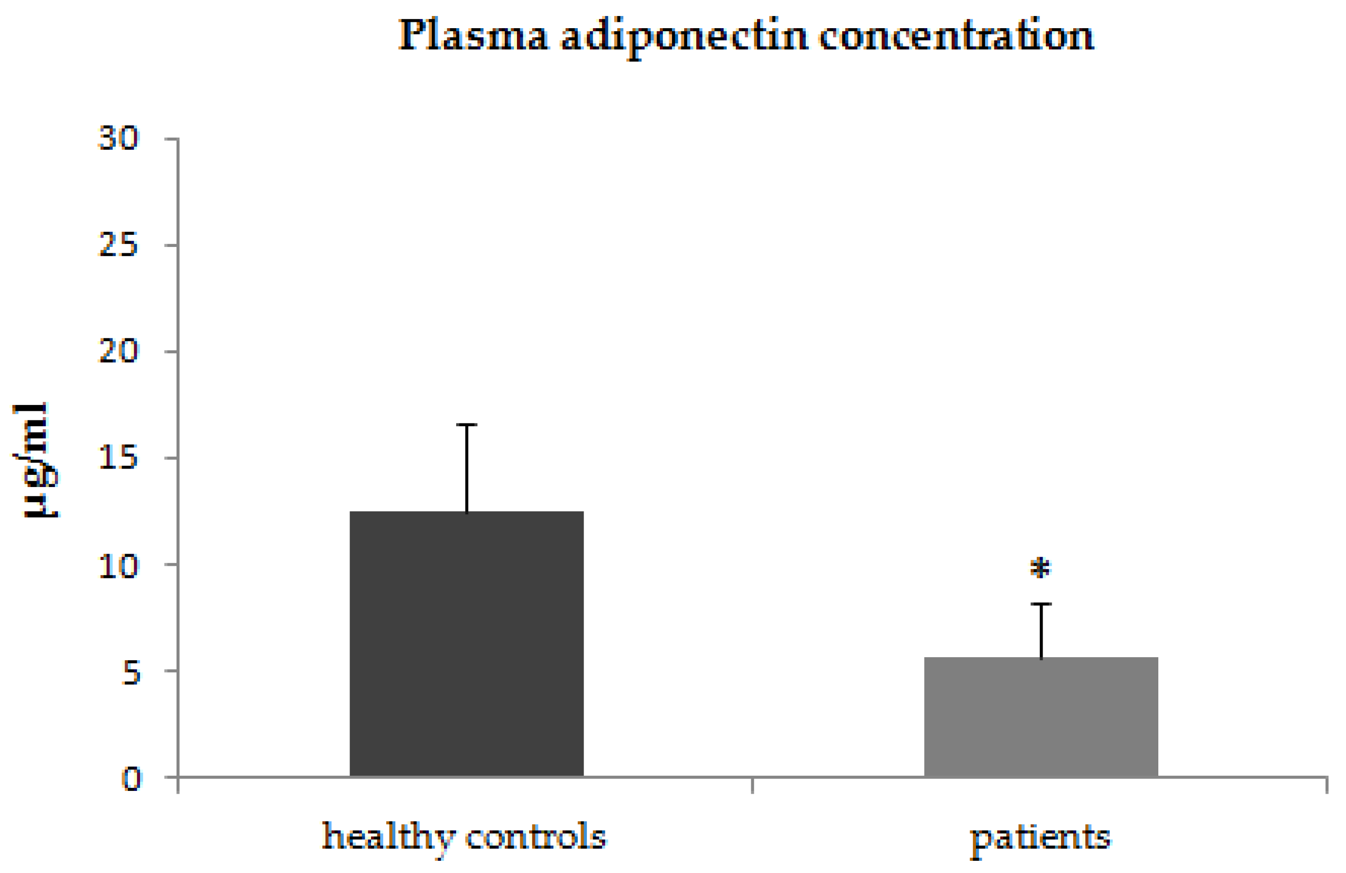
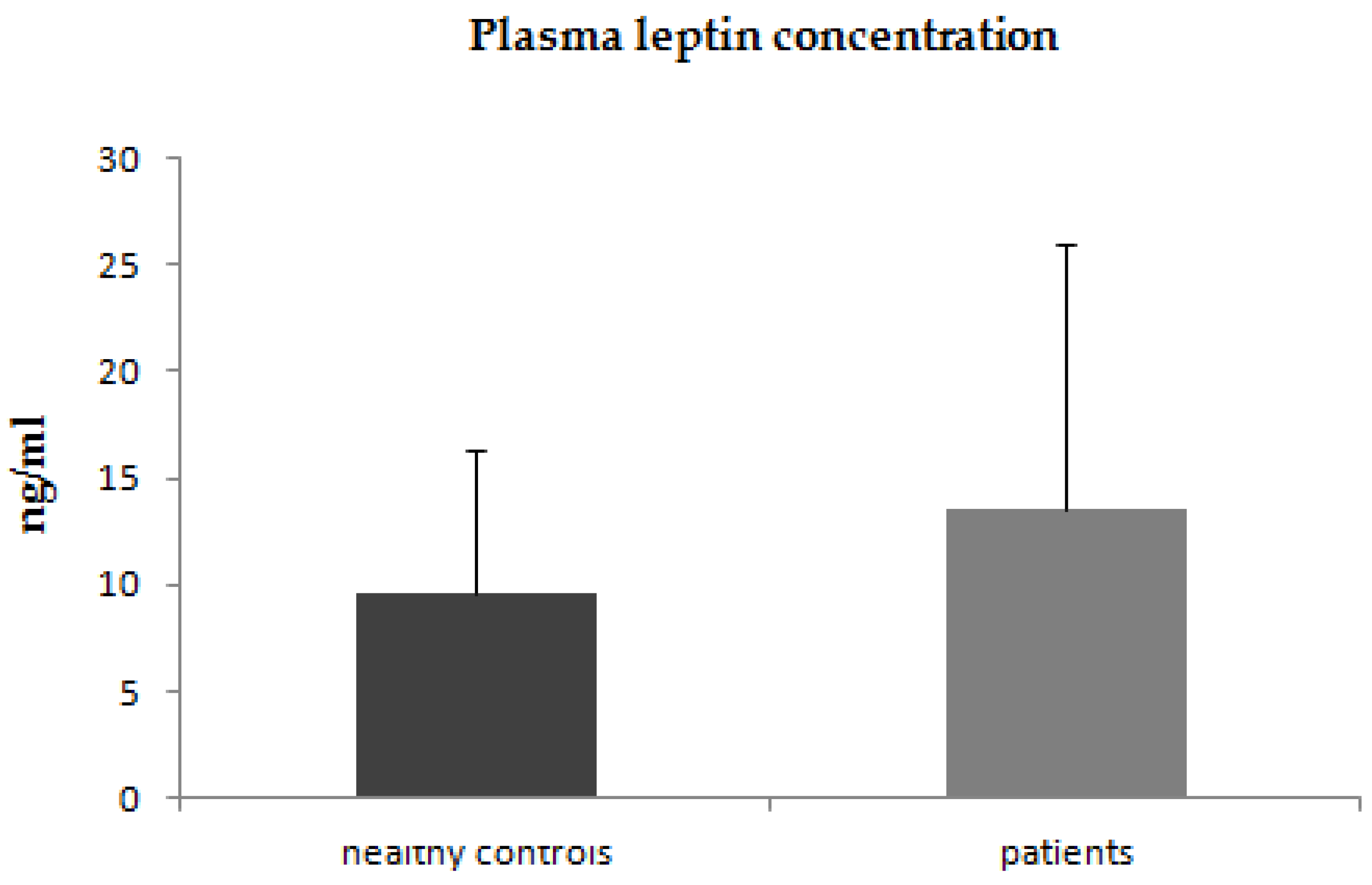
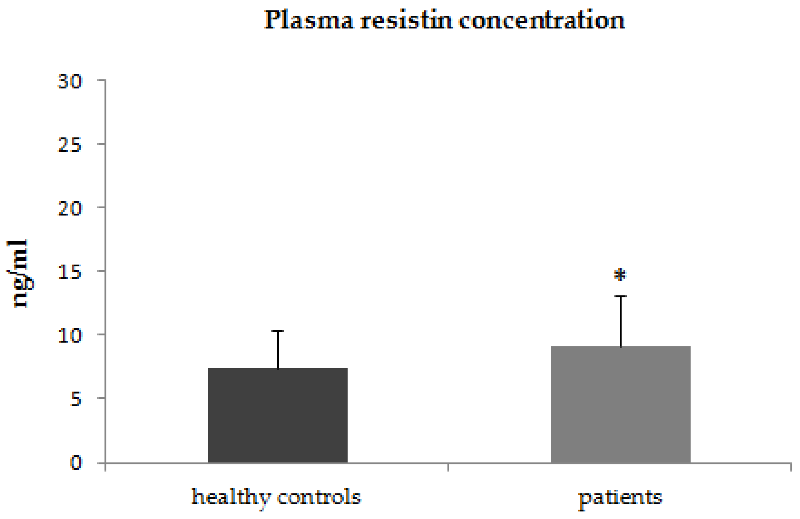
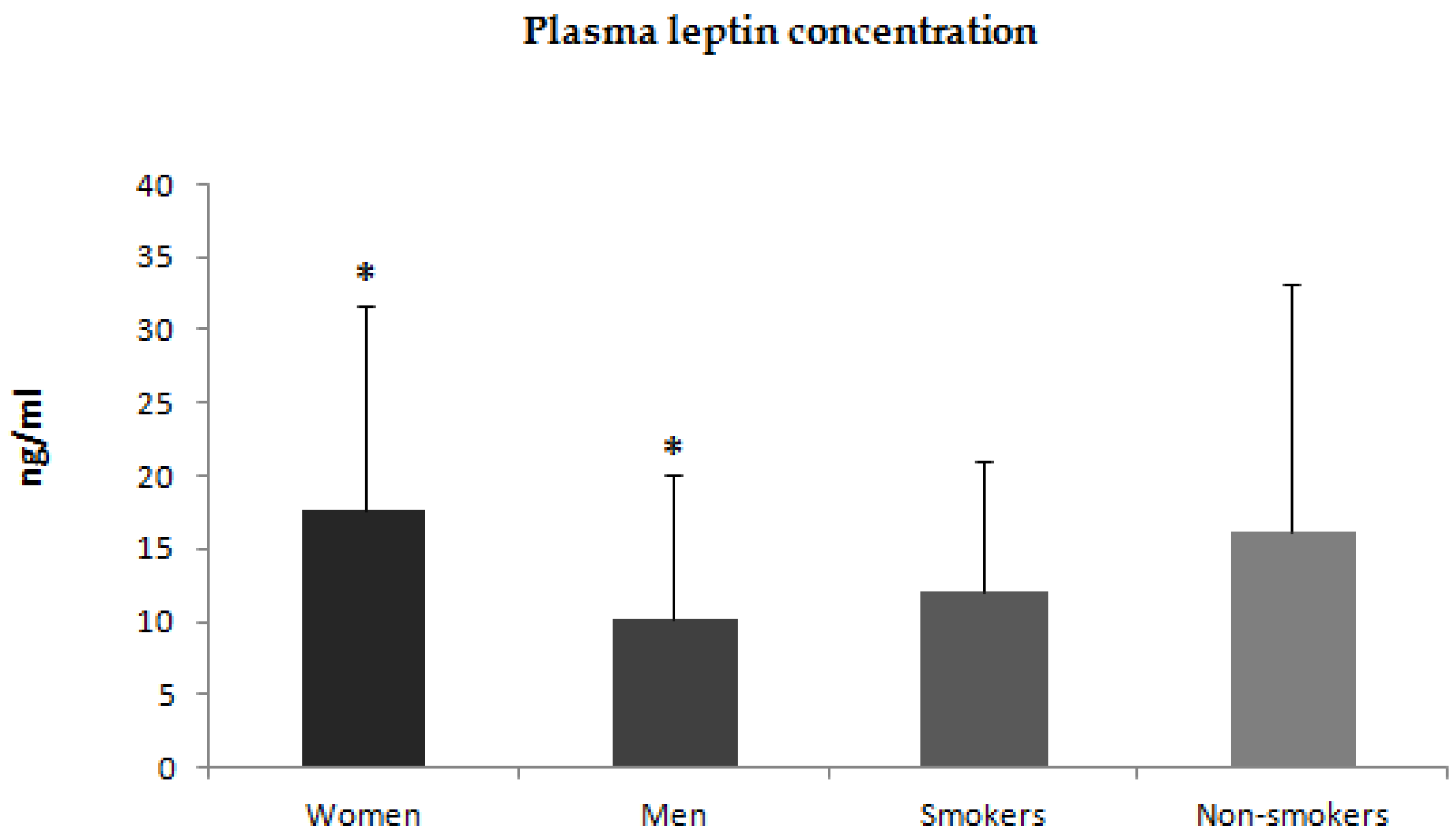
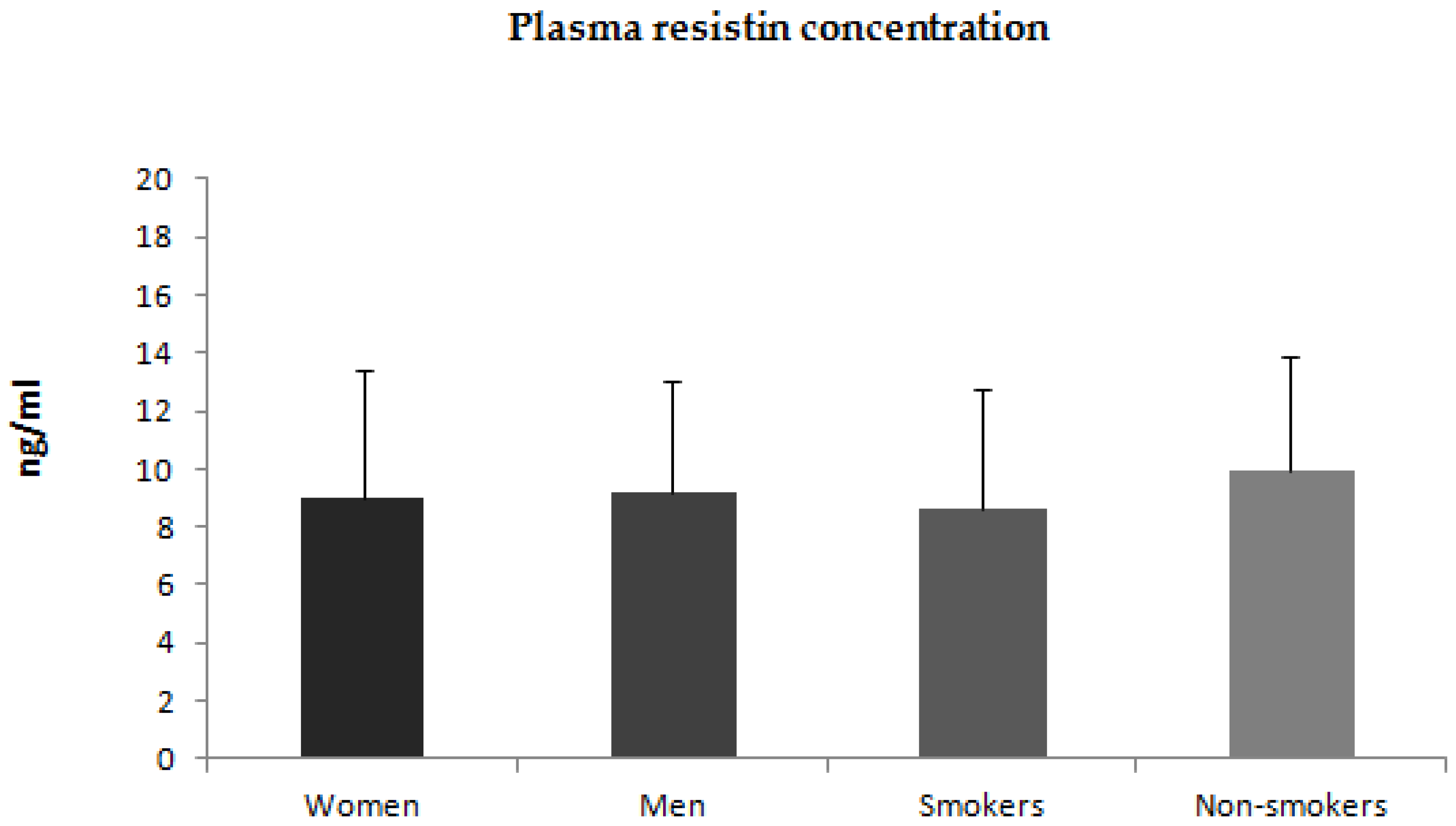
3.1. Assessment of Adiponectin, Leptin and Resistin Plasma Concentrations in Patients with Psoriasis and Control Subjects

In the first step of our study, we compared the plasma concentrations of adiponectin, leptin, and resistin between patients with psoriasis and healthy control subjects. The plasma concentrations of adiponectin were significantly lower in patients with psoriasis (p < 0.001) than in the control group (Figure 1). The plasma concentrations of leptin were higher in patients with psoriasis, however, due to high intra-patient variability of leptin plasma concentrations these differences did not reach statistical significance (p = 0.2) (Figure 2). The plasma concentrations of resistin were significantly increased in patients with psoriasis compared to healthy controls (p = 0.02) (Figure 3).
Additionally, we compared plasma concentrations of adiponectin, leptin, and resistin between men and women. Plasma concentrations of adiponectin and leptin were higher in women than in men (Figure 4 and Figure 5). There was no statistically significant difference in plasma concentration of resistin between men and women (Figure 6). We also compared plasma concentrations of adiponectin, leptin, and resistin between smoking and non-smoking patients. These differences were statistically nonsignificant (Figure 4, Figure 5 and Figure 6).

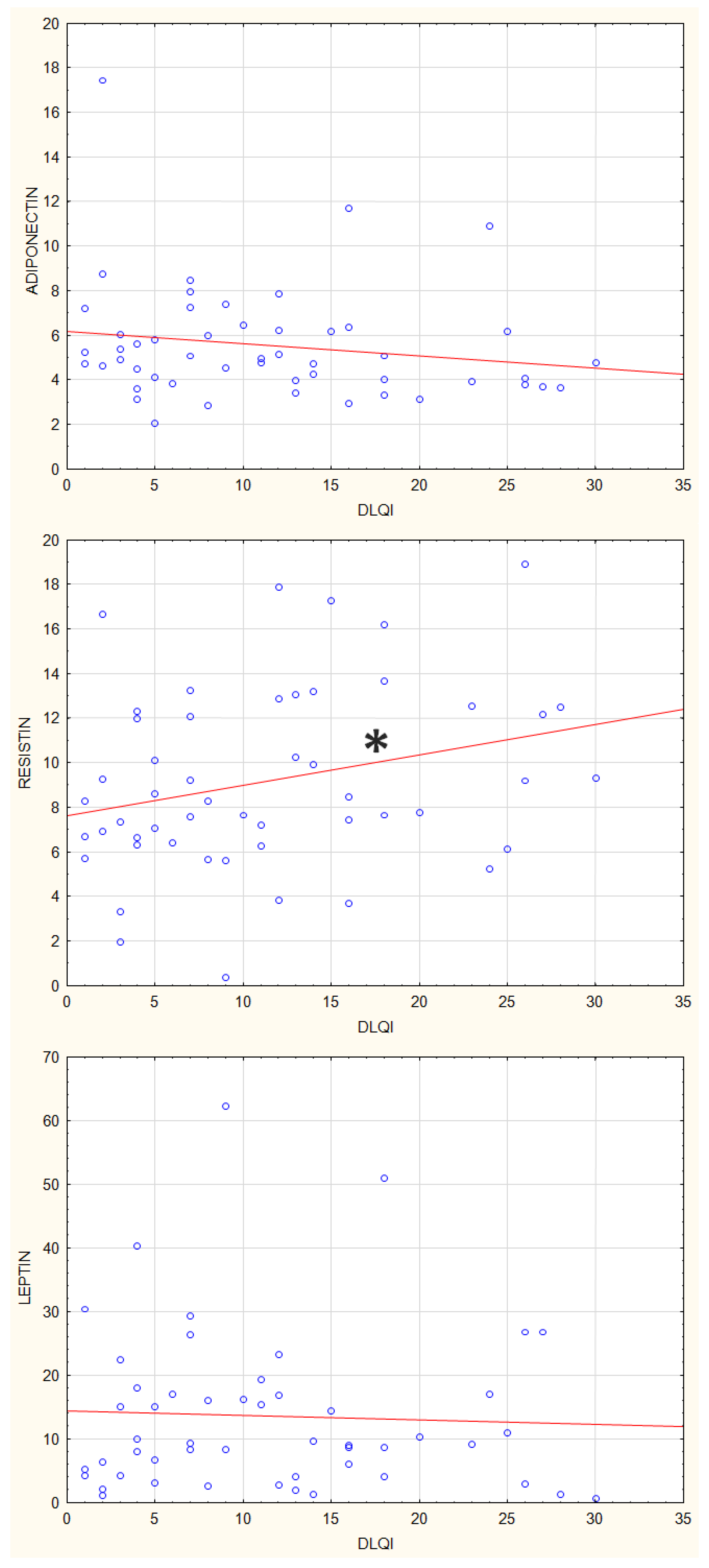
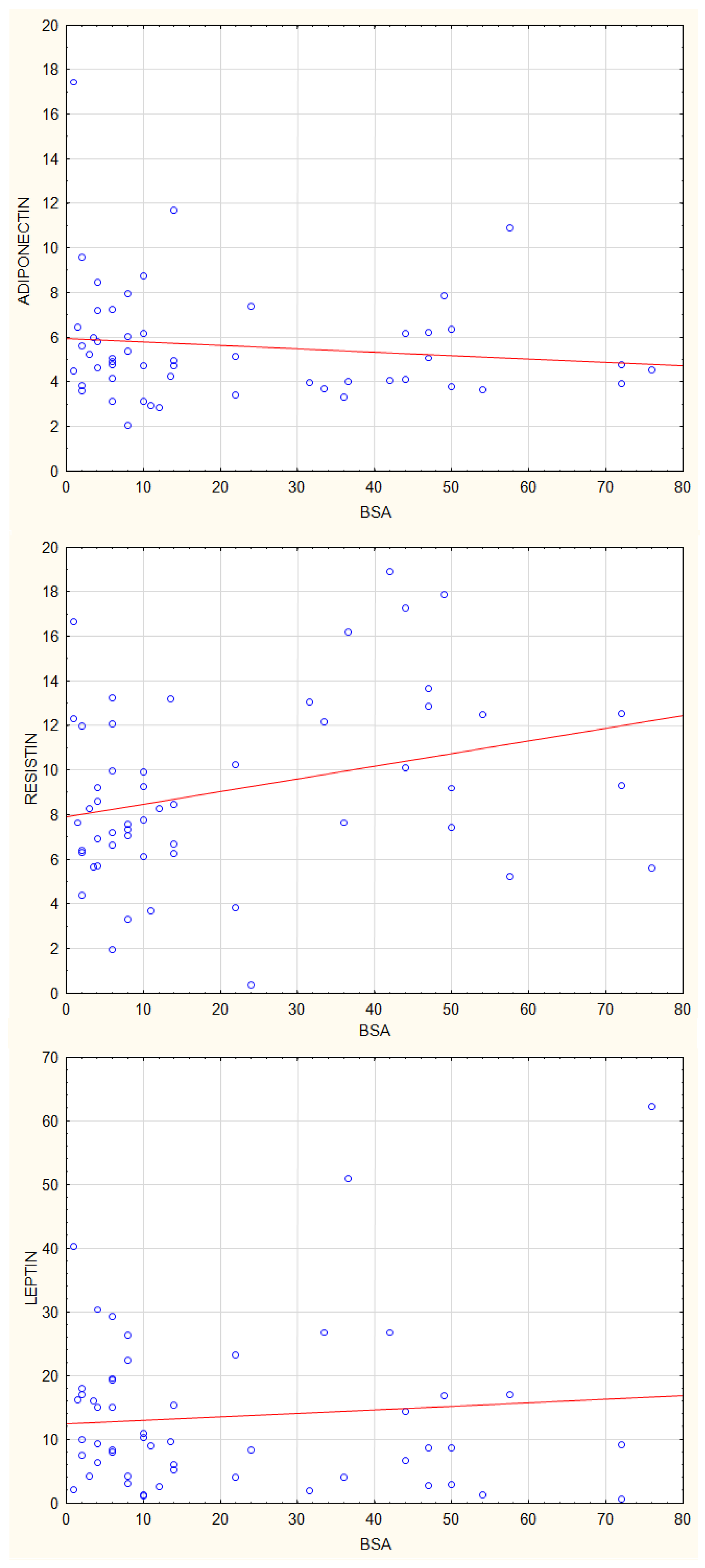
3.2. Correlations between Plasma Concentrations of Adiponectin, Leptin and Resistin and Disease Activity Parameters in Patients with Psoriasis

Then we analysed the correlations between plasma concentrations of adiponectin, leptin, and resistin, and parameters of psoriasis activity using the Dermatology Life Quality Index (DLQI), Psoriasis Activity Severity Index (PASI), and Body Surface Area (BSA) index. There were no statistically significant correlations between adiponectin and leptin plasma concentrations and values of, DLQI, PASI and BSA (Figure 7, Figure 8 and Figure 9). The resistin plasma concentrations correlated significantly with DLQI values (p = 0.03) (Figure 7).

3.3. Correlations between Plasma Concentrations of Adiponectin, Leptin, and Resistin and Clinical Parameters in Patients with Psoriasis

Additionally, we examined the correlations between adiponectin, leptin, and resistin plasma concentrations and selected clinical parameters. As shown in Table 1, plasma concentrations of adiponectin correlated significantly with the age of patients, whereas there were no statistically significant correlations between plasma levels of adiponectin, leptin and resistin and the age of disease diagnosis.
Plasma concentrations of adiponectin correlated significantly with CRP values and ALT values. Leptin plasma concentrations correlated significantly with BMI and creatinine values. There were no statistically significant correlations between plasma concentrations of adiponectin, leptin, and resistin and lipid parameters as well as sodium and potassium plasma concentrations. A positive correlation was found between leptin levels and plasma glucose levels, which had borderline statistical significance (p = 0.05) (Table 2).

4. Discussion

Adipose tissue has the very important endocrine functions of secreting adipokines and numerous cytokines [15]. Adipokines have multidirectional effects on the development of such diseases as dyslipidaemia, insulin resistance, diabetes, cardiovascular disease, rheumatoid arthritis, and kidney disease [16]. It has been shown that the action of adipokines can be multidirectional, they can both exacerbate the development of some diseases, as well as inhibit others. Previous studies suggest that adiponectin may have anti-inflammatory effects in psoriasis, slowing the development of the disease, while leptin and resistin may increase inflammation, exacerbating the symptoms of psoriasis [11,12]. Our study examined plasma concentrations of adiponectin, leptin, and resistin in patients with psoriasis. The concentrations of leptin and resistin were higher in patients with psoriasis than in the control group, while adiponectin concentrations were lower. There were no statistically significant correlations between plasma levels of adiponectin and leptin and disease severity parameters (PASI, DLQI, and BSA), while resistin plasma concentrations correlated significantly with DLQI values.
Adipokines affect metabolic processes and appear to provide a pathophysiological link between skin inflammation and metabolic changes in patients with psoriasis [15,16]. Adipokines are secreted by adipocytes and other cells such as macrophages [11,12]. In addition, it has been found that macrophages and monocytes outside adipose tissue, as well as endothelial and epithelial cells, can secrete adipokines [11,12]. Adipokines can exert very multidirectional effects. Resistin contributes to insulin resistance and diabetes. Leptin can lead to uncontrolled eating and weight gain promoting insulin resistance. In turn, adiponectin increases insulin sensitivity, decreasing insulin resistance [11,12].
Leptin increases the synthesis of pro-inflammatory mediators that play an important role in the development of psoriasis, such as TNF-α and CXCL8 [17,18]. These mediators can induce the activation of immune cells involved in the development of inflammation in the skin and psoriatic lesions [19]. The results indicate that adiponectin exerts anti-inflammatory effects in psoriasis by reducing the production of IL-6 and TNF-α [16,20]. On the other hand, TNF-α has been shown to inhibit adiponectin production, which may be one of the factors for reduced adiponectin levels in patients with psoriasis [21]. Studies suggest that adipokines in patients with psoriasis may contribute to the metabolic syndrome, regardless of BMI, and to the development of psoriatic skin lesions [22,23,24,25,26].
Previous studies have suggested that adipokines, may both exacerbate and attenuate skin inflammation in patients with psoriasis, as supported by reports of correlations between adipokines and psoriasis severity [25]. Adiponectin plays an anti-inflammatory role in keratinocytes by inhibiting the synthesis of pro-inflammatory cytokines, while increasing the secretion of anti-inflammatory cytokines [26,27,28,29]. Previous studies have shown lower adiponectin levels in patients with psoriasis than in healthy individuals [30,31,32]. Our results also showed lower levels of adiponectin in patients with psoriasis compared to healthy controls. We also found a positive correlation between plasma adiponectin levels and plasma C-reactive protein (CRP) levels. While the first studies suggested a negative correlation between plasma adiponectin levels and CRP levels, subsequent studies have shown a positive correlation [33,34,35,36]. This is explained by the stimulation of adiponectin synthesis by pro-inflammatory factors such as CRP. In our previous study, we demonstrated the stimulation of adiponectin expression in fibroblast-like synoviocytes by pro-inflammatory mediators such as TNF and LPS [37].
Adiponectin has also been shown to exert its anti-inflammatory effects in psoriasis patients by restoring the balance between Th1-Th17/Th2 and inhibiting IL-17A synthesis [38,39]. The role of adiponectin in the development of psoriasis has also been confirmed in experimental animal models. Mice lacking adiponectin develop severe psoriasis-like dermatitis and elevated levels of the Th17-related cytokines IL-17A, IL-17F, and IL-22 [40]. In our study, we found no correlation of plasma adiponectin setpoint with parameters of psoriasis activity, which is consistent with most clinical observations [30,35], although there are reports of a negative correlation between plasma adiponectin concentrations and PASI score [40,41,42]. Recent studies suggest that adiponectin is involved in sodium excretion [43,44]. In our study, we did not find statistically significant correlations between adiponectin and sodium plasma concentrations.
Previous studies have shown higher plasma leptin levels in psoriasis patients compared to healthy subjects [45,46,47]. In addition, a marked increase in leptin has been found in obese individuals, especially in those with the coexistence of psoriasis and obesity [48,49]. Furthermore, Mitsuyama et al. showed that leptin mRNA expression was significantly increased in subcutaneous adipose tissue of obese subjects with psoriasis compared to those with a normal BMI value [49]. Interestingly, leptin levels in non-obese patients with psoriasis did not differ significantly from non-obese controls. Leptin has been shown to increase the synthesis of pro-inflammatory mediators involved in the pathogenesis of psoriasis, such as IL-1, IL-6, TNF-α, and CXCL8 [50,51]. These mediators stimulate the Th1/Th17 axis, leading to increased IL-17/IL-23 levels [46,47,48,49,50,51,52,53]. The role of leptin in the pathogenesis of psoriasis has also been confirmed in numerous animal model studies. In our study, we observed increased plasma leptin levels in patients with psoriasis, but due to large intra-individual differences, these differences did not reach statistical significance. In addition, plasma leptin levels correlated significantly with BMI values.
Resistin is an adipokine synthesized mainly by macrophages and monocytes in adipose tissue. It has been shown that pro-inflammatory factors can increase its production [54]. Resistin, in turn, can stimulate B lymphocytes to secrete other pro-inflammatory factors that stimulate keratinocyte proliferation and T-cell recruitment to the skin. In addition, resistin can inhibit the proliferation of Foxp3+ regulatory T (Treg) cells in psoriasis [55]. The results of studies suggest that resistin plays an important role in the development of psoriasis by affecting inflammatory factors such as TNF-α and reducing the population of Foxp3+ Treg cells [55]. Previous studies have shown elevated plasma resistin levels in patients with psoriasis, which correlated with disease activity parameters [48,56,57]. Our study also found that elevated plasma resistin levels in patients with psoriasis correlated with the DLQI index.
Previous studies suggest an effect of diet on the development and course of psoriasis. A low-calorie diet has been shown to significantly improve patients’ clinical status and quality of life, as reflected in improvements in PASI and DLQI [58,59]. A low-calorie diet reduces the production of pro-inflammatory leukotriene B4. In addition, there is a decrease in the activity of CD4+ lymphocytes, and an increase in the production of IL-4, which has an anti-inflammatory effect in psoriasis. A low-calorie diet also reduces free radicals and causes a decrease in oxidative stress [60].
Saturated fatty acids have been shown to exacerbate skin inflammation, independent of obesity-related parameters such as increased fat mass, adipokine, and glucose levels. In patients with psoriasis, saturated fatty acid levels correlated with the activity of the disease process [61]. Saturated fatty acids intensify the inflammatory response leading to the activation of keratinocytes and macrophages. Palmitic acid has been shown to activate intracellular signaling pathways in epidermal keratinocytes, leading to increased synthesis of CCL20, CXCL8, and IL-1β [62]. The same processes can also be activated by leptin. Studies suggest that dietary intake, especially of saturated fatty acids, has an impact on the activation of inflammatory processes in the skin [61]. In addition, a low-calorie diet has been shown to increase plasma adiponectin concentrations together with a decrease in leptin concentrations [63].

5. Conclusions

In our study, we found increased plasma concentrations of leptin and resistin in patients with psoriasis, while adiponectin levels were significantly lower than in healthy subjects. There was no statistically significant correlation between adiponectin and leptin levels and disease severity parameters (PASI, DLQI, and BSA), though resistin plasma concentrations correlated significantly with DLQI values. The results of our study confirm the role of adiponectin, leptin, and resistin in the pathogenesis of psoriasis.

Author Contributions

Investigation: S.S.-G., M.S., M.M., E.D., K.Ł. and A.P.; software: K.S.; formal analysis: B.M. and A.P.; manuscript preparation: A.P.; conceptualization: S.S.-G. and A.P. All authors have read and agreed to the published version of the manuscript.

Funding

This research received no external funding.

Institutional Review Board Statement

The study was approved by the Ethics Committee of Pomeranian Medical University, Szczecin, Poland KB-0012/105/17.

Informed Consent Statement

Informed consent was obtained from all subjects involved in the study.

Data Availability Statement

Not applicable.

Conflicts of Interest

The authors declare no conflict of interest.

References

  1. Di Meglio, P.; Villanova, F.; Nestle, F.O. Psoriasis. Cold Spring Harb. Perspect. Med. 2014, 4, a015354. [Google Scholar] [CrossRef] [PubMed] [Green Version]
  2. Parisi, R.; Symmons, D.P.; Griffiths, C.E.; Ashcroft, D.M. Identification and Management of Psoriasis and Associated ComorbidiTy (IMPACT) project team. Global epidemiology of psoriasis: A systematic review of incidence and prevalence. J. Investig. Dermatol. 2013, 133, 377–385. [Google Scholar] [CrossRef] [PubMed] [Green Version]
  3. Griffiths, C.E.; Barker, J.N. Pathogenesis and clinical features of psoriasis. Lancet 2007, 370, 263–271. [Google Scholar] [CrossRef] [PubMed]
  4. Alexis, A.F.; Blackcloud, P. Psoriasis in skin of color: Epidemiology, genetics, clinical presentation, and treatment nuances. J. Clin. Aesthet. Dermatol. 2014, 7, 16–24. [Google Scholar]
  5. Tokura, Y.; Phadungsaksawasdi, P.; Kurihara, K.; Fujiyama, T.; Honda, T. Pathophysiology of Skin Resident Memory T Cells. Front. Immunol. 2021, 11, 618897. [Google Scholar] [CrossRef]
  6. Boehncke, W.H.; Brembilla, N.C. Autoreactive T-Lymphocytes in Inflammatory Skin Diseases. Front. Immunol. 2019, 10, 1198. [Google Scholar] [CrossRef] [PubMed] [Green Version]
  7. Hu, P.; Wang, M.; Gao, H.; Zheng, A.; Li, J.; Mu, D.; Tong, J. The Role of Helper T Cells in Psoriasis. Front. Immunol. 2021, 12, 788940. [Google Scholar] [CrossRef] [PubMed]
  8. Kelsen, J.; Dige, A.; Christensen, M.; D’Amore, F.; Iversen, L. Frequency and clonality of peripheral γδ T cells in psoriasis patients receiving anti-tumour necrosis factor-α therapy. Clin. Exp. Immunol. 2014, 177, 142–148. [Google Scholar] [CrossRef]
  9. Fantuzzi, G. Adipose tissue, adipokines, and inflammation. J. Allergy Clin. Immunol. 2005, 115, 911–919. [Google Scholar] [CrossRef]
  10. Perera, G.K.; Di Meglio, P.; Nestle, F.O. Psoriasis. Annu. Rev. Pathol. 2012, 7, 385–422. [Google Scholar] [CrossRef]
  11. Lynch, M.; Ahern, T.; Sweeney, C.M.; Malara, A.; Tobin, A.M.; O’Shea, D.; Kirby, B. Adipokines, psoriasis, systemic inflammation, and endothelial dysfunction. Int. J. Dermatol. 2017, 56, 1103–1118. [Google Scholar] [CrossRef] [PubMed]
  12. Gerdes, S.; Rostami-Yazdi, M.; Mrowietz, U. Adipokines and psoriasis. Exp. Dermatol. 2011, 20, 81–87. [Google Scholar] [CrossRef] [PubMed]
  13. Seminara, N.; Abuabara, K.; Shin, D.; Langan, S.; Kimmel, S.; Margolis, D.; Troxel, A.; Gelfand, J. Validity of The Health Improvement Network (THIN) for the study of psoriasis. Br. J. Dermatol. 2011, 164, 602–609. [Google Scholar] [CrossRef] [Green Version]
  14. Słuczanowska-Głabowska, S.; Jaworska, W.; Staniszewska, M.; Tkacz, M.; Safranow, K.; Łuczkowska, K.; Zagrodnik, E.; Stecewicz, I.; Machaliński, B.; Pawlik, A. Plasma Levels of Interleukins 36α, 36β, and 37 in Patients with Psoriasis and Their Correlation with Disease Activity Parameters. J. Clin. Med. 2022, 11, 5254. [Google Scholar] [CrossRef] [PubMed]
  15. Hao, Y.; Zhu, Y.J.; Zou, S.; Zhou, P.; Hu, Y.W.; Zhao, Q.X.; Gu, L.N.; Zhang, H.Z.; Wang, Z.; Li, J. Metabolic Syndrome and Psoriasis: Mechanisms and Future Directions. Front. Immunol. 2021, 12, 711060. [Google Scholar] [CrossRef] [PubMed]
  16. Farkhondeh, T.; Llorens, S.; Pourbagher-Shahri, A.M.; Ashrafizadeh, M.; Talebi, M.; Shakibaei, M.; Samarghandian, S. An Overview of the Role of Adipokines in Cardiometabolic Diseases. Molecules 2020, 25, 5218. [Google Scholar] [CrossRef]
  17. Johnston, A.; Arnadottir, S.; Gudjonsson, J.E.; Aphale, A.; Sigmarsdottir, A.A.; Gunnarsson, S.I.; Steinsson, J.T.; Elder, J.T.; Valdimarsson, H. Obesity in psoriasis: Leptin and resistin as mediators of cutaneous inflammation. Br. J. Dermatol. 2008, 159, 342–350. [Google Scholar] [CrossRef] [Green Version]
  18. Su, X.; Zhang, G.; Cheng, Y.; Wang, B. Leptin in skin disease modulation. Clin. Chim. Acta 2021, 516, 8–14. [Google Scholar] [CrossRef]
  19. Su, X.; Cheng, Y.; Chang, D. The Important Role of Leptin in Modulating the Risk of Dermatological Diseases. Front. Immunol. 2021, 11, 593564. [Google Scholar] [CrossRef]
  20. Bahceci, M.; Gokalp, D.; Bahceci, S.; Tuzcu, A.; Atmaca, S.; Arikan, S. The correlation between adiposity and adiponectin, tumor necrosis factor alpha, interleukin-6 and high sensitivity C-reactive protein levels. Is adipocyte size associated with inflammation in adults? J. Endocrinol. Investig. 2007, 30, 210–214. [Google Scholar] [CrossRef]
  21. He, Y.; Lu, L.; Wei, X.; Jin, D.; Qian, T.; Yu, A.; Sun, J.; Cui, J.; Yang, Z. The multimerization and secretion of adiponectin are regulated by TNF-alpha. Endocrine 2016, 51, 456–468. [Google Scholar] [CrossRef] [PubMed]
  22. Hulthe, J.; Hultén, L.M.; Fagerberg, B. Low adipocyte-derived plasma protein adiponectin concentrations are associated with the metabolic syndrome and small dense low-density lipoprotein particles: Atherosclerosis and insulin resistance study. Metabolism 2003, 52, 1612–1614. [Google Scholar] [CrossRef] [PubMed]
  23. Spranger, J.; Kroke, A.; Möhlig, M.; Bergmann, M.M.; Ristow, M.; Boeing, H.; Pfeiffer, A.F. Adiponectin and protection against type 2 diabetes mellitus. Lancet 2003, 361, 226–228, Erratum in Lancet 2002, 361, 1060. [Google Scholar] [CrossRef] [PubMed]
  24. Chen, Y.J.; Wu, C.Y.; Shen, J.L.; Chu, S.Y.; Chen, C.K.; Chang, Y.T.; Chen, C.M. Psoriasis independently associated with hyperleptinemia contributing to metabolic syndrome. Arch. Dermatol. 2008, 144, 1571–1575. [Google Scholar] [CrossRef] [Green Version]
  25. Wolk, K.; Sabat, R. Adipokines in psoriasis: An important link between skin inflammation and metabolic alterations. Rev. Endocr. Metab. Disord. 2016, 17, 305–317. [Google Scholar] [CrossRef] [PubMed]
  26. Coimbra, S.; Catarino, C.; Santos-Silva, A. The triad psoriasis-obesity-adipokine profile. J. Eur. Acad. Dermatol. Venereol. 2016, 30, 1876–1885. [Google Scholar] [CrossRef]
  27. Versini, M.; Jeandel, P.Y.; Rosenthal, E.; Shoenfeld, Y. Obesity in autoimmune diseases: Not a passive bystander. Autoimmun. Rev. 2014, 13, 981–1000. [Google Scholar] [CrossRef]
  28. Sterry, W.; Strober, B.E.; Menter, A.; International Psoriasis Council. Obesity in psoriasis: The metabolic, clinical and therapeutic implications. Report of an interdisciplinary conference and review. Br. J. Dermatol. 2007, 157, 649–655. [Google Scholar] [CrossRef]
  29. Wong, Y.; Nakamizo, S.; Tan, K.J.; Kabashima, K. An Update on the Role of Adipose Tissues in Psoriasis. Front. Immunol. 2019, 10, 1507. [Google Scholar] [CrossRef]
  30. Bai, F.; Zheng, W.; Dong, Y.; Wang, J.; Garstka, M.A.; Li, R.; An, J.; Ma, H. Serum levels of adipokines and cytokines in psoriasis patients: A systematic review and meta-analysis. Oncotarget 2017, 9, 1266–1278. [Google Scholar] [CrossRef] [Green Version]
  31. Gerdes, S.; Osadtschy, S.; Rostami-Yazdi, M.; Buhles, N.; Weichenthal, M.; Mrowietz, U. Leptin, adiponectin, visfatin and retinol-binding protein-4-mediators of comorbidities in patients with psoriasis? Exp. Dermatol. 2012, 21, 43–47. [Google Scholar] [CrossRef] [PubMed]
  32. Shibata, S.; Saeki, H.; Tada, Y.; Karakawa, M.; Komine, M.; Tamaki, K. Serum high molecular weight adiponectin levels are decreased in psoriasis patients. J. Dermatol. Sci. 2009, 55, 62–63. [Google Scholar] [CrossRef] [PubMed]
  33. Ouchi, N.; Kihara, S.; Funahashi, T.; Nakamura, T.; Nishida, M.; Kumada, M.; Okamoto, Y.; Ohashi, K.; Nagaretani, H.; Kishida, K.; et al. Reciprocal association of C-reactive protein with adiponectin in blood stream and adipose tissue. Circulation 2003, 107, 671–674. [Google Scholar] [CrossRef] [Green Version]
  34. Fantuzzi, G. Adiponectin and inflammation: Consensus and controversy. J. Allergy Clin. Immunol. 2008, 121, 326–330. [Google Scholar] [CrossRef]
  35. Baran, A.; Flisiak, I.; Jaroszewicz, J.; Świderska, M. Effect of psoriasis activity on serum adiponectin and leptin levels. Postepy. Dermatol. Alergol. 2015, 32, 101–106. [Google Scholar] [CrossRef] [PubMed] [Green Version]
  36. Ghoshal, K.; Chatterjee, T.; Chowdhury, S.; Sengupta, S.; Bhattacharyya, M. Adiponectin Genetic Variant and Expression Coupled with Lipid Peroxidation Reveal New Signatures in Diabetic Dyslipidemia. Biochem. Genet. 2021, 59, 781–798. [Google Scholar] [CrossRef] [PubMed]
  37. Łączna, M.; Kopytko, P.; Tkacz, M.; Zgutka, K.; Czerewaty, M.; Tarnowski, M.; Larysz, D.; Tkacz, R.; Kotrych, D.; Piotrowska, K.; et al. Adiponectin Is a Component of the Inflammatory Cascade in Rheumatoid Arthritis. J. Clin. Med. 2022, 11, 2740. [Google Scholar] [CrossRef]
  38. Campanati, A.; Orciani, M.; Consales, V.; Lazzarini, R.; Ganzetti, G.; Di Benedetto, G.; Di Primio, R.; Offidani, A. Characterization and profiling of immunomodulatory genes in resident mesenchymal stem cells reflect the Th1-Th17/Th2 imbalance of psoriasis. Arch. Dermatol. Res. 2014, 306, 915–920. [Google Scholar] [CrossRef]
  39. Surendar, J.; Frohberger, S.J.; Karunakaran, I.; Schmitt, V.; Stamminger, W.; Neumann, A.L.; Wilhelm, C.; Hoerauf, A.; Hübner, M.P. Adiponectin Limits IFN-γ and IL-17 Producing CD4 T Cells in Obesity by Restraining Cell Intrinsic Glycolysis. Front. Immunol. 2019, 10, 2555. [Google Scholar] [CrossRef] [Green Version]
  40. Shibata, S.; Tada, Y.; Hau, C.S.; Mitsui, A.; Kamata, M.; Asano, Y.; Sugaya, M.; Kadono, T.; Masamoto, Y.; Kurokawa, M.; et al. Adiponectin regulates psoriasiform skin inflammation by suppressing IL-17 production from γδ-T cells. Nat. Commun. 2015, 6, 7687. [Google Scholar] [CrossRef] [Green Version]
  41. Takahashi, H.; Tsuji, H.; Takahashi, I.; Hashimoto, Y.; Ishida-Yamamoto, A.; Iizuka, H. Plasma adiponectin and leptin levels in Japanese patients with psoriasis. Br. J. Dermatol. 2008, 159, 1207–1208. [Google Scholar] [CrossRef] [PubMed]
  42. Eder, L.; Jayakar, J.; Pollock, R.; Pellett, F.; Thavaneswaran, A.; Chandran, V.; Rosen, C.F.; Gladman, D.D. Serum adipokines in patients with psoriatic arthritis and psoriasis alone and their correlation with disease activity. Ann. Rheum. Dis. 2013, 72, 1956–1961. [Google Scholar] [CrossRef] [PubMed]
  43. Huang, H.; Han, Y.; Jose, P.A.; Zeng, C. Loss of Adiponectin-Induced Renal Sodium Excretion in Hypertension. J. Am. Soc. Hypertens. 2016, 10 (Suppl. S1), e4. [Google Scholar] [CrossRef] [PubMed]
  44. Zhang, Y.; Wang, S.; Huang, H.; Zeng, A.; Han, Y.; Zeng, C.; Zheng, S.; Ren, H.; Wang, Y.; Huang, Y.; et al. GRK4-mediated adiponectin receptor-1 phosphorylative desensitization as a novel mechanism of reduced renal sodium excretion in hypertension. Clin. Sci. Lond. 2020, 134, 2453–2467. [Google Scholar] [CrossRef] [PubMed]
  45. Cerman, A.A.; Bozkurt, S.; Sav, A.; Tulunay, A.; Elbaşi, M.O.; Ergun, T. Serum leptin levels, skin leptin and leptin receptor expression in psoriasis. Br. J. Dermatol. 2008, 159, 820–826. [Google Scholar] [CrossRef] [PubMed]
  46. Wang, Y.; Chen, J.; Zhao, Y.; Geng, L.; Song, F.; Chen, H.D. Psoriasis is associated with increased levels of serum leptin. Br. J. Dermatol. 2008, 158, 1134–1135. [Google Scholar] [CrossRef] [PubMed]
  47. Coimbra, S.; Oliveira, H.; Reis, F.; Belo, L.; Rocha, S.; Quintanilha, A.; Figueiredo, A.; Teixeira, F.; Castro, E.; Rocha-Pereira, P.; et al. Circulating adipokine levels in Portuguese patients with psoriasis vulgaris according to body mass index, severity and therapy. J. Eur. Acad. Dermatol. Venereol. 2010, 24, 1386–1394. [Google Scholar] [CrossRef]
  48. Kyriakou, A.; Patsatsi, A.; Sotiriadis, D.; Goulis, D.G. Serum Leptin, Resistin, and Adiponectin Concentrations in Psoriasis: A Meta-Analysis of Observational Studies. Dermatology 2017, 233, 378–389. [Google Scholar] [CrossRef]
  49. Mitsuyama, S.; Abe, F.; Kimura, M.; Yoshida, M.; Higuchi, T. Association between leptin gene expression in subcutaneous adipose tissue and circulating leptin levels in obese patients with psoriasis. Arch. Dermatol. Res. 2015, 307, 539–544. [Google Scholar] [CrossRef]
  50. El-Boghdady, N.A.; Ismail, M.F.; Abd-Alhameed, M.F.; Ahmed, A.S.; Ahmed, H.H. Bidirectional Association Between Psoriasis and Obesity: Benefits and Risks. J. Interferon. Cytokine. Res. 2018, 38, 12–19. [Google Scholar] [CrossRef]
  51. Nakajima, H.; Nakajima, K.; Tarutani, M.; Sano, S. Clear association between serum levels of adipokines and T-helper 17-related cytokines in patients with psoriasis. Clin. Exp. Dermatol. 2013, 38, 66–70. [Google Scholar] [CrossRef] [PubMed]
  52. Xue, K.; Liu, H.; Jian, Q.; Liu, B.; Zhu, D.; Zhang, M.; Gao, L.; Li, C. Leptin induces secretion of pro-inflammatory cytokines by human keratinocytes in vitro--a possible reason for increased severity of psoriasis in patients with a high body mass index. Exp. Dermatol. 2013, 22, 406–410. [Google Scholar] [CrossRef] [PubMed]
  53. Zhu, K.J.; Zhang, C.; Li, M.; Zhu, C.Y.; Shi, G.; Fan, Y.M. Leptin levels in patients with psoriasis: A meta-analysis. Clin. Exp. Dermatol. 2013, 38, 478–483. [Google Scholar] [CrossRef]
  54. Tripathi, D.; Kant, S.; Pandey, S.; Ehtesham, N.Z. Resistin in metabolism, inflammation, and disease. FEBS J. 2020, 287, 3141–3149. [Google Scholar] [CrossRef] [Green Version]
  55. Zhang, L.; Yang, X.Q.; Cheng, J.; Hui, R.S.; Gao, T.W. Increased Th17 cells are accompanied by FoxP3(+) Treg cell accumulation and correlated with psoriasis disease severity. Clin. Immunol. 2010, 135, 108–117. [Google Scholar] [CrossRef] [PubMed]
  56. Seth, D.; Ehlert, A.N.; Golden, J.B.; Damiani, G.; McCormick, T.S.; Cameron, M.J.; Cooper, K.D. Interaction of Resistin and Systolic Blood Pressure in Psoriasis Severity. J. Invest. Dermatol. 2020, 140, 1279–1282. [Google Scholar] [CrossRef]
  57. Huang, H.; Shen, E.; Tang, S.; Tan, X.; Guo, X.; Wang, Q.; Ding, H. Increased serum resistin levels correlate with psoriasis: A meta-analysis. Lipids Health Dis. 2015, 14, 44. [Google Scholar] [CrossRef] [Green Version]
  58. Pona, A.; Haidari, W.; Kolli, S.S.; Feldman, S.R. Diet and psoriasis. Dermatol. Online J. 2019, 25, 13030/qt1p37435s. [Google Scholar] [CrossRef]
  59. Wu, A.G.; Weinberg, J.M. The impact of diet on psoriasis. Cutis 2019, 104, 7–10, Erratum in Cutis 2019, 104, 241. [Google Scholar]
  60. Garbicz, J.; Całyniuk, B.; Górski, M.; Buczkowska, M.; Piecuch, M.; Kulik, A.; Rozentryt, P. Nutritional Therapy in Persons Suffering from Psoriasis. Nutrients 2021, 14, 119. [Google Scholar] [CrossRef]
  61. Herbert, D.; Franz, S.; Popkova, Y.; Anderegg, U.; Schiller, J.; Schwede, K.; Lorz, A.; Simon, J.C.; Saalbach, A. High-Fat Diet Exacerbates Early Psoriatic Skin Inflammation Independent of Obesity: Saturated Fatty Acids as Key Players. J. Investig. Dermatol. 2018, 138, 1999–2009. [Google Scholar] [CrossRef] [PubMed] [Green Version]
  62. Ikeda, K.; Morizane, S.; Akagi, T.; Hiramatsu-Asano, S.; Tachibana, K.; Yahagi, A.; Iseki, M.; Kaneto, H.; Wada, J.; Ishihara, K.; et al. Obesity and Dyslipidemia Synergistically Exacerbate Psoriatic Skin Inflammation. Int. J. Mol. Sci. 2022, 23, 4312. [Google Scholar] [CrossRef] [PubMed]
  63. Salehi-Abargouei, A.; Izadi, V.; Azadbakht, L. The effect of low calorie diet on adiponectin concentration: A systematic review and meta-analysis. Horm. Metab. Res. 2015, 47, 549–555. [Google Scholar] [CrossRef] [PubMed]
Figure 1. Plasma concentrations of adiponectin in patients with psoriasis and control group * p < 0.001.
Jcm 12 00663 g001
Figure 2. Plasma concentrations of leptin in patients with psoriasis and control group.
Jcm 12 00663 g002
Figure 3. Plasma concentrations of resistin in patients with psoriasis and control group * p = 0.02.
Jcm 12 00663 g003
Figure 4. Plasma concentrations of adiponectin in women and men with psoriasis and smoking and non-smoking patients * p < 0.001.
Jcm 12 00663 g004
Figure 5. Plasma concentrations of leptin in women and men with psoriasis and smoking and non-smoking patients * p = 0.002.
Jcm 12 00663 g005
Figure 6. Plasma concentrations of resistin in women and men with psoriasis and smoking and non-smoking patients.
Jcm 12 00663 g006
Figure 7. Correlations between plasma concentrations of adiponectin, leptin, and resistin and DLQI index, * p = 0.03.
Jcm 12 00663 g007
Figure 8. Correlations between plasma concentrations of adiponectin, leptin and resistin and PASI index.
Jcm 12 00663 g008
Figure 9. Correlations between plasma concentrations of adiponectin, leptin and resistin and BSA index.
Jcm 12 00663 g009
Table 1. Clinical characteristics of patients and control group.
ParametersPatients with Psoriasis
n-53
Control Group
n-31
Male2917
Female2414
Age [years]49.7 ± 17.648.5 ± 14.2
Age of disease onset [years]34.03 ± 20.08-
Smoking1912
DLQI11.47 ± 8.19-
PASI11.28 ± 10.87-
BSA21.33 ± 21.18-
Table 2. Correlations between adiponectin, leptin and resistin plasma concentrations and clinical parameters.
AdiponectinLeptinResistin
ParameterRspRspRsp
Age0.280.030.020.830.20.14
Age of disease onset0.050.710.050.67−0.060.62
BMI−0.330.140.560.010.240.29
Leukocytes0.020.920.210.330.220.29
ESR0.350.090.360.080.120.58
CRP0.430.040.270.200.330.11
AST−0.220.290.120.580.100.62
ALT−0.440.030.170.410.100.64
Creatinin0.030.980.450.030.090.66
Sodium0.080.740.130.570.080.73
Potassium0.260.270.190.410.060.79
Cholesterol−0.230.280.010.950.300.16
LDL−0.400.070.050.830.010.97
Triglycerides−0.020.940.290.19−0.010.97
Glucose−0.020.920.430.050.240.29
Disclaimer/Publisher’s Note: The statements, opinions and data contained in all publications are solely those of the individual author(s) and contributor(s) and not of MDPI and/or the editor(s). MDPI and/or the editor(s) disclaim responsibility for any injury to people or property resulting from any ideas, methods, instructions or products referred to in the content.

