
Sentinel Lymph Node Biopsy in Malignant Melanoma of the Head and Neck: A Single Center Experience

 , , , , and
Abstract
:1. Introduction
2. Methods
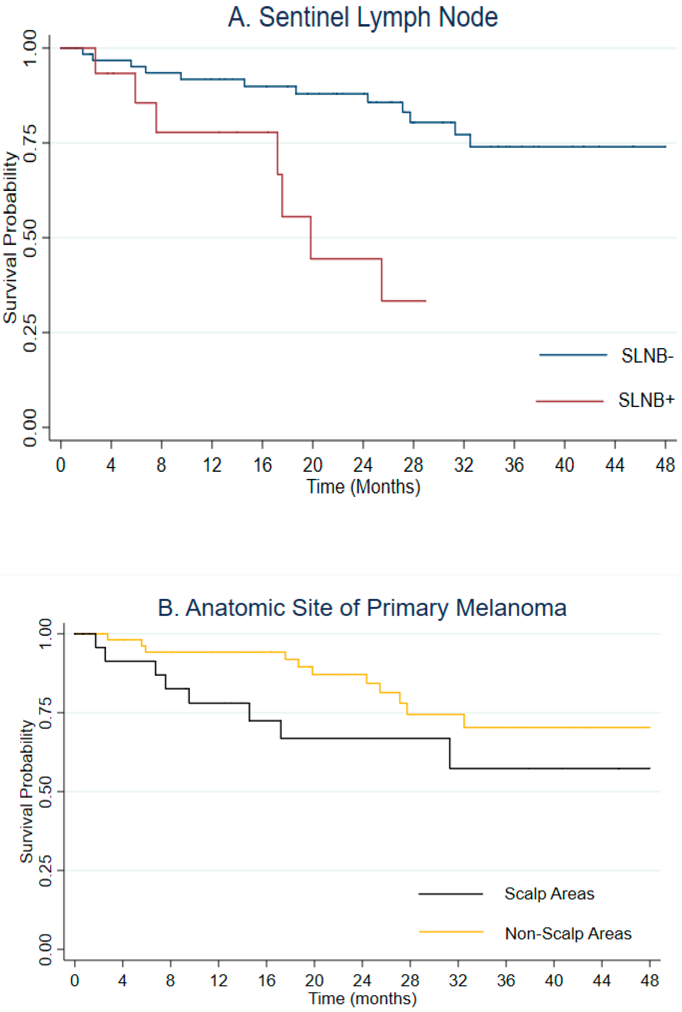
3. Results
4. Discussion
5. Conclusions
Author Contributions
Funding
Institutional Review Board Statement
Informed Consent Statement
Data Availability Statement
Conflicts of Interest
References
- Olla, D.; Tufaro, A.P.; Neumeister, M.W. Extirpative Considerations of Melanoma of the Head and Neck. Clin. Plast. Surg. 2021, 48, 659–668. [Google Scholar] [CrossRef] [PubMed]
- Kraft, S.; Granter, S.R. Molecular pathology of cutaneous neoplasms of the head and neck. Arch. Pathol. Lab. Med. 2014, 138, 759–787. [Google Scholar] [CrossRef] [PubMed]
- Haenssle, H.A.; Hoffmann, S.; Buhl, T.; Emmert, S.; Schön, M.P.; Bertsch, H.P.; Rosenberger, A. Assessment of melanoma histotypes and patient-associated factors: Basis for a predictive statistical model. J. Dtsch. Dermatol. Ges. 2015, 13, 37–45. [Google Scholar] [CrossRef] [PubMed]
- Ribero, S.; Stucci, L.S.; Marra, E.; Marconcini, R.; Spagnolo, F.; Orgiano, L.; Picasso, V.; Queirolo, P.; Palmieri, G.; Quaglino, P.; et al. Effect of Age on Melanoma Risk, Prognosis and Treatment Response. Acta Derm. Venereol. 2018, 98, 624–629. [Google Scholar] [CrossRef] [Green Version]
- Callender, G.G.; Egger, M.E.; Burton, A.L.; Scoggins, C.R.; Ross, M.I.; Stromberg, A.J.; Hagendoorn, L.; Martin, R.C., 2nd; McMasters, K.M. Prognostic implications of anatomic location of primary cutaneous melanoma of 1 mm or thicker. Am. J. Surg. 2011, 202, 659–664; discussion 664–665. [Google Scholar] [CrossRef]
- Fadaki, N.; Li, R.; Parrett, B.; Sanders, G.; Thummala, S.; Martineau, L.; Cardona-Huerta, S.; Miranda, S.; Cheng, S.T.; Miller, J.R., 3rd; et al. Is head and neck melanoma different from trunk and extremity melanomas with respect to sentinel lymph node status and clinical outcome? Ann. Surg. Oncol. 2013, 20, 3089–3097. [Google Scholar] [CrossRef] [PubMed]
- Licata, G.; Scharf, C.; Ronchi, A.; Pellerone, S.; Argenziano, G.; Verolino, P.; Moscarella, E. Diagnosis and Management of Melanoma of the Scalp: A Review of the Literature. Clin. Cosmet Investig. Dermatol. 2021, 14, 1435–1447. [Google Scholar] [CrossRef]
- Gibbs, P.; Robinson, W.A.; Pearlman, N.; Raben, D.; Walsh, P. Management of primary cutaneous melanoma of the head and neck: The University of Colorado experience and a review of the literature. J. Surg. Oncol. 2001, 77, 179–185; discussion 86–87. [Google Scholar] [CrossRef]
- Ettl, T.; Irga, S.; Müller, S.; Rohrmeier, C.; Reichert, T.E.; Schreml, S.; Gosau, M. Value of anatomic site, histology and clinicopathological parameters for prediction of lymph node metastasis and overall survival in head and neck melanomas. J. Craniomaxillofac. Surg. 2014, 42, e252–e258. [Google Scholar] [CrossRef]
- Boada, A.; Tejera-Vaquerizo, A.; Ribero, S.; Puig, S.; Moreno-Ramírez, D.; Descalzo-Gallego, M.A.; Fierro, M.T.; Quaglino, P.; Carrera, C.; Malvehy, J.; et al. Sentinel lymph node biopsy versus observation in thick melanoma: A multicenter propensity score matching study. Int. J. Cancer 2018, 142, 641–648. [Google Scholar] [CrossRef]
- Quaglino, P.; Ribero, S.; Osella-Abate, S.; Macrì, L.; Grassi, M.; Caliendo, V.; Asioli, S.; Sapino, A.; Macripò, G.; Savoia, P.; et al. Clinico-pathologic features of primary melanoma and sentinel lymph node predictive for non-sentinel lymph node involvement and overall survival in melanoma patients: A single centre observational cohort study. Surg. Oncol. 2011, 20, 259–264. [Google Scholar] [CrossRef] [PubMed]
- Aiom Melanoma Guidelines 2021. Available online: https://www.aiom.it/linee-guida-aiom-2021-melanoma/ (accessed on 5 January 2023).
- Coit, D.G.; Thompson, J.A.; Albertini, M.R.; Barker, C.; Carson, W.E.; Contreras, C.; Daniels, G.A.; DiMaio, D.; Fields, R.C.; Fleming, M.D.; et al. Cutaneous melanoma, version 2.2019, NCCN clinical practice guidelines in oncology. J. Natl. Compr. Canc. Netw. 2019, 17, 367–402. [Google Scholar] [CrossRef] [PubMed] [Green Version]
- Huang, K.; Misra, S.; Lemini, R.; Chen, Y.; Speicher, L.L.; Dawson, N.L.; Tolaymat, L.M.; Bagaria, S.P.; Gabriel, E.M. Completion lymph node dissection in patients with sentinel lymph node positive cutaneous head and neck melanoma. J. Surg. Oncol. 2020, 122, 1057–1065. [Google Scholar] [CrossRef] [PubMed]
- Leiter, U.; Stadler, R.; Mauch, C.; Hohenberger, W.; Brockmeyer, N.; Berking, C.; Sunderkötter, C.; Kaatz, M.; Schulte, K.W.; Lehmann, P.; et al. Complete lymph node dissection versus no dissection in patients with sentinel lymph node biopsy positive melanoma (DeCOG-SLT): A multicentre, randomised, phase 3 trial. Lancet Oncol. 2016, 17, 757–767. [Google Scholar] [CrossRef] [PubMed]
- López-Rodríguez, E.; García-Gómez, F.J.; Álvarez-Pérez, R.M.; Martínez-Castillo, R.; Borrego-Dorado, I.; Fernández-Ortega, P.; Zulueta-Dorado, T. Role of SPECT-CT in sentinel lymph node biopsy in patients diagnosed with head and neck melanoma. Rev. Esp. Med. Nucl. Imagen Mol. 2016, 35, 22–28. [Google Scholar] [CrossRef]
- Bachar, G.; Tzelnick, S.; Amiti, N.; Gutman, H. Patterns of failure in patients with cutaneous head and neck melanoma. Eur. J. Surg. Oncol. 2020, 46, 914–917. [Google Scholar] [CrossRef]
- Saaiq, M.; Zalaudek, I.; Rao, B.; Lee, Y.; Rudnicka, L.; Czuwara, J.; Giuffrida, R.; Wollina, U.; Jafferany, M.; Lotti, T.; et al. A brief synopsis on scalp melanoma. Dermatol. Ther. 2020, 33, e13795. [Google Scholar] [CrossRef]
- Mandalà, M.; Galli, F.; Cattaneo, L.; Merelli, B.; Rulli, E.; Ribero, S.; Quaglino, P.; De Giorgi, V.; Pigozzo, J.; Sileni, V.C.; et al. Italian Melanoma Intergroup. Mitotic rate correlates with sentinel lymph node status and outcome in cutaneous melanoma greater than 1 millimeter in thickness: A multi-institutional study of 1524 cases. J. Am. Acad. Dermatol. 2017, 76, 264–273.e2. [Google Scholar] [CrossRef]
- Roy, J.M.; Whitfield, R.J.; Gill, P.G. Review of the role of sentinel node biopsy in cutaneous head and neck melanoma. ANZ J. Surg. 2016, 86, 348–355. [Google Scholar] [CrossRef]
- Huang, K.; Misra, S.; Lemini, R.; Chen, Y.; Speicher, L.L.; Dawson, N.L.; Tolaymat, L.M.; Bagaria, S.P.; Gabriel, E.M. Melanoma patients with positive sentinel lymph nodes who did not undergo completion lymphadenectomy: A multi-institutional study. Ann. Surg. Oncol. 2006, 13, 809–816. [Google Scholar]
- Faries, M.B.; Thompson, J.F.; Cochran, A.J.; Andtbacka, R.H.; Mozzillo, N.; Zager, J.S.; Jahkola, T.; Bowles, T.L.; Testori, A.; Beitsch, P.D.; et al. Completion dissection or observation for sentinel lymph node metastasis in melanoma. N. Engl. J. Med. 2017, 376, 2211–2222. [Google Scholar] [CrossRef] [PubMed]
- Testori, A.A.E.; Ribero, S.; Indini, A.; Mandalà, M. Adjuvant Treatment of Melanoma: Recent Developments and Future Perspectives. Am. J. Clin. Dermatol. 2019, 20, 817–827. [Google Scholar] [CrossRef] [PubMed]
- Ollila, D.W.; Foshag, L.J.; Essner, R.; Stern, S.L.; Morton, D.L. Parotid region lymphatic mapping and sentinel lymphadenectomy for cutaneous melanoma. Ann. Surg. Oncol. 1999, 6, 150–154. [Google Scholar] [CrossRef]
- Lin, D.; Franc, B.L.; Kashani-Sabet, M.; Singer, M.I. Lymphatic drainage patterns of head and neck cutaneous melanoma observed on lymphoscintigraphy and sentinel lymph node biopsy. Head Neck. 2006, 28, 249–255. [Google Scholar] [CrossRef] [PubMed]
- Jensen, J.D.; Gray, R.J.; Wasif, N.; Roarke, M.C.; Casey, W.J.; Kreymerman, P.; Pockaj, B.A. Can lymphatic drainage of head and neck melanoma be predicted? J. Surg. Oncol. 2011, 103, 751–755. [Google Scholar] [CrossRef] [PubMed]
- Douglas, R.G.; Shaw, J.H.F. Melanoma of the head and neck in Auckland. N. Z. Med. J. 1987, 100, 584–587. [Google Scholar] [PubMed]
- Pereira, A.R.; Collgros, H.; Guitera, P.; Benati, E.; Longo, C.; Argenziano, G.; Dika, E.; Lambertini, M.; Menzies, S.W.; Lobato Williams, A.; et al. Melanomas of the scalp: Is hair coverage preventing early diagnosis? Int. J. Dermatol. 2021, 60, 340–346. [Google Scholar] [CrossRef]
- Chapman, B.C.; Gleisner, A.; Kwak, J.J.; Hosokawa, P.; Paniccia, A.; Merkow, J.S.; Koo, P.J.; Gajdos, C.; Pearlman, N.W.; McCarter, M.D.; et al. SPECT/CT Improves Detection of Metastatic Sentinel Lymph Nodes in Patients with Head and Neck Melanoma. Ann. Surg. Oncol. 2016, 23, 2652–2657. [Google Scholar] [CrossRef]
- Quartuccio, N.; Garau, L.M.; Arnone, A.; Pappalardo, M.; Rubello, D.; Arnone, G.; Manca, G. Comparison of 99mTc-Labeled Colloid SPECT/CT and Planar Lymphoscintigraphy in Sentinel Lymph Node Detection in Patients with Melanoma: A Meta-Analysis. J. Clin. Med. 2020, 9, 1680. [Google Scholar] [CrossRef]
- Vermeeren, L.; Valdés Olmos, R.A.; Klop, W.M.; van der Ploeg, I.M.; Nieweg, O.E.; Balm, A.J.; van den Brekel, M.W. SPECT/CT for sentinel lymph node mapping in head and neck melanoma. Head Neck. 2011, 33, 1–6. [Google Scholar] [CrossRef]


| Nodal Status | Anatomic Site | ||||||
|---|---|---|---|---|---|---|---|
| Variable | Overall (N = 93) | Nodal Negative (N = 75) | Nodal Positive (N = 18) | pa | Non-Scalp (N = 64) | Scalp (N = 29) | pa |
| Age, median (IQR) | 58 (50–70) | 59 (51–71) | 52 (41–61) | 0.04 | 53 (45–69) | 63 (55–72) | 0.01 |
| Sex | |||||||
| Male | 45 (51.6) | 40 (53.3) | 8 (44.4) | 0.49 | 31 (48.4) | 17 (58.6) | 0.36 |
| Female | 48 (48.4) | 35 (46.7) | 10 (55.6) | 33 (51.6) | 12 (41.4) | ||
| Breslow, median (IQR) | 1.8 (1.2–3.5) | 1.8 (1.1–3.0) | 2.25 (1.8–5.0) | 0.04 | 1.8 (1.2–2.6) | 2.5 (1.4–4.2) | 0.04 |
| T Stage | |||||||
| T1 | 13 (14.3) | 13 (17.3) | 0 (0.0) | 0.10 | 10 (15.6) | 3 (11.1) | 0.33 |
| T2 | 43 (47.3) | 35 (46.7) | 8 (44.4) | 33 (51.6) | 10 (37.0) | ||
| T3 | 15 (16.5) | 12 (16.0) | 3 (16.7) | 10 (15.6) | 5 (18.5) | ||
| T4 | 20 (22.0) | 13 (17.3) | 7 (38.9) | 11 (17.2) | 9 (33.3) | ||
| Missing | 2 | 2 | 0 | 0 | 2 | ||
| Ulceration | |||||||
| Absent | 57 (66.3) | 45 (66.2) | 12 (66.7) | 0.97 | 40 (67.8) | 17 (63.0) | 0.66 |
| Present | 29 (33.7) | 23 (33.8) | 6 (33.3) | 19 (32.2) | 10 (37.0) | ||
| Missing | 7 | 7 | 0 | 5 | 2 | ||
| Anatomic site | |||||||
| Scalp | 29 (31.5) | 22 (29.7) | 7 (38.9) | 0.63 | / | / | / |
| Face | 35 (38.0) | 27 (36.4) | 8 (44.4) | / | / | ||
| Ear | 19 (20.7) | 17 (23.0) | 2 (11.1) | / | / | ||
| Neck | 9 (9.8) | 8 (10.8) | 1 (5.6) | / | / | ||
| Drainage | |||||||
| Monolateral | 79 (86.0%) | 64 (86.5) | 15 (83.3) | 0.49 | 56 (88.9) | 23 (79.3) | 0.33 |
| Bilateral | 13 (14.1%) | 10 (13.5) | 3 (16.7) | 7 (11.1) | 6 (20.7) | ||
| Missing | 1 | 1 | 0 | 1 | 0 | ||
| Nodal Disease | |||||||
| Yes | 75 (80.6) | / | / | / | 53 (82.0) | 22 (75.9) | 0.43 |
| No | 18 (19.4) | / | / | 11 (18.0) | 7 (24.1) | ||
| Variable | HR 1 | 95% CIS 1 | HR 2 | 95% CIS 2 |
|---|---|---|---|---|
| Age (1 year increase) | 1.04 | 1.00–1.08 | 1.04 | 1.00–1.08 |
| Sex | ||||
| Females | Ref | Ref | Ref | Ref |
| Males | 1.85 | 0.72–4.71 | / | / |
| Breslow (1 mm increase) | 1.40 | 1.20–1.63 | 1.29 | 1.03–1.65 |
| T stage | ||||
| T1,T2 | Ref | Ref | Ref | Ref |
| T3,T4 | 4.09 | 1.54–10.8 | 1.20 | 0.31–4.60 |
| Ulceration | ||||
| No | Ref | Ref | Ref | Ref |
| Yes | 1.23 | 0.45–3.39 | / | / |
| SLNB | ||||
| Negative | Ref | Ref | Ref | Ref |
| Positive | 4.90 | 1.80–13.30 | 2.65 | 0.88–7.95 |
| Scalp | ||||
| Non scalp | Ref | Ref | Ref | Ref |
| Scalp | 1.92 | 0.77–4.78 | 1.31 | 0.47–3.63 |
Disclaimer/Publisher’s Note: The statements, opinions and data contained in all publications are solely those of the individual author(s) and contributor(s) and not of MDPI and/or the editor(s). MDPI and/or the editor(s) disclaim responsibility for any injury to people or property resulting from any ideas, methods, instructions or products referred to in the content. |
© 2023 by the authors. Licensee MDPI, Basel, Switzerland. This article is an open access article distributed under the terms and conditions of the Creative Commons Attribution (CC BY) license (https://creativecommons.org/licenses/by/4.0/).
Share and Cite
Rubatto, M.; Picciotto, F.; Moirano, G.; Fruttero, E.; Caliendo, V.; Borriello, S.; Sciamarrelli, N.; Fava, P.; Senetta, R.; Lesca, A.; et al. Sentinel Lymph Node Biopsy in Malignant Melanoma of the Head and Neck: A Single Center Experience. J. Clin. Med. 2023, 12, 553. https://doi.org/10.3390/jcm12020553
Rubatto M, Picciotto F, Moirano G, Fruttero E, Caliendo V, Borriello S, Sciamarrelli N, Fava P, Senetta R, Lesca A, et al. Sentinel Lymph Node Biopsy in Malignant Melanoma of the Head and Neck: A Single Center Experience. Journal of Clinical Medicine. 2023; 12(2):553. https://doi.org/10.3390/jcm12020553
Chicago/Turabian StyleRubatto, Marco, Franco Picciotto, Giovenale Moirano, Enrico Fruttero, Virginia Caliendo, Silvia Borriello, Nadia Sciamarrelli, Paolo Fava, Rebecca Senetta, Adriana Lesca, and et al. 2023. "Sentinel Lymph Node Biopsy in Malignant Melanoma of the Head and Neck: A Single Center Experience" Journal of Clinical Medicine 12, no. 2: 553. https://doi.org/10.3390/jcm12020553