
Factors Influencing Acromial and Scapular Spine Strain after Reverse Total Shoulder Arthroplasty: A Systematic Review of Biomechanical Studies

 , , , and
Abstract
:1. Introduction
2. Material and Methods
2.1. Search Strategy
2.2. Selection Process
- (1)
- Biomechanical in-vitro or in-silico studies;
- (2)
- Studies reporting on acromion or scapular spine fracture, strain, and stress;
- (3)
- Studies in the German or English language;
- (4)
- Studies released between January 2015 and May 2021.
2.3. Data Interpretation
2.4. Study Quality Assessment
3. Results
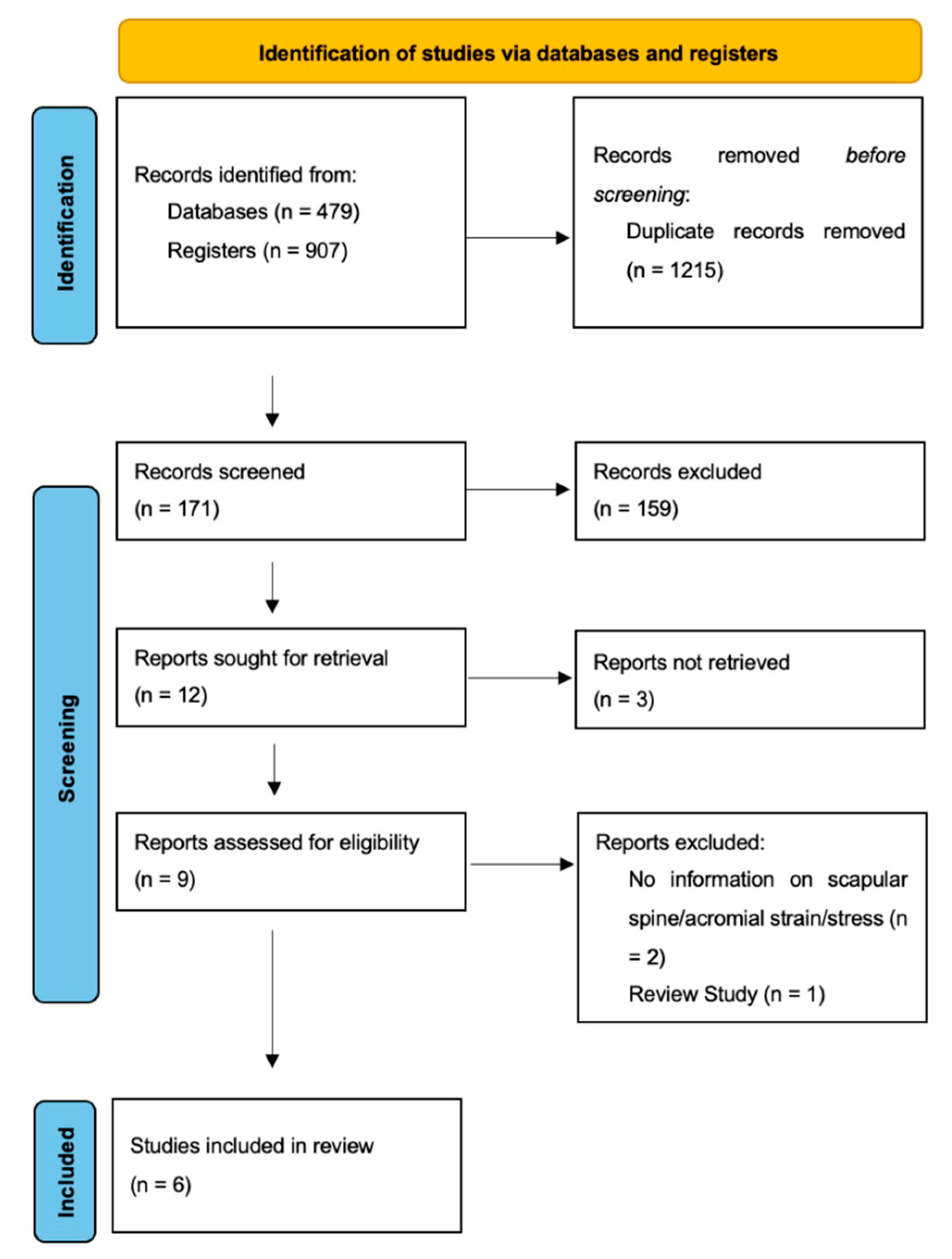
3.1. Search Results
3.2. Study Quality Assessment
3.3. Study Characteristics
3.4. Glenoid Lateralization
3.5. Glenoid Inferiorization
3.6. Humeral Lateralization
3.7. Deltoid Lengthening
3.8. Neck-Shaft Angle
3.9. Acromial Morphology
3.10. Coracoacromial Ligament
4. Discussion
5. Conclusions
Supplementary Materials
Author Contributions
Funding
Conflicts of Interest
References
- Drake, G.N.; O’Connor, D.P.; Edwards, T.B. Indications for reverse total shoulder arthroplasty in rotator cuff disease. Clin. Orthop. Relat. Res. 2010, 468, 1526–1533. [Google Scholar] [CrossRef] [Green Version]
- Ek, E.T.; Neukom, L.; Catanzaro, S.; Gerber, C. Reverse total shoulder arthroplasty for massive irreparable rotator cuff tears in patients younger than 65 years old: Results after five to fifteen years. J. Shoulder Elb. Surg. 2013, 22, 1199–1208. [Google Scholar] [CrossRef] [Green Version]
- Ernstbrunner, L.; Andronic, O.; Grubhofer, F.; Camenzind, R.S.; Wieser, K.; Gerber, C. Long-term results of reverse total shoulder arthroplasty for rotator cuff dysfunction: A systematic review of longitudinal outcomes. J. Shoulder Elb. Surg. 2019, 28, 774–781. [Google Scholar] [CrossRef]
- Ernstbrunner, L.; Suter, A.; Catanzaro, S.; Rahm, S.; Gerber, C. Reverse Total Shoulder Arthroplasty for Massive, Irreparable Rotator Cuff Tears Before the Age of 60 Years: Long-Term Results. J. Bone Jt. Surg. 2017, 99, 1721–1729. [Google Scholar] [CrossRef] [PubMed] [Green Version]
- Ernstbrunner, L.; Werthel, J.D.; Wagner, E.; Hatta, T.; Sperling, J.W.; Cofield, R.H. Glenoid bone grafting in primary reverse total shoulder arthroplasty. J. Shoulder Elb. Surg. 2017, 26, 1441–1447. [Google Scholar] [CrossRef]
- Gerber, C.; Canonica, S.; Catanzaro, S.; Ernstbrunner, L. Longitudinal observational study of reverse total shoulder arthroplasty for irreparable rotator cuff dysfunction: Results after 15 years. J. Shoulder Elb. Surg. Surg. 2018, 28, 774–781. [Google Scholar] [CrossRef] [PubMed]
- Yahuaca, B.I.; Simon, P.; Christmas, K.N.; Patel, S.; Gorman, R.A.; Mighell, M.A.; Frankle, M.A. Acute surgical management of proximal humerus fractures: ORIF vs. hemiarthroplasty vs. reverse shoulder arthroplasty. J. Shoulder Elb. Surg. 2020, 29, S32–S40. [Google Scholar] [CrossRef]
- Sabah, Y.; Decroocq, L.; Gauci, M.O.; Bonnevialle, N.; Lemmex, D.B.; Chelli, M.; Valenti, P.; Boileau, P. Clinical and radiological outcomes of reverse shoulder arthroplasty for acute fracture in the elderly. Int. Orthop. 2021, 45, 1775–1781. [Google Scholar] [CrossRef]
- Cazeneuve, J.F.; Cristofari, D.J. Delta III reverse shoulder arthroplasty: Radiological outcome for acute complex fractures of the proximal humerus in elderly patients. Orthop. Traumatol. Surg. Res. OTSR 2009, 95, 325–329. [Google Scholar] [CrossRef] [Green Version]
- Ernstbrunner, L.; Rahm, S.; Suter, A.; Imam, M.; Catanzaro, S.; Grubhofer, F.; Gerber, C. Salvage reverse total shoulder arthroplasty for failed operative treatment of proximal humeral fractures in patients younger than 60 years: Long-term results. J. Shoulder Elb. Surg. 2019, 29, 561–570. [Google Scholar] [CrossRef] [PubMed]
- Flury, M.P.; Frey, P.; Goldhahn, J.; Schwyzer, H.-K.; Simmen, B.R. Reverse shoulder arthroplasty as a salvage procedure for failed conventional shoulder replacement due to cuff failure--midterm results. Int. Orthop. 2011, 35, 53–60. [Google Scholar] [CrossRef] [Green Version]
- Grammont, P.; Trouilloud, P.; Laffay, J.; Deries, X. Etude et réalisation d’une nouvelle prothèse d’épaule. Rhumatologie 1987, 39, 407–418. [Google Scholar]
- Boileau, P.; Watkinson, D.J.; Hatzidakis, A.M.; Balg, F. Grammont reverse prosthesis: Design, rationale, and biomechanics. J. Shoulder Elb. Surg. 2005, 14, 147S–161S. [Google Scholar] [CrossRef]
- Lockhart, J.S.; Wong, M.T.; Langohr, G.D.G.; Athwal, G.S.; Johnson, J.A. The effect of arm loading and plane of elevation on acromial stress after reverse shoulder arthroplasty. J Orthop. Res 2017, 35, 388–395. [Google Scholar]
- Crosby, L.A.; Hamilton, A.; Twiss, T. Scapula fractures after reverse total shoulder arthroplasty: Classification and treatment. Clin. Orthop. Relat. Res. 2011, 469, 2544–2549. [Google Scholar] [CrossRef] [PubMed] [Green Version]
- Hamid, N.; Connor, P.M.; Fleischli, J.F.; D’Alessandro, D.F. Acromial fracture after reverse shoulder arthroplasty. Am. J. Orthop. 2011, 40, E125–E129. [Google Scholar] [PubMed]
- Hattrup, S.J. The influence of postoperative acromial and scapular spine fractures on the results of reverse shoulder arthroplasty. Orthopedics 2010, 33, 302. [Google Scholar] [CrossRef]
- Walch, G.; Mottier, F.; Wall, B.; Boileau, P.; Molé, D.; Favard, L. Acromial insufficiency in reverse shoulder arthroplasties. J. Shoulder Elb. Surg. 2009, 18, 495–502. [Google Scholar] [CrossRef]
- Cuff, D.; Pupello, D.; Virani, N.; Levy, J.; Frankle, M. Reverse shoulder arthroplasty for the treatment of rotator cuff deficiency. J. Bone Jt. Surg. Am. 2008, 90, 1244–1251. [Google Scholar] [CrossRef]
- Levy, J.C.; Anderson, C.; Samson, A. Classification of postoperative acromial fractures following reverse shoulder arthroplasty. J. Bone Jt. Surg. Am. 2013, 95, e104. [Google Scholar] [CrossRef]
- Zhou, H.S.; Chung, J.S.; Yi, P.H.; Li, X.; Price, M.D. Management of complications after reverse shoulder arthroplasty. Curr. Rev. Musculoskelet. Med. 2015, 8, 92–97. [Google Scholar] [CrossRef] [Green Version]
- Teusink, M.J.; Otto, R.J.; Cottrell, B.J.; Frankle, M.A. What is the effect of postoperative scapular fracture on outcomes of reverse shoulder arthroplasty? J. Shoulder Elb. Surg. 2014, 23, 782–790. [Google Scholar] [CrossRef] [PubMed]
- Neyton, L.; Erickson, J.; Ascione, F.; Bugelli, G.; Lunini, E.; Walch, G. Grammont Award 2018: Scapular fractures in reverse shoulder arthroplasty (Grammont style): Prevalence, functional, and radiographic results with minimum 5-year follow-up. J. Shoulder Elb. Surg. 2019, 28, 260–267. [Google Scholar] [CrossRef]
- Mayne, I.P.; Bell, S.N.; Wright, W.; Coghlan, J.A. Acromial and scapular spine fractures after reverse total shoulder arthroplasty. Shoulder Elb. 2016, 8, 90–100. [Google Scholar] [CrossRef] [PubMed] [Green Version]
- Patterson, D.C.; Chi, D.; Parsons, B.O.; Cagle, P.J. Acromial spine fracture after reverse total shoulder arthroplasty: A systematic review. J. Shoulder Elb. Surg. 2019, 28, 792–801. [Google Scholar] [CrossRef] [PubMed]
- Frankle, M.; Siegal, S.; Pupello, D.; Saleem, A.; Mighell, M.; Vasey, M. The Reverse Shoulder Prosthesis for glenohumeral arthritis associated with severe rotator cuff deficiency. A minimum two-year follow-up study of sixty patients. J. Bone Jt. Surg. Am. Vol. 2005, 87, 1697–1705. [Google Scholar] [CrossRef]
- Farshad, M.; Gerber, C. Reverse total shoulder arthroplasty-from the most to the least common complication. Int. Orthop. 2010, 34, 1075–1082. [Google Scholar] [CrossRef] [PubMed] [Green Version]
- Moverman, M.A.; Menendez, M.E.; Mahendraraj, K.A.; Polisetty, T.; Jawa, A.; Levy, J.C. Patient risk factors for acromial stress fractures after reverse shoulder arthroplasty: A multicenter study. J. Shoulder Elb. Surg. 2021, 30, 1619–1625. [Google Scholar] [CrossRef] [PubMed]
- Shah, S.S.; Gentile, J.; Chen, X.; Kontaxis, A.; Dines, D.M.; Warren, R.F.; Taylor, S.A.; Jahandar, A.; Gulotta, L.V. Influence of implant design and parasagittal acromial morphology on acromial and scapular spine strain after reverse total shoulder arthroplasty: A cadaveric and computer-based biomechanical analysis. J. Shoulder Elb. Surg. 2020, 29, 2395–2405. [Google Scholar] [CrossRef]
- Taylor, S.A.; Shah, S.S.; Chen, X.; Gentile, J.; Gulotta, L.V.; Dines, J.S.; Dines, D.M.; Cordasco, F.A.; Warren, R.F.; Kontaxis, A. Scapular Ring Preservation: Coracoacromial Ligament Transection Increases Scapular Spine Strains Following Reverse Total Shoulder Arthroplasty. J. Bone Jt. Surg. 2020, 102, 1358–1364. [Google Scholar] [CrossRef] [PubMed]
- Giles, J.W.; Langohr, G.D.; Johnson, J.A.; Athwal, G.S. Implant Design Variations in Reverse Total Shoulder Arthroplasty Influence the Required Deltoid Force and Resultant Joint Load. Clin. Orthop. Relat. Res. 2015, 473, 3615–3626. [Google Scholar] [CrossRef] [Green Version]
- Henninger, H.B.; Barg, A.; Anderson, A.E.; Bachus, K.N.; Burks, R.T.; Tashjian, R.Z. Effect of lateral offset center of rotation in reverse total shoulder arthroplasty: A biomechanical study. J. Shoulder Elb. Surg. 2012, 21, 1128–1135. [Google Scholar] [CrossRef]
- Schenk, P.; Aichmair, A.; Beeler, S.; Ernstbrunner, L.; Meyer, D.C.; Gerber, C. Acromial Fractures Following Reverse Total Shoulder Arthroplasty: A Cohort Controlled Analysis. Orthopedics 2020, 43, 15–22. [Google Scholar] [CrossRef] [PubMed]
- Werthel, J.D.; Schoch, B.S.; van Veen, S.C.; Elhassan, B.T.; An, K.N.; Cofield, R.H.; Sperling, J.W. Acromial Fractures in Reverse Shoulder Arthroplasty: A Clinical and Radiographic Analysis. J. Shoulder Elb. Arthroplast. 2018, 2, 2471549218777628. [Google Scholar] [CrossRef] [Green Version]
- Page, M.J.; McKenzie, J.E.; Bossuyt, P.M.; Boutron, I.; Hoffmann, T.C.; Mulrow, C.D.; Shamseer, L.; Tetzlaff, J.M.; Akl, E.A.; Brennan, S.E.; et al. The PRISMA 2020 statement: An updated guideline for reporting systematic reviews. BMJ 2021, 372, n71. [Google Scholar] [CrossRef]
- Werner, B.S.; Ascione, F.; Bugelli, G.; Walch, G. Does arm lengthening affect the functional outcome in onlay reverse shoulder arthroplasty? J. Shoulder Elb. Surg. 2017, 26, 2152–2157. [Google Scholar] [CrossRef] [PubMed]
- Hodgson, S. AO principles of fracture management. Ann. R. Coll. Surg. Engl. 2009, 91, 448–449. [Google Scholar] [CrossRef]
- Downs, S.H.; Black, N. The feasibility of creating a checklist for the assessment of the methodological quality both of randomised and non-randomised studies of health care interventions. J. Epidemiol. Community Health 1998, 52, 377–384. [Google Scholar] [CrossRef] [PubMed] [Green Version]
- Hik, F.; Ackland, D.C. The moment arms of the muscles spanning the glenohumeral joint: A systematic review. J. Anat. 2019, 234, 1–15. [Google Scholar] [CrossRef] [Green Version]
- Hansen, M.; Nayak, A.; Sathia, M.; Worhacz, K.; Stowell, R.; Jacofsky, M.; Roche, C. The impact of a novel proximal humerus muscle augment on deltoid and posterior rotator cuff force requirements and the overall joint reaction force with reverse total shoulder arthroplasty. J. Orthop. Res. 2016, 34, S1. [Google Scholar]
- Lewicki, K.A.; Bell, J.E.; Van Citters, D.W. The influence of reverse shoulder lateralization on deltoid activation and scapular fracture: A modeling study. J. Orthop. Res. 2017, 35. [Google Scholar]
- Onstot, B.R.; Jacofsky, M.C.; Hansen, M.L. Muscle force and excursion requirements and moment arm analysis of a posterior-superior offset reverse total shoulder prosthesis. Bull. Hosp. Jt. Dis. 2013, 71, S25–S30. [Google Scholar]
- Ott, N.; Alikah, A.; Hackl, M.; Seybold, D.; Müller, L.P.; Wegmann, K. The effect of glenoid lateralization and glenosphere size in reverse shoulder arthroplasty on deltoid load: A biomechanical cadaveric study. J. Orthop. 2021, 25, 107–111. [Google Scholar] [CrossRef]
- Humphrey, C.S.; Kelly Ii, J.D.; Norris, T.R. Optimizing glenosphere position and fixation in reverse shoulder arthroplasty, Part Two: The three-column concept. J. Shoulder Elb. Surg. 2008, 17, 595–601. [Google Scholar] [CrossRef]
- Wright, M.A.; Murthi, A.M. Offset in Reverse Shoulder Arthroplasty: Where, When, and How Much. J. Am. Acad. Orthop. Surg. 2021, 29, 89–99. [Google Scholar] [CrossRef]
- Kerrigan, A.M.; Reeves, J.M.; Langohr, G.D.G.; Johnson, J.A.; Athwal, G.S. The influence of reverse arthroplasty humeral component design features on scapular spine strain. J. Shoulder Elb. Surg. 2021, 30, 572–579. [Google Scholar] [CrossRef] [PubMed]
- Wong, M.T.; Daniel, G.; Langohr, G.; Athwal, G.S.; Johnson, J.A. Implant positioning has an effect on acromial stresses in reverse shoulder arthroplasty. J. Orthop. Res. 2016, 34, 1889–1895. [Google Scholar]
- Zeng, W.; Lewicki, K.A.; Chen, Z.; Van Citters, D.W. The evaluation of reverse shoulder lateralization on deltoid forces and scapular fracture risk: A computational study. Med. Nov. Technol. Devices 2021, 11, 100076. [Google Scholar] [CrossRef]
- Wong, M.T.; Langohr, G.D.G.; Athwal, G.S.; Johnson, J.A. Implant positioning in reverse shoulder arthroplasty has an impact on acromial stresses. J. Shoulder Elb. Surg. 2016, 25, 1889–1895. [Google Scholar] [CrossRef] [PubMed]
- Franceschetti, E.; de Sanctis, E.G.; Ranieri, R.; Palumbo, A.; Paciotti, M.; Franceschi, F. The role of the subscapularis tendon in a lateralized reverse total shoulder arthroplasty: Repair versus nonrepair. Int. Orthop. 2019, 43, 2579–2586. [Google Scholar] [CrossRef]
- Costantini, O.; Choi, D.S.; Kontaxis, A.; Gulotta, L.V. The effects of progressive lateralization of the joint center of rotation of reverse total shoulder implants. J. Shoulder Elb. Surg. 2015, 24, 1120–1128. [Google Scholar] [CrossRef]
- Langohr, G.D.; Giles, J.W.; Athwal, G.S.; Johnson, J.A. The effect of glenosphere diameter in reverse shoulder arthroplasty on muscle force, joint load, and range of motion. J. Shoulder Elb. Surg. 2015, 24, 972–979. [Google Scholar] [CrossRef]
- Routman, H.D.; Flurin, P.H.; Wright, T.W.; Zuckerman, J.D.; Hamilton, M.A.; Roche, C.P. Reverse Shoulder Arthroplasty Prosthesis Design Classification System. Bull. Hosp. Jt. Dis. 2015, 73, S5–S14. [Google Scholar]
- Roche, C.P.; Diep, P.; Hamilton, M.; Crosby, L.A.; Flurin, P.H.; Wright, T.W.; Zuckerman, J.D.; Routman, H.D. Impact of inferior glenoid tilt, humeral retroversion, bone grafting, and design parameters on muscle length and deltoid wrapping in reverse shoulder arthroplasty. Bull. Hosp. Jt. Dis. 2013, 71, 284–293. [Google Scholar]
- Hoenecke, H.R.; Flores-Hernandez, C.; D’Lima, D.D. Reverse total shoulder arthroplasty component center of rotation affects muscle function. J. Shoulder Elb. Surg. 2014, 23, 1128–1135. [Google Scholar] [CrossRef]
- Nolte, P.C.; Miles, J.W.; Tanghe, K.K.; Brady, A.W.; Midtgaard, K.S.; Cooper, J.D.; Lacheta, L.; Provencher, M.T.; Millett, P.J. The effect of glenosphere lateralization and inferiorization on deltoid force in reverse total shoulder arthroplasty. J. Shoulder Elb. Surg. 2021, 30, 1817–1826. [Google Scholar] [CrossRef] [PubMed]
- Oh, J.H.; Shin, S.J.; McGarry, M.H.; Scott, J.H.; Heckmann, N.; Lee, T.Q. Biomechanical effects of humeral neck-shaft angle and subscapularis integrity in reverse total shoulder arthroplasty. J. Shoulder Elb. Surg. 2014, 23, 1091–1098. [Google Scholar] [CrossRef] [PubMed]
- Helmkamp, J.K.; Bullock, G.S.; Amilo, N.R.; Guerrero, E.M.; Ledbetter, L.S.; Sell, T.C.; Garrigues, G.E. The clinical and radiographic impact of center of rotation lateralization in reverse shoulder arthroplasty: A systematic review. J. Shoulder Elb. Surg. 2018, 27, 2099–2107. [Google Scholar] [CrossRef] [PubMed]
- Gorman, R.A.; Christmas, K.N.; Simon, P.; Mighell, M.A.; Frankle, M.A. A cohort comparison of humeral implant designs in reverse shoulder arthroplasty: Does implant design lead to lower rates of complications and revision? J. Shoulder Elb. Surg. 2021, 30, 850–857. [Google Scholar] [CrossRef] [PubMed]
- Ladermann, A.; Denard, P.J.; Boileau, P.; Farron, A.; Deransart, P.; Terrier, A.; Ston, J.; Walch, G. Effect of humeral stem design on humeral position and range of motion in reverse shoulder arthroplasty. Int. Orthop. 2015, 39, 2205–2213. [Google Scholar] [CrossRef] [PubMed] [Green Version]
- Nelson, R.; Lowe, J.T.; Lawler, S.M.; Fitzgerald, M.; Mantell, M.T.; Jawa, A. Lateralized center of rotation and lower neck-shaft angle are associated with lower rates of scapular notching and heterotopic ossification and improved pain for reverse shoulder arthroplasty at 1 year. Orthopedics 2018, 41, 230–236. [Google Scholar] [CrossRef]
- Hamilton, M.A.; Roche, C.P.; Diep, P.; Flurin, P.H.; Routman, H.D. Effect of prosthesis design on muscle length and moment arms in reverse total shoulder arthroplasty. Bull. Hosp. Jt. Dis. 2013, 71, S31–S35. [Google Scholar]
- Reintgen, C.; Armington, S.; Vigan, M.; Werthel, J.D.; Patrick, M.; King, J.; Wright, T.; Schoch, B. Influence of Thoracic Kyphosis on Reverse Total Shoulder Arthroplasty Outcomes. J. Am. Acad. Orthop. Surg. 2021, 29, 840–847. [Google Scholar] [CrossRef]
- Putz, R.; Liebermann, J.; Reichelt, A. The function of the coracoacromial ligament. Acta Anat. 1988, 131, 140–145. [Google Scholar] [CrossRef] [PubMed] [Green Version]
- Gallino, M.; Battiston, B.; Annaratone, G.; Terragnoli, F. Coracoacromial ligament: A comparative arthroscopic and anatomic study. Arthroscopy 1995, 11, 564–567. [Google Scholar] [CrossRef]
- Baek Md, C.H.; Kim Md, J.G.; Lee Md, D.H.; Baek, G.R. Does Preservation of Coracoacromial Ligament Reduce the Acromial Stress Pathology Following Reverse Total Shoulder Arthroplasty? J. Shoulder Elb. Arthroplast. 2021, 5, 24715492211022171. [Google Scholar] [CrossRef] [PubMed]
- Lópiz, Y.; Rodríguez-González, A.; García-Fernández, C.; Marco, F. Scapula insufficiency fractures after reverse total shoulder arthroplasty in rotator cuff arthropathy: What is their functional impact? Rev. Esp. Cir. Ortop. Traumatol. 2015, 59, 318–325. [Google Scholar] [CrossRef]
- Stevens, C.G.; Murphy, M.R.; Stevens, T.D.; Bryant, T.L.; Wright, T.W. Bilateral scapular fractures after reverse shoulder arthroplasties. J. Shoulder Elb. Surg. 2015, 24, e50–e55. [Google Scholar] [CrossRef]
- Wahlquist, T.C.; Hunt, A.F.; Braman, J.P. Acromial base fractures after reverse total shoulder arthroplasty: Report of five cases. J. Shoulder Elb. Surg. 2011, 20, 1178–1183. [Google Scholar] [CrossRef]
- Rouleau, D.M.; Gaudelli, C. Successful treatment of fractures of the base of the acromion after reverse shoulder arthroplasty: Case report and review of the literature. Int. J. Shoulder Surg. 2013, 7, 149–152. [Google Scholar] [CrossRef] [Green Version]
- Group, A.C.o.R.R.; Mahendraraj, K.A.; Abboud, J.; Armstrong, A.; Austin, L.; Brolin, T.; Entezari, V.; Friedman, L.; Garrigues, G.E.; Grawe, B.; et al. Predictors of acromial and scapular stress fracture after reverse shoulder arthroplasty: A study by the ASES Complications of RSA Multicenter Research Group. J. Shoulder Elb. Surg. 2021, 30, 2296–2305. [Google Scholar] [CrossRef]


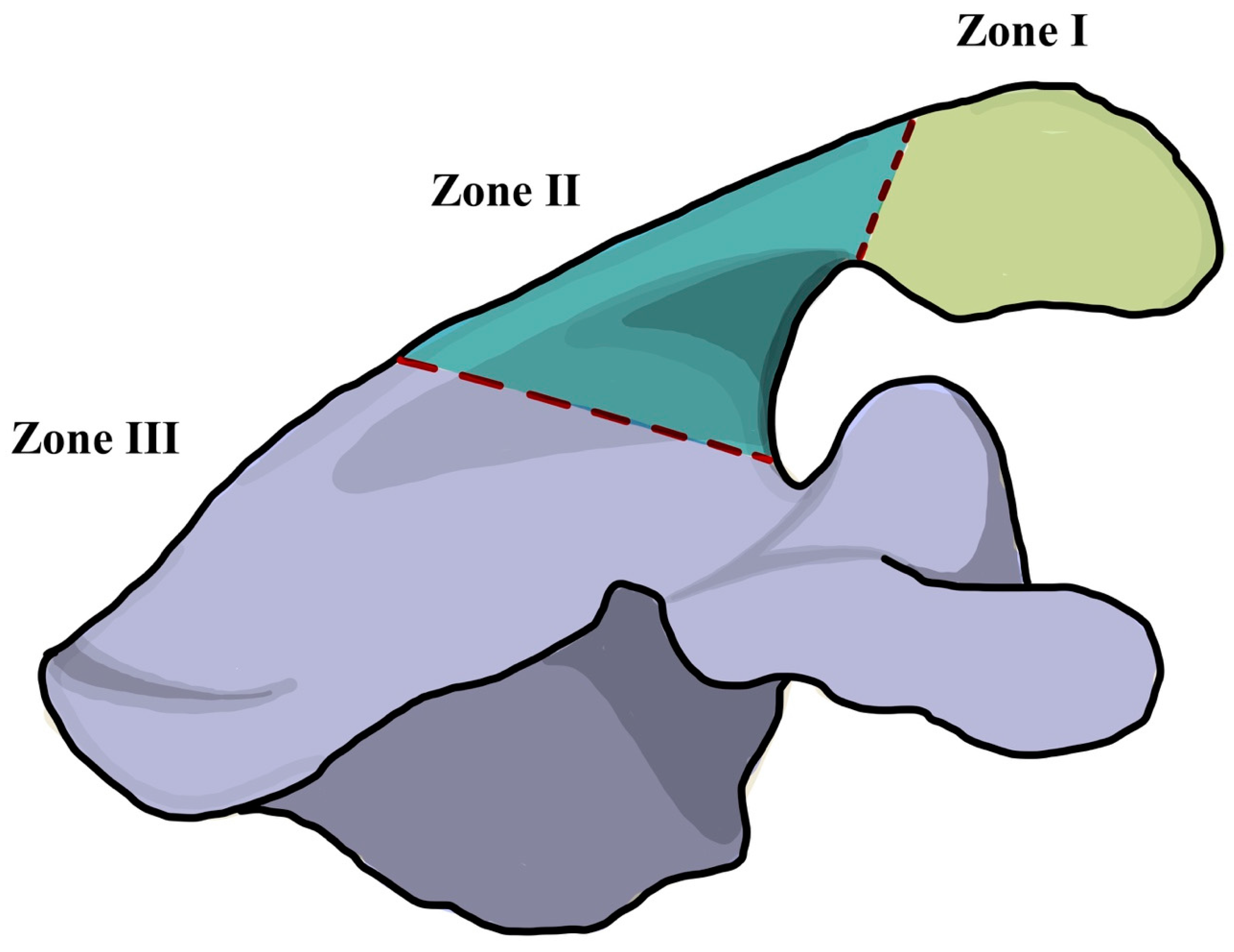
| Study | Specimen Type, Number, Age (Range) | Study Type | Implant Used | NSA (°) | Humeral Offset (mm) * | Glenosphere Offset (mm) * | Outcome(s) Assessed | Scapular Strain/Stress Location † |
|---|---|---|---|---|---|---|---|---|
| Kerrigan (2021) [46], Canada, DCOI | Cadaveric, 8, 73 (61–88) | In-vitro biomechanical study (quasi-static) | Custom modular 42 mm glenosphere Onlay humeral tray. No further specifications | 135, 145, 155 | −5.0, +5.0, +15.0 | Lateralization: +5.0 | Scapular strain during: A: Abduction (0–90°) in scapular plane and forward elevation (0–90°); B: Humeral lateralization; C: Varying neck shaft angles | Levy region I, II, and III |
| Lockhart (2020) [14], Canada | CT images of cadaveric shoulders, 10, 68 (49–87) | In-silico finite element modelling (quasi-static) | 38 mm glenosphere Onlay humeral tray. No further specifications | 155 | +15.0, +20.0, +25.0 | Inferiorization: 0, 2.5, 5.0 Lateralization: 0, +5.0, +10.0 | Acromial stress during: A: Abduction (0°), scapular plane elevation (30°), forward elevation (60°) B: Loading (0, 2.5, 5 kg) C: Glenosphere lateralization D: Glenosphere inferiorization E: Humeral medialization and lateralization | Levy region I, II, and III |
| Shah (2020) [29], USA | Cadaveric, 10, 53.2 (37–63) | In-vitro biomechanical study (quasi-static) | Zimmer Biomet Comprehensive 36 mm glenosphere. Onlay humeral tray | 147 | +3.0, +5.0, +8.0, +10.0, +13.0 | Lateralization: 0, +6.0 | Acromial and scapular strain and deltoid lengthening: A: Based on anatomical orientation of acromion B: During glenosphere lateralization C: During humeral lateralization | Levy region II and III |
| Taylor (2020) [30], USA | Cadaveric, 8, 68 (56.9–79.1) | In-vitro biomechanical study (dynamic) | Zimmer Biomet Comprehensive 36 mm glenosphere. Onlay humeral tray | 147 | +3.0 | No change in offset | Maximal principal strains on the acromion and scapular strain when: A: Coracoacromial ligament intact B: Coracoacromial ligament transacted | Levy region II and III |
| Wong (2016) [47], Canada, DCOI | Cadaveric, 10, 68 (49–87) | In-silico finite element modelling (dynamic) | Delta Xtend, Depuy Synthes 38 mm glenosphere. Onlay humeral tray | 155 | −5.0, 0, +5.0 | Inferiorization:0, 2.5, 5.0 Lateralization: 0, +5.0, +10.0 | Acromial stress during: A: Abduction (0–120°) B: Glenosphere inferiorization C: Glenosphere lateralization D: Humeral medialization and lateralization | |
| Zeng (2021) [48], USA | CT images of representative female subject, 1 | In-silico finite element modelling (dynamic) | Zimmer Anatomical Reverse 36 mm glenosphere. Onlay humeral tray | - | - | Lateralization: 0, +6.0, +12.0 | A: Maximal principal strain, stress and von Milses stress on scapula during glenosphere lateralization B: Deltoid muscle forces during glenosphere lateralization | Levy region I, II, and III |
Publisher’s Note: MDPI stays neutral with regard to jurisdictional claims in published maps and institutional affiliations. |
© 2022 by the authors. Licensee MDPI, Basel, Switzerland. This article is an open access article distributed under the terms and conditions of the Creative Commons Attribution (CC BY) license (https://creativecommons.org/licenses/by/4.0/).
Share and Cite
Paszicsnyek, A.; Jo, O.; Rupasinghe, H.S.; Ackland, D.C.; Treseder, T.; Pullen, C.; Hoy, G.; Ek, E.T.; Ernstbrunner, L. Factors Influencing Acromial and Scapular Spine Strain after Reverse Total Shoulder Arthroplasty: A Systematic Review of Biomechanical Studies. J. Clin. Med. 2022, 11, 361. https://doi.org/10.3390/jcm11020361
Paszicsnyek A, Jo O, Rupasinghe HS, Ackland DC, Treseder T, Pullen C, Hoy G, Ek ET, Ernstbrunner L. Factors Influencing Acromial and Scapular Spine Strain after Reverse Total Shoulder Arthroplasty: A Systematic Review of Biomechanical Studies. Journal of Clinical Medicine. 2022; 11(2):361. https://doi.org/10.3390/jcm11020361
Chicago/Turabian StylePaszicsnyek, Alexander, Olivia Jo, Harshi Sandeepa Rupasinghe, David C. Ackland, Thomas Treseder, Christopher Pullen, Greg Hoy, Eugene T. Ek, and Lukas Ernstbrunner. 2022. "Factors Influencing Acromial and Scapular Spine Strain after Reverse Total Shoulder Arthroplasty: A Systematic Review of Biomechanical Studies" Journal of Clinical Medicine 11, no. 2: 361. https://doi.org/10.3390/jcm11020361