Article

Influence of MAD Application on Episodes of Obstructive Apnea and Bruxism during Sleep—A Prospective Study

by
Monika Wojda
* and
Jolanta Kostrzewa-Janicka
Department of Prosthodontics, Medical University of Warsaw, 02-097 Warsaw, Poland
*
Author to whom correspondence should be addressed.
J. Clin. Med. 2022, 11(19), 5809; https://doi.org/10.3390/jcm11195809
Submission received: 29 August 2022 / Revised: 19 September 2022 / Accepted: 27 September 2022 / Published: 30 September 2022
(This article belongs to the Special Issue (Generalized) Bruxism: Current Challenges and Innovative Treatment)

Abstract

:
The condition of sleep bruxism (SB) is defined by many authors as the body’s response to obstructive sleep apnea (OSA). In the conservative treatment of OSA, mandibular advancement devices (MADs) have found their application. The aim of the study iso assess the impact of MADs on the occurrence of episodes and the intensity of OSA and SB. The study sample consisted of eight patients with OSA and SB diagnosed with these conditions on the basis of clinical examinations and polysomnography (PSG). The prospective study was designed to assess the use of MADs for OSA and SB. MADs were prepared for the patients who subsequently underwent control examinations after one week of wear, and another PSG (PSG II) with an MAD was performed in conditions resembling the first qualification examination (PSG I). The same parameters were assessed in both PSG examinations. Following treatment with the MAD, a favorable lowering of the mean values of the examined parameters was observed. The statistically significant differences were demonstrated only for the apnea–hypopnea index (AHI), the oxygen desaturation index (ODI), and the number of apneas and hypopneas, obstructive apneas, apneas in OSA, and phasic episodes of bruxism. The application of MADs in patients with OSA has a beneficial effect on the same manifestations of OSA and SB, even though only the number of phasic episodes of bruxism was statistically significant.

1. Introduction

Obstructive sleep apnea (OSA) occurs in about 9–38% of the adult population [1], and sleep bruxism (SB) occurs in about 13%. Both of these medical conditions occur during sleep, but associations between them have never been definitely explained [2,3]. OSA is characterized by episodes of complete collapse (apnea) or narrowing (hypopnea) of the upper respiratory tract at the pharyngeal level whilst maintaining and, in most cases, even increasing the functioning of the respiratory muscles. Such episodes generally lead to diminished arterial blood oxygen saturation and usually cause the patient to wake up, although they are often unaware of that fact despite loud snoring, as the respiration is being restored [4]. According to the 2018 International Consensus, bruxism is defined as masticatory muscle activity that occurs during sleep (characterized as rhythmic or non-rhythmic) and wakefulness (characterized by repetitive or sustained tooth contact and/or by bracing or thrusting of the mandible), respectively [2].
The gold standard in the diagnostics of OSA and SB is polysomnography (PSG), which enables the observation of both conditions simultaneously. Additionally, audio–video recordings facilitate the verification of the character of sounds that can be heard during the examination (gnashing, tapping, snoring, and others) and the kinds of movements (sighing, swallowing, coughing, myoclonus, and others).
In the conservative treatment of OSA, devices that produce continuous positive airway pressure (CPAP) are the method of choice. In individuals with a mild and moderate type of OSA and in patients with severe OSA who fail to tolerate treatment with CPAP, mandibular advancement devices (MADs) have also found their application [5,6,7,8]. MADs work on the principle of maintaining the mandible in the protruded position, displacing the tongue forward by means of the genioglossus muscle and changing the position of the hyoid bone to widen the upper respiratory tract [6]. Lavigne et al. presented a sequence of physiological events in OSA. Before the masticatory muscles become active as a consequence of the activity of the sympathetic system, the pulse becomes more rapid and a rise in alpha activity is observed on an EEG, after which the suprahyoid muscles that lower the mandible contract, which increases the airway patency. Then, the masseters contract to produce the phenomenon of grinding. For many authors, bruxism, as masticatory muscle activity, is the body’s response to the increase in sympathetic activity [9,10,11,12]. Thus, investigating whether MAD-wearing individuals still experience episodes of bruxism merits research. In view of the inconclusive results of many studies on the association of bruxism with sleep apnea, the impact of devices used in the treatment of OSA on both of these conditions should be examined [9,11,13]. The aim of the study was to evaluate the effect of MADs on the occurrence of episodes and the severity of OSA and SB.

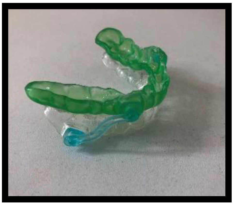
2. Materials and Methods

The study protocol gained the approval of the Bioethics Committee of the Medical University of Warsaw (KB/139/2018). A group of 8 patients (all male) with OSA and SB, who had been diagnosed on the basis of clinical and polysomnographic examinations, constituted the study material. At the Sleep Disorder Clinic of the Internal Diseases, Pneumology, and Allergology Department, the patients completed their own questionnaires concerning obstructive sleep apnea, which additionally included the Epworth Sleepiness Scale (ESS) and bruxism prevalence questionnaires. The ESS was based on the patient’s self-assessment to determine the probability of falling asleep in eight different life situations. The respondent had four options to choose from, with 0 indicating an inability to fall asleep in a given situation and 3 indicating a high probability of falling asleep. The number of points to score was between 0 and 24. Excessive daytime sleepiness, which is one of the criteria for suspected OSA, was determined with scores higher than 10. The bruxism questionnaire consisted of five questions concerning the occurrence of clenching and grinding in patients in a waking state and during sleep, as well as a reported sensation of stiffness or clenched jaws upon waking in the morning. Nocturnal grinding of the teeth during sleep was diagnosed either on the basis of the patients’ own convictions that they indeed suffered from this condition or because somebody had told them about it [14].
The patients with suspected OSA and SB who qualified for the study (had positive questionnaire results for OSA and SB) underwent the first PSG examination with electrodes additionally fixed on the masseters to assess SB (PSG I). The occurrence of the episodes of bruxism was verified with the analysis of audio–video recordings to distinguish the types of audible sounds (gnashing, tapping, snoring, etc.) and to eliminate certain types of movements not related to SB (sighing, swallowing, coughing, myoclonus, and others). The PSG documentation included a six-channel electroencephalogram, a bilateral electrooculogram, an electromyogram from the chin, anterior tibial muscle, and masseter area bilaterally, an electrocardiogram, a record of chest and abdomen movements, position of the body during sleep, airflow through the airways, and audio–video recordings. The PSG examination and the audio–video recordings were assessed by two independent physicians, taking into account the PSG assessment criteria for OSA and SB. Both examiners had received the same training. The evaluation of the frequency of the episodes of bruxism as registered during the PSG was performed in accordance with the criteria set by Lavigne et al. [15]. For this purpose, the bruxism episode index (BEI), phasic, tonic, and mixed episodes of bruxism, and a summary number of all types of episodes of bruxism were considered. The episodes of apnea were assessed on the basis of the American Academy of Sleep Medicine (AASM) criteria [16]. To assess OSA in the PSG, the apnea–hypopnea index (AHI), the oxygen desaturation index (ODI), the number of all types of apnea–hypopnea episodes, the number of obstructive apnea, mixed apnea, hypopnea, and apnea episodes, and the mean and minimal values of SpO2 were calculated.
For patients diagnosed with OSA and SB on the PSG I, a dental examination was conducted to assess the number and condition of the teeth retained in the mouth, the periodontal condition, and the presence of temporomandibular dysfunctions so that an MAD could be provided.
Following the analysis of inclusion and exclusion criteria, the study group consisted of eight patients with moderate-to-severe forms of OSA and sleep bruxism (Table 1), who were subsequently provided with MADs.
In this study, Silensor-sl (Erkodent) single-unit mandibular protruding devices were used, consisting of splints for the upper and lower dental arch with connectors in a registered constructive bite, enabling unobstructed mouth breathing. The constructive bite was determined with the George bite registration device (Scheu Dental Technology) with the advanced mandibular position at 60% of the maximum protrusion. The splint, performed with the pressing technique, covered the occlusal surfaces and incisal edges of all teeth, including the vestibular regions to ¾ of their height and the lingual surfaces of the teeth, extending to the alveolar mucosa of the right-sided ridge of the oral cavity (Figure 1).
For patients who had been provided with their MADs, a checkup examination and a second PSG with the MAD in the mouth (PSG II) were arranged. The first checkup visit took place a week after the patients had been given their MADs. During that visit, the device was adjusted to relieve the areas of excessive pressure to improve the comfort of wear. The adaptation period lasted a month, after which the second PSG with an MAD in the mouth was performed. The conditions of this second examination were similar to those of the first qualifying examination (PSG I). During the PSG II, the same parameters were examined as in the previous PSG examination.
The statistical analysis consisted of a comparison of the effects of the MAD application on the eight patients in the scope of fourteen parameters. The data included parameter values of the same patient prior to and following an MAD application obtained in the PSG I and PSG II examinations. The analysis was based on a Student’s t-test for the dependent groups or a Wilcoxon signed-rank test. All the examined variables were measured on a continuous scale. In the case of a normal distribution, the Student’s t-test for the dependent variables was employed as the test of choice. Otherwise, a non-parametric Wilcoxon test was used. The analysis started with checking the distribution, and then the proper test was employed. Additionally, Wilcoxon tests were used for all of the examined parameters to confirm or reveal other significant associations. The correlations of the dependent samples were analyzed by determining the Pearson correlation coefficient.

3. Results

The PSG results at the baseline and after the MAD application indicate positive changes in the OSA and SB parameters, as revealed during the PSG II in comparison with the baseline (PSG I) (Table 2). The MAD application resulted in a favorable decrease in the mean values of the AHI and ODI indices, the number of apnea and hypopnea episodes, the number of hypopneas, the number of apneas, obstructive apneas, and mixed apneas, the BEI, the number of bruxism episodes, and the number of phasic, tonic, and mixed episodes of bruxism, and a favorable increase in the mean values of the minimal and mean saturation.
The Statistical analysis with the Student’s t-test and the Wilcoxon test was sufficient to state that the statistically significant changes of the mean values of the examined parameters after the MAD application were revealed only in relation to the AHI and ODI, the number of apneas and hypopneas, the number of obstructive apneas, the number of apneas, and the number of episodes of phasic bruxism (Table 3). As for the MAD application, the following statistically significant reductions were noted: the AHI was reduced on average by 11.05 ± 8.35 (p = 0.007); the ODI was reduced on average by 15.28 ± 12.0 (p = 0.009); the number of apneas and hypopneas was reduced on average by 90.75 ± 59.07 (p = 0.003); the number of obstructive apneas was reduced on average by 72.86 ± 68.94 (p = 0.020); the number of apneas was reduced on average by 90.5 ± 74.09 (p = 0.011; the number of phasic episodes of bruxism was reduced on average by 6.38 ± 8.75 (p = 0.034).
For the number of mixed apneas, no statistically significant differences were found between the number of mixed apneas on the PSG with and without an MAD (PSG I and II) (p = 0.24). Similar results were obtained for the hypopneas (p = 0.78), the mean SpO2 (p = 0.230), the minimal SpO2 (p = 0.155), the BEI (p = 0.77), the number of episodes of bruxism (p = 0.150), the number of tonic bruxism episodes (p = 0.401), and the number of mixed bruxism episodes (p = 0.068).
In order to verify whether extreme and outlier values impacted the significance of the results, the tests were repeated, each time following the elimination of one case. The elimination of extreme and outlier cases did not alter the results, meaning that no additional significant correlations were found.
A very strong correlation in the AHI with and without an MAD was revealed (r = 0.898, p = 0.002). Similarly, strong correlations relating to the examinations with and without the device were found for the following: the ODI (r = 0.740, p = 0.036), the number of apneas and hypopneas (r = 0.885, p = 0.003), and the mean SpO2 (r = 0.846, p = 0.008). Another finding was an almost functional association of the number of rhythmic episodes of bruxism with and without an MAD and hypopneas with and without an MAD at r = 0.948 (p = 0.000) and r = 0.905 (p = 0.002), respectively. All of these significant correlations were positive (Table 4). Yet, a much larger sample is required to examine the effect of the improved parameters on the therapeutic effectiveness of MADs.

4. Discussion

There are studies confirming an association between OSA and SB [9,11,13,17,18] and the therapeutic efficacy of MADs in the management of OSA [19,20,21]. Hosoya et al. and the authors of the present study demonstrated a positive correlation between the incidence of OSA and the episodes of sleep bruxism in a group of patients diagnosed with OSA during a PSG examination. The present study revealed that sleep bruxism episodes occurred decisively more often in patients with OSA than in the controls, and they were observed during microarousals as a consequence of an OSA episode [13,17]. In their studies, Aarab et al. found a difference between the baseline AHI parameters and the results after treatment with continuous positive airway pressure (CPAP) above 20 and a difference of 15 in the number of apnea episodes before and after treatment with an MAD, which indicates that the number of apnea episodes decreased significantly in comparison with the baseline results for both the MAD and CPAP treatment modalities. The differences in the AHI were significant when comparisons were performed with a placebo splint group. Additionally, excessive daytime sleepiness manifested a downward tendency when both types of devices were used [19].
The association between OSA and SB, as well as the therapeutic efficacy of MADs in patients with OSA, prompted the commencement of studies on the impact of MADs on SB. Because of the varied etiology of bruxism, patients can benefit from the following three therapeutic strategies: occlusal, pharmacological, and behavioral psychotherapy. In occlusal therapy, we can use nightguards, which protect the patient’s dentition from wear due to attrition, relax the muscles, and reduce the effects of parafunctions. Since they do not protrude the mandible, it would seem that they should not impact on the occurrence of OSA and, as a consequence, SB, provided that in this case, apnea is due to bruxism. Management based on a single method is usually insufficient to protect the peripheral tissues due to SB. Studies have demonstrated the therapeutic efficacy of MADs with regard to SB patients with OSA [22,23,24,25,26,27]. There are also studies that confirm the superior effectiveness of MADs in reducing the number of SB episodes in comparison with the application of occlusal splints [25,26,27]. Martynowicz et al. reported a reduction in BEI in both groups of patients treated with CPAP and MADs [22]. It has also been demonstrated that a variety of intra-oral devices lower the frequency of bruxism episodes [23,24], although in the presented study, only the number of episodes of phasic bruxism was reduced. Several factors could have influenced the obtained results. One of them could be insufficient mandibular protrusion. Another was that there was no correlation between OSA and SB in the examined patients. Although both conditions were confirmed in the study group subjects, it is not known if SB was due to OSA; thus, the effect of the MADs on bruxism cannot be verified. The absence of positive effects of the MADs on SB may be due to an insufficient number of PSG examinations with the MADs in the mouth and patients’ individual determinants.
A prerequisite for an MAD application in an OSA treatment is meeting specific criteria during the dental examination. They are as follows: at least eight stabile teeth preserved in each jaw, a healthy periodontium, and the ability to set a construction bite with the mandibular position at 50–75% of the maximum protrusion and leaving a space between the incisors of about 3–5 mm, enabling free breathing through the mouth [28]. The effectiveness of the device increases with the degree of the mandibular protrusion, but the patient’s tolerance comparably suffers [29]. Studies on the correlation of the therapeutic effect with the degree of protrusion revealed that the latter is the fundamental factor influencing patients’ betterment [30,31]. Attaining 70% of the maximal mandibular protrusion is, according to some authors, a compromise between the device’s effectiveness and the occurrence of potential side effects of its application [32]. In the present study, the mandible was protruded at 60% of its maximal range so that the device was better tolerated and the adverse effects of the MAD application were avoided. Possible adverse effects include painful sensations in the TMJ, changes in overjet and overbite, disorders within Angle’s classes, and changes in the inclination of the maxillary incisor to the cranial base (1/NS) and the angle between the Sella-Nasion-Supramentale (SNB) [33,34]. Regarding the vertical dimension of the construction bite, it is claimed that it should be retained at a minimum level since increasing the vertical dimension through mandibular abduction leads to a down-backward displacement of the tongue, which reduces the patency of the airways [35]. The devices used in the present study facilitated mandibular abduction and prevented mandibular retrusion, but the degree of protrusion may have been insufficient.
The patients’ response to the treatment varied considerably, which could be due to many factors. One of them is the degree of the apnea intensity prior to treatment, which cannot be verified after one PSG examination. According to Aarab et al., apnea intensity varies depending on a particular night—from a normal condition to severe apnea [36]. Response to treatment with a specific method can be conditioned by a varied morphological structure of the craniofacial region and the upper airways, as well as by the duration of the condition and its treatment.
Undoubtedly, the strength of the study presented here was in the use of polysomnography to assess for OSA and SB. However, the limitations of the study were the small number of patients, the reduction in the number of PSG examinations to one with an MAD and one without an MAD, and the inability to adjust the degree of the mandibular protrusion in the device itself on consecutive nights.
In this regard, it seems reasonable to conduct similar studies on larger samples with diagnosed OSA and SB and include several PSG examinations with and without MADs to assess changes in the parameters used to describe OSA and SB. Conducting several PSG examinations with an MAD with different degrees of mandibular protrusion is also worth considering.

5. Conclusions

The application of MADs in patients with OSA has a beneficial effect on the manifestations of OSA and SB, even though only the number of phasic episodes of bruxism was statistically significant.

Author Contributions

Conceptualization, M.W. and J.K.-J.; methodology, M.W. and J.K.-J.; investigation, M.W. and J.K.-J.; data curation, M.W. and J.K.-J.; writing—original draft preparation, M.W. and J.K.-J.; writing—review and editing, M.W. and J.K.-J.; supervision, J.K.-J.; funding acquisition M.W. and J.K.-J. All authors have read and agreed to the published version of the manuscript.

Funding

This research received no external funding.

Institutional Review Board Statement

The study was conducted in accordance with the Declaration of Helsinki and approved by the Institutional Review Board (or Ethics Committee)—the Bioethics Committee at the Medical University of Warsaw (KB/139/2018) for studies involving humans.

Informed Consent Statement

Informed consent was obtained from all subjects involved in the study.

Data Availability Statement

The data presented in the study are available on reasonable request from the authors of this article.

Conflicts of Interest

The authors declare no conflict of interest.

References

  1. Senaratna, C.V.; Perret, J.L.; Lodge, C.J.; Lowe, A.J.; Campbell, B.E.; Matheson, M.C.; Hamilton, G.S.; Dharmage, S.C. Prevalence of obstructive sleep apnea in the general population: A systematic review. Sleep Med. Rev. 2017, 34, 70–81. [Google Scholar] [CrossRef] [PubMed]
  2. Lobbezoo, F.; Ahlberg, J.; Raphael, K.G.; Wetselaar, P.; Glaros, A.G.; Kato, T.; Santiago, V.; Winocur, E.; De Laat, A.; De Leeuw, R.; et al. International consensus on the assessment of bruxism: Report of a work in progress. J. Oral Rehabil. 2018, 45, 837–844. [Google Scholar] [CrossRef] [PubMed]
  3. Jurkowski, P.; Kostrzewa-Janicka, J.; Mierzwińska-Nastalska, E. Bruksizm—Patologia, zaburzenie czy zjawisko fizjologiczne? Przegląd piśmiennictwa. Cześć I—Definicja, epidemiologia, diagnostyka bruksizmu. Protet. Stomat. 2013, 6, 450–458. [Google Scholar] [CrossRef]
  4. American Academy of Sleep Medicine. International Classification of Sleep Disorders, 3rd ed.; American Academy of Sleep Medicine: Darien, IL, USA, 2014. [Google Scholar]
  5. Standards of Practice Committee of American Sleep Disorders Association. Practice parameters for the treatment of snoring and obstructive sleep apnea with oral appliance. Sleep 1995, 18, 511–513. [Google Scholar] [CrossRef] [PubMed]
  6. Pływaczewski, R.; Brzecka, A.; Bielicki, P.; Czajkowska-Malinowska, M.; Cofta, S.; Jonczak, L.; Radliński, J.; Tażbirek, M.; Wasilewska, J. Zalecenia Polskiego Towarzystwa Chorób Płuc dotyczące rozpoznawania i leczenia zaburzeń oddychania w czasie snu u dorosłych. Pneumonol. i Alergol. Pol. 2013, 81, 221–258. [Google Scholar] [CrossRef]
  7. Alessandri-Bonetti, A.; Bortolotti, F.; Moreno-Hay, I.; Michelotti, A.; Cordaro, M.; Alessandri-Bonetti, G.; Okeson, J.P. Effects of mandibular advancement device for obstructive sleep apnea on temporomandibular disorders: A systematic review and meta-analysis. Sleep Med. Rev. 2019, 48, 101211. [Google Scholar] [CrossRef]
  8. Segù, M.; Campagnoli, G.; Di Blasio, M.; Santagostini, A.; Pollis, M.; Levrini, L. Pilot Study of a New Mandibular Advancement Device. Dent. J. 2022, 10, 99. [Google Scholar] [CrossRef]
  9. Kato, T.; Montplaisir, J.Y.; Guitard, F.; Sessle, B.J.; Lund, J.P.; Lavigne, G.J. Evidence that experimentally induced sleep bruxism is a consequence of transient arousal. J. Dent. Res. 2003, 82, 284–288. [Google Scholar] [CrossRef]
  10. Lagana, G.; Malara, A.; Koumoulis, A.; Tepedino, M.; Venza, N.; Cozza, P. Bruxism, perceived anxiety and stress in university students. J. Biol. Regul. Homeost. Agents 2021, 35, 787–790. [Google Scholar]
  11. Adachi, K.; Rompre, S.; Yao, D.; Lavigne, G.; Sessle, B.J. Loss of corticobulbar motor exciability during sleep in primates: Preliminary findings. In Proceedings of the Society for Neuroscience 35th Meeting, Washington, DC, USA, 12–16 November 2005; Volume 399, p. 17. [Google Scholar]
  12. Lavigne, G.J.; Huynh, N.; Kato, T.; Okura, K.; Adachi, K.; Yao, D.; Sessle, B. Genesis of sleep bruxism: Motor and autonomic-cardiac interactions. Arch. Oral Biol. 2007, 52, 381–384. [Google Scholar] [CrossRef]
  13. Hosoya, H.; Kitaura, H.; Hashimoto, T.; Ito, M.; Kinbara, M.; Deguchi, T.; Irokawa, T.; Ohisa, N.; Ogawa, H.; Takano-Yamamoto, T. Relationship between sleep bruxism and sleep respiratory events in patients with obstructive sleep apnea syndrome. Sleep Breath 2014, 18, 837–844. [Google Scholar] [CrossRef] [PubMed]
  14. Pintado, M.R.; Anderson, G.C.; DeLong, R.; Douglas, W.H. Variation in tooth wear in young adults over a two-year period. J. Prosthet. Dent. 1997, 77, 313–320. [Google Scholar] [CrossRef]
  15. Lavigne, G.J.; Rompre, P.H.; Montplaisir, J.Y. Sleep bruxism: Validity of clinical research diagnostic criteria in a controlled polysomnographic study. J Dent. Res. 1996, 75, 546–552. [Google Scholar] [CrossRef] [PubMed]
  16. Berry, R.B.; Brooks, R.; Gamaldo, C.E. The AASM Manual for the Scoring of Sleep and Associated Events: Rules, Terminology and Technical Specifications Version 26; American Academy of Sleep Medicine: Darien, IL, USA, 2020. [Google Scholar]
  17. Wojda, M.; Bielicki, P.; Kostrzewa-Janicka, J. Ocena zależności pomiędzy bruksizmem a obturacyjnym bezdechem w czasie snu. Prot. Stomat. 2022, 72, 50–58. [Google Scholar] [CrossRef]
  18. Lagana, G.; Osmanagiq, V.; Malara, A.; Venza, N.; Cozza, P. Sleep bruxism and sdb in albanian growing subjects: A cross-sectional study. Dent. J. 2021, 9, 25. [Google Scholar] [CrossRef]
  19. Aarab, G.; Lobbezzo, F.; Hamburger, H.L.; Naeije, M. Oral appliance therapy versus nasal continuous positive airway pressure in obstructive sleep apnea; a randomized, placebo-controlled trial. Respiration 2011, 81, 411–419. [Google Scholar] [CrossRef]
  20. Okuno, K.; Sato, K.; Arisaka, T.; Hosohama, K.; Gotoh, M.; Taga, H. The effect of oral appliances that advanced the mandible forward and limited mouth opening in patients with obstructive sleep apnea: A systematic review and meta-analysis of randomised controlled trials. J. Oral Rehabil. 2014, 41, 542–554. [Google Scholar] [CrossRef]
  21. Hoffstein, V. Review of oral appliance for treatment of sleep-disordered breathing. Sleep Breath 2007, 11, 1–22. [Google Scholar] [CrossRef]
  22. Martynowicz, H.; Wieczorek, T.; Macek, P.; Wojakowska, A.; Poręba, R.; Gać, P.; Mazur, G.; Skomro, R.; Smardz, J.; Więckiewicz, M. The effect of continuous positive airway pressure and mandibular advancement device on sleep bruxism intensity in obstructive sleep apnea patients. Chronic Respir. Dis. 2022, 19, 14799731211052301. [Google Scholar] [CrossRef]
  23. Franco, L.; Rompre, P.H.; de Grandmont, P.; Abe, S.; Lavigne, G.J. A mandibular advancement appliance reduces pain and rhythmic masticatory muscle activity in patients with morning headache. J. Orofac Pain 2011, 25, 240–249. [Google Scholar]
  24. DuPont, J.S., Jr.; Brown, C. Management of nocturnal bruxism with an anterior stop point appliance. J. Tenn. Dent. Assoc. 2008, 88, 20–24. [Google Scholar] [PubMed]
  25. Landry, M.L.; Rompre, P.H.; Manzini, C.; Guitard, F.; de Grandmont, P.; Lavigne, G.J. Reduction of sleep bruxism using a mandibular advancement device: An experimental controlled study. Int. J. Prosthodont. 2006, 19, 549–556. [Google Scholar] [PubMed]
  26. Landry-Schönbeck, A.; de Grandmont, P.; Rompre, P.H.; Lavigne, G.L. Effect of an adjustable mandibular advancement appliance on sleep bruxism: A crossover sleep laboratory study. Int. J. Prosthodont. 2009, 22, 251–259. [Google Scholar] [PubMed]
  27. Solanki, N.; Singh, B.P.; Chand, P.; Siddharth, R.; Arya, D.; Kumar, L.; Tripathi, S.; Jivanani, H.; Dubey, A. Effect of mandibular advancement device on sleep bruxism score and sleep quality. J. Prosthet. Dent. 2017, 117, 67–72. [Google Scholar] [CrossRef]
  28. Johal, A.; Bottegal, J.M. Current principles in the management of obstructive sleep apnea with mandibular advancement appliances. Br. Dent. J. 2001, 190, 532–536. [Google Scholar] [CrossRef]
  29. Petelle, B.; Vincent, G.; Gagnadoux, F.; Rakotonanahary, B.; Meyer, B.; Fleury, B. One-night mandibular advancement titration for obstructive sleep apnea syndrome. Am. J. Respir. Crit. Care Med. 2002, 165, 1150–1153. [Google Scholar] [CrossRef]
  30. Petri, N.; Svanholt, P.; Solow, B.; Wildschiodtz, G.; Winkel, P. Mandibular advancement appliance for obstructive sleep apnea: Results of a randomized placebo controlled trial using parallel group design. J. Sleep Res. 2008, 17, 221–229. [Google Scholar] [CrossRef]
  31. Blanco, J.; Zamarron, C.; Abeleira Pazos, M.T.; Lamela, C.; Suarez Quintanilla, D. Prospective evaluation of an oral appliance in the treatment of obstructive sleep apnea syndrome. Sleep Breath 2005, 9, 20–25. [Google Scholar] [CrossRef]
  32. Bernhold, M.; Bondemark, L. A magnetic appliance for treatment of snoring patients with and without obstructive sleep apnea. Am. J. Orthod. Dentofacial Orthop. 1998, 113, 144–155. [Google Scholar] [CrossRef]
  33. Fritsch, K.M.; Iseli, A.; Russi, E.W.; Bloch, K.E. Side effects of mandibular advancement devices for sleep apnea treatment. Am. J. Respir. Crit. Care Med. 2001, 164, 813–818. [Google Scholar] [CrossRef]
  34. Mehta, A.; Qian, J.; Petocz, P.; Darendeliler, M.A.; Cistulli, P.A. A randomized, controlled study of a mandibular advancement splint for obstructive sleep apnea. Am. J. Respir. Crit. Care Med. 2001, 163, 1457–1461. [Google Scholar] [CrossRef] [PubMed]
  35. Pitsit, A.J.; Darendeliler, M.; Gotsopoulos, H.; Petocz, P.; Cistulli, P.A. Effect of vertical dimension on efficacy of oral appliance therapy in obstructive sleep apnea. Am. J. Respir. Crit. Care Med. 2002, 166, 860–864. [Google Scholar] [CrossRef] [PubMed] [Green Version]
  36. Aarab, G.; Lobbezzo, F.; Hamburger, H.L.; Naeije, M. Variability in the apnea-hypopnea index and its consequences for diagnosis and therapy evaluation. Respiration 2009, 77, 32–37. [Google Scholar] [CrossRef] [PubMed]
Figure 1. Mandibular advancement devices, Silensor-sl (Erkodent).
Jcm 11 05809 g001
Table 1. Inclusion and exclusion criteria.
InclusionExclusion
  • Age > 18
  • AHI > 15—moderate-to-severe form of OSA
  • At least eight teeth each in the upper and lower dental arch
  • No periodontal conditions
  • No temporomandibular dysfunctions
Medical:
Sleep/Respiratory disorder other than OSA
Taking of medications disrupting sleep or respiration
Previous treatment with MAD
Morphological disorders of the upper respiratory tract
Mental disorders
Dental:
Temporomandibular dysfunctions
Untreated periodontal or mucosal diseases
Absence of at least eight teeth each in the upper and lower dental arch
AHI apnea-hypopnea index, OSA obstructive sleep apnea, MAD mandibular advancement devices.
Table 2. Comparison of all of the variables examined before and after the MAD application (N = 8).
Parameter Mean ± SDMin–Max
Before44.9 ± 18.617.5–70.0
AHIwith MAD33.8 ± 18.515.5–66.7
Before40.7 ± 17.815.9–66.8
ODIwith MAD25.4 ± 13.95.1–50.9
Before288.3 ± 122.2108.0–463.0
Apneas + Hypopneaswith MAD197.5 ± 92.078.0–378.0
Before166.8 ± 66.068.0–271.0
Obstructive apneaswith MAD93.9 ± 61.922.0–199.0
Before24.1 ± 49.90.0–144.0
Mixed apneaswith MAD1.9 ± 2.60.0–6.0
Before94.4 ± 101.236–342
Hypopneaswith MAD94.1 ± 72.434–258
Before193.9 ± 97.769.0–378
Apneaswith MAD103.4 ± 59.222.0–199.0
Before93.3 ± 1.690.5–95.5
Mean SpO2with MAD93.8 ± 0.992.5–95.4
before78.4 ± 4.569.0–83.0
Min SpO2with MAD80.4 ± 4.574.0–86.0
before4.4 ± 6.32.0–18.4
BEIwith MAD3.3 ± 3.40.0–9.8
before36.0 ± 40.31.0–114.0
Episodes of bruxismwith MAD11.8 ± 11.20.0–34.0
before8.4 ± 11.90.0–35.0
Phasicwith MAD2.0 ± 3.40.0–10.0
before17.6 ± 20.51.0–64.0
Tonicwith MAD9.0 ± 11.90.0–33.0
before10.0 ± 15.00.0–39.0
Mixedwith MAD0.8 ± 0.70.0–2.0
AHI apnea-hypopnea index, ODI oxygen desaturation index, BEI bruxism episodes index, MAD mandibular advancement devices.
Table 3. Mean changes of the values of the parameters assessed with polysomnographic examinations following MAD applications.
ParameterMADp/T-Student Testp/Wilcoxon Test
AHI−11.05 ± 8.30.0070.012
ODI−15.28 ± 12.00.0090.017
Apneas + Hypopneas−90.75 ± 59.070.0030.012
Obstructive apneas−72.86 ± 68.940.0200.036
Mixed apneas−22.25 ± 50.41-0.237
Hypopneas−0.25 ± 47.06-0.779
Apneas−90.5 ± 74.090.0110.012
Mean SpO20.44 ± 0.950.2300.441
Min SpO22.0 ± 3.550.1550.260
BEI−1.14 ± 7.9-0.779
Episodes of bruxism−24.25 ± 42.410.1500.233
Phasic−6.38 ± 8.75-0.034
Tonic−8.63 ± 23.87-0.401
Mixed−9.25 ± 15.04-0.068
AHI apnea-hypopnea index, ODI oxygen desaturation index, BEI bruxism episodes index.
Table 4. Correlations of the dependent samples with and without an MAD (N = 8).
ParameterCorrelationSignificance
AHI0.8980.002
ODI0.7400.036
apneas + hypopneas0.8850.003
obstructive apneas0.4200.300
mixed apneas−0.1650.697
WE confirmhypopneas0.9050.002
apneas0.6530.079
mean SpO20.8460.008
min SpO20.6920.057
BEI−0.2660.524
episodes of bruxism−0.0540.899
phasic episodes of bruxism0.9480.000
tonic episodes of bruxism−0.0160.970
mixed episodes of bruxism−0.0810.849
AHI apnea-hypopnea index, ODI oxygen desaturation index, BEI bruxism episodes index.
Publisher’s Note: MDPI stays neutral with regard to jurisdictional claims in published maps and institutional affiliations.

