Article

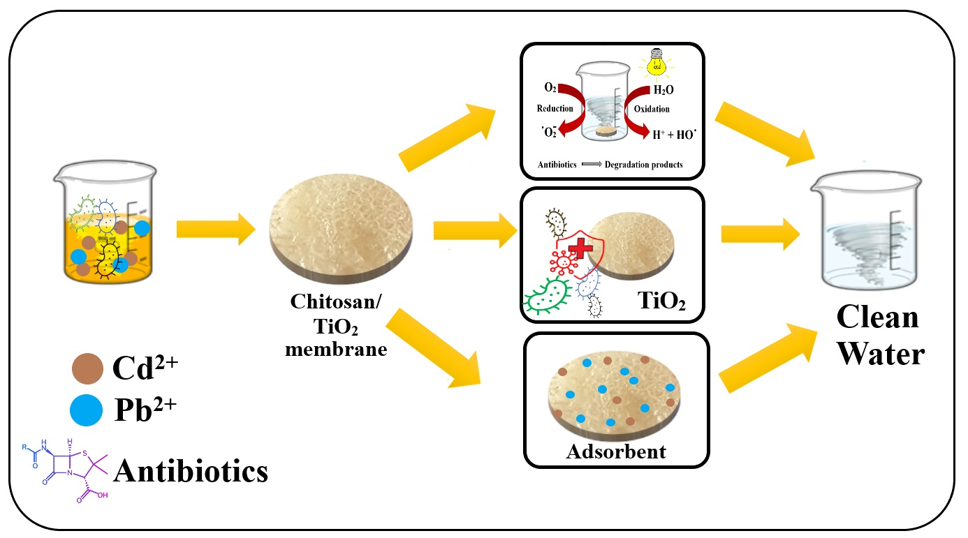
Preparation and Characterization of Chitosan/TiO2 Composite Membranes as Adsorbent Materials for Water Purification

by
Angela Spoială
1,2,3,
Cornelia-Ioana Ilie
1,2,3,
Georgiana Dolete
1,2,3,
Alexa-Maria Croitoru
1,2,3,
Vasile-Adrian Surdu
1,2,3,
Roxana-Doina Trușcă
2,3,
Ludmila Motelica
2,3,
Ovidiu-Cristian Oprea
2,3,4,5,*,
Denisa Ficai
2,3,4,
Anton Ficai
1,2,3,5,
Ecaterina Andronescu
1,2,3,5 and
Lia-Mara Dițu
6
1
Department of Science and Engineering of Oxide Materials and Nanomaterials, Faculty of Chemical Engineering and Biotechnologies, University Politehnica of Bucharest, 1-7 Gh Polizu Street, 011061 Bucharest, Romania
2
National Centre of Micro and Nanomaterials, Faculty of Chemical Engineering and Biotechnologies, University Politehnica of Bucharest, Spl. Indendentei 313, 060042 Bucharest, Romania
3
National Center for Scientific Research for Food Safety, University Politehnica of Bucharest, Spl. Indendentei 313, 060042 Bucharest, Romania
4
Department of Inorganic Chemistry, Physical Chemistry and Electrochemistry, Faculty of Chemical Engineering and Biotechnologies, University Politehnica of Bucharest, 1-7 Gh Polizu Street, 050054 Bucharest, Romania
5
Academy of Romanian Scientists, 3 Ilfov Street, 050045 Bucharest, Romania
6
Faculty of Biology, University of Bucharest, 1-3 Aleea Portocalelor, 060101 Bucharest, Romania
*
Author to whom correspondence should be addressed.
Membranes 2022, 12(8), 804; https://doi.org/10.3390/membranes12080804
Submission received: 2 August 2022 / Revised: 14 August 2022 / Accepted: 17 August 2022 / Published: 20 August 2022
(This article belongs to the Special Issue Chitosan-Based Membranes and Films for Water Purification)

Abstract

:
As it is used in all aspects of human life, water has become more and more polluted. For the past few decades, researchers and scientists have focused on developing innovative composite adsorbent membranes for water purification. The purpose of this research was to synthesize a novel composite adsorbent membrane for the removal of toxic pollutants (namely heavy metals, antibiotics and microorganisms). The as-synthesized chitosan/TiO2 composite membranes were successfully prepared through a simple casting method. The TiO2 nanoparticle concentration from the composite membranes was kept low, at 1% and 5%, in order not to block the functional groups of chitosan, which are responsible for the adsorption of metal ions. Nevertheless, the concentration of TiO2 must be high enough to bestow good photocatalytic and antimicrobial activities. The synthesized composite membranes were characterized by Fourier transform infrared spectroscopy (FTIR), X-ray diffraction (XRD), scanning electron microscopy (SEM), thermogravimetric analysis (TGA) and swelling capacity. The antibacterial activity was determined against four strains, Escherichia coli, Citrobacter spp., Enterococcus faecalis and Staphylococcus aureus. For the Gram-negative strains, a reduction of more than 5 units log CFU/mL was obtained. The adsorption capacity for heavy metal ions was maximum for the chitosan/TiO2 1% composite membrane, the retention values being 297 mg/g for Pb2+ and 315 mg/g for Cd2+ ions. These values were higher for the chitosan/TiO2 1% than for chitosan/TiO2 5%, indicating that a high content of TiO2 can be one of the reasons for modest results reported previously in the literature. The photocatalytic degradation of a five-antibiotic mixture led to removal efficiencies of over 98% for tetracycline and meropenem, while for vancomycin and erythromycin the efficiencies were 86% and 88%, respectively. These values indicate that the chitosan/TiO2 composite membranes exhibit excellent photocatalytic activity under visible light irradiation. The obtained composite membranes can be used for complex water purification processes (removal of heavy metal ions, antibiotics and microorganisms).

Graphical Abstract

1. Introduction

In the last several decades, water pollution has become a paramount concern that equally affects the environmental system and humans [1,2]. Because of various pollutants (antibiotics [3], pesticides [4], heavy metals [5], dyes [6], fluoride [7], chlorinated hydrocarbons [8], etc.) present in wastewaters, there is a need to develop innovative membranes to help mitigate this problem [9]. Therefore, removing these toxic pollutants from wastewater is of primordial importance [10,11]. Produced water is a by-product of the oil and gas industries that needs to be treated before it can be used in agriculture or recirculated towards other industries. Composite membrane solutions based on the zeolitic imidazole framework and polyacrylonitrile are reported in the literature [12]. The presence of pollutants in water systems significantly affects the surroundings and human health [2,13,14,15]. Researchers and scientists are trying to overcome this water issue by developing new alternative materials with excellent adsorbent properties [16,17,18]. Numerous traditional methods are used to remove these toxic pollutants [19], such as reduction, co-precipitation, ultrafiltration, sedimentation, photocatalysis, distillation and adsorption methods [2,13]. Mixed matrix membranes (MMMs) consisting of a polymeric matrix and an inorganic filler have the potential to achieve high performance in water depollution by mixing the capabilities of the organic polymer (such as adsorption of heavy metals) with those of the inorganic nanoparticles (such as antimicrobial action leading to fouling mitigation) [7,20]. The membranes are not only used for removal of pollutants. In the biotechnology, food and pharmaceutical industries, ultrafiltration membranes, such as polyethersulfone hollow fibers, are used for the efficient separation of biomolecules [21,22].
Among materials proposed for water purification, chitosan-based ones present a great interest because of their free amino and hydroxyl groups, good biocompatibility, biodegradability, nontoxicity, reactivity, hydrophilicity and cost effectiveness [2,11,23,24]. The fact that chitosan presents hydroxyl and amine groups on its surface is the reason for its wide use in heavy metal removal from wastewaters [25,26,27,28,29,30,31]. However, it does have some inconveniences regarding its low stability, thermo-mechanical properties and porosity [32]. Scientists have developed chitosan-based adsorbent membranes to overcome these difficulties through diverse methods [33,34,35,36,37]. Functionalization with glyoxal, glutaraldehyde or epichlorohydrin can reinforce the structure of chitosan through cross-linking, enhancing its mechanical properties. Studies have reported that through cross-linking, the adsorption capacity of chitosan membranes has improved substantially [38,39,40,41]. Various chitosan-based membranes have been developed for the removal of pollutants from wastewaters [42]. Many studies have demonstrated that chitosan exhibits some weak antibacterial and antioxidant properties, which can also help in the water purification processes [43,44,45].
Titanium dioxide (TiO2) is an industrial pigment, disinfectant agent and photocatalyst, having excellent thermo-stability and low toxicity. TiO2 has presented great performance in environmental applications. In addition to photocatalytic and antibacterial properties, TiO2 presented potential in water treatment applications due to the induced porosity to the composite membrane [23,46,47,48,49]. Gonzalez-Calderon et al. [50] and Li et al. [51] found that TiO2 incorporation in chitosan membranes improves the mechanical, physicochemical, thermal and UV protection of the composite membrane, and also that TiO2 substantially enhances the antimicrobial activity against E. coli and S. aureus. However, it has been proven that a considerable concentration of TiO2 on the biopolymer matrix could cause the aggregation of inorganic nanoparticles onto the surface of the composite, thus affecting the mechanical properties [52]. Razzaz et al. [53] reported a composite chitosan/TiO2 membrane that demonstrated high adsorption capacity for removing Cu(II) and Pb(II) ions from water systems. Elsewhere, Samadi et al. [54] presented a novel Cu–TiO2/chitosan hybrid thin film used to remove heavy metals from aquatic media. The study of Chien et al. [55] on chitosan/TiO2 composites confirmed the adsorption capacity of this solution, but the capacity was not impressive. In another study, the authors developed a novel magnetic EDTA/chitosan/TiO2 (MECT) nanocomposite to remove Cd(II) metal ions and phenol as hazardous materials from aqueous solutions [56]. In fact, many literature studies report adsorption values under 100 mg/g, mainly because the composite ratio of chitosan to TiO2 is close to 1:1 or even more as it is in [55]. Such high TiO2 content in the composite membrane will adversely affect the capacity of the chitosan to further bind the heavy metal ions, but in theory should improve the photocatalytic activity of the membrane. Using a lower TiO2 concentration for the composite membrane should allow a better adsorption capacity vs. heavy metal ions, with only a small diminishing of the photocatalytic activity.
Most of the literature reports regarding the photocatalytic activity of TiO2 are based on UV [57,58,59] or simulated sun light [60,61]. In theory, introducing intermediary electronic levels into the semiconductor’s band gap allows photocatalytic activity under visible light irradiation. Such intermediary electronic levels can be generated by doping or by obtaining a high surface defect density on the TiO2 nanoparticles during synthesis.
TiO2, like many other oxides with photocatalytic activity [62,63], generates reactive oxygen species (ROS), which ensures antimicrobial activity against various microorganisms. Chitosan also presents weak antimicrobial activity on its own [44]. Therefore, such a composite membrane should exhibit antimicrobial activity, but due to synergism a strong bactericidal action can arise.
Our aim was to develop novel chitosan/TiO2 adsorbent composite membranes, with low TiO2 content, for complex water purification. To our knowledge, this is the first time that this membrane is reported to be used as an adsorbent for heavy metal ions, as a photocatalytic system against an antibiotic mixture and as an antibacterial agent, accomplishing thus a complex water purification process. We used previously synthesized TiO2 through a simple sol-gel method [64] that yields impurity-free nanoparticles, but with high surface defect density, which allowed the use of visible light in photocatalysis. The chitosan/TiO2 composite membranes were prepared through a simple casting method and further characterized by FTIR, XRD, TG-DSC-FTIR and SEM. Swelling capacity, heavy metal ion adsorption capacity, photocatalytic degradation of a five-antibiotic mixture and antibacterial activity were also determined.

2. Materials and Methods
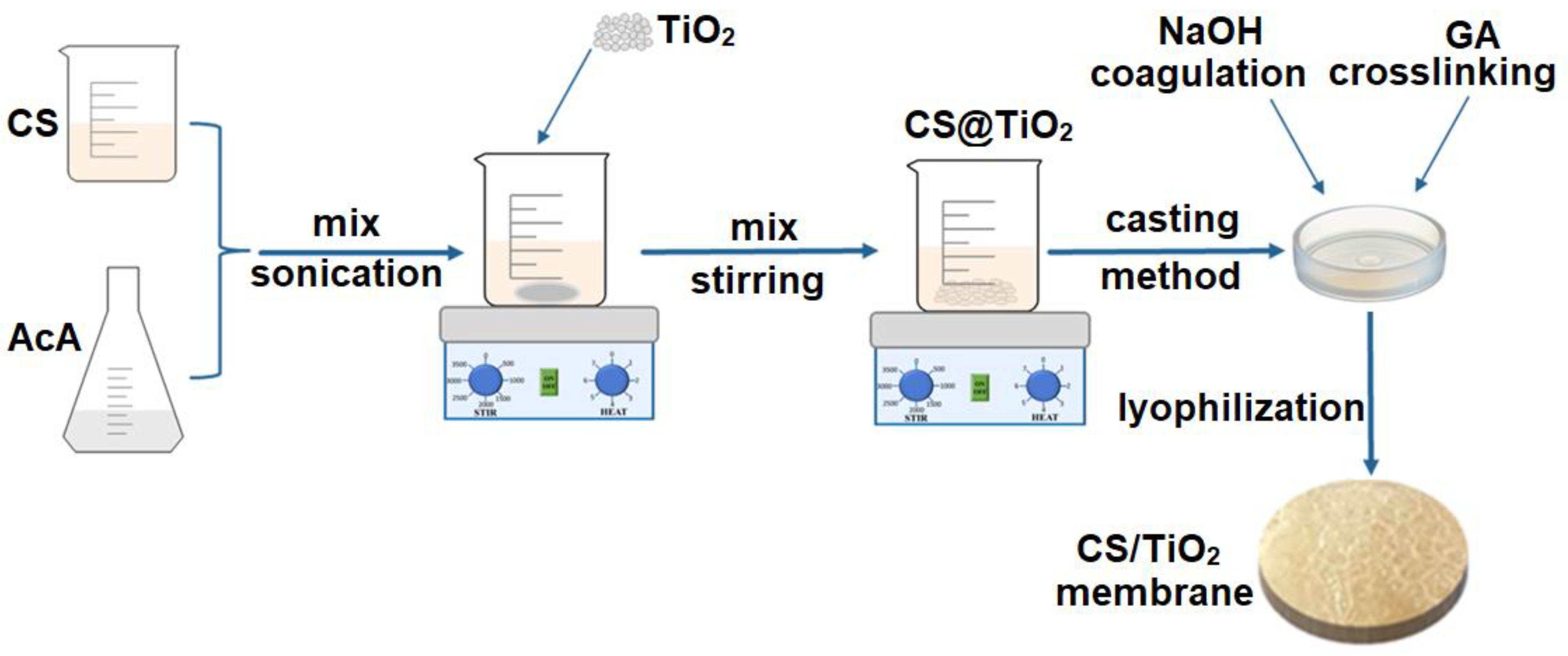
2.1. Experimental

Titanium isopropoxide, having ≥ 97.0% purity, was acquired from Sigma Aldrich (Merck, Burlington, MA, USA). Isopropanol (2-propanol) with 99.99% purity was obtained from Sigma Aldrich (Merck, Burlington, MA, USA). Nitric acid 65% was from Sigma Aldrich (Merck, Burlington, MA, USA). Chitosan (CS) (molecular weight 100.000–300.000, Acros Organics, Geel, Belgium), glacial acetic acid (AcA) (Chimreactiv, Bucharest, Romania) and sodium hydroxide ≥ 97.0% were from Sigma Aldrich (Merck, Burlington, MA, USA). Glutaraldehyde (GA) (50% in water) was purchased from Sigma Aldrich (Merck, Burlington, MA, USA) and distilled water was used. All chemicals used in the present study were of analytical grade without further purification.
The microbiological activity was performed using Nutrient Broth No. 2 and agar, acquired from Sigma-Aldrich (Darmstadt, Germany). All strains tested in this study were provided by the Microorganisms Collection of the Department of Microbiology, Faculty of Biology and Research Institute of the University of Bucharest.
TiO2 was synthesized using a simple sol-gel method as described in [64]. Briefly, titanium isopropoxide was added drop by drop into a solution containing isopropanol and distilled water to obtain titanium dioxide nanoparticles. The obtained solution was magnetically stirred on a hot plate at almost 80 °C. After 1 h of stirring, a solution containing concentrated nitric acid and distilled water was added to the previous solution and kept under constant stirring on a hot plate at 60 °C for 6 h until a white sol-gel appeared. The appearance of the white sol-gel indicates the formation of titanium dioxide nanoparticles. After the precipitate was washed several times for residual removal, it was heated in an electric oven at 300 °C for 2 h. The powders were then placed in a furnace at 550 °C for 5 h. The as-obtained TiO2 nanoparticles were further used to develop the composite membranes described below.
Figure 1 illustrates a schematic chart of the preparation of chitosan/TiO2 composite membranes through a simple casting method. First, chitosan (2 g) was dissolved in a 1% acetic acid solution. Then, TiO2 was added to the as-obtained chitosan solution previously synthesized through the sol-gel method; afterwards, the mixture was magnetically stirred at room temperature for 24 h and then sonicated for 4 h at 35 °C to obtain a homogenous solution. For this experiment, two polymer solutions with 1% and 5% (w/w) TiO2 nanoparticles were obtained. The solutions were cast into Petri dishes and dried in an electric oven at 50 °C overnight. The obtained membranes were treated with NaOH solution for 24 h to coagulate the chitosan; afterwards, they were washed with distilled water to remove side products and excess NaOH. To cross-link the obtained membranes, they were placed in a diluted glutaraldehyde solution (200 mL, 2% v/v) for 24 h under magnetic stirring. This was followed by washing with distilled water to remove any remains from the glutaraldehyde. The synthesized composite membranes were further lyophilized and analyzed through proper techniques.

2.2. Characterization

The synthesized membranes were characterized by Fourier transform infrared spectroscopy (FTIR), X-ray diffraction (XRD), thermogravimetric analysis (TG) and differential scanning calorimetry (DSC) coupled with the FTIR analysis of the evolved gases and scanning electron microscopy (SEM).
The Fourier transform infrared spectroscopy (FTIR) measurements were performed using a Nicolet iS50R spectrometer (Thermo Fisher Scientific, Waltham, MA, USA). The spectra were recorded at room temperature using the attenuated total reflection (ATR) (Thermo Fisher Scientific, Waltham, MA, USA), with 32 scans between 4000 and 400 cm−1 at a resolution of 4 cm−1, with the scanning time being 47 s.
FTIR 2D maps were recorded with a Nicolet iS50R FTIR microscope (Thermo Fisher Scientific Inc., Waltham, MA, USA), with a DTGS detector, in the wavenumber range 4000–600 cm−1. The 2D FTIR maps were used to obtain information about the spatial distribution of the components.
X-ray diffraction (XRD) experiments were carried out on a Panalytical Empyrean instrument (Malvern Panalytical, Malvern, UK) with Ni-filtered Cu radiation (λ = 0.15406 Å) equipped with a 1/4° fixed divergence slit and a 1/2° anti-scatter slit on the incidence beam side, and a 1/2° anti-scatter slit mounted on a PIXCel3D detector (Malvern Panalytical, Malvern, UK) on the diffracted beam side. Data reduction and analysis of the patterns were performed in HighScore Plus 3.0.e software (Malvern Panalytical, Malvern, UK) coupled with the ICDD PDF4 + 2021 database.
The thermogravimetric analysis (TG-DSC) was performed with an STA 449 F3 Jupiter apparatus from Netzsch (Selb, Germany) coupled with an FTIR Tensor 27 from Bruker. Approximately 10 mg of dry powder was placed in an open alumina crucible and heated up to 900 °C with a 10 °C min−1 rate under a flow of 50 mL min−1 of dried air. As a reference, an empty alumina crucible was used.
The electron microscopy images (SEM) were obtained using a Quanta Inspect F50 (FEI Company, Eindhoven, The Netherlands) equipped with a field emission gun (FEG) with a 1.2 nm resolution and an energy dispersive X-ray spectrometer (EDS) with an MnK resolution of 133 eV Kα.
The heavy metal ion retention capacity of the chitosan/TiO2 composite membranes was evaluated by inductively coupled plasma–mass spectrometry (ICP-MS). Before ICP-MS analysis, samples of each membrane (~1 g) were exposed for 2 h to 50 mL 1% and 5% lead nitrate solutions and 1% and 5% cadmium nitrate solutions (both from Sigma Aldrich, Darmstadt, Germany). Nitrate solutions have been used as sources of heavy metals found in polluting aqueous media, having a substantial negative impact on the environment, including animal and human health. After exposure to the previously mentioned solutions, our membranes were removed from the Pb(NO3)2 and Cd(NO3)2 solutions, washed with distilled water and placed in the oven at 105 °C until complete drying.
The retention of heavy metals on the composite membranes was evaluated considering cadmium and lead concentrations. Cadmium and lead concentrations were determined using an Agilent 8800 Triple Quadrupole ICP-MS (Agilent Technologies, Tokyo, Japan), equipped with an ASX500 autosampler, MicroMist concentric nebulizer, Peltier cooling spray-chamber (2 °C), 2.5 mm internal diameter torch, nickel sampler and skimmer cones. Operating conditions of ICP-MS included 1550 W RF power, 1 L/min carrier gas flow, 0.7 mL/min He flow and nebulizer pump set to 0.1 RPS. Before ICP-MS analysis, the dried membranes were subjected to digestion. Three samples of ~200 mg of each previously treated membrane were digested with 8 mL HNO3 in a microwave oven (Ethos UP, Milestone Inc., Sorisole, Italy), applying a dedicated program for high organic content matrices (200 °C heating, 1800 W microwave power) for 35 min. After cooling, the digestion samples were diluted with Milli-Q water up to 50 mL and filtered through 0.45 μm pore size syringe filters. To achieve the best sensibility, samples were diluted 100,000 times with ultrapure water. Before measurements, ICP-MS was tuned according to the manufacturer and calibrated with five calibration standards ranging from 0.1 to 5 µg/L Cd and Pb. Our results were verified by a certified reference material (SRM 1567b) subjected to the same preparation steps as the samples and calculating the percent recovery. Calibration curves of the two elements proved linear in the operating range of 0.1–5 μg/L, with correlation coefficients greater than 0.999. Cadmium recovery from the SRM 1567b sample was 98%, and lead recovery was 107%, considered acceptable in instrumental analysis. The final metal concentration reported for dry membrane mass was calculated using the following formula:
M e t a l   f i n a l   c o n c e n t r a t i o n   ( µ g · m g 1 ) = C × F d × V M
where C is the raw concentration read by ICP-MS, Fd is the dilution factor (100,000), V is the final digestion volume (0.05 L) and M is the dry sample mass (mg).
The photocatalytic activity of the chitosan/TiO2 membranes was determined against a solution with five antibiotics (50 ppb vancomycin, meropenem, tetracycline, clindamycin and erythromycin) by irradiation with a LOHUIS® (Ulmi, Romania), commercially available, and a fluorescent lamp of 160 W/2900 lm (lumen), with a color temperature of 3200 K and color rendering index > 60, placed at 20 cm distance. The chitosan/TiO2 samples of the ~40.0 mg disk were inserted into a 50 mL solution of antibiotic mix. After irradiation, at defined time intervals, a sample of 1 mL was analyzed by LC-MS to quantify the quantity of the antibiotic. Three groups of parallel experiments were set up.
The antibiotic degradation by the tested membranes was evaluated with liquid chromatography–mass spectrometry (LC-MS). The LC-MS analyses were performed on Agilent Technologies 6540 UHD Accurate-Mass Q-TOF LC/MS equipped with a reversed-phase Zorbax Eclipse C18 (Agilent, 50 × 4.6 mm, 2.7 μm particle size). Mobile phases were 95% water (A) and 5% acetonitrile (B). Antibiotics were separated following a gradient program: initial conditions were 95% A, then the gradient was from 100% B to 5% B in 5 min, and finally, solvents were maintained at 95% A and 5% B for 4 min. The total run time was 14 min. The column temperature was 60 °C, and the flow rate was 0.15 mL/min. The sample injection volume was set at 5 μL. The mass spectrometer was a Q-TOF system with a Dual ESI ion source operated in positive ionization mode. The operating parameters were ion spray voltage 5300 V, drying gas, 7 L/min, nebulizer gas 21 psig and probe temperature 300 °C. The acquisition rate was 1.1 spectra/s, 909.1 ms/spectrum for each compound.
The swelling capacity was determined by immersing square pieces 2 cm × 2 cm in 200 mL water. Each piece was weighed before starting the experiment and at fixed time intervals: 0.25, 0.5, 1, 2, 4, 6, 12 and 24 h. The experiments were performed in triplicate. The following formula calculated the water uptake capacity of the samples:
Water retention (%) = M h , t M i M i × 100 , where Mi is the initial weight and Mh,t is the weight after immersion in water.
The antibacterial assessments of the composite membranes were made to evaluate their potential use in water purification applications. The anti-adherent capacity of the membranes obtained in this study was conducted by determining the colony-forming units/mL values (CFU/mL). The antibacterial activity was evaluated against the Staphylococcus aureus MRSA 5578, Enterococcus faecalis VRE 2566, Escherichia coli ATCC 25922 and Citrobacter sp 2021. The samples (1 cm/1 cm) were previously sterilized under UV radiation for 30 min on each side in order to eliminate any possible contamination. To confirm the sterility of the tested samples before the antibacterial assay, each type of membrane was maintained in nutrient broth media for 24 h at 37 °C. The clarity of the broth media confirmed the sterility of the samples.
Bacterial cell suspensions (1.5 × 10⁸ CFU/mL) were made in a sterile physiological buffer from fresh cultures (18–24 h). The anti-adherent capacity of the samples was performed using the method described in the previous studies [65,66,67] and according to the CLSI standard [68]. The negative control was considered the sterile media, and the positive control (C+) was the broth media inoculated with microbial suspensions. The CFU/mL values were expressed as the average of the total number of colonies × 1/D (D = decimal dilution, for which the number of total colonies was to be determined). The assays were performed in three independent experiments.
Antibacterial assessments were performed in triplicate and were analyzed using GraphPad Prism 9 by GraphPad Software, San Diego, CA, USA. We compared the ability of selected strains to adhere to the surface of the membranes using analysis of variance (ANOVA) and Dunnett’s multiple comparisons test. A p-value < 0.05 is considered statistically significant.

3. Results and Discussion

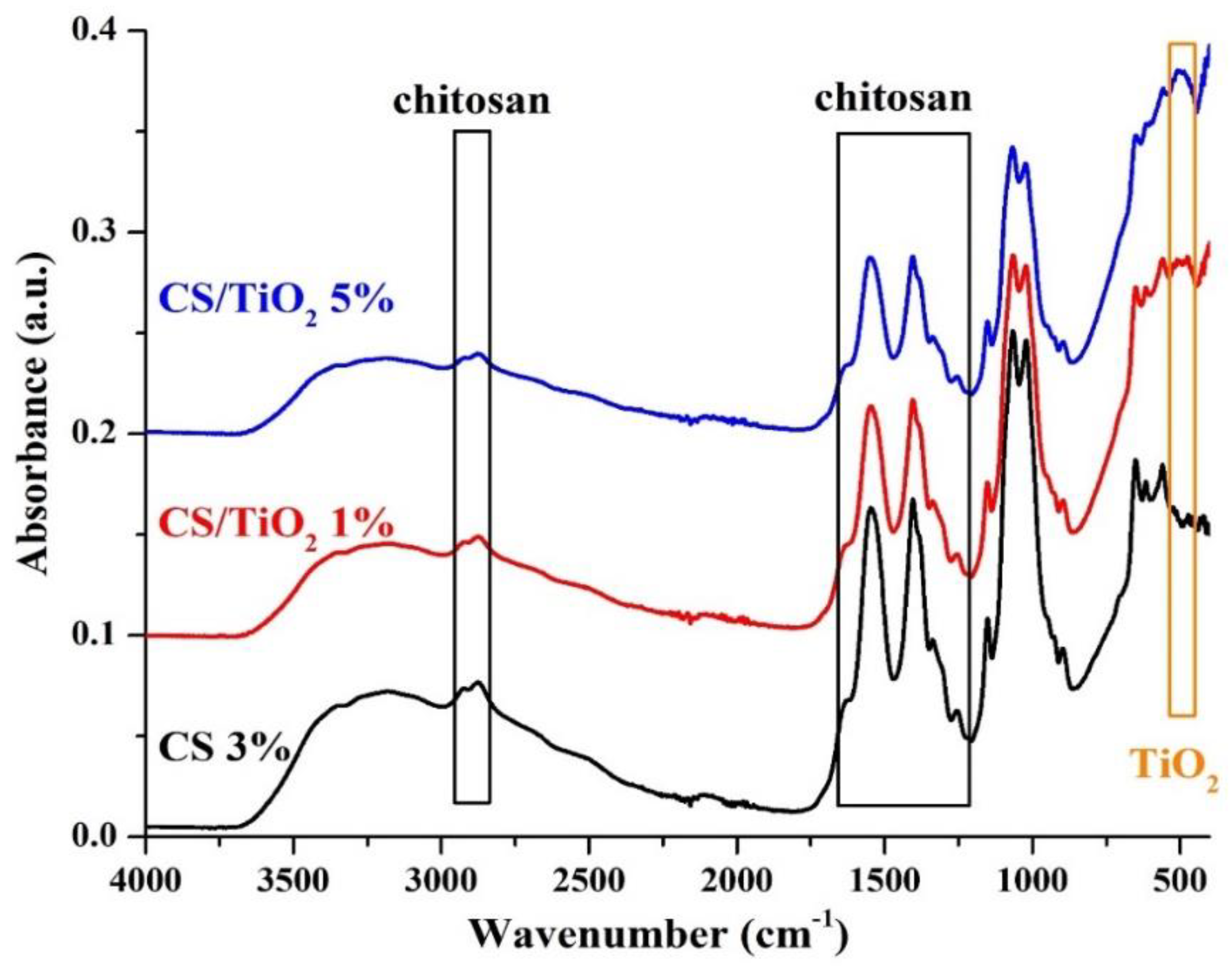
3.1. Fourier Transform Infrared Spectroscopy (FTIR) Analysis

FTIR analysis is a valuable technique for determining and detecting chemical bonds inside a material. The FTIR spectra of chitosan and chitosan/TiO2 composite membranes are presented in Figure 2, and the assignment of the relevant peaks is made in Table 1. The chitosan exhibits several peaks corresponding to the functional groups: -OH and –NH2 in the 3100–3500 cm−1 region, the C-H stretching vibration at 2875–2925 cm−1 (symmetric and asymmetric), amide band I, II and III in the region 1630–1300 cm−1 and asymmetric vibration of a C-O-C group at ~1150 cm−1 [69]. These peaks are observable for all samples.
Additionally, the presence of TiO2 produces a double peak at ~500 cm−1 due to the Ti-O stretching vibration (observable only in chitosan/TiO2 samples). Comparison between the chitosan and chitosan/TiO2 composite membranes indicates their interactions; this interaction is represented by the formation of Ti-O-Ti stretching bonds at wavelengths ranging from 476 cm−1 to 508 cm−1 [70,71].
The spatial distribution of TiO2 nanoparticles inside the chitosan matrix can be investigated by FTIR microscopy. The microscope records the full FTIR spectra for each point, creating a 2D map that can be displayed for any chosen wavelength. The FTIR maps recorded at 3200, 1630 and 610 cm−1 for the chitosan control and the composite membranes (chitosan/TiO2 1% and chitosan/TiO2 5%) are presented in Figure 3. Interactions between various components of a composite sample will lead to zonal modification of absorption at certain characteristic wavelengths. When 2D FTIR maps present distinct zones at different characteristic wavelengths, it can be interpreted as a localized interaction proof between components. Mono-component or highly homogeneous mixtures will yield similar 2D maps for different wavelengths, and for inhomogeneous samples some differences between maps can be visualized. The membranes present a high degree of homogeneity, with some agglomerated clusters and surface defects. As expected, the FTIR maps recorded for the simple chitosan membrane are consistent at all three wavenumbers. In the case of the composite membranes, chitosan/TiO2, the maps recorded at 3200 and 1630 cm−1 are similar, with only minor differences. Nevertheless, the maps recorded at 610 cm−1 present noticeable differences for the composite membranes compared with the 3200 and 1630 cm−1 maps, indicating the presence of TiO2 nanoparticles that interact with the chitosan matrix and induce shifts in the position of the absorption peaks. Based on the information provided by FTIR microscopy, the spatial distribution of the TiO2 nanoparticles in the composite membranes can be considered good, with occasional agglomerations on the micrometer level.

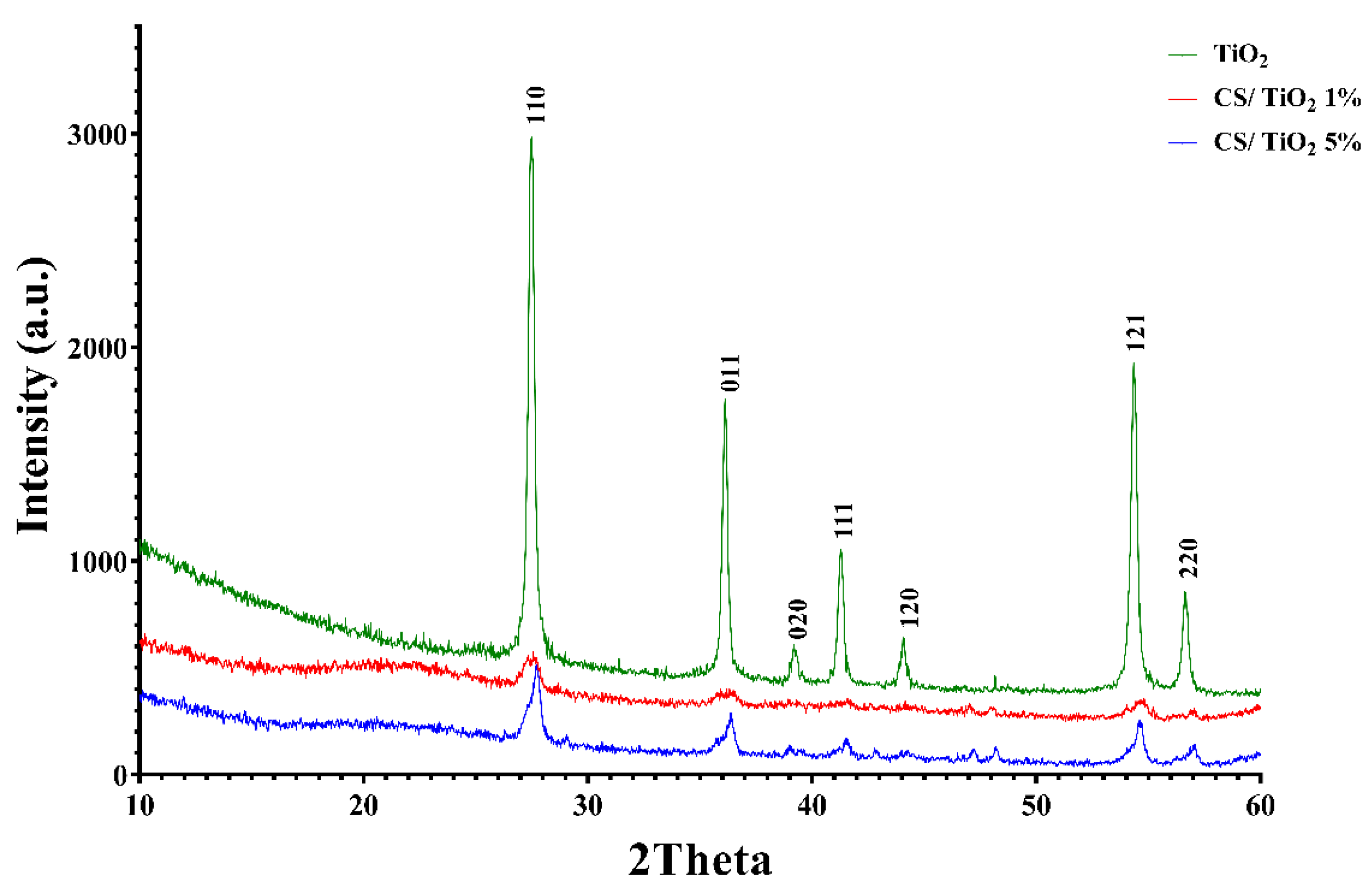
3.2. X-ray Diffraction (XRD) Analysis

An XRD analysis was carried out (Figure 4) to reveal the crystalline structure of the synthesized chitosan and chitosan/TiO2 composite membranes.
The Scherrer equation was used to determine the crystallite size (~31.36 nm) of TiO2 in rutile form. The XRD diagram confirms the presence of common peaks of TiO2 (crystalline) and a broad phase ranging from 10 to 25, corresponding to chitosan (slightly amorphous) [72].

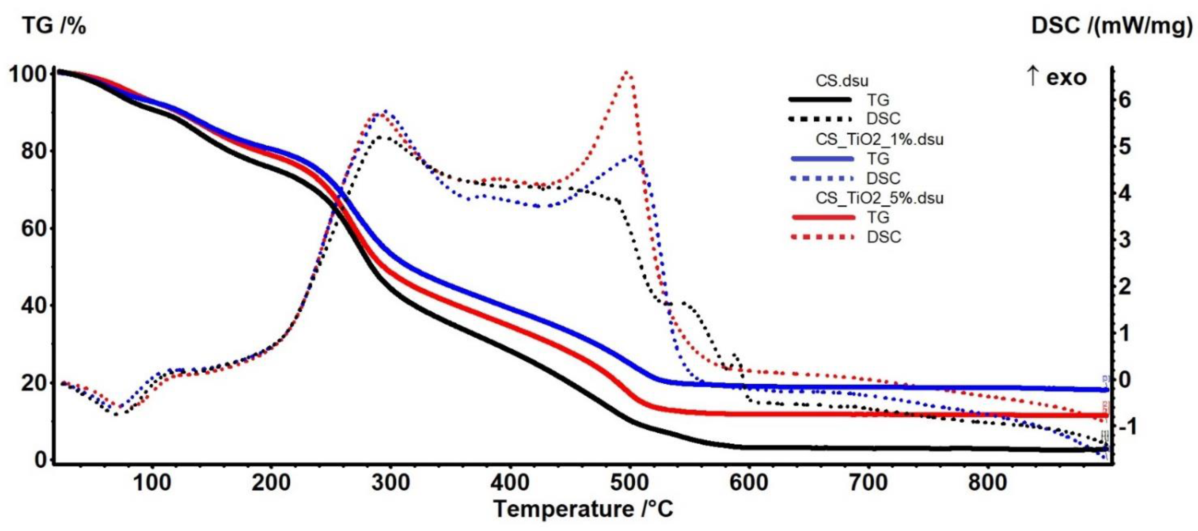
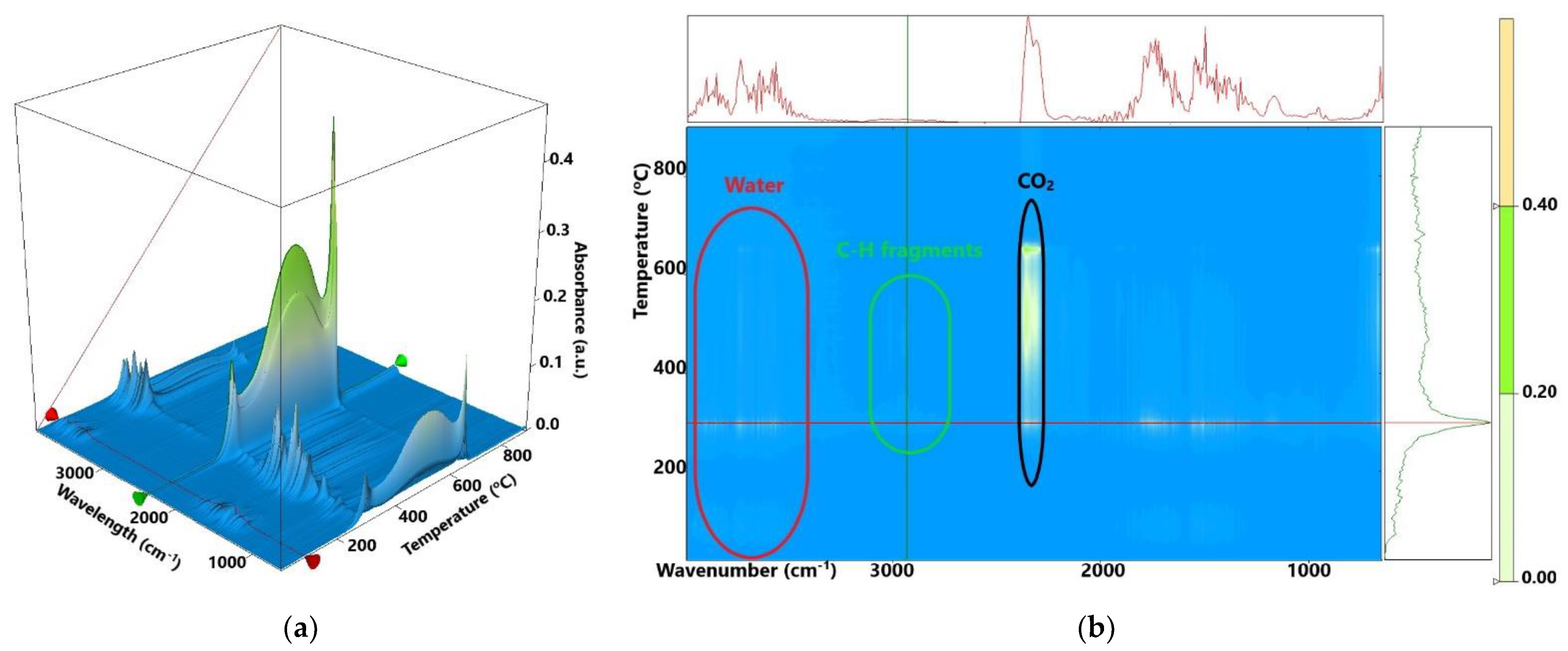
3.3. Thermal (TG-DSC) Analysis

The thermal analysis results are presented in Figure 5 for chitosan and chitosan/TiO2 composite membranes. The comparison between the samples can be made based on the data from Table 2. The samples are losing residual water molecules up to 105 °C (~7–10%), and a weak endothermic effect accompanies the process on the DSC curve [73].
The chitosan control sample exhibits a higher mass loss, which is expected as there is no inorganic part in it. The degradative process starts after 150 °C, when chitosan molecular chains break free and residual acetic acid is eliminated (~11–14%).
After 200 °C up to 370 °C, the samples lose ~40% of their mass in a complex degradative-oxidative process. The polysaccharide chains are broken, and the smaller fragments are oxidized. This process is accompanied by a strong, sizeable exothermic effect, with a maximum at ~290 °C generated by the oxidation of the organic fragments. The FTIR spectra of the evolved gases (Figure 6) permit identification of water, CO2 and hydrocarbon fragments (such as acetic acid) in this temperature interval. The larger organic fragments are slowly oxidized, and residual carbonaceous mass is burned after 370 °C, the corresponding effects on the DSC curve being exothermic (~500 °C).
As expected, the residual mass is higher as the proportion of inorganic TiO2 in the sample increases up to 5%.
The 3D FTIR plot (Figure 6a) presents the evolution of the FTIR spectrum vs. temperature. By projecting this map in 2D space (wavenumber vs. temperature—like a topographical map) we can easily identify the components and temperature intervals when they are eliminated from the sample (Figure 6b). The FTIR spectra recorded for the evolved gases indicate the presence of water and carbon dioxide molecules, but also some traces of carbon monoxide. Hydrocarbon fragments can be identified starting from 250 °C.

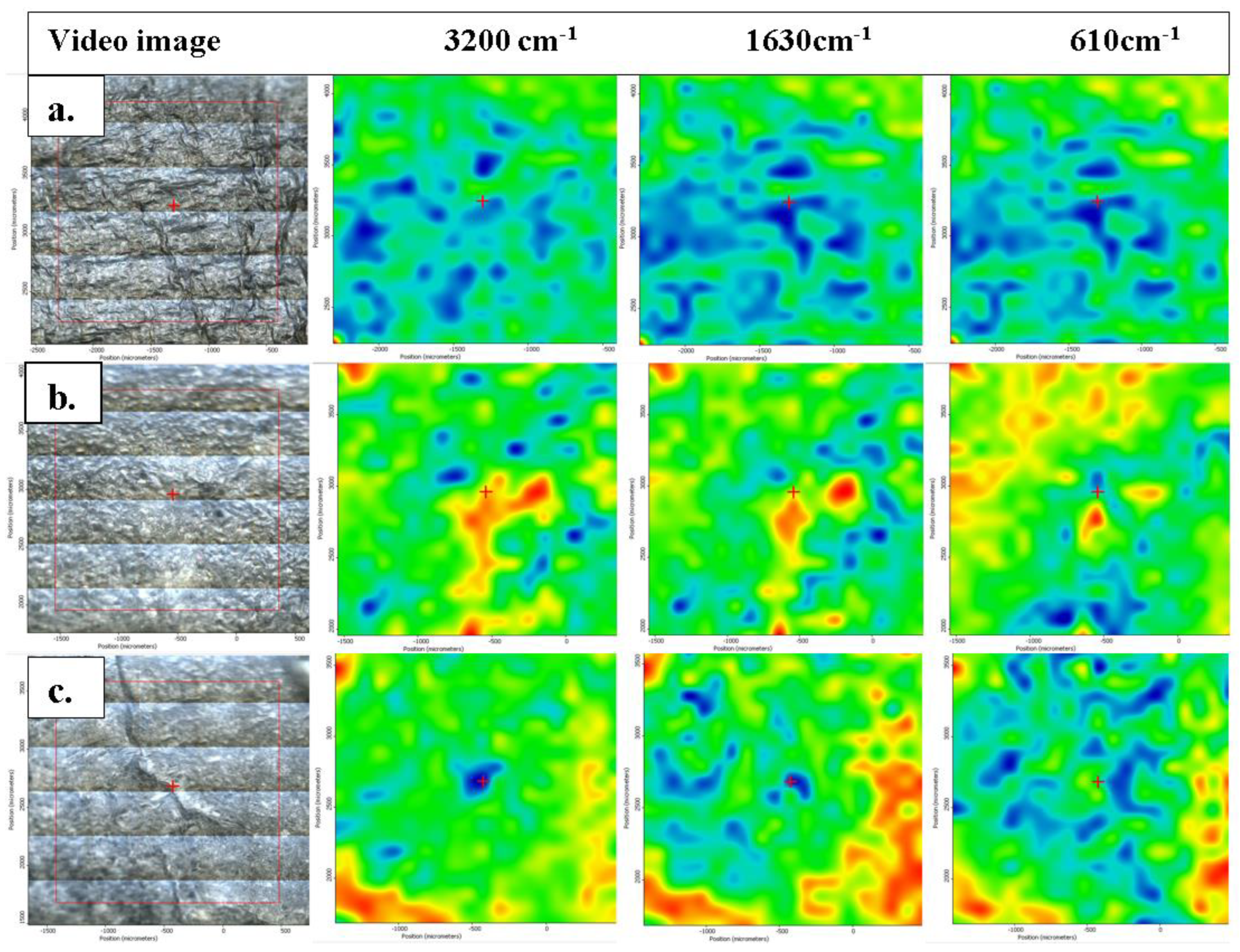
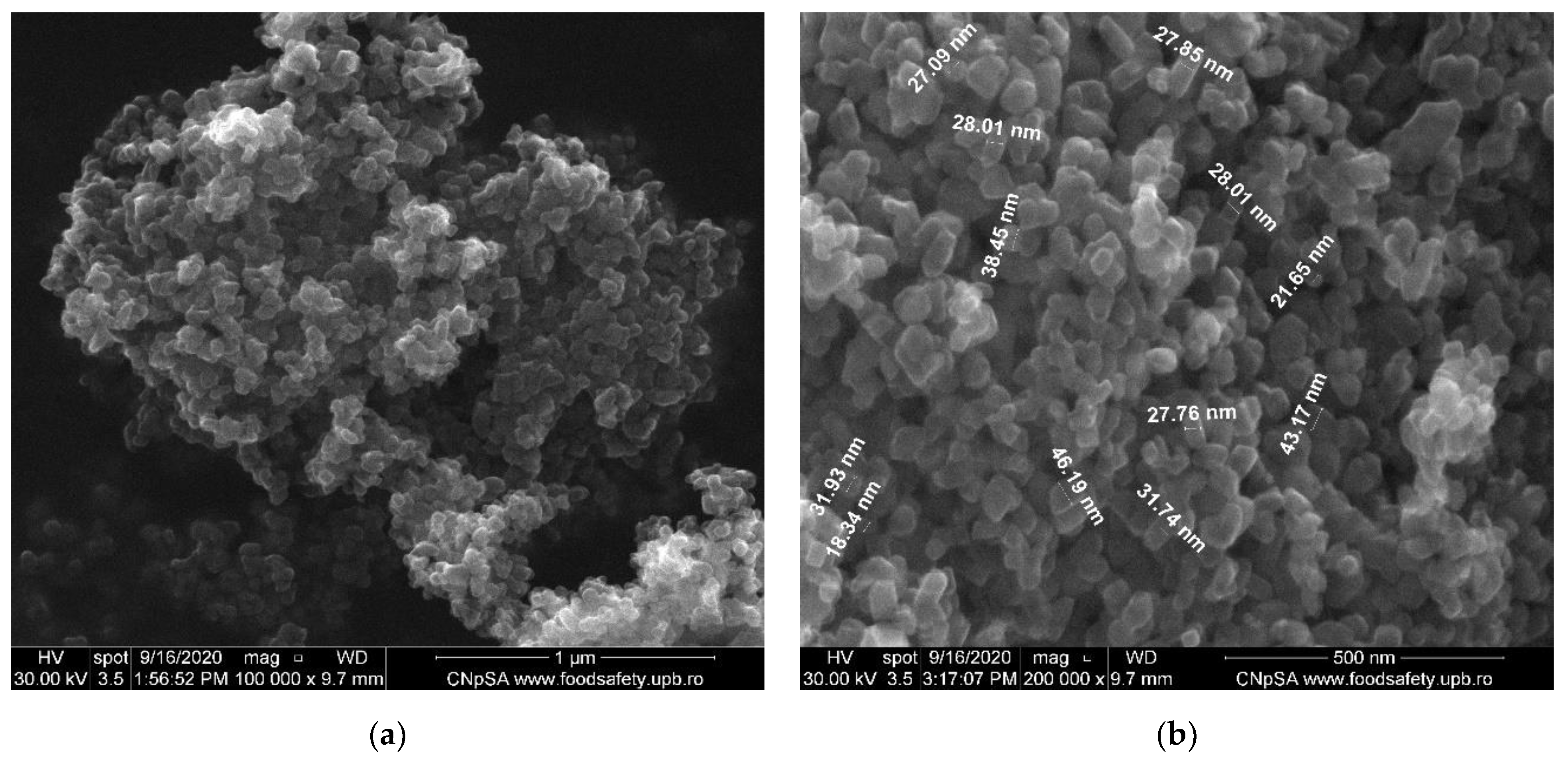
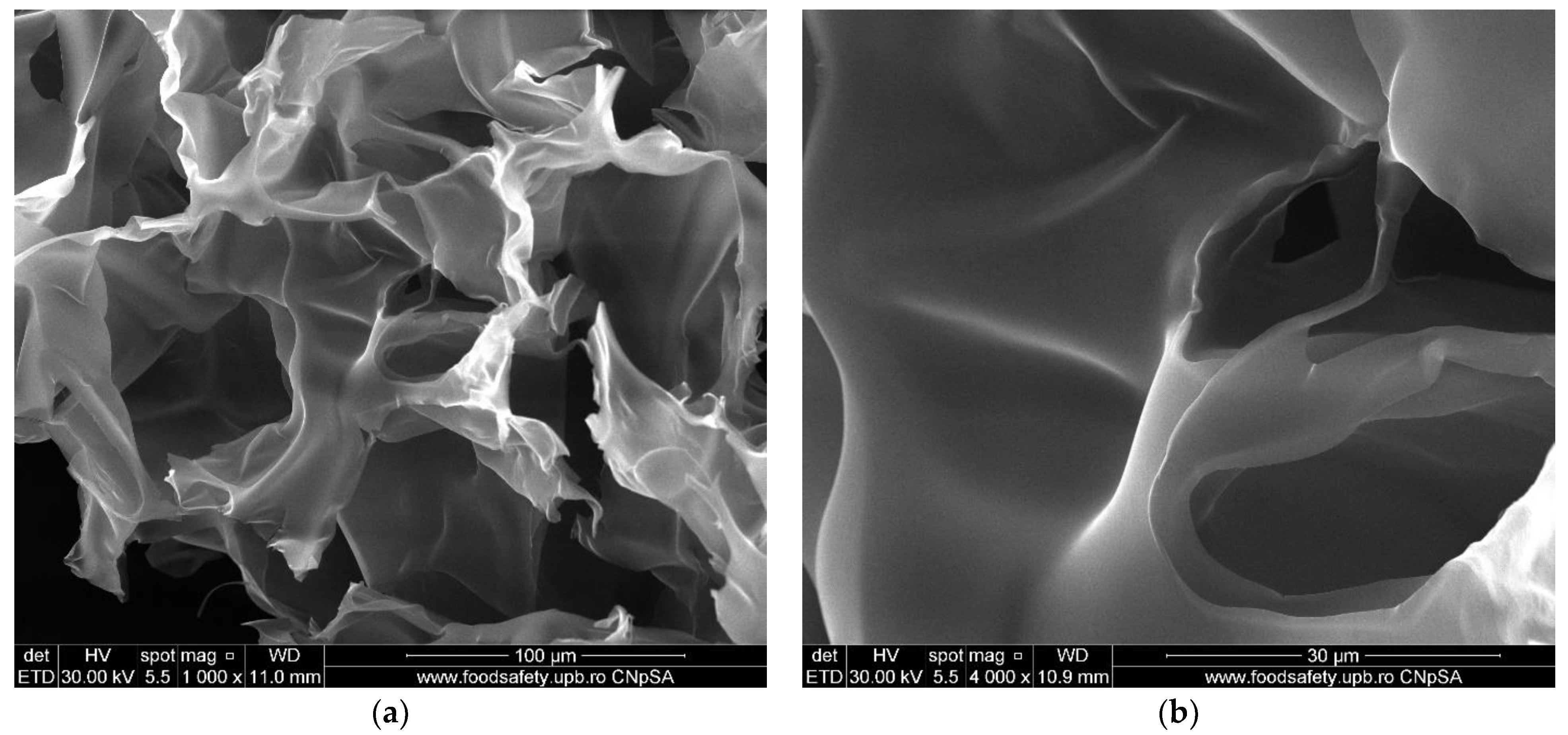
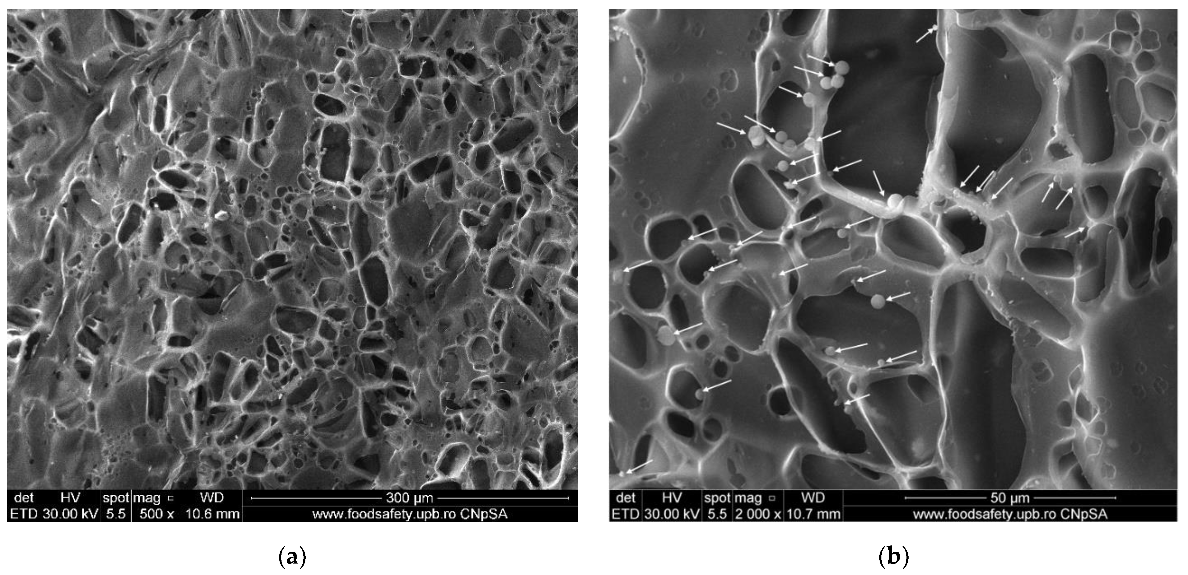
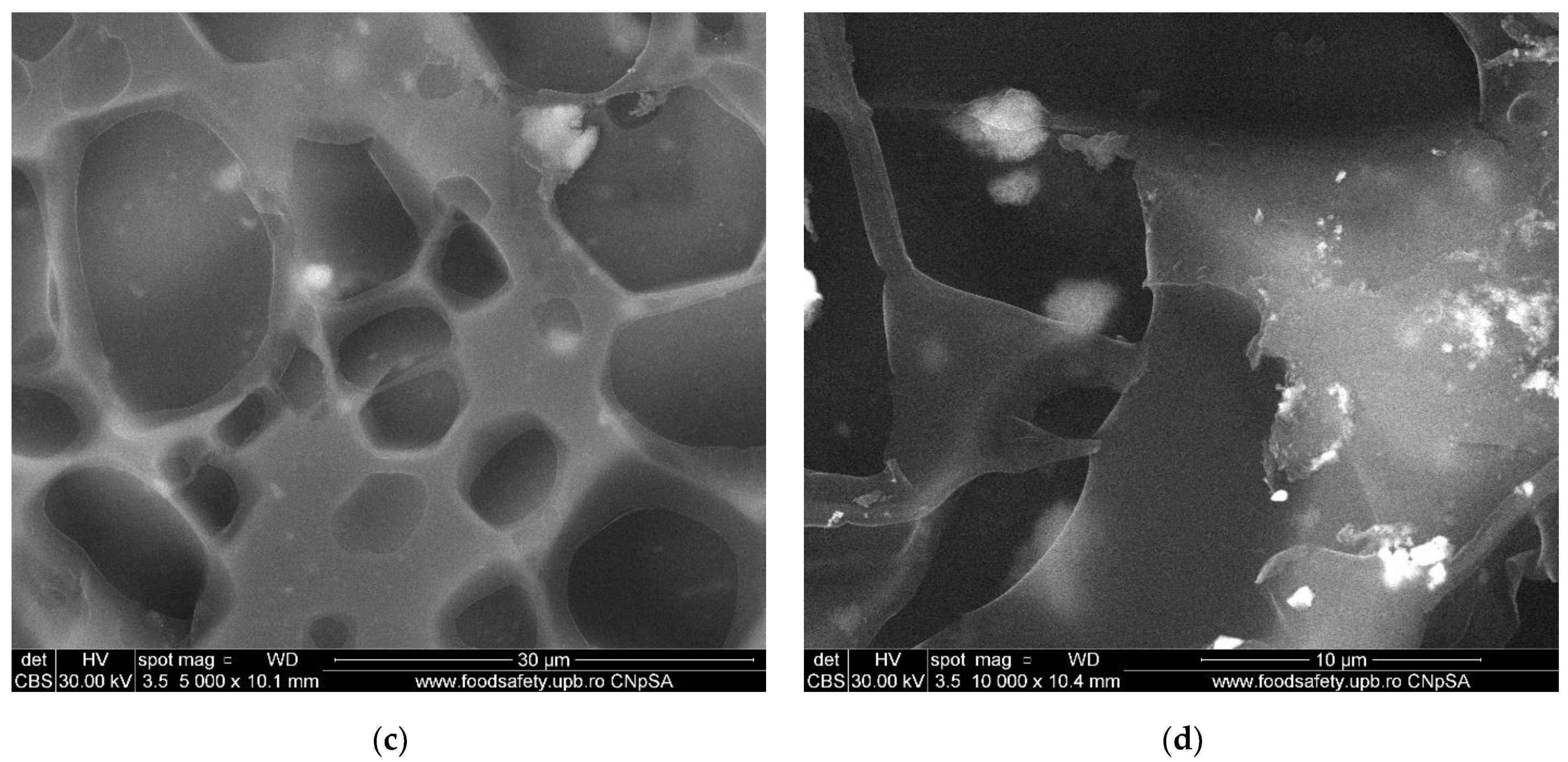
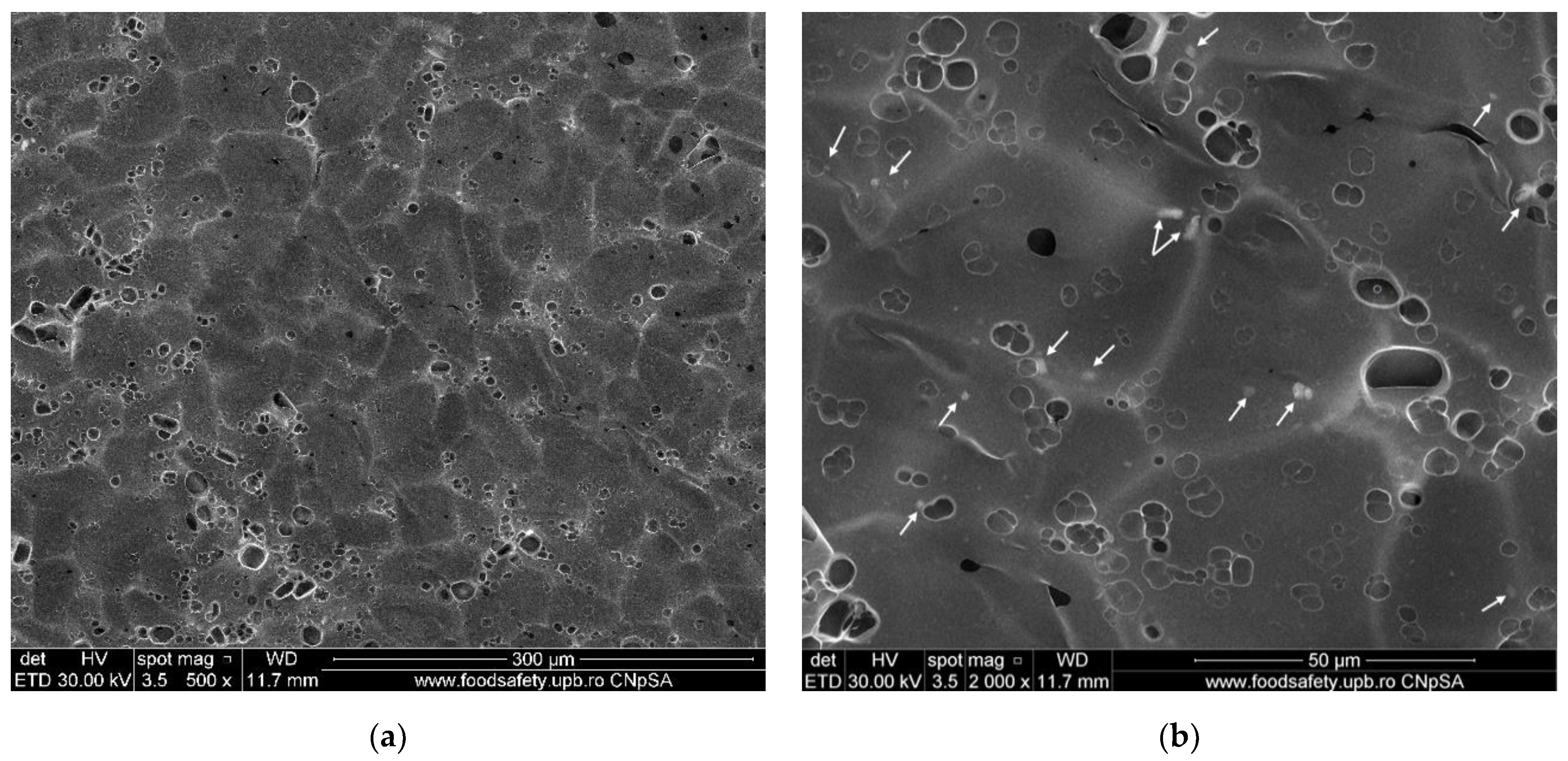
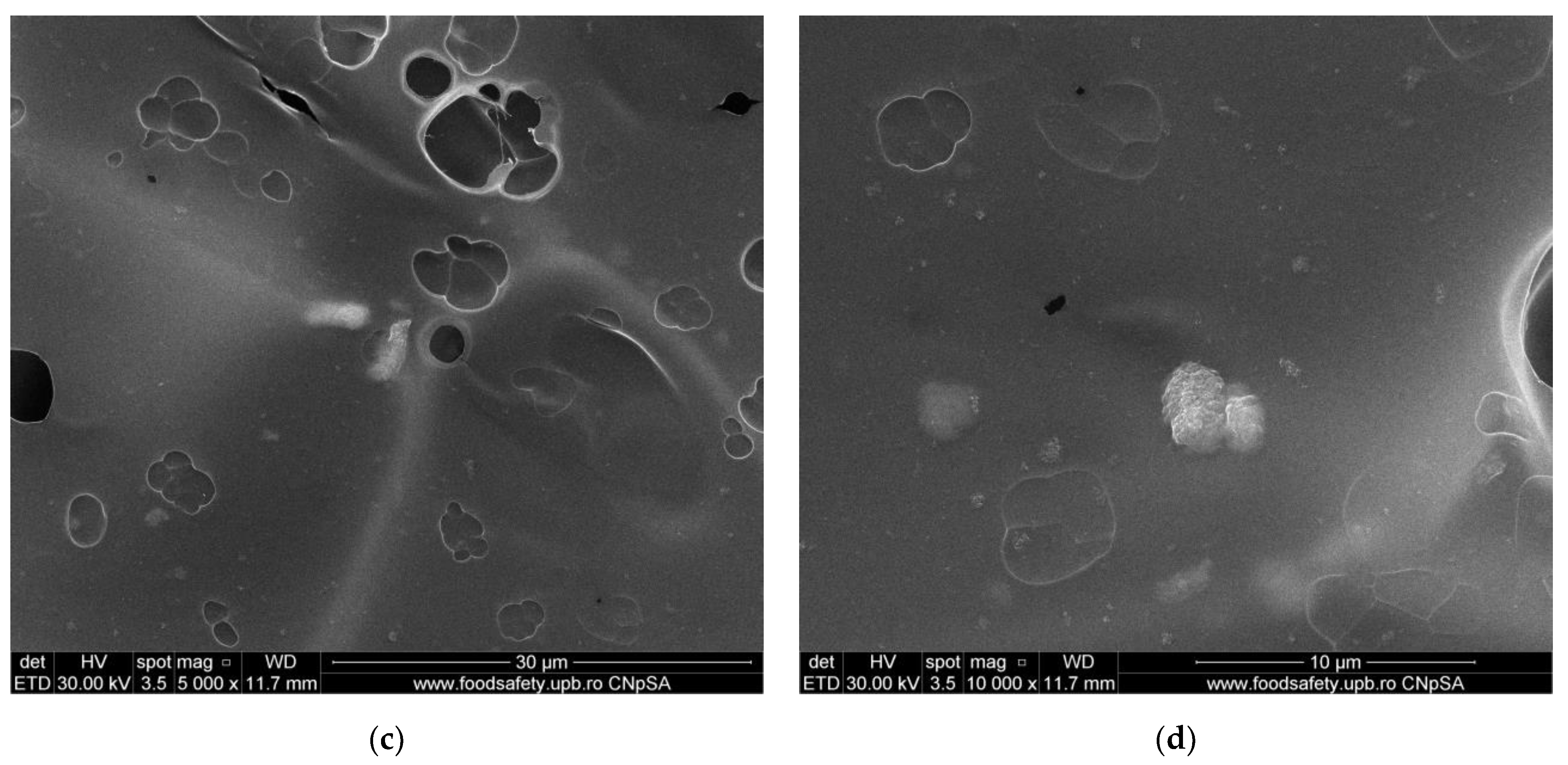
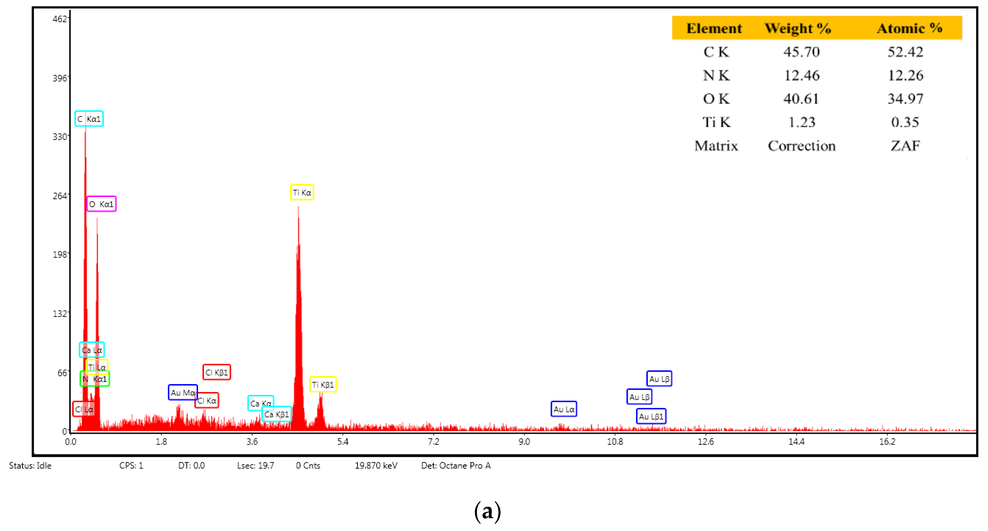
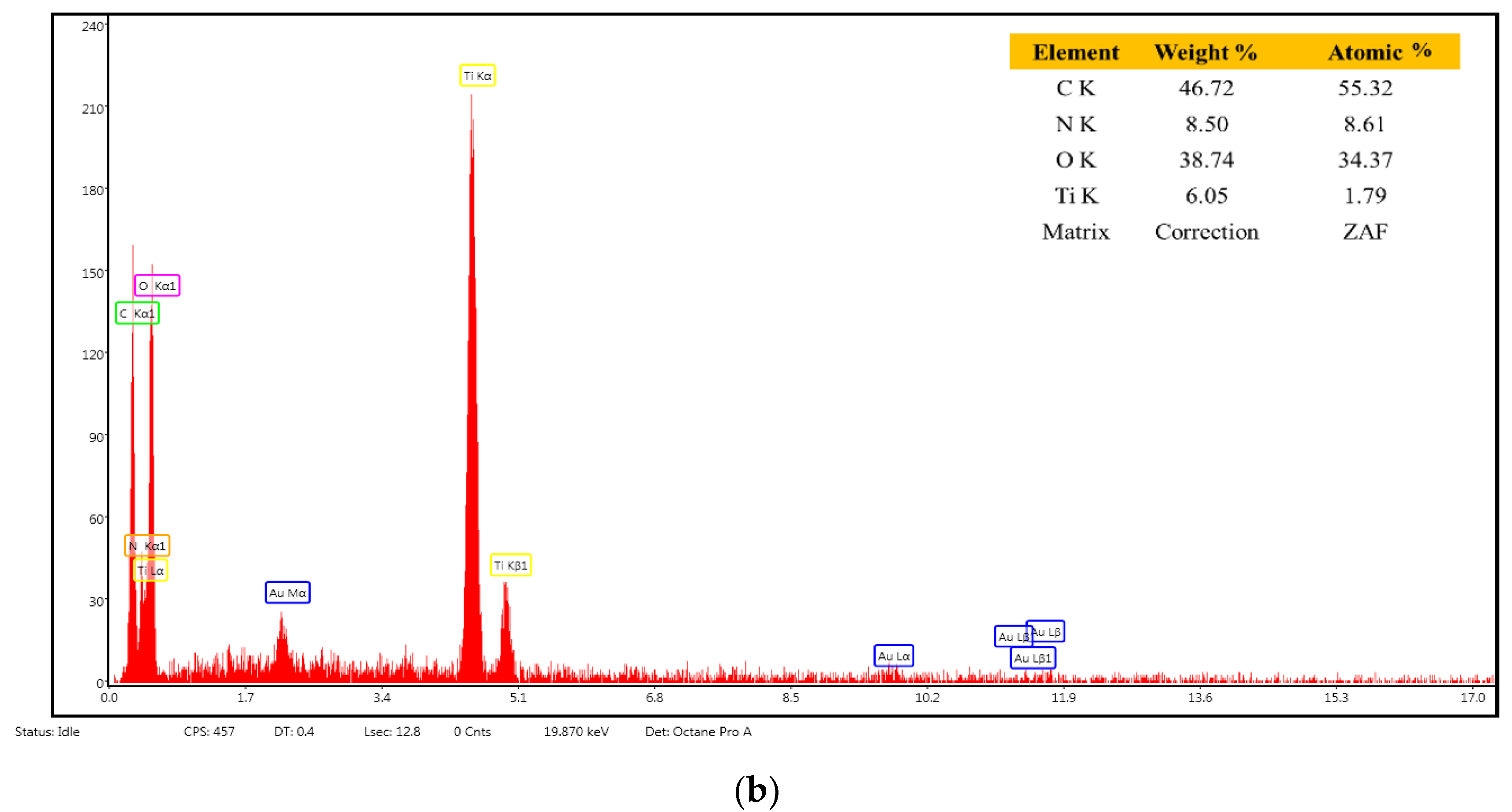
3.4. Scanning Electron Microscopy (SEM) Characterization

The SEM analysis gives us information regarding the surface morphology and possible fractures that could appear inside the membranes. For the SEM characterization, the samples were coated with a thin layer of gold before using them for analysis.
The as-synthesized TiO2 nanoparticles were examined by SEM (shown in Figure 7). They have polyhedral shapes and a tendency to form agglomerates. The individual TiO2 nanoparticles presented uniformity in size (average of 30.8 nm) and shape. Correlating the values from SEM with those calculated by the Scherrer equation from XRD, we can conclude that each nanoparticle contains a single crystallite grain.
The morphology of the simple chitosan (CS) membrane is presented in Figure 8. The membrane is highly porous, with labyrinthic pores. Such pores are the result of the coagulation and cross-linking processes when large flakes become fused under various shapes. The lyophilization process also plays an important role in forming the smaller pores by forcing the water out of the structure. The result is a sponge-like appearance of the membrane, similar to that in other literature reports [44,74,75].
From the analysis of SEM micrographs, one may observe that the as-prepared composite membranes have a different structure, which may be caused by the higher concentration of nanoparticles within the chitosan matrix (for chitosan/TiO2 5%). In the first case, the chitosan/TiO2 1%, the well-interconnected and homogenous porous microstructure generated during the lyophilization process can be observed (Figure 9a). This porous structure is decorated with TiO2 nanoparticle agglomerations (marked with arrows in Figure 9b). Higher magnifications (Figure 9c,d) reveal the stand alone nature of the pores (with no interconnections). The pores are smaller than those of the simple chitosan membrane, but still numerous and large enough, due to the presence of the TiO2 nanoparticles that act as cross-linking points, stitching the chitosan polymer chains together. Multiple nanoparticle agglomerates are visible on the pore surfaces (Figure 9d). The pore formation within the membranes might lead to enhanced adsorption capacity vs. various pollutants presented in wastewaters, which is significant in developing adsorbent membranes for water purification applications.
The chitosan/TiO2 5% has a smoother surface (Figure 10a), on which numerous smaller pores are visible (smaller and less numerous when compared with CS or chitosan/TiO2 1% membranes). Adding a higher concentration of TiO2 nanoparticles has reduced the porosity due to the increased density of TiO2 nanoparticles within the chitosan matrix. The high availability of TiO2 nanoparticles, which act as cross-linking points, leads to tighter packing of the polymer matrix, with smaller pores, and less numerous when compared with previous membranes. Higher magnification images (Figure 10c,d) indicate that many of these pores are shallow, lacking depth in the membrane structure. Therefore, this composite membrane might present a lower adsorption capacity for pollutants. The identified TiO2 nanoparticle agglomerates seem embedded into the chitosan matrix, and well dispersed across the membrane’s surface (Figure 10b).
EDX analysis (Figure 11a,b) indicates the elemental composition of the tested samples. The results confirm that a higher Ti atom concentration is found in the composite membrane chitosan/TiO2 5%, for both samples the percentage being close to the theoretical one.

3.5. Adsorption of Heavy Metal Ions (Cd and Pb)

The composite samples were immersed in cadmium or lead nitrate solutions to assess the retention capacity of each obtained composite membrane. Determinations of the Cd and Pb ion quantity adsorbed by the membranes were conducted by ICP-MS analysis. Table 3 presents the obtained results verified by a certified reference material based on heavy metal solutions.
The experimental results confirmed the SEM data and our starting hypothesis. The sample chitosan/TiO2 1%, having a higher porosity and more free functional groups than the chitosan matrix, is exhibiting a higher value for the removal capacity for both Cd and Pb when compared with the sample chitosan/TiO2 5%. In addition to the different membrane porosity as revealed by SEM micrographs, the higher TiO2 content leads to more functional groups of chitosan being involved in interactions with the nanoparticle surfaces. This diminishes the available groups that can be involved in heavy metal ion adsorption, thus confirming our starting hypothesis.
The calculated removal efficiency (1% solutions) for the chitosan/TiO2 1% membrane (81.9% for Pb and 38.1% for Cd) was higher than for the chitosan/TiO2 5% one (58.3% for Pb and 35.4% for Cd), due to the higher porosity as seen in SEM micrographs and higher number of free amino and hydroxyl functional groups. Table 4 presents a literature comparison with previously reported results.
The literature reports quite a few methods for regeneration of the chitosan-based membranes used, with the aim of enhancing the reusability and preserving the performance level [80,81,82]. For regeneration and reuse, depending on the nature of the adsorbate, the desorbing agent can be acid, salt, base, chelating agent, etc. For cations, usually an acidic solution is used for regeneration. By adding an acidic eluent, the amino groups in chitosan become protonated, and this favors the desorption of cations [83]. Efficient regeneration of the membrane, without major loss in the adsorption capacity, photocatalytic or antimicrobial activities, is highly desirable due to operating costs and elimination of secondary waste. Therefore, further studies are required to determine the regeneration and reusability capacity of these membranes.

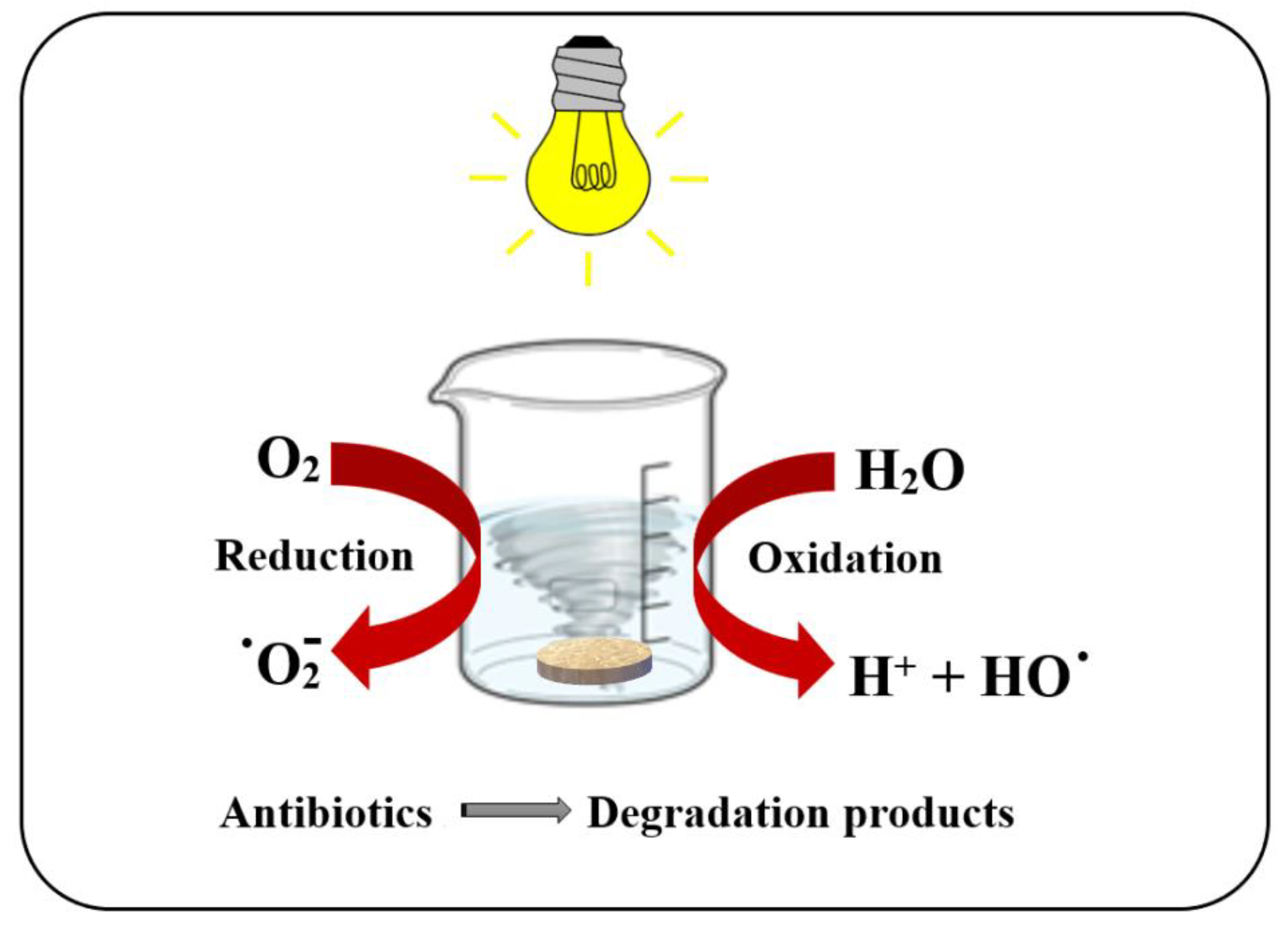
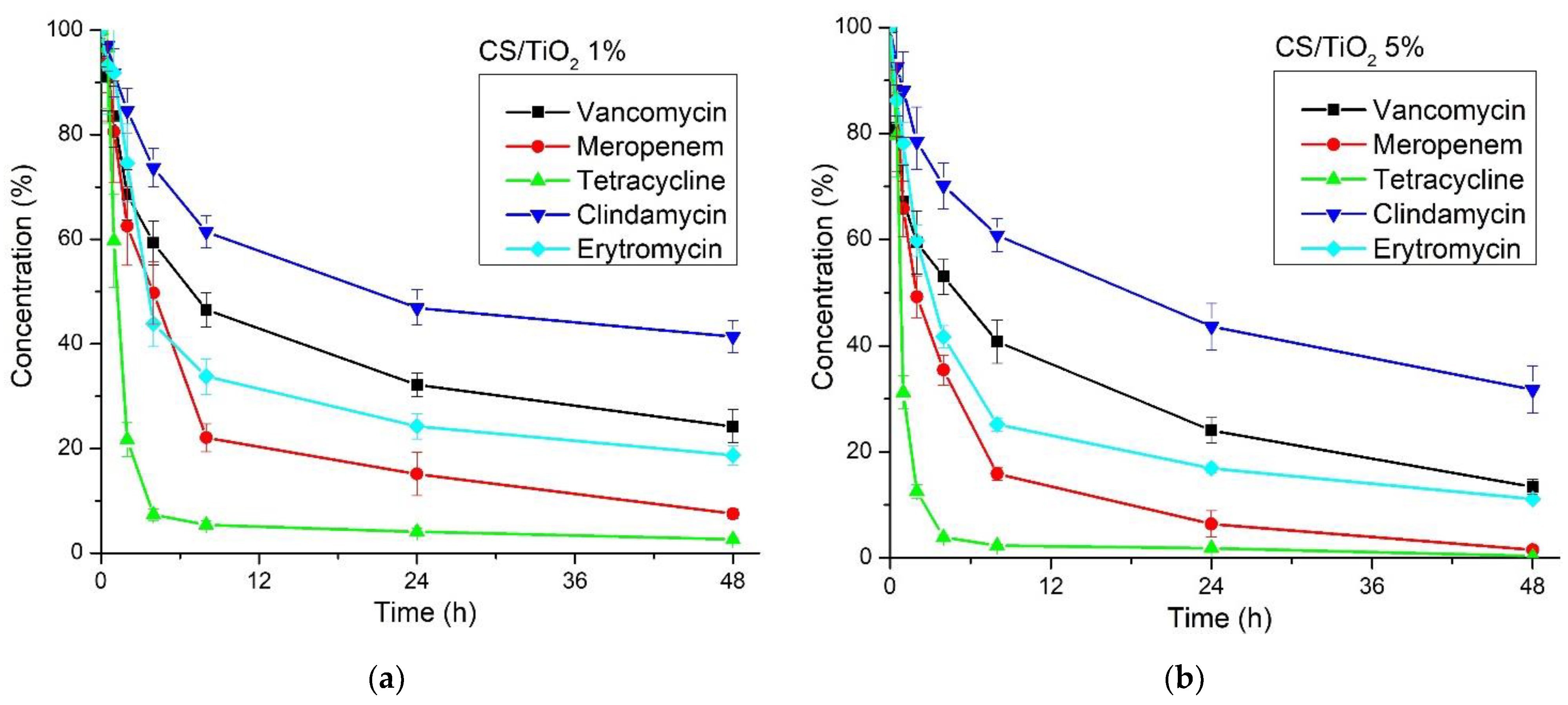
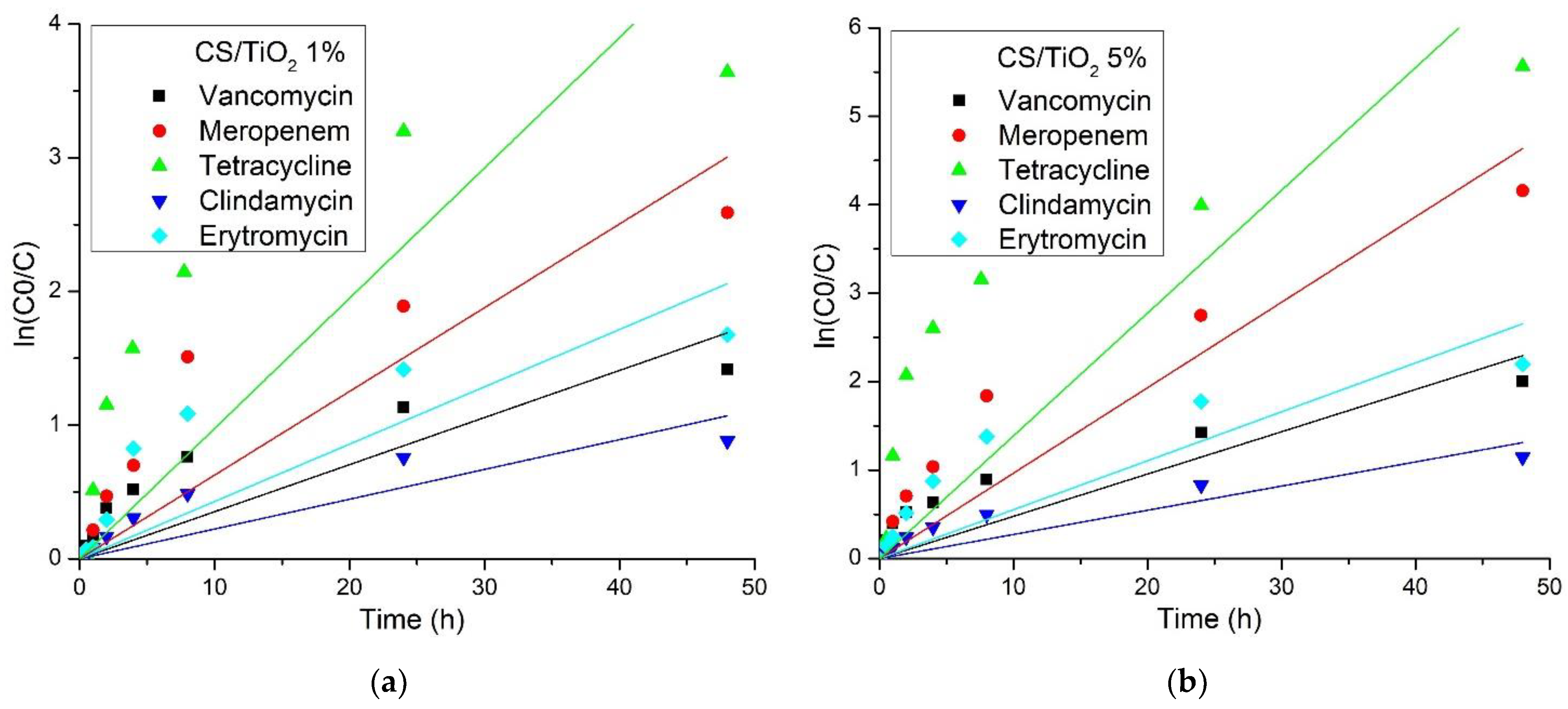
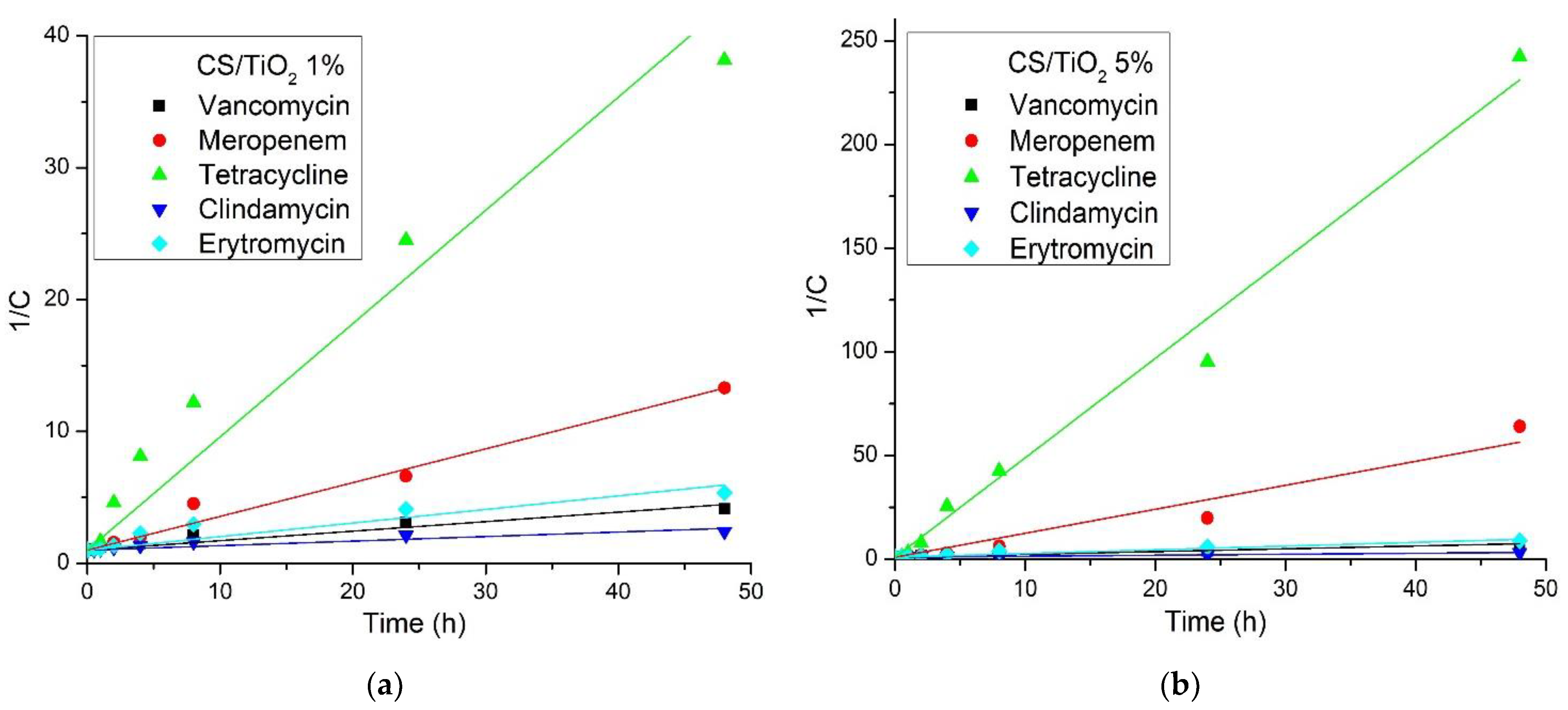
3.6. Photocatalytic Activity Determination
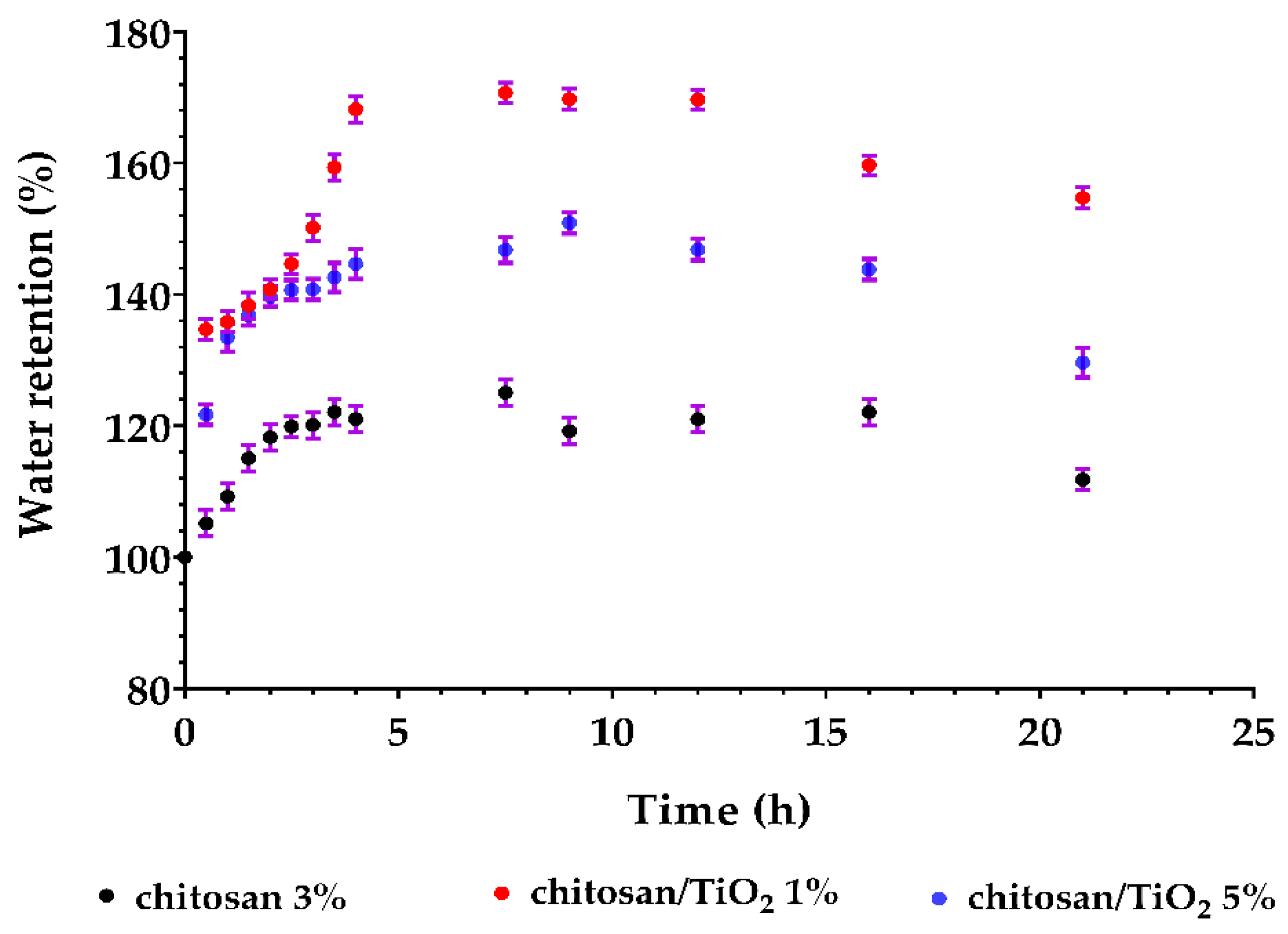
The photocatalytic activity of the chitosan/TiO2 composite membranes was tested against a solution containing a mix of antibiotics (50 ppb each) by using visible light, as indicated in Figure 12.
TiO2 is a well-known photocatalyst under UV light [84,85]. In this research, we have proven that it can be used in composite materials to purify the water under visible light irradiation. The results obtained after LC-MS analysis (Figure 13) indicated that both membranes have an excellent capacity to degrade the antibiotics under visible light irradiation. Nevertheless, the membrane chitosan/TiO2 5% has a superior performance.
The removal efficiency values, calculated as the percent of initial antibiotic concentration removed during 48 h of irradiation, are presented in Table 5.
The best removal efficiency was obtained for tetracycline, followed by meropenem, while the lowest degradation performance was recorded for clindamycin (still ~68% for the chitosan/TiO2 5% membrane).
In order to compare our results with the previous ones reported in the literature (there are available reports on chitosan/TiO2 composites for tetracycline only), the rate constant must be determined. The literature reports pseudo-first- and pseudo-second-order kinetics for the photocatalytic degradation of various organics by TiO2 [59,86,87,88]. To establish the best fitting model, we represented graphically both types of reactions according to the following equations:
ln(C0/C) = kobs1∙t     for a pseudo-first-order kinetic and,
1/C = 1/C0 + kobs2∙t     for a pseudo-second-order kinetic,
where C0 and C represent initial concentration and the concentration at time t, and kobs1 and kobs2 represent the pseudo-first-order and pseudo-second-order rate constants. The plots ln(C0/C) and 1/C vs time (t) are presented in Figure 14 and Figure 15, respectively. Figure 14a and Figure 15a are present the plots for the chitosan/TiO2 1% membrane and Figure 14b and Figure 15b present the plots for the chitosan/TiO2 1% membrane.
The values calculated for kobs1 and kobs2 are presented in Table 6, together with the corresponding correlation coefficients. From the obtained results, we can see that the pseudo-second-order model gave the better fit to the experimental data, with an R22 between 0.9003 and 0.9925.
Some studies on the photocatalytic degradation of tetracycline with chitosan/TiO2 composites [59] have indicated that the rate constant for a pseudo-first-order reaction is dependent on antibiotic concentration, with the best value of 0.0117 min−1 for 20 mg/L tetracycline concentration (20 ppm). This rate is in fact quite low due to the introduction of some heteropolyacids into the composite material; the best efficiency reported is only 66.67%. A higher removal rate of 85% under simulated sun radiation was reported in [61], against a 10 mg/L tetracycline solution (10 ppm), with a pseudo-first-order rate constant of 0.0322 min−1. This rate constant is still only a fraction of the ones we reported in the present study.
By using N- and S- doped TiO2, Farhadian et al., [89] obtained a constant rate of 0.048 min−1 for photocatalytic degradation of a 25 ppm tetracycline solution. In another study, [90], the authors report a pseudo-second-order rate constant of 0.0014 L∙mg−1∙min−1 for the best removal efficiency of 77% in the case of tetracycline in a photoreactor with UV irradiation and bubbling O2. A 97.2% removal efficiency of tetracycline from a 20 ppm solution under UV irradiation is reported in [91] by using a composite chitosan/TiO2 + ZnO/graphene, in this case ZnO being the main photocatalyst. The complete removal of tetracycline is reported in [92]. This was achieved under UV irradiation and in the presence of H2O2 with a composite made from alginate/chitosan/TiO2. Comparing the results from the literature with those reported in the present study indicates that our composite membranes exhibited a better performance, under visible light irradiation, with no added O2 or H2O2, with the best removal efficiency of 99.62% and rate constants with orders of magnitude higher.
For meropenem, for which we report here a removal efficiency of 98.44%, the literature is scarce. In [93], the authors report a removal efficiency of 80% under direct solar irradiation (concentrated with mirrors), using 50 mg pure TiO2 for a starting solution with a concentration identical with ours (only meropenem—50 ppb).

3.7. Swelling Study

To evaluate the sample stability, the composite membranes were subjected to swelling measurements to determine the weight change during water immersion. Figure 16 illustrates the water uptake capacity of the obtained composite membranes. It can be observed that in the first 4–6 h, the membrane mass is increasing up to a maximum value for each membrane type. After saturation is attained, the mass is relatively constant up to 8–10 h, and afterwards a slight decrease is recorded.
This behavior is caused by the partial disintegration/dissolution of the membrane surface, which will lead to some mass loss, as can be observed in the time interval 10–24 h. Similar mass loss during swelling tests are reported in the literature for other polysaccharide membranes [18,94]. The porous nature of the chitosan/TiO2 1% membrane allows it a higher swelling capacity (~170%), while the chitosan/TiO2 5% membrane, with fewer and smaller pores, exhibits a smaller water retention capacity (~150%).

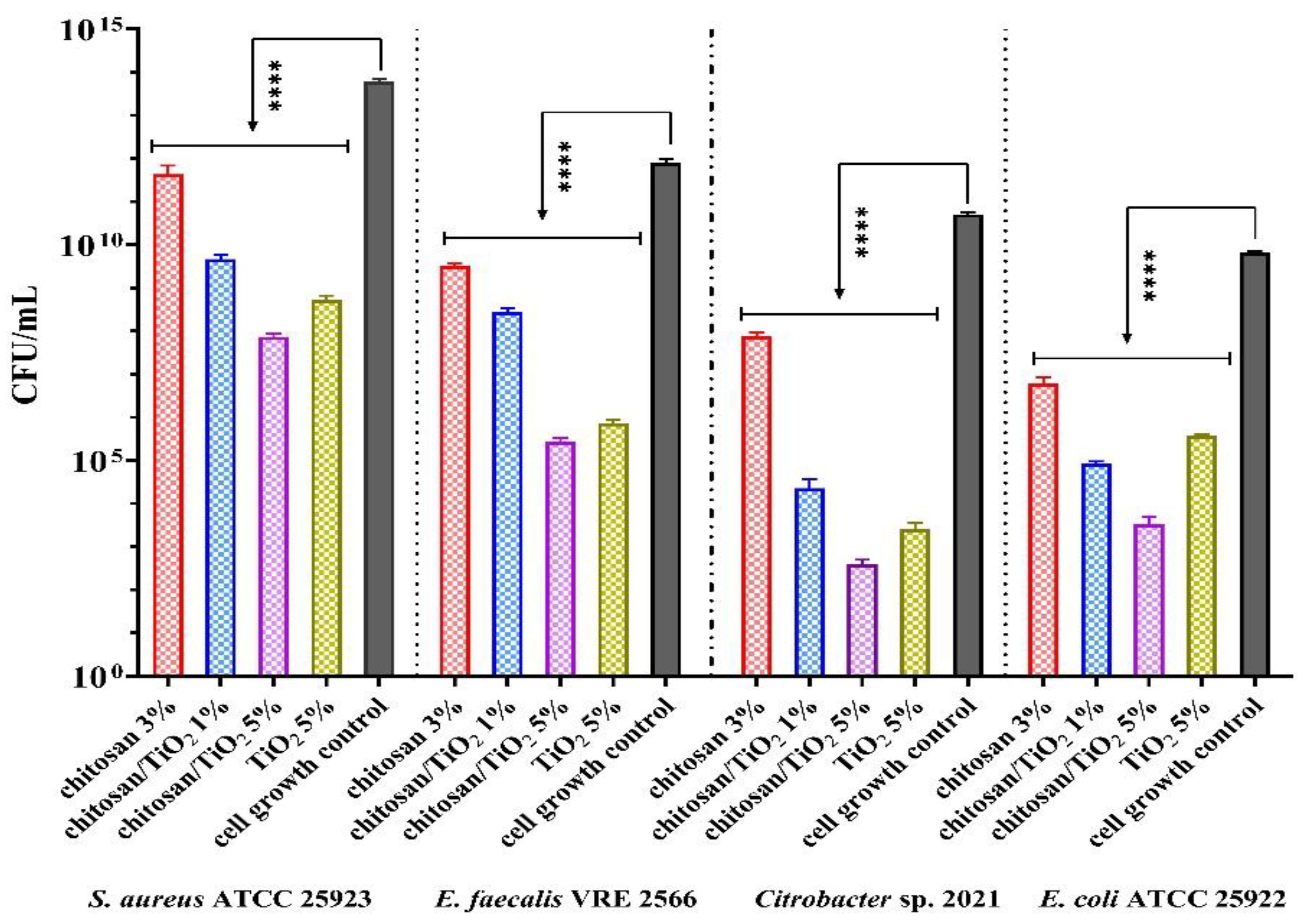
3.8. The Antibacterial Assessments

The antibacterial activity of TiO2 nanoparticles depends on their size, shape, morphology, crystalline structure and photocatalytic activity [95,96,97]. The size, shape and crystal structure are the most important properties that influence the physicochemical properties and antibacterial activity of TiO2 nanoparticles [96,97]. TiO2 acts on microbial cells again by affecting the cell wall and membrane by damaging DNA and decreasing/stopping the DNA replication and protein processes [98]. In another way, the cells exposed to TiO2 show a rapid inactivation at the regulatory and signaling levels, affecting the coenzyme-independent respiratory chains and assimilating processes of transport ions, respectively, damaging the biosynthesis of Fe-S clusters [99,100]. Moreover, the antibacterial activity of biodegradable chitosan/TiO2 composite membranes can also be explained by the bacteriostatic effect of the chitosan [101,102].
In this study, the antibacterial activity of the composite membranes was evaluated against Gram-negative and Gram-positive bacteria. The chitosan/TiO2 membranes presented bacteriostatic activity for all strains tested. The most sensitive strain on the action of TiO2 nanoparticles was E. coli ATCC 25922, followed by Citrobacter sp. 2021 (clinical strain). The TiO2 determined a sensitivity to both Gram-negative bacteria, with a decrease of at least 5 logarithmic units of CFU/mL compared to the cell growth control (C+). The chitosan presents moderate antimicrobial activity on all strains.
Figure 17 shows a synergic and significant antibacterial effect of both chitosan/TiO2 composite membranes. The antibacterial activity was enhanced due to the chitosan and TiO2 combination. For Citrobacter sp., the chitosan/TiO2 1% and chitosan/TiO2 5% have a pronounced high degree of inhibition (more than 8 logarithmic units of CFU/mL of C+). In addition, these samples showed a stronger antibacterial effect than TiO2 or chitosan. In the case of the Gram-positive bacteria, the composite membranes presented moderate antibacterial activity against both drug-resistance strains. The E. faecalis was more sensitive to the influence of the tested membranes.
The sensitivity of the bacterial strains can also be explained by the small size of the TiO2 nanoparticles (~30 nm), which have a higher interaction with the bacterial cells and activate the damage processes [96].
In another study [103], the antibacterial activity of chitosan/TiO2 composite film was evaluated for E. coli, S. aureus, C. albicans and A. niger strains (~106 CFU/mL) under visible light. The most sensitive strain was E coli, with a final bactericidal ratio of approx. 100%. In addition, Siripatrawan et al. [104] reported a higher growth inhibition of the tested bacteria and fungi (S. aureus, E. coli, Salmonella, P. aeruginosa, Aspergillus, etc.) for the exposure of chitosan/TiO2 films to UV radiation than those samples without exposure.
The lower capacity of the composite membranes to adhere to the surface determined the perspective/potential of using them in water purification applications.

4. Conclusions

In conclusion, we successfully synthesized the composite membranes based on chitosan and TiO2 nanoparticles. These composite membranes were used to remove toxic pollutants from wastewater in a complex water purification process (adsorption of heavy metal ions, photocatalytic degradation a five-antibiotic mix and antibacterial activity against four bacterial strains). The SEM images showed TiO2 nanoparticle agglomerations on the surface of the membrane’s porous structure.
The heavy metal ion adsorption capacity indicated high values for Cd (315 mg/g) and Pb (297 mg/g) for the chitosan/TiO2 1% membrane. This confirms the hypothesis that a decrease in TiO2 quantity in the composite membrane will free more of the chitosan’s functional groups, enhancing the adsorption capacity. Nevertheless, both membranes have good adsorption capacity, with the values obtained for chitosan/TiO2 5% being around 255 mg/g for both Cd and Pb.
The photocatalytic activity of the membranes was determined against a mix of five antibiotic solutions under visible light irradiation. The composite membranes practically removed both tetracycline and meropenem with efficiencies over 98%, while for vancomycin and erythromycin the efficiencies were 86% and 88%, respectively.
The antibacterial assessment indicated a pronounced degree of inhibition in the case of Gram-negative bacteria due to a synergic activity of chitosan and TiO2, with a reduction of 8 units log CFU/mL obtained for Citrobacter sp. In the case of Gram-positive bacteria, the tested composite membranes presented moderate antibacterial activity against both strains.
In conclusion, it can be stated that both tested composite membranes can be used for complex water purification.

Author Contributions

Conceptualization, A.S., O.-C.O. and A.F.; methodology, A.S., C.-I.I., O.-C.O., D.F. and A.F.; validation, A.S., O.-C.O., D.F., A.F. and L.-M.D.; formal analysis, C.-I.I., G.D., A.-M.C., V.-A.S., R.-D.T., L.M., O.-C.O. and L.-M.D.; investigation, A.S. and A.F.; writing—original draft preparation, A.S., C.-I.I., L.M. and O.-C.O.; writing—review and editing, A.S., L.M., O.-C.O. and A.F.; visualization, A.S., O.-C.O., D.F., A.F. and E.A.; supervision, O.-C.O., A.F. and E.A.; project administration, A.S.; funding acquisition, E.A. All authors have read and agreed to the published version of the manuscript.

Funding

This work was supported by a grant from the Romanian Ministry of Education and Research, CNCS-UEFISCDI, project number PN-III-P1-1.1-PD-2019-1156, within PNCD III.

Institutional Review Board Statement

Not applicable.

Informed Consent Statement

Not applicable.

Data Availability Statement

Not applicable.

Conflicts of Interest

The authors declare no conflict of interest.

References

  1. Mihaly, M.; Comanescu, A.F.; Rogozea, E.A.; Meghea, A. Nonionic microemulsion extraction of Ni (II) from wastewater. Mol. Cryst. Liq. Cryst. 2010, 523, 63–72. [Google Scholar] [CrossRef]
  2. Spoială, A.; Ilie, C.-I.; Dolete, G.; Trușcă, R.-D.; Motelică, L.; Oprea, O.-C.; Ficai, D.; Ficai, A.; Andronescu, E.; Dițu, L.-M. The development of antimicrobial chitosan/ ZnO nanocomposite membranes for water purification. Rev. Română Mater. Rom. J. Mater. 2022, 52, 17–25. [Google Scholar]
  3. Voicu, G.; Oprea, O.; Vasile, B.S.; Andronescu, E. Antibacterial activity of zinc oxide—Gentamicin hybrid material. Dig. J. Nanomater. Biostructures 2013, 8, 1191–1203. [Google Scholar]
  4. Enache, D.F.; Vasile, E.; Simonescu, C.M.; Razvan, A.; Nicolescu, A.; Nechifor, A.C.; Oprea, O.; Patescu, R.E.; Onose, C.; Dumitru, F. Cysteine-functionalized silica-coated magnetite nanoparticles as potential nano adsorbents. J. Solid State Chem. 2017, 253, 318–328. [Google Scholar] [CrossRef]
  5. Enache, D.F.; Vasile, E.; Simonescu, C.M.; Culita, D.; Vasile, E.; Oprea, O.; Pandele, A.M.; Razvan, A.; Dumitru, F.; Nechifor, G. Schiff base-functionalized mesoporous silicas (MCM-41, HMS) as Pb(II) adsorbents. RSC Adv. 2018, 8, 176–189. [Google Scholar] [CrossRef] [Green Version]
  6. Motelica, L.; Marinof, L.; Holban, A.; Vasile, B.S.; Ficai, A. Optical, photocatalytic and antibacterial properties of zinc oxide nanoparticles obtained by a solvothermal method. UPB Sci. Bull. Ser. B 2020, 82, 59–70. [Google Scholar]
  7. Chaudhary, M.; Maiti, A. Fe-Al-Mn@chitosan based metal oxides blended cellulose acetate mixed matrix membrane for fluoride decontamination from water: Removal mechanisms and antibacterial behavior. J. Membr. Sci. 2020, 611, 118372. [Google Scholar] [CrossRef]
  8. Modi, A.; Bellare, J. Efficient removal of 2,4-dichlorophenol from contaminated water and alleviation of membrane fouling by high flux polysulfone-iron oxide/graphene oxide composite hollow fiber membranes. J. Water Process Eng. 2020, 33, 101113. [Google Scholar]
  9. Mihaly, M.; Lacatusu, I.; Enesca, I.A.; Meghea, A. Hybride nanomaterials based on silica coated C-60 clusters obtained by microemulsion technique. Mol. Cryst. Liq. Cryst. 2008, 483, 205–215. [Google Scholar] [CrossRef]
  10. Mihaly, M.; Lacatusu, I.; Meghea, A. Sulphonephtalein chromophore as molecular probe in micelle systems. Rev. Chim.-Buchar. 2007, 58, 929–932. [Google Scholar]
  11. Wang, J.; Zhuang, S. Removal of various pollutants from water and wastewater by modified chitosan adsorbents. Crit. Rev. Environ. Sci. Technol. 2018, 47, 2331–2386. [Google Scholar] [CrossRef]
  12. Modi, A.; Jiang, Z.Y.; Kasher, R. Hydrostable ZIF-8 layer on polyacrylonitrile membrane for efficient treatment of oilfield produced water. Chem. Eng. J. 2022, 434, 133513. [Google Scholar] [CrossRef]
  13. Croitoru, A.M.; Ficai, A.; Ficai, D.; Trusca, R.; Dolete, G.; Andronescu, E.; Turculet, S.C. Chitosan/graphene oxide nanocomposite membranes as adsorbents with applications in water purification. Materials 2020, 13, 1687. [Google Scholar] [CrossRef] [PubMed] [Green Version]
  14. Jamshaid, M.; Arshad Khan, A.; Ahmed, K.; Saleem, M. Heavy metal in drinking water its effect on human health and its treatment techniques-a review. Int. J. Biosci. 2018, 12, 223–240. [Google Scholar]
  15. Qa, M.; Ms, K. Effect on human health due to drinking water contaminated with heavy metals. J. Pollut. Eff. Control. 2016, 5. [Google Scholar] [CrossRef] [Green Version]
  16. Culita, D.C.; Simonescu, C.M.; Patescu, R.E.; Preda, S.; Stanica, N.; Munteanu, C.; Oprea, O. Polyamine functionalized magnetite nanoparticles as novel adsorbents for Cu(II) removal from aqueous solutions. J. Inorg. Organomet. Polym. Mater. 2017, 27, 490–502. [Google Scholar] [CrossRef]
  17. Motelica, L.; Ficai, D.; Oprea, O.C.; Ficai, A.; Ene, V.L.; Vasile, B.S.; Andronescu, E.; Holban, A.M. Antibacterial biodegradable films based on alginate with silver nanoparticles and lemongrass essential oil-innovative packaging for cheese. Nanomaterials 2021, 11, 2377. [Google Scholar] [CrossRef]
  18. Motelica, L.; Ficai, D.; Oprea, O.; Ficai, A.; Trusca, R.D.; Andronescu, E.; Holban, A.M. Biodegradable alginate films with ZnO nanoparticles and citronella essential oil-a novel antimicrobial structure. Pharmaceutics 2021, 13, 1020. [Google Scholar] [CrossRef]
  19. Spoiala, A.; Ilie, C.I.; Trusca, R.D.; Oprea, O.C.; Surdu, V.A.; Vasile, B.S.; Ficai, A.; Ficai, D.; Andronescu, E.; Ditu, L.M. Zinc oxide nanoparticles for water purification. Materials 2021, 14, 4747. [Google Scholar] [CrossRef]
  20. Ly, Q.V.; Matindi, C.N.; Kuvarega, A.T.; Le, Q.V.; Tran, V.S.; Hu, Y.X.; Li, J.X. Organic fouling assessment of novel PES/SPSf/double layered hydroxide mixed matrix membrane for water treatment application. J. Water Process Eng. 2020, 37, 101526. [Google Scholar] [CrossRef]
  21. Modi, A.; Bellare, J. Efficient separation of biological macromolecular proteins by polyethersulfone hollow fiber ultrafiltration membranes modified with Fe3O4 nanoparticles-decorated carboxylated graphene oxide nanosheets. Int. J. Biol. Macromol. 2019, 135, 798–807. [Google Scholar] [CrossRef] [PubMed]
  22. Verma, S.K.; Modi, A.; Singh, A.K.; Teotia, R.; Kadam, S.; Bellare, J. Functionally coated polyethersulfone hollow fiber membranes: A substrate for enhanced HepG2/C3A functions. Colloids Surf. B 2018, 164, 358–369. [Google Scholar] [CrossRef] [PubMed]
  23. Spoială, A.; Ilie, C.I.; Ficai, D.; Ficai, A.; Andronescu, E. Chitosan-based nanocomposite polymeric membranes for water purification-a review. Materials 2021, 14, 2091. [Google Scholar] [CrossRef]
  24. Crini, G.; Badot, P.-M. Application of chitosan, a natural aminopolysaccharide, for dye removal from aqueous solutions by adsorption processes using batch studies: A review of recent literature. Prog. Polym. Sci. 2008, 33, 399–447. [Google Scholar] [CrossRef]
  25. Aliabadi, M.; Irani, M.; Ismaeili, J.; Piri, H.; Parnian, M.J. Electrospun nanofiber membrane of PEO/Chitosan for the adsorption of nickel, cadmium, lead and copper ions from aqueous solution. Chem. Eng. J. 2013, 220, 237–243. [Google Scholar] [CrossRef]
  26. Aliabadi, M.; Irani, M.; Ismaeili, J.; Najafzadeh, S. Design and evaluation of chitosan/hydroxyapatite composite nanofiber membrane for the removal of heavy metal ions from aqueous solution. J. Taiwan Inst. Chem. Eng. 2014, 45, 518–526. [Google Scholar] [CrossRef]
  27. Haider, S.; Park, S.-Y. Preparation of the electrospun chitosan nanofibers and their applications to the adsorption of Cu(II) and Pb(ii) ions from an aqueous solution. J. Membr. Sci. 2009, 328, 90–96. [Google Scholar] [CrossRef]
  28. Gupta, N.; Kushwaha, A.K.; Chattopadhyaya, M.C. Adsorptive removal of Pb2+, Co2+ and Ni2+ by hydroxyapatite/chitosan composite from aqueous solution. J. Taiwan Inst. Chem. Eng. 2012, 43, 125–131. [Google Scholar] [CrossRef]
  29. Chen, A.-H.; Liu, S.-C.; Chen, C.-Y.; Chen, C.-Y. Comparative adsorption of Cu(II), Zn(II), and Pb(II) ions in aqueous solution on the crosslinked chitosan with epichlorohydrin. J. Hazard. Mater. 2008, 154, 184–191. [Google Scholar] [CrossRef]
  30. Mousa, N.E.; Simonescu, C.M.; Patescu, R.E.; Onose, C.; Tardei, C.; Culita, D.C.; Oprea, O.; Patroi, D.; Lavric, V. Pb2+ removal from aqueous synthetic solutions by calcium alginate and chitosan coated calcium alginate. React. Funct. Polym. 2016, 109, 137–150. [Google Scholar] [CrossRef]
  31. Simonescu, C.M.; Marin, I.; Tardei, C.; Marinescu, V.; Oprea, O.; Capatina, C. Chitosan and chitosan modified with glutaraldehyde microparticles for Pb(II) biosorption I. Microparticles preparation and characterization. Rev. Chim.-Buchar. 2014, 65, 627–632. [Google Scholar]
  32. Vakili, M.; Rafatullah, M.; Salamatinia, B.; Abdullah, A.Z.; Ibrahim, M.H.; Tan, K.B.; Gholami, Z.; Amouzgar, P. Application of chitosan and its derivatives as adsorbents for dye removal from water and wastewater: A review. Carbohydr. Polym. 2014, 113, 115–130. [Google Scholar] [CrossRef] [PubMed]
  33. Sapna; Sharma, R.; Kumar, D. Chitosan-based membranes for wastewater desalination and heavy metal detoxification. Micro Nano Technol. 2019, 799–814. [Google Scholar] [CrossRef]
  34. Li, M.; Hong, Y.; Wang, Z.; Chen, S.; Gao, M.; Kwok, R.T.K.; Qin, W.; Lam, J.W.Y.; Zheng, Q.; Tang, B.Z. Fabrication of chitosan nanoparticles with aggregation-induced emission characteristics and their applications in long-term live cell imaging. Macromol. Rapid Commun. 2013, 34, 767–771. [Google Scholar] [CrossRef] [PubMed]
  35. Jiang, W.; Wang, W.; Pan, B.; Zhang, Q.; Zhang, W.; Lv, L. Facile fabrication of magnetic chitosan beads of fast kinetics and high capacity for copper removal. ACS Appl. Mater. Interfaces 2014, 6, 3421–3426. [Google Scholar] [CrossRef] [PubMed]
  36. Jaiswal, A.; Ghsoh, S.S.; Chattopadhyay, A. Quantum dot impregnated-chitosan film for heavy metal ion sensing and removal. Langmuir 2012, 28, 15687–15696. [Google Scholar] [CrossRef] [PubMed]
  37. Nayak, V.; Jyothi, M.S.; Balakrishna, R.G.; Padaki, M.; Ismail, A.F. Preparation and characterization of chitosan thin films on mixed-matrix membranes for complete removal of chromium. ChemistryOpen 2015, 4, 278–287. [Google Scholar] [CrossRef]
  38. Kim, T.-Y.; Park, S.-S.; Cho, S.-Y. Adsorption characteristics of reactive black 5 onto chitosan beads cross-linked with epichlorohydrin. J. Ind. Eng. Chem. 2012, 18, 1458–1464. [Google Scholar] [CrossRef]
  39. Li, X.-Q.; Tang, R.-C. Crosslinking of chitosan fiber by a water-soluble diepoxy crosslinker for enhanced acid resistance and its impact on fiber structures and properties. React. Funct. Polym. 2016, 100, 116–122. [Google Scholar] [CrossRef]
  40. Poon, L.; Wilson, L.D.; Headley, J.V. Chitosan-glutaraldehyde copolymers and their sorption properties. Carbohydr. Polym. 2014, 109, 92–101. [Google Scholar] [CrossRef]
  41. Riegger, B.R.; Baurer, B.; Mirzayeva, A.; Tovar, G.E.M.; Bach, M. A systematic approach of chitosan nanoparticle preparation via emulsion crosslinking as potential adsorbent in wastewater treatment. Carbohydr. Polym. 2018, 180, 46–54. [Google Scholar] [CrossRef] [PubMed]
  42. Singh, P.; Chauhan, K.; Priya, V.; Singhal, R.K. A greener approach for impressive removal of As(III)/As(V) from an ultra-low concentration using a highly efficient chitosan thiomer as a new adsorbent. RSC Adv. 2016, 6, 64946–64961. [Google Scholar] [CrossRef]
  43. Ardila, N.; Daigle, F.; Heuzey, M.C.; Ajji, A. Antibacterial activity of neat chitosan powder and flakes. Molecules 2017, 22, 100. [Google Scholar] [CrossRef] [PubMed]
  44. Motelica, L.; Ficai, D.; Ficai, A.; Trusca, R.D.; Ilie, C.I.; Oprea, O.C.; Andronescu, E. Innovative antimicrobial chitosan/ZnO/Ag NPs/citronella essential oil nanocomposite—Potential coating for grapes. Foods 2020, 9, 1801. [Google Scholar] [CrossRef] [PubMed]
  45. Radulescu, M.; Ficai, D.; Oprea, O.; Ficai, A.; Andronescu, E.; Holban, A.M. Antimicrobial chitosan based formulations with impact on different biomedical applications. Curr. Pharm. Biotechnol. 2015, 16, 128–136. [Google Scholar] [CrossRef] [PubMed]
  46. Marino, T.; Boerrigter, M.; Faccini, M.; Chaumette, C.; Arockiasamy, L.; Bundschuh, J.; Figoli, A. Photocatalytic activity and synthesis procedures of TiO2 nanoparticles for potential applications in membranes. In Application of Nanotechnology in Membranes for Water Treatment; CRC Press: Boca Raton, FL, USA, 2017; pp. 127–146. [Google Scholar]
  47. Park, J.Y.; Lee, C.; Jung, K.W.; Jung, D. Structure related photocatalytic properties of TiO2. Bull. Korean Chem. Soc. 2009, 30, 402–404. [Google Scholar]
  48. Ficai, D.; Oprea, O.; Ficai, A.; Holban, A.M. Metal oxide nanoparticles: Potential uses in biomedical applications. Curr. Proteom. 2014, 11, 139–149. [Google Scholar] [CrossRef]
  49. Vaja, F.; Comanescu, C.; Oprea, O.; Ficai, D.; Guran, C. Effects of ZnO nanoparticles on the wet scrub resistance and photocatalytic properties of acrylic coatings. Rev. Chim.-Buchar. 2012, 63, 722–726. [Google Scholar]
  50. González-Calderon, J.A.; Vallejo-Montesinos, J.; Martínez-Martínez, H.N.; Cerecero-Enríquez, R.; López-Zamora, L. Effect of chemical modification of titanium dioxide particles via silanization under properties of chitosan/potato starch film. Rev. Mex. Ing. Química 2019, 18, 913–927. [Google Scholar] [CrossRef]
  51. Li, W.; Zheng, K.; Chen, H.; Feng, S.; Wang, W.; Qin, C. Influence of nano titanium dioxide and clove oil on chitosan-starch film characteristics. Polymers 2019, 11, 1418. [Google Scholar] [CrossRef] [Green Version]
  52. Anaya-Esparza, L.M.; Ruvalcaba-Gomez, J.M.; Maytorena-Verdugo, C.I.; Gonzalez-Silva, N.; Romero-Toledo, R.; Aguilera-Aguirre, S.; Perez-Larios, A.; Montalvo-Gonzalez, A.E. Chitosan-TiO2: A versatile hybrid composite. Materials 2020, 13, 811. [Google Scholar]
  53. Razzaz, A.; Ghorban, S.; Hosayni, L.; Irani, M.; Aliabadi, M. Chitosan nanofibers functionalized by TiO2 nanoparticles for the removal of heavy metal ions. J. Taiwan Inst. Chem. Eng. 2016, 58, 333–343. [Google Scholar] [CrossRef]
  54. Samadi, S.; Khalilian, F.; Tabatabaee, A. Synthesis, characterization and application of Cu–TiO2/chitosan nanocomposite thin film for the removal of some heavy metals from aquatic media. J. Nanostructure Chem. 2014, 4, 84. [Google Scholar] [CrossRef] [Green Version]
  55. Chien, S.; Abiyu Kerebo, B.; Kuo-Pin, y. Chitosan@TiO2 composites for the adsorption of copper(ii) and antibacterial applications. Sustain. Environ. Res. 2022, 32, 27. [Google Scholar]
  56. Alizadeh, B.; Delnavaz, M.; Shakeri, A. Removal of Cd(II) and phenol using novel cross-linked magnetic EDTA/chitosan/TiO2 nanocomposite. Carbohydr. Polym. 2018, 181, 675–683. [Google Scholar] [CrossRef]
  57. Nguyen, U.N.T.; Do, K.H.; Jang, B.; Kim, K.S.; Kim, J.H.; Lee, S.M. Always-on photocatalytic antibacterial facemask with mini uv-led array. Mater. Today Sustain. 2022, 18, 100117. [Google Scholar] [CrossRef]
  58. Onkani, S.P.; Diagboya, P.N.; Mtunzi, F.M.; Klink, M.J.; Olu-Owolabi, B.I.; Pakade, V. Comparative study of the photocatalytic degradation of 2-chlorophenol under uv irradiation using pristine and ag-doped species of TiO2, ZnO and ZnS photocatalysts. J. Environ. Manag. 2020, 260, 110145. [Google Scholar]
  59. Yu, X.N.; Lu, Z.Y.; Wu, D.; Yu, P.; He, M.; Chen, T.T.; Shi, W.D.; Huo, P.W.; Yan, Y.S.; Feng, Y.X. Heteropolyacid-chitosan/TiO2 composites for the degradation of tetracycline hydrochloride solution. React. Kinet. Mech. Catal. 2014, 111, 347–360. [Google Scholar]
  60. Wu, S.J.; Wang, F.; Li, Q.; Wang, J.; Zhou, Y.; Duan, N.; Niazi, S.; Wang, Z.P. Photocatalysis and degradation products identification of deoxynivalenol in wheat using upconversion nanoparticles@TiO2 composite. Food Chem. 2020, 323, 126823. [Google Scholar] [CrossRef]
  61. Hou, C.T.; Liu, H.Y.; Li, Y.J. The preparation of three-dimensional flower-like TiO2/TiOF2 photocatalyst and its efficient degradation of tetracycline hydrochloride. RSC Adv. 2021, 11, 14957–14969. [Google Scholar] [CrossRef]
  62. Motelica, L.; Popescu, A.; Razvan, A.G.; Oprea, O.; Trusca, R.D.; Vasile, B.S.; Dumitru, F.; Holban, A.M. Facile use of Zno nanopowders to protect old manual paper documents. Materials 2020, 13, 5452. [Google Scholar]
  63. Comanescu, A.-F.; Mihaly, M.; Meghea, A. Photocatalytic degradation of organic pollutants using nio based materials. UPB Sci. Bull. Ser. B 2012, 74, 49–60. [Google Scholar]
  64. Sharma, A.; Karn, R.K.; Pandiyan, S.K. Synthesis of TiO2 nanoparticles by sol-gel method and their characterization. J. Basic Appl. Eng. Res. 2014, 1, 1–5. [Google Scholar]
  65. Lemnaru Popa, G.M.; Trusca, R.D.; Ilie, C.I.; Tiplea, R.E.; Ficai, D.; Oprea, O.; Stoica-Guzun, A.; Ficai, A.; Ditu, L.M. Antibacterial activity of bacterial cellulose loaded with bacitracin and amoxicillin: In vitro studies. Molecules 2020, 25, 4069. [Google Scholar] [CrossRef] [PubMed]
  66. Anghel, I.; Holban, A.M.; Grumezescu, A.M.; Andronescu, E.; Ficai, A.; Anghel, A.G.; Maganu, M.; Laz, R.V.; Chifiriuc, M.C. Modified wound dressing with phyto-nanostructured coating to prevent staphylococcal and pseudomonal biofilm development. Nanoscale Res. Lett. 2012, 7, 690. [Google Scholar] [PubMed] [Green Version]
  67. Cotar, A.I.; Grumezescu, A.M.; Andronescu, E.; Voicu, G.; Ficai, A.; Ou, K.-L.; Huang, K.-S.; Chifiriuc, M.C. Nanotechnological solution for improving the antibiotic e_ciency against biofilms developed by gram-negative bacterial strains. Lett. Appl. NanoBioSci 2013, 2, 97–104. [Google Scholar]
  68. CLSI. Performance Standards for Antimicrobial Susceptibility Testing; Clinical and Laboratory Standards Institute: Berwyn, PA, USA, 2021. [Google Scholar]
  69. Branca, C.; D’Angelo, G.; Crupi, C.; Khouzami, K.; Rifici, S.; Ruello, G.; Wanderlingh, U. Role of the oh and nh vibrational groups in polysaccharide-nanocomposite interactions: A ftir-atr study on chitosan and chitosan/clay films. Polymer 2016, 99, 614–622. [Google Scholar]
  70. Kustiningsih, I.; Ridwan, A.; Abriyani, D.; Syairazy, M.; Kurniawan, T.; Barleany, D.R. Development of chitosan-TiO2 nanocomposite for packaging film and its ability to inactive staphylococcus aureus. Orient. J. Chem. 2019, 35, 1132–1137. [Google Scholar]
  71. Humelnicu, A.C.; Samoila, P.; Asandulesa, M.; Cojocaru, C.; Bele, A.; Marinoiu, A.T.; Sacca, A.; Harabagiu, V. Chitosan-sulfated titania composite membranes with potential applications in fuel cell: Influence of cross-linker nature. Polymers 2020, 12, 1125. [Google Scholar]
  72. Kumar, P. Nano-TiO2 doped chitosan scaffold for the bone tissue engineering applications. Int. J. Biomater. 2018, 2018, 6576157. [Google Scholar] [CrossRef] [Green Version]
  73. Mindru, I.; Gingasu, D.; Patron, L.; Marinescu, G.; Calderon-Moreno, J.M.; Preda, S.; Oprea, O.; Nita, S. Copper aluminate spinel by soft chemical routes. Ceram. Int. 2016, 42, 154–164. [Google Scholar] [CrossRef]
  74. Vasile, B.S.; Oprea, O.; Voicu, G.; Ficai, A.; Andronescu, E.; Teodorescu, A.; Holban, A. Synthesis and characterization of a novel controlled release zinc oxide/gentamicin-chitosan composite with potential applications in wounds care. Int. J. Pharm. 2014, 463, 161–169. [Google Scholar]
  75. Rosli, N.A.H.; Loh, K.S.; Wong, W.Y.; Lee, T.K.; Ahmad, A. Hybrid composite membrane of phosphorylated chitosan/poly (vinyl alcohol)/silica as a proton exchange membrane. Membranes 2021, 11, 675. [Google Scholar] [CrossRef]
  76. Chen, M.H.; Yu, M.D.; Kang, R.F.; Sun, H.M.; Zhang, W.; Wang, S.S.; Wang, N.; Wang, J. Removal of Pb (ii) and V (v) from aqueous solution by glutaraldehyde crosslinked chitosan and nanocomposites. Chemosphere 2022, 297, 134084. [Google Scholar] [CrossRef] [PubMed]
  77. Tao, Y.G.; Ye, L.B.; Pan, J.; Wang, Y.M.; Tang, B. Removal of Pb(II) from aqueous solution on chitosan/TiO2 hybrid film. J. Hazard. Mater. 2009, 161, 718–722. [Google Scholar] [CrossRef]
  78. Chen, A.W.; Zeng, G.M.; Chen, G.Q.; Hu, X.J.; Yan, M.; Guan, S.; Shang, C.; Lu, L.H.; Zou, Z.J.; Xie, G.X. Novel thiourea-modified magnetic ion-imprinted chitosan/TiO2 composite for simultaneous removal of cadmium and 2,4-dichlorophenol. Chem. Eng. J. 2012, 191, 85–94. [Google Scholar] [CrossRef]
  79. Wu, S.P.; Kan, J.R.; Dai, X.Z.; Shen, X.J.; Zhang, K.; Zhu, M.Y. Ternary carboxymethyl chitosan-hemicellulose-nanosized TiO2 composite as effective adsorbent for removal of heavy metal contaminants from water. Fibers Polym. 2017, 18, 22–32. [Google Scholar] [CrossRef]
  80. Chen, Y.S.; Ooi, C.W.; Show, P.L.; Hoe, B.C.; Chai, W.S.; Chiu, C.Y.; Wang, S.S.S.; Chang, Y.K. Removal of ionic dyes by nanofiber membrane functionalized with chitosan and egg white proteins: Membrane preparation and adsorption efficiency. Membranes 2022, 12, 63. [Google Scholar]
  81. Popa, A.; Visa, A.; Maranescu, B.; Hulka, I.; Lupa, L. Chemical modification of chitosan for removal of Pb(II) ions from aqueous solutions. Materials 2021, 14, 7894. [Google Scholar] [CrossRef]
  82. Rathinam, K.; Kou, X.W.; Hobby, R.; Panglisch, S. Sustainable development of magnetic chitosan core-shell network for the removal of organic dyes from aqueous solutions. Materials 2021, 14, 7701. [Google Scholar] [CrossRef]
  83. Alves, D.C.D.; Healy, B.; Yu, T.; Breslin, C.B. Graphene-based materials immobilized within chitosan: Applications as adsorbents for the removal of aquatic pollutants. Materials 2021, 14, 3655. [Google Scholar] [CrossRef] [PubMed]
  84. Zanfir, A.V.; Voicu, G.; Badanoiu, A.I.; Gogan, D.; Oprea, O.; Vasile, E. Synthesis and characterization of titania-silica fume composites and their influence on the strength of self-cleaning mortar. Compos. Part B Eng. 2018, 140, 157–163. [Google Scholar] [CrossRef]
  85. Totu, E.E.; Cristache, C.M.; Voicila, E.; Oprea, O.; Agir, I.; Tavukcuoglu, O.; Didilescu, A.C. On physical and chemical characteristics of poly (methylmethacrylate) nanocomposites for dental applications. I. Mater. Plast 2017, 54, 666–672. [Google Scholar] [CrossRef]
  86. Jawad, A.H.; Mubarak, N.S.A.; Ishak, M.A.M.; Ismail, K.; Nawawi, W.I. Kinetics of photocatalytic decolourization of cationic dye using porous TiO2 film. J. Taibah Univ. Sci. 2016, 10, 352–362. [Google Scholar] [CrossRef] [Green Version]
  87. Rytwo, G.; Zelkind, A.L. Evaluation of kinetic pseudo-order in the photocatalytic degradation of ofloxacin. Catalysts 2022, 12, 24. [Google Scholar] [CrossRef]
  88. Alghamdi, Y.G.; Krishnakumar, B.; Malik, M.A.; Alhayyani, S. Design and preparation of biomass-derived activated carbon loaded TiO2 photocatalyst for photocatalytic degradation of reactive red 120 and ofloxacin. Polymers 2022, 14, 880. [Google Scholar] [CrossRef]
  89. Farhadian, N.; Akbarzadeh, R.; Pirsaheb, M.; Jen, T.C.; Fakhri, Y.; Asadi, A. Chitosan modified N, S-doped TiO2 and N, S-doped ZnO for visible light photocatalytic degradation of tetracycline. Int. J. Biol. Macromol. 2019, 132, 360–373. [Google Scholar] [CrossRef]
  90. Ikhlef-Taguelmimt, T.; Hamiche, A.; Yahiaoui, I.; Bendellali, T.; Lebik-Elhadi, H.; Ait-Amar, H.; Aissani-Benissad, F. Tetracycline hydrochloride degradation by heterogeneous photocatalysis using TiO2(P25) immobilized in biopolymer (chitosan) under uv irradiation. Water Sci. Technol. 2020, 82, 1570–1578. [Google Scholar] [CrossRef]
  91. Patehkhor, H.A.; Fattahi, M.; Khosravi-Nikou, M. Synthesis and characterization of ternary chitosan-TiO2-ZnO over graphene for photocatalytic degradation of tetracycline from pharmaceutical wastewater. Sci. Rep. 2021, 11, 24177. [Google Scholar] [CrossRef]
  92. Rizzi, V.; Gubitosa, J.; Fini, P.; Petrella, A.; Romita, R.; Agostiano, A.; Cosma, P. A “classic” material for capture and detoxification of emergent contaminants for water purification: The case of tetracycline. Environ. Technol. Innov. 2020, 19, 100812. [Google Scholar] [CrossRef]
  93. Cabrera-Reina, A.; Martinez-Piernas, A.B.; Bertakis, Y.; Xekoukoulotakis, N.P.; Aguera, A.; Perez, J.A.S. TiO2 photocatalysis under natural solar radiation for the degradation of the carbapenem antibiotics imipenem and meropenem in aqueous solutions at pilot plant scale. Water Res. 2019, 166, 115037. [Google Scholar] [CrossRef] [PubMed]
  94. Zhang, H.; Zhu, J.Y.; Hu, Y.Q.; Chen, A.N.; Zhou, L.; Gao, H.; Liu, Y.M.; Liu, S.Q. Study on photocatalytic antibacterial and sustained-release properties of cellulose/TiO2/beta-CD composite hydrogel. J. Nanomater. 2019, 2019. [Google Scholar] [CrossRef]
  95. Rokicka-Konieczna, P.; Wanag, A.; Sienkiewicz, A.; Kusiak-Nejman, E.; Morawski, A.W. Antibacterial effect of TiO2 nanoparticles modified with aptes. Catal. Commun. 2020, 134, 105862. [Google Scholar] [CrossRef]
  96. López de Dicastillo, C.; Correa, M.G.; Martínez, F.B.; Streitt, C.; Galotto, M.J. Antimicrobial effect of titanium dioxide nanoparticles. In Antimicrobial Resistance—A One Health Perspective; IntechOpen: London, UK, 2020; pp. 1–19. [Google Scholar]
  97. Lopez de Dicastillo, C.; Patino, C.; Galotto, M.J.; Palma, J.L.; Alburquenque, D.; Escrig, J. Novel antimicrobial titanium dioxide nanotubes obtained through a combination of atomic layer deposition and electrospinning technologies. Nanomaterials 2018, 8, 128. [Google Scholar] [CrossRef] [PubMed] [Green Version]
  98. Azizi-Lalabadi, M.; Ehsani, A.; Divband, B.; Alizadeh-Sani, M. Antimicrobial activity of titanium dioxide and zinc oxide nanoparticles supported in 4a zeolite and evaluation the morphological characteristic. Sci. Rep. 2019, 9, 17439. [Google Scholar] [CrossRef] [Green Version]
  99. Khashan, K.S.; Sulaiman, G.M.; Abdulameer, F.A.; Albukhaty, S.; Ibrahem, M.A.; Al-Muhimeed, T.; AlObaid, A.A. Antibacterial activity of TiO2 nanoparticles prepared by one-step laser ablation in liquid. Appl. Sci. 2021, 11, 4623. [Google Scholar] [CrossRef]
  100. Kubacka, A.; Diez, M.S.; Rojo, D.; Bargiela, R.; Ciordia, S.; Zapico, I.; Albar, J.P.; Barbas, C.; Martins dos Santos, V.A.; Fernandez-Garcia, M.; et al. Understanding the antimicrobial mechanism of TiO2-based nanocomposite films in a pathogenic bacterium. Sci. Rep. 2014, 4, 4134. [Google Scholar] [CrossRef] [Green Version]
  101. Sahariah, P.; Másson, M. Antimicrobial chitosan and chitosan derivatives: A review of the structure–activity relationship. Biomacromolecules 2017, 18, 3846–3868. [Google Scholar] [CrossRef]
  102. Goy, R.C.; Morais, S.T.B.; Assis, O.B.G. Evaluation of the antimicrobial activity of chitosan and its quaternized derivative on E. coli and S. aureus growth. Rev. Bras. Farmacogn. 2016, 26, 122–127. [Google Scholar] [CrossRef] [Green Version]
  103. Zhang, X.; Xiao, G.; Wang, Y.; Zhao, Y.; Su, H.; Tan, T. Preparation of chitosan-TiO2 composite film with efficient antimicrobial activities under visible light for food packaging applications. Carbohydr. Polym. 2017, 169, 101–107. [Google Scholar] [CrossRef]
  104. Siripatrawan, U.; Kaewklin, P. Fabrication and characterization of chitosan-titanium dioxide nanocomposite film as ethylene scavenging and antimicrobial active food packaging. Food Hydrocolloid 2018, 84, 125–134. [Google Scholar] [CrossRef]
Figure 1. Schematic representation of the preparation of chitosan/TiO2 composite membranes (CS—chitosan; AcA—acetic acid; GA—glutaraldehyde).
Membranes 12 00804 g001
Figure 2. FTIR analysis of chitosan and chitosan/TiO2 composite membranes.
Membranes 12 00804 g002
Figure 3. FTIR microscopy images recorded of the samples: (a) CS 3%; composite membranes (b) chitosan/TiO2 1% and (c) chitosan/TiO2 5%;the red indicates the zones with high absorbance, while the blue corresponds to the zones with low absorbance.
Membranes 12 00804 g003
Figure 4. XRD analysis of TiO2 and chitosan/TiO2 composite membranes.
Membranes 12 00804 g004
Figure 5. TG-DSC curves of chitosan and chitosan/TiO2 composite membranes.
Membranes 12 00804 g005
Figure 6. The evolved gases FTIR 3D diagram for the CS/TiO2 5% sample (a) and its 2D projection with assigned identification/temperature zones (b); the yellow-green indicates the zones with high absorbance, while the light shade of blue corresponds to the zones with low absorbance.
Membranes 12 00804 g006
Figure 7. SEM images of TiO2 nanoparticles: (a) 100.000× magnification, (b) 200.000× magnification.
Membranes 12 00804 g007
Figure 8. SEM images of simple chitosan CS membrane: (a) 1000× magnification, (b) 4000× magnification.
Membranes 12 00804 g008
Figure 9. SEM images of chitosan/TiO2 1%: (a) 500× magnification, (b) 2000× magnification, (c) 5000× magnification and (d) 10,000× magnification.
Membranes 12 00804 g009aMembranes 12 00804 g009b
Figure 10. SEM images of chitosan/TiO2 5%: (a) 500× magnification, (b) 2000× magnification, (c) 5000× magnification and (d) 10,000× magnification.
Membranes 12 00804 g010aMembranes 12 00804 g010b
Figure 11. EDX spectra and elemental composition: (a) chitosan/TiO2 1%, (b) chitosan/TiO2 5%.
Membranes 12 00804 g011aMembranes 12 00804 g011b
Figure 12. The schematic setup for the photocatalytic test.
Membranes 12 00804 g012
Figure 13. The percentile concentration of each antibiotic after irradiation in the presence of (a) CS/TiO2 1% and (b) CS/TiO2 5% membranes.
Membranes 12 00804 g013
Figure 14. The plots of ln(C0/C) vs. irradiation time (t) for (a) CS/TiO2 1% and (b) CS/TiO2 5% membranes.
Membranes 12 00804 g014
Figure 15. The plots of 1/C vs. irradiation time (t) for (a) CS/TiO2 1% and (b) CS/TiO2 5% membranes.
Membranes 12 00804 g015
Figure 16. Swelling behavior for the chitosan and chitosan/TiO2 composite membranes.
Membranes 12 00804 g016
Figure 17. The influence of chitosan-based membranes against Gram-positive and Gram-negative bacteria. The significant differences between groups and cell wall control were statistically analyzed using one-way ANOVA, followed by Dunnett’s multiple comparisons test (**** p < 0.0001).
Membranes 12 00804 g017
Table 1. Assignment of relevant IR absorption bands of chitosan and TiO2.
No.Characteristic Functional GroupsCS 3% (cm−1)CS/TiO2 1% (cm−1)CS/TiO2 5% (cm−1)
1O-H stretching vibration335233443356
N-H stretching vibration318231783187
2Asymmetric stretching of Csp3-H292429242924
Symmetric stretching of Csp3-H287528752875
3Amide I band
C=O stretching
1631 sh1627 sh1627 sh
4Amide II band in plane N-H bending154215471551
5δ C-H140414081408
6Asymmetric stretching C-O-C115011501150
7C-O stretching106510651069
102110211021
8Ti-O-Ti stretching vibrations-485504
-480504
Table 2. Information regarding the TG/DSC analysis of chitosan/TiO2 composite membranes.
SampleMass Loss RT-105 °CMass Loss 105–200 °CMass Loss
200–370 °C
Residual Mass at 900 °CEndoExo IExo II
CS 3%9.80%14.67%43.14%2.63%71.6 °C293.1 °C490.8 °C
CS/TiO2 1%7.73%13.42%40.68%11.35%78.8 °C288.8 °C498.3 °C
CS/TiO2 5%7.76%11.89%37.80%18.00%69.0 °C294.0 °C504.2 °C
Table 3. Cd and Pb removal capacity of chitosan/TiO2 composite membranes.
SampleMetal Final Concentration
Pb (µg/mg)Cd (µg/mg)
CS/TiO2 1% Pb 1%256.1 ± 3.1
CS/TiO2 1% Pb 5%297.0 ± 4.8
CS/TiO2 1% Cd 1% 90.7 ± 1.6
CS/TiO2 1% Cd 5% 315.1 ± 2.7
CS/TiO2 5% Pb 1%182.2 ± 1.9
CS/TiO2 5% Pb 5%255.1 ± 4.2
CS/TiO2 5% Cd 1% 84.2 ± 1.5
CS/TiO2 5% Cd 5% 255.0 ± 3.3
Table 4. Comparison with previously reported results from literature for chitosan/TiO2 membranes.
Composite MembraneRemoval Capacity (u.m.)PollutantReference
Chitosan/TiO2 (1%)297.0 mg/gPb(II)This study
315.1 mg/gCd(II)
Chitosan/TiO2 (5%)255.1 mg/gPb(II)This study
255.0 mg/gCd(II)
Chitosan/TiO232.1 mg/gPb(II)[76]
Chitosan/TiO2 hybrid film36.8 mg/gPb(II)[77]
EDTA/Chitosan/TiO2 nanocomposite209 mg/gCd(II)[56]
Chitosan/TiO2 composite256 mg/gCd(II)[78]
Chitosan-Hemicellulose-TiO2 composite27.6 mg/gCd(II)[79]
Table 5. Antibiotic removal efficiency (%) of chitosan/TiO2 composite membranes at 48 h.
AntibioticVancomycinMeropenemTetracyclineClindamycinErythromycin
CS/TiO2 1%75.79%92.49%97.38%58.64%81.31%
CS/TiO2 5%86.55%98.44%99.62%68.26%88.89%
Table 6. Values for the rate constants kobs1 and kobs2 and corresponding correlation coefficients.
Membrane
Parameter/
Antibiotic
CS/TiO2 1%CS/TiO2 5%
kobs1∙10−3 (min−1)R12kobs2 (L∙mg−1∙min−1)R22kobs1∙10−3 (min−1)R12kobs2 (L∙mg−1∙min−1)R22
Clindamycin0.37150.83660.01150.98030.45550.88660.01590.9905
Vancomycin0.58720.82690.02400.97560.79670.85830.04520.9925
Erythromycin0.71480.77750.03430.94990.92170.79930.05990.9708
Meropenem1.04350.85510.08540.98871.60950.91030.38530.9337
Tetracycline1.62430.63370.28630.90032.31220.70181.59820.9256
Publisher’s Note: MDPI stays neutral with regard to jurisdictional claims in published maps and institutional affiliations.

