
Large-Scale Study of Antibody Titer Decay following BNT162b2 mRNA Vaccine or SARS-CoV-2 Infection

 , ,
, ,
Abstract
:1. Introduction
2. Methods
2.1. Study Subjects and Study Design
2.2. SARS-CoV-2 Testing by Real-Time RT-PCR
2.3. SARS-CoV-2 IgG Testing
2.4. Statistical Analyses
2.5. Software
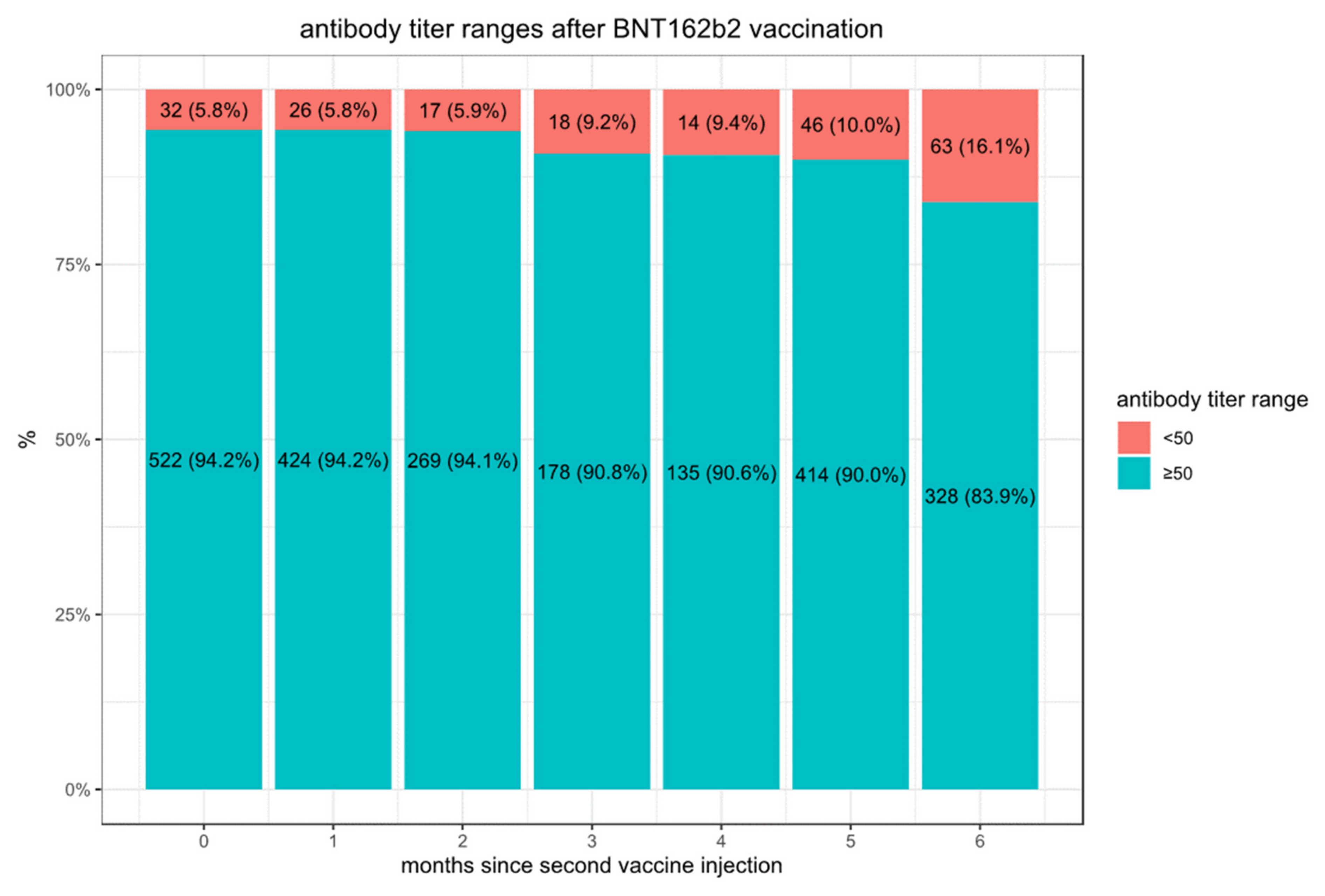
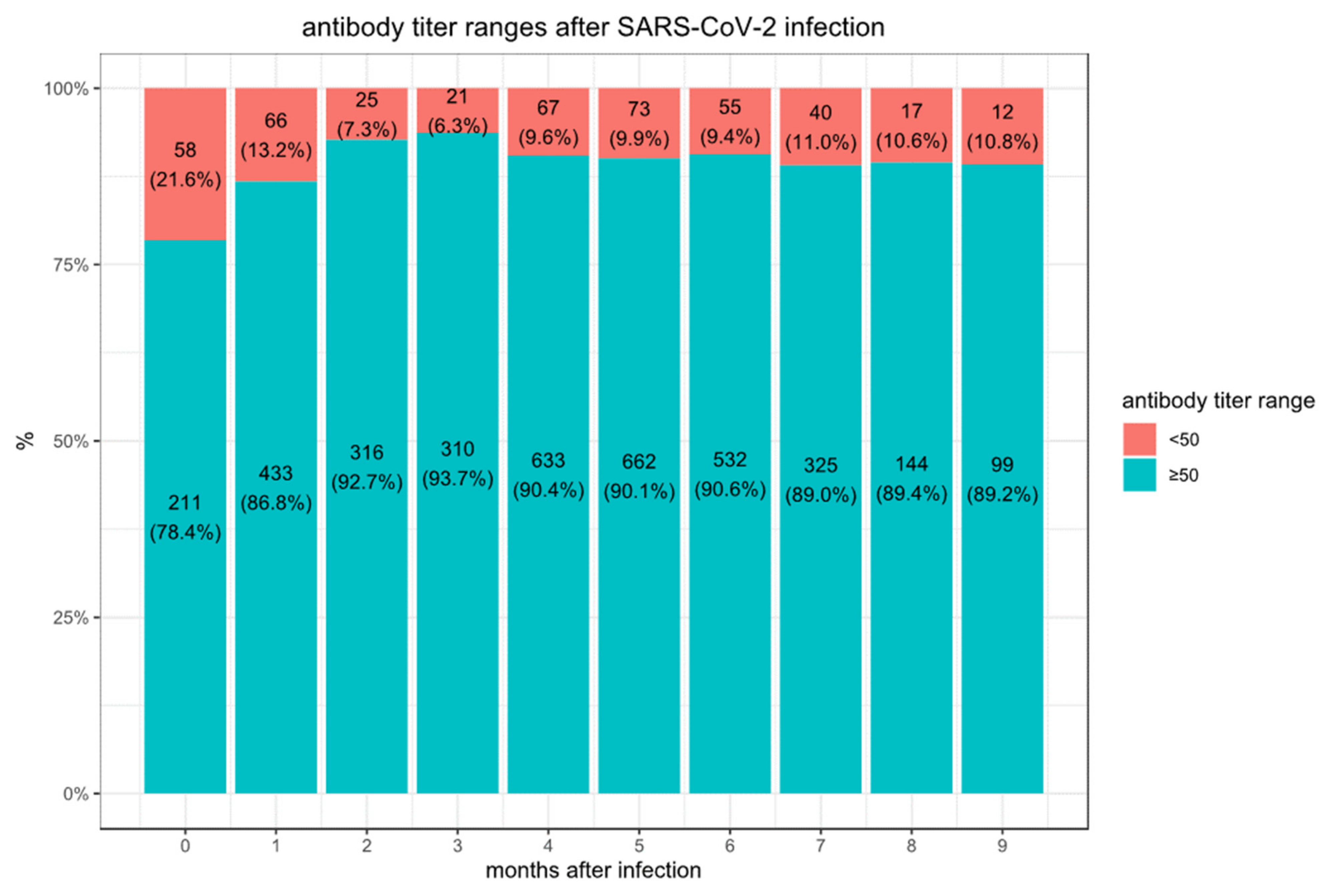
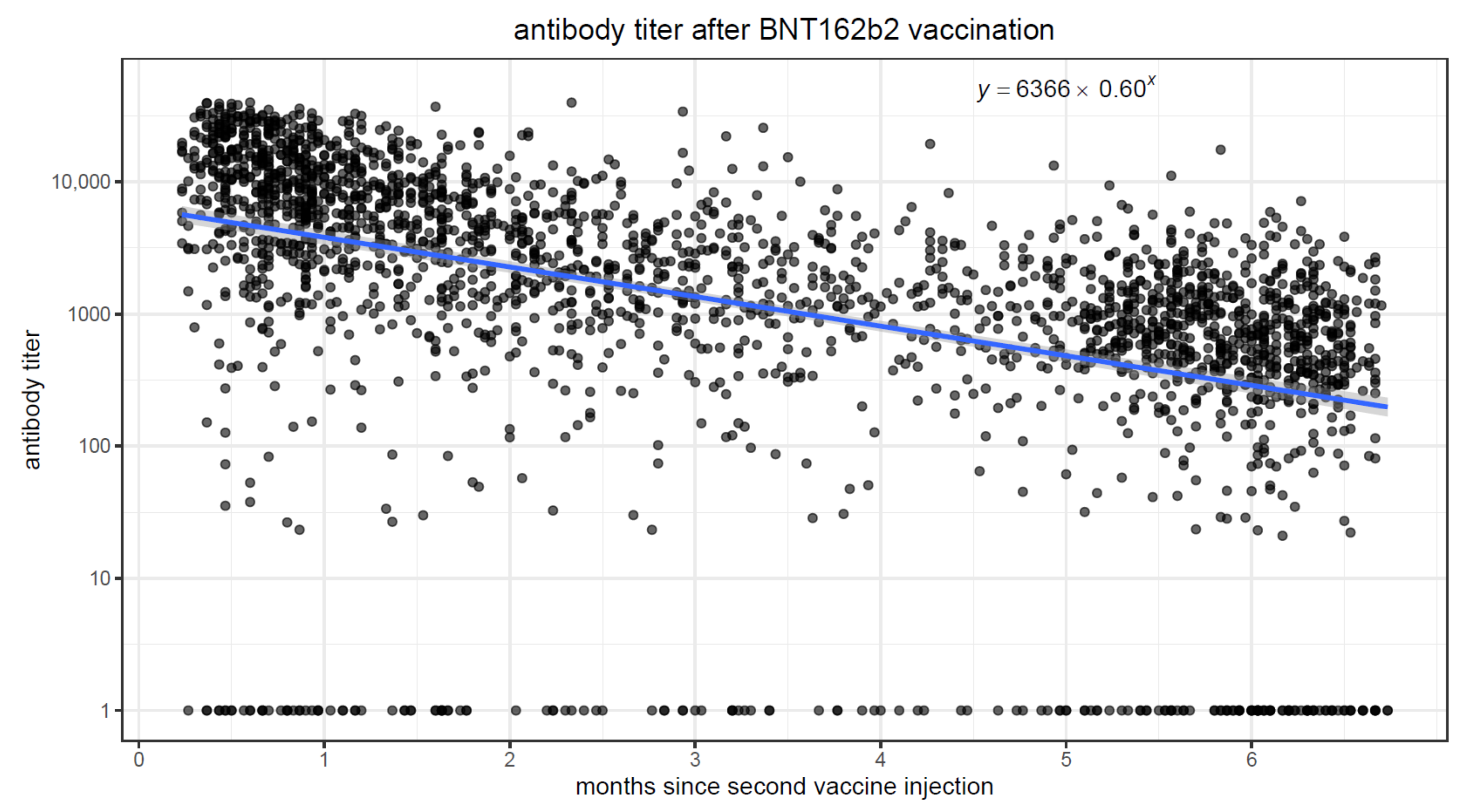
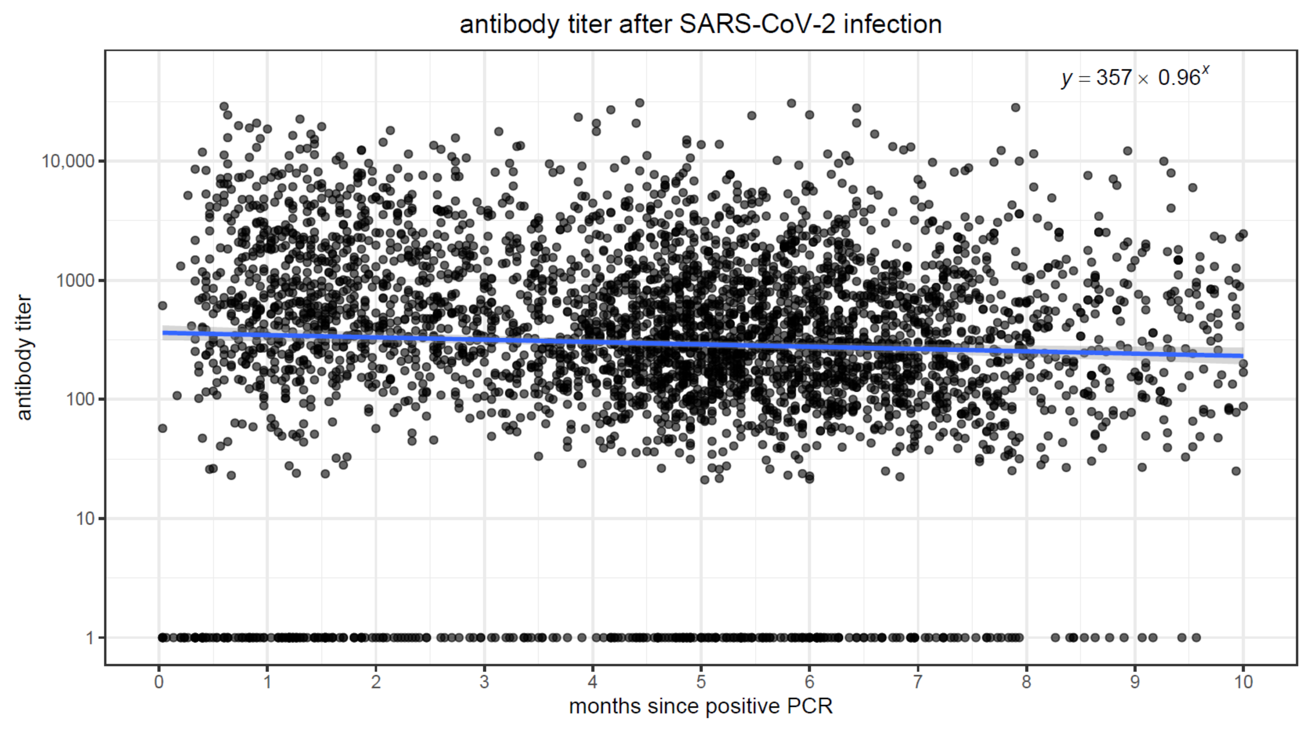
3. Results
4. Discussion
4.1. Study Strengths
4.2. Study Limitations
4.3. Related Work
4.4. Importance of Antibodies in Protection against COVID-19
5. Conclusions
Author Contributions
Funding
Institutional Review Board Statement
Informed Consent Statement
Data Availability Statement
Conflicts of Interest
References
- Khoury, D.S.; Cromer, D.; Reynaldi, A.; Schlub, T.E.; Wheatley, A.K.; Juno, J.A.; Subbarao, K.; Kent, S.J.; Triccas, J.A.; Davenport, M.P. Neutralizing antibody levels are highly predictive of immune protection from symptomatic SARS-CoV-2 infection. Nat. Med. 2021, 27, 1205–1211. [Google Scholar] [CrossRef]
- Lumley, S. Antibody status and incidence of SARS-CoV-2 infection in health care workers. N. Engl. J. Med. 2021, 384, 533–540. [Google Scholar] [CrossRef]
- Kim, J.H.; Marks, F.; Clemens, J.D. Looking beyond COVID-19 vaccine phase 3 trials. Nat. Med. 2021, 27, 205–211. [Google Scholar] [CrossRef]
- Gaebler, C.; Wang, Z.; Lorenzi, J.C.C.; Muecksch, F.; Finkin, S.; Tokuyama, M.; Cho, A.; Jankovic, M.; Schaefer-Babajew, D.; Oliveira, T.Y.; et al. Evolution of antibody immunity to SARS-CoV-2. Nature 2021, 591, 639–644. [Google Scholar] [CrossRef]
- Wheatley, A.K.; Juno, J.A.; Wang, J.J.; Selva, K.J.; Reynaldi, A.; Tan, H.-X.; Lee, W.S.; Wragg, K.M.; Kelly, H.G.; Esterbauer, R.; et al. Evolution of immune responses to SARS-CoV-2 in mild-moderate COVID-19. Nat. Commun. 2021, 12, 1162. [Google Scholar] [CrossRef]
- Wang, P.; Nair, M.S.; Liu, L.; Iketani, S.; Luo, Y.; Guo, Y.; Wang, M.; Yu, J.; Zhang, B.; Kwong, P.D.; et al. Antibody resistance of SARS-CoV-2 variants B.1.351 and B.1.1.7. Nature 2021, 593, 130–135. [Google Scholar] [CrossRef]
- Raz, A.; Keshet, Y.; Popper-Giveon, A.; Karkabi, M.S. One size does not fit all: Lessons from Israel’s COVID-19 vaccination drive and hesitancy. Vaccine 2021, 39, 4027–4028. [Google Scholar] [CrossRef]
- COVID-19 Dashboard. Available online: https://datadashboard.health.gov.il/COVID-19/ (accessed on 16 August 2021).
- Chakraborty, C.; Bhattacharya, M.; Sharma, A.R. Present variants of concern and variants of interest of severe acute respiratory syndrome coronavirus 2: Their significant mutations in S-glycoprotein, infectivity, re-infectivity, immune escape, and vaccines activity. Rev. Med. Virol. 2021, e2270. [Google Scholar] [CrossRef]
- Faulkner, N.; Ng, K.W.; Wu, M.Y.; Harvey, R.; Margaritis, M.; Paraskevopoulou, S.; Houlihan, C.; Hussain, S.; Greco, M.; Bolland, W.; et al. Reduced antibody cross-reactivity following infection with B.1.1.7 than with parental SARS-CoV-2 strains. eLife 2021, 10, e69317. [Google Scholar] [CrossRef]
- Martínez-Flores, D.; Zepeda-Cervantes, J.; Cruz-Reséndiz, A.; Aguirre-Sampieri, S.; Sampieri, A.; Vaca, L. SARS-CoV-2 Vaccines Based on the Spike Glycoprotein and Implications of New Viral Variants. Front. Immunol. 2021, 12, 701501. [Google Scholar] [CrossRef]
- Israel, A.; Merzon, E.; Schäffer, A.A.; Shenhar, Y.; Green, I.; Golan-Cohen, A.; Ruppin, E.; Magen, E.; Vinker, S. Elapsed time since BNT162b2 vaccine and risk of SARS-CoV-2 infection in a large cohort: Test negative design study. BMJ 2021, 375, e067873. [Google Scholar] [CrossRef]
- Muhsen, K.; Na’aminh, W.; Lapidot, Y.; Goren, S.; Amir, Y.; Perlman, S.; Green, M.S.; Chodick, G.; Cohen, D. A nationwide analysis of population group differences in the COVID-19 epidemic in Israel, February 2020–February 2021. Lancet Reg. Health Eur. 2021, 7, 100130. [Google Scholar] [CrossRef]
- Rennert, G.; Peterburg, Y. Prevalence of selected chronic diseases in Israel. Isr. Med. Assoc. J. 2001, 3, 404–408. [Google Scholar]
- Hamood, R.; Hamood, H.; Merhasin, I.; Keinan-Boker, L. A feasibility study to assess the validity of administrative data sources and self-reported information of breast cancer survivors. Isr. J. Health Policy Res. 2016, 5, 50. [Google Scholar] [CrossRef] [Green Version]
- Abbott Receives FDA Emergency Use Authorization for COVID-19 Antibody Blood Test on AlinityTM i System—11 May 2020. Available online: https://abbott.mediaroom.com/2020-05-11-Abbott-Receives-FDA-Emergency-Use-Authorization-for-COVID-19-Antibody-Blood-Test-on-Alinity-TM-i-System (accessed on 6 August 2021).
- Grupel, D.; Gazit, S.; Schreiber, L.; Nadler, V.; Wolf, T.; Lazar, R.; Supino-Rosin, L.; Perez, G.; Peretz, A.; Ben Tov, A.; et al. Kinetics of SARS-CoV-2 anti-S IgG after BNT162b2 vaccination. Vaccine 2021, 39, 5337–5340. [Google Scholar] [CrossRef]
- Amanat, F.; Thapa, M.; Lei, T.; Ahmed, S.M.S.; Adelsberg, D.C.; Carreño, J.M.; Strohmeier, S.; Schmitz, A.J.; Zafar, S.; Zhou, J.Q.; et al. SARS-CoV-2 mRNA vaccination induces functionally diverse antibodies to NTD, RBD, and S2. Cell 2021, 184, 3936–3948. [Google Scholar] [CrossRef]
- Turner, J.S.; O’Halloran, J.A.; Kalaidina, E.; Kim, W.; Schmitz, A.J.; Zhou, J.Q.; Lei, T.; Thapa, M.; Chen, R.E.; Case, J.B.; et al. SARS-CoV-2 mRNA vaccines induce persistent human germinal centre responses. Nature 2021, 596, 109–113. [Google Scholar] [CrossRef]
- Breuer, A.; Raphael, A.; Stern, H.; Odeh, M.; Fiszlinski, J.; Algur, N.; Magen, S.; Megged, O.; Schlesinger, Y.; Barak-Corren, Y.; et al. SARS-CoV-2 antibodies started to decline just four months after COVID-19 infection in a paediatric population. Acta Paediatr. 2021, 110, 3054–3062. [Google Scholar] [CrossRef]
- L’Huillier, A.G.; Meyer, B.; Andrey, D.O.; Arm-Vernez, I.; Baggio, S.; Didierlaurent, A.; Eberhardt, C.S.; Eckerle, I.; Grasset-Salomon, C.; Huttner, A.; et al. Antibody persistence in the first 6 months following SARS-CoV-2 infection among hospital workers: A prospective longitudinal study. Clin. Microbiol. Infect. 2021, 27, 784.e1–784.e8. [Google Scholar] [CrossRef]
- Cho, A.; Muecksch, F.; Schaefer-Babajew, D.; Wang, Z.; Finkin, S.; Gaebler, C.; Ramos, V.; Cipolla, M.; Mendoza, P.; Agudelo, M.; et al. Anti-SARS-CoV-2 receptor-binding domain antibody evolution after mRNA vaccination. Nature 2021, 600, 517–521. [Google Scholar] [CrossRef]
- Ebinger, J.E.; Fert-Bober, J.; Printsev, I.; Wu, M.; Sun, N.; Prostko, J.C.; Frias, E.C.; Stewart, J.L.; Van Eyk, J.E.; Braun, J.G.; et al. Antibody responses to the BNT162b2 mRNA vaccine in individuals previously infected with SARS-CoV-2. Nat. Med. 2021, 27, 981–984. [Google Scholar] [CrossRef]
- Padoan, A.; Dall’Olmo, L.; Rocca, F.D.; Barbaro, F.; Cosma, C.; Basso, D.; Cattelan, A.; Cianci, V.; Plebani, M. Antibody response to first and second dose of BNT162b2 in a cohort of characterized healthcare workers. Clin. Chim. Acta 2021, 519, 60–63. [Google Scholar] [CrossRef]
- Tré-Hardy, M.; Cupaiolo, R.; Papleux, E.; Wilmet, A.; Horeanga, A.; Antoine-Moussiaux, T.; Della Vecchia, A.; Beukinga, I.; Vekemans, M.; Blairon, L. Reactogenicity, safety and antibody response, after one and two doses of mRNA-1273 in seronegative and seropositive healthcare workers. J. Infect. 2021, 83, 237–279. [Google Scholar] [CrossRef]
- Glück, V.; Grobecker, S.; Tydykov, L.; Salzberger, B.; Glück, T.; Weidlich, T.; Bertok, M.; Gottwald, C.; Wenzel, J.J.; Gessner, A.; et al. SARS-CoV-2-directed antibodies persist for more than six months in a cohort with mild to moderate COVID-19. Infection 2021, 49, 739–746. [Google Scholar] [CrossRef]
- Wang, Z.; Muecksch, F.; Schaefer-Babajew, D.; Finkin, S.; Viant, C.; Gaebler, C.; Hoffmann, H.-H.; Barnes, C.O.; Cipolla, M.; Ramos, V.; et al. Naturally enhanced neutralizing breadth against SARS-CoV-2 one year after infection. Nature 2021, 595, 426–431. [Google Scholar] [CrossRef]
- Schoof, M.; Faust, B.; Saunders, R.A.; Sangwan, S.; Rezelj, V.; Hoppe, N.; Boone, M.; Billesbølle, C.B.; Puchades, C.; Azumaya, C.M.; et al. An ultrapotent synthetic nanobody neutralizes SARS-CoV-2 by stabilizing inactive Spike. Science 2020, 370, 1473–1479. [Google Scholar] [CrossRef]
- De Gasparo, R.; Pedotti, M.; Simonelli, L.; Nickl, P.; Muecksch, F.; Cassaniti, I.; Percivalle, E.; Lorenzi, J.C.C.; Mazzola, F.; Magrì, D.; et al. Bispecific IgG neutralizes SARS-CoV-2 variants and prevents escape in mice. Nature 2021, 593, 424–428. [Google Scholar] [CrossRef]
- Xu, J.; Xu, K.; Jung, S.; Conte, A.; Lieberman, J.; Muecksch, F.; Lorenzi, J.C.C.; Park, S.; Schmidt, F.; Wang, Z.; et al. Nanobodies from camelid mice and llamas neutralize SARS-CoV-2 variants. Nature 2021, 595, 278–282. [Google Scholar] [CrossRef]
- Iwasaki, A. Exploiting Mucosal Immunity for Antiviral Vaccines. Annu. Rev. Immunol. 2016, 34, 575–608. [Google Scholar] [CrossRef]
- Li, C.; Yu, D.; Wu, X.; Liang, H.; Zhou, Z.; Xie, Y.; Li, T.; Wu, J.; Lu, F.; Feng, L.; et al. Twelve-month specific IgG response to SARS-CoV-2 receptor-binding domain among COVID-19 convalescent plasma donors in Wuhan. Nat. Commun. 2021, 12, 4846. [Google Scholar] [CrossRef]
- Ibarrondo, F.J.; Fulcher, J.A.; Goodman-Meza, D.; Elliott, J.; Hofmann, C.; Hausner, M.A.; Ferbas, K.G.; Tobin, N.H.; Aldrovandi, G.M.; Yang, O.O. Rapid Decay of Anti–SARS-CoV-2 Antibodies in Persons with Mild COVID-19. N. Engl. J. Med. 2020, 383, 1085–1087. [Google Scholar] [CrossRef]
- Seow, J.; Graham, C.; Merrick, B.; Acors, S.; Pickering, S.; Steel, K.J.A.; Hemmings, O.; O’Byrne, A.; Kouphou, N.; Galao, R.P.; et al. Longitudinal observation and decline of neutralizing antibody responses in the three months following SARS-CoV-2 infection in humans. Nat. Microbiol. 2020, 5, 1598–1607. [Google Scholar] [CrossRef]
- Li, D.; Edwards, R.J.; Manne, K.; Martinez, D.R.; Schäfer, A.; Alam, S.M.; Wiehe, K.; Lu, X.; Parks, R.; Sutherland, L.L.; et al. In vitro and in vivo functions of SARS-CoV-2 infection-enhancing and neutralizing antibodies. Cell 2021, 184, 4203–4219. [Google Scholar] [CrossRef]
- Pegu, A.; O’Connell, S.E.; Schmidt, S.D.; O’Dell, S.; Talana, C.A.; Lai, L.; Albert, J.; Anderson, E.; Bennett, H.; Corbett, K.S.; et al. Durability of mRNA-1273 vaccine-induced antibodies against SARS-CoV-2 variants. Science 2021, 373, 1372–1377. [Google Scholar] [CrossRef]
- COVID Data Tracker. Available online: https://covid.cdc.gov/covid-data-tracker/#rates-by-vaccine-status (accessed on 26 November 2021).
- Iyer, A.S.; Jones, F.K.; Nodoushani, A.; Kelly, M.; Becker, M.; Slater, D.; Mills, R.; Teng, E.; Kamruzzaman, M.; Garcia-Beltran, W.F.; et al. Persistence and decay of human antibody responses to the receptor binding domain of SARS-CoV-2 spike protein in COVID-19 patients. Sci. Immunol. 2020, 5, 367. [Google Scholar] [CrossRef]
- Gudbjartsson, D.F.; Norddahl, G.L.; Melsted, P.; Gunnarsdottir, K.; Holm, H.; Eythorsson, E.; Arnthorsson, A.O.; Helgason, D.; Bjarnadottir, K.; Ingvarsson, R.F.; et al. Humoral Immune Response to SARS-CoV-2 in Iceland. N. Engl. J. Med. 2020, 383, 1724–1734. [Google Scholar] [CrossRef]
- Sahin, U.; Muik, A.; Derhovanessian, E.; Vogler, I.; Kranz, L.M.; Vormehr, M.; Baum, A.; Pascal, K.; Quandt, J.; Maurus, D.; et al. COVID-19 vaccine BNT162b1 elicits human antibody and TH1 T cell responses. Nature 2020, 586, 594–599. [Google Scholar] [CrossRef]
- Hellerstein, M. What are the roles of antibodies versus a durable, high-quality T-cell response in protective immunity against SARS-CoV-2? Vaccine 2020, 6, 100076. [Google Scholar] [CrossRef]
- McMahan, K.; Yu, J.; Mercado, N.B.; Loos, C.; Tostanoski, L.H.; Chandrashekar, A.; Liu, J.; Peter, L.; Atyeo, C.; Zhu, A.; et al. Correlates of protection against SARS-CoV-2 in rhesus macaques. Nature 2020, 590, 630–634. [Google Scholar] [CrossRef]
- Gilbert, P.B.; Montefiori, D.C.; McDermott, A.B.; Fong, Y.; Benkeser, D.; Deng, W.; Zhou, H.; Houchens, C.R.; Martins, K.; Jayashankar, L.; et al. Immune correlates analysis of the mRNA-1273 COVID-19 vaccine efficacy clinical trial. Science, 2021; in press. [Google Scholar]


| Vaccinated | Convalescent | ||
|---|---|---|---|
| N | 2653 | 4361 | |
| Age (in years) | mean (SD) | 56.45 (15.87) | 41.99 (16.09) |
| Age group n (%) | 18–59 years | 1296 (48.9%) | 3663 (84.0%) |
| ≥60 years | 1357 (51.1%) | 698 (16.0%) | |
| Sex, n (%) | Female | 1604 (60.5%) | 2728 (62.6%) |
| Male | 1049 (39.5%) | 1633 (37.4%) | |
| Demographic group, n (%) | Arab | 248 (10.9%) | 615 (14.1%) |
| General (mostly Jewish) | 1633 (71.9%) | 1959 (44.9%) | |
| Jewish Ultra-orthodox | 389 (17.1%) | 1787 (41.0%) | |
| SES, mean (SD) | 9.88 (3.70) | 7.57 (3.55) | |
| *missing* | 179 (7.24%) | 253 (6.16%) | |
| Body mass index (BMI) | mean (SD) | 27.79 (5.26) | 27.20 (5.74) |
| *missing* | 35 (1.34%) | 92 (2.16%) | |
| BMI category, n (%) | <18.5 Underweight | 46 (1.8%) | 135 (3.2%) |
| 18.5–25 Normal | 780 (29.8%) | 1468 (34.4%) | |
| 25–30 Overweight | 994 (38.0%) | 1470 (34.5%) | |
| 30–35 Obese I | 550 (21.0%) | 781 (18.3%) | |
| 35–40 Obese II | 195 (7.5%) | 304 (7.1%) | |
| 40–Obese III | 51 (1.9%) | 105 (2.5%) | |
| *missing* | 37 (1.4%) | 98 (2.2%) | |
| co-morbidities, n (%) | diabetes mellitus | 659 (24.8%) | 493 (11.3%) |
| hypertension | 1140 (43.0%) | 808 (18.5%) | |
| asthma | 299 (11.3%) | 409 (9.4%) | |
| COPD | 265 (10.0%) | 137 (3.1%) | |
| ischemic heart disease | 325 (12.3%) | 183 (4.2%) | |
| solid tumor | 342 (12.9%) | 172 (3.9%) | |
| chronic renal disease | 199 (7.5%) | 58 (1.3%) | |
| Time (in days) since… mean (SD) | 2nd vaccination | 101.35 (65.73) | - |
| first positive PCR | - | 151.17 (82.32) | |
| Time (in days) since vaccination or positive PCR by 30 days intervals, n (%) | 0–29 | 556 (21.0%) | 269 (6.2%) |
| 30–59 | 456 (17.2%) | 499 (11.4%) | |
| 60–89 | 289 (10.9%) | 341 (7.8%) | |
| 90–119 | 200 (7.5%) | 331 (7.6%) | |
| 120–149 | 170 (6.4%) | 700 (16.1%) | |
| 150–179 | 542 (20.4%) | 735 (16.9%) | |
| 180–209 | 440 (16.6%) | 587 (13.5%) | |
| 210–239 | - | 365 (8.4%) | |
| 240–269 | - | 161 (3.7%) | |
| 270– | - | 373 (8.6%) |
| Time Since Second Vaccine Injection (in Days) | 0–29 | 30–59 | 60–89 | 90–119 | 120–149 | 150–179 | 180– | |
|---|---|---|---|---|---|---|---|---|
| N | 556 | 456 | 289 | 200 | 170 | 542 | 440 | |
| Age (in years) | 53.76 (16.90) | 55.41 (16.66) | 55.64 (16.52) | 53.31 (16.75) | 53.29 (16.29) | 56.34 (13.76) | 64.23 (12.30) | |
| Sex, n (%) | Female | 318 (57.2%) | 273 (59.9%) | 173 (59.9%) | 129 (64.5%) | 104 (61.2%) | 358 (66.1%) | 249 (56.6%) |
| Male | 238 (42.8%) | 183 (40.1%) | 116 (40.1%) | 71 (35.5%) | 66 (38.8%) | 184 (33.9%) | 191 (43.4%) | |
| SARS-CoV-2 IgG antibody level | mean (SD) | 12,153 (9875) | 6848 (6340) | 3476 (4582) | 2383 (3266) | 1552 (2103) | 1122 (1431) | 765 (948) |
| median [IQR] | 9913 [3650–18,733] | 5106 [2109–9601] | 2159 [1039–4169] | 1323 [549–3126] | 1071 [471–1901] | 764 [385–1343] | 447 [205–966] | |
| Time Since First Positive PCR (in Days) | 0–29 | 30–59 | 60–89 | 90–119 | 120–149 | 150–179 | 180–209 | 210–239 | 240–269 | 270– | |
|---|---|---|---|---|---|---|---|---|---|---|---|
| N | 269 | 499 | 341 | 331 | 700 | 735 | 587 | 365 | 161 | 373 | |
| Age (in years) | 39.96 (14.78) | 41.10 (14.95) | 45.07 (16.53) | 43.52 (16.32) | 42.90 (16.23) | 40.95 (16.13) | 41.39 (16.14) | 40.59 (15.32) | 41.75 (17.42) | 43.18 (17.10) | |
| Sex, n (%) | Female | 157 (58.4%) | 326 (65.3%) | 234 (68.6%) | 219 (66.2%) | 400 (57.1%) | 461 (62.7%) | 371 (63.2%) | 230 (63.0%) | 112 (69.6%) | 218 (58.4%) |
| Male | 112 (41.6%) | 173 (34.7%) | 107 (31.4%) | 112 (33.8%) | 300 (42.9%) | 274 (37.3%) | 216 (36.8%) | 135 (37.0%) | 49 (30.4%) | 155 (41.6%) | |
| SARS-CoV-2 IgG antibody level | mean (SD) | 1914 (3870) | 1739 (2972) | 1552 (2522) | 1195 (2406) | 1079 (2556) | 860 (1962) | 904 (2384) | 850 (2104) | 901 (1739) | 731 (1280) |
| median [IQR] | 490 [109–1869] | 586 [212–1908] | 538 [247–1723] | 377 [165–1080] | 329 [140–886] | 312 [138–8301] | 278 [125–751] | 278 [105–727] | 351 [124–919] | 314 [116–783] | |
| Vaccinated | Convalescent | ||||||
|---|---|---|---|---|---|---|---|
| Factor | 95% CI | p | Factor | 95% CI | p | ||
| (Intercept) | 10,598 | [4889–22,975] | <0.001 | 234 | [143–384] | <0.001 | |
| Each month since vaccination (for vaccinated) | 0.623 | [0.599–0.649] | <0.001 | ||||
| Each month since first positive (for convalescent) | 0.960 | [0.939–0.982] | <0.001 | ||||
| was symptomatic (for convalescent) | 1.811 | [1.531–2.142] | <0.001 | ||||
| was hospitalized (for convalescent) | 3.323 | [2.217–4.980] | <0.001 | ||||
| Age | ≥60 (vs. <60) | 0.790 | [0.644–0.969] | 0.024 | 1.546 | [1.269–1.884] | <0.001 |
| Sex | Female (vs. Male) | 1.243 | [1.035–1.492] | 0.020 | 0.923 | [0.812–1.048] | 0.215 |
| Socio-economic status (SES) | 0.995 | [0.966–1.024] | 0.723 | 0.995 | [0.973–1.018] | 0.662 | |
| Demographic grp. (vs. general) | Arab | 1.525 | [1.101–2.113] | 0.011 | 0.982 | [0.789–1.222] | 0.871 |
| Ultra-orthodox | 1.436 | [1.099–1.877] | 0.008 | 1.261 | [1.065–1.492] | 0.007 | |
| Body Mass Index (BMI) | <18.5 Underweight | 0.359 | [0.144–0.893] | 0.028 | 0.683 | [0.404–1.157] | 0.156 |
| 18.5–25 Normal | 0.757 | [0.374–1.534] | 0.440 | 0.880 | [0.581–1.333] | 0.547 | |
| 25–30 Overweight | 0.774 | [0.383–1.564] | 0.475 | 1.143 | [0.754–1.733] | 0.529 | |
| 30–35 Obese | 0.804 | [0.393–1.645] | 0.550 | 1.429 | [0.930–2.194] | 0.103 | |
| ≥35 Obese II+ | 0.647 | [0.307–1.363] | 0.252 | 1.839 | [1.166–2.899] | 0.009 | |
| Comorbidity | diabetes mellitus | 0.720 | [0.579–0.894] | 0.003 | 1.354 | [1.093–1.678] | 0.006 |
| hypertension | 0.786 | [0.639–0.966] | 0.022 | 1.254 | [1.036–1.518] | 0.020 | |
| asthma | 1.200 | [0.911–1.582] | 0.195 | 1.046 | [0.848–1.290] | 0.676 | |
| COPD | 0.643 | [0.479–0.863] | 0.003 | 0.798 | [0.562–1.133] | 0.207 | |
| ischemic heart disease | 0.869 | [0.655–1.152] | 0.328 | 1.322 | [0.956–1.828] | 0.091 | |
| solid tumor | 0.642 | [0.494–0.834] | 0.001 | 1.048 | [0.760–1.444] | 0.777 | |
| chronic renal disease | 0.200 | [0.143–0.281] | <0.001 | 1.965 | [1.134–3.407] | 0.016 | |
| Vaccinated | Convalescent | ||||||
|---|---|---|---|---|---|---|---|
| Factor | 95% CI | p | Factor | 95% CI | p | ||
| days since vaccination (for vaccinated) | 0–29 | 1.000 | ref. | ||||
| 30–60 | 0.645 | [0.497–0.838] | 0.001 | ||||
| 60–90 | 0.328 | [0.243–0.442] | <0.001 | ||||
| 90–120 | 0.174 | [0.124–0.245] | <0.001 | ||||
| 120–150 | 0.131 | [0.089–0.191] | <0.001 | ||||
| 150–180 | 0.100 | [0.077–0.129] | <0.001 | ||||
| 180– | 0.064 | [0.048–0.084] | <0.001 | ||||
| days since first positive (for convalescent) | 0–89 | 1.000 | ref. | ||||
| 90–120 | 0.964 | [0.754–1.232] | 0.770 | ||||
| 120–150 | 0.786 | [0.650–0.950] | 0.013 | ||||
| 150–180 | 0.773 | [0.641–0.932] | 0.007 | ||||
| 180–210 | 0.720 | [0.589–0.880] | 0.001 | ||||
| 210–240 | 0.736 | [0.581–0.934] | 0.012 | ||||
| 240–270 | 0.868 | [0.622–1.210] | 0.403 | ||||
| 270– | 0.691 | [0.544–0.878] | 0.002 | ||||
| Age | ≥60 (vs. <60) | 0.804 | [0.656–0.984] | 0.035 | 1.581 | [1.300–1.921] | <0.001 |
| Sex | Female (vs. Male) | 1.251 | [1.047–1.495] | 0.014 | 0.919 | [0.811–1.041] | 0.184 |
| Socio-economic status (SES) | 1.006 | [0.979–1.033] | 0.680 | 0.989 | [0.967–1.010] | 0.301 | |
| Demographic grp. (vs. general) | Arab | 1.731 | [1.266–2.368] | 0.001 | 0.916 | [0.744–1.128] | 0.408 |
| Ultra-orthodox | 1.527 | [1.185–1.967] | 0.001 | 1.186 | [1.011–1.391] | 0.036 | |
| Body Mass Index (BMI) | <18.5 Underweight | 0.335 | [0.135–0.831] | 0.018 | 0.630 | [0.374–1.061] | 0.082 |
| 18.5–25 Normal | 0.680 | [0.339–1.362] | 0.276 | 0.829 | [0.550–1.250] | 0.371 | |
| 25–30 Overweight | 0.704 | [0.352–1.408] | 0.321 | 1.072 | [0.711–1.617] | 0.739 | |
| 30–35 Obese | 0.699 | [0.345–1.416] | 0.320 | 1.321 | [0.864–2.018] | 0.198 | |
| ≥35 Obese II+ | 0.591 | [0.284–1.232] | 0.161 | 1.762 | [1.126–2.757] | 0.013 | |
| Vaccinated | Convalescent | ||||||
|---|---|---|---|---|---|---|---|
| Factor | 95% CI | p | Factor | 95% CI | p | ||
| (Intercept) | 3907 | [592–25,761] | <0.001 | 185 | [39–888] | <0.001 | |
| Each month since vaccination (for vaccinated) | 0.618 | [0.585–0.653] | <0.001 | ||||
| Each month since first positive (for convalescent) | 0.889 | [0.839–0.943] | <0.001 | ||||
| was symptomatic (for convalescent) | 1.746 | [1.183–2.576] | <0.005 | ||||
| was hospitalized (for convalescent) | 2.947 | [1.597–5.436] | <0.001 | ||||
| Sex | Female (vs. Male) | 1.401 | [1.067–1.841] | 0.015 | 0.865 | [0.628–1.191] | 0.373 |
| Socio-economic status (SES) | 0.989 | [0.950–1.030] | 0.593 | 1.035 | [0.980–1.884] | 0.218 | |
| Demographic grp. (vs. general) | Arab | 1.782 | [0.989–3.212] | 0.055 | 1.020 | [0.579–1.797] | 0.945 |
| Ultra-orthodox | 1.475 | [0.987–2.206] | 0.058 | 1.352 | [0.881–2.073] | 0.167 | |
| Body Mass Index (BMI) | <18.5 Underweight | 0.147 | [0.017–1.312] | 0.086 | 0.476 | [0.040–5.713] | 0.558 |
| 18.5–25 Normal | 1.662 | [0.269–10.246] | 0.584 | 2.735 | [0.624–11.991] | 0.182 | |
| 25–30 Overweight | 1.558 | [0.254–9.544] | 0.631 | 2.850 | [0.669–12.144] | 0.156 | |
| 30–35 Obese | 0.804 | [0.275–10.502] | 0.569 | 3.060 | [0.708–13.226] | 0.134 | |
| ≥35 Obese II+ | 0.647 | [0.200–8.116] | 0.797 | 5.173 | [1.161–23.042] | 0.031 | |
| Comorbidity | diabetes mellitus | 0.747 | [0.570–0.979] | 0.035 | 1.213 | [0.864–1.704] | 0.265 |
| hypertension | 0.782 | [0.594–1.030] | 0.080 | 1.084 | [0.773–1.519] | 0.640 | |
| asthma | 1.284 | [0.874–1.887] | 0.202 | 1.386 | [0.868–2.213] | 0.171 | |
| COPD | 0.723 | [0.501–1.042] | 0.082 | 0.790 | [0.462–1.349] | 0.387 | |
| ischemic heart disease | 0.821 | [0.592–1.139] | 0.238 | 1.207 | [0.796–1.831] | 0.376 | |
| solid tumor | 0.574 | [0.420–0.783] | <0.001 | 0.867 | [0.546–1.379] | 0.547 | |
| chronic renal disease | 0.329 | [0.217–0.498] | <0.001 | 2.776 | [1.301–5.921] | 0.008 | |
Publisher’s Note: MDPI stays neutral with regard to jurisdictional claims in published maps and institutional affiliations. |
© 2021 by the authors. Licensee MDPI, Basel, Switzerland. This article is an open access article distributed under the terms and conditions of the Creative Commons Attribution (CC BY) license (https://creativecommons.org/licenses/by/4.0/).
Share and Cite
Israel, A.; Shenhar, Y.; Green, I.; Merzon, E.; Golan-Cohen, A.; Schäffer, A.A.; Ruppin, E.; Vinker, S.; Magen, E. Large-Scale Study of Antibody Titer Decay following BNT162b2 mRNA Vaccine or SARS-CoV-2 Infection. Vaccines 2022, 10, 64. https://doi.org/10.3390/vaccines10010064
Israel A, Shenhar Y, Green I, Merzon E, Golan-Cohen A, Schäffer AA, Ruppin E, Vinker S, Magen E. Large-Scale Study of Antibody Titer Decay following BNT162b2 mRNA Vaccine or SARS-CoV-2 Infection. Vaccines. 2022; 10(1):64. https://doi.org/10.3390/vaccines10010064
Chicago/Turabian StyleIsrael, Ariel, Yotam Shenhar, Ilan Green, Eugene Merzon, Avivit Golan-Cohen, Alejandro A. Schäffer, Eytan Ruppin, Shlomo Vinker, and Eli Magen. 2022. "Large-Scale Study of Antibody Titer Decay following BNT162b2 mRNA Vaccine or SARS-CoV-2 Infection" Vaccines 10, no. 1: 64. https://doi.org/10.3390/vaccines10010064