Next Issue
Volume 9, October
Previous Issue
Volume 9, August


Antioxidants, Volume 9, Issue 9 (September 2020) – 135 articles

Cover Story (view full-size image): 
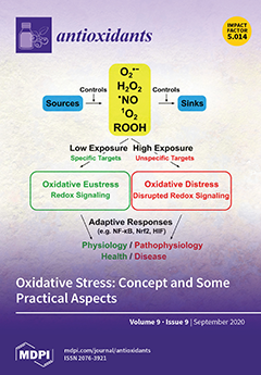
Oxidative stress is defined as “an imbalance between oxidants and antioxidants in favor of the oxidants, leading to a disruption of redox signaling and control and/or molecular damage”. The commentary presents the basic features of this global concept, which has attracted interest in biology and medicine. The term “antioxidants” in cellular defense against oxidants predominantly includes antioxidant enzymes with their substrates and coenzymes. Exogenous low-molecular-mass compounds also have a role, but this is more limited. Multiple biomarkers of damage due to oxidative stress have been identified for different molecular classes (protein, lipid, carbohydrate, DNA/RNA), and the current state of practical aspects in health and disease is delineated. View this paper
  • Issues are regarded as officially published after their release is announced to the table of contents alert mailing list.
  • You may sign up for e-mail alerts to receive table of contents of newly released issues.
  • PDF is the official format for papers published in both, html and pdf forms. To view the papers in pdf format, click on the "PDF Full-text" link, and use the free Adobe Reader to open them.
Order results
Result details
Section
Select all
Export citation of selected articles as:
19 pages, 4805 KiB  
Article
Epigenetic Scanning of KEAP1 CpG Sites Uncovers New Molecular-Driven Patterns in Lung Adeno and Squamous Cell Carcinomas
by Federico Pio Fabrizio, Tommaso Mazza, Stefano Castellana, Angelo Sparaneo and Lucia Anna Muscarella
Antioxidants 2020, 9(9), 904; https://doi.org/10.3390/antiox9090904 - 22 Sep 2020
Cited by 8 | Viewed by 2604
Abstract
Background: The KEAP1/NRF2 (Kelch-like ECH-associated protein 1/nuclear factor erythroid 2–related factor 2) pathway modulates detoxification processes and participates in the resistance of solid tumors to therapy. Scientific evidence about the presence of genetic and epigenetic abnormalities of the KEAP1 gene was firstly reported [...] Read more.
Background: The KEAP1/NRF2 (Kelch-like ECH-associated protein 1/nuclear factor erythroid 2–related factor 2) pathway modulates detoxification processes and participates in the resistance of solid tumors to therapy. Scientific evidence about the presence of genetic and epigenetic abnormalities of the KEAP1 gene was firstly reported in non-small-cell lung cancer (NSCLC) and then described in other tumors. At present, the prognostic role of aberrant methylation at cytosine-guanine dinucleotide (CpG) sites of the KEAP1 gene promoter is debated in NSCLC, and its correlation with transcriptional changes and protein levels remains to be defined in large sample cohorts. Methods: We evaluated and compared multiple KEAP1 omics data (methylation, transcript, and protein expression levels) from The Cancer Genome Atlas (TCGA) to explore the role of CpGs located in different portions of KEAP1 and the correlation between methylation, transcription, and protein levels. Data from two subsets of lung adenocarcinoma (LUAD, n = 617) and lung squamous cell carcinoma (LUSC, n = 571) cohorts of NSCLC patients with different disease stages were evaluated. Results: We found that the methylation levels of many KEAP1 CpGs at various promoter and intragenic locations showed a significant inverse correlation with the transcript levels. Interestingly, these results were limited to the KRAS wild-type LUSC and LUAD cohorts, whereas in LUAD the effect of the epigenetic silencing of KEAP1 on its transcription was also observed in the EGFR mutated subpopulation. Conclusions: These results support the idea that the prognostic role of KEAP1 CpG sites warrants more in-depth investigation and that the impact of their changes in methylation levels may differ among specific NSCLC histologies and molecular backgrounds. Moreover, the observed impact of epigenetic silencing on KEAP1 expression in specific KRAS and EGFR settings may suggest a potential role of KEAP1 methylation as a predictive marker for NSCLC patients for whom anti-EGFR treatments are considered. Full article
(This article belongs to the Special Issue Keap1/Nrf2 Signaling Pathway)
Show Figures

Graphical abstract

23 pages, 1325 KiB  
Article
A Chemometric Approach to Oxidative Stability and Physicochemical Quality of Raw Ground Chicken Meat Affected by Black Seed and Other Spice Extracts
by Małgorzata Muzolf-Panek, Anna Kaczmarek, Jolanta Tomaszewska-Gras, Renata Cegielska-Radziejewska, Tomasz Szablewski, Małgorzata Majcher and Kinga Stuper-Szablewska
Antioxidants 2020, 9(9), 903; https://doi.org/10.3390/antiox9090903 - 22 Sep 2020
Cited by 18 | Viewed by 3298
Abstract
The effects of black seed (Nigella sativa), allspice, bay leaf, caraway, cardamom, clove, and nutmeg extracts on the quality of raw ground chicken legs stored at 4 °C were investigated. During 12 days of storage, conjugated diene (CD) content, thiobarbituric acid [...] Read more.
The effects of black seed (Nigella sativa), allspice, bay leaf, caraway, cardamom, clove, and nutmeg extracts on the quality of raw ground chicken legs stored at 4 °C were investigated. During 12 days of storage, conjugated diene (CD) content, thiobarbituric acid reactive substances (TBARS), oxidation induction time (IP) by DSC (differential scanning calorimetry), hexanal content by GC-SPME-MS, thiol group (SH) content were determined. Moreover, microbial growth, pH and color of the samples were investigated. Sensory analysis was also realized. All extracts increased oxidative stability and safety of meat, significantly changed the color of the samples, stabilized the pH and increased their sensory scores (except color of samples with bay leaf and black seed) when comparing to control. Black seed, allspice and clove extracts showed high antioxidant capacity in lipid (CD = 0.23%, 0.28%, and 0.37%, respectively; TBARS = 0.55, 0.50, and 0.48 mg/kg, respectively) and protein fraction (SH content = 47.9, 52.1 and 52.7 nmol/g, respectively), although the ABTS•+ radical scavenging activity of black seed (33.1 µM/g) was significantly lower than the cloves (2496 µM/g) and allspice (815 µM/g). In the sensory analysis the highest scores were ascribed to the sample with cardamom followed by cloves. Principal component analysis (PCA) indicated complex and inseparable interrelationship among lipid and protein oxidation processes and the relationship of the protein oxidation on the lightness of meat. The results enabled to discriminate the meat samples, showing a great impact of the extracts on the final quality of raw chicken meat with black seed being potent antioxidant active additive. Full article
(This article belongs to the Special Issue Antioxidative Properties in Meat and Meat Products)
Show Figures

Graphical abstract

21 pages, 3571 KiB  
Review
Antioxidant and Neuroprotective Effects of Caffeine against Alzheimer’s and Parkinson’s Disease: Insight into the Role of Nrf-2 and A2AR Signaling
by Muhammad Ikram, Tae Ju Park, Tahir Ali and Myeong Ok Kim
Antioxidants 2020, 9(9), 902; https://doi.org/10.3390/antiox9090902 - 22 Sep 2020
Cited by 57 | Viewed by 9301
Abstract
This paper reviews the results of studies conducted on the role of caffeine in the management of different neurological disorders, such as Parkinson’s disease (PD) and Alzheimer’s disease (AD). To highlight the potential role of caffeine in managing different neurodegenerative diseases, we identified [...] Read more.
This paper reviews the results of studies conducted on the role of caffeine in the management of different neurological disorders, such as Parkinson’s disease (PD) and Alzheimer’s disease (AD). To highlight the potential role of caffeine in managing different neurodegenerative diseases, we identified studies by searching PubMed, Web of Science, and Google Scholar by scrutinizing the lists of pertinent publications. According to the collected overall findings, caffeine may reduce the elevated oxidative stress; inhibit the activation of adenosine A2A, thereby regulating the accumulation of Aβ; reduce the hyperphosphorylation of tau; and reduce the accumulation of misfolded proteins, such as α-synuclein, in Alzheimer’s and Parkinson’s diseases. The studies have suggested that caffeine has promising protective effects against different neurodegenerative diseases and that these effects may be used to tackle the neurological diseases and/or their consequences. Here, we review the ongoing research on the role of caffeine in the management of different neurodegenerative disorders, focusing on AD and PD. The current findings suggest that caffeine produces potent antioxidant, inflammatory, and anti-apoptotic effects against different models of neurodegenerative disease, including AD, PD, and other neurodegenerative disorders. Caffeine has shown strong antagonistic effects against the adenosine A2A receptor, which is a microglial receptor, and strong agonistic effects against nuclear-related factor-2 (Nrf-2), thereby regulating the cellular homeostasis at the brain by reducing oxidative stress, neuroinflammation, regulating the accumulation of α-synuclein in PD and tau hyperphosphorylation, amyloidogenesis, and synaptic deficits in AD, which are the cardinal features of these neurodegenerative diseases. Full article
(This article belongs to the Special Issue Role of Antioxidant Molecules and Melatonin in Cellular Protection)
Show Figures

Figure 1

16 pages, 1469 KiB  
Review
Oxidative Stress, Neuroinflammation and Mitochondria in the Pathophysiology of Amyotrophic Lateral Sclerosis
by Elena Obrador, Rosario Salvador, Rafael López-Blanch, Ali Jihad-Jebbar, Soraya L. Vallés and José M. Estrela
Antioxidants 2020, 9(9), 901; https://doi.org/10.3390/antiox9090901 - 22 Sep 2020
Cited by 59 | Viewed by 5527
Abstract
Amyotrophic lateral sclerosis (ALS) is a progressive motor neuron (MN) disease. Its primary cause remains elusive, although a combination of different causal factors cannot be ruled out. There is no cure, and prognosis is poor. Most patients with ALS die due to disease-related [...] Read more.
Amyotrophic lateral sclerosis (ALS) is a progressive motor neuron (MN) disease. Its primary cause remains elusive, although a combination of different causal factors cannot be ruled out. There is no cure, and prognosis is poor. Most patients with ALS die due to disease-related complications, such as respiratory failure, within three years of diagnosis. While the underlying mechanisms are unclear, different cell types (microglia, astrocytes, macrophages and T cell subsets) appear to play key roles in the pathophysiology of the disease. Neuroinflammation and oxidative stress pave the way leading to neurodegeneration and MN death. ALS-associated mitochondrial dysfunction occurs at different levels, and these organelles are involved in the mechanism of MN death. Molecular and cellular interactions are presented here as a sequential cascade of events. Based on our present knowledge, the discussion leads to the idea that feasible therapeutic strategies should focus in interfering with the pathophysiology of the disease at different steps. Full article
(This article belongs to the Special Issue Oxidative Stress and Inflammation in the Nervous System)
Show Figures

Figure 1

26 pages, 1776 KiB  
Review
The Role of Bilirubin and the Other “Yellow Players” in Neurodegenerative Diseases
by Sri Jayanti, Libor Vítek, Claudio Tiribelli and Silvia Gazzin
Antioxidants 2020, 9(9), 900; https://doi.org/10.3390/antiox9090900 - 22 Sep 2020
Cited by 20 | Viewed by 6395
Abstract
Bilirubin is a yellow endogenous derivate of the heme catabolism. Since the 1980s, it has been recognized as one of the most potent antioxidants in nature, able to counteract 10,000× higher intracellular concentrations of H2O2. In the recent years, [...] Read more.
Bilirubin is a yellow endogenous derivate of the heme catabolism. Since the 1980s, it has been recognized as one of the most potent antioxidants in nature, able to counteract 10,000× higher intracellular concentrations of H2O2. In the recent years, not only bilirubin, but also its precursor biliverdin, and the enzymes involved in their productions (namely heme oxygenase and biliverdin reductase; altogether the “yellow players”—YPs) have been recognized playing a protective role in diseases characterized by a chronic prooxidant status. Based on that, there is an ongoing effort in inducing their activity as a therapeutic option. Nevertheless, the understanding of their specific contributions to pathological conditions of the central nervous system (CNS) and their role in these diseases are limited. In this review, we will focus on the most recent evidence linking the role of the YPs specifically to neurodegenerative and neurological conditions. Both the protective, as well as potentially worsening effects of the YP’s activity will be discussed. Full article
(This article belongs to the Special Issue Oxidative Stress and Neurodegenerative Disorders)
Show Figures

Graphical abstract

13 pages, 619 KiB  
Article
Paraoxonase 1 Phenotype and Protein N-Homocysteinylation in Patients with Rheumatoid Arthritis: Implications for Cardiovascular Disease
by Jolanta Parada-Turska, Grażyna Wójcicka and Jerzy Beltowski
Antioxidants 2020, 9(9), 899; https://doi.org/10.3390/antiox9090899 - 21 Sep 2020
Cited by 9 | Viewed by 2585
Abstract
Paraoxonase 1 (PON1) is the high density lipoprotein-associated esterase which inhibits the development of atherosclerosis by metabolizing lipid peroxidation products as well as hydrolyzing proatherogenic metabolite of homocysteine (Hcy), Hcy thiolactone, which otherwise reacts with lysine groups of proteins, thus forming N-Hcy-protein in [...] Read more.
Paraoxonase 1 (PON1) is the high density lipoprotein-associated esterase which inhibits the development of atherosclerosis by metabolizing lipid peroxidation products as well as hydrolyzing proatherogenic metabolite of homocysteine (Hcy), Hcy thiolactone, which otherwise reacts with lysine groups of proteins, thus forming N-Hcy-protein in a process referred to as protein N-homocysteinylation. Rheumatoid arthritis (RA) is the chronic inflammatory autoimmune disease associated with increased risk of cardiovascular complications, but the underlying mechanisms are incompletely understood. We examined PON1 status and N-homocysteinylation of serum proteins in patients with RA. Blood was collected from 74 RA patients and 70 control subjects. PON1 activity was measured toward synthetic (paraoxon, phenyl acetate) and natural (Hcy thiolactone) substrates. PON1 protein concentration was measured by ELISA. Total Hcy as well as N-Hcy-protein were measured in serum as well. PON1 activity toward Hcy thiolactone was lower in RA patients than in control subjects which was accompanied by increased concentration of N-Hcy-protein despite normal total Hcy concentration. PON1 protein concentration was unchanged in the RA group, but the specific enzyme activity was reduced. When RA patients were categorized according to the DAS28-ESR score, PON1 concentration and enzymatic activity were lower whereas N-Hcy-protein was higher in those with high disease activity. PON1 activity and Hcy thiolactone were correlated with DAS28-ESR score and myeloperoxidase concentration. In conclusion, RA is associated with deficiency of PON1 activity and increased protein N-homocyseinylation which may contribute to accelerated development of cardiovascular diseases. Full article
(This article belongs to the Section Antioxidant Enzyme Systems)
Show Figures

Figure 1

23 pages, 1083 KiB  
Review
How to Improve the Antioxidant Defense in Asphyxiated Newborns—Lessons from Animal Models
by Hanna Kletkiewicz, Maciej Klimiuk, Alina Woźniak, Celestyna Mila-Kierzenkowska, Karol Dokladny and Justyna Rogalska
Antioxidants 2020, 9(9), 898; https://doi.org/10.3390/antiox9090898 - 21 Sep 2020
Cited by 6 | Viewed by 3227
Abstract
Oxygen free radicals have been implicated in brain damage after neonatal asphyxia. In the early phase of asphyxia/reoxygenation, changes in antioxidant enzyme activity play a pivotal role in switching on and off the cascade of events that can kill the neurons. Hypoxia/ischemia (H/I) [...] Read more.
Oxygen free radicals have been implicated in brain damage after neonatal asphyxia. In the early phase of asphyxia/reoxygenation, changes in antioxidant enzyme activity play a pivotal role in switching on and off the cascade of events that can kill the neurons. Hypoxia/ischemia (H/I) forces the brain to activate endogenous mechanisms (e.g., antioxidant enzymes) to compensate for the lost or broken neural circuits. It is important to evaluate therapies to enhance the self-protective capacity of the brain. In animal models, decreased body temperature during neonatal asphyxia has been shown to increase cerebral antioxidant capacity. However, in preterm or severely asphyxiated newborns this therapy, rather than beneficial seems to be harmful. Thus, seeking new therapeutic approaches to prevent anoxia-induced complications is crucial. Pharmacotherapy with deferoxamine (DFO) is commonly recognized as a beneficial regimen for H/I insult. DFO, via iron chelation, reduces oxidative stress. It also assures an optimal antioxidant protection minimizing depletion of the antioxidant enzymes as well as low molecular antioxidants. In the present review, some aspects of recently acquired insight into the therapeutic effects of hypothermia and DFO in promoting neuronal survival after H/I are discussed. Full article
(This article belongs to the Special Issue Hypoxia-Induced Oxidative Stress in the Brain)
Show Figures

Graphical abstract

21 pages, 865 KiB  
Review
Implications of Oxidative Stress and Potential Role of Mitochondrial Dysfunction in COVID-19: Therapeutic Effects of Vitamin D
by Natalia de las Heras, Virna Margarita Martín Giménez, León Ferder, Walter Manucha and Vicente Lahera
Antioxidants 2020, 9(9), 897; https://doi.org/10.3390/antiox9090897 - 21 Sep 2020
Cited by 87 | Viewed by 11429
Abstract
Due to its high degree of contagiousness and like almost no other virus, SARS-CoV-2 has put the health of the world population on alert. COVID-19 can provoke an acute inflammatory process and uncontrolled oxidative stress, which predisposes one to respiratory syndrome, and in [...] Read more.
Due to its high degree of contagiousness and like almost no other virus, SARS-CoV-2 has put the health of the world population on alert. COVID-19 can provoke an acute inflammatory process and uncontrolled oxidative stress, which predisposes one to respiratory syndrome, and in the worst case, death. Recent evidence suggests the mechanistic role of mitochondria and vitamin D in the development of COVID-19. Indeed, mitochondrial dynamics contribute to the maintenance of cellular homeostasis, and its uncoupling involves pathological situations. SARS-CoV-2 infection is associated with altered mitochondrial dynamics with consequent oxidative stress, pro-inflammatory state, cytokine production, and cell death. Furthermore, vitamin D deficiency seems to be associated with increased COVID-19 risk. In contrast, vitamin D can normalize mitochondrial dynamics, which would improve oxidative stress, pro-inflammatory state, and cytokine production. Furthermore, vitamin D reduces renin–angiotensin–aldosterone system activation and, consequently, decreases ROS generation and improves the prognosis of SARS-CoV-2 infection. Thus, the purpose of this review is to deepen the knowledge about the role of mitochondria and vitamin D directly involved in the regulation of oxidative stress and the inflammatory state in SARS-CoV-2 infection. As future prospects, evidence suggests enhancing the vitamin D levels of the world population, especially of those individuals with additional risk factors that predispose to the lethal consequences of SARS-CoV-2 infection. Full article
(This article belongs to the Special Issue Oxidative Stress in Vascular Pathophysiology)
Show Figures

Figure 1

17 pages, 14805 KiB  
Article
Endotoxin Tolerance in Abdominal Aortic Aneurysm Macrophages, In Vitro: A Case–Control Study
by Lara T. Meital, Mark T. Windsor, Alesiya E. Maynard, Karl Schulze, Rebecca Magee, Jill O’Donnell, Pankaj Jha, Chaim Y. Meital, Maria Perissiou, Steven Coverdale, Jonathan Golledge, Anna V. Kuballa, Tom G. Bailey, Christopher D. Askew and Fraser D. Russell
Antioxidants 2020, 9(9), 896; https://doi.org/10.3390/antiox9090896 - 21 Sep 2020
Cited by 8 | Viewed by 3040
Abstract
Macrophages are implicated in the pathogenesis of abdominal aortic aneurysm (AAA). This study examined the environmentally conditioned responses of AAA macrophages to inflammatory stimuli. Plasma- and blood-derived monocytes were separated from the whole blood of patients with AAA (30–45 mm diameter; n = [...] Read more.
Macrophages are implicated in the pathogenesis of abdominal aortic aneurysm (AAA). This study examined the environmentally conditioned responses of AAA macrophages to inflammatory stimuli. Plasma- and blood-derived monocytes were separated from the whole blood of patients with AAA (30–45 mm diameter; n = 33) and sex-matched control participants (n = 44). Increased concentrations of pro-inflammatory and pro-oxidant biomarkers were detected in the plasma of AAA patients, consistent with systemic inflammation and oxidative stress. However, in monocyte-derived macrophages, a suppressed cytokine response was observed in AAA compared to the control following stimulation with lipopolysaccharide (LPS) (tumor necrosis factor alpha (TNF-α) 26.9 ± 3.3 vs. 15.5 ± 3.2 ng/mL, p < 0.05; IL-6 3.2 ± 0.6 vs. 1.4 ± 0.3 ng/mL, p < 0.01). LPS-stimulated production of 8-isoprostane, a biomarker of oxidative stress, was also markedly lower in AAA compared to control participants. These findings are consistent with developed tolerance in human AAA macrophages. As Toll-like receptor 4 (TLR4) has been implicated in tolerance, macrophages were examined for changes in TLR4 expression and distribution. Although TLR4 mRNA and protein expression were unaltered in AAA, cytosolic internalization of receptors and lipid rafts was found. These findings suggest the inflamed, pro-oxidant AAA microenvironment favors macrophages with an endotoxin-tolerant-like phenotype characterized by a diminished capacity to produce pro-inflammatory mediators that enhance the immune response. Full article
(This article belongs to the Section Health Outcomes of Antioxidants and Oxidative Stress)
Show Figures

Graphical abstract

17 pages, 3776 KiB  
Article
Neuroprotective Activity of Polyphenol-Rich Ribes diacanthum Pall against Oxidative Stress in Glutamate-Stimulated HT-22 Cells and a Scopolamine-Induced Amnesia Animal Model
by Hyun Jeong Kim, Seung Yeon Baek, Dai-Eun Sok, Kun Jong Lee, Young-Jun Kim and Mee Ree Kim
Antioxidants 2020, 9(9), 895; https://doi.org/10.3390/antiox9090895 - 21 Sep 2020
Cited by 12 | Viewed by 3617
Abstract
Ribes diacanthum Pall, a native Mongolian medicinal plant, has been reported to show antioxidant activities due to its polyphenol and flavonoid content, and is especially rich in the ethyl acetate fraction from an 80% methanol extraction (RDP). We assessed the cytoprotective effect of [...] Read more.
Ribes diacanthum Pall, a native Mongolian medicinal plant, has been reported to show antioxidant activities due to its polyphenol and flavonoid content, and is especially rich in the ethyl acetate fraction from an 80% methanol extraction (RDP). We assessed the cytoprotective effect of RDP on glutamate-caused oxidative stress and apoptosis in mouse hippocampal neuronal cells (HT-22 cells). Cell viability was significantly recovered by RDP treatment. Also, RDP effectively decreased the glutamate-induced production of intracellular reactive oxygen species (ROS). In flow cytometric analysis, apoptotic cells and the mitochondrial membrane potential were suppressed by RDP. In the Western blotting analysis, we found that RDP not only decreased the release of apoptotic proteins but also recovered anti-apoptotic protein. Additionally, RDP enhanced the antioxidant defense system by regulating the expression of antioxidant enzymes. Furthermore, treatment with RDP activated the BDNF/TrkB pathway. In accordance with the in vitro results, RDP meliorated memory deficit by defending hippocampal neuronal cells against oxidative damage in scopolamine-injected mice. Taken together, our present study showed that RDP exerted antioxidant and neuroprotective actions against oxidative stress. Therefore, RDP might facilitate the development of candidates for functional health foods for neurodegenerative disorders. Full article
(This article belongs to the Special Issue Antioxidants of Natural Product)
Show Figures

Figure 1

19 pages, 3400 KiB  
Review
Oxidative Stress, Proton Fluxes, and Chloroquine/Hydroxychloroquine Treatment for COVID-19
by Christina B. Klouda and William L. Stone
Antioxidants 2020, 9(9), 894; https://doi.org/10.3390/antiox9090894 - 21 Sep 2020
Cited by 18 | Viewed by 5730
Abstract
Chloroquine (CQ) and hydroxychloroquine (HCQ) have been proposed as treatments for COVID-19. These drugs have been studied for many decades, primarily in the context of their use as antimalarials, where they induce oxidative stress-killing of the malarial parasite. Less appreciated, however, is evidence [...] Read more.
Chloroquine (CQ) and hydroxychloroquine (HCQ) have been proposed as treatments for COVID-19. These drugs have been studied for many decades, primarily in the context of their use as antimalarials, where they induce oxidative stress-killing of the malarial parasite. Less appreciated, however, is evidence showing that CQ/HCQ causes systemic oxidative stress. In vitro and observational data suggest that CQ/HCQ can be repurposed as potential antiviral medications. This review focuses on the potential health concerns of CQ/HCQ induced by oxidative stress, particularly in the hyperinflammatory stage of COVID-19 disease. The pathophysiological role of oxidative stress in acute respiratory distress syndrome (ARDS) has been well-documented. Additional oxidative stress caused by CQ/HCQ during ARDS could be problematic. In vitro data showing that CQ forms a complex with free-heme that promotes lipid peroxidation of phospholipid bilayers are also relevant to COVID-19. Free-heme induced oxidative stress is implicated as a systemic activator of coagulation, which is increasingly recognized as a contributor to COVID-19 morbidity. This review will also provide a brief overview of CQ/HCQ pharmacology with an emphasis on how these drugs alter proton fluxes in subcellular organelles. CQ/HCQ-induced alterations in proton fluxes influence the type and chemical reactivity of reactive oxygen species (ROS). Full article
(This article belongs to the Special Issue ROS and COVID)
Show Figures

Graphical abstract

17 pages, 1783 KiB  
Article
Modulating Laying Hens Productivity and Immune Performance in Response to Oxidative Stress Induced by E. coli Challenge Using Dietary Propolis Supplementation
by Ahmed O. Abbas, Abdulaziz A. Alaqil, Hossam S. El-Beltagi, Hanaa K. Abd El-Atty and Nancy N. Kamel
Antioxidants 2020, 9(9), 893; https://doi.org/10.3390/antiox9090893 - 21 Sep 2020
Cited by 25 | Viewed by 5405
Abstract
Propolis (PR) is a resin product of bee colonies that has rich bioactive antioxidant and bactericidal compounds. Endotoxin, a byproduct of bacterial growth, is reported to cause progressive induction of endogenous oxidative stress and has negative impacts on individual health and wellbeing. Hereby, [...] Read more.
Propolis (PR) is a resin product of bee colonies that has rich bioactive antioxidant and bactericidal compounds. Endotoxin, a byproduct of bacterial growth, is reported to cause progressive induction of endogenous oxidative stress and has negative impacts on individual health and wellbeing. Hereby, we investigated the ability of PR to alleviate the oxidative stress and immunosuppression imposed by avian pathogenic Escherichia coli using laying hen as a based model. In this study, PR was dietary supplemented to hens for 4 weeks at a concentration of 0.1%. At the beginning of the 4th week of the experiment, hens from control and PR treatment were injected with E. coli (O157:H7; 107 colonies/hen) or saline. The results showed significant (p < 0.05) negative impact of E. coli challenge on antioxidant status, immune response and productive performance. PR supplementation reduced (p < 0.05) inflammation markers levels (tumor necrosis factor α (TNFα) and interleukin 1β (IL-1β)) and plasma corticosterone concentration. The antioxidant status was ameliorated with dietary PR supplementation to challenged hens, showing significant (p < 0.05) reduction in malondialdehyde (MDA) levels and increasing total antioxidant capacity (TAC) concentrations. Cell mediated, as well as, humeral immune response improved significantly (p < 0.05) with dietary PR verified by the enhancement of T- and B-lymphocyte proliferation and the positive respond to phytohemagglutinin (PHA). Leucocyte cells viability increased significantly and the apoptotic factor forkhead box O3 (Foxo3) was reduced with PR supplementation. The current study revealed that dietary PR supplementation can effectively be used as an organic feed additive to overcome the endogenous oxidative stress induced by endotoxins challenge. Full article
(This article belongs to the Special Issue Effect of Antioxidant Therapy on Oxidative Stress In Vivo)
Show Figures

Graphical abstract

14 pages, 407 KiB  
Article
Systemic Oxidative Stress and Visceral Adipose Tissue Mediators of NLRP3 Inflammasome and Autophagy Are Reduced in Obese Type 2 Diabetic Patients Treated with Metformin
by Zaida Abad-Jiménez, Sandra López-Domènech, Rubén Díaz-Rúa, Francesca Iannantuoni, Segundo Ángel Gómez-Abril, Dolores Periañez-Gómez, Carlos Morillas, Víctor M. Víctor and Milagros Rocha
Antioxidants 2020, 9(9), 892; https://doi.org/10.3390/antiox9090892 - 21 Sep 2020
Cited by 13 | Viewed by 3205
Abstract
Obesity is a low-grade inflammatory condition affecting a range of individuals, from metabolically healthy obese (MHO) subjects to type 2 diabetes (T2D) patients. Metformin has been shown to display anti-inflammatory properties, though the underlying molecular mechanisms are unclear. To study whether the effects [...] Read more.
Obesity is a low-grade inflammatory condition affecting a range of individuals, from metabolically healthy obese (MHO) subjects to type 2 diabetes (T2D) patients. Metformin has been shown to display anti-inflammatory properties, though the underlying molecular mechanisms are unclear. To study whether the effects of metformin are mediated by changes in the inflammasome complex and autophagy in visceral adipose tissue (VAT) of obese patients, a biopsy of VAT was obtained from a total of 68 obese patients undergoing gastric bypass surgery. The patients were clustered into two groups: MHO patients and T2D patients treated with metformin. Patients treated with metformin showed decreased levels of all analyzed serum pro-inflammatory markers (TNFα, IL6, IL1β and MCP1) and a downwards trend in IL18 levels associated with a lower production of oxidative stress markers in leukocytes (mitochondrial ROS and myeloperoxidase (MPO)). A reduction in protein levels of MCP1, NFκB, NLRP3, ASC, ATG5, Beclin1 and CHOP and an increase in p62 were also observed in the VAT of the diabetic group. This downregulation of both the NLRP3 inflammasome and autophagy in VAT may be associated with the improved inflammatory profile and leukocyte homeostasis seen in obese T2D patients treated with metformin with respect to MHO subjects and endorses the cardiometabolic protective effect of this drug. Full article
(This article belongs to the Special Issue Oxidative Stress in Obesity)
Show Figures

Graphical abstract

36 pages, 1715 KiB  
Review
Importance of the Use of Oxidative Stress Biomarkers and Inflammatory Profile in Aqueous and Vitreous Humor in Diabetic Retinopathy
by Ana Karen López-Contreras, María Guadalupe Martínez-Ruiz, Cecilia Olvera-Montaño, Ricardo Raúl Robles-Rivera, Diana Esperanza Arévalo-Simental, José Alberto Castellanos-González, Abel Hernández-Chávez, Selene Guadalupe Huerta-Olvera, Ernesto German Cardona-Muñoz and Adolfo Daniel Rodríguez-Carrizalez
Antioxidants 2020, 9(9), 891; https://doi.org/10.3390/antiox9090891 - 20 Sep 2020
Cited by 28 | Viewed by 7582
Abstract
Diabetic retinopathy is one of the leading causes of visual impairment and morbidity worldwide, being the number one cause of blindness in people between 27 and 75 years old. It is estimated that ~191 million people will be diagnosed with this microvascular complication [...] Read more.
Diabetic retinopathy is one of the leading causes of visual impairment and morbidity worldwide, being the number one cause of blindness in people between 27 and 75 years old. It is estimated that ~191 million people will be diagnosed with this microvascular complication by 2030. Its pathogenesis is due to alterations in the retinal microvasculature as a result of a high concentration of glucose in the blood for a long time which generates numerous molecular changes like oxidative stress. Therefore, this narrative review aims to approach various biomarkers associated with the development of diabetic retinopathy. Focusing on the molecules showing promise as detection tools, among them we consider markers of oxidative stress (TAC, LPO, MDA, 4-HNE, SOD, GPx, and catalase), inflammation (IL-6, IL-1ß, IL-8, IL-10, IL-17A, TNF-α, and MMPs), apoptosis (NF-kB, cyt-c, and caspases), and recently those that have to do with epigenetic modifications, their measurement in different biological matrices obtained from the eye, including importance, obtaining process, handling, and storage of these matrices in order to have the ability to detect the disease in its early stages. Full article
(This article belongs to the Special Issue Oxidative Stress in Diabetic Retinopathy)
Show Figures

Figure 1

23 pages, 6578 KiB  
Article
Microparticles-Mediated Vascular Inflammation and its Amelioration by Antioxidant Activity of Baicalin
by Keshav Raj Paudel and Dong-Wook Kim
Antioxidants 2020, 9(9), 890; https://doi.org/10.3390/antiox9090890 - 20 Sep 2020
Cited by 32 | Viewed by 3998
Abstract
Microparticles (MPs) are extracellular vesicles (0.1–1.0 μm in size), released in response to cell activation or apoptosis. Endothelial microparticles (EC-MP), vascular smooth muscle cell microparticles (VSMC-MP), and macrophage microparticles (MØ-MP) are key hallmarks of atherosclerosis progression. In our current study, we investigated the [...] Read more.
Microparticles (MPs) are extracellular vesicles (0.1–1.0 μm in size), released in response to cell activation or apoptosis. Endothelial microparticles (EC-MP), vascular smooth muscle cell microparticles (VSMC-MP), and macrophage microparticles (MØ-MP) are key hallmarks of atherosclerosis progression. In our current study, we investigated the potent antioxidant activity of baicalin to ameliorate MP-induced vascular smooth muscle cell (VSMC) dysfunction and endothelial cell (EC) dysfunction, as well as the production of inflammatory mediators in macrophage (RAW264.7). In our study, baicalin suppressed the apoptosis, reactive oxygen species (ROS) generation, NO production, foam cell formation, protein expression of inducible nitric oxide synthase and cyclooxygenase-2 in MØ-MP-induced RAW264.7. In addition, VSMC migration induced by VSMC-MP was dose-dependently inhibited by baicalin. Likewise, baicalin inhibits metalloproteinase-9 expression and suppresses VSMC-MP-induced VSMC proliferation by down-regulation of mitogen-activated protein kinase and proliferating cell nuclear antigen protein expressions. Baicalin also inhibited ROS production and apoptosis in VSMC. In EC, the marker of endothelial dysfunction (endothelial senescence, upregulation of ICAM, and ROS production) induced by EC-MP was halted by baicalin. Our results suggested that baicalin exerts potent biological activity to restore the function of EC and VSMC altered by their corresponding microparticles and inhibits the release of inflammation markers from activated macrophages. Full article
Show Figures

Graphical abstract

13 pages, 2923 KiB  
Article
Rats Genetically Selected for High Aerobic Exercise Capacity Have Elevated Plasma Bilirubin by Upregulation of Hepatic Biliverdin Reductase-A (BVRA) and Suppression of UGT1A1
by Terry D. Hinds, Jr., Justin F. Creeden, Darren M. Gordon, Adam C. Spegele, Steven L. Britton, Lauren G. Koch and David E. Stec
Antioxidants 2020, 9(9), 889; https://doi.org/10.3390/antiox9090889 - 19 Sep 2020
Cited by 25 | Viewed by 3722
Abstract
Exercise in humans and animals increases plasma bilirubin levels, but the mechanism by which this occurs is unknown. In the present study, we utilized rats genetically selected for high capacity running (HCR) and low capacity running (LCR) to determine pathways in the liver [...] Read more.
Exercise in humans and animals increases plasma bilirubin levels, but the mechanism by which this occurs is unknown. In the present study, we utilized rats genetically selected for high capacity running (HCR) and low capacity running (LCR) to determine pathways in the liver that aerobic exercise modifies to control plasma bilirubin. The HCR rats, compared to the LCR, exhibited significantly higher levels of plasma bilirubin and the hepatic enzyme that produces it, biliverdin reductase-A (BVRA). The HCR also had reduced expression of the glucuronyl hepatic enzyme UGT1A1, which lowers plasma bilirubin. Recently, bilirubin has been shown to activate the peroxisome proliferator-activated receptor-α (PPARα), a ligand-induced transcription factor, and the higher bilirubin HCR rats had significantly increased PPARα-target genes Fgf21, Abcd3, and Gys2. These are known to promote liver function and glycogen storage, which we found by Periodic acid–Schiff (PAS) staining that hepatic glycogen content was higher in the HCR versus the LCR. Our results demonstrate that exercise stimulates pathways that raise plasma bilirubin through alterations in hepatic enzymes involved in bilirubin synthesis and metabolism, improving liver function, and glycogen content. These mechanisms may explain the beneficial effects of exercise on plasma bilirubin levels and health in humans. Full article
(This article belongs to the Special Issue Pharmacological and Clinical Significance of Heme Oxygenase-1)
Show Figures

Graphical abstract

35 pages, 5323 KiB  
Review
Allium Flavonols: Health Benefits, Molecular Targets, and Bioavailability
by Damini Kothari, Woo-Do Lee and Soo-Ki Kim
Antioxidants 2020, 9(9), 888; https://doi.org/10.3390/antiox9090888 - 19 Sep 2020
Cited by 62 | Viewed by 7824
Abstract
Allium species are revered worldwide as vegetables, condiments, and spices as well as the therapeutic agents in traditional medicine. The bioactive compounds in alliums mainly include organosulfur compounds, polyphenols, dietary fibers, and saponins. Flavonoids, particularly flavonols from alliums, have been demonstrated to have [...] Read more.
Allium species are revered worldwide as vegetables, condiments, and spices as well as the therapeutic agents in traditional medicine. The bioactive compounds in alliums mainly include organosulfur compounds, polyphenols, dietary fibers, and saponins. Flavonoids, particularly flavonols from alliums, have been demonstrated to have the antioxidant, anticancer, hypolipidemic, anti-diabetic, cardioprotective, neuroprotective, and antimicrobial activities. However, flavonols are mostly characterized from onions and have not been comprehensively reviewed across different species. This article therefore focuses on flavonol profiles from different Allium species, their health effects, underlying molecular mechanisms, and bioavailability. Intriguingly, the functional health effects of flavonols were mainly ascribed to their antioxidant and anti-inflammatory activities involving a cascade of multiple signaling pathways. Although the Allium-derived flavonols offer tremendous potential in preventing chronic disease risks, in-depth studies are needed to translate their clinical application. Full article
Show Figures

Graphical abstract

16 pages, 2399 KiB  
Article
Impairment of PGC-1 Alpha Up-Regulation Enhances Nitrosative Stress in the Liver during Acute Pancreatitis in Obese Mice
by Sergio Rius-Pérez, Isabel Torres-Cuevas, María Monsalve, Francisco J. Miranda and Salvador Pérez
Antioxidants 2020, 9(9), 887; https://doi.org/10.3390/antiox9090887 - 19 Sep 2020
Cited by 6 | Viewed by 2571
Abstract
Acute pancreatitis is an inflammatory process of the pancreatic tissue that often leads to distant organ dysfunction. Although liver injury is uncommon in acute pancreatitis, obesity is a risk factor for the development of hepatic complications. The aim of this work was to [...] Read more.
Acute pancreatitis is an inflammatory process of the pancreatic tissue that often leads to distant organ dysfunction. Although liver injury is uncommon in acute pancreatitis, obesity is a risk factor for the development of hepatic complications. The aim of this work was to evaluate the role of PGC-1α in inflammatory response regulation in the liver and its contribution to the detrimental effect of obesity on the liver during acute pancreatitis. For this purpose, we induced acute pancreatitis by cerulein in not only wild-type (WT) and PGC-1α knockout (KO) mice, but also in lean and obese mice. PGC-1α levels were up-regulated in the mice livers with pancreatitis. The increased PGC-1α levels were bound to p65 to restrain its transcriptional activity toward Nos2. Lack of PGC-1α favored the assembly of the p65/phospho-STAT3 complex, which promoted Nos2 expression during acute pancreatitis. The increased transcript Nos2 levels and the pro-oxidant liver status caused by the down-regulated expression of the PGC-1α-dependent antioxidant genes enhanced nitrosative stress and decreased energy charge in the livers of the PGC-1α KO mice with pancreatitis. It is noteworthy that the PGC-1α levels lowered in the obese mice livers, which increased the Nos2 mRNA expression and protein nitration levels and decreased energy charge during pancreatitis. In conclusion, obesity impairs PGC-1α up-regulation in the liver to cause nitrosative stress during acute pancreatitis. Full article
(This article belongs to the Special Issue Cellular Oxidative Stress)
Show Figures

Graphical abstract

18 pages, 452 KiB  
Review
A Short Overview of Changes in Inflammatory Cytokines and Oxidative Stress in Response to Physical Activity and Antioxidant Supplementation
by Shima Taherkhani, Katsuhiko Suzuki and Lindy Castell
Antioxidants 2020, 9(9), 886; https://doi.org/10.3390/antiox9090886 - 18 Sep 2020
Cited by 39 | Viewed by 6600
Abstract
Excessive release of inflammatory cytokines and oxidative stress (OS) are triggering factors in the onset of chronic diseases. One of the factors that can ensure health in humans is regular physical activity. This type of activity can enhance immune function and dramatically prevent [...] Read more.
Excessive release of inflammatory cytokines and oxidative stress (OS) are triggering factors in the onset of chronic diseases. One of the factors that can ensure health in humans is regular physical activity. This type of activity can enhance immune function and dramatically prevent the spread of the cytokine response and OS. However, if physical activity is done intensely at irregular intervals, it is not only unhealthy but can also lead to muscle damage, OS, and inflammation. In this review, the response of cytokines and OS to exercise is described. In addition, it is focused predominantly on the role of reactive oxygen and nitrogen species (RONS) generated from muscle metabolism and damage during exercise and on the modulatory effects of antioxidant supplements. Furthermore, the influence of factors such as age, sex, and type of exercise protocol (volume, duration, and intensity of training) is analyzed. The effect of antioxidant supplements on improving OS and inflammatory cytokines is somewhat ambiguous. More research is needed to understand this issue, considering in greater detail factors such as level of training, health status, age, sex, disease, and type of exercise protocol. Full article
32 pages, 6289 KiB  
Article
Retinoprotective Effect of Wild Olive (Acebuche) Oil-Enriched Diet against Ocular Oxidative Stress Induced by Arterial Hypertension
by Álvaro Santana-Garrido, Claudia Reyes-Goya, M. Carmen Pérez-Camino, Helder André, Alfonso Mate and Carmen M. Vázquez
Antioxidants 2020, 9(9), 885; https://doi.org/10.3390/antiox9090885 - 18 Sep 2020
Cited by 11 | Viewed by 5996
Abstract
Oxidative stress plays an important role in the pathogenesis of ocular diseases, including hypertensive eye diseases. The beneficial effects of olive oil on cardiovascular diseases might rely on minor constituents. Currently, very little is known about the chemical composition and/or therapeutic effects of [...] Read more.
Oxidative stress plays an important role in the pathogenesis of ocular diseases, including hypertensive eye diseases. The beneficial effects of olive oil on cardiovascular diseases might rely on minor constituents. Currently, very little is known about the chemical composition and/or therapeutic effects of the cultivated olive tree’s counterpart, wild olive (also known in Spain as acebuche—ACE). Here, we aimed to analyze the antioxidant and retinoprotective effects of ACE oil on the eye of hypertensive mice made hypertensive via administration of NG-nitro-L-arginine-methyl-ester (L-NAME), which were subjected to a dietary supplementation with either ACE oil or extra virgin olive oil (EVOO) for comparison purposes. Deep analyses of major and minor compounds present in both oils was accompanied by blood pressure monitoring, morphometric analyses, as well as different determinations of oxidative stress-related parameters in retinal layers. Aside from its antihypertensive effect, an ACE oil-enriched diet reduced NADPH (nicotinamide adenine dinucleotide phosphate) oxidase activity/gene/protein expression (with a major implication of NADPH oxidase (NOX)2 isoform) in the retinas of hypertensive mice. Supplementation with ACE oil in hypertensive animals also improved alterations in nitric oxide bioavailability and in antioxidant enzyme profile. Interestingly, our findings show that the use of ACE oil resulted in better outcomes, compared with reference EVOO, against hypertension-related oxidative retinal damage. Full article
(This article belongs to the Special Issue Oxidative Stress in Vascular Pathophysiology)
Show Figures

Graphical abstract

21 pages, 6118 KiB  
Article
The Transcriptomic Response of the Murine Thyroid Gland to Iodide Overload and the Role of the Nrf2 Antioxidant System
by Dionysios V. Chartoumpekis, Panos G. Ziros, Ilias Georgakopoulos-Soares, Adam A. T. Smith, Ana Claudia Marques, Mark Ibberson, Peter A. Kopp, Ioannis Habeos, Ioannis P. Trougakos, Nicholas K. H. Khoo and Gerasimos P. Sykiotis
Antioxidants 2020, 9(9), 884; https://doi.org/10.3390/antiox9090884 - 18 Sep 2020
Cited by 8 | Viewed by 4177
Abstract
Background: Thyroid follicular cells have physiologically high levels of reactive oxygen species because oxidation of iodide is essential for the iodination of thyroglobulin (Tg) during thyroid hormone synthesis. Thyroid follicles (the functional units of the thyroid) also utilize incompletely understood autoregulatory mechanisms to [...] Read more.
Background: Thyroid follicular cells have physiologically high levels of reactive oxygen species because oxidation of iodide is essential for the iodination of thyroglobulin (Tg) during thyroid hormone synthesis. Thyroid follicles (the functional units of the thyroid) also utilize incompletely understood autoregulatory mechanisms to defend against exposure to excess iodide. To date, no transcriptomic studies have investigated these phenomena in vivo. Nuclear erythroid factor 2 like 2 (Nrf2 or Nfe2l2) is a transcription factor that regulates the expression of numerous antioxidant and other cytoprotective genes. We showed previously that the Nrf2 pathway regulates the antioxidant defense of follicular cells, as well as Tg transcription and Tg iodination. We, thus, hypothesized that Nrf2 might be involved in the transcriptional response to iodide overload. Methods: C57BL6/J wild-type (WT) or Nrf2 knockout (KO) male mice were administered regular water or water supplemented with 0.05% sodium iodide for seven days. RNA from their thyroids was prepared for next-generation RNA sequencing (RNA-Seq). Gene expression changes were assessed and pathway analyses were performed on the sets of differentially expressed genes. Results: Analysis of differentially expressed messenger RNAs (mRNAs) indicated that iodide overload upregulates inflammatory-, immune-, fibrosis- and oxidative stress-related pathways, including the Nrf2 pathway. Nrf2 KO mice showed a more pronounced inflammatory–autoimmune transcriptional response to iodide than WT mice. Compared to previously published datasets, the response patterns observed in WT mice had strong similarities with the patterns typical of Graves’ disease and papillary thyroid carcinoma (PTC). Long non-coding RNAs (lncRNAs) and microRNAs (miRNAs) also responded to iodide overload, with the latter targeting mRNAs that participate mainly in inflammation pathways. Conclusions: Iodide overload induces the Nrf2 cytoprotective response and upregulates inflammatory, immune, and fibrosis pathways similar to autoimmune hyperthyroidism (Graves’ disease) and PTC. Full article
(This article belongs to the Special Issue Keap1/Nrf2 Signaling Pathway)
Show Figures

Figure 1

18 pages, 2537 KiB  
Article
Chlorella sorokiniana Dietary Supplementation Increases Antioxidant Capacities and Reduces ROS Release in Mitochondria of Hyperthyroid Rat Liver
by Gaetana Napolitano, Gianluca Fasciolo, Giovanna Salbitani and Paola Venditti
Antioxidants 2020, 9(9), 883; https://doi.org/10.3390/antiox9090883 - 17 Sep 2020
Cited by 19 | Viewed by 3571
Abstract
The ability of aerobic organisms to cope with the attack of radicals and other reactive oxygen species improves by feeding on foods containing antioxidants. Microalgae contain many molecules showing in vitro antioxidant capacity, and their food consumption can protect cells from oxidative insults. [...] Read more.
The ability of aerobic organisms to cope with the attack of radicals and other reactive oxygen species improves by feeding on foods containing antioxidants. Microalgae contain many molecules showing in vitro antioxidant capacity, and their food consumption can protect cells from oxidative insults. We evaluated the capacity of dietary supplementation with 1% dried Chlorella sorokiniana strain 211/8k, an alga rich in glutathione, α-tocopherol, and carotenoids, to counteract an oxidative attack in vivo. We used the hyperthyroid rat as a model of oxidative stress, in which the increase in metabolic capacities is associated with an increase in the release of mitochondrial reactive oxygen species (ROS) and the susceptibility to oxidative insult. Chlorella sorokiniana supplementation prevents the increases in oxidative stress markers and basal oxygen consumption in hyperthyroid rat livers. It also mitigates the thyroid hormone-induced increase in maximal aerobic capacities, the mitochondrial ROS release, and the susceptibility to oxidative stress. Finally, alga influences the thyroid hormone-induced changes in the factors involved in mitochondrial biogenesis peroxisomal proliferator-activated receptor-γ coactivator (PGC1-1) and nuclear respiratory factor 2 (NRF-2). Our results suggest that Chlorella sorokiniana dietary supplementation has beneficial effects in counteracting oxidative stress and that it works primarily by preserving mitochondrial function. Thus, it can be useful in preventing dysfunctions in which mitochondrial oxidative damage and ROS production play a putative role. Full article
(This article belongs to the Special Issue Feature Papers in Antioxidants in 2020)
Show Figures

Figure 1

30 pages, 1620 KiB  
Review
Use of Spectroscopic Techniques to Monitor Changes in Food Quality during Application of Natural Preservatives: A Review
by Abdo Hassoun, Maria Carpena, Miguel A. Prieto, Jesus Simal-Gandara, Fatih Özogul, Yeşim Özogul, Özlem Emir Çoban, María Guðjónsdóttir, Francisco J. Barba, Francisco J. Marti-Quijal, Anet Režek Jambrak, Nadica Maltar-Strmečki, Jasenka Gajdoš Kljusurić and Joe M. Regenstein
Antioxidants 2020, 9(9), 882; https://doi.org/10.3390/antiox9090882 - 17 Sep 2020
Cited by 33 | Viewed by 7501
Abstract
Consumer demand for food of high quality has driven research for alternative methods of food preservation on the one hand, and the development of new and rapid quality assessment techniques on the other hand. Recently, there has been a growing need and interest [...] Read more.
Consumer demand for food of high quality has driven research for alternative methods of food preservation on the one hand, and the development of new and rapid quality assessment techniques on the other hand. Recently, there has been a growing need and interest in healthier food products, which has led to an increased interest in natural preservatives, such as essential oils, plant extracts, and edible films and coatings. Several studies have shown the potential of using biopreservation, natural antimicrobials, and antioxidant agents in place of other processing and preservation techniques (e.g., thermal and non-thermal treatments, freezing, or synthetic chemicals). Changes in food quality induced by the application of natural preservatives have been commonly evaluated using a range of traditional methods, including microbiology, sensory, and physicochemical measurements. Several spectroscopic techniques have been proposed as promising alternatives to the traditional time-consuming and destructive methods. This review will provide an overview of recent studies and highlight the potential of spectroscopic techniques to evaluate quality changes in food products following the application of natural preservatives. Full article
(This article belongs to the Special Issue Plant Antioxidants for Food Safety and Quality)
Show Figures

Graphical abstract

16 pages, 2501 KiB  
Article
Catalase T-Deficient Fission Yeast Meiocytes Show Resistance to Ionizing Radiation
by Razan Muhtadi, Alexander Lorenz, Samantha J. Mpaulo, Christian Siebenwirth and Harry Scherthan
Antioxidants 2020, 9(9), 881; https://doi.org/10.3390/antiox9090881 - 17 Sep 2020
Cited by 2 | Viewed by 2686
Abstract
Environmental stress, reactive oxygen species (ROS), or ionizing radiation (IR) can induce adverse effects in organisms and their cells, including mutations and premature aging. DNA damage and its faulty repair can lead to cell death or promote cancer through the accumulation of mutations. [...] Read more.
Environmental stress, reactive oxygen species (ROS), or ionizing radiation (IR) can induce adverse effects in organisms and their cells, including mutations and premature aging. DNA damage and its faulty repair can lead to cell death or promote cancer through the accumulation of mutations. Misrepair in germ cells is particularly dangerous as it may lead to alterations in developmental programs and genetic disease in the offspring. DNA damage pathways and radical defense mechanisms mediate resistance to genotoxic stresses. Here, we investigated, in the fission yeast Schizosaccharomyces pombe, the role of the H2O2-detoxifying enzyme cytosolic catalase T (Ctt1) and the Fe2+/Mn2+ symporter Pcl1 in protecting meiotic chromosome dynamics and gamete formation from radicals generated by ROS and IR. We found that wild-type and pcl1-deficient cells respond similarly to X ray doses of up to 300 Gy, while ctt1∆ meiocytes showed a moderate sensitivity to IR but a hypersensitivity to hydrogen peroxide with cells dying at >0.4 mM H2O2. Meiocytes deficient for pcl1, on the other hand, showed a resistance to hydrogen peroxide similar to that of the wild type, surviving doses >40 mM. In all, it appears that in the absence of the main H2O2-detoxifying pathway S. pombe meiocytes are able to survive significant doses of IR-induced radicals. Full article
(This article belongs to the Special Issue Enzymatic and Non-Enzymatic Molecules with Antioxidant Function)
Show Figures

Figure 1

18 pages, 5000 KiB  
Article
Using C-doped TiO2 Nanoparticles as a Novel Sonosensitizer for Cancer Treatment
by Chun-Chen Yang, Chong-Xuan Wang, Che-Yung Kuan, Chih-Ying Chi, Ching-Yun Chen, Yu-Ying Lin, Gin-Shin Chen, Chun-Han Hou and Feng-Huei Lin
Antioxidants 2020, 9(9), 880; https://doi.org/10.3390/antiox9090880 - 17 Sep 2020
Cited by 42 | Viewed by 3982
Abstract
Sonodynamic therapy is an effective treatment for eliminating tumor cells by irradiating sonosentitizer in a patient’s body with higher penetration ultrasound and inducing the free radicals. Titanium dioxide has attracted the most attention due to its properties among many nanosensitizers. Hence, in this [...] Read more.
Sonodynamic therapy is an effective treatment for eliminating tumor cells by irradiating sonosentitizer in a patient’s body with higher penetration ultrasound and inducing the free radicals. Titanium dioxide has attracted the most attention due to its properties among many nanosensitizers. Hence, in this study, carbon doped titanium dioxide, one of inorganic materials, is applied to avoid the foregoing, and furthermore, carbon doped titanium dioxide is used to generate ROS under ultrasound irradiation to eliminate tumor cells. Spherical carbon doped titanium dioxide nanoparticles are synthesized by the sol-gel process. The forming of C-Ti-O bond may also induce defects in lattice which would be beneficial for the phenomenon of sonoluminescence to improve the effectiveness of sonodynamic therapy. By dint of DCFDA, WST-1, LDH and the Live/Dead test, carbon doped titanium dioxide nanoparticles are shown to be a biocompatible material which may induce ROS radicals to suppress the proliferation of 4T1 breast cancer cells under ultrasound treatment. From in vivo study, carbon doped titanium dioxide nanoparticles activated by ultrasound may inhibit the growth of the 4T1 tumor, and it showed a significant difference between sonodynamic therapy (SDT) and the other groups on the seventh day of the treatment. Full article
(This article belongs to the Special Issue Paradox Role of Oxidative Stress in Cancer: State of the Art)
Show Figures

Figure 1

20 pages, 3105 KiB  
Article
Vitamin C and E Treatment Blunts Sprint Interval Training–Induced Changes in Inflammatory Mediator-, Calcium-, and Mitochondria-Related Signaling in Recreationally Active Elderly Humans
by Victoria L. Wyckelsma, Tomas Venckunas, Marius Brazaitis, Stefano Gastaldello, Audrius Snieckus, Nerijus Eimantas, Neringa Baranauskiene, Andrejus Subocius, Albertas Skurvydas, Mati Pääsuke, Helena Gapeyeva, Priit Kaasik, Reedik Pääsuke, Jaak Jürimäe, Brigitte A. Graf, Bengt Kayser, Nicolas Place, Daniel C. Andersson, Sigitas Kamandulis and Håkan Westerblad
Antioxidants 2020, 9(9), 879; https://doi.org/10.3390/antiox9090879 - 17 Sep 2020
Cited by 21 | Viewed by 4467
Abstract
Sprint interval training (SIT) has emerged as a time-efficient training regimen for young individuals. Here, we studied whether SIT is effective also in elderly individuals and whether the training response was affected by treatment with the antioxidants vitamin C and E. Recreationally active [...] Read more.
Sprint interval training (SIT) has emerged as a time-efficient training regimen for young individuals. Here, we studied whether SIT is effective also in elderly individuals and whether the training response was affected by treatment with the antioxidants vitamin C and E. Recreationally active elderly (mean age 65) men received either vitamin C (1 g/day) and vitamin E (235 mg/day) or placebo. Training consisted of nine SIT sessions (three sessions/week for three weeks of 4-6 repetitions of 30-s all-out cycling sprints) interposed by 4 min rest. Vastus lateralis muscle biopsies were taken before, 1 h after, and 24 h after the first and last SIT sessions. At the end of the three weeks of training, SIT-induced changes in relative mRNA expression of reactive oxygen/nitrogen species (ROS)- and mitochondria-related proteins, inflammatory mediators, and the sarcoplasmic reticulum Ca2+ channel, the ryanodine receptor 1 (RyR1), were blunted in the vitamin treated group. Western blots frequently showed a major (>50%) decrease in the full-length expression of RyR1 24 h after SIT sessions; in the trained state, vitamin treatment seemed to provide protection against this severe RyR1 modification. Power at exhaustion during an incremental cycling test was increased by ~5% at the end of the training period, whereas maximal oxygen uptake remained unchanged; vitamin treatment did not affect these measures. In conclusion, treatment with the antioxidants vitamin C and E blunts SIT-induced cellular signaling in skeletal muscle of elderly individuals, while the present training regimen was too short or too intense for the changes in signaling to be translated into a clear-cut change in physical performance. Full article
Show Figures

Figure 1

19 pages, 2929 KiB  
Article
Antioxidant Profile of Pepper (Capsicum annuum L.) Fruits Containing Diverse Levels of Capsaicinoids
by José M. Palma, Fátima Terán, Alba Contreras-Ruiz, Marta Rodríguez-Ruiz and Francisco J. Corpas
Antioxidants 2020, 9(9), 878; https://doi.org/10.3390/antiox9090878 - 17 Sep 2020
Cited by 27 | Viewed by 6919
Abstract
Capsicum is the genus where a number of species and varieties have pungent features due to the exclusive content of capsaicinoids such as capsaicin and dihydrocapsaicin. In this work, the main enzymatic and non-enzymatic systems in pepper fruits from four varieties with different [...] Read more.
Capsicum is the genus where a number of species and varieties have pungent features due to the exclusive content of capsaicinoids such as capsaicin and dihydrocapsaicin. In this work, the main enzymatic and non-enzymatic systems in pepper fruits from four varieties with different pungent capacity have been investigated at two ripening stages. Thus, a sweet pepper variety (Melchor) from California-type fruits and three autochthonous Spanish varieties which have different pungency levels were used, including Piquillo, Padrón and Alegría riojana. The capsaicinoids contents were determined in the pericarp and placenta from fruits, showing that these phenyl-propanoids were mainly localized in placenta. The activity profiles of catalase, total and isoenzymatic superoxide dismutase (SOD), the enzymes of the ascorbate–glutathione cycle (AGC) and four NADP-dehydrogenases indicate that some interaction with capsaicinoid metabolism seems to occur. Among the results obtained on enzymatic antioxidants, the role of Fe-SOD and the glutathione reductase from the AGC is highlighted. Additionally, it was found that ascorbate and glutathione contents were higher in those pepper fruits which displayed the greater contents of capsaicinoids. Taken together, all these data indicate that antioxidants may contribute to preserve capsaicinoids metabolism to maintain their functionality in a framework where NADPH is perhaps playing an essential role. Full article
(This article belongs to the Special Issue Antioxidants in Foods)
Show Figures

Graphical abstract

17 pages, 2695 KiB  
Article
N-Acetylaspartylglutamate (NAAG) Pretreatment Reduces Hypoxic-Ischemic Brain Damage and Oxidative Stress in Neonatal Rats
by Ewelina Bratek, Apolonia Ziembowicz and Elzbieta Salinska
Antioxidants 2020, 9(9), 877; https://doi.org/10.3390/antiox9090877 - 17 Sep 2020
Cited by 10 | Viewed by 2503
Abstract
N-acetylaspartylglutamate (NAAG), the most abundant peptide transmitter in the mammalian nervous system, activates mGluR3 at presynaptic sites, inhibiting the release of glutamate, and acts on mGluR3 on astrocytes, stimulating the release of neuroprotective growth factors (TGF-β). NAAG can also affect N-methyl- [...] Read more.
N-acetylaspartylglutamate (NAAG), the most abundant peptide transmitter in the mammalian nervous system, activates mGluR3 at presynaptic sites, inhibiting the release of glutamate, and acts on mGluR3 on astrocytes, stimulating the release of neuroprotective growth factors (TGF-β). NAAG can also affect N-methyl-d-aspartate (NMDA) receptors in both synaptic and extrasynaptic regions. NAAG reduces neurodegeneration in a neonatal rat model of hypoxia-ischemia (HI), although the exact mechanism is not fully recognized. In the present study, the effect of NAAG application 24 or 1 h before experimental birth asphyxia on oxidative stress markers and the potential mechanisms of neuroprotection on 7-day old rats was investigated. The intraperitoneal application of NAAG at either time point before HI significantly reduced the weight deficit of the ischemic brain hemisphere, radical oxygen species (ROS) content and activity of antioxidant enzymes, and increased the concentration of reduced glutathione (GSH). No additional increase in the TGF-β concentration was observed after NAAG application. The fast metabolism of NAAG and the decrease in TGF-β concentration that resulted from NAAG pretreatment, performed up to 24 h before HI, excluded the involvement mGluR3 in neuroprotection. The observed effect may be explained by the activation of NMDA receptors induced by NAAG pretreatment 24 h before HI. Inhibition of the NAAG effect by memantine supports this conclusion. NAAG preconditioning 1 h before HI results in a mixture of mGluR3 and NMDA receptor activation. Preconditioning with NAAG induces the antioxidative defense system triggered by mild excitotoxicity in neurons. Moreover, this response to NAAG pretreatment is consistent with the commonly accepted mechanism of preconditioning. However, this theory requires further investigation. Full article
(This article belongs to the Special Issue Hypoxia-Induced Oxidative Stress in the Brain)
Show Figures

Figure 1

15 pages, 4624 KiB  
Article
Combined Treatment with Low Cytotoxic Ethyl Acetate Nepenthes Extract and Ultraviolet-C Improves Antiproliferation to Oral Cancer Cells via Oxidative Stress
by Sheng-Yao Peng, Li-Ching Lin, Zhe-Wei Yang, Fang-Rong Chang, Yuan-Bin Cheng, Jen-Yang Tang and Hsueh-Wei Chang
Antioxidants 2020, 9(9), 876; https://doi.org/10.3390/antiox9090876 - 16 Sep 2020
Cited by 9 | Viewed by 3335
Abstract
Ultraviolet-C (UVC) irradiation provides an alternative radiotherapy to X-ray. UVC sensitizer from natural products may improve radiotherapy at low cytotoxic side effects. The aim of this study is to assess the regulation for oral cancer cell proliferation by a combined treatment of UVC [...] Read more.
Ultraviolet-C (UVC) irradiation provides an alternative radiotherapy to X-ray. UVC sensitizer from natural products may improve radiotherapy at low cytotoxic side effects. The aim of this study is to assess the regulation for oral cancer cell proliferation by a combined treatment of UVC and our previously reported anti-oral cancer natural product (ethyl acetate extract of Nepenthes adrianii × clipeata; EANA). The detailed possible UVC sensitizing mechanisms of EANA such as effects on cell proliferation, cell cycle, apoptosis, and DNA damage are investigated individually and in combination using 3-(4,5-Dimethylthiazol-2-yl)-2,5-diphenyltetrazolium bromide (MTS) assay, flow cytometry, and western blotting at low dose conditions. In a 24 h MTS assay, the low dose EANA (5 μg/mL) and low dose UVC (12 J/m2) individually show 80% and combinedly 57% cell proliferation in oral cancer Ca9-22 cells; but no cytotoxicity to normal oral HGF-1 cells. Mechanistically, low dose EANA and low dose UVC individually induce apoptosis (subG1 accumulation, pancaspase activation, and caspases 3, 8, 9), oxidative stress (reactive oxygen species, mitochondrial superoxide, and mitochondrial membrane potential depletion), and DNA damage (γH2AX and 8-hydroxy-2′-deoxyguanosine). Moreover, the combined treatment (UVC/EANA) synergistically induces these changes. Combined low dose treatment-induced antiproliferation, apoptosis, oxidative stress, and DNA damage were suppressed by the ROS scavenger N-acetylcysteine. In conclusion, UVC/EANA shows synergistic antiproliferation, oxidative stress, apoptosis, and DNA damage to oral cancer cells in an oxidative stress-dependent manner. With the selective killing properties of low dose EANA and low dose UVC, EANA provides a novel UVC sensitizing agent to improve the anti-oral cancer therapy. Full article
(This article belongs to the Special Issue Oxidative stress and Applied Biology)
Show Figures

Figure 1

23 pages, 1298 KiB  
Review
Phototherapy as a Rational Antioxidant Treatment Modality in COVID-19 Management; New Concept and Strategic Approach: Critical Review
by Reem Hanna, Snehal Dalvi, Tudor Sălăgean, Ioana Roxana Bordea and Stefano Benedicenti
Antioxidants 2020, 9(9), 875; https://doi.org/10.3390/antiox9090875 - 16 Sep 2020
Cited by 20 | Viewed by 6277
Abstract
The COVID-19 pandemic has taken the entire globe by storm. The pathogenesis of this virus has shown a cytokine storm release, which contributes to critical or severe multi-organ failure. Currently the ultimate treatment is palliative; however, many modalities have been introduced with effective [...] Read more.
The COVID-19 pandemic has taken the entire globe by storm. The pathogenesis of this virus has shown a cytokine storm release, which contributes to critical or severe multi-organ failure. Currently the ultimate treatment is palliative; however, many modalities have been introduced with effective or minimal outcomes. Meanwhile, enormous efforts are ongoing to produce safe vaccines and therapies. Phototherapy has a wide range of clinical applications against various maladies. This necessitates the exploration of the role of phototherapy, if any, for COVID-19. This critical review was conducted to understand COVID-19 disease and highlights the prevailing facts that link phototherapy utilisation as a potential treatment modality for SARS-CoV-2 viral infection. The results demonstrated phototherapy’s efficacy in regulating cytokines and inflammatory mediators, increasing angiogenesis and enhancing healing in chronic pulmonary inflammatory diseases. In conclusion, this review answered the following research question. Which molecular and cellular mechanisms of action of phototherapy have demonstrated great potential in enhancing the immune response and reducing host–viral interaction in COVID-19 patients? Therefore, phototherapy is a promising treatment modality, which needs to be validated further for COVID-19 by robust and rigorous randomised, double blind, placebo-controlled, clinical trials to evaluate its impartial outcomes and safety. Full article
Show Figures

Graphical abstract

Previous Issue
Next Issue
Back to TopTop