Next Issue
Volume 9, July
Previous Issue
Volume 9, May


Antioxidants, Volume 9, Issue 6 (June 2020) – 105 articles

Cover Story (view full-size image): 
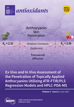
Anthocyanins are natural colorants with antioxidant properties and have been shown to inhibit photoaging reactions and reduce the symptoms of some skin diseases. We investigated their ability to penetrate the stratum corneum, a prerequisite for bioactivity in the skin when used in a lipstick formulation. Simple and complex anthocyanins were able to permeate through the stratum corneum in vivo and reach depths relevant for their use as beneficial ingredients in skin care products. This work provides insight on the permeation behavior of different anthocyanins through the skin, demonstrating both lateral and transversal diffusion when applied topically in a lipophilic delivery system. These findings further contribute to the understanding of anthocyanin delivery in other topical formulations. View this paper.
  • Issues are regarded as officially published after their release is announced to the table of contents alert mailing list.
  • You may sign up for e-mail alerts to receive table of contents of newly released issues.
  • PDF is the official format for papers published in both, html and pdf forms. To view the papers in pdf format, click on the "PDF Full-text" link, and use the free Adobe Reader to open them.
Order results
Result details
Section
Select all
Export citation of selected articles as:
23 pages, 384 KiB  
Review
Update on the Effects of Antioxidants on Diabetic Retinopathy: In Vitro Experiments, Animal Studies and Clinical Trials
by Jose Javier Garcia-Medina, Elena Rubio-Velazquez, Elisa Foulquie-Moreno, Ricardo P Casaroli-Marano, Maria Dolores Pinazo-Duran, Vicente Zanon-Moreno and Monica del-Rio-Vellosillo
Antioxidants 2020, 9(6), 561; https://doi.org/10.3390/antiox9060561 - 26 Jun 2020
Cited by 39 | Viewed by 4535
Abstract
Current therapies for diabetic retinopathy (DR) incorporate blood glucose and blood pressure control, vitrectomy, photocoagulation, and intravitreal injections of anti-vascular endothelial growth factors or corticosteroids. Nonetheless, these techniques have not been demonstrated to completely stop the evolution of this disorder. The pathophysiology of [...] Read more.
Current therapies for diabetic retinopathy (DR) incorporate blood glucose and blood pressure control, vitrectomy, photocoagulation, and intravitreal injections of anti-vascular endothelial growth factors or corticosteroids. Nonetheless, these techniques have not been demonstrated to completely stop the evolution of this disorder. The pathophysiology of DR is not fully known, but there is more and more evidence indicating that oxidative stress is an important mechanism in the progression of DR. In this sense, antioxidants have been suggested as a possible therapy to reduce the complications of DR. In this review we aim to assemble updated information in relation to in vitro experiments, animal studies and clinical trials dealing with the effect of the antioxidants on DR. Full article
11 pages, 1719 KiB  
Article
Platelet-Rich Fibrin Can Neutralize Hydrogen Peroxide-Induced Cell Death in Gingival Fibroblasts
by Zahra Kargarpour, Jila Nasirzade, Francesca Di Summa, Layla Panahipour, Richard J. Miron and Reinhard Gruber
Antioxidants 2020, 9(6), 560; https://doi.org/10.3390/antiox9060560 - 26 Jun 2020
Cited by 17 | Viewed by 3307
Abstract
Hydrogen peroxide is a damage signal at sites of chronic inflammation. The question arises whether platelet-rich fibrin (PRF), platelet-poor plasma (PPP), and the buffy coat can neutralize hydrogen peroxide toxicity and thereby counteract local oxidative stress. In the present study, gingival fibroblasts cells [...] Read more.
Hydrogen peroxide is a damage signal at sites of chronic inflammation. The question arises whether platelet-rich fibrin (PRF), platelet-poor plasma (PPP), and the buffy coat can neutralize hydrogen peroxide toxicity and thereby counteract local oxidative stress. In the present study, gingival fibroblasts cells were exposed to hydrogen peroxide with and without lysates obtained from PRF membranes, PPP, heated PPP (75 °C for 10 min), and the buffy coat. Cell viability was examined by trypan blue staining, live-dead staining, and formazan crystal formation. Cell apoptosis was assessed by cleaved caspase-3 Western blot analysis. Reverse transcription-quantitative polymerase chain reaction (RT-PCR) was utilized to determine the impact of PRF lysates on the expression of catalase in fibroblasts. It was reported that lysates from PRF, PPP, and the buffy coat—but not heated PPP—abolished the hydrogen peroxide-induced toxicity in gingival fibroblasts. Necrosis was confirmed by a loss of membrane integrity and apoptosis was ruled out by the lack of cleavage of caspase-3. Aminotriazole, an inhibitor of catalase, reduced the cytoprotective activity of PRF lysates yet blocking of glutathione peroxidase by mercaptosuccinate did not show the same effect. PRF lysates had no impact on the expression of catalase in gingival fibroblasts. These findings suggest that PRF, PPP, and the buffy coat can neutralize hydrogen peroxide through the release of heat-sensitive catalase. Full article
(This article belongs to the Special Issue Enzymatic and Non-Enzymatic Molecules with Antioxidant Function)
Show Figures

Graphical abstract

19 pages, 458 KiB  
Article
Vitamin D3 Supplementation in Drinking Water Prior to Slaughter Improves Oxidative Status, Physiological Stress, and Quality of Pork
by Ana I. Rey, José Francisco Segura, David Castejón, Encarnación Fernández-Valle, Mª Isabel Cambero and Luis Calvo
Antioxidants 2020, 9(6), 559; https://doi.org/10.3390/antiox9060559 - 26 Jun 2020
Cited by 17 | Viewed by 3619
Abstract
The aims of this study were to investigate the effect of vitamin D3 administration in drinking water during lairage time prior to slaughter on physiological stress, oxidative status, and pork quality characteristics. Two experiments were carried out. The first one was performed [...] Read more.
The aims of this study were to investigate the effect of vitamin D3 administration in drinking water during lairage time prior to slaughter on physiological stress, oxidative status, and pork quality characteristics. Two experiments were carried out. The first one was performed to examine the effect of vitamin D3 supplementation in drinking water, and the second one to check the effect of supplementation dose (500,000 IU/L vs. 700,000 IU/L). Serum calcium concentration was greater in pigs receiving vitamin D3 in water when compared to the control group. In experiment 1, a 40% α-tocopherol increase in meat from the group supplemented with vitamin D3 (500,000 IU/animal) was observed, that resulted in a tendency of decreased meat malondialdehyde (MDA) values at days 5 and 8 after refrigerated storage. In experiment 2, since water intake was higher (800,000 IU and 1,120,000 IU/animal of vitamin D3 consumption) effects on oxidative status were more profound and vitamin D3 supplementation increased serum α-tocopherol and decreased cortisol and serum TBARS. These effects were also observed in meat; TBARS levels were decreased after 3 days of refrigerated storage. In both experiments meat from pigs that received vitamin D3 in drinking water had a lower proportion of total free-polyunsaturated fatty acids (mainly n-6) when compared to the unsupplemented pigs, and these were positively correlated with TBARS production at day 5 of refrigerated storage (r = 0.53 and 0.38 for experiments 1 and 2, respectively). Meat from pigs receiving vitamin D3 in water showed reduced cohesiveness, gumminess, and chewiness values compared to the control group. The magnetic resonance imaging study of muscle confirmed the effects on water retention with lesser transverse relaxation time in pigs supplemented with vitamin D3. No vitamin D3 dose effect was observed, apart from muscle α-tocopherol concentration that was higher in pigs supplemented with 700,000 compared to those supplemented with 500,000 UI/L. Full article
Show Figures

Graphical abstract

12 pages, 5723 KiB  
Article
Effects of Darkness and Light Spectra on Nutrients and Pigments in Radish, Soybean, Mung Bean and Pumpkin Sprouts
by Linda Mastropasqua, Nunzio Dipierro and Costantino Paciolla
Antioxidants 2020, 9(6), 558; https://doi.org/10.3390/antiox9060558 - 26 Jun 2020
Cited by 37 | Viewed by 7725
Abstract
Fresh sprouts are an important source of antioxidant compounds and contain useful phytonutrients in the human diet. Many factors, such as the time of germination and types of light, influence the physiological processes and biosynthetic pathways in sprouts. The effect of red, blue [...] Read more.
Fresh sprouts are an important source of antioxidant compounds and contain useful phytonutrients in the human diet. Many factors, such as the time of germination and types of light, influence the physiological processes and biosynthetic pathways in sprouts. The effect of red, blue and white light vs. dark conditions on the quality parameters in different sprout species after 5 d of germination was evaluated. Total ascorbate, soluble proteins, sugars, phenolic compounds, and pigments, such as carotenoids, chlorophylls, and anthocyanins, were investigated in radishes, soybeans, mung beans, and pumpkin sprouts. The light treatments increased the contents of vitamin C and the various pigments in all sprouts, conversely, they increased the soluble proteins and sugars, including d-glucose, d-fructose and sucrose, in soybeans and pumpkins, respectively. The dark treatment prevented the decrease in dry matter due to the lighting, while the red light induced an increase in polyphenols in soybean. These results suggest that the nutritional content of different sprouts grown under different light conditions depend on the dark or specific spectral wavelength used for their growth. The manuscript may increase the knowledge on light use for the industrialized food production aiming at preserving the phytonutrient content of vegetables, increasing the consumer health, or developing tailored diets for specific nutritional needs. Full article
(This article belongs to the Special Issue Designing the Antioxidant Properties of Low-Processed Food)
Show Figures

Graphical abstract

15 pages, 1534 KiB  
Article
Antioxidant, Physicochemical, and Cellular Secretion of Glucagon-Like Peptide-1 Properties of Oat Bran Protein Hydrolysates
by Mallory E. Walters, William G. Willmore and Apollinaire Tsopmo
Antioxidants 2020, 9(6), 557; https://doi.org/10.3390/antiox9060557 - 26 Jun 2020
Cited by 18 | Viewed by 3463
Abstract
The aim of this work was to determine the physicochemical and biological activities of hydrolyzed proteins from sonicated oat brans. In addition to the control bran sample, two types of pre-treatment procedures—namely, ultrasonic bath and probe-type sonication—were performed to extract proteins, followed by [...] Read more.
The aim of this work was to determine the physicochemical and biological activities of hydrolyzed proteins from sonicated oat brans. In addition to the control bran sample, two types of pre-treatment procedures—namely, ultrasonic bath and probe-type sonication—were performed to extract proteins, followed by hydrolysis with various proteases. Physicochemical analyses showed that Flavourzyme-hydrolysates had greater amounts of aromatic amino acids, Papain-hydrolysates low surface charges (−0.78 to −1.32 mV) compared to the others (−3.67 to −9.17 mV), and Alcalase-hydrolysates a higher surface hydrophobicity. The hydrolysates had good radical scavenging activities but, as the ultrasonic pre-treatment of the brans showed, in certain cases there was a reduction in activities of up to 22% for ROO and HO and 15% for O2•− radicals. In anti-diabetic tests, the maximum inhibition of α-amylase was 31.8%, while that of dipeptidyl peptidase-4 was 53.6%. In addition, the secretion of glucagon-like peptide-1 in NCI-H716 cells was enhanced by 11.5% in the presence of hydrolysates. Full article
(This article belongs to the Special Issue Current Trends in Food-Derived Peptidic Antioxidants)
Show Figures

Figure 1

16 pages, 4419 KiB  
Article
Biochemical Characterization of Traditional Varieties of Sweet Pepper (Capsicum annuum L.) of the Campania Region, Southern Italy
by Florinda Fratianni, Antonio d’Acierno, Autilia Cozzolino, Patrizia Spigno, Riccardo Riccardi, Francesco Raimo, Catello Pane, Massimo Zaccardelli, Valentina Tranchida Lombardo, Marina Tucci, Stefania Grillo, Raffaele Coppola and Filomena Nazzaro
Antioxidants 2020, 9(6), 556; https://doi.org/10.3390/antiox9060556 - 26 Jun 2020
Cited by 30 | Viewed by 6444
Abstract
Bioactive compounds of different Campania native sweet pepper varieties were evaluated. Polyphenols ranged between 1.37 mmol g−1 and 3.42 mmol g−1, β-carotene was abundant in the red variety “Cazzone” (7.05 μg g−1). Yellow and red varieties showed a [...] Read more.
Bioactive compounds of different Campania native sweet pepper varieties were evaluated. Polyphenols ranged between 1.37 mmol g−1 and 3.42 mmol g−1, β-carotene was abundant in the red variety “Cazzone” (7.05 μg g−1). Yellow and red varieties showed a content of ascorbic acid not inferior to 0.82 mg g−1, while in some green varieties the presence of ascorbic acid was almost inconsistent. Interrelationships between the parameters analyzed and the varieties showed that ascorbic acid could represent the factor mostly influencing the antioxidant activity. Polyphenol profile was different among the varieties, with a general prevalence of acidic phenols in yellow varieties and of flavonoids in red varieties. Principal Component Analysis, applied to ascorbic acid, total polyphenols and β-carotene, revealed that two of the green varieties (“Friariello napoletano” and “Friariello Sigaretta”) were well clustered and that the yellow variety “Corno di capra” showed similarity with the green varieties, in particular with “Friariello Nocerese”. This was confirmed by the interrelationships applied to polyphenol composition, which let us to light on a clustering of several red and yellow varieties, and that mainly the yellow ”Corno di capra” was closer to the green varieties of “Friariello”. Full article
(This article belongs to the Special Issue Antioxidants in Foods)
Show Figures

Graphical abstract

16 pages, 1740 KiB  
Article
The Hypoxia Tolerance of the Goldfish (Carassius auratus) Heart: The NOS/NO System and Beyond
by Mariacristina Filice, Rosa Mazza, Serena Leo, Alfonsina Gattuso, Maria Carmela Cerra and Sandra Imbrogno
Antioxidants 2020, 9(6), 555; https://doi.org/10.3390/antiox9060555 - 26 Jun 2020
Cited by 11 | Viewed by 3355
Abstract
The extraordinary capacity of the goldfish (Carassius auratus) to increase its cardiac performance under acute hypoxia is crucial in ensuring adequate oxygen supply to tissues and organs. However, the underlying physiological mechanisms are not yet completely elucidated. By employing an ex [...] Read more.
The extraordinary capacity of the goldfish (Carassius auratus) to increase its cardiac performance under acute hypoxia is crucial in ensuring adequate oxygen supply to tissues and organs. However, the underlying physiological mechanisms are not yet completely elucidated. By employing an ex vivo working heart preparation, we observed that the time-dependent enhancement of contractility, distinctive of the hypoxic goldfish heart, is abolished by the Nitric Oxide Synthase (NOS) antagonist L-NMMA, the Nitric Oxide (NO) scavenger PTIO, as well as by the PI3-kinase (PI3-K) and sarco/endoplasmic reticulum Ca2+-ATPase 2a (SERCA2a) pumps’ inhibition by Wortmannin and Thapsigargin, respectively. In goldfish hearts exposed to hypoxia, an ELISA test revealed no changes in cGMP levels, while Western Blotting analysis showed an enhanced expression of the phosphorylated protein kinase B (pAkt) and of the NADPH oxidase catalytic subunit Nox2 (gp91phox). A significant decrease of protein S-nitrosylation was observed by Biotin Switch assay in hypoxic hearts. Results suggest a role for a PI3-K/Akt-mediated activation of the NOS-dependent NO production, and SERCA2a pumps in the mechanisms conferring benefits to the goldfish heart under hypoxia. They also propose protein denitrosylation, and the possibility of nitration, as parallel intracellular events. Full article
Show Figures

Figure 1

24 pages, 2127 KiB  
Article
In vitro Antioxidant, Anti-inflammatory, Anti-metabolic Syndrome, Antimicrobial, and Anticancer Effect of Phenolic Acids Isolated from Fresh Lovage Leaves [Levisticum officinale Koch] Elicited with Jasmonic Acid and Yeast Extract
by Anna Jakubczyk, Urszula Złotek, Urszula Szymanowska, Kamila Rybczyńska-Tkaczyk, Krystyna Jęderka and Sławomir Lewicki
Antioxidants 2020, 9(6), 554; https://doi.org/10.3390/antiox9060554 - 25 Jun 2020
Cited by 10 | Viewed by 3924
Abstract
Lovage seedlings were elicited with jasmonic acid (JA) and yeast extract (YE) to induce the synthesis of biologically active compounds. A simulated digestion process was carried out to determine the potential bioavailability of phenolic acids. Buffer extracts were prepared for comparison. The ability [...] Read more.
Lovage seedlings were elicited with jasmonic acid (JA) and yeast extract (YE) to induce the synthesis of biologically active compounds. A simulated digestion process was carried out to determine the potential bioavailability of phenolic acids. Buffer extracts were prepared for comparison. The ability to neutralize ABTS radicals was higher in all samples after the in vitro digestion, compared to that in the buffer extracts. However, the elicitation resulted in a significant increase only in the value of the reduction power of the potentially bioavailable fraction of phenolic acids. The effect of the elicitation on the activity of the potentially bioavailable fraction of phenolic acids towards the enzymes involved in the pathogenesis of the metabolic syndrome, i.e., ACE, lipase, amylase, and glucosidase, was analyzed as well. The in vitro digestion caused a significant increase in the ability to inhibit the activity of these enzymes; moreover, the inhibitory activity against alpha-amylase was revealed only after the digestion process. The potential anti-inflammatory effect of the analyzed extracts was defined as the ability to inhibit key pro-inflammatory enzymes, i.e., lipoxygenase and cyclooxygenase 2. The buffer extracts from the YE-elicited lovage inhibited the LOX and COX-2 activity more effectively than the extracts from the control plants. A significant increase in the anti-inflammatory and antimicrobial properties was noted after the simulated digestion. Full article
(This article belongs to the Special Issue Feature Papers in Antioxidants in 2020)
Show Figures

Figure 1

16 pages, 1135 KiB  
Article
ApoE Genotype-Dependent Response to Antioxidant and Exercise Interventions on Brain Function
by Kiran Chaudhari, Jessica M. Wong, Philip H. Vann, Tori Como, Sid E. O’Bryant and Nathalie Sumien
Antioxidants 2020, 9(6), 553; https://doi.org/10.3390/antiox9060553 - 25 Jun 2020
Cited by 4 | Viewed by 3552
Abstract
This study determined whether antioxidant supplementation is a viable complement to exercise regimens in improving cognitive and motor performance in a mouse model of Alzheimer’s disease risk. Starting at 12 months of age, separate groups of male and female mice expressing human Apolipoprotein [...] Read more.
This study determined whether antioxidant supplementation is a viable complement to exercise regimens in improving cognitive and motor performance in a mouse model of Alzheimer’s disease risk. Starting at 12 months of age, separate groups of male and female mice expressing human Apolipoprotein E3 (GFAP-ApoE3) or E4 (GFAP-ApoE4) were fed either a control diet or a diet supplemented with vitamins E and C. The mice were further separated into a sedentary group or a group that followed a daily exercise regimen. After 8 weeks on the treatments, the mice were administered a battery of functional tests including tests to measure reflex and motor, cognitive, and affective function while remaining on their treatment. Subsequently, plasma inflammatory markers and catalase activity in brain regions were measured. Overall, the GFAP-ApoE4 mice exhibited poorer motor function and spatial learning and memory. The treatments improved balance, learning, and cognitive flexibility in the GFAP-ApoE3 mice and overall the GFAP-ApoE4 mice were not responsive. The addition of antioxidants to supplement a training regimen only provided further benefits to the active avoidance task, and there was no antagonistic interaction between the two interventions. These outcomes are indicative that there is a window of opportunity for treatment and that genotype plays an important role in response to interventions. Full article
(This article belongs to the Special Issue Oxidative Stress and Exercise)
Show Figures

Figure 1

17 pages, 3860 KiB  
Article
Plant-Derived Natural Biomolecule Picein Attenuates Menadione Induced Oxidative Stress on Neuroblastoma Cell Mitochondria
by Kavindra Kumar Kesari, Anupam Dhasmana, Shruti Shandilya, Neeraj Prabhakar, Ahmed Shaukat, Jinze Dou, Jessica M. Rosenholm, Tapani Vuorinen and Janne Ruokolainen
Antioxidants 2020, 9(6), 552; https://doi.org/10.3390/antiox9060552 - 25 Jun 2020
Cited by 19 | Viewed by 5091
Abstract
Several bioactive compounds are in use for the treatment of neurodegenerative disorders, such as Alzheimer’s and Parkinson’s disease. Historically, willow (salix sp.) bark has been an important source of salisylic acid and other natural compounds with anti-inflammatory, antipyretic and analgesic properties. Among [...] Read more.
Several bioactive compounds are in use for the treatment of neurodegenerative disorders, such as Alzheimer’s and Parkinson’s disease. Historically, willow (salix sp.) bark has been an important source of salisylic acid and other natural compounds with anti-inflammatory, antipyretic and analgesic properties. Among these, picein isolated from hot water extract of willow bark, has been found to act as a natural secondary metabolite antioxidant. The aim of this study was to investigate the unrevealed pharmacological action of picein. In silico studies were utilized to direct the investigation towards the neuroprotection abilities of picein. Our in vitro studies demonstrate the neuroprotective properties of picein by blocking the oxidative stress effects, induced by free radical generator 2-methyl-1,4-naphthoquinone (menadione, MQ), in neuroblastoma SH-SY5Y cells. Several oxidative stress-related parameters were evaluated to measure the protection for mitochondrial integrity, such as mitochondrial superoxide production, mitochondrial activity (MTT), reactive oxygen species (ROS) and live-cell imaging. A significant increase in the ROS level and mitochondrial superoxide production were measured after MQ treatment, however, a subsequent treatment with picein was able to mitigate this effect by decreasing their levels. Additionally, the mitochondrial activity was significantly decreased by MQ exposure, but a follow-up treatment with picein recovered the normal metabolic activity. In conclusion, the presented results demonstrate that picein can significantly reduce the level of MQ-induced oxidative stress on mitochondria, and thereby plays a role as a potent neuroprotectant. Full article
(This article belongs to the Special Issue Oxidative Stress and Neurodegenerative Disorders)
Show Figures

Graphical abstract

16 pages, 2750 KiB  
Article
Esculetin as a Bifunctional Antioxidant Prevents and Counteracts the Oxidative Stress and Neuronal Death Induced by Amyloid Protein in SH-SY5Y Cells
by Letizia Pruccoli, Fabiana Morroni, Giulia Sita, Patrizia Hrelia and Andrea Tarozzi
Antioxidants 2020, 9(6), 551; https://doi.org/10.3390/antiox9060551 - 25 Jun 2020
Cited by 36 | Viewed by 3143
Abstract
Oxidative stress (OS) appears to be an important determinant during the different stages of progression of Alzheimer’s Disease (AD). In particular, impaired antioxidant defense mechanisms, such as the decrease of glutathione (GSH) and nuclear factor erythroid 2 (NF-E2)-related factor 2 (Nrf2), a master [...] Read more.
Oxidative stress (OS) appears to be an important determinant during the different stages of progression of Alzheimer’s Disease (AD). In particular, impaired antioxidant defense mechanisms, such as the decrease of glutathione (GSH) and nuclear factor erythroid 2 (NF-E2)-related factor 2 (Nrf2), a master regulator of antioxidant genes, including those for GSH, are associated with OS in the human AD brain. Among the neuropathological hallmarks of AD, the soluble oligomers of amyloid beta (A) peptides seem to promote neuronal death through mitochondrial dysfunction and OS. In this regard, bifunctional antioxidants can exert a dual neuroprotective role by scavenging reactive oxygen species (ROS) directly and concomitant induction of antioxidant genes. In this study, among natural coumarins (esculetin, scopoletin, fraxetin and daphnetin), we demonstrated the ability of esculetin (ESC) to prevent and counteract ROS formation in neuronal SH-SY5Y cells, suggesting its profile as a bifunctional antioxidant. In particular, ESC increased the resistance of the SH-SY5Y cells against OS through the activation of Nrf2 and increase of GSH. In similar experimental conditions, ESC could also protect the SH-SY5Y cells from the OS and neuronal death evoked by oligomers of A1–42 peptides. Further, the use of the inhibitors PD98059 and LY294002 also showed that Erk1/2 and Akt signaling pathways were involved in the neuroprotection mediated by ESC. These results encourage further research in AD models to explore the efficacy and safety profile of ESC as a novel neuroprotective agent. Full article
Show Figures

Figure 1

18 pages, 4580 KiB  
Article
Impact of the APE1 Redox Function Inhibitor E3330 in Non-Small Cell Lung Cancer Cells Exposed to Cisplatin: Increased Cytotoxicity and Impairment of Cell Migration and Invasion
by Rita Manguinhas, Ana S. Fernandes, João G. Costa, Nuno Saraiva, Sérgio P. Camões, Nuno Gil, Rafael Rosell, Matilde Castro, Joana P. Miranda and Nuno G. Oliveira
Antioxidants 2020, 9(6), 550; https://doi.org/10.3390/antiox9060550 - 24 Jun 2020
Cited by 21 | Viewed by 3650
Abstract
Elevated expression levels of the apurinic/apyrimidinic endonuclease 1 (APE1) have been correlated with the more aggressive phenotypes and poor prognosis of non-small cell lung cancer (NSCLC). This study aimed to assess the impact of the inhibition of the redox function of APE1 with [...] Read more.
Elevated expression levels of the apurinic/apyrimidinic endonuclease 1 (APE1) have been correlated with the more aggressive phenotypes and poor prognosis of non-small cell lung cancer (NSCLC). This study aimed to assess the impact of the inhibition of the redox function of APE1 with E3330 either alone or in combination with cisplatin in NSCLC cells. For this purpose, complementary endpoints focusing on cell viability, apoptosis, cell cycle distribution, and migration/invasion were studied. Cisplatin decreased the viability of H1975 cells in a time- and concentration-dependent manner, with IC50 values of 9.6 µM for crystal violet assay and 15.9 µM for 3-(4,5-Dimethylthiazol-2-yl)-5-(3-carboxymethoxyphenyl)-2-(4-sulfophenyl)-2H-tetrazolium (MTS) assay. E3330 was clearly cytotoxic for concentrations above 30 µM. The co-incubation of E3330 and cisplatin significantly decreased cell viability compared to cisplatin alone. Regarding cell cycle distribution, cisplatin led to an increase in sub-G1, whereas the co-treatment with E3330 did not change this profile, which was then confirmed in terms of % apoptotic cells. In addition, the combination of E3330 and cisplatin at low concentrations decreased collective and chemotactic migration, and also chemoinvasion, by reducing these capabilities up to 20%. Overall, these results point to E3330 as a promising compound to boost cisplatin therapy that warrants further investigation in NSCLC. Full article
(This article belongs to the Special Issue Redox-Active Molecules as Therapeutic Agents)
Show Figures

Graphical abstract

22 pages, 3394 KiB  
Article
Reversible Oxidative Modifications in Myoglobin and Functional Implications
by Mark H. Mannino, Rishi S. Patel, Amanda M. Eccardt, Blythe E. Janowiak, David C. Wood, Fahu He and Jonathan S. Fisher
Antioxidants 2020, 9(6), 549; https://doi.org/10.3390/antiox9060549 - 24 Jun 2020
Cited by 13 | Viewed by 4411
Abstract
Myoglobin (Mb), an oxygen-binding heme protein highly expressed in heart and skeletal muscle, has been shown to undergo oxidative modifications on both an inter- and intramolecular level when exposed to hydrogen peroxide (H2O2) in vitro. Here, we show that [...] Read more.
Myoglobin (Mb), an oxygen-binding heme protein highly expressed in heart and skeletal muscle, has been shown to undergo oxidative modifications on both an inter- and intramolecular level when exposed to hydrogen peroxide (H2O2) in vitro. Here, we show that exposure to H2O2 increases the peroxidase activity of Mb. Reaction of Mb with H2O2 causes covalent binding of heme to the Mb protein (Mb-X), corresponding to an increase in peroxidase activity when ascorbic acid is the reducing co-substrate. Treatment of H2O2-reacted Mb with ascorbic acid reverses the Mb-X crosslink. Reaction with H2O2 causes Mb to form dimers, trimers, and larger molecular weight Mb aggregates, and treatment with ascorbic acid regenerates Mb monomers. Reaction of Mb with H2O2 causes formation of dityrosine crosslinks, though the labile nature of the crosslinks broken by treatment with ascorbic acid suggests that the reversible aggregation of Mb is mediated by crosslinks other than dityrosine. Disappearance of a peptide containing a tryptophan residue when Mb is treated with H2O2 and the peptide’s reappearance after subsequent treatment with ascorbic acid suggest that tryptophan side chains might participate in the labile crosslinking. Taken together, these data suggest that while exposure to H2O2 causes Mb-X formation, increases Mb peroxidase activity, and causes Mb aggregation, these oxidative modifications are reversible by treatment with ascorbic acid. A caveat is that future studies should demonstrate that these and other in vitro findings regarding properties of Mb have relevance in the intracellular milieu, especially in regard to actual concentrations of metMb, H2O2, and ascorbate that would be found in vivo. Full article
(This article belongs to the Special Issue Redox Signalling and Exercise)
Show Figures

Graphical abstract

18 pages, 3184 KiB  
Article
Antioxidative Effects of Thymus quinquecostatus CELAK through Mitochondrial Biogenesis Improvement in RAW 264.7 Macrophages
by Jin Young Hong, Hyunseong Kim, Wan-Jin Jeon, Seungho Baek and In-Hyuk Ha
Antioxidants 2020, 9(6), 548; https://doi.org/10.3390/antiox9060548 - 23 Jun 2020
Cited by 9 | Viewed by 4257
Abstract
Oxidative stress plays a key role in the pathogenesis of several diseases, including neurodegenerative diseases. Recent studies have reported that mitochondrial dysfunction is a leading cause of the overproduction of reactive oxygen species and oxidative stress. Mitochondrial changes play an important role in [...] Read more.
Oxidative stress plays a key role in the pathogenesis of several diseases, including neurodegenerative diseases. Recent studies have reported that mitochondrial dysfunction is a leading cause of the overproduction of reactive oxygen species and oxidative stress. Mitochondrial changes play an important role in preventing oxidative stress. However, there is a lack of experimental evidence supporting this hypothesis. Thymus quinquecostatus CELAK (TQC) extract is a plant from China belonging to the thymus species, which can mediate the inflammatory response and prevent cell damage through its antioxidant activities. This study examines whether TQC can scavenge excess ROS originating from the mitochondria in RAW 264.7 macrophages. We used lipopolysaccharide (LPS) to induce inflammation and oxidative stress in RAW 264.7 macrophages and performed an immunocytochemistry dot blot of 8-hydroxy-2′-deoxyguanosine (8-OHdG) and real-time PCR to analyze the expression levels of genes involved in mitochondrial biogenesis and oxidative metabolism. TQC was found to significantly reduce the intensity of immunostained MitoSOX and 8-OHdG levels in the total genomic DNA within the mitochondria in RAW 264.7 macrophages. The HO-1 and Nrf2 mRNA levels were also significantly increased in the TQC groups. Therefore, we verified that TQC improves mitochondrial function and attenuates oxidative stress induced by LPS. Our results can provide reference for the effect of TQC to develop new therapeutic strategies for various diseases. Full article
(This article belongs to the Special Issue Antioxidant and Biological Properties of Plant Extracts)
Show Figures

Figure 1

22 pages, 980 KiB  
Review
Polyphenols as Possible Agents for Pancreatic Diseases
by Uroš Gašić, Ivanka Ćirić, Tomislav Pejčić, Dejan Radenković, Vladimir Djordjević, Siniša Radulović and Živoslav Tešić
Antioxidants 2020, 9(6), 547; https://doi.org/10.3390/antiox9060547 - 23 Jun 2020
Cited by 15 | Viewed by 4870
Abstract
Pancreatic cancer (PC) is very aggressive and it is estimated that it kills nearly 50% of patients within the first six months. The lack of symptoms specific to this disease prevents early diagnosis and treatment. Today, gemcitabine alone or in combination with other [...] Read more.
Pancreatic cancer (PC) is very aggressive and it is estimated that it kills nearly 50% of patients within the first six months. The lack of symptoms specific to this disease prevents early diagnosis and treatment. Today, gemcitabine alone or in combination with other cytostatic agents such as cisplatin (Cis), 5-fluorouracil (5-FU), irinotecan, capecitabine, or oxaliplatin (Oxa) is used in conventional therapy. Outgoing literature provides data on the use of polyphenols, biologically active compounds, in the treatment of pancreatic cancer and the prevention of acute pancreatitis. Therefore, the first part of this review gives a brief overview of the state of pancreatic disease as well as the procedures for its treatment. The second part provides a detailed overview of the research regarding the anticancer effects of both pure polyphenols and their plant extracts. The results regarding the antiproliferative, antimetastatic, as well as inhibitory effects of polyphenols against PC cell lines as well as the prevention of acute pancreatitis are presented in detail. Finally, particular emphasis is given to the polyphenolic profiles of apples, berries, cherries, sour cherries, and grapes, given the fact that these fruits are rich in polyphenols and anthocyanins. Polyphenolic profiles, the content of individual polyphenols, and their relationships are discussed. Based on this, significant data can be obtained regarding the amount of fruit that should be consumed daily to achieve a therapeutic effect. Full article
(This article belongs to the Special Issue Phenolics as Antioxidant Agents)
Show Figures

Figure 1

19 pages, 12178 KiB  
Article
Antiarrhythmic Effects of Melatonin and Omega-3 Are Linked with Protection of Myocardial Cx43 Topology and Suppression of Fibrosis in Catecholamine Stressed Normotensive and Hypertensive Rats
by Barbara Szeiffova Bacova, Csilla Viczenczova, Katarina Andelova, Matus Sykora, Kiranj Chaudagar, Miroslav Barancik, Michaela Adamcova, Vladimir Knezl, Tamara Egan Benova, Peter Weismann, Jan Slezak and Narcisa Tribulova
Antioxidants 2020, 9(6), 546; https://doi.org/10.3390/antiox9060546 - 22 Jun 2020
Cited by 30 | Viewed by 3013
Abstract
Cardiac β-adrenergic overstimulation results in oxidative stress, hypertrophy, ischemia, lesion, and fibrosis rendering the heart vulnerable to malignant arrhythmias. We aimed to explore the anti-arrhythmic efficacy of the anti-oxidative and anti-inflammatory compounds, melatonin, and omega-3, and their mechanisms of actions in normotensive and [...] Read more.
Cardiac β-adrenergic overstimulation results in oxidative stress, hypertrophy, ischemia, lesion, and fibrosis rendering the heart vulnerable to malignant arrhythmias. We aimed to explore the anti-arrhythmic efficacy of the anti-oxidative and anti-inflammatory compounds, melatonin, and omega-3, and their mechanisms of actions in normotensive and hypertensive rats exposed to isoproterenol (ISO) induced β-adrenergic overdrive. Eight-month-old, male SHR, and Wistar rats were injected during 7 days with ISO (cumulative dose, 118 mg/kg). ISO rats were either untreated or concomitantly treated with melatonin (10 mg/kg/day) or omega-3 (Omacor, 1.68 g/kg/day) until 60 days of ISO withdrawal and compared to non-ISO controls. Findings showed that both melatonin and omega-3 increased threshold current to induce ventricular fibrillation (VF) in ISO rats regardless of the strain. Prolonged treatment with these compounds resulted in significant suppression of ISO-induced extracellular matrix alterations, as indicated by reduced areas of diffuse fibrosis and decline of hydroxyproline, collagen-1, SMAD2/3, and TGF-β1 protein levels. Importantly, the highly pro-arrhythmic ISO-induced disordered cardiomyocyte distribution of electrical coupling protein, connexin-43 (Cx43), and its remodeling (lateralization) were significantly attenuated by melatonin and omega-3 in Wistar as well as SHR hearts. In parallel, both compounds prevented the post-ISO-related increase in Cx43 variant phosphorylated at serine 368 along with PKCε, which are known to modulate Cx43 remodeling. Melatonin and omega-3 increased SOD1 or SOD2 protein levels in ISO-exposed rats of both strains. Altogether, the results indicate that anti-arrhythmic effects of melatonin and omega-3 might be attributed to the protection of myocardial Cx43 topology and suppression of fibrosis in the setting of oxidative stress induced by catecholamine overdrive in normotensive and hypertensive rats. Full article
(This article belongs to the Special Issue Role of Antioxidant Molecules and Melatonin in Cellular Protection)
Show Figures

Figure 1

20 pages, 1848 KiB  
Article
Neuroprotection by Phytoestrogens in the Model of Deprivation and Resupply of Oxygen and Glucose In Vitro: The Contribution of Autophagy and Related Signaling Mechanisms
by Giuseppe Abbruzzese, Javier Morón-Oset, Sabela Díaz-Castroverde, Nuria García-Font, Cesáreo Roncero, Francisco López-Muñoz, José Luis Marco Contelles and María Jesús Oset-Gasque
Antioxidants 2020, 9(6), 545; https://doi.org/10.3390/antiox9060545 - 22 Jun 2020
Cited by 13 | Viewed by 3050
Abstract
Phytoestrogens can have a neuroprotective effect towards ischemia-reperfusion-induced neuronal damage. However, their mechanism of action has not been well described. In this work, we investigate the type of neuronal cell death induced by oxygen and glucose deprivation (OGD) and resupply (OGDR) and pinpoint [...] Read more.
Phytoestrogens can have a neuroprotective effect towards ischemia-reperfusion-induced neuronal damage. However, their mechanism of action has not been well described. In this work, we investigate the type of neuronal cell death induced by oxygen and glucose deprivation (OGD) and resupply (OGDR) and pinpoint some of the signaling mechanisms whereby the neuroprotective effects of phytoestrogens occur in these conditions. First, we found that autophagy initiation affords neuronal protection upon neuronal damage induced by OGD and OGDR. The mammalian target of rapamycin/ribosomal S6 kinase (mTOR/S6K) pathway is blocked in these conditions, and we provide evidence that this is mediated by modulation of both the 5′ AMP-activated protein kinase (AMPK) and phosphatidylinositol-3-kinase/protein kinase B (PI3K/AKT) pathways. These are dampened up or down, respectively, under OGDR-induced neuronal damage. In contrast, the MAPK-Erk kinase/extracellular signal-regulated kinase (MEK/ERK) pathway is increased under these conditions. Regarding the pathways affected by phytoestrogens, we show that their protective properties require autophagy initiation, but at later stages, they decrease mitogen-activated protein kinase (MAPK) and AMPK activation and increase mTOR/S6K activation. Collectively, our results put forward a novel mode of action where phytoestrogens play a dual role in the regulation of autophagy by acting as autophagy initiation enhancers when autophagy is a neuroprotective and pro-survival mechanism, and as autophagy initiation inhibitors when autophagy is a pro-death mechanism. Finally, our results support the therapeutic potential of phytoestrogens in brain ischemia by modulating autophagy. Full article
Show Figures

Graphical abstract

33 pages, 3625 KiB  
Article
Oxylipins Associated to Current Diseases Detected for the First Time in the Oxidation of Corn Oil as a Model System of Oils Rich in Omega-6 Polyunsaturated Groups. A Global, Broad and in-Depth Study by 1H NMR Spectroscopy
by Jon Alberdi-Cedeño, María L. Ibargoitia and María D. Guillén
Antioxidants 2020, 9(6), 544; https://doi.org/10.3390/antiox9060544 - 20 Jun 2020
Cited by 12 | Viewed by 3120
Abstract
For the first time, an important number of oxylipins have been identified and quantified in corn oil submitted to mild oxidative conditions at each time of their oxidation process. This oil can be considered as a model system of edible oils rich in [...] Read more.
For the first time, an important number of oxylipins have been identified and quantified in corn oil submitted to mild oxidative conditions at each time of their oxidation process. This oil can be considered as a model system of edible oils rich in polyunsaturated omega-6 groups. The study was carried out using 1H nuclear magnetic resonance spectroscopy (1H NMR), which does not require chemical modification of the sample. These newly detected oxylipins include dihydroperoxy-non-conjugated-dienes, hydroperoxy-epoxy-, hydroxy-epoxy- and keto-epoxy-monoenes as well as E-epoxy-monoenes, some of which have been associated with several diseases. Furthermore, the formation of other functional groups such as poly-formates, poly-hydroxy and poly-ether groups has also been proven. These are responsible for the polymerization and increased viscosity of the oil. Simultaneously, monitoring of the formation of well-known oxylipins, such as hydroperoxy-, hydroxy-, and keto-dienes, and of different kinds of oxygenated-alpha,beta-unsaturated aldehydes such as 4-hydroperoxy-, 4-hydroxy-, 4-oxo-2E-nonenal and 4,5-epoxy-2E-decenal, which are also related to different degenerative diseases, has been carried out. The provided data regarding the compounds identification and their sequence and kinetics of formation constitute valuable information for future studies in which lipid oxidation is involved, both in food and in other scientific fields. Full article
(This article belongs to the Special Issue Lipid Oxidation and Antioxidants in Food)
Show Figures

Graphical abstract

29 pages, 565 KiB  
Article
Study of the In Vitro Digestion of Olive Oil Enriched or Not with Antioxidant Phenolic Compounds. Relationships between Bioaccessibility of Main Components of Different Oils and Their Composition
by Jon Alberdi-Cedeño, María L. Ibargoitia and María D. Guillén
Antioxidants 2020, 9(6), 543; https://doi.org/10.3390/antiox9060543 - 20 Jun 2020
Cited by 13 | Viewed by 2955
Abstract
The changes provoked by in vitro digestion in the lipids of olive oil enriched or not with different phenolic compounds were studied by proton nuclear magnetic resonance (1H NMR) and solid phase microextraction followed by gas chromatography/mass spectrometry (SPME-GC/MS). These changes [...] Read more.
The changes provoked by in vitro digestion in the lipids of olive oil enriched or not with different phenolic compounds were studied by proton nuclear magnetic resonance (1H NMR) and solid phase microextraction followed by gas chromatography/mass spectrometry (SPME-GC/MS). These changes were compared with those provoked in the lipids of corn oil and of virgin flaxseed oil submitted to the same digestive conditions. Lipolysis and oxidation were the two reactions under consideration. The bioaccessibility of main and minor components of olive oil, of phenolic compounds added, and of compounds formed as consequence of the oxidation, if any, were matters of attention. Enrichment of olive oil with antioxidant phenolic compounds does not affect the extent of lipolysis, but reduces the oxidation degree to minimum values or avoids it almost entirely. The in vitro bioaccessibility of nutritional and bioactive compounds was greater in the olive oil digestate than in those of other oils, whereas that of compounds formed in oxidation was minimal, if any. Very close quantitative relationships were found between the composition of the oils in main components and their in vitro bioaccessibility. These relationships, some of which have predictive value, can help to design lipid diets for different nutritional purposes. Full article
Show Figures

Figure 1

28 pages, 406 KiB  
Review
A Review of the Potential Benefits of Plants Producing Berries in Skin Disorders
by Stefano Piazza, Marco Fumagalli, Saba Khalilpour, Giulia Martinelli, Andrea Magnavacca, Mario Dell’Agli and Enrico Sangiovanni
Antioxidants 2020, 9(6), 542; https://doi.org/10.3390/antiox9060542 - 20 Jun 2020
Cited by 12 | Viewed by 6138
Abstract
During the last 30 years, berries have gained great attention as functional food against several risk factors in chronic diseases. The number of related publications on Pubmed rose from 1000 items in 1990 to more than 11,000 in 2019. Despite the fact that [...] Read more.
During the last 30 years, berries have gained great attention as functional food against several risk factors in chronic diseases. The number of related publications on Pubmed rose from 1000 items in 1990 to more than 11,000 in 2019. Despite the fact that a common and clear definition of “berries” is not shared among different scientific areas, the phytochemical pattern of these fruits is mainly characterized by anthocyanins, flavanols, flavonols, and tannins, which showed antioxidant and anti-inflammatory properties in humans. Skin insults, like wounds, UV rays, and excessive inflammatory responses, may lead to chronic dermatological disorders, conditions often characterized by long-term treatments. The application of berries for skin protection is sustained by long traditional use, but many observations still require a clear pharmacological validation. This review summarizes the scientific evidence, published on EMBASE, MEDLINE, and Scholar, to identify extraction methods, way of administration, dose, and mechanism of action of berries for potential dermatological treatments. Promising in vitro and in vivo evidence of Punica granatum L. and Vitis vinifera L. supports wound healing and photoprotection, while Schisandra chinensis (Turcz.) Baill. and Vaccinium spp. showed clear immunomodulatory effects. Oral or topical administrations of these berries justify the evaluation of new translational studies to validate their efficacy in humans. Full article
(This article belongs to the Special Issue Feature Papers in Antioxidants in 2020)
24 pages, 350 KiB  
Review
Edible Leafy Plants from Mexico as Sources of Antioxidant Compounds, and Their Nutritional, Nutraceutical and Antimicrobial Potential: A Review
by Lourdes Mateos-Maces, José Luis Chávez-Servia, Araceli Minerva Vera-Guzmán, Elia Nora Aquino-Bolaños, Jimena E. Alba-Jiménez and Bethsabe Belem Villagómez-González
Antioxidants 2020, 9(6), 541; https://doi.org/10.3390/antiox9060541 - 20 Jun 2020
Cited by 25 | Viewed by 6712
Abstract
A review of indigenous Mexican plants with edible stems and leaves and their nutritional and nutraceutical potential was conducted, complemented by the authors’ experiences. In Mexico, more than 250 species with edible stems, leaves, vines and flowers, known as “quelites,” are [...] Read more.
A review of indigenous Mexican plants with edible stems and leaves and their nutritional and nutraceutical potential was conducted, complemented by the authors’ experiences. In Mexico, more than 250 species with edible stems, leaves, vines and flowers, known as “quelites,” are collected or are cultivated and consumed. The assessment of the quelite composition depends on the chemical characteristics of the compounds being evaluated; the protein quality is a direct function of the amino acid content, which is evaluated by high-performance liquid chromatography (HPLC), and the contribution of minerals is evaluated by atomic absorption spectrometry, inductively coupled plasma-optical emission spectrometry (ICP-OES) or ICP mass spectrometry. The total contents of phenols, flavonoids, carotenoids, saponins and other general compounds have been analyzed using UV-vis spectrophotometry and by HPLC. For the determination of specific compounds such as phenolic compounds, flavonoids, organic acids and other profiles, it is recommended to use HPLC-DAD, UHPLC-DAD, UFLC-PDA or gas chromatography-mass spectrometry. The current biochemical analysis and biological evaluations were performed to understand the mechanisms of action that lead to decreased glucose levels and lipid peroxidation, increased hypoglycemic and antitumor activity, immune system improvement, increased antibacterial and antifungal activity and, in some cases, anti-Helicobacter pylori activity. Full article
(This article belongs to the Special Issue Antioxidant capacity of Anthocyanins and Other Vegetal Pigments)
11 pages, 1538 KiB  
Viewpoint
Targeting the Heme-Heme Oxygenase System to Prevent Severe Complications Following COVID-19 Infections
by Frank A. D. T. G. Wagener, Peter Pickkers, Stephen J. Peterson, Stephan Immenschuh and Nader G. Abraham
Antioxidants 2020, 9(6), 540; https://doi.org/10.3390/antiox9060540 - 19 Jun 2020
Cited by 64 | Viewed by 10329
Abstract
SARS-CoV-2 is causing a pandemic resulting in high morbidity and mortality. COVID-19 patients suffering from acute respiratory distress syndrome (ARDS) are often critically ill and show lung injury and hemolysis. Heme is a prosthetic moiety crucial for the function of a wide variety [...] Read more.
SARS-CoV-2 is causing a pandemic resulting in high morbidity and mortality. COVID-19 patients suffering from acute respiratory distress syndrome (ARDS) are often critically ill and show lung injury and hemolysis. Heme is a prosthetic moiety crucial for the function of a wide variety of heme-proteins, including hemoglobin and cytochromes. However, injury-derived free heme promotes adhesion molecule expression, leukocyte recruitment, vascular permeabilization, platelet activation, complement activation, thrombosis, and fibrosis. Heme can be degraded by the anti-inflammatory enzyme heme oxygenase (HO) generating biliverdin/bilirubin, iron/ferritin, and carbon monoxide. We therefore postulate that free heme contributes to many of the inflammatory phenomena witnessed in critically ill COVID-19 patients, whilst induction of HO-1 or harnessing heme may provide protection. HO-activity not only degrades injurious heme, but its effector molecules possess also potent salutary anti-oxidative and anti-inflammatory properties. Until a vaccine against SARS-CoV-2 becomes available, we need to explore novel strategies to attenuate the pro-inflammatory, pro-thrombotic, and pro-fibrotic consequences of SARS-CoV-2 leading to morbidity and mortality. The heme-HO system represents an interesting target for novel “proof of concept” studies in the context of COVID-19. Full article
(This article belongs to the Special Issue Pharmacological and Clinical Significance of Heme Oxygenase-1)
Show Figures

Figure 1

17 pages, 2558 KiB  
Article
Coreolanceolins A–E, New Flavanones from the Flowers of Coreopsis lanceolate, and Their Antioxidant and Anti-Inflammatory Effects
by Hyoung-Geun Kim, Young Sung Jung, Seon Min Oh, Hyun-Ji Oh, Jung-Hwan Ko, Dae-Ok Kim, Se Chan Kang, Yeong-Geun Lee, Dae Young Lee and Nam-In Baek
Antioxidants 2020, 9(6), 539; https://doi.org/10.3390/antiox9060539 - 19 Jun 2020
Cited by 14 | Viewed by 3484
Abstract
(1) Background: Many flavonoids derived from natural sources have been reported to exhibit antioxidant and anti-inflammatory effects. Our preliminary study suggested that Coreopsis lanceolata flowers (CLFs) include high flavonoid content; (2) Methods: CLFs were extracted in 80% (v/v) aqueous methanol and fractionated into [...] Read more.
(1) Background: Many flavonoids derived from natural sources have been reported to exhibit antioxidant and anti-inflammatory effects. Our preliminary study suggested that Coreopsis lanceolata flowers (CLFs) include high flavonoid content; (2) Methods: CLFs were extracted in 80% (v/v) aqueous methanol and fractionated into ethyl acetate, n-butanol, and water fractions. Repeated column chromatographies for the organic fractions led to the isolation of seven flavanones. Quantitative analysis of the flavanones was carried out using reversed-phase high-performance liquid chromatography. All flavanones were evaluated for their antioxidant and pro-inflammatory inhibition effects; (3) Results: Spectroscopic analyses revealed the chemical structure of five new flavanones, coreolanceolins A–E, and two known ones. The content of the seven flavanones in extracts were determined from 0.8 ± 0.1 to 38.8 ± 0.3 mg/g. All flavanones showed radical scavenging activities (respectively 104.3 ± 1.9 to 20.5 ± 0.3 mg vitamin C equivalents (VCE)/100 mg and 1278.6 ± 26.8 to 325.6 ± 0.2 mg VCE/100 mg) in the DPPH and 2,2′-Azino-bis(3-ethylbenzothiazoline-6-sulfonic acid) (ABTS) assays and recovery activities in Caco-2 (59.7 to 41.1%), RAW264.7 (87.8 to 56.0%), and PC-12 (100.5 to 69.9%) cells against reactive oxygen species. Furthermore, all flavanones suppressed nitric oxide production (99.5% to 37.3%) and reduced iNOS and COX-2 expression in lipopolysaccharide-treated RAW 264.7 cells; (4) Conclusions: Five new and two known flavanones were isolated from CLF, and most of them showed high antioxidant and pro-inflammatory inhibition effects. Full article
(This article belongs to the Special Issue Phenolics as Antioxidant Agents)
Show Figures

Graphical abstract

25 pages, 3111 KiB  
Article
Alterations in the Antioxidant Enzyme Activities in the Neurodevelopmental Rat Model of Schizophrenia Induced by Glutathione Deficiency during Early Postnatal Life
by Magdalena Górny, Anna Bilska-Wilkosz, Małgorzata Iciek, Marta Hereta, Kinga Kamińska, Adrianna Kamińska, Grażyna Chwatko, Zofia Rogóż and Elżbieta Lorenc-Koci
Antioxidants 2020, 9(6), 538; https://doi.org/10.3390/antiox9060538 - 19 Jun 2020
Cited by 17 | Viewed by 7075
Abstract
The aim of the present study was to assess the effects of l-buthionine-(S,R)-sulfoximine (BSO), a glutathione (GSH) synthesis inhibitor, and GBR 12909, a dopamine reuptake inhibitor, administered alone or in combination to Sprague-Dawley rats during early postnatal development (p5–p16), on the levels [...] Read more.
The aim of the present study was to assess the effects of l-buthionine-(S,R)-sulfoximine (BSO), a glutathione (GSH) synthesis inhibitor, and GBR 12909, a dopamine reuptake inhibitor, administered alone or in combination to Sprague-Dawley rats during early postnatal development (p5–p16), on the levels of reactive oxygen species (ROS), lipid peroxidation (LP) and the activities of antioxidant enzymes: superoxide dismutase (SOD), catalase (CAT), glutathione peroxidase (GPx) and glutathione disulfide reductase (GR) in peripheral tissues (liver, kidney) and selected brain structures (prefrontal cortex, PFC; hippocampus, HIP; and striatum, STR) of 16-day-old rats. The studied parameters were analyzed with reference to the content of GSH and sulfur amino acids, methionine (Met) and cysteine (Cys) described in our previous study. This analysis showed that treatment with a BSO + GBR 12909 combination caused significant decreases in the lipid peroxidation levels in the PFC and HIP, in spite of there being no changes in ROS. The reduction of lipid peroxidation indicates a weakening of the oxidative power of the cells, and a shift in balance in favor of reducing processes. Such changes in cellular redox signaling in the PFC and HIP during early postnatal development may result in functional changes in adulthood. Full article
(This article belongs to the Special Issue Oxidative stress and Applied Biology)
Show Figures

Figure 1

18 pages, 3131 KiB  
Article
Analysis of the Status of the Cutaneous Endogenous and Exogenous Antioxidative System of Smokers and the Short-Term Effect of Defined Smoking Thereon
by Silke B. Lohan, Karl Bühring, Anna-Christina Lauer, Annette Friedrich, Jürgen Lademann, Annette Buss, Robert Sabat, Kerstin Wolk and Martina C. Meinke
Antioxidants 2020, 9(6), 537; https://doi.org/10.3390/antiox9060537 - 19 Jun 2020
Cited by 8 | Viewed by 2919
Abstract
The daily consumption of tobacco products leads to a boost in free radical production in tissues, promoting the risk for malignancies, metabolic alterations and chronic-inflammatory diseases. This study aimed to broaden the knowledge of the status of the antioxidative (AO) system in the [...] Read more.
The daily consumption of tobacco products leads to a boost in free radical production in tissues, promoting the risk for malignancies, metabolic alterations and chronic-inflammatory diseases. This study aimed to broaden the knowledge of the status of the antioxidative (AO) system in the skin, compared to the blood, of healthy appearing smokers. Both, the basic status compared to non-smokers and the short-term impact of controlled cigarette consumption in smokers were analyzed. Our study showed that the basic level of the AO system of smokers significantly differed from that of non-smokers. As determined by resonant Raman spectroscopy (RRS), the levels of exogenous AOs were decreased in both, the skin, in vivo (β-carotene and lycopene), and blood plasma (β-carotene only). In contrast, the levels of glutathione (GSH), the prototypical endogenous AO, which were analyzed by fluorimetric assays in cutaneous tape strips and blood plasma, were increased in the skin, although unchanged in the blood of smokers. Elevated cutaneous GSH levels were reflected by an elevated overall radical scavenging activity in the skin, as quantified by non-invasive electron paramagnetic resonance (EPR) spectroscopy. Analysis of the expression of selected stress-associated genes in blood immune cells by quantitative RT-PCR in subgroups of non-smokers and smokers additionally demonstrated the downregulation of AKR1C2 in smokers, and its negative correlation with blood plasma levels of the protective immune mediator interleukin-22, assessed by the ELISA technique. Controlled cigarette consumption did not alter exogenous or endogenous AOs in the skin of smokers, but decreased lycopene levels in blood plasma. Moreover, there was a decline in blood IL-22 levels, while no relevant response of blood cell gene expressions was found after the considered short time. Our data therefore demonstrate a strengthened endogenous AO status in the skin of smokers, which may indicate a long-term adaptation to chronic oxidative stress in this specific organ. While this effect was not clearly visible in the blood, this compartment seems to be useful as an immediate indicator of the body’s AO consumption. Moreover, decreased levels of AKR1C2, which we show for the first time to be expressed in immune cells, may be a candidate marker for long-term smoking. In addition, this study demonstrates that the rate constant of a spin probe decline determined by EPR spectroscopy mainly represents the endogenous AO status of a tissue. Full article
Show Figures

Graphical abstract

15 pages, 2521 KiB  
Article
Glutamine Cooperatively Upregulates Lipopolysaccharide-Induced Nitric Oxide Production in BV2 Microglial Cells through the ERK and Nrf-2/HO-1 Signaling Pathway
by Rajapaksha Gedara Prasad Tharanga Jayasooriya, Ilandarage Menu Neelaka Molagoda, Matharage Gayani Dilshara, Yung Hyun Choi and Gi-Young Kim
Antioxidants 2020, 9(6), 536; https://doi.org/10.3390/antiox9060536 - 19 Jun 2020
Cited by 2 | Viewed by 3022
Abstract
Glutamine (Gln) is a nonessential α-amino acid for protein biosynthesis. However, the mechanism through which Gln regulates NO production in microglial cells is still unclear. In this study, we investigated whether the presence or absence of Gln affects NO production in lipopolysaccharide (LPS)-stimulated [...] Read more.
Glutamine (Gln) is a nonessential α-amino acid for protein biosynthesis. However, the mechanism through which Gln regulates NO production in microglial cells is still unclear. In this study, we investigated whether the presence or absence of Gln affects NO production in lipopolysaccharide (LPS)-stimulated BV2 microglial cells. Our data revealed that Gln depletion decreased cell viability accompanied by mild cytotoxicity, and blocked LPS-induced NO production concomitant with a significant decrease in inducible NO synthase (iNOS) expression. Additionally, Gln depletion for 24 h blocked the restoration of LPS-mediated NO production in the presence of Gln, suggesting that Gln depletion caused long-term immune deprivation. In particular, sodium-coupled amino acid transporter 1 and 2 (SNAT1 and SNAT2), which are the main Gln transporters, were highly upregulated in LPS-stimulated BV2 microglial cells, in the presence of Gln accompanied by NO production. Regardless of the presence of Gln, LPS positively stimulated nuclear factor erythroid 2-related factor 2 (Nrf2) and heme oxygenase-1 (HO-1) expression, and transient Nrf2 knockdown and HO-1 inhibition stimulated LPS-induced NO production and iNOS expression; however, transient Nrf2 knockdown did not affect SNAT1 and SNAT2 expression, indicating that Gln transporters, SNAT1 and SNAT2, were not regulated by Nrf2, which downregulated the HO-1-mediated NO production. Moreover, Gln depletion significantly reduced LPS-induced extracellular signal-regulated kinase (ERK) phosphorylation; furthermore, a specific ERK inhibitor, PD98059, and transient ERK knockdown attenuated LPS-stimulated NO production and iNOS expression, in the presence of Gln, accompanied by downregulation of SNAT1 and SNAT2, suggesting that the ERK signaling pathway was related to LPS-mediated NO production via SNAT1 and SNAT2. Altogether, our data indicated that extracellular Gln is vital for NO production from microglia in inflammatory conditions. Full article
Show Figures

Figure 1

13 pages, 863 KiB  
Article
Sunflower (Helianthus annuus L.) Plants at Various Growth Stages Subjected to Extraction—Comparison of the Antioxidant Activity and Phenolic Profile
by Francesco Gai, Magdalena Karamać, Michał A. Janiak, Ryszard Amarowicz and Pier Giorgio Peiretti
Antioxidants 2020, 9(6), 535; https://doi.org/10.3390/antiox9060535 - 19 Jun 2020
Cited by 18 | Viewed by 6772
Abstract
The aim of this study was to evaluate the differences in the antioxidant activity and phenolic profile of sunflower (Helianthus annuus L.) extracts obtained from the aerial parts of plants harvested at five growth stages. In vitro assays were used to determine [...] Read more.
The aim of this study was to evaluate the differences in the antioxidant activity and phenolic profile of sunflower (Helianthus annuus L.) extracts obtained from the aerial parts of plants harvested at five growth stages. In vitro assays were used to determine the antioxidant activity, i.e., ABTS•+ and DPPH scavenging activity, the ferric-reducing antioxidant power (FRAP) and the ability to inhibit β-carotene–linoleic acid emulsion oxidation. Phenolic compounds, such as mono- and dicaffeoylquinic acid isomers and caffeic acid hexose, were identified using the LC–TOF–MS/MS technique. The predominant compound during the growth cycle of the plant was 3,5-di-O-caffeoylquinic acid, whose content was the highest at the mid-flowering stage. The total phenolic content was also the highest in sunflowers at the mid-flowering stage. The main phenolic compound contents were closely correlated with ABTS•+ and DPPH scavenging activity and FRAP. No significant correlation was found between the total phenolic content and the antioxidant activity in the emulsion system. The highest antiradical activity and FRAP were generally determined in older plants (mid-flowering and late flowering stages). In conclusion, the aerial parts of sunflowers, in particular those harvested at the mid-flowering stage, are a good plant material from which to obtain phenolic compound extracts, albeit mainly of one class (esters of caffeic acid and quinic acid), with high antioxidant activity. Full article
(This article belongs to the Special Issue Antioxidant and Biological Properties of Plant Extracts)
Show Figures

Graphical abstract

28 pages, 2249 KiB  
Article
Impact of Different Pasteurization Techniques and Subsequent Ultrasonication on the In Vitro Bioaccessibility of Carotenoids in Valencia Orange (Citrus sinensis (L.) Osbeck) Juice
by Lara Etzbach, Ruth Stolle, Kerstin Anheuser, Volker Herdegen, Andreas Schieber and Fabian Weber
Antioxidants 2020, 9(6), 534; https://doi.org/10.3390/antiox9060534 - 18 Jun 2020
Cited by 12 | Viewed by 4254
Abstract
The effects of traditional pasteurization (low pasteurization, conventional pasteurization, hot filling) and alternative pasteurization (pulsed electric fields, high pressure processing), followed by ultrasonication on the carotenoid content, carotenoid profile, and on the in vitro carotenoid bioaccessibility of orange juice were investigated. There was [...] Read more.
The effects of traditional pasteurization (low pasteurization, conventional pasteurization, hot filling) and alternative pasteurization (pulsed electric fields, high pressure processing), followed by ultrasonication on the carotenoid content, carotenoid profile, and on the in vitro carotenoid bioaccessibility of orange juice were investigated. There was no significant difference in the total carotenoid content between the untreated juice (879.74 µg/100 g juice) and all pasteurized juices. Significantly lower contents of violaxanthin esters were found in the high thermally-treated juices (conventional pasteurization, hot filling) compared to the untreated juice, owing to heat-induced epoxy-furanoid rearrangement. The additional ultrasonication had almost no effects on the carotenoid content and profile of the orange juices. However, the in vitro solubilization and the micellarization efficiency were strongly increased by ultrasound, the latter by approximately 85.3–159.5%. Therefore, among the applied processing techniques, ultrasonication might be a promising technology to enhance the in vitro bioaccessibility of carotenoids and, thus, the nutritional value of orange juice. Full article
Show Figures

Graphical abstract

37 pages, 5011 KiB  
Article
Identification of Chemical Profiles and Biological Properties of Rhizophora racemosa G. Mey. Extracts Obtained by Different Methods and Solvents
by Annalisa Chiavaroli, Koaudio Ibrahime Sinan, Gokhan Zengin, Mohamad Fawzi Mahomoodally, Nabeelah Bibi Sadeer, Ouattara Katinan Etienne, Zoltán Cziáky, József Jekő, Jasmina Glamočlija, Marina Soković, Lucia Recinella, Luigi Brunetti, Sheila Leone, Hassan H. Abdallah, Paola Angelini, Giancarlo Angeles Flores, Roberto Venanzoni, Luigi Menghini, Giustino Orlando and Claudio Ferrante
Antioxidants 2020, 9(6), 533; https://doi.org/10.3390/antiox9060533 - 18 Jun 2020
Cited by 17 | Viewed by 5099
Abstract
Mangrove forests exemplify a multifaceted ecosystem since they do not only play a crucial ecological role but also possess medicinal properties. Methanolic, ethyl acetate and aqueous leaf and bark extracts were prepared using homogenizer-assisted extraction (HAE), infusion and maceration (with and without stirring). [...] Read more.
Mangrove forests exemplify a multifaceted ecosystem since they do not only play a crucial ecological role but also possess medicinal properties. Methanolic, ethyl acetate and aqueous leaf and bark extracts were prepared using homogenizer-assisted extraction (HAE), infusion and maceration (with and without stirring). The different extracts were screened for phytochemical profiling and antioxidant capacities in terms of radical scavenging (DPPH, ABTS), reducing potential (CUPRAC, FRAP), total antioxidant capacity and chelating power. Additionally, R. racemosa was evaluated for its anti-diabetic (α-amylase, α-glucosidase), anti-tyrosinase and anti-cholinesterase (AChE, BChE) activities. Additionally, antimycotic and antibacterial effects were investigated against Eescherichia coli, Pseudomonas aeruginosa, Salmonella typhimurium, Listeria monocytogenes, Enterobacter cloacae, Bacillus cereus, Micrococcus luteus, Staphylococcus aureus, Aspergillus fumigatus, Aspergillus niger, Trichoderma viride, Penicillium funiculosum, Penicillium ochrochloron and Penicillium verrucosum. Finally, based on phytochemical fingerprint, in silico studies, including bioinformatics, network pharmacology and docking approaches were conducted to predict the putative targets, namely tyrosinase, lanosterol-14-α-demethylase and E. coli DNA gyrase, underlying the observed bio-pharmacological and microbiological effects. The methanolic leave and bark extracts (prepared by both HAE and maceration) abounded with phenolics, flavonoids, phenolic acids and flavonols. Results displayed that both methanolic leaf and bark extracts (prepared by HAE) exhibited the highest radical scavenging, reducing potential and total antioxidant capacity. Furthermore, our findings showed that the highest enzymatic inhibitory activity recorded was with the tyrosinase enzyme. In this context, bioinformatics analysis predicted putative interactions between tyrosinase and multiple secondary metabolites including apigenin, luteolin, vitexin, isovitexin, procyanidin B, quercetin and methoxy-trihydroxyflavone. The same compounds were also docked against lanosterol-14α-demethylase and E. Coli DNA gyrase, yielding affinities in the submicromolar–micromolar range that further support the observed anti-microbial effects exerted by the extracts. In conclusion, extracts of R. racemosa may be considered as novel sources of phytoanti-oxidants and enzyme inhibitors that can be exploited as future first-line pharmacophores. Full article
Show Figures

Graphical abstract

17 pages, 1953 KiB  
Review
Pro-oxidant Actions of Carotenoids in Triggering Apoptosis of Cancer Cells: A Review of Emerging Evidence
by Juhyun Shin, Min-Ho Song, Jae-Wook Oh, Young-Soo Keum and Ramesh Kumar Saini
Antioxidants 2020, 9(6), 532; https://doi.org/10.3390/antiox9060532 - 17 Jun 2020
Cited by 95 | Viewed by 11671
Abstract
Carotenoids are well known for their potent antioxidant function in the cellular system. However, in cancer cells with an innately high level of intracellular reactive oxygen species (ROS), carotenoids may act as potent pro-oxidant molecules and trigger ROS-mediated apoptosis. In recent years, the [...] Read more.
Carotenoids are well known for their potent antioxidant function in the cellular system. However, in cancer cells with an innately high level of intracellular reactive oxygen species (ROS), carotenoids may act as potent pro-oxidant molecules and trigger ROS-mediated apoptosis. In recent years, the pro-oxidant function of several common dietary carotenoids, including astaxanthin, β-carotene, fucoxanthin, and lycopene, has been investigated for their effective killing effects on various cancer cell lines. Besides, when carotenoids are delivered with ROS-inducing cytotoxic drugs (e.g., anthracyclines), they can minimize the adverse effects of these drugs on normal cells by acting as antioxidants without interfering with their cytotoxic effects on cancer cells as pro-oxidants. These dynamic actions of carotenoids can optimize oxidative stress in normal cells while enhancing oxidative stress in cancer cells. This review discusses possible mechanisms of carotenoid-triggered ROS production in cancer cells, the activation of pro-apoptotic signaling by ROS, and apoptotic cell death. Moreover, synergistic actions of carotenoids with ROS-inducing anti-cancer drugs are discussed, and research gaps are suggested. Full article
(This article belongs to the Special Issue Carotenoids, Oxidative Stress and Disease)
Show Figures

Graphical abstract

Previous Issue
Next Issue
Back to TopTop