Next Issue
Volume 7, April
Previous Issue
Volume 7, February


Antioxidants, Volume 7, Issue 3 (March 2018) – 11 articles

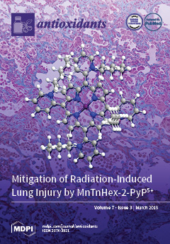
Cover Story (view full-size image): Lipophilic superoxide dismutase mimetics are effective mitigators of many disease processes that involve oxidative injury. New data presented here demonstrate the development of a nonhuman primate model of radiation-induced lung injury, with high genetic and phenotypic similarity to human beings. The findings indicate the potential of MnTnHex-2-PyP5+ to mitigate radiation-induced lung injury. Both inflammation and pro-fibrotic mechanisms are involved, and further studies are needed to understand and maximize the mitigating effects. View this paper
  • Issues are regarded as officially published after their release is announced to the table of contents alert mailing list.
  • You may sign up for e-mail alerts to receive table of contents of newly released issues.
  • PDF is the official format for papers published in both, html and pdf forms. To view the papers in pdf format, click on the "PDF Full-text" link, and use the free Adobe Reader to open them.
Order results
Result details
Section
Select all
Export citation of selected articles as:
2 pages, 162 KiB  
Editorial
Vitamin E
by Volker Böhm
Antioxidants 2018, 7(3), 44; https://doi.org/10.3390/antiox7030044 - 20 Mar 2018
Cited by 15 | Viewed by 5343
Abstract
Vitamin E is the major lipid-soluble antioxidant in the cell antioxidant system and is exclusively obtained from the diet[...] Full article
(This article belongs to the Special Issue Vitamin E)
12 pages, 887 KiB  
Article
Microbial Biotransformation of a Polyphenol-Rich Potato Extract Affects Antioxidant Capacity in a Simulated Gastrointestinal Model
by Joelle Khairallah, Shima Sadeghi Ekbatan, Kebba Sabally, Michèle M. Iskandar, Raza Hussain, Atef Nassar, Lekha Sleno, Laetitia Rodes, Satya Prakash, Danielle J. Donnelly and Stan Kubow
Antioxidants 2018, 7(3), 43; https://doi.org/10.3390/antiox7030043 - 20 Mar 2018
Cited by 7 | Viewed by 5594
Abstract
A multistage human gastrointestinal model was used to digest a polyphenol-rich potato extract containing chlorogenic acid, caffeic acid, ferulic acid, and rutin as the primary polyphenols, to assess for their microbial biotransformation and to measure changes in antioxidant capacity in up to 24 [...] Read more.
A multistage human gastrointestinal model was used to digest a polyphenol-rich potato extract containing chlorogenic acid, caffeic acid, ferulic acid, and rutin as the primary polyphenols, to assess for their microbial biotransformation and to measure changes in antioxidant capacity in up to 24 h of digestion. The biotransformation of polyphenols was assessed by liquid chromatography–mass spectrometry. Antioxidant capacity was measured by the ferric reducing antioxidant power (FRAP) assay. Among the colonic reactors, parent (poly)phenols were detected in the ascending (AC), but not the transverse (TC) or descending (DC) colons. The most abundant microbial phenolic metabolites in all colonic reactors included derivatives of propionic acid, acetic acid, and benzoic acid. As compared to the baseline, an earlier increase in antioxidant capacity (T = 8 h) was seen in the stomach and small intestine vessels as compared to the AC (T = 16 h) and TC and DC (T = 24 h). The increase in antioxidant capacity observed in the DC and TC can be linked to the accumulation of microbial smaller-molecular-weight phenolic catabolites, as the parent polyphenolics had completely degraded in those vessels. The colonic microbial digestion of potato-based polyphenols could lead to improved colonic health, as this generates phenolic metabolites with significant antioxidant potential. Full article
(This article belongs to the Special Issue Dietary Antioxidants and Prevention of Non-Communicable Diseases)
Show Figures

Graphical abstract

16 pages, 4936 KiB  
Article
Redistribution of Extracellular Superoxide Dismutase Causes Neonatal Pulmonary Vascular Remodeling and PH but Protects Against Experimental Bronchopulmonary Dysplasia
by Laurie G. Sherlock, Ashley Trumpie, Laura Hernandez-Lagunas, Sarah McKenna, Susan Fisher, Russell Bowler, Clyde J. Wright, Cassidy Delaney and Eva Nozik-Grayck
Antioxidants 2018, 7(3), 42; https://doi.org/10.3390/antiox7030042 - 14 Mar 2018
Cited by 12 | Viewed by 5149
Abstract
Background: A naturally occurring single nucleotide polymorphism (SNP), (R213G), in extracellular superoxide dismutase (SOD3), decreases SOD3 matrix binding affinity. Humans and mature mice expressing the R213G SNP exhibit increased cardiovascular disease but decreased lung disease. The impact of this [...] Read more.
Background: A naturally occurring single nucleotide polymorphism (SNP), (R213G), in extracellular superoxide dismutase (SOD3), decreases SOD3 matrix binding affinity. Humans and mature mice expressing the R213G SNP exhibit increased cardiovascular disease but decreased lung disease. The impact of this SNP on the neonatal lung at baseline or with injury is unknown. Methods: Wild type and homozygous R213G mice were injected with intraperitoneal bleomycin or phosphate buffered saline (PBS) three times weekly for three weeks and tissue harvested at 22 days of life. Vascular and alveolar development were evaluated by morphometric analysis and immunostaining of lung sections. Pulmonary hypertension (PH) was assessed by right ventricular hypertrophy (RVH). Lung protein expression for superoxide dismutase (SOD) isoforms, catalase, vascular endothelial growth factor receptor 2 (VEGFR2), endothelial nitric oxide synthase (eNOS) and guanosine triphosphate cyclohydrolase-1 (GTPCH-1) was evaluated by western blot. SOD activity and SOD3 expression were measured in serum. Results: In R213G mice, SOD3 lung protein expression decreased, serum SOD3 protein expression and SOD serum activity increased compared to wild type (WT) mice. Under control conditions, R213G mice developed pulmonary vascular remodeling (decreased vessel density and increased medial wall thickness) and PH; alveolar development was similar between strains. After bleomycin injury, in contrast to WT, R213G mice were protected from impaired alveolar development and their vascular abnormalities and PH did not worsen. Bleomycin decreased VEGFR2 and GTPCH-1 only in WT mice. Conclusion: R213G neonatal mice demonstrate impaired vascular development and PH at baseline without alveolar simplification, yet are protected from bleomycin induced lung injury and worsening of pulmonary vascular remodeling and PH. These results show that vessel bound SOD3 is essential in normal pulmonary vascular development, and increased serum SOD3 expression and SOD activity prevent lung injury in experimental bronchopulmonary dysplasia (BPD) and PH. Full article
(This article belongs to the Special Issue Superoxide Dismutase (SOD) Enzymes, Mimetics and Oxygen Radicals)
Show Figures

Figure 1

14 pages, 634 KiB  
Review
Influence of Vitamin C on Lymphocytes: An Overview
by Gwendolyn N. Y. Van Gorkom, Roel G. J. Klein Wolterink, Catharina H. M. J. Van Elssen, Lotte Wieten, Wilfred T. V. Germeraad and Gerard M. J. Bos
Antioxidants 2018, 7(3), 41; https://doi.org/10.3390/antiox7030041 - 10 Mar 2018
Cited by 92 | Viewed by 17762
Abstract
Vitamin C or ascorbic acid (AA) is implicated in many biological processes and has been proposed as a supplement for various conditions, including cancer. In this review, we discuss the effects of AA on the development and function of lymphocytes. This is important [...] Read more.
Vitamin C or ascorbic acid (AA) is implicated in many biological processes and has been proposed as a supplement for various conditions, including cancer. In this review, we discuss the effects of AA on the development and function of lymphocytes. This is important in the light of cancer treatment, as the immune system needs to regenerate following chemotherapy or stem cell transplantation, while cancer patients are often AA-deficient. We focus on lymphocytes, as these white blood cells are the slowest to restore, rendering patients susceptible to often lethal infections. T lymphocytes mediate cellular immunity and have been most extensively studied in the context of AA biology. In vitro studies demonstrate that T cell development requires AA, while AA also enhances T cell proliferation and may influence T cell function. There are limited and opposing data on the effects of AA on B lymphocytes that mediate humoral immunity. However, AA enhances the proliferation of NK cells, a group of cytotoxic innate lymphocytes. The influence of AA on natural killer (NK) cell function is less clear. In summary, an increasing body of evidence indicates that AA positively influences lymphocyte development and function. Since AA is a safe and cheap nutritional supplement, it is worthwhile to further explore its potential benefits for immune reconstitution of cancer patients treated with immunotoxic drugs. Full article
(This article belongs to the Special Issue Vitamin C: Current Concepts in Human Physiology)
Show Figures

Figure 1

17 pages, 5882 KiB  
Article
Post-Irradiation Treatment with a Superoxide Dismutase Mimic, MnTnHex-2-PyP5+, Mitigates Radiation Injury in the Lungs of Non-Human Primates after Whole-Thorax Exposure to Ionizing Radiation
by John Mark Cline, Greg Dugan, John Daniel Bourland, Donna L. Perry, Joel D. Stitzel, Ashley A. Weaver, Chen Jiang, Artak Tovmasyan, Kouros Owzar, Ivan Spasojevic, Ines Batinic-Haberle and Zeljko Vujaskovic
Antioxidants 2018, 7(3), 40; https://doi.org/10.3390/antiox7030040 - 07 Mar 2018
Cited by 27 | Viewed by 5603
Abstract
Radiation injury to the lung is the result of acute and chronic free radical formation, and there are currently few effective means of mitigating such injury. Studies in rodents indicate that superoxide dismutase mimetics may be effective in this regard; however, studies in [...] Read more.
Radiation injury to the lung is the result of acute and chronic free radical formation, and there are currently few effective means of mitigating such injury. Studies in rodents indicate that superoxide dismutase mimetics may be effective in this regard; however, studies in humans or large animals are lacking. We hypothesized that post-exposure treatment with the lipophilic mitochondrial superoxide dismutase mimetic, MnTnHex-2-PyP5+ (hexyl), would reduce radiation-induced pneumonitis and fibrosis in the lungs of nonhuman primates. Rhesus monkeys (Macaca mulatta) received 10 Gy whole thorax irradiation, 10 Gy + hexyl treatment, sham irradiation, or sham irradiation + hexyl. Hexyl was given twice daily, subcutaneously, at 0.05 mg/kg, for 2 months. Animals were monitored daily, and respiratory rates, pulse oximetry, hematology and serum chemistry panels were performed weekly. Computed tomography scans were performed at 0, 2, and 4 months after irradiation. Supportive fluid therapy, corticosteroids, analgesics, and antibiotics were given as needed. All animals were humanely euthanized 4.5 months after irradiation, and pathologic assessments were made. Multifocal, progressive lung lesions were seen at 2 and 4 months in both irradiated groups. Hexyl treatment delayed the onset of radiation-induced lung lesions, reduced elevations of respiratory rate, and reduced pathologic increases in lung weight. No adverse effects of hexyl treatment were found. These results demonstrate (1) development of a nonhuman primate model of radiation-induced lung injury, (2) a significant mitigating effect of hexyl treatment on lung pathology in this model, and (3) no evidence for toxicity of hexyl at the dose studied. Full article
(This article belongs to the Special Issue Superoxide Dismutase (SOD) Enzymes, Mimetics and Oxygen Radicals)
Show Figures

Graphical abstract

17 pages, 2371 KiB  
Article
Systems-Level Feedbacks of NRF2 Controlling Autophagy upon Oxidative Stress Response
by Orsolya Kapuy, Diána Papp, Tibor Vellai, Gábor Bánhegyi and Tamás Korcsmáros
Antioxidants 2018, 7(3), 39; https://doi.org/10.3390/antiox7030039 - 05 Mar 2018
Cited by 55 | Viewed by 6726
Abstract
Although the primary role of autophagy-dependent cellular self-eating is cytoprotective upon various stress events (such as starvation, oxidative stress, and high temperatures), sustained autophagy might lead to cell death. A transcription factor called NRF2 (nuclear factor erythroid-related factor 2) seems to be essential [...] Read more.
Although the primary role of autophagy-dependent cellular self-eating is cytoprotective upon various stress events (such as starvation, oxidative stress, and high temperatures), sustained autophagy might lead to cell death. A transcription factor called NRF2 (nuclear factor erythroid-related factor 2) seems to be essential in maintaining cellular homeostasis in the presence of either reactive oxygen or nitrogen species generated by internal metabolism or external exposure. Accumulating experimental evidence reveals that oxidative stress also influences the balance of the 5′ AMP-activated protein kinase (AMPK)/rapamycin (mammalian kinase target of rapamycin or mTOR) signaling pathway, thereby inducing autophagy. Based on computational modeling here we propose that the regulatory triangle of AMPK, NRF2 and mTOR guaranties a precise oxidative stress response mechanism comprising of autophagy. We suggest that under conditions of oxidative stress, AMPK is crucial for autophagy induction via mTOR down-regulation, while NRF2 fine-tunes the process of autophagy according to the level of oxidative stress. We claim that the cellular oxidative stress response mechanism achieves an incoherently amplified negative feedback loop involving NRF2, mTOR and AMPK. The mTOR-NRF2 double negative feedback generates bistability, supporting the proper separation of two alternative steady states, called autophagy-dependent survival (at low stress) and cell death (at high stress). In addition, an AMPK-mTOR-NRF2 negative feedback loop suggests an oscillatory characteristic of autophagy upon prolonged intermediate levels of oxidative stress, resulting in new rounds of autophagy stimulation until the stress events cannot be dissolved. Our results indicate that AMPK-, NRF2- and mTOR-controlled autophagy induction provides a dynamic adaptation to altering environmental conditions, assuming their new frontier in biomedicine. Full article
(This article belongs to the Special Issue Oxidative Stress and Cancer: The Nrf2 Enigma)
Show Figures

Figure 1

20 pages, 3628 KiB  
Article
Synthetic Lignan Secoisolariciresinol Diglucoside (LGM2605) Reduces Asbestos-Induced Cytotoxicity in an Nrf2-Dependent and -Independent Manner
by Ralph A. Pietrofesa, Shampa Chatterjee, Kyewon Park, Evguenia Arguiri, Steven M. Albelda and Melpo Christofidou-Solomidou
Antioxidants 2018, 7(3), 38; https://doi.org/10.3390/antiox7030038 - 02 Mar 2018
Cited by 18 | Viewed by 4235
Abstract
Asbestos exposure triggers inflammatory processes associated with oxidative stress and tissue damage linked to malignancy. LGM2605 is the synthetic lignan secoisolariciresinol diglucoside (SDG) with free radical scavenging, antioxidant, and anti-inflammatory properties in diverse inflammatory cell and mouse models, including exposure to asbestos fibers. [...] Read more.
Asbestos exposure triggers inflammatory processes associated with oxidative stress and tissue damage linked to malignancy. LGM2605 is the synthetic lignan secoisolariciresinol diglucoside (SDG) with free radical scavenging, antioxidant, and anti-inflammatory properties in diverse inflammatory cell and mouse models, including exposure to asbestos fibers. Nuclear factor-E2 related factor 2 (Nrf2) activation and boosting of endogenous tissue defenses were associated with the protective action of LGM2605 from asbestos-induced cellular damage. To elucidate the role of Nrf2 induction by LGM2605 in protection from asbestos-induced cellular damage, we evaluated LGM2605 in asbestos-exposed macrophages from wild-type (WT) and Nrf2 disrupted (Nrf2/) mice. Cells were pretreated with LGM2605 (50 µM and 100 µM) and exposed to asbestos fibers (20 µg/cm2) and evaluated 8 h and 24 h later for inflammasome activation, secreted cytokine levels (interleukin-1β (IL-1β), interleukin-18 (IL-18), interleukin-6 (IL-6), and tumor necrosis factor alpha (TNFα)), cytotoxicity and cell death, nitrosative stress, and Nrf2-regulated enzyme levels. Asbestos exposure induced robust oxidative and nitrosative stress, cell death and cytotoxicity, which were equally mitigated by LGM2605. Inflammasome activation was significantly attenuated in Nrf2−/− macrophages compared to WT, and the protective action of LGM2605 was seen only in WT cells. In conclusion, in a cell model of asbestos-induced toxicity, LGM2605 acts via protective mechanisms that may not involve Nrf2 activation. Full article
(This article belongs to the Special Issue Oxidative Stress and Cancer: The Nrf2 Enigma)
Show Figures

Figure 1

13 pages, 600 KiB  
Review
The Energy Costs of Prematurity and the Neonatal Intensive Care Unit (NICU) Experience
by John B. C. Tan, Danilo S. Boskovic and Danilyn M. Angeles
Antioxidants 2018, 7(3), 37; https://doi.org/10.3390/antiox7030037 - 02 Mar 2018
Cited by 18 | Viewed by 5811
Abstract
Premature neonates are in an energy deficient state due to (1) oxygen desaturation and hypoxia events, (2) painful and stressful stimuli, (3) illness, and (4) neurodevelopmental energy requirements. Failure to correct energy deficiency in premature infants may lead to adverse effects such as [...] Read more.
Premature neonates are in an energy deficient state due to (1) oxygen desaturation and hypoxia events, (2) painful and stressful stimuli, (3) illness, and (4) neurodevelopmental energy requirements. Failure to correct energy deficiency in premature infants may lead to adverse effects such as neurodevelopmental delay and negative long-term metabolic and cardiovascular outcomes. The effects of energy dysregulation and the challenges that clinicians in the Neonatal Intensive Care Unit (NICU) face in meeting the premature infant’s metabolic demands are discussed. Specifically, the focus is on the effects of pain and stress on energy homeostasis. Energy deficiency is a complex problem and requires a multi-faceted solution to promote optimum development of premature infants. Full article
(This article belongs to the Special Issue Antioxidants: Infant Nutrition)
Show Figures

Figure 1

12 pages, 466 KiB  
Review
Selenium and Selenoproteins in Gut Inflammation—A Review
by Shaneice K. Nettleford and K. Sandeep Prabhu
Antioxidants 2018, 7(3), 36; https://doi.org/10.3390/antiox7030036 - 01 Mar 2018
Cited by 55 | Viewed by 8934
Abstract
Inflammatory bowel disease (IBD), characterized by severe flares and remissions, is a debilitating condition. While the etiology is unknown, many immune cells, such as macrophages, T cells and innate lymphoid cells, are implicated in the pathogenesis of the disease. Previous studies have shown [...] Read more.
Inflammatory bowel disease (IBD), characterized by severe flares and remissions, is a debilitating condition. While the etiology is unknown, many immune cells, such as macrophages, T cells and innate lymphoid cells, are implicated in the pathogenesis of the disease. Previous studies have shown the ability of micronutrient selenium (Se) and selenoproteins to impact inflammatory signaling pathways implicated in the pathogenesis of the disease. In particular, two transcription factors, nuclear factor-κB (NF-κB), and peroxisome proliferator activated receptor (PPAR)γ, which are involved in the activation of immune cells, and are also implicated in various stages of inflammation and resolution, respectively, are impacted by Se status. Available therapies for IBD produce detrimental side effects, resulting in the need for alternative therapies. Here, we review the current understanding of the role of NF-κB and PPARγ in the activation of immune cells during IBD, and how Se and selenoproteins modulate effective resolution of inflammation to be considered as a promising alternative to treat IBD. Full article
(This article belongs to the Special Issue Selenium and Selenoproteins for Optimal Health)
Show Figures

Figure 1

32 pages, 1480 KiB  
Article
A Possible Role for Singlet Oxygen in the Degradation of Various Antioxidants. A Meta-Analysis and Review of Literature Data
by Athinoula L. Petrou, Petros L. Petrou, Theodoros Ntanos and Antonis Liapis
Antioxidants 2018, 7(3), 35; https://doi.org/10.3390/antiox7030035 - 27 Feb 2018
Cited by 26 | Viewed by 8135
Abstract
The thermodynamic parameters Eact, ΔH, ΔS, and ΔG for various processes involving antioxidants were calculated using literature kinetic data (k, T). The ΔG values of the antioxidants’ processes vary in the range 91.27–116.46 kJmol−1 [...] Read more.
The thermodynamic parameters Eact, ΔH, ΔS, and ΔG for various processes involving antioxidants were calculated using literature kinetic data (k, T). The ΔG values of the antioxidants’ processes vary in the range 91.27–116.46 kJmol−1 at 310 K. The similarity of the ΔG values (for all of the antioxidants studied) is supported to be an indication that a common mechanism in the above antioxidant processes may be taking place. A value of about 10–30 kJmol−1 is the activation energy for the diffusion of reactants depending on the reaction and the medium. The energy 92 kJmol−1 is needed for the excitation of O2 from the ground to the first excited state (1Δg, singlet oxygen). We suggest the same role of the oxidative stress and specifically of singlet oxygen to the processes of antioxidants as in the processes of proteinaceous diseases. We therefore suggest a competition between the various antioxidants and the proteins of proteinaceous diseases in capturing singlet oxygen’s empty π* orbital. The concentration of the antioxidants could be a crucial factor for the competition. Also, the structures of the antioxidant molecules play a significant role since the various structures have a different number of regions of high electron density. Full article
Show Figures

Figure 1

12 pages, 296 KiB  
Article
Quantification of Phenolic Compounds and In Vitro Radical Scavenging Abilities with Leaf Extracts from Two Varieties of Psidium guajava L.
by Julio César Camarena-Tello, Héctor Eduardo Martínez-Flores, Ma. Guadalupe Garnica-Romo, José Saúl Padilla-Ramírez, Alfredo Saavedra-Molina, Osvaldo Alvarez-Cortes, María Carmen Bartolomé-Camacho and José Octavio Rodiles-López
Antioxidants 2018, 7(3), 34; https://doi.org/10.3390/antiox7030034 - 27 Feb 2018
Cited by 40 | Viewed by 7688
Abstract
Guava leaf (Psidium guajava L.) extracts are used in both traditional medicine and the pharmaceutical industry. The antioxidant compounds in P. guajava leaves can have positive effects including anti-inflammatory, anti-hyperglycemic, hepatoprotective, analgesic, anti-cancer effects, as well as protecting against cardiovascular diseases. In [...] Read more.
Guava leaf (Psidium guajava L.) extracts are used in both traditional medicine and the pharmaceutical industry. The antioxidant compounds in P. guajava leaves can have positive effects including anti-inflammatory, anti-hyperglycemic, hepatoprotective, analgesic, anti-cancer effects, as well as protecting against cardiovascular diseases. In the present study, phenolic compounds and in vitro antioxidant capacity were measured in extracts obtained with polar and non-polar solvents from leaves of two varieties of guava, Calvillo Siglo XXI and Hidrozac. The quantity of total phenolics and total flavonoids were expressed as equivalents of gallic acid and quercetin, respectively. Hydroxyl radical, 2,2′-azino-bis(3-ethylbenzothiazoline-6-sulphonic acid) (ABTS), 2,2-diphenyl-1-picrylhydrazyl (DPPH), and Oxygen Radical Absorbance Capacity using fluorescein (ORAC-FL) in vitro tests were used to assess the radical scavenging abilities of the extracts. The total phenolics were higher in the aqueous fraction of the variety Calvillo Siglo XXI, while in the Hidrozac variety total phenolics were higher in the acetone and chloroform fractions. Total flavonoids were higher in all fractions in the variety Calvillo Siglo XXI. Total phenolics showed a highly positive correlation for ORAC-FL, and a moderately positive correlation with hydroxyl radicals. Finally, total flavonoids showed a slightly positive correlation for ORAC-FL and hydroxyl radicals. Both varieties of guava leaf extract showed excellent antioxidant properties. Full article
Previous Issue
Next Issue
Back to TopTop