Next Issue
Volume 6, September
Previous Issue
Volume 6, March


Antioxidants, Volume 6, Issue 2 (June 2017) – 22 articles

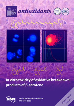
Cover Story (view full-size image): The background shows a primary pneumocyte culture. In the center, there are two pictures representing a mitosis (upper left), a micronucleus (upper right), an apoptosis (lower left) and necrotic nuclei (lower right). On the right, the COMET assay is addressed showing a nucleoid with DNA fragments distributed in the tail. Furthermore, on the top of the cover, the structural formula of beta-carotin is given; on the bottom, three structures of volatile oxidative degradation products are shown. View this paper
  • Issues are regarded as officially published after their release is announced to the table of contents alert mailing list.
  • You may sign up for e-mail alerts to receive table of contents of newly released issues.
  • PDF is the official format for papers published in both, html and pdf forms. To view the papers in pdf format, click on the "PDF Full-text" link, and use the free Adobe Reader to open them.
Order results
Result details
Section
Select all
Export citation of selected articles as:
4228 KiB  
Article
NOX2-Induced Activation of Arginase and Diabetes-Induced Retinal Endothelial Cell Senescence
by Modesto Rojas, Tahira Lemtalsi, Haroldo A. Toque, Zhimin Xu, David Fulton, Robert William Caldwell and Ruth B. Caldwell
Antioxidants 2017, 6(2), 43; https://doi.org/10.3390/antiox6020043 - 15 Jun 2017
Cited by 49 | Viewed by 7582
Abstract
Increases in reactive oxygen species (ROS) and decreases in nitric oxide (NO) have been linked to vascular dysfunction during diabetic retinopathy (DR). Diabetes can reduce NO by increasing ROS and by increasing activity of arginase, which competes with nitric oxide synthase (NOS) for [...] Read more.
Increases in reactive oxygen species (ROS) and decreases in nitric oxide (NO) have been linked to vascular dysfunction during diabetic retinopathy (DR). Diabetes can reduce NO by increasing ROS and by increasing activity of arginase, which competes with nitric oxide synthase (NOS) for their commons substrate l-arginine. Increased ROS and decreased NO can cause premature endothelial cell (EC) senescence leading to defective vascular repair. We have previously demonstrated the involvement of NADPH oxidase 2 (NOX2)-derived ROS, decreased NO and overactive arginase in DR. Here, we investigated their impact on diabetes-induced EC senescence. Studies using diabetic mice and retinal ECs treated with high glucose or H2O2 showed that increases in ROS formation, elevated arginase expression and activity, and decreased NO formation led to premature EC senescence. NOX2 blockade or arginase inhibition prevented these effects. EC senescence was also increased by inhibition of NOS activity and this was prevented by treatment with a NO donor. These results indicate that diabetes/high glucose-induced activation of arginase and decreases in NO bioavailability accelerate EC senescence. NOX2-generated ROS contribute importantly to this process. Blockade of NOX2 or arginase represents a strategy to prevent diabetes-induced premature EC senescence by preserving NO bioavailability. Full article
(This article belongs to the Special Issue ROS Derived from NADPH Oxidase (NOX) in Angiogenesis)
Show Figures

Figure 1

950 KiB  
Review
The Role of NOX4 and TRX2 in Angiogenesis and Their Potential Cross-Talk
by Chaofei Chen, Li Li, Huanjiao Jenny Zhou and Wang Min
Antioxidants 2017, 6(2), 42; https://doi.org/10.3390/antiox6020042 - 08 Jun 2017
Cited by 34 | Viewed by 10289
Abstract
The nicotinamide adenine dinucleotide phosphate (NADPH) oxidase (NOX) family is the major source of reactive oxygen species (ROS) in the vascular system. In this family, NOX4, a constitutive active form of NOXs, plays an important role in angiogenesis. Thioredoxin 2 (TRX2) is a [...] Read more.
The nicotinamide adenine dinucleotide phosphate (NADPH) oxidase (NOX) family is the major source of reactive oxygen species (ROS) in the vascular system. In this family, NOX4, a constitutive active form of NOXs, plays an important role in angiogenesis. Thioredoxin 2 (TRX2) is a key mitochondrial redox protein that maintains normal protein function and also provides electrons to peroxiredoxin 3 (PRX3) to scavenge H2O2 in mitochondria. Angiogenesis, a process of new blood vessel formation, is involved in a variety of physiological processes and pathological conditions. It seems to be paradoxical for ROS-producing NOX4 and ROS-scavenging TRX2 to have a similar role in promoting angiogenesis. In this review, we will focus on data supporting the role of NOX4 and TRX2 in angiogenesis and their cross-talks and discuss how ROS can positively or negatively regulate angiogenesis, depending on their species, levels and locations. NOX4 and TRX2-mediated ROS signaling could be promising targets for the treatment of angiogenesis-related diseases. Full article
(This article belongs to the Special Issue ROS Derived from NADPH Oxidase (NOX) in Angiogenesis)
Show Figures

Figure 1

678 KiB  
Review
Undernutrition and Overnutrition Burden for Diseases in Developing Countries: The Role of Oxidative Stress Biomarkers to Assess Disease Risk and Interventional Strategies
by Francesca Mastorci, Cristina Vassalle, Kyriazoula Chatzianagnostou, Claudio Marabotti, Khawer Siddiqui, Ahmed Ould Eba, Soueid Ahmed Sidi Mhamed, Arun Bandopadhyay, Marco Stefano Nazzaro, Mirko Passera and Alessandro Pingitore
Antioxidants 2017, 6(2), 41; https://doi.org/10.3390/antiox6020041 - 08 Jun 2017
Cited by 19 | Viewed by 6540
Abstract
The increased life expectancy, urbanization, and unhealthy lifestyle characterized by a shift towards a sedentary lifestyle and decreased energy expenditure are considered the main drivers of epidemiological transition. In particular, developing countries are facing a double burden caused by coexisting under- and over-nutrition, [...] Read more.
The increased life expectancy, urbanization, and unhealthy lifestyle characterized by a shift towards a sedentary lifestyle and decreased energy expenditure are considered the main drivers of epidemiological transition. In particular, developing countries are facing a double burden caused by coexisting under- and over-nutrition, which causes a change in the disease profile from infectious diseases to a chronic degenerative pattern. This review discusses the under- and over-nutrition context in Mauritania and India, two countries that are experiencing a nutritional transition, and where we began a collaboration with local medical staff to integrate interventional and diagnostic guidelines. If many studies about diet and its relationship to non-communicable diseases are available for India, very few nutrition and cardiovascular risk studies have been conducted in Mauritania. Presently, with the exponential increase of nutrition-related diseases, targeted approaches are needed to provide balanced diets in parallel with the development of national preventive health systems and screening programs adapted to local needs. In this context, the measurement of oxidative stress biomarkers could be promising as an additive tool to assess cardiovascular (CV) risk in general population, and ameliorating prevention in patients at CV risk or with overt CV disease. Moreover, the possibility of improving the outcome by the direct employment of antioxidant remains plausible. Moreover, studies on the content of antioxidant in different foods may be helpful to develop a balanced diet, and achieve the maximal nutritional and functional properties of cultivars with benefits for human health. Full article
(This article belongs to the Special Issue Dietary Antioxidants and Health Promotion)
Show Figures

Figure 1

1871 KiB  
Review
Roles of Nicotinamide Adenine Dinucleotide Phosphate (NADPH) Oxidase in Angiogenesis: Isoform-Specific Effects
by Haibo Wang and M. Elizabeth Hartnett
Antioxidants 2017, 6(2), 40; https://doi.org/10.3390/antiox6020040 - 03 Jun 2017
Cited by 34 | Viewed by 7819
Abstract
Angiogenesis is the formation of new blood vessels from preexisting ones and is implicated in physiologic vascular development, pathologic blood vessel growth, and vascular restoration. This is in contrast to vasculogenesis, which is de novo growth of vessels from vascular precursors, or from [...] Read more.
Angiogenesis is the formation of new blood vessels from preexisting ones and is implicated in physiologic vascular development, pathologic blood vessel growth, and vascular restoration. This is in contrast to vasculogenesis, which is de novo growth of vessels from vascular precursors, or from vascular repair that occurs when circulating endothelial progenitor cells home into an area and develop into blood vessels. The objective of this review is to discuss the isoform-specific role of nicotinamide adenine dinucleotide phosphate (NADPH) oxidase (NOX) in physiologic and pathologic angiogenesis and vascular repair, but will not specifically address vasculogenesis. As the major source of reactive oxygen species (ROS) in vascular endothelial cells (ECs), NOX has gained increasing attention in angiogenesis. Activation of NOX leads to events necessary for physiologic and pathologic angiogenesis, including EC migration, proliferation and tube formation. However, activation of different NOX isoforms has different effects in angiogenesis. Activation of NOX2 promotes pathologic angiogenesis and vascular inflammation, but may be beneficial in revascularization in the hindlimb ischemic model. In contrast, activation of NOX4 appears to promote physiologic angiogenesis mainly by protecting the vasculature during ischemia, hypoxia and inflammation and by restoring vascularization, except in models of oxygen-induced retinopathy and diabetes where NOX4 activation leads to pathologic angiogenesis. Full article
(This article belongs to the Special Issue ROS Derived from NADPH Oxidase (NOX) in Angiogenesis)
Show Figures

Figure 1

699 KiB  
Communication
Is Root Catalase a Bifunctional Catalase-Peroxidase?
by Vasileia Chioti and George Zervoudakis
Antioxidants 2017, 6(2), 39; https://doi.org/10.3390/antiox6020039 - 25 May 2017
Cited by 10 | Viewed by 8521
Abstract
Plant catalases exhibit spatial and temporal distribution of their activity. Moreover, except from the typical monofunctional catalase, a bifunctional catalase-peroxidase has been reported. The aim of this study was to investigate whether the leaf and root catalases from six different plant species ( [...] Read more.
Plant catalases exhibit spatial and temporal distribution of their activity. Moreover, except from the typical monofunctional catalase, a bifunctional catalase-peroxidase has been reported. The aim of this study was to investigate whether the leaf and root catalases from six different plant species (Lactuca sativa, Cichorium endivia, Apium graveolens, Petroselinum crispum, Lycopersicon esculentum, and Solanum melongena) correspond to the monofunctional or the bifunctional type based on their sensitivity to the inhibitor 3-amino-1,2,4-triazole (3-AT). The leaf catalases from all species seem to be monofunctional since they are very sensitive to 3-AT. On the other hand, the root enzymes from Lactuca sativa, Cichorium endivia, Lycopersicon esculentum, and Solanum melongena seem to be bifunctional catalase-peroxidases, considering that they are relatively insensitive to 3-AT, whereas the catalases from Apium graveolens and Petroselinum crispum display the same monofunctional characteristics as the leaves’ enzymes. The leaf catalase activity is usually higher (Lactuca sativa, Petroselinum crispum, and Solanum melongena) or similar (Cichorium endivia and Apium graveolens) to the root one, except for the enzyme from Lycopersicon esculentum, while in all plant species the leaf protein concentration is significantly higher than the root protein concentration. These results suggest that there are differences between leaf and root catalases—differences that may correspond to their physiological role. Full article
Show Figures

Figure 1

1630 KiB  
Review
The Reactive Sulfur Species Concept: 15 Years On
by Gregory I. Giles, Muhammad Jawad Nasim, Wesam Ali and Claus Jacob
Antioxidants 2017, 6(2), 38; https://doi.org/10.3390/antiox6020038 - 23 May 2017
Cited by 75 | Viewed by 10235
Abstract
Fifteen years ago, in 2001, the concept of “Reactive Sulfur Species” or RSS was advocated as a working hypothesis. Since then various organic as well as inorganic RSS have attracted considerable interest and stimulated many new and often unexpected avenues in research and [...] Read more.
Fifteen years ago, in 2001, the concept of “Reactive Sulfur Species” or RSS was advocated as a working hypothesis. Since then various organic as well as inorganic RSS have attracted considerable interest and stimulated many new and often unexpected avenues in research and product development. During this time, it has become apparent that molecules with sulfur-containing functional groups are not just the passive “victims” of oxidative stress or simple conveyors of signals in cells, but can also be stressors in their own right, with pivotal roles in cellular function and homeostasis. Many “exotic” sulfur-based compounds, often of natural origin, have entered the fray in the context of nutrition, ageing, chemoprevention and therapy. In parallel, the field of inorganic RSS has come to the forefront of research, with short-lived yet metabolically important intermediates, such as various sulfur-nitrogen species and polysulfides (Sx2−), playing important roles. Between 2003 and 2005 several breath-taking discoveries emerged characterising unusual sulfur redox states in biology, and since then the truly unique role of sulfur-dependent redox systems has become apparent. Following these discoveries, over the last decade a “hunt” and, more recently, mining for such modifications has begun—and still continues—often in conjunction with new, innovative and complex labelling and analytical methods to capture the (entire) sulfur “redoxome”. A key distinction for RSS is that, unlike oxygen or nitrogen, sulfur not only forms a plethora of specific reactive species, but sulfur also targets itself, as sulfur containing molecules, i.e., peptides, proteins and enzymes, preferentially react with RSS. Not surprisingly, today this sulfur-centred redox signalling and control inside the living cell is a burning issue, which has moved on from the predominantly thiol/disulfide biochemistry of the past to a complex labyrinth of interacting signalling and control pathways which involve various sulfur oxidation states, sulfur species and reactions. RSS are omnipresent and, in some instances, are even considered as the true bearers of redox control, perhaps being more important than the Reactive Oxygen Species (ROS) or Reactive Nitrogen Species (RNS) which for decades have dominated the redox field. In other(s) words, in 2017, sulfur redox is “on the rise”, and the idea of RSS resonates throughout the Life Sciences. Still, the RSS story isn’t over yet. Many RSS are at the heart of “mistaken identities” which urgently require clarification and may even provide the foundations for further scientific revolutions in the years to come. In light of these developments, it is therefore the perfect time to revisit the original hypotheses, to select highlights in the field and to question and eventually update our concept of “Reactive Sulfur Species”. Full article
Show Figures

Figure 1

1975 KiB  
Article
Effects of β-Carotene and Its Cleavage Products in Primary Pneumocyte Type II Cells
by Cornelia Haider, Franziska Ferk, Ekramije Bojaxhi, Giuseppe Martano, Hanno Stutz, Nikolaus Bresgen, Siegfried Knasmüller, Avdulla Alija and Peter M. Eckl
Antioxidants 2017, 6(2), 37; https://doi.org/10.3390/antiox6020037 - 21 May 2017
Cited by 15 | Viewed by 6165
Abstract
β-Carotene has been shown to increase the risk of developing lung cancer in smokers and asbestos workers in two large scale trails, the Beta-Carotene and Retinol Efficacy Trial (CARET) and the Alpha-Tocopherol Beta-carotene Cancer Prevention Trial (ATBC). Based on this observation, it was [...] Read more.
β-Carotene has been shown to increase the risk of developing lung cancer in smokers and asbestos workers in two large scale trails, the Beta-Carotene and Retinol Efficacy Trial (CARET) and the Alpha-Tocopherol Beta-carotene Cancer Prevention Trial (ATBC). Based on this observation, it was proposed that genotoxic oxidative breakdown products may cause this effect. In support of this assumption, increased levels of sister chromatid exchanges, micronuclei, and chromosomal aberrations were found in primary hepatocyte cultures treated with a mixture of cleavage products (CPs) and the major product apo-8′carotenal. However, because these findings cannot directly be transferred to the lung due to the exceptional biotransformation capacity of the liver, potential genotoxic and cytotoxic effects of β-carotene under oxidative stress and its CPs were investigated in primary pneumocyte type II cells. The results indicate that increased concentrations of β-carotene in the presence of the redox cycling quinone dimethoxynaphthoquinone (DMNQ) exhibit a cytotoxic potential, as evidenced by an increase of apoptotic cells and loss of cell density at concentrations > 10 µM. On the other hand, the analysis of micronucleated cells gave no clear picture due to the cytotoxicity related reduction of mitotic cells. Last, although CPs induced significant levels of DNA strand breaks even at concentrations ≥ 1 µM and 5 µM, respectively, β-carotene in the presence of DMNQ did not cause DNA damage. Instead, β-carotene appeared to act as an antioxidant. These findings are in contrast with what was demonstrated for primary hepatocytes and may reflect different sensitivities to and different metabolism of β-carotene in the two cell types. Full article
(This article belongs to the Special Issue Carotenoids—Antioxidant Properties)
Show Figures

Figure 1

1708 KiB  
Article
Hormetic Property of Ginseng Steroids on Anti-Oxidant Status against Exercise Challenge in Rat Skeletal Muscle
by Ming-Fen Hsu, Szu-Hsien Yu, Mallikarjuna Korivi, Wei-Horng Jean, Shin-Da Lee, Chih-Yang Huang, Yi-Hung Liao, Jessica Lu and Chia-Hua Kuo
Antioxidants 2017, 6(2), 36; https://doi.org/10.3390/antiox6020036 - 19 May 2017
Cited by 10 | Viewed by 5098
Abstract
Background: Existing literature on anti-oxidant capacity of ginseng has been inconsistent due to variance in the profile of ginseng steroids (Ginsenosides) that is because of differences in seasons and species. Methods: We used various doses of ginseng steroids to determine its effect on [...] Read more.
Background: Existing literature on anti-oxidant capacity of ginseng has been inconsistent due to variance in the profile of ginseng steroids (Ginsenosides) that is because of differences in seasons and species. Methods: We used various doses of ginseng steroids to determine its effect on oxidative stress and anti-oxidant capacity of rat skeletal muscle against exercise. Results: Under non-exercise conditions, we found increased thiobarbituric acid reactive substance (TBARS) levels and decreased reduced/oxidized glutathione ratio (GSH/GSSG) in rat skeletal muscle as dose increases (p < 0.05), which indicates the pro-oxidant property of ginseng steroids at baseline. Intriguingly, exhaustive exercise-induced increased TBARS and decreased GSH/GSSG ratio were attenuated with low and medium doses of ginseng steroids (20 and 40 mg per kg), but not with high dose (120 mg per kg). At rest, anti-oxidant enzyme activities, including catalase (CAT), glutathione reductase (GR) and glutathione S-transferase (GST) were increased above vehicle-treated level, but not with the high dose, suggesting a hormetic dose-response of ginseng steroids. Conclusion: The results of this study provide an explanation for the inconsistent findings on anti-oxidative property among previous ginseng studies. For optimizing the anti-oxidant outcome, ginseng supplementation at high dose should be avoided. Full article
(This article belongs to the Special Issue Exercise Induced Muscle Damage and Oxidative Stress)
Show Figures

Figure 1

6561 KiB  
Article
Green Synthesized Zinc Oxide (ZnO) Nanoparticles Induce Oxidative Stress and DNA Damage in Lathyrus sativus L. Root Bioassay System
by Kamal K. Panda, Dambaru Golari, A. Venugopal, V. Mohan M. Achary, Ganngam Phaomei, Narasimham L. Parinandi, Hrushi K. Sahu and Brahma B. Panda
Antioxidants 2017, 6(2), 35; https://doi.org/10.3390/antiox6020035 - 18 May 2017
Cited by 45 | Viewed by 10088
Abstract
Zinc oxide nanoparticles (ZnONP-GS) were synthesised from the precursor zinc acetate (Zn(CH3COO)2) through the green route using the milky latex from milk weed (Calotropis gigantea L. R. Br) by alkaline precipitation. Formation of the ZnONP-GS was monitored by [...] Read more.
Zinc oxide nanoparticles (ZnONP-GS) were synthesised from the precursor zinc acetate (Zn(CH3COO)2) through the green route using the milky latex from milk weed (Calotropis gigantea L. R. Br) by alkaline precipitation. Formation of the ZnONP-GS was monitored by UV-visible spectroscopy followed by characterization and confirmation by energy-dispersive X-ray spectroscopy (EDX), transmission electron microscopy (TEM), and X-ray diffraction (XRD). Both the ZnONP-GS and the commercially available ZnONP-S (Sigma-Aldrich) and cationic Zn2+ from Zn(CH3COO)2 were tested in a dose range of 0–100 mg·L−1 for their potency (i) to induce oxidative stress as measured by the generation reactive oxygen species (ROS: O2•−, H2O2 and OH), cell death, and lipid peroxidation; (ii) to modulate the activities of antioxidant enzymes: catalase (CAT), superoxide dismutase (SOD), guaiacol peroxidase (GPX), and ascorbate peroxidase (APX); and (iii) to cause DNA damage as determined by Comet assay in Lathyrus sativus L. root bioassay system. Antioxidants such as Tiron and dimethylthiourea significantly attenuated the ZnONP-induced oxidative and DNA damage, suggesting the involvement of ROS therein. Our study demonstrated that both ZnONP-GS and ZnONP-S induced oxidative stress and DNA damage to a similar extent but were significantly less potent than Zn2+ alone. Full article
(This article belongs to the Special Issue Nanomaterial Oxidative Stress)
Show Figures

Figure 1

1098 KiB  
Article
A Study of the Protective Properties of Iraqi Olive Leaves against Oxidation and Pathogenic Bacteria in Food Applications
by Ammar B. Altemimi
Antioxidants 2017, 6(2), 34; https://doi.org/10.3390/antiox6020034 - 17 May 2017
Cited by 7 | Viewed by 6241
Abstract
There is an ancient and prodigious history of olive trees because of their nutritional, medicinal, and traditional uses. Intensive studies have been conducted on olive leaves because they have many positive and beneficial effects for human health. In this study, different solvents were [...] Read more.
There is an ancient and prodigious history of olive trees because of their nutritional, medicinal, and traditional uses. Intensive studies have been conducted on olive leaves because they have many positive and beneficial effects for human health. In this study, different solvents were used to examine the olive leaves for their antioxidant and antimicrobial activities and their possible food applications. The obtained results showed that the amounts of phenolic compounds of the olive leaf were 190.44 ± 0.50, 173 ± 1.72, 147.78 ± 0.69, and 147.50 ± 0.05 mg gallic acid/g extracts using methanol, ethanol, diethyl ether, and hexanol, respectively. The statistical analysis revealed that there was a significant difference in the phenolic contents in terms of the used solvents. The stability of the olive leaves extraction was also studied and the results indicated that increasing the storage temperature could negatively affect and encourage the degradation of the phenolic compounds. Furthermore, the olive leaf extraction was applied to raw sheep meat slides at 0.5%, 1.5%, and 2.5% (w/v) in order to test its antioxidant and antimicrobial effects. The results obviously showed that the sample treated with 2.5% olive leaf extract had the significantly (p < 0.05) lowest Thiobarbituric Acid (TBA) values of 1.92 ± 0.12 (mg Malonaldehyde MDA/kg) throughout 12 days of cold storage. Moreover, the results showed that the sample, which was treated with 2.5% olive leaf extract, had low total bacterial count and total coliform bacteria (6.23 ± 0.05, 5.2 ± 0.35 log colony forming unit (CFU)/g, respectively) among the control, 0.5%, and 1.5% olive leaf treated samples throughout 12 days of storage. The phenolic extracts from the olive leaf extract had significant antioxidant and antimicrobial activities, which could be used as a source of potential antioxidant and antimicrobial agents. Full article
(This article belongs to the Special Issue Bioactive Phenolic Compounds)
Show Figures

Figure 1

924 KiB  
Review
The Interplay between Oncogenic Signaling Networks and Mitochondrial Dynamics
by Sarbajeet Nagdas and David F. Kashatus
Antioxidants 2017, 6(2), 33; https://doi.org/10.3390/antiox6020033 - 17 May 2017
Cited by 33 | Viewed by 7363
Abstract
Mitochondria are dynamic organelles that alter their organization in response to a variety of cellular cues. Mitochondria are central in many biologic processes, such as cellular bioenergetics and apoptosis, and mitochondrial network morphology can contribute to those physiologic processes. Some of the biologic [...] Read more.
Mitochondria are dynamic organelles that alter their organization in response to a variety of cellular cues. Mitochondria are central in many biologic processes, such as cellular bioenergetics and apoptosis, and mitochondrial network morphology can contribute to those physiologic processes. Some of the biologic processes that are in part governed by mitochondria are also commonly deregulated in cancers. Furthermore, patient tumor samples from a variety of cancers have revealed that mitochondrial dynamics machinery may be deregulated in tumors. In this review, we will discuss how commonly mutated oncogenes and their downstream effector pathways regulate the mitochondrial dynamics machinery to promote changes in mitochondrial morphology as well as the physiologic consequences of altered mitochondrial morphology for tumorigenic growth. Full article
(This article belongs to the Special Issue Mitochondrial Shape Change in Physio-Pathology)
Show Figures

Figure 1

853 KiB  
Review
The Importance of NADPH Oxidases and Redox Signaling in Angiogenesis
by Rodrigo Prieto-Bermejo and Angel Hernández-Hernández
Antioxidants 2017, 6(2), 32; https://doi.org/10.3390/antiox6020032 - 13 May 2017
Cited by 49 | Viewed by 9783
Abstract
Eukaryotic cells have to cope with the constant generation of reactive oxygen species (ROS). Although the excessive production of ROS might be deleterious for cell biology, there is a plethora of evidence showing that moderate levels of ROS are important for the control [...] Read more.
Eukaryotic cells have to cope with the constant generation of reactive oxygen species (ROS). Although the excessive production of ROS might be deleterious for cell biology, there is a plethora of evidence showing that moderate levels of ROS are important for the control of cell signaling and gene expression. The family of the nicotinamide adenine dinucleotide phosphate oxidases (NADPH oxidases or Nox) has evolved to produce ROS in response to different signals; therefore, they fulfil a central role in the control of redox signaling. The role of NADPH oxidases in vascular physiology has been a field of intense study over the last two decades. In this review we will briefly analyze how ROS can regulate signaling and gene expression. We will address the implication of NADPH oxidases and redox signaling in angiogenesis, and finally, the therapeutic possibilities derived from this knowledge will be discussed. Full article
(This article belongs to the Special Issue ROS Derived from NADPH Oxidase (NOX) in Angiogenesis)
Show Figures

Figure 1

1142 KiB  
Article
Phenolic Compositions and Antioxidant Properties in Bark, Flower, Inner Skin, Kernel and Leaf Extracts of Castanea crenata Sieb. et Zucc
by Phung Thi Tuyen, Tran Dang Xuan, Do Tan Khang, Ateeque Ahmad, Nguyen Van Quan, Truong Thi Tu Anh, La Hoang Anh and Truong Ngoc Minh
Antioxidants 2017, 6(2), 31; https://doi.org/10.3390/antiox6020031 - 05 May 2017
Cited by 57 | Viewed by 7470
Abstract
In this study, different plant parts (barks, flowers, inner skins, kernels and leaves) of Castanea crenata (Japanese chestnut) were analyzed for total phenolic, flavonoid, and tannin contents. Antioxidant properties were evaluated by using 1,1-diphenyl-2-picrylhydrazyl (DPPH), 2,2′-azinobis (3-ethylbenzothiazoline-6-sulfonic acid) diammonium salt (ABTS), reducing power, [...] Read more.
In this study, different plant parts (barks, flowers, inner skins, kernels and leaves) of Castanea crenata (Japanese chestnut) were analyzed for total phenolic, flavonoid, and tannin contents. Antioxidant properties were evaluated by using 1,1-diphenyl-2-picrylhydrazyl (DPPH), 2,2′-azinobis (3-ethylbenzothiazoline-6-sulfonic acid) diammonium salt (ABTS), reducing power, and β-carotene bleaching methods. The highest total phenolic and tannin contents were found in the inner skins (1034 ± 7.21 mg gallic acid equivalent/g extract and 253.89 ± 5.59 mg catechin equivalent/g extract, respectively). The maximum total flavonoid content was observed in the flowers (147.41 ± 1.61 mg rutin equivalent/g extract). The inner skins showed the strongest antioxidant activities in all evaluated assays. Thirteen phenolic acids and eight flavonoids were detected and quantified for the first time. Major phenolic acids were gallic, ellagic, sinapic, and p-coumaric acids, while the principal flavonoids were myricetin and isoquercitrin. The inner skin extract was further fractionated by column chromatography to yield four fractions, of which fraction F3 exhibited the most remarkable DPPH scavenging capacity. These results suggest that C. crenata provides promising antioxidant capacities, and is a potential natural preservative agent in food and pharmaceutical industries. Full article
(This article belongs to the Special Issue Bioactive Phenolic Compounds)
Show Figures

Figure 1

853 KiB  
Review
HO-1 Induction in Cancer Progression: A Matter of Cell Adaptation
by Mariapaola Nitti, Sabrina Piras, Umberto M. Marinari, Lorenzo Moretta, Maria A. Pronzato and Anna Lisa Furfaro
Antioxidants 2017, 6(2), 29; https://doi.org/10.3390/antiox6020029 - 05 May 2017
Cited by 157 | Viewed by 14838
Abstract
The upregulation of heme oxygenase-1 (HO-1) is one of the most important mechanisms of cell adaptation to stress. Indeed, the redox sensitive transcription factor Nrf2 is the pivotal regulator of HO-1 induction. Through the antioxidant, antiapoptotic, and antinflammatory properties of its metabolic products, [...] Read more.
The upregulation of heme oxygenase-1 (HO-1) is one of the most important mechanisms of cell adaptation to stress. Indeed, the redox sensitive transcription factor Nrf2 is the pivotal regulator of HO-1 induction. Through the antioxidant, antiapoptotic, and antinflammatory properties of its metabolic products, HO-1 plays a key role in healthy cells in maintaining redox homeostasis and in preventing carcinogenesis. Nevertheless, several lines of evidence have highlighted the role of HO-1 in cancer progression and its expression correlates with tumor growth, aggressiveness, metastatic and angiogenetic potential, resistance to therapy, tumor escape, and poor prognosis, even though a tumor- and tissue-specific activity has been observed. In this review, we summarize the current literature regarding the pro-tumorigenic role of HO-1 dependent tumor progression as a promising target in anticancer strategy. Full article
(This article belongs to the Special Issue Oxidative Stress and Cancer: The Nrf2 Enigma)
Show Figures

Figure 1

2032 KiB  
Review
Natural Phenol Polymers: Recent Advances in Food and Health Applications
by Lucia Panzella and Alessandra Napolitano
Antioxidants 2017, 6(2), 30; https://doi.org/10.3390/antiox6020030 - 14 Apr 2017
Cited by 78 | Viewed by 13366
Abstract
Natural phenol polymers are widely represented in nature and include a variety of classes including tannins and lignins as the most prominent. Largely consumed foods are rich sources of phenol polymers, notably black foods traditionally used in East Asia, but other non-edible, easily [...] Read more.
Natural phenol polymers are widely represented in nature and include a variety of classes including tannins and lignins as the most prominent. Largely consumed foods are rich sources of phenol polymers, notably black foods traditionally used in East Asia, but other non-edible, easily accessible sources, e.g., seaweeds and wood, have been considered with increasing interest together with waste materials from agro-based industries, primarily grape pomace and other byproducts of fruit and coffee processing. Not in all cases were the main structural components of these materials identified because of their highly heterogeneous nature. The great beneficial effects of natural phenol-based polymers on human health and their potential in improving the quality of food were largely explored, and this review critically addresses the most interesting and innovative reports in the field of nutrition and biomedicine that have appeared in the last five years. Several in vivo human and animal trials supported the proposed use of these materials as food supplements and for amelioration of the health and production of livestock. Biocompatible and stable functional polymers prepared by peroxidase-catalyzed polymerization of natural phenols, as well as natural phenol polymers were exploited as conventional and green plastic additives in smart packaging and food-spoilage prevention applications. The potential of natural phenol polymers in regenerative biomedicine as additives of biomaterials to promote growth and differentiation of osteoblasts is also discussed. Full article
Show Figures

Figure 1

1758 KiB  
Article
Acute Pre-/Post-Treatment with 8th Day SOD-Like Supreme (a Free Radical Scavenging Health Product) Protects against Oxidant-Induced Injury in Cultured Cardiomyocytes and Hepatocytes In Vitro as Well as in Mouse Myocardium and Liver In Vivo
by Pou Kuan Leong, Jihang Chen, Wing Man Chan, Hoi Yan Leung, Lincoln Chan and Kam Ming Ko
Antioxidants 2017, 6(2), 28; https://doi.org/10.3390/antiox6020028 - 10 Apr 2017
Cited by 9 | Viewed by 5253
Abstract
8th Day superoxide dismutase (SOD)-Like Supreme (SOD-Like Supreme, a free radical scavenging health product) is an antioxidant-enriched fermentation preparation with free radical scavenging properties. In the present study, the cellular/tissue protective actions of SOD-Like Supreme against menadione toxicity in cultured H9c2 cardiomyocytes and [...] Read more.
8th Day superoxide dismutase (SOD)-Like Supreme (SOD-Like Supreme, a free radical scavenging health product) is an antioxidant-enriched fermentation preparation with free radical scavenging properties. In the present study, the cellular/tissue protective actions of SOD-Like Supreme against menadione toxicity in cultured H9c2 cardiomyocytes and in AML12 hepatocytes as well as oxidant-induced injury in the mouse myocardium and liver were investigated. SOD-Like Supreme was found to possess potent free radical scavenging activity in vitro as assessed by an oxygen radical absorbance capacity assay. Incubation with SOD-Like Supreme (0.5–3% (v/v)) was shown to protect against menadione-induced toxicity in H9c2 and AML12 cells, as evidenced by increases in cell viability. The ability of SOD-Like Supreme to protect against menadione cytotoxicity was associated with an elevation in the cellular reduced glutathione (GSH)/oxidized glutathione (GSSG) ratio in menadione-challenged cells. Consistent with the cell-based studies, pre-/post-treatment with SOD-Like Supreme (0.69 and 2.06 mL/kg, three intermittent doses per day for two consecutive days) was found to protect against isoproterenol-induced myocardial injury and carbon tetrachloride hepatotoxicity in mice. The cardio/hepatoprotection afforded by SOD-Like Supreme was also paralleled by increases in myocardial/hepatic mitochondrial GSH/GSSG ratios in the SOD-Like Supreme-treated/oxidant-challenged mice. In conclusion, incubation/treatment with SOD-Like Supreme was found to protect against oxidant-induced injury in vitro and in vivo, presumably by virtue of its free radical scavenging activity. Full article
(This article belongs to the Special Issue Superoxide Dismutase (SOD) Enzymes, Mimetics and Oxygen Radicals)
Show Figures

Figure 1

530 KiB  
Review
Strange Bedfellows: Nuclear Factor, Erythroid 2-Like 2 (Nrf2) and Hypoxia-Inducible Factor 1 (HIF-1) in Tumor Hypoxia
by Rachel K. Toth and Noel A. Warfel
Antioxidants 2017, 6(2), 27; https://doi.org/10.3390/antiox6020027 - 06 Apr 2017
Cited by 84 | Viewed by 14305
Abstract
The importance of the tumor microenvironment for cancer progression and therapeutic resistance is an emerging focus of cancer biology. Hypoxia, or low oxygen, is a hallmark of solid tumors that promotes metastasis and represents a significant obstacle to successful cancer therapy. In response [...] Read more.
The importance of the tumor microenvironment for cancer progression and therapeutic resistance is an emerging focus of cancer biology. Hypoxia, or low oxygen, is a hallmark of solid tumors that promotes metastasis and represents a significant obstacle to successful cancer therapy. In response to hypoxia, cancer cells activate a transcriptional program that allows them to survive and thrive in this harsh microenvironment. Hypoxia-inducible factor 1 (HIF-1) is considered the main effector of the cellular response to hypoxia, stimulating the transcription of genes involved in promoting angiogenesis and altering cellular metabolism. However, growing evidence suggests that the cellular response to hypoxia is much more complex, involving coordinated signaling through stress response pathways. One key signaling molecule that is activated in response to hypoxia is nuclear factor, erythroid 2 like-2 (Nrf2). Nrf2 is a transcription factor that controls the expression of antioxidant-response genes, allowing the cell to regulate reactive oxygen species. Nrf2 is also activated in various cancer types due to genetic and epigenetic alterations, and is associated with poor survival and resistance to therapy. Emerging evidence suggests that coordinated signaling through Nrf2 and HIF-1 is critical for tumor survival and progression. In this review, we discuss the distinct and overlapping roles of HIF-1 and Nrf2 in the cellular response to hypoxia, with a focus on how targeting Nrf2 could provide novel chemotherapeutic modalities for treating solid tumors. Full article
(This article belongs to the Special Issue Oxidative Stress and Cancer: The Nrf2 Enigma)
Show Figures

Figure 1

681 KiB  
Review
Green Tea Catechins for Prostate Cancer Prevention: Present Achievements and Future Challenges
by Valeria Naponelli, Ileana Ramazzina, Chiara Lenzi, Saverio Bettuzzi and Federica Rizzi
Antioxidants 2017, 6(2), 26; https://doi.org/10.3390/antiox6020026 - 05 Apr 2017
Cited by 35 | Viewed by 9069
Abstract
Green tea catechins (GTCs) are a family of chemically related compounds usually classified as antioxidant molecules. Epidemiological evidences, supported by interventional studies, highlighted a more than promising role for GTCs in human prostate cancer (PCa) chemoprevention. In the last decades, many efforts have [...] Read more.
Green tea catechins (GTCs) are a family of chemically related compounds usually classified as antioxidant molecules. Epidemiological evidences, supported by interventional studies, highlighted a more than promising role for GTCs in human prostate cancer (PCa) chemoprevention. In the last decades, many efforts have been made to gain new insights into the mechanism of action of GTCs. Now it is clear that GTCs’ anticancer action can no longer be simplistically limited to their direct antioxidant/pro-oxidant properties. Recent contributions to the advancement of knowledge in this field have shown that GTCs specifically interact with cellular targets, including cell surface receptors, lipid rafts, and endoplasmic reticulum, modulate gene expression through direct effect on transcription factors or indirect epigenetic mechanisms, and interfere with intracellular proteostasis at various levels. Many of the effects observed in vitro are dose and cell context dependent and take place at concentrations that cannot be achieved in vivo. Poor intestinal absorption together with an extensive systemic and enteric metabolism influence GTCs’ bioavailability through still poorly understood mechanisms. Recent efforts to develop delivery systems that increase GTCs’ overall bioavailability, by means of biopolymeric nanoparticles, represent the main way to translate preclinical results in a real clinical scenario for PCa chemoprevention. Full article
(This article belongs to the Special Issue Plant Polyphenols in Cancer Chemoprevention)
Show Figures

Figure 1

811 KiB  
Review
Abnormalities of Mitochondrial Dynamics in Neurodegenerative Diseases
by Ju Gao, Luwen Wang, Jingyi Liu, Fei Xie, Bo Su and Xinglong Wang
Antioxidants 2017, 6(2), 25; https://doi.org/10.3390/antiox6020025 - 05 Apr 2017
Cited by 177 | Viewed by 15226
Abstract
Neurodegenerative diseases are incurable and devastating neurological disorders characterized by the progressive loss of the structure and function of neurons in the central nervous system or peripheral nervous system. Mitochondria, organelles found in most eukaryotic cells, are essential for neuronal survival and are [...] Read more.
Neurodegenerative diseases are incurable and devastating neurological disorders characterized by the progressive loss of the structure and function of neurons in the central nervous system or peripheral nervous system. Mitochondria, organelles found in most eukaryotic cells, are essential for neuronal survival and are involved in a number of neuronal functions. Mitochondrial dysfunction has long been demonstrated as a common prominent early pathological feature of a variety of common neurodegenerative diseases, including Alzheimer’s disease (AD), Parkinson’s disease (PD), amyotrophic lateral sclerosis (ALS), and Huntington’s disease (HD). Mitochondria are highly dynamic organelles that undergo continuous fusion, fission, and transport, the processes of which not only control mitochondrial morphology and number but also regulate mitochondrial function and location. The importance of mitochondrial dynamics in the pathogenesis of neurodegenerative diseases has been increasingly unraveled after the identification of several key fusion and fission regulators such as Drp1, OPA1, and mitofusins. In this review, after a brief discussion of molecular mechanisms regulating mitochondrial fusion, fission, distribution, and trafficking, as well as the important role of mitochondrial dynamics for neuronal function, we review previous and the most recent studies about mitochondrial dynamic abnormalities observed in various major neurodegenerative diseases and discuss the possibility of targeting mitochondrial dynamics as a likely novel therapeutic strategy for neurodegenerative diseases. Full article
(This article belongs to the Special Issue Mitochondrial Shape Change in Physio-Pathology)
Show Figures

Figure 1

509 KiB  
Review
Zinc and Oxidative Stress: Current Mechanisms
by Dilina Do Nascimento Marreiro, Kyria Jayanne Clímaco Cruz, Jennifer Beatriz Silva Morais, Jéssica Batista Beserra, Juliana Soares Severo and Ana Raquel Soares De Oliveira
Antioxidants 2017, 6(2), 24; https://doi.org/10.3390/antiox6020024 - 29 Mar 2017
Cited by 345 | Viewed by 30166
Abstract
Oxidative stress is a metabolic dysfunction that favors the oxidation of biomolecules, contributing to the oxidative damage of cells and tissues. This consequently contributes to the development of several chronic diseases. In particular, zinc is one of the most relevant minerals to human [...] Read more.
Oxidative stress is a metabolic dysfunction that favors the oxidation of biomolecules, contributing to the oxidative damage of cells and tissues. This consequently contributes to the development of several chronic diseases. In particular, zinc is one of the most relevant minerals to human health, because of its antioxidant properties. This review aims to provide updated information about the mechanisms involved in the protective role of zinc against oxidative stress. Zinc acts as a co-factor for important enzymes involved in the proper functioning of the antioxidant defense system. In addition, zinc protects cells against oxidative damage, acts in the stabilization of membranes and inhibits the enzyme nicotinamide adenine dinucleotide phosphate oxidase (NADPH-Oxidase). Zinc also induces the synthesis of metallothioneins, which are proteins effective in reducing hydroxyl radicals and sequestering reactive oxygen species (ROS) produced in stressful situations, such as in type 2 diabetes, obesity and cancer. Literature provides strong evidence for the role of zinc in the protection against oxidative stress in several diseases. Full article
(This article belongs to the Special Issue Oxidative Stress and Metals Metabolism)
Show Figures

Figure 1

2507 KiB  
Article
Antioxidant Properties of Selenophene, Thiophene and Their Aminocarbonitrile Derivatives
by Levon A. Tavadyan, Zaruhi H. Manukyan, Lusik H. Harutyunyan, Makich V. Musayelyan, Adrine D. Sahakyan and Hakob G. Tonikyan
Antioxidants 2017, 6(2), 22; https://doi.org/10.3390/antiox6020022 - 24 Mar 2017
Cited by 32 | Viewed by 5973
Abstract
The oxygen radical absorbance capacity (ORAC) method was used to detect the antiperoxyradical ability of organoselenium compounds: selenophene and its derivative, 2-amino-4,5,6,7-tetrahydro-1-selenophene-3-carbonitrile (ATSe); while as a comparison, the sulfur-containing analogue of selenophene—thiophene and its derivative—2-amino-4,5,6,7-tetrahydro-1-thiophene-3-carbonitrile (ATS)—was selected. Cyclic voltammetry (CV), differential pulse voltammetry [...] Read more.
The oxygen radical absorbance capacity (ORAC) method was used to detect the antiperoxyradical ability of organoselenium compounds: selenophene and its derivative, 2-amino-4,5,6,7-tetrahydro-1-selenophene-3-carbonitrile (ATSe); while as a comparison, the sulfur-containing analogue of selenophene—thiophene and its derivative—2-amino-4,5,6,7-tetrahydro-1-thiophene-3-carbonitrile (ATS)—was selected. Cyclic voltammetry (CV), differential pulse voltammetry (DPV) and squarewave voltammetry (SWV) methods were used to determine the redox characteristics of organoselenium and organosulfur compounds. The antiradical activity and capacity of the studied compounds were also measured by using stable radical 2,2ʹ-diphenyl-1-picrylhydrazyl (DPPH). Detected anodic peaks of the oxidation of selenophene, thiophene and their derivatives in CV, DPV and SWV in the interval of −1200 ÷ (+1600) mV potentials in regard to the Ag/Ag+ medium of acetonitrile prove the presence of antiperoxyradical activity in regard to oxidizers, i.e., peroxyradicals. The chemical mechanism of the antiperoxyradical ability of selenophene, thiophene and their organic derivatives is proposed. Full article
(This article belongs to the Special Issue Selenium and Selenoproteins for Optimal Health)
Show Figures

Figure 1

703 KiB  
Review
Effects of Reactive Oxygen Species on Tubular Transport along the Nephron
by Agustin Gonzalez-Vicente and Jeffrey L. Garvin
Antioxidants 2017, 6(2), 23; https://doi.org/10.3390/antiox6020023 - 23 Mar 2017
Cited by 26 | Viewed by 7546
Abstract
Reactive oxygen species (ROS) are oxygen-containing molecules naturally occurring in both inorganic and biological chemical systems. Due to their high reactivity and potentially damaging effects to biomolecules, cells express a battery of enzymes to rapidly metabolize them to innocuous intermediaries. Initially, ROS were [...] Read more.
Reactive oxygen species (ROS) are oxygen-containing molecules naturally occurring in both inorganic and biological chemical systems. Due to their high reactivity and potentially damaging effects to biomolecules, cells express a battery of enzymes to rapidly metabolize them to innocuous intermediaries. Initially, ROS were considered by biologists as dangerous byproducts of respiration capable of causing oxidative stress, a condition in which overproduction of ROS leads to a reduction in protective molecules and enzymes and consequent damage to lipids, proteins, and DNA. In fact, ROS are used by immune systems to kill virus and bacteria, causing inflammation and local tissue damage. Today, we know that the functions of ROS are not so limited, and that they also act as signaling molecules mediating processes as diverse as gene expression, mechanosensation, and epithelial transport. In the kidney, ROS such as nitric oxide (NO), superoxide (O2), and their derivative molecules hydrogen peroxide (H2O2) and peroxynitrite (ONO2) regulate solute and water reabsorption, which is vital to maintain electrolyte homeostasis and extracellular fluid volume. This article reviews the effects of NO, O2, ONO2, and H2O2 on water and electrolyte reabsorption in proximal tubules, thick ascending limbs, and collecting ducts, and the effects of NO and O2 in the macula densa on tubuloglomerular feedback. Full article
(This article belongs to the Special Issue Oxidative Stress and Salt-Sensitive Hypertension)
Show Figures

Figure 1

Previous Issue
Next Issue
Back to TopTop