Article

CoQ10Phytosomes Improve Cellular Ubiquinone Uptake in Skeletal Muscle Cells: An Ex Vivo Study Using CoQ10-Enriched Low-Density Lipoproteins Obtained in a Randomized Crossover Study

1
Department of Life and Environmental Sciences, Polytechnic University of Marche, Via Brecce Bianche, 60131 Ancona, Italy
2
Indena SpA, Viale Ortles, 20139 Milan, Italy
*
Author to whom correspondence should be addressed.
Antioxidants 2023, 12(4), 964; https://doi.org/10.3390/antiox12040964
Submission received: 17 March 2023 / Revised: 13 April 2023 / Accepted: 17 April 2023 / Published: 20 April 2023
(This article belongs to the Special Issue The Ubiquitous and Multifaceted Coenzyme Q)

Abstract

:
Coenzyme Q10 (CoQ10) bioavailability in vivo is limited due to its lipophilic nature. Moreover, a large body of evidence in the literature shows that muscle CoQ10 uptake is limited. In order to address cell specific differences in CoQ uptake, we compared cellular CoQ10 content in cultured human dermal fibroblasts and murine skeletal muscle cells that were incubated with lipoproteins from healthy volunteers and enriched with different formulations of CoQ10 following oral supplementation. Using a crossover design, eight volunteers were randomized to supplement 100 mg/daily CoQ10 for two weeks, delivered both in phytosome form (UBQ) as a lecithin formulation and in CoQ10 crystalline form. After supplementation, plasma was collected for CoQ10 determination. In the same samples, low density lipoproteins (LDL) were extracted and normalized for CoQ10 content, and 0.5 µg/mL in the medium were incubated with the two cell lines for 24 h. The results show that while both formulations were substantially equivalent in terms of plasma bioavailability in vivo, UBQ-enriched lipoproteins showed a higher bioavailability compared with crystalline CoQ10-enriched ones both in human dermal fibroblasts (+103%) and in murine skeletal myoblasts (+48%). Our data suggest that phytosome carriers might provide a specific advantage in delivering CoQ10 to skin and muscle tissues.

1. Introduction

Ubiquinone or Coenzyme Q10 (CoQ10) is a ubiquitous, endogenous lipophilic cofactor that is well known for its bioenergetic and antioxidant functions within cells. In particular, CoQ10 as a component of the mitochondrial electron transport chainacts as an electron shuttle between complex I, II, and complex III [1,2,3,4]. Within the mitochondria, CoQ10 has been shown to modulate the permeability transition pore and activity of uncoupling protein [5,6,7]. Moreover, CoQ10 in its reduced form ubiquinolis endowed with antioxidant properties in all biological membranes and acts synergistically with vitamin E and vitamin C to protect against lipid peroxidation [2].
Mitochondria-rich skeletal and cardiac muscle tissues have the highest content in CoQ10 compared with other tissues, and its levels are shown to be directly related to muscle functionality. Moreover, CoQ10 content is decreased in these tissues in senescence as well as under pathological conditions which has important prognostic implications [8]. For these reasons, numerous CoQ10 interventions have been designed to contrast cardiovascular pathologies in relation to both its bioenergetic function as well as antioxidant and anti-inflammatory activities [9].
In the skeletal muscle, several CoQ interventions investigated its role as a dietary supplement in sport nutrition as an antioxidant to contrast physical exercise-induced oxidative stress or as a bioenergetic support [10,11].
In fact, although a healthy organism produces sufficient amounts of CoQ10 for its normal function, secondary CoQ deficits are not uncommon and are related to increased consumption duringintense physical exercise [12], pathologically impaired redox status, and lowered biosynthesis that is associated with senescence [13,14,15]. Dietary intake accounts for approximately −5 mg/day [16]. CoQ-rich foods include meat, fish, and nuts, and the highest concentration of CoQ is found in heart tissue [16]; however, that is not frequently consumed in the human diet.
Moreover, CoQ in general shows limited gastrointestinal absorption and bioavailability due to its chemical–physical properties [17]. Dietary CoQ10 distribution shows tissue specific differences in uptakewith plasma showing the most increases and muscle tissue showing the least [18,19]. This represents a unique feature of CoQ10 that behaves differently from similar lipophilic molecules such as tocopherol that seem to be less tissue specific [20].
Accordingly, understanding the molecular mechanisms regulating CoQ uptake in the muscle represents a priority for the realization of innovative formulations that could promote enhanced bioavailability in these refractory tissues. A recent study by Drobnic et al. [21] did show that CoQ10 formulated in phytosomes as Ubiqsome® (standardized in the coenzyme Q1018–22% by HPLC), led to a significant increase in quinone in skeletal muscle tissue in vivo.
The present pilot study exploits anex vivodesign to verify the bioavailability of CoQ10 phytosome compared with crystalline CoQ10 in different cellular models characterized by a different ability to absorb exogenous CoQ10—namely, dermal fibroblasts and skeletal myoblasts. In order to mimic thein vitroinvestigation within vivoprocessesas much as possible, CoQ10 was given in the form of enriched lipoproteins that were normalized in their CoQ10 content following HPLC determination and appropriately diluted in cell culture medium to 0.5 µg/mL, as shown in Figure 1.

2. Materials and Methods

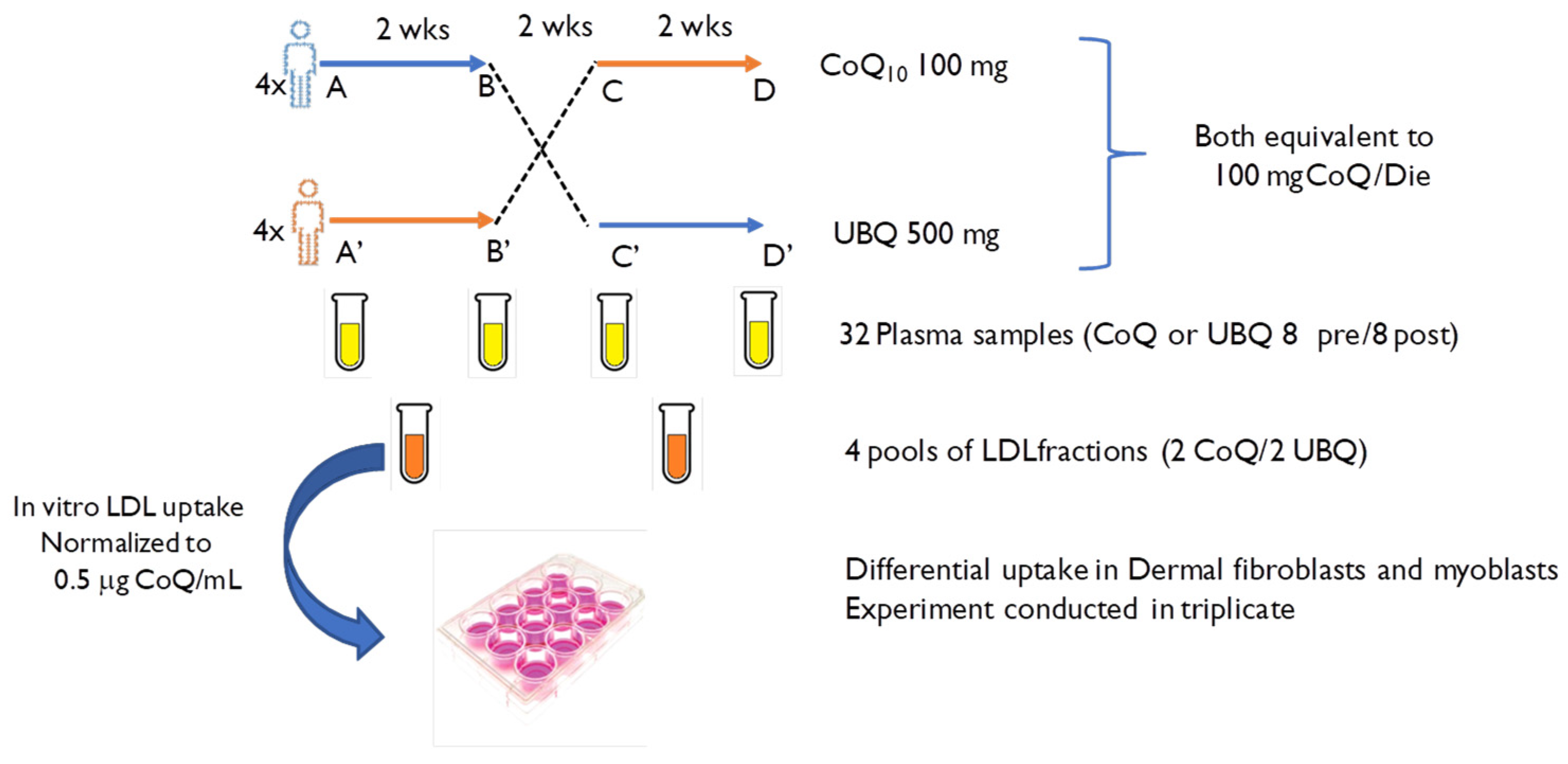
2.1. Experimental Design

This human study was conducted between September and November 2022 using atwo arms randomized crossover design with a wash-out phase that enrolled 8 subjects following the layout reported in Table 1. Specifically, the enrolled subjects were 4 males (age 32 ± 2) and 4 females (age 29 ± 1). The inclusion criteria included healthy subjects aged <40 years with a BMI between 18.5 and 24.9 kg/m2.
At the beginning of the study, the participants were randomized into two groups using the free Research Randomizer tool (www.randomizer.org accessed on 1 September 2022). One group received 500 mg/day of phytosome Coenzyme Q10 in capsules (UBQ) for two weeks, which is equivalent to 100 mg of CoQ10 (INDENA, Italy), and the second group received 100 mg/day of crystallized CoQ10 in capsules (CoQ)for two weeks. Both crystalline and Ubiqsome were provided by INDENA. Containers that were labelled as A or B were sequentially numbered and matched with the randomization list and were provided to blinded operators who allocated the containers to volunteers. The generation of randomization codes and the labelling of containers was conducted by independent operators who were not directly involved in the experimental procedures. Volunteers and researchers were blinded to the allocation sequence, and plasma and LDL extracts were classified based on generic labelling (A or B) in order to allow LDL pooling for exvivo study. Volunteers were instructed to take supplements with a meal. None of the participants took any medication or dietary supplements within 1 month before the beginning of the study. Following the two weeks of supplementation, the volunteers had two weeks of a washout phase. Subsequently, the groups were inverted so that all the subjects involved in the study took both formulations at subsequent times. The primary endpoint of thein vivo clinical study was the evaluation of Q10 plasma bioavailability. The isolated lipoproteins were subsequently used for exvivo studies in the cell culture models described.

2.2. Blood Samples and Extraction of Enriched LDL from Plasma

Blood (20 mL) was collected under fasting conditions in lithiumheparin vacutainers from each subject at the base line and at the end of each treatment phase at the Department of Life and Environmental Sciences (DISVA, UNIVPM) by qualified operators. Plasma (approximately 12 mL from each subject) was obtained by centrifugation at 1600× g for 5 min at 4 °C within 20 min after blood withdrawal. 250 µL of plasma was immediately stored at −80 °C to evaluate the CoQ10 amount and oxidative status forin vivobioavailability determination. The remaining fresh plasma was pooled among the volunteers taking the same formulation within the same arm of the study.
Subsequently, low-density lipoproteins were extracted from the pooled plasma using a heparin trisodium citrate solution, as previously reported by Weiland et al. [22], and the insoluble LDLs’ pellets were resuspended in cold PBS (0.1 M sodium phosphate buffer, pH7.4, containing 0.9% NaCl).In order to purify and concentrate the LDL, the solution was centrifuged and filtered at 2900 g for 20 min at 10 °C in a tube equipped with a filter membrane with a 10,000 kDa cutoff (centrifugal filter devices Amicon Ultra 4 mL, Millipore, Burlington, MA, USA). The enriched LDL samples were divided into 2 mL aliquots and stored at −80 °C in order to limit freeze–thawing procedures that are known to affect LDL oxidation.
In vitrostudies were conducted using 4 pools of LDL fraction, including 2 for each formulation, as reported in Figure 2. In particular, enriched LDLs were derived from 8 plasma aliquots B, D (CoQ10 crystallized post-treatment) and 8 plasma aliquots B’, D’ (Ubiqsome post-treatment).

2.3. Cell Culture and LDL Treatment

Human dermal fibroblasts (HDF) were purchased from the Istituto Zooprofilattico Sperimentale (Brescia, Italy) as a pooled sample from female subjects (40 years).HDFs were cultured in MEM with Earl salts (Carlo Erba, Italy) that were supplemented with 10% fetalbovine serum (South American Origin, Euroclone, Pero, Italy), 1% antimycin (10,000 U/mL) and streptomycin (10 mg/mL), stable glutamine (200 mM), and amphotericin B (250 µg/mL, Euroclone, Pero, Italy) and maintained in a 5% CO2 atmosphere at 37 °C. The complete medium was replaced every two days.
Mouse immortalised skeletal muscle myoblasts (C2C12), kindly provided by Prof. Michele Guescini, University of Urbino (Italy), were cultured in Dulbecco’s Modified Eagle Medium (DMEM) which was supplemented with 10% heat-inactivated fetal bovine serum (FBS, South American Origin, Euroclone, Pero, Italy), 1% glutamine (200 mM), 1% penicillin (10,000 U/mL), and 1% streptomycin (10 mg/mL) and maintained in a 5% CO2 atmosphere at 37 °C.
The LDL treatment was performed for both cell types at the sub-confluence state. Pooled LDLs with different formulations (UBQ and CoQ) were added to the complete medium to reach equal ubiquinol (QH) concentration (0.5 µg/mL). To avoid external contaminations, the solutions of media that were supplemented with pooled LDL were filtered through a 0.2 µm filter using a syringe and subsequently injected into the HPLC system to verify whether the ubiquinol concentrations were affected by filtration. Cells treated with only the complete medium were used as negative controls. All experiments were conducted independently three times.

2.4. Coenzyme Q10Level and Its Oxidative Status Determination in Plasma and Cell Samples

Plasma and cellular CoQ10 content and their oxidative statuses were analyzed by electrochemical detection using high performance liquid chromatography (Nanospace HPLC-ECD, Shiseido, Tokyo, Japan) associated with a post-chromatographic reducing column (Shiseido CQR, Tokyo, Japan) that simultaneously measured both the oxidized and reduced forms of CoQ, as previously described [12]. In particular, to quantify the total CoQ (CoQ9 + CoQ10) in the mouse myoblasts, the HPLC method, described by Andreani et al. [23], was used to discriminate the four peaks relative to the reduced and oxidized forms of CoQ9 and CoQ10.
CoQ10 plasma content was expressed as total plasma CoQ10 levels (µg/mL) or plasma nmol CoQ10/mmol cholesterol. The CoQ oxidative status was expressed as the percentage of oxidized CoQ with respect to the total CoQ.
Cellular CoQ levels were normalized by protein content that was assessed using the BCA protein assay kit (Thermofisher, Waltham, MA, USA), and content was expressed as CoQ10 ng/mg protein or total CoQ ng/mg protein.

2.5. Sample Size and Statistical Analysis

Sample size determination was based on mean values observed in similar studies that were conducted in our laboratory by evaluating the plasma bioavailability of different CoQ10 formulations.
In particular, the expected mean difference was used as a reference value. Considering a mean plasma change of 0.5 ± 0.34 µg/mL in the treated groups, 8 subjects would be required to detect a difference with 80% power and a 5% two-sided type I error rate.
Data from primary outcomes were expressed as means (SDs) and followed a normal distribution; therefore, unpaired t-tests with Welch’s correction were used. Two-way ANOVA was performed on the exvivo study in cell culture models using Tukey’s multiple comparisons test to compare control (Ctrl) with CoQ10 cells. Statistical significance was defined as a two-sided p value < 0.05.

3. Results

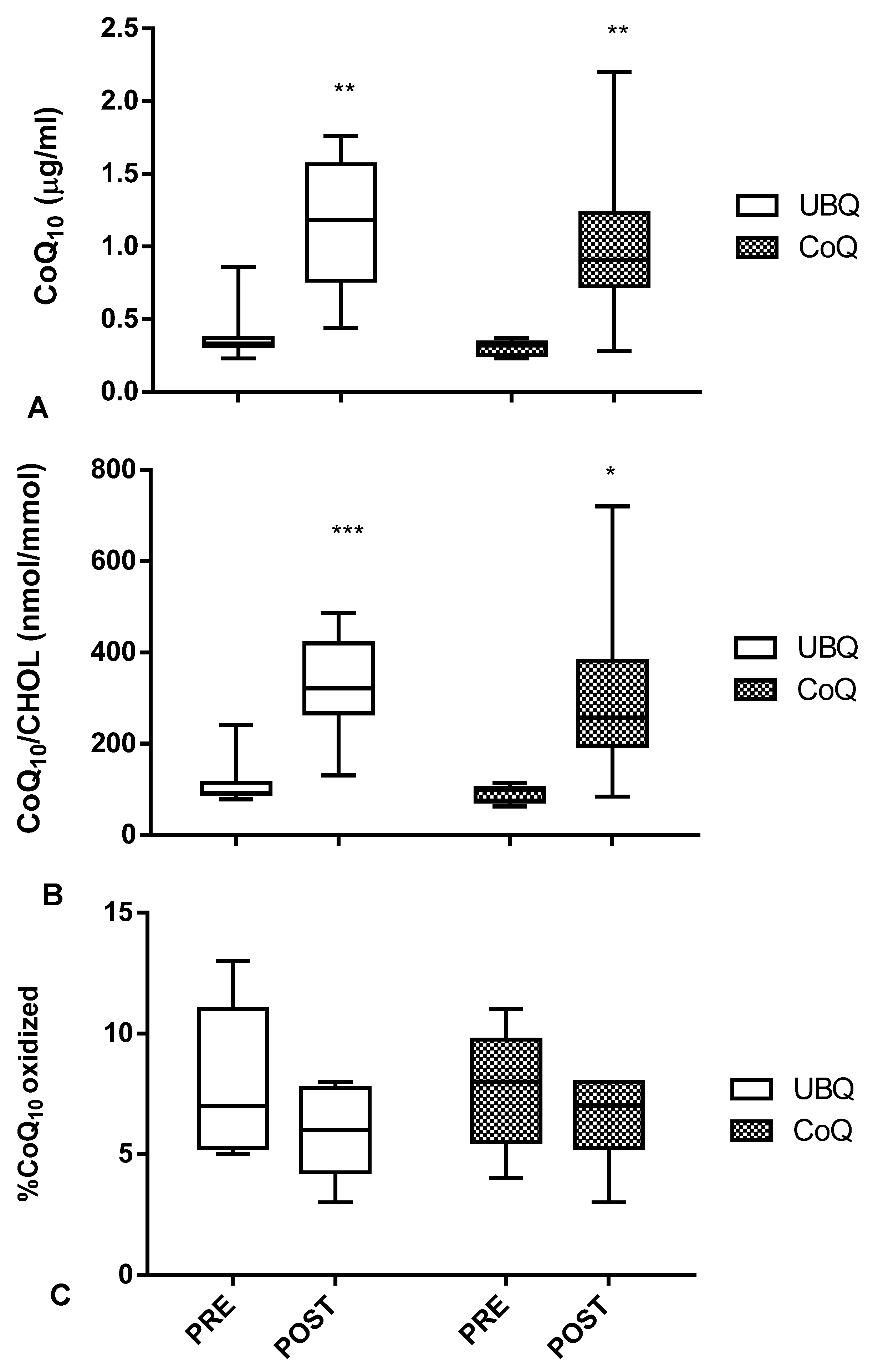
3.1. Both Crystalline CoQ10 and Ubiqsome Supplementation Produced a Significant Increase in CoQ10Plasma Levelsand Improved Its Oxidative Status

The consort 2010 flow chart describing the different steps of the invivo study is reported in Figure 1. All volunteers who participated in the study successfully completed the trial and no withdrawal or any side effects were reported. All data reported in theinvivostudy refers to the mean of eight values at each time point taking into consideration the crossover design. Two weeks of supplementation with UBQ resulted in a significant absolute plasma CoQ10 increase from 0.4 ± 0.2 µg/mL to 1.2 ± 0.5 µg/mL (p < 0.01) (Figure 3A). Crystalline CoQ10 supplementation also produced a significant increase in plasma levels (from 0.3 ± 0.1 µg/mL to 1.0 ± 0.6 µg/mL (p < 0.01). The total amount of plasma CoQ10 levels in each volunteer ranged from 0.2 to 0.9 µg/mL for basal conditions, while plasma CoQ10 levels ranged from 0.4 to 2.2 µg/mL following supplementation.
Since CoQ10 is transported in plasma by lipoproteins, mainly LDL, variation in cholesterolemia could affect absolute CoQ10 plasma determination. Nonetheless, a similar response was also observed following total cholesterol normalization in plasma CoQ10 normalized to cholesterol, with variations for UBQ and crystalline CoQ10changing from 114 ± 53 nmol CoQ10/mmol CHOL to 320 ± 111 nmol CoQ10/mmol CHOL (+181%, p < 0.001) and from 93 ± 18 nmol CoQ10/mmol CHOL to 312± 192 nmol CoQ10/mmol CHOL (+235%, p < 0.05), respectively (Figure 3B).
Dietary supplementation also resulted in a slight improvement in plasma CoQ10 oxidative status. The baseline percentage of oxidized CoQ10 in volunteers was 8%. The percentage of oxidized CoQ10 decreased following UBQ (6%) and crystalline CoQ10 (6.5%) supplementation, while no significant differences were detected between the two types of intervention (−0.5%; p = 0.51) (Figure 3C).
Indeed, the large majority of exogenous CoQ10 upon transfer through the gastrointestinal barrier is converted to ubiquinol and incorporated in lipoproteins. This biochemically mediated invivo transformation of ubiquinone to ubiquinol, together with the complexity of the lipoprotein environment, represents a critical advantage of the exvivo experimental design used in the present study.

3.2. CoQ10-Enriched LDL from Ubiqsome Supplemented Subjects Are More Efficient Vectors of CoQ10 to Cultured Cells

The incorporation of exogenous CoQ10 was quantified by HPLC following 24 h incubation both in human dermal fibroblasts and mouse skeletal myoblasts (Figure 4A,B). The results demonstrate that LDL from UBQ-supplemented volunteers were able to better deliver CoQ10 in both cellular models.
In particular, in human dermal fibroblasts exposed to LDL from UBQ-supplemented subjects, cellular CoQ10 increased by 9-fold compared withthe basal level recorded in untreated cells as it went from 5.6 ± 1.6 ng CoQ/mg protein to 51.4 ± 15.6 ng CoQ/mg prot (p < 0.0001) (Figure 4A). Significantly lower increases (4.5-fold; p < 0.0001) were observed in the same dermal fibroblasts incubated for the same time and at the same concentration of CoQ in the presence of LDL isolated from crystalline CoQ10-supplemented subjects. While the total increase was also substantially halved in this case, a highly significant increase from the baseline values was recorded, with cellular CoQ content ranging in this case from 5.6 ± 1.6 ng CoQ/mg protein to 25.3 ± 9.8 ng CoQ/mg protein (p < 0.0001) (Figure 4A).
An even more divergent behavior in CoQ10 delivering efficacy between LDL from UBQ- and CoQ-supplemented subjects was observed in murine skeletal myoblasts. In these cells, 24 hrs incubation with cell culture medium containing 0.5 µg CoQ10/mL from UBQ-enriched LDL produced a 12-fold increase in cellular CoQ10 content increasing from 2.6 ± 0.5 ng CoQ10/mg protein to 30.2 ± 12.6 ng CoQ10/mg protein (p < 0.0001) (Figure 4A). On the contrary, myoblasts incubated under the same experimental conditions using LDL from CoQ10-supplemented volunteers increased the cellular CoQ10 amount only 3-fold (from 2.6 ± 0.5 ng CoQ10/mg protein to 7.9 ± 2.8 ng CoQ10/mg protein; p = 0.34) (Figure 4A). In summary, in murine skeletal myoblasts, UBQ formulation was 4-fold more bioavailable in comparison with crystalline CoQ10 (p < 0.0001) (Figure 4A).
The murine cells data described in this study refer only to the CoQ10 fraction which is a minor component of the total Coenzyme Q pool of these cells, as shown in Table 1. Indeed, it is important to note that two cellular models, human and murine, are different in terms of their CoQ composition. While human cells contain only CoQ10, on the contrary, murine cells contain mainly CoQ9 and a far lower content of CoQ10. Interestingly, if we consider the total cellular CoQ content in murine cells (CoQ9 + CoQ10), as expected, this is higher in mitochondria rich skeletal muscle cells (66.1 ± 43.2 ng CoQ/mg protein) compared with dermal fibroblasts (5.6 ± 1.6 ng CoQ/mg prot).
Following incubation with LDL from the UBQ-supplemented subjects, the total CoQ cellular content in the skeletal muscle cells increased to 113.5 ± 37.4 ng CoQ/mg protein (1.7-fold increase, p < 0.001) (Figure 3B). However, in the same cells incubated with LDL from crystalline CoQ10-supplemented subjects, the total CoQ content increased only to 76.8 ± 30.5 ng CoQ/mg protein (1.2-fold increase, p = 0.62). In conclusion, in relation to total CoQ cellular content in skeletal myoblast, UBQ was 1.4-fold more bioavailable in comparison with crystalline CoQ10 (p < 0.0001) (Figure 4B).

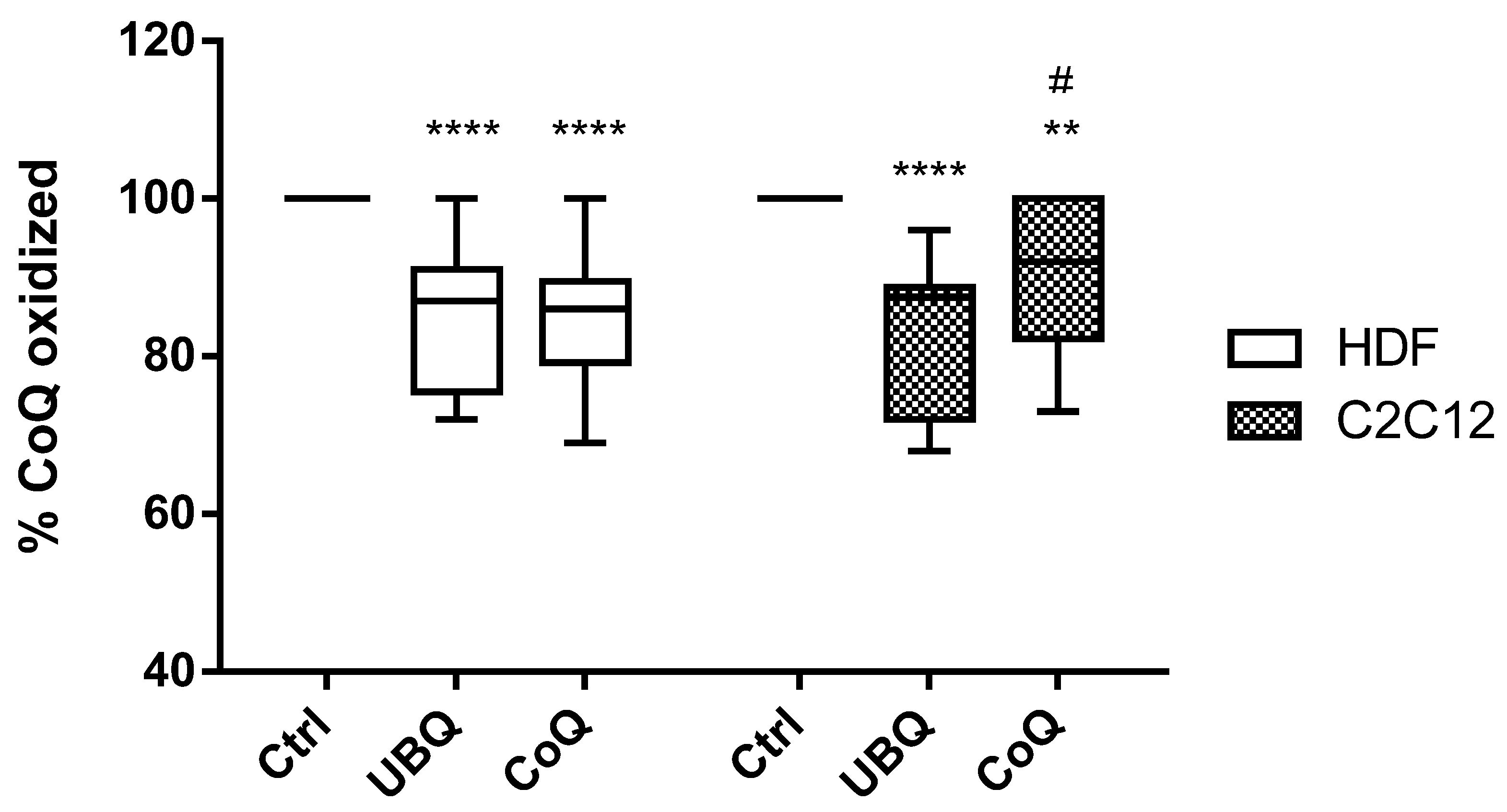
3.3. Ubiqsome Enriched LDL Were More Effective in Improving Cellular CoQ10Oxidative Status in Murine Myoblasts

In untreated human dermal fibroblasts, the whole CoQ content in cellular extracts was in the oxidized form (Figure 5). Supplementation with UBQ- or crystalline CoQ10-enriched LDL was able to decrease cellular CoQ10 oxidation to 85 ± 8% (p < 0.0001) (Figure 5), and no significant differences were observed between the two interventions (p = 0.99).
When mouse skeletal myoblasts were treated with enriched LDL, the difference between the two formulations was detectable. In fact, in crystalline CoQ10-treated cells, the level of oxidation was 91 ± 10 (Figure 5, p < 0.01 compared with the baseline), while in the UBQ-exposed myoblasts, the percentage of oxidized CoQ was 83 ± 10% (p < 0.0001 compared with the baseline; p < 0.014 compared with crystalline CoQ10).

4. Discussion

Coenzyme Q10 is an established nutritional supplement for improving human health in different clinical conditions. Indeed, CoQ10 secondary deficiency is not a rare condition and is associated with different pathological conditions characterized by increased oxidative stress [24], such as cardiovascular and metabolic diseases [25,26], drugs interfering with its biosynthesis, such as statins [27,28], aging, and senescence-related organ dysfunction, such as sarcopenia [24,29].
The beneficial effects of CoQ10 are well documented, both as a potent antioxidant in the lipid environment [2], as an anti-inflammatory agent [30,31], as well as a promotor of mitochondrial energy metabolism [32,33].
However, a major issue associated with CoQ10 use in clinical settings is related to its poor bioavailability and limited cellular uptak; in particular, muscle and cardiac tissues are the most refractory tissues to exogenous CoQ10 uptake [23,34], and the efficacy of oral supplementation in these districts is highly debated [23,34,35]. Onlyone study byKamzalovet al. observed a significant increase in muscle CoQ10 derived from homogenate and mitochondria in mice that were treated with CoQ10 at 148 and 654 mg/kg for 11 weeks [36].
Accordingly, several research efforts have addressed the topic of enhancing CoQ10 bioavailability; in particular, in terms of the development of novel delivery formulas, particle size reduction (nanoparticles), the solid dispersion of water insoluble drugs, microemulsion systems, cyclodextrin complexes, and liposomes [37,38,39]. In the present study, we investigated a new form of carrier involving a lecithin-based formulation also known as Phytosome. Phytosomes represent a functional solid dispersion in a phospholipid matrix. Ubiqsome is a phytosome standardized in coenzyme Q1018–22% by HPLC which previously showed enhanced plasmatic levels of CoQ10 after administration in a single dose [40].
Ubiqsome formulations have already been tested in invitro models using rat cardiac and human epithelial cells in relation to tissue CoQ10 bioavailability, cellular redox state, and cellular bioenergetics in comparison with pure CoQ10 form. Notably, 100 nM of Ubiqsome for 24 h was able to increase cellular and mitochondrial CoQ10 content that is associated with increased antioxidant defences (decreased lipid membrane peroxidation and ferroptosis) and improved mitochondria functionality (increased ATP production, spared respiratory capacity, and mitochondrial membrane potential) in both cell lines. On the contrary, the same dose of pure CoQ10 did not show any increase in tissue CoQ10 bioavailability, cellular antioxidant capacities, or bioenergetic parameters. Additionally, the authors reported that tissue CoQ10 uptake in both cell lines likely involved macropinocytosis mechanisms [41].
Moreover, in a recent study conducted in healthy aged athletes exposed to intense physical exercise, UBQ supplementation (equivalent to 100 mg CoQ10 per day for 1 month) was able to increase both plasma and muscular CoQ10 content. In particular, the study reported a remarkable and unique 36% increase in muscle CoQ10 levels following supplementation with UBQ [21]. In the supplemented subjects, increases in CoQ10 content were associated with protection from physical exercise-induced oxidative damage (decline in plasma malonyl dialdehyde levels) and a concomitant increase in plasma total antioxidant capacity. Moreover, the exercise-induced proinflammatory plasmatic markers IL6 and IL10 also significantly decreased.
In order to further investigate the peculiar bioavailability of Ubiqsome in muscle cells in more detail, we developed anex vivostudy design involving, as a first step, the oral supplementation of healthy subjects with either a standard crystalline CoQ10 formulation or Ubiqsome using equimolar doses of CoQ10 (100 mg/day for 2 weeks). Subsequently, CoQ10-enriched LDL deriving from subjects after supplementation with both products were used to treat human dermal fibroblasts known to be able to incorporate efficiently exogenous CoQ10 and murine myoblasts that, on the contrary, are more refractory to exogenous CoQ10 uptake. The proposed experimental model is able to mimic, in a more rigorous approach, the physiological exposure of tissues to CoQ10 that are essentially transported by lipoproteins. This model incorporates all biological transformations that take place at the gastrointestinal layer, such as a reduction in CoQ10 and lipoprotein assembly, and systemically, such as the maturation of LDL that constitute the primary carriers of CoQ10 in the circulation.
Our data are in agreement with the results obtained in an in vivo study conducted by Drobnic et al. [21] and may provide a simple approach to compare the cellular bioavailability of CoQ10 from LDL enriched with different formulations on the very same cellular system, thus reducing biological variability and limiting invasive procedures such as muscle biopsy.
Intriguingly, whileplasma bioavailability showed that both CoQ10 formulations were able to significantly increase plasma levels and its oxidative status in a statistically similar manner (Figure 3A,B), when we used pooled LDL isolated from the human plasma of volunteers who were treated with different formulations and then normalized for their CoQ10 content for the ex vivo supplementation of cultured cells, different bioavailability profiles for UBQ and CoQ10 formulas were observed in both cell lines used(Figure 3A,B). Specifically, UBQ-enriched LDL were more efficient in delivering CoQ10 in both human dermal fibroblasts and muscle myoblasts. The increase in cellular CoQ10 levels was significantly higher with respect to that obtained incubating cells with crystalline CoQ10-enriched LDL using identical CoQ10 dosages and times of exposure (Figure 4A). Dermal fibroblasts constitute the main cellular component of the dermis, a tissue characterized by lower CoQ10 content compared with other tissues, such as skeletal and cardiac muscle, and this might contribute to a higher tendency to acquire the exogenous CoQ10that is necessary for its critical role in cellular bioenergetics and antioxidant protection.
Surprisingly, skeletal muscle cells, which are well known for their refractoriness to exogenous CoQ10 uptake, also significantly increased their CoQ10 content when exposed to UBQ-enriched LDL at a much higher extent then the cells exposed to crystalline CoQ10-enriched LDL (Figure 3A,B). Moreover, UBQ in murine skeletal muscle cells was able to significantly improve cellular CoQ oxidative status, while no significant effects were observed in cells incubated with crystalline CoQ10-enriched LDL (Figure 5). This different redox improvement was tissue specific; in fact, in human dermal fibroblasts, both formulations were equally effective at increasing the percentage of cellular reduced CoQ10 oxidative status (Figure 5).
Enhanced muscle delivery of CoQ10 provided by Ubiqsome could be associated with the physio–chemical properties of the lipid components of phytosomes that could be better absorbed by plasma membranes of muscle cells or to more complex biochemical processes possibly linked with a promotion of mitochondrial biogenesis.
Concerning the phytosome composition, we did not analyze the protein and lipid composition of the pooled Ubiqsome-LDLs which could play a critical role. We aim to address this in future studies. Furthermore, other functional parameters related to mitochondrial/cellular oxidative statuses and oxidative stress resistance, which were not investigated in the present work, will be addressed in future experiments in order to validate the effect of increased CoQ bioavailability on cellular metabolism.
Indeed, mitochondria-rich tissues are characterized by elevated CoQ10 content but also by their limited ability to absorb exogenous CoQ10. The fact that under physiological conditions, mitochondria CoQ10 content is not saturating but is in the range of the Km of mitochondrial respiratory complexes [42] suggests that these tissues may have developed a selective permeability to CoQ10 that deserves particular attention.
In line with this observation, Kamzalov et al. report that exogenous CoQ10 uptake in mice homogenate is tissue specific as it is at its maximum in the liver and its minimum in skeletal muscle [36]. In fact, unlike other lipophilic endogenous and dietary molecules, very little is known about specific CoQ10 translocation proteins. To date, molecular mechanisms involved in tissue CoQ10 uptake remain not fully characterized, although recent studies using different cellular models to identify novel CoQ10 transporters including CoQ biosynthetic proteins have been suggested to be involved in intracellular trafficking.
In particular, in yeast cells, Cqd1 and Cqd2 proteins belonging to UbiB family (homologues to human COQ8A and B) were identified as main carriers in intracellular CoQ distribution. Specifically, Cqd1 and Cqd2 have been suggested to mediate CoQ transport from the mitochondria to the cytosolic environment and viceversa [43]. Another study conducted on yeast and bacteria cells analyzed a novel CoQ9 protein function involved in the inner mitochondrial membrane (IMM) permeability of CoQ precursors. In fact, thanks to biochemical, structural, and computational data, the authors show how CoQ9 is able to deliver both CoQ precursors and promote its translocation to mitochondrial CoQ biosynthetic enzymes (CoQ7) [44]. Mitochondria mass might therefore represent a targetable regulator of CoQ10 uptake that could be triggered using inducers of mitochondrial biogenesis. Further studies are required to verify this hypothesis and to evaluate whether the induction of mitochondria biogenesis underlies increased CoQ10 uptake evoked by Ubiqsome.

5. Conclusions

This exvivo study confirms invivo evidence of improved CoQ10phytosome muscle bioavailability which is important considering their well-described resistance to exogenous CoQ uptake. Further experiments are required in order to provide mechanistic insights beyond phytosome-improved bioavailability. The proposed model could provide a useful tool to investigate the mechanisms underlying tissue-dependent CoQ translocation that still remain unexplained.

Author Contributions

F.M. performed the experiments, F.M., S.S. and I.C. acquired and analyzed the data, and wrote the manuscript. F.O., P.O. and L.T. designed the layout of this study. A.R. and G.P. contributed to writing, reviewing, and editing the paper. All authors have read and agreed to the published version of the manuscript.

Funding

This research received no external funding.

Institutional Review Board Statement

The study was evaluated and approved by the Research EthicsCommittee of the Polytechnic University of Marche (Prot.n. 0162240 dated 5 September 2022).

Informed Consent Statement

Informed consent was obtained from all subjects involved in the study.

Data Availability Statement

All data presented in the study are available from the corresponding author (L.T.) upon request.

Conflicts of Interest

F.O., A.R. and G.P. are Indena’s employees.

References

  1. Lenaz, G.; Genova, M.L. Structure and organization of mitochondrial respiratory complexes: A new understanding of an old subject. Antioxid. Redox Signal. 2010, 12, 961–1008. [Google Scholar] [CrossRef] [PubMed]
  2. Bentinger, M.; Brismar, K.; Dallner, G. The antioxidant role of coenzyme Q. Mitochondrion 2007, 7, 41–50. [Google Scholar] [CrossRef]
  3. Navas, P.; Villalba, J.M.; Cabo, R.D. The importance of plasma membrane coenzyme Q in aging and stress responses. Mitochondrion 2007, 7, 34–40. [Google Scholar] [CrossRef] [PubMed]
  4. Cabo, R.D.; Cabello, R.; Rios, M.; Lopez-Lluch, G.; Ingram, D.K.; Lane, M.A.; Navas, P. Calorie restriction attenuates age-related alterations in the plasma membrane antioxidant system in rat liver. Exp. Gerontol. 2004, 39, 297–304. [Google Scholar] [CrossRef] [PubMed]
  5. Echtay, K.S.; Winkler, E.; Klingenberg, M. Coenzyme Q is an obligatory cofactor for uncoupling protein function. Nature 2000, 408, 609–613. [Google Scholar] [CrossRef]
  6. Sluse, F.E.; Jarmuszkiewicz, W.; Navet, R.; Douette, P.; Mathy, G.; Sluse-Goffart, C.M. Mitochondrial UCPs: New insights into regulation and impact. Biochim. Biophys. Acta 2006, 1757, 480–485. [Google Scholar] [CrossRef] [PubMed]
  7. Belliere, J.; Devun, F.; Cottet-Rousselle, C.; Batandier, C.; Leverve, X.; Fontaine, E. Prerequisites for ubiquinone analogs to prevent mitochondrial permeability transition-induced cell death. J. Bioenerg. Biomembr. 2012, 44, 207–212. [Google Scholar] [CrossRef]
  8. Molyneux, S.L.; Florkowski, C.M.; George, P.M.; Pilbrow, A.P.; Frampton, C.M.; Lever, M.; Richards, A.M. Coenzyme Q10: An independent predictor of mortality in chronic heart failure. J. Am. Coll. Cardiol. 2008, 52, 1435–1441. [Google Scholar] [CrossRef]
  9. Cirilli, I.; Damiani, E.; Dludla, P.; Hargreaves, I.; Marcheggiani, F.; Millichap, L.E.; Orlando, P.; Silvestri, S.; Tiano, L. Role of Coenzyme Q10 in Health and Disease: An Update on the Last 10 Years (2010–2020). Antioxidants 2021, 10, 1325. [Google Scholar] [CrossRef]
  10. Kon, M.; Tanabe, K.; Akimoto, T.; Fuminori, K.; Yuko, T.; Kazuhiro, S.; Tadashi, O.; Ichiro, K. Reducing exercise-induced muscular injury in kendo athletes with supplementation of coenzyme Q10. Br. J. Nutr. 2008, 100, 903–909. [Google Scholar] [CrossRef]
  11. Cooke, M.; Iosia, M.; Buford, T.; Shelmadine, B.; Hudson, G.; Kerksick, C.; Rasmussen, C.; Greenwood, M.; Leutholtz, B.; Willoughby, D.; et al. Effects of acute and 14-day coenzyme Q10 supplementation on exercise performance in both trained and untrained individuals. J. Int. Soc. Sport. Nutr. 2008, 4, 5–8. [Google Scholar] [CrossRef] [PubMed]
  12. Orlando, P.; Silvestri, S.; Galeazzi, R.; Antonicelli, R.; Marcheggiani, F.; Cirilli, I.; Bacchetti, T.; Tiano, L. Effect of ubiquinol supplementation on biochemical and oxidative stress indexes after intense exercise in youngathletes. Redox Rep. 2018, 23, 136–145. [Google Scholar] [CrossRef]
  13. Marcheggiani, F.; Kordes, S.; Cirilli, I.; Orlando, P.; Silvestri, S.; Vogelsang, A.; Möller, N.; Blatt, T.; Weise, J.M.; Damiani, E.; et al. Anti-ageing effects of ubiquinone and ubiquinol in a senescence model of human dermal fibroblasts. Free Radic. Biol. Med. 2021, 165, 282–288. [Google Scholar] [CrossRef] [PubMed]
  14. Fazakerley, D.J.; Chaudhuri, R.; Yang, P.; Maghzal, G.J.; Thomas, K.C.; Krycer, J.R.; Humphrey, S.J.; Parker, B.L.; Fisher-Wellman, K.H.; Meoli, C.C.; et al. Mitochondrial CoQ deficiency is a common driver of mitochondrial oxidants and insulin resistance. Elife 2018, 6, 7. [Google Scholar] [CrossRef] [PubMed]
  15. Shimizu, M.; Miyazaki, T.; Takagi, A.; Sugita, Y.; Yatsu, S.; Murata, A.; Kato, T.; Suda, S.; Ouchi, S.; Aikawa, T.; et al. Low circulating coenzyme Q10 during acutephase is associated with inflammation, malnutrition, and in-hospital mortality in patients admitted to the coronary careunit. Heart Vessel. 2017, 32, 668–673. [Google Scholar] [CrossRef] [PubMed]
  16. Pravst, I.; Žmitek, K.; Žmitek, J. Coenzyme Q10 Contents in Foods and Fortification Strategies. Crit. Rev. Food Sci. Nutr. 2010, 50, 269–280. [Google Scholar] [CrossRef] [PubMed]
  17. Bhagavan, H.N.; Chopra, R.K. Coenzyme Q10: Absorption, tissue uptake, metabolism and pharmacokinetics. Free Radic. Res. 2006, 40, 445–453. [Google Scholar] [CrossRef]
  18. Bentinger, M.; Dallner, G.; Chojnacki, T.; Swiezewska, E. Distribution and breakdown of labeled coenzyme Q10 in rat. Free Radic. Biol. Med. 2003, 34, 563–575. [Google Scholar] [CrossRef]
  19. Zhang, Y.; Åberg, F.; Appelkvist, E.L.; Dallner, G.; Ernster, L. Uptake of dietary coenzyme Q supplement is limited in rats. J. Nutr. 1995, 125, 446–453. [Google Scholar]
  20. Zhang, Y.; Turunen, M.; Appelkvist, E.L. Restricted uptake of dietary coenzyme Q is in contrast to the unrestricted uptake of alpha-tocopherol into rat organs and cells. J. Nutr. 1996, 26, 2089–2097. [Google Scholar] [CrossRef]
  21. Drobnic, F.; Riera, J.; Artuch, V.; Jou, C.; Codina, A.; Montero, R.; Paredes-Fuentes, A.J.; Domingo, J.C.; Banquells, M.; Riva, A.; et al. Efficient Muscle Distribution Reflects the Positive Influence of Coenzyme Q10 Phytosome in Healthy Aging Athletes after Stressing Exercise. J. Food Sci. Nutr. Res. 2020, 3, 262–275. [Google Scholar] [CrossRef]
  22. Weiland, H.; Seidel, D. A simple specific method for precipitation of low density lipoproteins. J. Lipid Res. 1983, 24, 904–909. [Google Scholar] [CrossRef]
  23. Andreani, C.; Bartolacci, C.; Guescini, M.; Battistelli, M.; Stocchi, V.; Orlando, F.; Provinciali, M.; Amici, A.; Marchini, C.; Tiano, L.; et al. Combination of Coenzyme Q(10) Intake and Moderate Physical Activity Counteracts Mitochondrial Dysfunctions in a SAMP8 Mouse Model. Oxidative Med. Cell. Longev. 2018, 2018, 8936251. [Google Scholar] [CrossRef] [PubMed]
  24. Fischer, A.; Onur, S.; Niklowitz, P.; Menke, T.; Laudes, M.; Rimbach, G.; Döring, F. Coenzyme Q10 status as a determinant of muscular strength in two independent cohorts. PLoS ONE 2016, 11, e0167124. [Google Scholar] [CrossRef]
  25. Martinefski, M.; Samassa, P.; Buontempo, F.; Hocht, C.; Lucangioli, S.; Tripodi, V. Relative bioavailability of coenzyme Q10 formulation for paediatric individualized therapy. J. Pharm. Pharmacol. 2016, 69, 567–573. [Google Scholar] [CrossRef]
  26. Langsjoen, P.H.; Langsjoen, A.M. Overview of the use of CoQ10 in cardiovascular disease. Biofactors 1999, 2, 273–284. [Google Scholar] [CrossRef]
  27. La Guardia, P.G.; Alberici, L.C.; Ravagnani, F.G.; Catharino, R.R.; Vercesi, A.E. Protection of rat skeletal muscle fibers by either L-carnitine or coenzyme Q10 against statins toxicity mediated by mitochondrial reactive oxygen generation. Front. Physiol. 2013, 4, 103. [Google Scholar] [CrossRef] [PubMed]
  28. Wang, L.W.; Jabbour, A.; Hayward, C.S.; Furlong, T.J.; Girgis, L.; Macdonald, P.S.; Keogh, A.M. Potential role of coenzyme Q10 in facilitating recovery from statin-induced rhabdomyolysis. Intern. Med. J. 2015, 45, 451–453. [Google Scholar] [CrossRef]
  29. Lopez-Lluch, G.; Rodriguez-Aguilera, J.C.; Santos-Ocana, C.; Navas, P. Is coenzyme Q a key factor in aging? Mech. Ageing Dev. 2010, 131, 225–235. [Google Scholar] [CrossRef]
  30. Olivieri, F.; Lazzarini, R.; Babini, L.; Prattichizzo, F.; Rippo, M.R.; Tiano, L.; Di Nuzzo, S.; Graciotti, L.; Festa, R.; Bruge, F.; et al. Anti-inflammatory effect of ubiquinol-10 on young and senescent endothelial cells via miR-146a modulation. Free Radic. Biol. Med. 2013, 63, 410–420. [Google Scholar] [CrossRef]
  31. Lee, B.J.; Tseng, Y.F.; Yen, C.H.; Lin, P.T. Effects of coenzyme Q10 supplementation (300 mg/day) on antioxidation and antiinflammation in coronary artery disease patients during statins therapy: A randomized, placebo-controlled trial. Nutr. J. 2013, 12, 142. [Google Scholar] [CrossRef] [PubMed]
  32. Stone, L.H.; Chappuis, E.; Wright, C.; Kelly, R.F.; McFalls, O.E. CoQ10 enhances PGC1α and increases expression of mitochondrial antioxidant proteins in chronically ischemic swine myocardium. Nutr. Metab. 2019, 16, 92. [Google Scholar] [CrossRef]
  33. Bergamini, C.; Moruzzi, N.; Sblendido, A.; Lenaz, G.; Fato, R. A Water Soluble CoQ10 Formulation Improves Intracellular Distribution and Promotes Mitochondrial Respiration in Cultured Cells. PLoS ONE 2012, 7, e33712. [Google Scholar] [CrossRef]
  34. Orlando, P.; Sabbatinelli, J.; Silvestri, S.; Marcheggiani, F.; Cirilli, I.; Dludla, P.V.; Molardi, A.; Nicolini, F.; Tiano, L. Ubiquinol supplementation in elderly patients undergoing aortic valve replacement: Biochemical and clinical aspects. Aging 2020, 12, 15514–15531. [Google Scholar] [CrossRef]
  35. Rosenfeldt, F.L.; Pepe, S.; Linnane, A.; Nagley, P.; Rowland, M.; Ou, R.; Marasco, S.; Lyon, W.; Esmore, D. Coenzyme Q10 protects the aging heart against stress: Studies in rats, human tissues, and patients. Ann. N. Y. Acad. Sci. 2002, 959, 355–359. [Google Scholar] [CrossRef] [PubMed]
  36. Kamzalov, S.; Sumien, N.; Forster, M.J.; Sohal, R.S. Coenzyme Q intake elevates the mitochondrial and tissue levels of Coenzyme Q and alpha-tocopherol in young mice. J. Nutr. 2003, 133, 3175–3180. [Google Scholar] [CrossRef]
  37. Zaki, N.M. Strategies for oral delivery and mitochondrial targeting of CoQ10. Drug Deliv. 2016, 6, 1868–1881. [Google Scholar] [CrossRef]
  38. Zmitek, J.; Zmitek, K.; Pravst, I. Improving the bioavailability of coenzyme Q10: From theory to practice. Agro-Food Ind. 2008, 19, 8–10. [Google Scholar]
  39. Xia, S.; Xu, S.; Zhang, X.; Zhong, F.; Wang, Z. Nanoliposomes mediate coenzyme Q10 transport and accumulation across human intestinal Caco-2 cell monolayer. J. Agric. Food Chem. 2009, 57, 7989–7996. [Google Scholar] [CrossRef]
  40. Petrangolini, G.; Ronchi, M.; Frattini, E.; De Combarieu, E.; Allegrini, P.; Riva, A. A New Food-grade Coenzyme Q10 Formulation Improves Bioavailability: Single and Repeated Pharmacokinetic Studies in Healthy Volunteers. Curr. Drug Deliv. 2019, 16, 759–767. [Google Scholar] [CrossRef]
  41. Rizzardi, N.; Liparulo, I.; Antonelli, G.; Orsini, F.; Riva, A.; Bergamini, C.; Fato, R. Coenzyme Q10 Phytosome Formulation Improves CoQ10 Bioavailability and Mitochondrial Functionality in Cultured Cells. Antioxidants 2021, 10, 927. [Google Scholar] [CrossRef] [PubMed]
  42. Lenaz, G.; Fato, R.; Castelluccio, C.; Genova, M.L.; Bovina, C.; Estornell, E.; Valls, V.; Pallotti, F.; Castelli, G.P. The function of coenzyme Q in mitochondria. Clin. Investig. 1993, 71, S66–S70. [Google Scholar] [CrossRef] [PubMed]
  43. Kemmerer, Z.A.; Robinson, K.P.; Schmitz, J.M.; Manicki, M.; Paulson, B.R.; Jochem, A.; Hutchins, P.D.; Coon, J.J.; Pagliarini, D.J. UbiB proteins regulate cellular CoQ distribution in Saccharomyces cerevisiae. Nat. Commun. 2021, 6, 4769. [Google Scholar] [CrossRef] [PubMed]
  44. Lohman, D.C.; Aydin, D.; Von Bank, H.C.; Smith, R.W.; Linke, V.; Weisenhorn, E.; McDevitt, M.T.; Hutchins, P.; Wilkerson, E.M.; Wancewicz, B.; et al. An Isoprene Lipid-Binding Protein Promotes Eukaryotic Coenzyme Q Biosynthesis. Mol. Cell 2019, 21, 763–774. [Google Scholar] [CrossRef]
Figure 1. CONSORT flowchart. A total number of 8 patients were enrolled. Volunteers were randomized in 2 groups (CoQ10, crystallized CoQ10 and UBQ, Ubiqsome) according to a crossover design. For for LDL extraction plasma was subsequently pooled into 2 groups, each one containing plasma from 4 patients.
Antioxidants 12 00964 g001
Figure 2. Layout of the study design. A and A’: pre-supplementation; B and B’: post-supplementation; C and C’: pre-supplementation after wash-out; D and D’: post-supplementation post wash-out. After collecting the blood samples, a total of 32 plasma samples were obtained and divided into 16 plasma samples (pre- and post-supplementation) each for crystalline CoQ10 (CoQ) and Ubiqsome (UBQ). From these plasma samples, 4 pools of enriched LDLs were extracted and divided into 2 each for CoQ and UBQ.
Antioxidants 12 00964 g002
Figure 3. Plasma CoQ10 levels. (A) CoQ10 normalized to cholesterol (CHOL) levels (B) and percent of oxidized CoQ10 (C) in human healthy volunteers supplemented for 2 weeks with UBQ and crystalline CoQ10 (CoQ) at the same dose (i.e., 100 mg/day of CoQ10). Data are expressed as boxes and bar plots (mean value) (n = 8). Statistical significance was calculated using unpaired t-tests with Welch’s correction compared with pre-supplementation (PRE) (* p < 0.05, ** p < 0.01, *** p < 0.001).
Antioxidants 12 00964 g003
Figure 4. Cellular CoQ10 (A) and total CoQ levels (B) in human dermal fibroblasts (white clear box plots) and murine skeletal myoblasts (small dot box plots) treated with p medium (controls, Ctrl), enriched LDL derived from the plasma of volunteers supplemented with Ubiqsome (UBQ) or crystalline CoQ10 (CoQ) at the same concentration of CoQ10 0.5 µg/mL for 24 h. Data are expressed as boxes and bar plots (mean value) (n = 3). Statistical significance was calculated using two-way Anova with Tukey’s multiple comparisons tests compared with the control cells (Ctrl) (*** p < 0.001, **** p < 0.0001) or crystalline CoQ10 (CoQ) (## p < 0.01, #### p < 0.0001).
Antioxidants 12 00964 g004
Figure 5. Cellular CoQ10 oxidized levels in human dermal fibroblasts (white clear box plots) and mouse skeletal myoblasts (small dot box plots) treated with medium (controls, Ctrl), enriched LDL derived from the plasma of subjects supplemented with Ubiqsome (UBQ) or crystalline CoQ10 (CoQ) at the same concentration of CoQ10 0.5 µg/mL for 24 h. Data are expressed as boxes and bar plots (median value) (n = 3). Statistical significance was calculated using two-way Anova with Tukey’s multiple comparisons tests compared with the control cells (Ctrl) (** p < 0.01, **** p < 0.0001) or crystalline CoQ10 (CoQ) (# p <0.05).
Antioxidants 12 00964 g005
Table 1. CoQ9 and CoQ10 values (ng/mg protein) in mouse skeletal cells that were exposed to different treatments with pooled LDL extracted from volunteers who were supplemented with Ubiqsome (UBQ) and crystallized CoQ10(CoQ). a p < 0.0001 compared with the control; b p < 0.0001 compared with CoQ.
TreatmentCoQ9 (ng/mg Protein)CoQ10 (ng/mg Protein)
Ctrl63.5 ± 42.92.6 ± 0.5
UBQ83.3 ± 38.330.2 ± 12.6 (a;b)
CoQ68.9 ± 30.17.9 ± 2.8
Disclaimer/Publisher’s Note: The statements, opinions and data contained in all publications are solely those of the individual author(s) and contributor(s) and not of MDPI and/or the editor(s). MDPI and/or the editor(s) disclaim responsibility for any injury to people or property resulting from any ideas, methods, instructions or products referred to in the content.

